Mechanism-Based Approach to New Antibiotic Producers ...
17
Citation: Volynkina, I.A.; Zakalyukina, Y.V.; Alferova, V.A.; Belik, A.R.; Yagoda, D.K.; Nikandrova, A.A.; Buyuklyan, Y.A.; Udalov, A.V.; Golovin, E.V.; Kryakvin, M.A.; et al. Mechanism-Based Approach to New Antibiotic Producers Screening among Actinomycetes in the Course of the Citizen Science Project. Antibiotics 2022, 11, 1198. https://doi.org/ 10.3390/antibiotics11091198 Academic Editor: Kui Zhu Received: 5 August 2022 Accepted: 2 September 2022 Published: 5 September 2022 Publisher’s Note: MDPI stays neutral with regard to jurisdictional claims in published maps and institutional affil- iations. Copyright: © 2022 by the authors. Licensee MDPI, Basel, Switzerland. This article is an open access article distributed under the terms and conditions of the Creative Commons Attribution (CC BY) license (https:// creativecommons.org/licenses/by/ 4.0/). antibiotics Article Mechanism-Based Approach to New Antibiotic Producers Screening among Actinomycetes in the Course of the Citizen Science Project Inna A. Volynkina 1,2, * , Yuliya V. Zakalyukina 3,4 , Vera A. Alferova 5,6 , Albina R. Belik 3 , Daria K. Yagoda 7 , Arina A. Nikandrova 1 , Yuliya A. Buyuklyan 3 , Andrei V. Udalov 3 , Evgenii V. Golovin 3 , Maxim A. Kryakvin 7 , Dmitrii A. Lukianov 1,2 , Mikhail V. Biryukov 1,3,8 , Petr V. Sergiev 1,2 , Olga A. Dontsova 1,2,5 and Ilya A. Osterman 1,2,3, * 1 Center of Life Sciences, Skolkovo Institute of Science and Technology, Bolshoy Boulevard 30, bld. 1, 121205 Moscow, Russia 2 Department of Chemistry, Lomonosov Moscow State University, Leninskie Gory 1, 119991 Moscow, Russia 3 Center for Translational Medicine, Sirius University of Science and Technology, Olympic Avenue 1, 354340 Sochi, Russia 4 Department of Soil Science, Lomonosov Moscow State University, Leninskie Gory 1, 119991 Moscow, Russia 5 Shemyakin-Ovchinnikov Institute of Bioorganic Chemistry, Miklukho-Maklaya 16/10, 117997 Moscow, Russia 6 Gause Institute of New Antibiotics, B. Pirogovskaya 11, 119021 Moscow, Russia 7 School of Bioengineering and Bioinformatics, Lomonosov Moscow State University, Leninskie Gory 1, 119991 Moscow, Russia 8 Department of Biology, Lomonosov Moscow State University, Leninskie Gory 1, 119991 Moscow, Russia * Correspondence: [email protected] (I.A.V.); [email protected] (I.A.O.) Abstract: Since the discovery of streptomycin, actinomycetes have been a useful source for new antibiotics, but there have been diminishing rates of new finds since the 1960s. The decreasing probability of identifying new active agents led to reduced interest in soil bacteria as a source for new antibiotics. At the same time, actinomycetes remain a promising reservoir for new active molecules. In this work, we present several reporter plasmids encoding visible fluorescent protein genes. These plasmids provide primary information about the action mechanism of antimicrobial agents at an early stage of screening. The reporters and the pipeline described have been optimized and designed to employ citizen scientists without specialized skills or equipment with the aim of essentially crowdsourcing the search for new antibiotic producers in the vast natural reservoir of soil bacteria. The combination of mechanism-based approaches and citizen science has proved its effectiveness in practice, revealing a significant increase in the screening rate. As a proof of concept, two new strains, Streptomyces sp. KB-1 and BV113, were found to produce the antibiotics pikromycin and chartreusin, respectively, demonstrating the efficiency of the pipeline. Keywords: citizen science; crowdsourcing; antibiotic producers screening; actinomycetes; reporter systems; chartreusin; pikromycin 1. Introduction The spread of antibiotic resistance is one of the major problems for modern antibacte- rial therapy. The most serious threats are methicillin-resistant Staphylococcus aureus (MRSA), vancomycin-resistant Enterococcus (VRE), multidrug-resistant (MDR) Acinetobacter bau- mannii and β-lactam-resistant Enterobacteriaceae [1]. The discovery of new antibacterial compounds may help to solve the problem, and soil bacteria still remain one of the most promising sources of bioactive natural products [2,3]. At the same time, traditional screening of soil actinomycetes for the production of antimicrobial agents often yields known molecules [4]. For this reason, new strategies for the discovery of new antibiotics have been developed [5–8]. One possible solution to Antibiotics 2022, 11, 1198. https://doi.org/10.3390/antibiotics11091198 https://www.mdpi.com/journal/antibiotics
-
Upload
khangminh22 -
Category
Documents
-
view
6 -
download
0
Transcript of Mechanism-Based Approach to New Antibiotic Producers ...

Citation: Volynkina, I.A.;
Zakalyukina, Y.V.; Alferova, V.A.;
Belik, A.R.; Yagoda, D.K.;
Nikandrova, A.A.; Buyuklyan, Y.A.;
Udalov, A.V.; Golovin, E.V.; Kryakvin,
M.A.; et al. Mechanism-Based
Approach to New Antibiotic
Producers Screening among
Actinomycetes in the Course of the
Citizen Science Project. Antibiotics
2022, 11, 1198. https://doi.org/
10.3390/antibiotics11091198
Academic Editor: Kui Zhu
Received: 5 August 2022
Accepted: 2 September 2022
Published: 5 September 2022
Publisher’s Note: MDPI stays neutral
with regard to jurisdictional claims in
published maps and institutional affil-
iations.
Copyright: © 2022 by the authors.
Licensee MDPI, Basel, Switzerland.
This article is an open access article
distributed under the terms and
conditions of the Creative Commons
Attribution (CC BY) license (https://
creativecommons.org/licenses/by/
4.0/).
antibiotics
Article
Mechanism-Based Approach to New Antibiotic ProducersScreening among Actinomycetes in the Course of the CitizenScience ProjectInna A. Volynkina 1,2,* , Yuliya V. Zakalyukina 3,4 , Vera A. Alferova 5,6 , Albina R. Belik 3, Daria K. Yagoda 7,Arina A. Nikandrova 1 , Yuliya A. Buyuklyan 3, Andrei V. Udalov 3, Evgenii V. Golovin 3, Maxim A. Kryakvin 7,Dmitrii A. Lukianov 1,2 , Mikhail V. Biryukov 1,3,8 , Petr V. Sergiev 1,2 , Olga A. Dontsova 1,2,5
and Ilya A. Osterman 1,2,3,*
1 Center of Life Sciences, Skolkovo Institute of Science and Technology, Bolshoy Boulevard 30, bld. 1,121205 Moscow, Russia
2 Department of Chemistry, Lomonosov Moscow State University, Leninskie Gory 1, 119991 Moscow, Russia3 Center for Translational Medicine, Sirius University of Science and Technology, Olympic Avenue 1,
354340 Sochi, Russia4 Department of Soil Science, Lomonosov Moscow State University, Leninskie Gory 1, 119991 Moscow, Russia5 Shemyakin-Ovchinnikov Institute of Bioorganic Chemistry, Miklukho-Maklaya 16/10, 117997 Moscow, Russia6 Gause Institute of New Antibiotics, B. Pirogovskaya 11, 119021 Moscow, Russia7 School of Bioengineering and Bioinformatics, Lomonosov Moscow State University, Leninskie Gory 1,
119991 Moscow, Russia8 Department of Biology, Lomonosov Moscow State University, Leninskie Gory 1, 119991 Moscow, Russia* Correspondence: [email protected] (I.A.V.); [email protected] (I.A.O.)
Abstract: Since the discovery of streptomycin, actinomycetes have been a useful source for newantibiotics, but there have been diminishing rates of new finds since the 1960s. The decreasingprobability of identifying new active agents led to reduced interest in soil bacteria as a source fornew antibiotics. At the same time, actinomycetes remain a promising reservoir for new activemolecules. In this work, we present several reporter plasmids encoding visible fluorescent proteingenes. These plasmids provide primary information about the action mechanism of antimicrobialagents at an early stage of screening. The reporters and the pipeline described have been optimizedand designed to employ citizen scientists without specialized skills or equipment with the aim ofessentially crowdsourcing the search for new antibiotic producers in the vast natural reservoir ofsoil bacteria. The combination of mechanism-based approaches and citizen science has proved itseffectiveness in practice, revealing a significant increase in the screening rate. As a proof of concept,two new strains, Streptomyces sp. KB-1 and BV113, were found to produce the antibiotics pikromycinand chartreusin, respectively, demonstrating the efficiency of the pipeline.
Keywords: citizen science; crowdsourcing; antibiotic producers screening; actinomycetes; reportersystems; chartreusin; pikromycin
1. Introduction
The spread of antibiotic resistance is one of the major problems for modern antibacte-rial therapy. The most serious threats are methicillin-resistant Staphylococcus aureus (MRSA),vancomycin-resistant Enterococcus (VRE), multidrug-resistant (MDR) Acinetobacter bau-mannii and β-lactam-resistant Enterobacteriaceae [1]. The discovery of new antibacterialcompounds may help to solve the problem, and soil bacteria still remain one of the mostpromising sources of bioactive natural products [2,3].
At the same time, traditional screening of soil actinomycetes for the production ofantimicrobial agents often yields known molecules [4]. For this reason, new strategiesfor the discovery of new antibiotics have been developed [5–8]. One possible solution to
Antibiotics 2022, 11, 1198. https://doi.org/10.3390/antibiotics11091198 https://www.mdpi.com/journal/antibiotics

Antibiotics 2022, 11, 1198 2 of 17
this so-called “rediscovery” issue is to collect samples from novel ecological niches andto isolate strains that occur there. Specifically, extreme environments such as those withhigh salinity or alkalinity are a promising reservoir of new antimicrobial producers [9].Moreover, “previously uncultured” microorganisms, that do not readily grow under lab-oratory conditions, may also serve as an untapped source of secondary metabolites [10].The advances in cultivation techniques led to the discovery of a new antibiotic, teixobactin,which inhibits cell wall biosynthesis [11]. Metagenomic libraries with DNA fragmentsextracted directly from soil and cloned in appropriate vectors, which are expressed in a cul-turable bacterium, enable researches to screen genes from “unculturable” microorganismsfor antimicrobial production [12,13]. Furthermore, whole-bacterial genome sequencingand bioinformatics tools for genome mining make it possible to identify biosynthetic geneclusters, even those predicted to encode novel antibiotics [14,15]. However, activation oftranscriptionally silent gene clusters is still a technical challenge.
Whereas some researchers are focused on developing new technically sophisticated,usually expensive and even more time-consuming approaches, including genome mining,bioinformatics analysis of omics data, screening of synthetic chemical libraries or target-based screening, others suggest expanding antimicrobial discovery activity through crowd-sourcing [16]. A widely known innovative program named the Small World Initiative (SWI,www.smallworldinitiative.org (accessed on 25 August 2022)) was formed at Yale Universityin 2012. As part of the citizen science project, students from around 150 participating schoolsisolated bacteria from their local environments, determined antibiotic production among thosestrains and extracted active compounds for further characterization. Later, SWI presentedan alternative pipeline, based on identification of biosynthetic gene clusters responsible forantimicrobial production phenotype, using transposon mutagenesis [17]. All the experimentswere carried out in well-equipped research laboratories. Inspired by the SWI, in 2015, theMicrobiology Society implemented a spin-off program, Antibiotics Unearthed, in the UK(www.microbiologysociety.org (accessed on 27 August 2022)). During the project, selectedschool or college students, as well as undergraduate students, analyzed their soil samples forantibacterial compounds and investigated any potential compounds found. Moreover, theMicrobiology Society held a series of events, giving the general public an opportunity to collecta soil sample and prepare it for scientific analysis during their visit. There are other citizenscience projects aimed at crowdsourcing only soil sampling: Citizen Science Soil CollectionProgram (University of Oklahoma, since 2010, www.whatsinyourbackyard.org (accessed on26 August 2022)), Swab and Send (since 2015, [16]) and Drugs From Dirt (Rockefeller University,since 2016, www.drugsfromdirt.org (accessed on 25 August 2022)). A similar Citizen Scienceproject is currently being carried out in Russia starting from 2021. In this project, we decided tocrowdsource not only sampling but also some primary experiments such as isolation of singlecolonies and testing them against indicator reporter strains. Therefore, we have designed anoptimized workflow that enables school students and the interested public to perform scientificexperiments in educational laboratories which are not specially equipped, or even at home.
One way to address the “rediscovery” issue is to determine the antimicrobial targetand mode of action during the screening. It might help to narrow down the pool of bioactivenatural products under consideration, and sometimes it might even help to prematurelyidentify the compound produced. Mechanism of action and antibiotic target could berevealed by different methods: direct measurement of the main cellular process inhibitionby incorporation of labeled metabolites [18], proteomic signature [19], cytological profilingof bacteria using fluorescence microscopy [20], selection of resistant clones followed bygenome sequencing [21], genome mining [22] and different reporter systems [23]. Weconsider reporter systems to be the most preferred tool for the Citizen Science project,since they can provide information at an early stage of screening. In addition, we recentlydeveloped such a mechanism-oriented reporter for high-throughput sorting of antibioticproducers by their mode of action [24]. In this study, we present a set of new constructsencoding visible fluorescent proteins or β-galactosidase, which can be detected withoutspecial equipment. With these reporter plasmids being transformed into antibiotic-sensitive

Antibiotics 2022, 11, 1198 3 of 17
indicator strains, citizen scientists could conduct a mechanism-oriented screening on theirown, in educational laboratories or even at home.
Here, we propose a novel pipeline of the Citizen Science project aimed at findingnew antibiotics, starting from a soil sample and ending with an antibiotic structure. Thewhole process could be divided into two stages: (a) isolation and screening for newproducing strains; and (b) identification of active compounds. Crowdsourcing at thefirst stage of the workflow significantly increases the speed of research and discoveryrates. The identification of active compounds (the second stage) should be conducted by aspecialist in the research laboratory, because it requires the usage of high-pressure liquidchromatography (HPLC), mass spectrometry, etc. The pipeline described here has beenillustrated with two examples: Streptomyces sp. KB-1 and BV113 isolates were found toproduce the antibiotics pikromycin (translation inhibitor) and chartreusin (SOS responseinducer), respectively. The combination of citizen science with the mechanism-basedapproach and modern analytical methods allows quick and efficient discovery of newantibiotic producers.
2. Results2.1. New Reporter Systems for Elucidating the Mode of Antibiotic Action in Application to theCitizen Science Project
Recently we developed a double reporter system, pDualrep2 [24], which consistsof two fluorescent protein genes, turbo-rfp and katushka2S (Figure 1A). The expressionof turbo-rfp is controlled by the SOS-inducible sulA gene promoter [25]. Whereas theexpression of katushka2S is regulated by the modified trpL attenuator sequence [26]. Po-tent antibiotics, which stall the ribosome on the trpL2A open reading frame, preventpremature transcription termination and promote transcription of the entire gene. Previ-ously, this system was successfully applied, resulting in the discovery of new antibioticproducers [27,28]. However, specific equipment is required to distinguish between thefluorescent signals from TurboRFP (553/574 nm) and Katushka2S (588/633 nm) (Figure 1B).
Antibiotics 2022, 11, x FOR PEER REVIEW 3 of 19
followed by genome sequencing [21], genome mining [22] and different reporter systems
[23]. We consider reporter systems to be the most preferred tool for the Citizen Science
project, since they can provide information at an early stage of screening. In addition, we
recently developed such a mechanism-oriented reporter for high-throughput sorting of
antibiotic producers by their mode of action [24]. In this study, we present a set of new
constructs encoding visible fluorescent proteins or β-galactosidase, which can be detected
without special equipment. With these reporter plasmids being transformed into antibi-
otic-sensitive indicator strains, citizen scientists could conduct a mechanism-oriented
screening on their own, in educational laboratories or even at home.
Here, we propose a novel pipeline of the Citizen Science project aimed at finding new
antibiotics, starting from a soil sample and ending with an antibiotic structure. The whole
process could be divided into two stages: (a) isolation and screening for new producing
strains; and (b) identification of active compounds. Crowdsourcing at the first stage of the
workflow significantly increases the speed of research and discovery rates. The identifi-
cation of active compounds (the second stage) should be conducted by a specialist in the
research laboratory, because it requires the usage of high-pressure liquid chromatography
(HPLC), mass spectrometry, etc. The pipeline described here has been illustrated with two
examples: Streptomyces sp. KB-1 and BV113 isolates were found to produce the antibiotics
pikromycin (translation inhibitor) and chartreusin (SOS response inducer), respectively.
The combination of citizen science with the mechanism-based approach and modern an-
alytical methods allows quick and efficient discovery of new antibiotic producers.
2. Results
2.1. New Reporter Systems for Elucidating the Mode of Antibiotic Action in Application to the
Citizen Science Project
Recently we developed a double reporter system, pDualrep2 [24], which consists of
two fluorescent protein genes, turbo-rfp and katushka2S (Figure 1A). The expression of
turbo-rfp is controlled by the SOS-inducible sulA gene promoter [25]. Whereas the expres-
sion of katushka2S is regulated by the modified trpL attenuator sequence [26]. Potent anti-
biotics, which stall the ribosome on the trpL2A open reading frame, prevent premature
transcription termination and promote transcription of the entire gene. Previously, this
system was successfully applied, resulting in the discovery of new antibiotic producers
[27,28]. However, specific equipment is required to distinguish between the fluorescent
signals from TurboRFP (553/574 nm) and Katushka2S (588/633 nm) (Figure 1B).
Figure 1. Double reporter system pDualrep2. (A) pDualrep2 plasmid map and the reporter scheme.
CDF_ori, CloDF13-derived CDF replicon; sulA, promoter of the sulA gene; T5, bacteriophage T5
promoter; trpL2A, modified trpL leader open reading frame carrying W10A and W11A substitu-
tions. Transcription start sites are shown by arrows. Transcription terminators are shown by vertical
dashes. (B) Comparison of fluorescence signals using an imaging system (left) and with the naked
eye (right). An agar plate was coated with the E. coli ΔtolC strain transformed with the pDualrep2
plasmid and spotted with erythromycin (Ery) and levofloxacin (Lev). The plate was scanned in Cy3
Figure 1. Double reporter system pDualrep2. (A) pDualrep2 plasmid map and the reporter scheme.CDF_ori, CloDF13-derived CDF replicon; sulA, promoter of the sulA gene; T5, bacteriophage T5promoter; trpL2A, modified trpL leader open reading frame carrying W10A and W11A substitutions.Transcription start sites are shown by arrows. Transcription terminators are shown by vertical dashes.(B) Comparison of fluorescence signals using an imaging system (left) and with the naked eye (right).An agar plate was coated with the E. coli ∆tolC strain transformed with the pDualrep2 plasmid andspotted with erythromycin (Ery) and levofloxacin (Lev). The plate was scanned in Cy3 (for TurboRFP)and Cy5 (for Katushka2S) channels, shown as green and red pseudocolors, respectively.
In order to keep the advantage of the dual reporter system pDualrep2, but to avoid thenecessity of specific equipment, the katushka2S gene was replaced by the β-galactosidasegene (lacZ), the expression of which could be visually detected by the blue-colored productin the presence of X-gal substrate (Supplementary Figure S1). The turbo-rfp gene was leftunmodified since its expression is clearly visible in orange-red color to the naked eye. Thenew plasmid was named pDualrep3. In practice, it demonstrated a high background signal

Antibiotics 2022, 11, 1198 4 of 17
due to the nonspecific hydrolysis of X-gal and, in part, leakage of the trpL2A regulatorysequence, so that only potent protein synthesis inhibitors (such as chloramphenicol andfusidic acid) could be detected with the pDualrep3 reporter.
This problem and the inability to resolve the situation when one sample inducesthe expression of both reporter genes, prompted us to design single reporter constructs.Based on fluorescent protein genes turbo-rfp and katushka2S, we created three new plasmids,named pTrpL2A-RFP, pTrpL2A-Katushka2S and pSulA-RFP, and validated them with a setof antibiotics (Figure 2). Both pTrpL2A-RFP and pTrpL2A-Katushka2S demonstrated strongreporter induction upon treatment with antibiotics that inhibit protein biosynthesis duringthe elongation step, such as chloramphenicol, puromycin, tetracycline, erythromycin,fusidic acid and lincomycin (Figure 2A,B). Consistent with previous data, spectinomycinand clindamycin demonstrated barely visible reporter induction, while streptomycin andkanamycin showed no induction at all [24]. It is worth considering that only antibioticsthat cause ribosome stalling on the trpL2A open reading frame could be detected by meansof these reporters. For this reason, aminoglycoside antibiotics which predominantly causemRNA misreading, such as streptomycin and kanamycin, would stay undercover [29]. Inaddition, we observed that cytotoxic antibiotic doxorubicin, which is known to intercalateDNA and inhibit topoisomerases in bacteria and eukaryotic cells [30–32], induced trpL2A-containing reporters. We assume that doxorubicin may bind to ribosomal RNA, thusimpeding the ribosome in synthesizing polypeptides.
Antibiotics 2022, 11, x FOR PEER REVIEW 5 of 19
Figure 2. New reporter constructs pTrpL2A-RFP (A), pTrpL2A-Katushka2S (B) and pSulA-RFP (C)
were created, transformed into the E. coli ΔtolC strain and validated with a panel of antibiotics. The
plates (A,C) were scanned in the Cy3 (TurboRFP) channel; the plate (B) was scanned in the Cy5
(Katushka2S) channel (middle). TurboRFP and Katushka2S are visible to the naked eye in orange-
red and lilac colors, respectively, in the zone of antibiotic sublethal concentrations (right). The panel
of antibiotics is as follows: chloramphenicol (Chl), puromycin (Puro), tetracycline (Tet), erythromy-
cin (Ery), rifampicin (Rif), doxorubicin (Dox), spectinomycin (Spm), streptomycin (Str), fusidic acid
(Fus), clindamycin (Clin), lincomycin (Linc), levofloxacin (Lev) and kanamycin (Kan).
2.2. Stocks of Freeze-Dried E. coli Reporter Cells Are Suitable for Application in the Citizen Sci-
ence Project
As usual, stocks of E. coli strains transformed with reporter plasmids are stored in
freezers at −80 °C until the experiment. It is almost impossible to equip all educational
laboratories with such freezers, not to mention the problem of stocks transportation. In
order to work with reporter cells far away from an equipped laboratory and in nonsterile
conditions, the freeze-drying method has been developed and tested in practice. This
method was previously described for E. coli cells by the ATCC organization (the American
Type Culture Collection, www.atcc.org (accessed on 20 August 2022)) and in some publi-
cations as well [36]. In the current study, we examined the freeze-drying procedure on the
E. coli ΔtolC strain transformed with the pDualrep2 plasmid. As a result, we observed that
Figure 2. New reporter constructs pTrpL2A-RFP (A), pTrpL2A-Katushka2S (B) and pSulA-RFP (C)were created, transformed into the E. coli ∆tolC strain and validated with a panel of antibiotics. Theplates (A,C) were scanned in the Cy3 (TurboRFP) channel; the plate (B) was scanned in the Cy5(Katushka2S) channel (middle). TurboRFP and Katushka2S are visible to the naked eye in orange-redand lilac colors, respectively, in the zone of antibiotic sublethal concentrations (right). The panel ofantibiotics is as follows: chloramphenicol (Chl), puromycin (Puro), tetracycline (Tet), erythromycin(Ery), rifampicin (Rif), doxorubicin (Dox), spectinomycin (Spm), streptomycin (Str), fusidic acid (Fus),clindamycin (Clin), lincomycin (Linc), levofloxacin (Lev) and kanamycin (Kan).

Antibiotics 2022, 11, 1198 5 of 17
The construct pSulA-RFP demonstrated clear reporter induction upon treatment withdoxorubicin and levofloxacin (Figure 2C). Both of them are known to function by inhibitingbacterial DNA gyrase, contributing to DNA damage and subsequent SOS response [30,33].Moreover, we observed a barely visible reporter induction in the case of rifampicin, whichis known to inhibit bacterial RNA polymerase [34].
Thus, plasmids pTrpL2A-Katushka2S and pSulA-RFP were chosen as the most con-venient for use in the Citizen Science project. Being transformed into antibiotic-sensitivebacterial strains [35], they may provide primary information about the action mechanismof antimicrobial agents produced by soil actinomycetes. Visually detectable induction ofreporter gene expression makes it possible to work with reporter strains in the absence ofspecial equipment, as for example in educational laboratories or at home.
2.2. Stocks of Freeze-Dried E. coli Reporter Cells Are Suitable for Application in the CitizenScience Project
As usual, stocks of E. coli strains transformed with reporter plasmids are stored infreezers at −80 ◦C until the experiment. It is almost impossible to equip all educationallaboratories with such freezers, not to mention the problem of stocks transportation. Inorder to work with reporter cells far away from an equipped laboratory and in nonsterileconditions, the freeze-drying method has been developed and tested in practice. Thismethod was previously described for E. coli cells by the ATCC organization (the AmericanType Culture Collection, www.atcc.org (accessed on 20 August 2022)) and in some pub-lications as well [36]. In the current study, we examined the freeze-drying procedure onthe E. coli ∆tolC strain transformed with the pDualrep2 plasmid. As a result, we observedthat freeze-dried cells retain reporter activity for at least eight and a half months (Figure 3).In addition, they can be stored both at room temperature and at 4 ◦C, which makes thestocks of freeze-dried reporter cells most suitable for application in the course of the CitizenScience project.
Antibiotics 2022, 11, x FOR PEER REVIEW 6 of 19
freeze-dried cells retain reporter activity for at least eight and a half months (Figure 3). In
addition, they can be stored both at room temperature and at 4 °C, which makes the stocks
of freeze-dried reporter cells most suitable for application in the course of the Citizen Sci-
ence project.
Figure 3. Comparison of fluorescence induction after the freeze-dried cells were kept for 8.5 months
(257 days) at room temperature (A), at 4 °C (B) and overnight culture as a control (C). Agar plates
were coated with the E. coli ΔtolC strain transformed with the pDualrep2 plasmid and spotted with
antibiotics at the following concentrations: ampicillin (Amp, 100 mg/mL), erythromycin at two con-
centrations (Ery 50, 50 mg/mL, and Ery 5, 5 mg/mL), levofloxacin (Lev, 25 µg/mL) and kanamycin
(Kan, 50 mg/mL). The plates were scanned in Cy3 (TurboRFP) and Cy5 (Katushka2S) channels,
shown as green and red pseudocolors, respectively.
2.3. The Pipeline of the Citizen Science Project
The Citizen Science project inspired by the Ministry of Science and Higher Education
of the Russian Federation (075-15-2021-1085) provides schoolchildren, students, teachers
and the interested public throughout Russian regions a unique opportunity to work with
scientists as part of the global initiative to discover new antibiotics from soil actinobacte-
ria.
The first and most essential stage of the project is to choose the environmental locus
from which sampling will be carried out. It can be a poorly studied natural niche, farm-
land, urban landscape or hard-to-reach area. Volunteers collect soil or sediment samples
in sterile containers (Figure 4A) and bring them to a school laboratory, educational center
or home. All information about the selected location, description of the territory, relief,
vegetation, etc., is recorded on a special website.
Using an isolation kit containing sterile plates and reagents, the volunteers prepare
serial 10-fold dilutions of collected samples and spread 50 μL of each suspension on iso-
lation agar plates supplemented with antibiotics that limit the growth of Gram-negative
bacteria and fungi. The inoculated plates should be wrapped with Parafilm® M and incu-
bated either in a thermostat at 28 °C or at room temperature for 2–3 weeks until noticeable
colonies of actinobacteria appear, which can be recognized by a fluffy, velvety or leathery
surface (Figure 4B).
Individual colonies are picked according to their cultural properties and restreaked
by sterile toothpicks onto the surface of two ISP3 agar plates (Supplementary Table S3)
[37]: one for further experiments and the other one for transfer to the Research Center.
After 10–14 days of cultivation, isolates from one ISP3 plate are restreaked on different
solid growth media known to promote the synthesis of natural products by actinomycete
strains (Figure 4C, Supplementary Table S3). No pharmaceutical antibiotics are added to
cultivation media at this stage. Acting carefully near an alcohol lamp or gas burner, the
citizen scientist can streak 4–5 isolates on one plate, arranging them by sectors. The second
ISP3 plate with actinobacteria lawns is transferred to the Research Center.
Figure 3. Comparison of fluorescence induction after the freeze-dried cells were kept for 8.5 months(257 days) at room temperature (A), at 4 ◦C (B) and overnight culture as a control (C). Agar plateswere coated with the E. coli ∆tolC strain transformed with the pDualrep2 plasmid and spottedwith antibiotics at the following concentrations: ampicillin (Amp, 100 mg/mL), erythromycin attwo concentrations (Ery 50, 50 mg/mL, and Ery 5, 5 mg/mL), levofloxacin (Lev, 25 µg/mL) andkanamycin (Kan, 50 mg/mL). The plates were scanned in Cy3 (TurboRFP) and Cy5 (Katushka2S)channels, shown as green and red pseudocolors, respectively.
2.3. The Pipeline of the Citizen Science Project
The Citizen Science project inspired by the Ministry of Science and Higher Educationof the Russian Federation (075-15-2021-1085) provides schoolchildren, students, teachersand the interested public throughout Russian regions a unique opportunity to work withscientists as part of the global initiative to discover new antibiotics from soil actinobacteria.
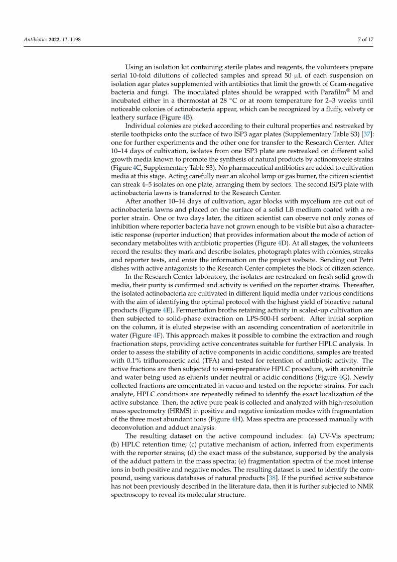
The first and most essential stage of the project is to choose the environmental locusfrom which sampling will be carried out. It can be a poorly studied natural niche, farmland,urban landscape or hard-to-reach area. Volunteers collect soil or sediment samples in sterile

Antibiotics 2022, 11, 1198 6 of 17
containers (Figure 4A) and bring them to a school laboratory, educational center or home.All information about the selected location, description of the territory, relief, vegetation,etc., is recorded on a special website.
Antibiotics 2022, 11, x FOR PEER REVIEW 7 of 19
Figure 4. Workflow diagram of the Citizen Science project. (A) Soil sampling. (B) Isolation of indi-
vidual actinomycete colonies through serial dilution. (C) Pure isolates are streaked onto different
selective growth media. One extra plate is transferred to the Research Center. (D) Activity-guided
selection of antimicrobial producers using agar block diffusion assay with the reporter strains. (E)
Determination of optimal cultivation conditions. (F) Solid-phase extraction of culture liquid
Figure 4. Workflow diagram of the Citizen Science project. (A) Soil sampling. (B) Isolation ofindividual actinomycete colonies through serial dilution. (C) Pure isolates are streaked onto differentselective growth media. One extra plate is transferred to the Research Center. (D) Activity-guidedselection of antimicrobial producers using agar block diffusion assay with the reporter strains.(E) Determination of optimal cultivation conditions. (F) Solid-phase extraction of culture liquidfollowed by fractions analysis using agar-well diffusion assay with the reporter strains. (G) Bioactivefractions are repeatedly subjected to reversed-phase HPLC separation. (H) Active pure HPLC peaksare analyzed with HRMS, revealing the exact mass of the antimicrobial compound.
Antibiotics 2022, 11, 1198 7 of 17
Using an isolation kit containing sterile plates and reagents, the volunteers prepareserial 10-fold dilutions of collected samples and spread 50 µL of each suspension onisolation agar plates supplemented with antibiotics that limit the growth of Gram-negativebacteria and fungi. The inoculated plates should be wrapped with Parafilm® M andincubated either in a thermostat at 28 ◦C or at room temperature for 2–3 weeks untilnoticeable colonies of actinobacteria appear, which can be recognized by a fluffy, velvety orleathery surface (Figure 4B).
Individual colonies are picked according to their cultural properties and restreaked bysterile toothpicks onto the surface of two ISP3 agar plates (Supplementary Table S3) [37]:one for further experiments and the other one for transfer to the Research Center. After10–14 days of cultivation, isolates from one ISP3 plate are restreaked on different solidgrowth media known to promote the synthesis of natural products by actinomycete strains(Figure 4C, Supplementary Table S3). No pharmaceutical antibiotics are added to cultivationmedia at this stage. Acting carefully near an alcohol lamp or gas burner, the citizen scientistcan streak 4–5 isolates on one plate, arranging them by sectors. The second ISP3 plate withactinobacteria lawns is transferred to the Research Center.
After another 10–14 days of cultivation, agar blocks with mycelium are cut out ofactinobacteria lawns and placed on the surface of a solid LB medium coated with a re-porter strain. One or two days later, the citizen scientist can observe not only zones ofinhibition where reporter bacteria have not grown enough to be visible but also a character-istic response (reporter induction) that provides information about the mode of action ofsecondary metabolites with antibiotic properties (Figure 4D). At all stages, the volunteersrecord the results: they mark and describe isolates, photograph plates with colonies, streaksand reporter tests, and enter the information on the project website. Sending out Petridishes with active antagonists to the Research Center completes the block of citizen science.
In the Research Center laboratory, the isolates are restreaked on fresh solid growthmedia, their purity is confirmed and activity is verified on the reporter strains. Thereafter,the isolated actinobacteria are cultivated in different liquid media under various conditionswith the aim of identifying the optimal protocol with the highest yield of bioactive naturalproducts (Figure 4E). Fermentation broths retaining activity in scaled-up cultivation arethen subjected to solid-phase extraction on LPS-500-H sorbent. After initial sorptionon the column, it is eluted stepwise with an ascending concentration of acetonitrile inwater (Figure 4F). This approach makes it possible to combine the extraction and roughfractionation steps, providing active concentrates suitable for further HPLC analysis. Inorder to assess the stability of active components in acidic conditions, samples are treatedwith 0.1% trifluoroacetic acid (TFA) and tested for retention of antibiotic activity. Theactive fractions are then subjected to semi-preparative HPLC procedure, with acetonitrileand water being used as eluents under neutral or acidic conditions (Figure 4G). Newlycollected fractions are concentrated in vacuo and tested on the reporter strains. For eachanalyte, HPLC conditions are repeatedly refined to identify the exact localization of theactive substance. Then, the active pure peak is collected and analyzed with high-resolutionmass spectrometry (HRMS) in positive and negative ionization modes with fragmentationof the three most abundant ions (Figure 4H). Mass spectra are processed manually withdeconvolution and adduct analysis.
The resulting dataset on the active compound includes: (a) UV-Vis spectrum;(b) HPLC retention time; (c) putative mechanism of action, inferred from experimentswith the reporter strains; (d) the exact mass of the substance, supported by the analysisof the adduct pattern in the mass spectra; (e) fragmentation spectra of the most intenseions in both positive and negative modes. The resulting dataset is used to identify the com-pound, using various databases of natural products [38]. If the purified active substancehas not been previously described in the literature data, then it is further subjected to NMRspectroscopy to reveal its molecular structure.

Antibiotics 2022, 11, 1198 8 of 17
Optionally, the pipeline can be supplemented with an additional analysis of fractionson a panel of bacterial strains specifically resistant to known antibiotics. Such an optionmay sometimes help to avoid wasting time on “rediscovering” already known compounds.
2.4. Phenotypic, Phylogenetic and Physiological Characteristics of Two New ProducingActinobacteria Strains
In the course of the Citizen Science project, two strains, KB-1 and BV113, were isolatedfrom the urban soil of Moscow and moss from Sochi, respectively. Both strains demonstratedprominent antibiotic activity in tests on the reporter cells (Supplementary Figure S2). KB-1exhibited strong pTrpL2A-Katushka2S reporter induction, indicating that the active compoundproduced by the isolate functions as an inhibitor of protein biosynthesis. Whereas BV113exhibited the induction of pSulA-RFP, which points to its ability to produce a substance thatelicits the SOS response in bacteria nearby. Using phenotypic features, the KB-1 and BV113strains were identified as mycelial actinobacteria (Figure 5, Supplementary Figures S3 and S4).
Antibiotics 2022, 11, x FOR PEER REVIEW 9 of 19
ability to produce a substance that elicits the SOS response in bacteria nearby. Using phe-
notypic features, the KB-1 and BV113 strains were identified as mycelial actinobacteria
(Figure 5, Supplementary Figures S3 and S4).
Figure 5. Morphological properties of Streptomyces sp. KB-1, grown on ISP4 medium at 28 °C for 14
days (A,B), and Streptomyces sp. BV113, grown on ISP3 medium at 28 °C for 14 days (C,D). (A,C)
Photographs of plates with strain streaks. (B,D) Mycelium micrographs taken on a scanning electron
microscope (SEM).
Comparative analysis of 16S rRNA sequences of KB-1 and BV113 strains with repre-
sentatives of the family Streptomycetaceae confirmed that the isolates are closely related
to species of the genus Streptomyces. The Maximum Likelihood tree (Figure 6) based on
16S rRNA gene sequences indicated that KB-1 forms a tight cluster with strains S. zaomy-
ceticus NRRL B-2038T, S. exfoliatus NBRC 13191T, S. venezuelae ATCC 10712T and S. virido-
brunneus NBRC 15902T (100% sequence similarity). The ability to synthesize pikromycin
was noted earlier in S. zaomyceticus [39] and S. venezuelae ATCC 15439 [40]. High levels of
16S rRNA gene sequence similarity were also found between the strain BV113 and a group
of S. osmaniensis OU-63T, S. longwoodensis NBRC 14251T and S. galbus JCM 4570T (99.7%),
which constitute a well-supported cluster on the phylogenetic tree, with a 91% bootstrap
value (Figure 6). The isolate BV113 was also found to share relatively high 16S rRNA gene
similarity with S. chartreusis JCM 4570T (99.2%), known as a producer of chartreusin [41].
The position of strains KB-1 and BV113 in the phylogenetic tree was unaffected by
the choice of tree-making algorithm or outgroup strains used.
Figure 5. Morphological properties of Streptomyces sp. KB-1, grown on ISP4 medium at 28 ◦C for14 days (A,B), and Streptomyces sp. BV113, grown on ISP3 medium at 28 ◦C for 14 days (C,D).(A,C) Photographs of plates with strain streaks. (B,D) Mycelium micrographs taken on a scanningelectron microscope (SEM).
Comparative analysis of 16S rRNA sequences of KB-1 and BV113 strains with repre-sentatives of the family Streptomycetaceae confirmed that the isolates are closely related tospecies of the genus Streptomyces. The Maximum Likelihood tree (Figure 6) based on 16SrRNA gene sequences indicated that KB-1 forms a tight cluster with strains S. zaomyceticusNRRL B-2038T, S. exfoliatus NBRC 13191T, S. venezuelae ATCC 10712T and S. viridobrun-neus NBRC 15902T (100% sequence similarity). The ability to synthesize pikromycin wasnoted earlier in S. zaomyceticus [39] and S. venezuelae ATCC 15439 [40]. High levels of 16SrRNA gene sequence similarity were also found between the strain BV113 and a group

Antibiotics 2022, 11, 1198 9 of 17
of S. osmaniensis OU-63T, S. longwoodensis NBRC 14251T and S. galbus JCM 4570T (99.7%),which constitute a well-supported cluster on the phylogenetic tree, with a 91% bootstrapvalue (Figure 6). The isolate BV113 was also found to share relatively high 16S rRNA genesimilarity with S. chartreusis JCM 4570T (99.2%), known as a producer of chartreusin [41].
Antibiotics 2022, 11, x FOR PEER REVIEW 10 of 19
Figure 6. Maximum Likelihood phylogenetic tree based on the Tamura–Nei model, showing the
relationship between isolated strains, KB-1 and BV113, and representative members of the genus
Streptomyces (based on 1147 unambiguously aligned nucleotides of 16S rRNA gene sequence). Num-
bers at nodes are bootstrap support percentages based on 1000 sampled datasets; only values above
60% are shown. Asterisks (*) indicate the branches that were also found in the Neighbor-Joining
tree. Bar, 0.01 substitutions per nucleotide position.
The physiological and biochemical characteristics of KB-1 and BV113 strains com-
pared with closely related strains are given in Supplementary Tables S1 and S2.
2.5. Identification of Active Compounds Produced by Streptomyces sp. KB-1 and BV113 Strains
The first object of the study was Streptomyces sp. KB-1, which demonstrated inhibi-
tion of protein biosynthesis on the reporter strains (Supplementary Figure S2). Its culture
liquid was subjected to solid-phase extraction, with an active fraction being eluted from
the LPS-500-H sorbent with a 20% aqueous acetonitrile (MeCN). Activity-guided HPLC
analysis revealed a specific active metabolite with a maximum UV absorption at 274 nm
(Figure 7). The mass spectrum of this compound contained the main adduct [M+H]+ with
m/z value 526.3370, exhibiting a sole fragment ion observed at m/z 158.1184 in the MS/MS
spectrum (Supplementary Figure S5). The exact mass of the detected metabolite corre-
sponds to the composition C28H47NO8 (the calculated m/z value for [M+H]+, 526.3374).
The search for candidates was carried out based on the accumulated data using the
NPAtlas, Dictionary of Natural Products and PubChem databases. The characteristic frag-
mentation allowed us to conclude that the active compound is a known inhibitor of pro-
tein biosynthesis, pikromycin (Figure 7) [42,43].
Figure 6. Maximum Likelihood phylogenetic tree based on the Tamura–Nei model, showing therelationship between isolated strains, KB-1 and BV113, and representative members of the genusStreptomyces (based on 1147 unambiguously aligned nucleotides of 16S rRNA gene sequence). Num-bers at nodes are bootstrap support percentages based on 1000 sampled datasets; only values above60% are shown. Asterisks (*) indicate the branches that were also found in the Neighbor-Joining tree.Bar, 0.01 substitutions per nucleotide position.
The position of strains KB-1 and BV113 in the phylogenetic tree was unaffected by thechoice of tree-making algorithm or outgroup strains used.
The physiological and biochemical characteristics of KB-1 and BV113 strains comparedwith closely related strains are given in Supplementary Tables S1 and S2.
2.5. Identification of Active Compounds Produced by Streptomyces sp. KB-1 and BV113 Strains
The first object of the study was Streptomyces sp. KB-1, which demonstrated inhibitionof protein biosynthesis on the reporter strains (Supplementary Figure S2). Its culture liquidwas subjected to solid-phase extraction, with an active fraction being eluted from the LPS-500-H sorbent with a 20% aqueous acetonitrile (MeCN). Activity-guided HPLC analysisrevealed a specific active metabolite with a maximum UV absorption at 274 nm (Figure 7).The mass spectrum of this compound contained the main adduct [M+H]+ with m/z value526.3370, exhibiting a sole fragment ion observed at m/z 158.1184 in the MS/MS spectrum(Supplementary Figure S5). The exact mass of the detected metabolite corresponds to thecomposition C28H47NO8 (the calculated m/z value for [M+H]+, 526.3374).

Antibiotics 2022, 11, 1198 10 of 17Antibiotics 2022, 11, x FOR PEER REVIEW 11 of 19
Figure 7. HPLC analysis of the active fraction of Streptomyces sp. KB-1 culture liquid. Elution with
70→95% MeCN in water for 9 min followed by 3 min of 95% MeCN. Red box indicates the active
component on the HPLC profile.
The second object of the study was Streptomyces sp. BV113, which demonstrated the
induction of SOS response in tests on the reporter strains (Supplementary Figure S2).
Solid-phase extraction of the Streptomyces sp. BV113 strain fermentation broth using the
LPS-500-H sorbent yielded an active fraction eluted with 30% acetonitrile in water. HPLC
analysis of the fraction (Figure 8) showed that the activity was associated with a hydro-
phobic compound with characteristic long wavelength maxima in the UV-Vis spectrum
(400, 422 nm). Mass spectrometric analysis of this compound revealed a prominent
[M+NH4]+ ion peak at m/z 658.213 in positive ion mode, corresponding to the molecular
formula C32H32O14 (the calculated m/z value for [M+NH4]+, 658.2130).
Glycosylated benzochromenone chartreusin (Figure 8) was selected as the most ap-
propriate candidate. The structural hypothesis was confirmed by comparing the fragmen-
tation of this compound with the literature data. Fragmentation of the [M+Na]+ ion with
m/z value 663.1677 contains key fragment ions with m/z values 329.1207 and 503.0943
(Supplementary Figure S6), which correspond to the loss of glycosidic fragments of the
molecule [44]. Consistent with the results of the reporter strains test, chartreusin is known
to bind to GC-rich tracts in DNA and cause single-strand breaks [45].
Figure 7. HPLC analysis of the active fraction of Streptomyces sp. KB-1 culture liquid. Elution with70→95% MeCN in water for 9 min followed by 3 min of 95% MeCN. Red box indicates the activecomponent on the HPLC profile.
The search for candidates was carried out based on the accumulated data using theNPAtlas, Dictionary of Natural Products and PubChem databases. The characteristicfragmentation allowed us to conclude that the active compound is a known inhibitor ofprotein biosynthesis, pikromycin (Figure 7) [42,43].
The second object of the study was Streptomyces sp. BV113, which demonstrated theinduction of SOS response in tests on the reporter strains (Supplementary Figure S2). Solid-phase extraction of the Streptomyces sp. BV113 strain fermentation broth using the LPS-500-H sorbent yielded an active fraction eluted with 30% acetonitrile in water. HPLC analysisof the fraction (Figure 8) showed that the activity was associated with a hydrophobiccompound with characteristic long wavelength maxima in the UV-Vis spectrum (400,422 nm). Mass spectrometric analysis of this compound revealed a prominent [M+NH4]+
ion peak at m/z 658.213 in positive ion mode, corresponding to the molecular formulaC32H32O14 (the calculated m/z value for [M+NH4]+, 658.2130).
Glycosylated benzochromenone chartreusin (Figure 8) was selected as the most ap-propriate candidate. The structural hypothesis was confirmed by comparing the fragmen-tation of this compound with the literature data. Fragmentation of the [M+Na]+ ion withm/z value 663.1677 contains key fragment ions with m/z values 329.1207 and 503.0943(Supplementary Figure S6), which correspond to the loss of glycosidic fragments of themolecule [44]. Consistent with the results of the reporter strains test, chartreusin is knownto bind to GC-rich tracts in DNA and cause single-strand breaks [45].

Antibiotics 2022, 11, 1198 11 of 17Antibiotics 2022, 11, x FOR PEER REVIEW 12 of 19
Figure 8. HPLC analysis of the active fraction of Streptomyces sp. BV113 culture liquid. Elution with
5→95% MeCN in water for 10 min followed by 2 min of 95% MeCN. Red box indicates the active
component on the HPLC profile.
3. Materials and Methods
3.1. Reporter Strains and Medium
E. coli JW5503 strain with deletion of the ΔtolC gene (referred to here as E. coli ΔtolC)
was kindly provided by Hironori Niki, National Institute of Genetics, Japan [46]. E. coli
BW25113 strain with partial deletion of the lptD gene, codons 330 to 352, (referred to here
as E. coli lptD) was kindly provided by Alexander S. Mankin, University of Illinois, Chi-
cago, USA [35]. Both strains were transformed with either new reporter plasmids or a
double reporter system pDualrep2 [24].
E. coli strains were grown at 37 °C in LB medium supplied with 100 μg/mL ampicillin,
if required.
3.2. Plasmids and Cloning
To create the construct pDualrep3, the vector backbone was amplified by high-fidel-
ity PCR from the pDualrep2 plasmid [24] using primers 5′-CCAGCACAGTGGTCGAAG-
3′ and 5′-CATATGTTGTGTTTGCATTGTTATTCTC-3′. The lacZ gene was amplified by
PCR from the pJC27 plasmid [47] with 5′-CAATGCAAACACAACATATGACCATGAT-
TACGCCAAGC-3′ forward and 5′-TTCTTCGACCACTGTGCTGGAATACGGGCAGA-
CATGGC-3′ reverse primers. The joining of two DNA fragments was performed with the
NEBuilder® HiFi DNA Assembly technique (NEB).
Figure 8. HPLC analysis of the active fraction of Streptomyces sp. BV113 culture liquid. Elution with5→95% MeCN in water for 10 min followed by 2 min of 95% MeCN. Red box indicates the activecomponent on the HPLC profile.
3. Materials and Methods3.1. Reporter Strains and Medium
E. coli JW5503 strain with deletion of the ∆tolC gene (referred to here as E. coli ∆tolC)was kindly provided by Hironori Niki, National Institute of Genetics, Japan [46]. E. coliBW25113 strain with partial deletion of the lptD gene, codons 330 to 352, (referred to hereas E. coli lptD) was kindly provided by Alexander S. Mankin, University of Illinois, Chicago,IL, USA [35]. Both strains were transformed with either new reporter plasmids or a doublereporter system pDualrep2 [24].
E. coli strains were grown at 37 ◦C in LB medium supplied with 100 µg/mL ampicillin,if required.
3.2. Plasmids and Cloning
To create the construct pDualrep3, the vector backbone was amplified by high-fidelityPCR from the pDualrep2 plasmid [24] using primers 5′-CCAGCACAGTGGTCGAAG-3′ and5′-CATATGTTGTGTTTGCATTGTTATTCTC-3′. The lacZ gene was amplified by PCR from thepJC27 plasmid [47] with 5′-CAATGCAAACACAACATATGACCATGATTACGCCAAGC-3′ for-ward and 5′-TTCTTCGACCACTGTGCTGGAATACGGGCAGACATGGC-3′ reverse primers.The joining of two DNA fragments was performed with the NEBuilder® HiFi DNA Assemblytechnique (NEB).

Antibiotics 2022, 11, 1198 12 of 17
The plasmid pTrpL2A-Katushka2S was obtained by PCR amplification with primers 5′-GGGCCCGCGACTCTAGATCATAATCA-3′ and 5′-GGTTCAGTAGAAAAGATCAAAGGATC-3′ using pDualrep2 as a template followed by blunt-end DNA ligation.
The plasmid pTrpL2A-RFP was obtained from pTrpL2A-Katushka2S by replacing the ka-tushka2S gene with turbo-rfp. The vector was amplified by PCR with primers 5′-CCAGCACAGTGGTCGAAG-3′ and 5′-TTCTCCTTGATCAGCTCGCCCATATGTTGTGTTTGCATTGTTATTCTC-3′. The turbo-rfp gene was amplified from the pDualrep2 plasmid with primers 5′-GGCGAGCTGATCAAGGAG-3′ and 5′-TTCTTCGACCACTGTGCTGGAAGCTTGTCGACCTGCAG-3′.The joining of two DNA fragments was performed using the NEBuilder® HiFi DNA Assemblytechnique (NEB).
The reporter construct pSulA-RFP was obtained from the pDualrep2 plasmid by PCRamplification with primers 5′-CCAGCACAGTGGTCGAAG-3′ and 5′-TTCTTCGACCACTGTGCTGGAAGCTTGTCGACCTGCAG-3′ and subsequent NEBuilder® HiFi DNA Assem-bly technique (NEB).
The E. coli JM109 strain was used for DNA cloning. Sequences of intermediate productsand final constructs were confirmed by sequencing with appropriate primers. Plasmidmaps were visualized using the program SnapGene® Viewer (version 5.2.4).
3.3. Reporter Cells Freeze-Drying and Storage
For freeze-drying, an overnight culture (OD600 0.9–1.0) of E. coli ∆tolC transformedwith the pDualrep2 plasmid was used. In total, 500 µL of the overnight culture wastransferred into sterile 2.0 mL centrifuge tubes, cells were harvested by centrifugation at7000 rpm for 2 min (~8400× g, Centrifuge 5418 R, Eppendorf, Hamburg, Germany),washed and resuspended in 500 µL of the lyophilization medium: 1% gelatin (w/v), 1%monosodium glutamate (w/v), 10% sucrose (w/v) and distilled H2O. Prepared lyophiliza-tion medium was sterilized by filtration through a 0.45 µm filter prior to use.
Opened centrifuge tubes with the suspension of reporter cells were covered withParafilm® M. Thereafter, tubes were subjected to gradual freezing: 20 min at 4 ◦C, thenfreezing in liquid nitrogen. Drying of samples was carried out at a pressure below0.370 mBar for 24 h (FreeZone Plus 2.5 Liter Cascade Benchtop Freeze Dry System,Labconco, Kansas City, MO, USA). The parafilm was removed from dried samples; halfof them were stored at room temperature and the other half at 4 ◦C. On the day of theexperiment, freeze-dried stocks were restored by adding 2 mL of fresh sterile LB medium.
3.4. Reporter Assays on Agar Plates
The overnight cultures of reporter cells diluted 5–10 times with fresh sterile LBmedium or the restored freeze-dried stocks were plated on LB agar medium suppliedwith 100 µg/mL ampicillin. The test samples were placed on the surface of dried agarplates covered with a reporter strain. For reporter activity validation tests, 1.5 µL of thefollowing antibiotics were used: chloramphenicol (1 mg/mL), puromycin (2 mg/mL), ery-thromycin (5 mg/mL), doxorubicin (2 mg/mL), spectinomycin (5 mg/mL), streptomycin(5 mg/mL), fusidic acid (5 mg/mL), clindamycin (10 mM), lincomycin (10 mM), lev-ofloxacin (30 µg/mL), kanamycin (5 mg/mL) and ampicillin (100 mg/mL); as well, diskssoaked in antibiotic solution were used for tetracycline (30 µg) and rifampicin (5 µg).
In order to perform reporter activity tests with actinobacteria isolates grown on dif-ferent nutrient media, agar blocks with mycelium were cut out of the grown lawns usingthe wide end of sterile 1000 µL pipette tips and were placed on the surface of dried platescoated with a reporter strain.
To test the reporter and antibacterial activity of liquid samples, such as culture liquidsor fractions obtained after solid-phase extraction or HPLC analysis, dried plates coveredwith a reporter strain were subjected to cutting wells out of the agar medium using thewide end of sterile 1000 µL pipette tips. The free volume of the resulting wells was100 µL. For culture liquids and HPLC fractions, 100 µL of solution per well was used; for

Antibiotics 2022, 11, 1198 13 of 17
solid-phase extraction eluates, 10 µL of solution mixed with 90 µL of distilled H2O per wellwas used in the agar diffusion assay.
Following overnight incubation at 37 ◦C or at room temperature (RT) for 1–2 days,the Petri dishes were photographed or, if possible, scanned by a ChemiDoc™ ImagingSystem (Bio-Rad, Hercules, CA, USA) with two channels: Cy3 (emission filter 605 ± 50 nm,green pseudocolor) for TurboRFP fluorescence and Cy5 (emission filter 695 ± 50 nm, redpseudocolor) for Katushka2S fluorescence.
3.5. Sampling and Isolation of Actinobacteria
Soils, sea sediments and plant samples were collected from different regions of Russiaduring the summer of 2021. The samples were placed into sterile containers to preventcontamination, delivered to an educational laboratory and stored at 4 ◦C until investigation.Sample solutions were prepared by dissolving 1 g of materials in 99 mL distilled water. Tofacilitate dissolution, sample flasks were shaken at 200 rpm for 10 min (Innova® 44 Shaker,New Brunswick Scientific, Edison, NJ, USA). Aliquots of serial 10-fold dilutions werespread on isolation media: mineral agar gauze 1 [48], organic medium 79 [49], M490 [50]and HV agar [51] (Table S3) supplemented with nystatin (250 µg/mL) and nalidixic acid(10 µg/mL) to prevent the growth of fungi and Gram-negative bacteria, respectively. After14–21 days of incubation at 28 ◦C or at room temperature (RT), the powdery-surfaced andleathery colonies were recognized as actinobacteria strains, picked, restreaked and grownon two fresh ISP3 agar plates (Table S3) [37]: one for further experiments and the other onefor transfer to the Research Center.
Thereafter, individual actinobacteria isolates were restreaked and grown on a set ofnutrient media: mineral agar gauze 1 (G1), organic medium 79 (Org79), glucose-asparagineagar (GA), soy flour mannitol agar (SFM) and oatmeal agar (ISP3) for 10 days at 28 ◦C orat RT (Table S3). Lawns of isolated strains were subsequently subjected to the agar blockdiffusion screening for antibiotic activity against reporter strains.
In the Research Center, the isolated actinobacteria strains were kept on oatmeal agar(ISP3) slants and stored as suspensions of spores in 20% glycerol (v/v) at −20 ◦C.
3.6. Phenotypic, Morphological and Physiological Characterization of New ProducingActinobacteria Strains
Aerial spore-mass color, substrate mycelial pigmentation, the production of diffusiblepigments and melanin were recorded after incubation at 28 ◦C for 14 days on media recom-mended by the International Streptomyces Project (ISP) [37]. Morphological characteristicsof aerial hyphae of KB-1 and BV113 strains were analyzed after 2 weeks of incubation at28 ◦C on ISP4 with CamScan S2 scanning electron microscope (Cambridge Instruments,Cambridge, UK) and on ISP3 medium with JSM-6380LA scanning electron microscope(JEOL Ltd., Akishima, Tokyo, Japan), respectively.
Carbon source utilization was assessed after incubation at 28 ◦C for 14 days on basalagar medium (ISP9) (Table S3) [37] supplemented with bromocresol purple indicator so-lution, 0.04% (w/v). Cellulose decomposition, starch hydrolysis, nitrate reduction, milkpeptonization, gelatin liquefaction and H2S production were examined as described previ-ously [52].
3.7. Phylogenetic Analysis of New Producing Actinobacteria Strains
Genomic DNA extraction from actinobacteria isolates was carried out accordingto the procedures described previously [53]. Amplification of the 16S rRNA gene wasperformed by high-fidelity PCR using primers 5′-GGATGAGCCCGCGGCCTA-3′ (243F)and 5′-CCAGCCCCACCTTCGAC-3′ (A3R) [28]. The sequencing was conducted with ABIPrism® BigDye™ Terminator v3.1 Cycle Sequencing Kit (Applied Biosystems, Waltham,MA, USA) and detected with Applied Biosystems® 3730 DNA Analyzer (Life Technologies,Carlsbad, CA, USA) in the Center for Collective Use “Genome” (Moscow, Russia). Thecontigs were processed and assembled with the GeneStudio™ Pro software (Version 2.2.0.0).

Antibiotics 2022, 11, 1198 14 of 17
GenBank accession numbers for the 16S rRNA gene sequences of Streptomyces sp. KB-1 andBV113 strains are OM780307.1 and OM801975.1, respectively.
Taxonomic affiliation of the isolates was assessed using the 16S rRNA gene sequencesas a query in the BLAST (https://blast.ncbi.nlm.nih.gov/Blast.cgi (accessed on 6 March2022)) and EzTaxon (www.ezbiocloud.net (accessed on 6 March 2022)) web services. The16S rRNA gene sequences of the most closely related species of the genus Streptomyces(more than 99% of identity) were used to build the alignment. In total, it comprised25 nucleotide sequences, with Amycolatopsis rifamycinica DSM 46095T as an outgroup. Thealignment was manually trimmed to 1147 bp and the phylogenetic tree was reconstructedby the Maximum Likelihood (ML) method based on the Tamura–Nei model [54], as well asby the Neighbor-Joining (NJ) method [55] based on the Kimura two-parameter model [56].The phylogenetic tree was visualized using MEGA 7.0 software [57].
3.8. Cultivation and Extraction of SECONDARY metabolites
Actinobacteria isolates were first inoculated in 20 mL of liquid organic medium 79 (Org79,no agar) and cultivated at 28 ◦C with constant shaking (180 rpm, Innova® 44 Shaker, NewBrunswick Scientific, Edison, NJ, USA) for 3 days. The resulting starters were used to inoculate200 mL of different liquid media (Table S3) and then suspensions were incubated at 28 ◦C withconstant shaking (220 rpm, Innova® 44 Shaker, New Brunswick Scientific, Edison, NJ, USA) in750 mL Erlenmeyer flasks to determine the optimal cultivation conditions.
For Streptomyces sp. KB-1, the greatest production of an antibacterial metabolite wasobserved upon cultivation in a modified ISP3 liquid medium for 6 days. Composition of themodified ISP3 medium (g/L) was as follows: oatmeal—20, powder chalk—5, FeSO4·7H2O—0.001, MnCl2·4H2O—0.001, ZnSO4·7H2O—0.001, pH 7.2.
For Streptomyces sp. BV113, the greatest production of an antibacterial metabolite wasobserved upon cultivation in a modified Org79 liquid medium for 4 days. Compositionof the modified Org79 medium (g/L) was as follows: maltose—10, peptone—10, caseinhydrolysate—2, yeast extract—2, NaCl—6, pH 7.2.
Culture liquids were separated from biomass by centrifugation at 20,000× g for 5 min(Centrifuge 5810 R, Rotor FA-45-6-30, Eppendorf, Hamburg, Germany). The supernatantswere subjected to solid-phase extraction and primary fractionation on LPS-500-H sorbent(LLC “Technosorbent”, Moscow, Russia) using water-acetonitrile mixtures as eluents.
3.9. Antibiotic Identification
HPLC analysis and fractionation were performed with the Vanquish Flex UHPLCSystem using the Diode Array Detector (Thermo Fisher Scientific, Waltham, MA, USA),equipped with Luna® 5 µm C18(2) 100 Å, 250 × 4.6 mm column (Phenomenex, Torrance,CA, USA).
Mass spectra were collected using maXis II 4G ETD mass spectrometer (Bruker Dal-tonics, Bremen, Germany) and UltiMate 3000 chromatograph (Thermo Fisher Scientific,Waltham, MA, USA), equipped with Acclaim RSLC 120 C18 2.2 µm 2.1 × 100 mm column(Thermo Fisher Scientific, Waltham, MA, USA). Spectrum registration mode: ESI ionizationmode, full scan from 100–1500 m/z, MS/MS with selection of the three most intense ions,dissociation type: CID 10–40 eV, nitrogen collision gas. Mass spectra were processed usingOpenChrom Lablicate Edition (1.4.0.202201211106), TOPPView v. 2.6.0 [58]. The chemicalstructures were identified using the GNPS [59], NPAtlas [60,61] and Dictionary of NaturalProducts 31.1 (https://dnp.chemnetbase.com (accessed on 10 March 2022)) [38] databases.
4. Conclusions
In order to speed up the screening for new antibiotic-producing strains, we createdreporter plasmids that can be utilized in the course of the Citizen Science project. Theability to detect reporter induction with the naked eye makes it possible to crowdsourcenot only sampling but also some primary experiments such as isolation of single coloniesand testing them against indicator reporter strains. Consistent with the idea of giving

Antibiotics 2022, 11, 1198 15 of 17
citizen scientists an opportunity to collect and analyze natural samples near their homes,we developed a special optimized pipeline that has been successfully applied in practice.Two examples of antibiotic-producing strains discovered de novo are presented in thearticle: Streptomyces sp. KB-1 (produces pikromycin) and BV113 (produces chartreusin).Through a proof of concept, we demonstrate that the novel workflow of the Citizen Scienceproject is an effective means to improve the screening productivity.
Supplementary Materials: The following supporting information can be downloaded at: https://www.mdpi.com/article/10.3390/antibiotics11091198/s1, Figure S1: double reporter system pDu-alrep3; Figure S2: agar block diffusion assay of isolated actinomycetes (BV113, KB-1, Nm4); Figure S3:utilization of different sugars (1.0%, w/v) as sole carbon source by strain KB-1; Figure S4: utilizationof different sugars (1.0%, w/v) as sole carbon source by strain BV113; Figure S5: MS/MS spectrum ofthe pikromycin [M+H]+ ion at m/z value 526.3370; Figure S6: MS/MS spectrum of the chartreusin[M+Na]+ ion at m/z value 663.1677; Table S1: physiological and biochemical characteristics of KB-1isolate and closely related Streptomyces sp. strains; Table S2: physiological and biochemical charac-teristics of BV113 isolate and closely related Streptomyces sp. strains; Table S3: composition of somenutrient media used. References [37,48–51,62–65] are cited in the supplementary materials.
Author Contributions: Conceptualization, I.A.O., O.A.D. and P.V.S.; methodology, I.A.O., M.V.B. andV.A.A.; software, Y.V.Z. and V.A.A.; validation, I.A.O., M.V.B. and V.A.A.; formal analysis, Y.V.Z. andV.A.A.; investigation, I.A.V., Y.V.Z., A.R.B., D.K.Y., A.A.N., Y.A.B., A.V.U., E.V.G., M.A.K., D.A.L. andM.V.B.; resources, Y.V.Z. and A.R.B.; data curation, Y.V.Z.; writing—original draft preparation, I.A.V.,Y.V.Z., V.A.A., D.A.L., M.V.B. and I.A.O.; writing—review and editing, I.A.V.; visualization, I.A.V.,Y.V.Z. and V.A.A.; supervision, I.A.O.; project administration, I.A.O. and M.V.B.; funding acquisition,I.A.O. All authors have read and agreed to the published version of the manuscript.
Funding: This research was funded by the Ministry of Science and Higher Education of the RussianFederation (Agreement NO. 075–15-2021-1085).
Institutional Review Board Statement: Not applicable.
Informed Consent Statement: Not applicable.
Data Availability Statement: Not applicable.
Acknowledgments: SEM studies were carried out at the Shared Research Facility “Electron mi-croscopy in life sciences” at Lomonosov Moscow State University (Unique Equipment “Three-dimensional electron microscopy and spectroscopy”).
Conflicts of Interest: The authors declare no conflict of interest. The funders had no role in the designof the study; in the collection, analyses, or interpretation of data; in the writing of the manuscript, orin the decision to publish the results.
References1. Zhu, Y.; Huang, W.E.; Yang, Q. Clinical Perspective of Antimicrobial Resistance in Bacteria. Infect. Drug Resist. 2022, 15, 735–746.
[CrossRef] [PubMed]2. Quinn, G.A.; Banat, A.M.; Abdelhameed, A.M.; Banat, I.M. Streptomyces from traditional medicine: Sources of new innovations
in antibiotic discovery. J. Med. Microbiol. 2020, 69, 1040–1048. [CrossRef] [PubMed]3. De Simeis, D.; Serra, S. Actinomycetes: A Never-Ending Source of Bioactive Compounds—An Overview on Antibiotics Production.
Antibiotics 2021, 10, 483. [CrossRef] [PubMed]4. Wright, G.D. Antibiotics: A new hope. Chem. Biol. 2012, 19, 3–10. [CrossRef]5. Hoffman, P.S. Antibacterial Discovery: 21st Century Challenges. Antibiotics 2020, 9, 213. [CrossRef]6. Herrmann, J.; Lukezic, T.; Kling, A.; Baumann, S.; Huttel, S.; Petkovic, H.; Muller, R. Strategies for the Discovery and Development
of New Antibiotics from Natural Products: Three Case Studies. Curr. Top. Microbiol. Immunol. 2016, 398, 339–363. [CrossRef]7. Cortes-Sanchez, E.; Hoskisson, P.A. New approaches for new antibiotics. Biochemist 2015, 37, 28–31. [CrossRef]8. Coates, A.R.; Hu, Y. Novel approaches to developing new antibiotics for bacterial infections. Br. J. Pharmacol. 2007, 152, 1147–1154.
[CrossRef]9. Trenozhnikova, L.; Azizan, A. Discovery of Actinomycetes from Extreme Environments with Potential to Produce Novel
Antibiotics. Cent. Asian J. Glob. Health 2018, 7, 337. [CrossRef]10. Lewis, K.; Epstein, S.; D’Onofrio, A.; Ling, L.L. Uncultured microorganisms as a source of secondary metabolites. J. Antibiot. 2010,
63, 468–476. [CrossRef]

Antibiotics 2022, 11, 1198 16 of 17
11. Ling, L.L.; Schneider, T.; Peoples, A.J.; Spoering, A.L.; Engels, I.; Conlon, B.P.; Mueller, A.; Schaberle, T.F.; Hughes, D.E.; Epstein,S.; et al. A new antibiotic kills pathogens without detectable resistance. Nature 2015, 517, 455–459. [CrossRef]
12. Schmeisser, C.; Steele, H.; Streit, W.R. Metagenomics, biotechnology with non-culturable microbes. Appl. Microbiol. Biotechnol.2007, 75, 955–962. [CrossRef]
13. Gillespie, D.E.; Brady, S.F.; Bettermann, A.D.; Cianciotto, N.P.; Liles, M.R.; Rondon, M.R.; Clardy, J.; Goodman, R.M.; Handelsman,J. Isolation of antibiotics turbomycin a and B from a metagenomic library of soil microbial DNA. Appl. Environ. Microbiol. 2002,68, 4301–4306. [CrossRef]
14. Krause, J. Applications and Restrictions of Integrated Genomic and Metabolomic Screening: An Accelerator for Drug Discoveryfrom Actinomycetes? Molecules 2021, 26, 5450. [CrossRef]
15. Foulston, L. Genome mining and prospects for antibiotic discovery. Curr. Opin. Microbiol. 2019, 51, 1–8. [CrossRef]16. Roberts, A.P. Swab and Send: A citizen science, antibiotic discovery project. Futur. Sci. OA 2020, 6, FSO477. [CrossRef]17. Davis, E.; Sloan, T.; Aurelius, K.; Barbour, A.; Bodey, E.; Clark, B.; Dennis, C.; Drown, R.; Fleming, M.; Humbert, A.; et al.
Antibiotic discovery throughout the Small World Initiative: A molecular strategy to identify biosynthetic gene clusters involvedin antagonistic activity. MicrobiologyOpen 2017, 6, e00435. [CrossRef]
18. Cunningham, M.L.; Kwan, B.P.; Nelson, K.J.; Bensen, D.C.; Shaw, K.J. Distinguishing on-target versus off-target activity in earlyantibacterial drug discovery using a macromolecular synthesis assay. J. Biomol. Screen. 2013, 18, 1018–1026. [CrossRef]
19. Wenzel, M.; Bandow, J.E. Proteomic signatures in antibiotic research. Proteomics 2011, 11, 3256–3268. [CrossRef]20. Holtkamp, W.; Cunha, C.E.; Peske, F.; Konevega, A.L.; Wintermeyer, W.; Rodnina, M.V. GTP hydrolysis by EF-G synchronizes
tRNA movement on small and large ribosomal subunits. EMBO J. 2014, 33, 1073–1085. [CrossRef]21. Guerillot, R.; Li, L.; Baines, S.; Howden, B.; Schultz, M.B.; Seemann, T.; Monk, I.; Pidot, S.J.; Gao, W.; Giulieri, S.; et al.
Comprehensive antibiotic-linked mutation assessment by resistance mutation sequencing (RM-seq). Genome Med. 2018, 10, 63.[CrossRef]
22. Alanjary, M.; Kronmiller, B.; Adamek, M.; Blin, K.; Weber, T.; Huson, D.; Philmus, B.; Ziemert, N. The Antibiotic Resistant TargetSeeker (ARTS), an exploration engine for antibiotic cluster prioritization and novel drug target discovery. Nucleic Acids Res. 2017,45, W42–W48. [CrossRef]
23. Melamed, S.; Lalush, C.; Elad, T.; Yagur-Kroll, S.; Belkin, S.; Pedahzur, R. A bacterial reporter panel for the detection andclassification of antibiotic substances. Microb. Biotechnol. 2012, 5, 536–548. [CrossRef]
24. Osterman, I.A.; Komarova, E.S.; Shiryaev, D.I.; Korniltsev, I.A.; Khven, I.M.; Lukyanov, D.A.; Tashlitsky, V.N.; Serebryakova, M.V.;Efremenkova, O.V.; Ivanenkov, Y.A.; et al. Sorting Out Antibiotics’ Mechanisms of Action: A Double Fluorescent Protein Reporterfor High-Throughput Screening of Ribosome and DNA Biosynthesis Inhibitors. Antimicrob. Agents Chemother. 2016, 60, 7481–7489.[CrossRef]
25. Ysern, P.; Clerch, B.; Castano, M.; Gibert, I.; Barbe, J.; Llagostera, M. Induction of SOS genes in Escherichia coli and mutagenesis inSalmonella typhimurium by fluoroquinolones. Mutagenesis 1990, 5, 63–66. [CrossRef]
26. Osterman, I.A.; Prokhorova, I.V.; Sysoev, V.O.; Boykova, Y.V.; Efremenkova, O.V.; Svetlov, M.S.; Kolb, V.A.; Bogdanov, A.A.;Sergiev, P.V.; Dontsova, O.A. Attenuation-based dual-fluorescent-protein reporter for screening translation inhibitors. Antimicrob.Agents Chemother. 2012, 56, 1774–1783. [CrossRef]
27. Osterman, I.A.; Wieland, M.; Maviza, T.P.; Lashkevich, K.A.; Lukianov, D.A.; Komarova, E.S.; Zakalyukina, Y.V.; Buschauer, R.;Shiriaev, D.I.; Leyn, S.A.; et al. Tetracenomycin X inhibits translation by binding within the ribosomal exit tunnel. Nat. Chem. Biol.2020, 16, 1071–1077. [CrossRef]
28. Zakalyukina, Y.V.; Birykov, M.V.; Lukianov, D.A.; Shiriaev, D.I.; Komarova, E.S.; Skvortsov, D.A.; Kostyukevich, Y.; Tashlitsky,V.N.; Polshakov, V.I.; Nikolaev, E.; et al. Nybomycin-producing Streptomyces isolated from carpenter ant Camponotus vagus.Biochimie 2019, 160, 93–99. [CrossRef] [PubMed]
29. Becker, B.; Cooper, M.A. Aminoglycoside antibiotics in the 21st century. ACS Chem. Biol. 2013, 8, 105–115. [CrossRef] [PubMed]30. Gl, B.; Rajput, R.; Gupta, M.; Dahiya, P.; Thakur, J.K.; Bhatnagar, R.; Grover, A. Structure-based drug repurposing to inhibit the
DNA gyrase of Mycobacterium tuberculosis. Biochem. J. 2020, 477, 4167–4190. [CrossRef] [PubMed]31. Tacar, O.; Sriamornsak, P.; Dass, C.R. Doxorubicin: An update on anticancer molecular action, toxicity and novel drug delivery
systems. J. Pharm. Pharmacol. 2013, 65, 157–170. [CrossRef]32. Frederick, C.A.; Williams, L.D.; Ughetto, G.; van der Marel, G.A.; van Boom, J.H.; Rich, A.; Wang, A.H. Structural comparison of
anticancer drug-DNA complexes: Adriamycin and daunomycin. Biochemistry 1990, 29, 2538–2549. [CrossRef]33. Tunitskaya, V.L.; Khomutov, A.R.; Kochetkov, S.N.; Kotovskaya, S.K.; Charushin, V.N. Inhibition of DNA gyrase by levofloxacin
and related fluorine-containing heterocyclic compounds. Acta Nat. 2011, 3, 94–99. [CrossRef]34. Campbell, E.A.; Korzheva, N.; Mustaev, A.; Murakami, K.; Nair, S.; Goldfarb, A.; Darst, S.A. Structural mechanism for rifampicin
inhibition of bacterial rna polymerase. Cell 2001, 104, 901–912. [CrossRef]35. Orelle, C.; Carlson, S.; Kaushal, B.; Almutairi, M.M.; Liu, H.; Ochabowicz, A.; Quan, S.; Pham, V.C.; Squires, C.L.; Murphy, B.T.;
et al. Tools for characterizing bacterial protein synthesis inhibitors. Antimicrob. Agents Chemother. 2013, 57, 5994–6004. [CrossRef]36. Sinskey, T.J.; Silverman, G.J. Characterization of injury incurred by Escherichia coli upon freeze-drying. J. Bacteriol. 1970, 101,
429–437. [CrossRef]37. Shirling, E.B.; Gottlieb, D. Methods for characterization of Streptomyces species. Int. J. Syst. Bacteriol. 1966, 16, 313–340. [CrossRef]

Antibiotics 2022, 11, 1198 17 of 17
38. Chen, Y.; de Bruyn Kops, C.; Kirchmair, J. Data Resources for the Computer-Guided Discovery of Bioactive Natural Products.J. Chem. Inf. Model. 2017, 57, 2099–2111. [CrossRef]
39. Maezawa, I.; Hori, T.; Kinumaki, A.; Suzuki, M. Biological conversion of narbonolide to picromycin. J. Antibiot. 1973, 26, 771–775.[CrossRef]
40. Maezawa, I.; Kinumaki, A.; Suzuki, M. Letter: Isolation and identification of picronolide, methynolide and neomethynolideproduced by Streptomyces venezuelae MCRL-0376. J. Antibiot. 1974, 27, 84–85. [CrossRef]
41. Leach, B.E.; Calhoun, K.M.; Johnson, L.E.; Teeters, C.M.; Jackson, W.G. Chartreusin, a New Antibiotic Produced by Streptomyceschartreusis, a New Species. J. Am. Chem. Soc. 1953, 75, 4011–4012. [CrossRef]
42. Almutairi, M.M.; Svetlov, M.S.; Hansen, D.A.; Khabibullina, N.F.; Klepacki, D.; Kang, H.Y.; Sherman, D.H.; Vazquez-Laslop, N.;Polikanov, Y.S.; Mankin, A.S. Co-produced natural ketolides methymycin and pikromycin inhibit bacterial growth by preventingsynthesis of a limited number of proteins. Nucleic Acids Res. 2017, 45, 9573–9582. [CrossRef] [PubMed]
43. Lee, S.K.; Basnet, D.B.; Choi, C.Y.; Sohng, J.K.; Ahn, J.S.; Yoon, Y.J. The role of erythromycin C-12 hydroxylase, EryK, as a substitutefor PikC hydroxylase in pikromycin biosynthesis. Bioorganic Chem. 2004, 32, 549–559. [CrossRef] [PubMed]
44. Xu, Z.; Jakobi, K.; Welzel, K.; Hertweck, C. Biosynthesis of the antitumor agent chartreusin involves the oxidative rearrangementof an anthracyclic polyketide. Chem. Biol. 2005, 12, 579–588. [CrossRef]
45. Portugal, J. Chartreusin, elsamicin A and related anti-cancer antibiotics. Curr. Med. Chem. Anti-Cancer Agents 2003, 3, 411–420.[CrossRef]
46. Baba, T.; Ara, T.; Hasegawa, M.; Takai, Y.; Okumura, Y.; Baba, M.; Datsenko, K.A.; Tomita, M.; Wanner, B.L.; Mori, H. Constructionof Escherichia coli K-12 in-frame, single-gene knockout mutants: The Keio collection. Mol. Syst. Biol. 2006, 2, 2006.0008. [CrossRef]
47. Manickam, N.; Joshi, K.; Bhatt, M.J.; Farabaugh, P.J. Effects of tRNA modification on translational accuracy depend on intrinsiccodon-anticodon strength. Nucleic Acids Res. 2016, 44, 1871–1881. [CrossRef]
48. Gause, G.F.; Preobrazhenskaya, T.P.; Sveshnikova, M.A.; Terekhova, L.P.; Maksimova, T.S. Guide for Determination of Actinomycetes:Genera Streptomyces, Streptoverticillium, and Chainia; Nauka: Moscow, Russia, 1983; Volume 1.
49. Prauser, H.; Falta, R. Phage sensitivity, cell wall composition and taxonomy of actinomyctes. Z. Allg. Mikrobiol. 1968, 8, 39–46.[CrossRef]
50. MacFaddin, J.F. Media for Isolation-Cultivation-Identification-Maintenance of Medical Bacteria; Williams & Wilkins: Baltimore, MD,USA, 1985; Volume 1.
51. Hayakawa, M.; Nonomura, H. Humic Acid-Vitamin Agar, a New Medium for the Selective Isolation of Soil Actinomycetes.J. Ferment. Technol. 1987, 65, 501–509. [CrossRef]
52. Williams, S.T.; Goodfellow, M.; Alderson, G.; Wellington, E.M.; Sneath, P.H.; Sackin, M.J. Numerical classification of Streptomycesand related genera. J. Gen. Microbiol. 1983, 129, 1743–1813. [CrossRef]
53. Zakalyukina, Y.V.; Biryukov, M.V.; Golichenkov, M.V.; Netrusov, A.I. Phenotypic and phylogenetic characterization of actino-mycetes isolated from Lasius niger and Formica cunicularia ants. Mosc. Univ. Biol. Sci. Bull. 2017, 72, 13–19. [CrossRef]
54. Tamura, K.; Nei, M. Estimation of the number of nucleotide substitutions in the control region of mitochondrial DNA in humansand chimpanzees. Mol. Biol. Evol. 1993, 10, 512–526. [CrossRef]
55. Saitou, N.; Nei, M. The neighbor-joining method: A new method for reconstructing phylogenetic trees. Mol. Biol. Evol. 1987, 4,406–425. [CrossRef]
56. Kimura, M. A simple method for estimating evolutionary rates of base substitutions through comparative studies of nucleotidesequences. J. Mol. Evol. 1980, 16, 111–120. [CrossRef]
57. Kumar, S.; Stecher, G.; Tamura, K. MEGA7: Molecular Evolutionary Genetics Analysis Version 7.0 for Bigger Datasets. Mol. Biol.Evol. 2016, 33, 1870–1874. [CrossRef]
58. Kohlbacher, O.; Reinert, K.; Gropl, C.; Lange, E.; Pfeifer, N.; Schulz-Trieglaff, O.; Sturm, M. TOPP—The OpenMS proteomicspipeline. Bioinformatics 2007, 23, e191–e197. [CrossRef]
59. Wang, M.; Carver, J.J.; Phelan, V.V.; Sanchez, L.M.; Garg, N.; Peng, Y.; Nguyen, D.D.; Watrous, J.; Kapono, C.A.; Luzzatto-Knaan,T.; et al. Sharing and community curation of mass spectrometry data with Global Natural Products Social Molecular Networking.Nat. Biotechnol. 2016, 34, 828–837. [CrossRef]
60. van Santen, J.A.; Poynton, E.F.; Iskakova, D.; McMann, E.; Alsup, T.A.; Clark, T.N.; Fergusson, C.H.; Fewer, D.P.; Hughes, A.H.;McCadden, C.A.; et al. The Natural Products Atlas 2.0: A database of microbially-derived natural products. Nucleic Acids Res.2022, 50, D1317–D1323. [CrossRef]
61. van Santen, J.A.; Jacob, G.; Singh, A.L.; Aniebok, V.; Balunas, M.J.; Bunsko, D.; Neto, F.C.; Castano-Espriu, L.; Chang, C.; Clark,T.N.; et al. The Natural Products Atlas: An Open Access Knowledge Base for Microbial Natural Products Discovery. ACS Cent.Sci. 2019, 5, 1824–1833. [CrossRef]
62. Shirling, E.B.; Gottlieb, D. Cooperative description of type cultures of Streptomyces. II. Species descriptions from first study. Int.J. Syst. Bacteriol. 1968, 18, 69–189. [CrossRef]
63. Reddy, T.V.K.; Mahmood, S.; Idris, M.M.; Ciesielski, S. Streptomyces osmaniensis sp. nov., isolated from soil. Int. J. Syst. Evol.Microbiol. 2010, 60, 1755–1759. [CrossRef]
64. Krainsky, A. Die Actinomyceten und ihre Bedeutung in der Natur. Zentr. Bakt. Parasitenk. 1914, 42, 649–688.65. Hobbs, G.; Frazer, C.M.; Gardner, D.C.; Cullum, J.A.; Oliver, S.G. Dispersed growth of Streptomyces in liquid culture. Appl.
Microbiol. Biotechnol. 1989, 31, 272–277. [CrossRef]