Mechanical Properties and Tensile Failure Analysis of Novel Bio-absorbable Mg-Zn-Cu and Mg-Zn-Se...
21
Mechanical Properties and Tensile Failure Analysis of Novel Bio- absorbable Mg-Zn-Cu and Mg-Zn-Se Alloys for Endovascular Applications Dharam Persaud-Sharma 1,* , Noah Budiansky 2 , and Anthony J. McGoron 3 Noah Budiansky: [email protected]; Anthony J. McGoron: [email protected] 1 Department of Biomedical Engineering, Florida International University, Miami, FL 33199, USA 2 Exponent Failure Analysis Consultants, Natick, MA 01760, USA 3 Florida International University, Department of Biomedical Engineering, Miami, FL 33199, USA Abstract In this paper, the mechanical properties and tensile failure mechanism of two novel bio-absorbable as-cast Mg-Zn-Se and Mg-Zn-Cu alloys for endovascular medical applications are characterized. Alloys were manufactured using an ARC melting process and tested as-cast with compositions of Mg-Zn-Se and Mg-Zn-Cu, being 98/1/1 wt.% respectively. Nanoindentation testing conducted at room temperature was used to characterize the elastic modulus (E) and surface hardness (H) for both the bare alloys and the air formed oxide layer. As compared to currently available shape memory alloys and degradable as-cast alloys, these experimental alloys possess superior as-cast mechanical properties that can increase their biocompatibility, degradation kinetics, and the potential for medical device creation. Keywords Mg-Zn-Se; Mg-Zn-Cu; magnesium alloys; fracture analysis; mechanical properties 1. Introduction Magnesium alloys present new opportunities for biomedical applications because of their high strength and lightweight properties with favorable biocompatibility [1]. Most endovascular stents currently available today are permanently implanted and made from corrosion resistant metals such as stainless steel, Nitinol, and cobalt-chromium alloys. A material’s mechanical properties used for such purposes dictate its ability to withstand the device forming processes. Stents are widely used because they can prevent or reduce the tendency for vessel restenosis after an angioplasty leading to shrinkage of the lumen [2]. Thus, careful material selection is required that allow it to expand narrowed vessels permitting the natural flow of fluids to perfuse an area of arterial damage and to act as a biological scaffold while maintaining the appropriate mechanical integrity to withstand interfacial shear stress induced by intraluminal blood flow. However, the widely used permanent prosthetic devices have several unfavorable clinical shortcomings when implanted in the human body that prevent them from being deemed as ideal devices. Some of these limitations include: long-term endothelial dysfunction, delayed re- endothelialization, thrombogenicity, permanent physical irritation, chronic inflammatory * Author to whom correspondence should be addressed; [email protected];. NIH Public Access Author Manuscript Metals (Basel). Author manuscript; available in PMC 2013 March 29. Published in final edited form as: Metals (Basel). 2013 March ; 3(1): 23–40. doi:10.3390/met3010023. NIH-PA Author Manuscript NIH-PA Author Manuscript NIH-PA Author Manuscript
Transcript of Mechanical Properties and Tensile Failure Analysis of Novel Bio-absorbable Mg-Zn-Cu and Mg-Zn-Se...
Mechanical Properties and Tensile Failure Analysis of Novel Bio-absorbable Mg-Zn-Cu and Mg-Zn-Se Alloys for EndovascularApplications
Dharam Persaud-Sharma1,*, Noah Budiansky2, and Anthony J. McGoron3
Noah Budiansky: [email protected]; Anthony J. McGoron: [email protected] of Biomedical Engineering, Florida International University, Miami, FL 33199, USA2Exponent Failure Analysis Consultants, Natick, MA 01760, USA3Florida International University, Department of Biomedical Engineering, Miami, FL 33199, USA
AbstractIn this paper, the mechanical properties and tensile failure mechanism of two novel bio-absorbableas-cast Mg-Zn-Se and Mg-Zn-Cu alloys for endovascular medical applications are characterized.Alloys were manufactured using an ARC melting process and tested as-cast with compositions ofMg-Zn-Se and Mg-Zn-Cu, being 98/1/1 wt.% respectively. Nanoindentation testing conducted atroom temperature was used to characterize the elastic modulus (E) and surface hardness (H) forboth the bare alloys and the air formed oxide layer. As compared to currently available shapememory alloys and degradable as-cast alloys, these experimental alloys possess superior as-castmechanical properties that can increase their biocompatibility, degradation kinetics, and thepotential for medical device creation.
KeywordsMg-Zn-Se; Mg-Zn-Cu; magnesium alloys; fracture analysis; mechanical properties
1. IntroductionMagnesium alloys present new opportunities for biomedical applications because of theirhigh strength and lightweight properties with favorable biocompatibility [1]. Mostendovascular stents currently available today are permanently implanted and made fromcorrosion resistant metals such as stainless steel, Nitinol, and cobalt-chromium alloys. Amaterial’s mechanical properties used for such purposes dictate its ability to withstand thedevice forming processes. Stents are widely used because they can prevent or reduce thetendency for vessel restenosis after an angioplasty leading to shrinkage of the lumen [2].Thus, careful material selection is required that allow it to expand narrowed vesselspermitting the natural flow of fluids to perfuse an area of arterial damage and to act as abiological scaffold while maintaining the appropriate mechanical integrity to withstandinterfacial shear stress induced by intraluminal blood flow. However, the widely usedpermanent prosthetic devices have several unfavorable clinical shortcomings whenimplanted in the human body that prevent them from being deemed as ideal devices. Someof these limitations include: long-term endothelial dysfunction, delayed re-endothelialization, thrombogenicity, permanent physical irritation, chronic inflammatory
*Author to whom correspondence should be addressed; [email protected];.
NIH Public AccessAuthor ManuscriptMetals (Basel). Author manuscript; available in PMC 2013 March 29.
Published in final edited form as:Metals (Basel). 2013 March ; 3(1): 23–40. doi:10.3390/met3010023.
NIH
-PA Author Manuscript
NIH
-PA Author Manuscript
NIH
-PA Author Manuscript
local reactions, mismatches in mechanical behavior between stented and non-stented vesselareas, inability to adapt to growth in young patients, and importantly non-permissive ordisadvantageous characteristics for later surgical revascularization [3,4].
Ideally, once a stent is implanted within a vessel, the walls of the stents become lined withendothelial cells preventing thrombosis, and after approximately 6–12 months, arterialremodeling and healing is achieved [3]. After healthy arterial healing and remodeling hasbeen accomplished, there is no longer a functional need for the stent. Recognizing such anidea, the development of biodegradable stents that can degrade once the objectives ofpermanent stents have been fulfilled is currently being explored [2,4–6]. Although, there arevery few metallic materials that can fulfill such physiological criteria; magnesium and itsalloys possess the ability to degrade once implanted and minimalize adverse biologicaleffects.
In this study, the mechanical properties and tensile failure mechanism of two novelbiodegradable alloys for endovascular medical applications Mg-Zn-Se and Mg-Zn-Cu arecharacterized. The elastic modulus (E) and surface hardness (H) for both the bare alloys andthe air formed oxide layer were characterized by nanoindentation methods. X-rayphotoelectron spectroscopy (XPS) was used to determine the chemical composition of theoxide layer. Tensile strength (σmax), and elongation at failure were determined usingtraditional tensile testing procedures and the poisson ratio (ν) for each alloy was calculated.
Additionally, the fracture mechanisms of the alloys have been investigated by scanningelectron microscopy (SEM) and energy-dispersive X-ray spectroscopy (EDS). Aftercarefully examining existing literature, this is the first paper to present these alloycompositions with supporting data and the potential application to serve as endovascularmedical devices.
2. Materials and Methods2.1. Alloy Manufacturing and Sample Preparation
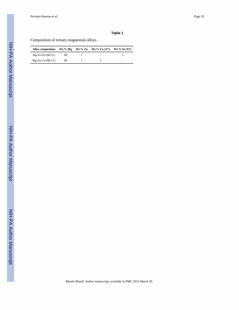
The alloy compositions were synthesized using an Arc-melting method in inert Argon gas atACI Alloys, Inc (CA, USA). Both alloy compositions were composed of weight percent (wt.%) ratios (98/1/1) Mg-Zn-X *. Materials were cast in a 1 in × 1 in × 6 in mold. Testspecimens were cut into ASTM-E8 sub-size specimen dimensions.
Samples were mechanically dry ground to #1200 grit finish using silicon carbide abrasivepaper. Samples were further polished to a 2 μm finish using an alumina suspension. Aftermechanical polishing, samples were cleaned in acetone and rinsed with deionized water andblown dry with nitrogen gas. Materials were stored in a plastic container until furthertesting.
2.2. XPS and Nanoindentation Analysis2.2.1. XPS Analysis—Samples were stored in a sealed plastic container for 4-months toallow an air formed oxide layer to develop on the surface of the polished alloys. The oxidelayer composition was characterized using a Physical Electronics 5400 ECSA (XPS) system,with a minimum analysis time of 40 min. A maximum radiation beam intensity of 1100 eVwas used for analysis on all samples.
2.2.2. Nanoindentation Analysis—Nanoindentation analysis was performed todetermine the elastic modulus (E) and surface hardness (H) of both the bare metal alloys andsamples with an air formed oxide layer. Testing was conducted using a TI 950 TriboIndenterat the Hysitron Testing Facility (MN, USA). The tests were performed using a diamond
Persaud-Sharma et al. Page 2
Metals (Basel). Author manuscript; available in PMC 2013 March 29.
NIH
-PA Author Manuscript
NIH
-PA Author Manuscript
NIH
-PA Author Manuscript
Berkovich indenter probe. Test parameters included using the load-controlled feedbackmode with a 5 s linear loading to a peak force of 10 mN, a 10 s hold at peak force, and a 5 slinear unloading duration. Tests were conducted in accordance with the Oliver-Pharranalysis method with a 5 × 5 analysis grid [7]. According to the Oliver-Pharr methodology,nanoindentation tests are performed by applying a force to drive an indenter probe into thesurface of a sample and then reducing the force to withdraw a probe. The applied load andindenter displacement into the sample are continuously monitored. After experimentation, aload vs. displacement curve was generated from collected data, allowing for the calculationof sample hardness (H) and elastic modulus (E). Indentation testing was performed on thesamples after the 4-month aging duration to determine the mechanical properties of theoxide layer. Later, the oxide layer was removed from the samples by mechanically polishingthe surface of the alloys and indentation measurements were gathered to determine (E) and(H) of the bare metal samples.
2.2.3. Material Density Calculation—The density of each experimental alloycomposition was determined using Archimedes principle as shown in Equation 1. Distilledwater was used as the liquid for testing with a density of 1.0 g·cm−3 (ρw). W is the weight ofthe object, and Wa is the apparent weight of the object in water.
(1)
2.3. Tensile Testing and Fracture Analysis2.3.1. Tensile Testing—Tensile testing specimens were prepared in accordance withASTM standard E8 sub-size specimen specifications. Testing was conducted using a MTSBionix 358.02 servohydraulic test system. Tensile tests were conducted at room temperaturewith a load cell of 1kN and a crosshead speed of 0.2 mm/min. From the stress-strain datacurves, 0.2% offset yield values were determined. Elongation at failure was calculated fromactual testing specimens and cross-referenced with data from stress-strain curves usingEquation 2, where L is the final stressed length and L0 is the unstressed length. PoissonRatio (ν) values for each alloy composition was calculated from physical measurements ofthe tensile specimens after testing according to Equation 3. The average gauge length valuefor all test specimens before testing was 30 mm, with an average width and thickness of 6.3mm and 2.1 mm, respectively. Tensile specimens were machined in the long dimension ofthe 1 in × 1 in × 6 in as-cast bar. Testing in additional directions was not possible due to thecasting mold used. Tensile testing was repeated on five samples, and the data is reported asthe statistical mean ± standard deviation (Table. 2).
(2)
(3)
2.3.2. Fracture Analysis by Scanning Electron Microscopy and EnergyDispersive X-Ray Spectroscopy—The fractured surfaces were examined after tensiletesting. The fractured surfaces were characterized using a JEOL JSM 5900 LV (SEM) andEDS-UTW Detector system (EDS). Images were analyzed in both secondary electronimaging (SEI) and backscatter imaging (BEI) modes at an accelerating voltage of 20 keV.
Persaud-Sharma et al. Page 3
Metals (Basel). Author manuscript; available in PMC 2013 March 29.
NIH
-PA Author Manuscript
NIH
-PA Author Manuscript
NIH
-PA Author Manuscript
3. Results3.1. Mechanical Properties of Bare Alloys and Air Formed Oxide Layer UsingNanoindentation
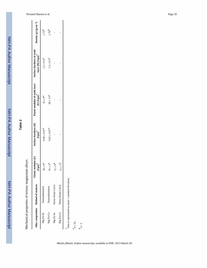
Table 2 summarizes the mechanical properties of the bare magnesium alloys and the airformed oxide layer. The bare Mg-Zn-Se alloy had a lower elastic modulus than Mg-Zn-Cu.Notably, the air formed oxide layers on both Mg-Zn-Se and Mg-Zn-Cu were revealed topossess higher (E) and (H) values than bare metal alloys. Reported oxide layer (H) valuesnearly doubled when compared with bare metal (H) values for both alloy compositions.
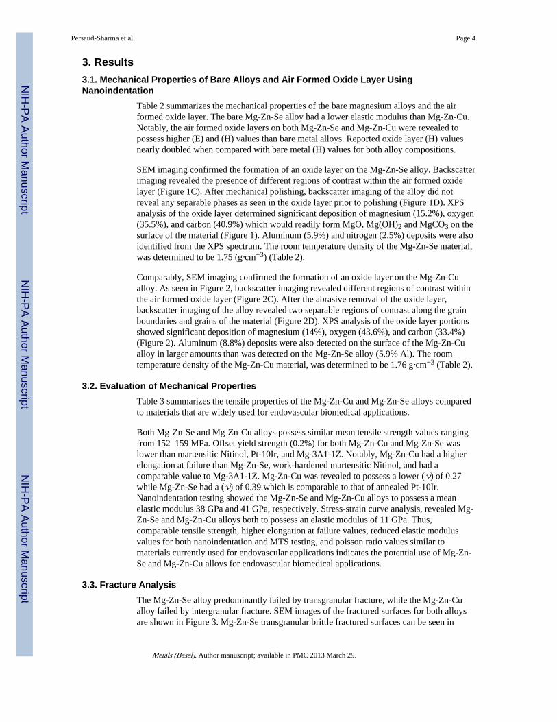
SEM imaging confirmed the formation of an oxide layer on the Mg-Zn-Se alloy. Backscatterimaging revealed the presence of different regions of contrast within the air formed oxidelayer (Figure 1C). After mechanical polishing, backscatter imaging of the alloy did notreveal any separable phases as seen in the oxide layer prior to polishing (Figure 1D). XPSanalysis of the oxide layer determined significant deposition of magnesium (15.2%), oxygen(35.5%), and carbon (40.9%) which would readily form MgO, Mg(OH)2 and MgCO3 on thesurface of the material (Figure 1). Aluminum (5.9%) and nitrogen (2.5%) deposits were alsoidentified from the XPS spectrum. The room temperature density of the Mg-Zn-Se material,was determined to be 1.75 (g·cm−3) (Table 2).
Comparably, SEM imaging confirmed the formation of an oxide layer on the Mg-Zn-Cualloy. As seen in Figure 2, backscatter imaging revealed different regions of contrast withinthe air formed oxide layer (Figure 2C). After the abrasive removal of the oxide layer,backscatter imaging of the alloy revealed two separable regions of contrast along the grainboundaries and grains of the material (Figure 2D). XPS analysis of the oxide layer portionsshowed significant deposition of magnesium (14%), oxygen (43.6%), and carbon (33.4%)(Figure 2). Aluminum (8.8%) deposits were also detected on the surface of the Mg-Zn-Cualloy in larger amounts than was detected on the Mg-Zn-Se alloy (5.9% Al). The roomtemperature density of the Mg-Zn-Cu material, was determined to be 1.76 g·cm−3 (Table 2).
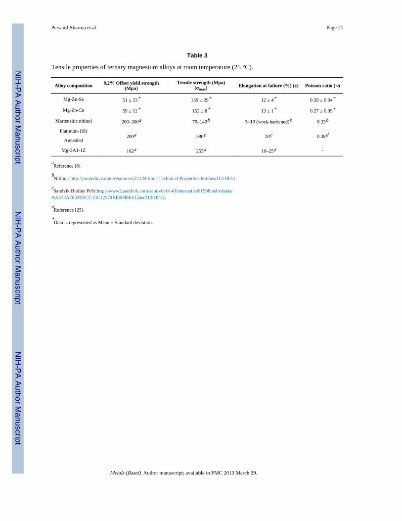
3.2. Evaluation of Mechanical PropertiesTable 3 summarizes the tensile properties of the Mg-Zn-Cu and Mg-Zn-Se alloys comparedto materials that are widely used for endovascular biomedical applications.
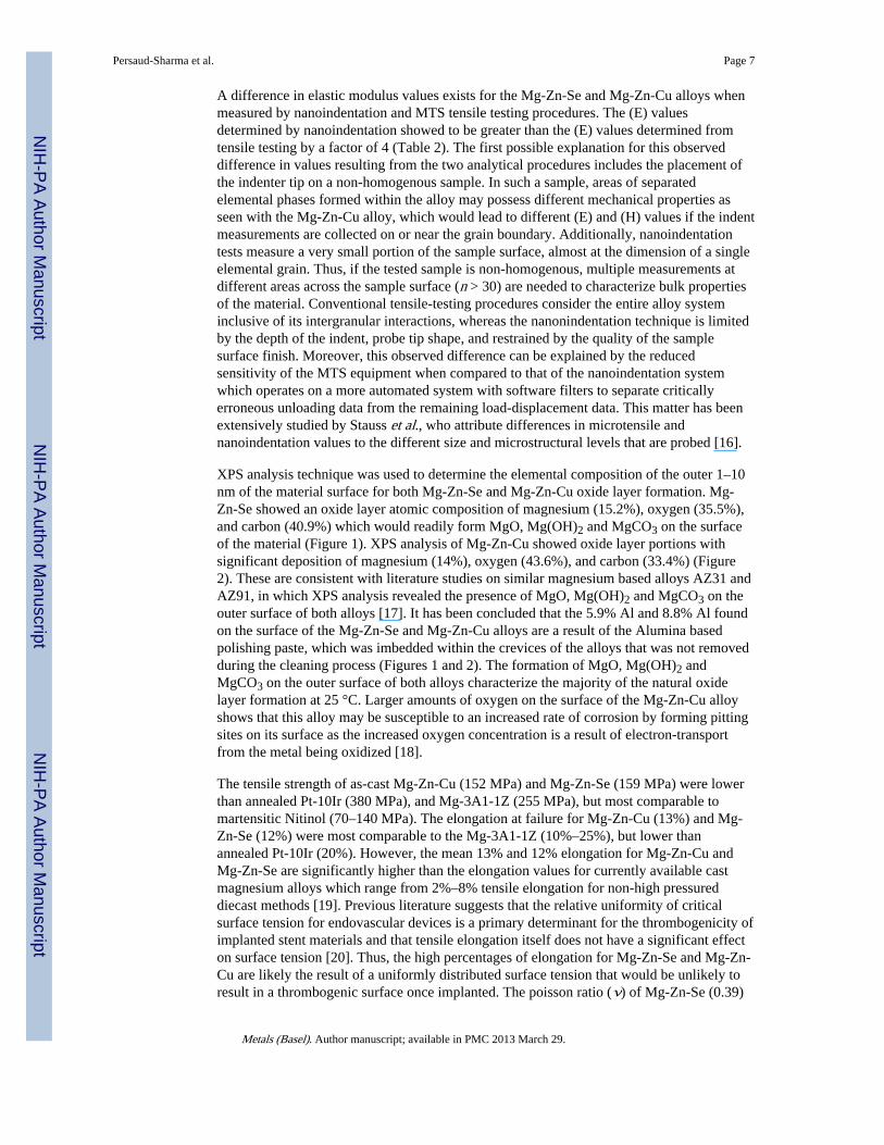
Both Mg-Zn-Se and Mg-Zn-Cu alloys possess similar mean tensile strength values rangingfrom 152–159 MPa. Offset yield strength (0.2%) for both Mg-Zn-Cu and Mg-Zn-Se waslower than martensitic Nitinol, Pt-10Ir, and Mg-3A1-1Z. Notably, Mg-Zn-Cu had a higherelongation at failure than Mg-Zn-Se, work-hardened martensitic Nitinol, and had acomparable value to Mg-3A1-1Z. Mg-Zn-Cu was revealed to possess a lower (ν) of 0.27while Mg-Zn-Se had a (ν) of 0.39 which is comparable to that of annealed Pt-10Ir.Nanoindentation testing showed the Mg-Zn-Se and Mg-Zn-Cu alloys to possess a meanelastic modulus 38 GPa and 41 GPa, respectively. Stress-strain curve analysis, revealed Mg-Zn-Se and Mg-Zn-Cu alloys both to possess an elastic modulus of 11 GPa. Thus,comparable tensile strength, higher elongation at failure values, reduced elastic modulusvalues for both nanoindentation and MTS testing, and poisson ratio values similar tomaterials currently used for endovascular applications indicates the potential use of Mg-Zn-Se and Mg-Zn-Cu alloys for endovascular biomedical applications.
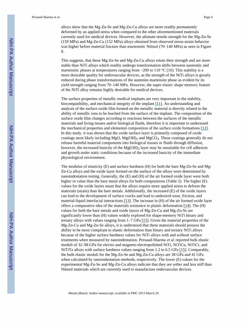
3.3. Fracture AnalysisThe Mg-Zn-Se alloy predominantly failed by transgranular fracture, while the Mg-Zn-Cualloy failed by intergranular fracture. SEM images of the fractured surfaces for both alloysare shown in Figure 3. Mg-Zn-Se transgranular brittle fractured surfaces can be seen in
Persaud-Sharma et al. Page 4
Metals (Basel). Author manuscript; available in PMC 2013 March 29.
NIH
-PA Author Manuscript
NIH
-PA Author Manuscript
NIH
-PA Author Manuscript
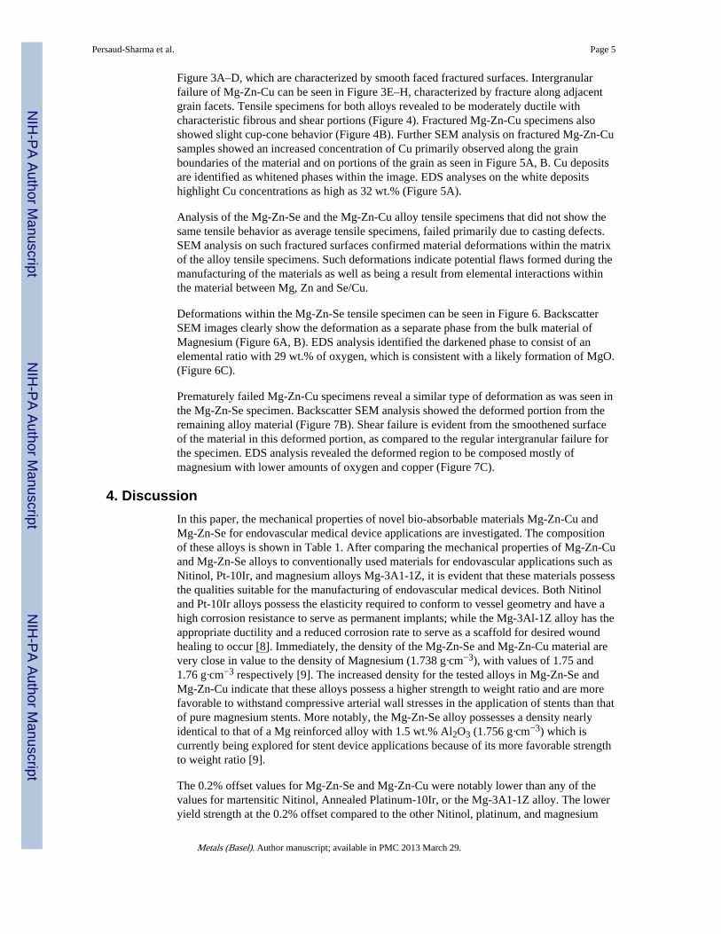
Figure 3A–D, which are characterized by smooth faced fractured surfaces. Intergranularfailure of Mg-Zn-Cu can be seen in Figure 3E–H, characterized by fracture along adjacentgrain facets. Tensile specimens for both alloys revealed to be moderately ductile withcharacteristic fibrous and shear portions (Figure 4). Fractured Mg-Zn-Cu specimens alsoshowed slight cup-cone behavior (Figure 4B). Further SEM analysis on fractured Mg-Zn-Cusamples showed an increased concentration of Cu primarily observed along the grainboundaries of the material and on portions of the grain as seen in Figure 5A, B. Cu depositsare identified as whitened phases within the image. EDS analyses on the white depositshighlight Cu concentrations as high as 32 wt.% (Figure 5A).
Analysis of the Mg-Zn-Se and the Mg-Zn-Cu alloy tensile specimens that did not show thesame tensile behavior as average tensile specimens, failed primarily due to casting defects.SEM analysis on such fractured surfaces confirmed material deformations within the matrixof the alloy tensile specimens. Such deformations indicate potential flaws formed during themanufacturing of the materials as well as being a result from elemental interactions withinthe material between Mg, Zn and Se/Cu.
Deformations within the Mg-Zn-Se tensile specimen can be seen in Figure 6. BackscatterSEM images clearly show the deformation as a separate phase from the bulk material ofMagnesium (Figure 6A, B). EDS analysis identified the darkened phase to consist of anelemental ratio with 29 wt.% of oxygen, which is consistent with a likely formation of MgO.(Figure 6C).
Prematurely failed Mg-Zn-Cu specimens reveal a similar type of deformation as was seen inthe Mg-Zn-Se specimen. Backscatter SEM analysis showed the deformed portion from theremaining alloy material (Figure 7B). Shear failure is evident from the smoothened surfaceof the material in this deformed portion, as compared to the regular intergranular failure forthe specimen. EDS analysis revealed the deformed region to be composed mostly ofmagnesium with lower amounts of oxygen and copper (Figure 7C).
4. DiscussionIn this paper, the mechanical properties of novel bio-absorbable materials Mg-Zn-Cu andMg-Zn-Se for endovascular medical device applications are investigated. The compositionof these alloys is shown in Table 1. After comparing the mechanical properties of Mg-Zn-Cuand Mg-Zn-Se alloys to conventionally used materials for endovascular applications such asNitinol, Pt-10Ir, and magnesium alloys Mg-3A1-1Z, it is evident that these materials possessthe qualities suitable for the manufacturing of endovascular medical devices. Both Nitinoland Pt-10Ir alloys possess the elasticity required to conform to vessel geometry and have ahigh corrosion resistance to serve as permanent implants; while the Mg-3Al-1Z alloy has theappropriate ductility and a reduced corrosion rate to serve as a scaffold for desired woundhealing to occur [8]. Immediately, the density of the Mg-Zn-Se and Mg-Zn-Cu material arevery close in value to the density of Magnesium (1.738 g·cm−3), with values of 1.75 and1.76 g·cm−3 respectively [9]. The increased density for the tested alloys in Mg-Zn-Se andMg-Zn-Cu indicate that these alloys possess a higher strength to weight ratio and are morefavorable to withstand compressive arterial wall stresses in the application of stents than thatof pure magnesium stents. More notably, the Mg-Zn-Se alloy possesses a density nearlyidentical to that of a Mg reinforced alloy with 1.5 wt.% Al2O3 (1.756 g·cm−3) which iscurrently being explored for stent device applications because of its more favorable strengthto weight ratio [9].
The 0.2% offset values for Mg-Zn-Se and Mg-Zn-Cu were notably lower than any of thevalues for martensitic Nitinol, Annealed Platinum-10Ir, or the Mg-3A1-1Z alloy. The loweryield strength at the 0.2% offset compared to the other Nitinol, platinum, and magnesium
Persaud-Sharma et al. Page 5
Metals (Basel). Author manuscript; available in PMC 2013 March 29.
NIH
-PA Author Manuscript
NIH
-PA Author Manuscript
NIH
-PA Author Manuscript
alloys show that the Mg-Zn-Se and Mg-Zn-Cu alloys are more readily permanentlydeformed by an applied stress when compared to the other aforementioned materialscurrently used for medical devices. However, the ultimate tensile strength for the Mg-Zn-Se(159 MPa) and Mg-Zn-Cu (152 MPa) alloys obtained from observed stress-strain behaviorwas higher before material fracture than martensitic Nitinol (70–140 MPa) as seen in Figure8.
This suggests, that these Mg-Zn-Se and Mg-Zn-Cu alloys retain their strength and are morestable than NiTi alloys which readily undergo transformation shifts between austenitic andmartensitic phases at temperatures ranging from −200 to 110 °C [10]. This stability is amore desirable quality for endovascular devices, as the strength of the NiTi alloys is greatlyreduced during phase transformations of the austenite-martensite phase as evident by itsyield strength ranging from 70–140 MPa. However, the super elastic shape memory featureof the NiTi alloy remains highly desirable for medical devices.
The surface properties of metallic medical implants are very important to the stability,biocompatibility, and mechanical integrity of the implant [11]. An understanding andanalysis of the surface oxide film formed on the metallic material is directly related to theability of metallic ions to be leached from the surface of the implant. The composition of thesurface oxide film changes according to reactions between the surfaces of the metallicmaterials and living tissues and/or biological fluids, therefore it is important to understandthe mechanical properties and elemental composition of the surface oxide formations [12].In this study, it was shown that the oxide surface layer is primarily composed of oxidecoatings most likely including MgO, Mg(OH)2 and MgCO3. These coatings generally do notrelease harmful material components into biological tissues or fluids through diffusion,however, the increased basicity of the Mg(OH)2 layer may be unsuitable for cell adhesionand growth under static conditions because of the increased basicity of the immediatephysiological environment.
The modulus of elasticity (E) and surface hardness (H) for both the bare Mg-Zn-Se and Mg-Zn-Cu alloys and the oxide layer formed on the surface of the alloys were determined bynanoindentation testing. Generally, the (E) and (H) of the air formed oxide layer were bothhigher in value than the bare metal alloys for both compositions (Table 2). The higher (E)values for the oxide layers mean that the alloys require more applied stress to deform thematerials (strain) than the bare metals. Additionally, the increased (E) of the oxide layerscan lead to the development of surface cracks and lead to undesired wear, friction, andmaterial-liquid interfacial interactions [13]. The increase in (H) of the air formed oxide layeroffers a comparative idea of the materials resistance to plastic deformation [14]. The (H)values for both the bare metals and oxide layers of Mg-Zn-Cu and Mg-Zn-Se aresignificantly lower than (H) values widely explored for shape-memory NiTi binary andternary alloys with values ranging from 1–7 GPa [15]. Given the material properties of theMg-Zn-Cu and Mg-Zn-Se alloys, it is understood that these materials should possess theability to be more compliant to elastic deformation than binary and ternary NiTi alloysbecause of the higher surface hardness values for NiTi alloys with and without surfacetreatments when measured by nanoindentation. Persaud-Sharma et al. reported bulk elasticmoduli of 32–98 GPa for electro and magneto-electropolished NiTi, NiTiCu, NiTiCr, andNiTiTa alloys with surface hardness values ranging from 1.2 to 6.5 GPa [15]. Comparably,the bulk elastic moduli for the Mg-Zn-Se and Mg-Zn-Cu alloys are 38 GPa and 41 GPawhen calculated by nanoindentation methods, respectively. The lower (E) values for theexperimental Mg-Zn-Se and Mg-Zn-Cu alloys indicate that they are softer and less stiff thanNitinol materials which are currently used to manufacture endovascular devices.
Persaud-Sharma et al. Page 6
Metals (Basel). Author manuscript; available in PMC 2013 March 29.
NIH
-PA Author Manuscript
NIH
-PA Author Manuscript
NIH
-PA Author Manuscript
A difference in elastic modulus values exists for the Mg-Zn-Se and Mg-Zn-Cu alloys whenmeasured by nanoindentation and MTS tensile testing procedures. The (E) valuesdetermined by nanoindentation showed to be greater than the (E) values determined fromtensile testing by a factor of 4 (Table 2). The first possible explanation for this observeddifference in values resulting from the two analytical procedures includes the placement ofthe indenter tip on a non-homogenous sample. In such a sample, areas of separatedelemental phases formed within the alloy may possess different mechanical properties asseen with the Mg-Zn-Cu alloy, which would lead to different (E) and (H) values if the indentmeasurements are collected on or near the grain boundary. Additionally, nanoindentationtests measure a very small portion of the sample surface, almost at the dimension of a singleelemental grain. Thus, if the tested sample is non-homogenous, multiple measurements atdifferent areas across the sample surface (n > 30) are needed to characterize bulk propertiesof the material. Conventional tensile-testing procedures consider the entire alloy systeminclusive of its intergranular interactions, whereas the nanonindentation technique is limitedby the depth of the indent, probe tip shape, and restrained by the quality of the samplesurface finish. Moreover, this observed difference can be explained by the reducedsensitivity of the MTS equipment when compared to that of the nanoindentation systemwhich operates on a more automated system with software filters to separate criticallyerroneous unloading data from the remaining load-displacement data. This matter has beenextensively studied by Stauss et al., who attribute differences in microtensile andnanoindentation values to the different size and microstructural levels that are probed [16].
XPS analysis technique was used to determine the elemental composition of the outer 1–10nm of the material surface for both Mg-Zn-Se and Mg-Zn-Cu oxide layer formation. Mg-Zn-Se showed an oxide layer atomic composition of magnesium (15.2%), oxygen (35.5%),and carbon (40.9%) which would readily form MgO, Mg(OH)2 and MgCO3 on the surfaceof the material (Figure 1). XPS analysis of Mg-Zn-Cu showed oxide layer portions withsignificant deposition of magnesium (14%), oxygen (43.6%), and carbon (33.4%) (Figure2). These are consistent with literature studies on similar magnesium based alloys AZ31 andAZ91, in which XPS analysis revealed the presence of MgO, Mg(OH)2 and MgCO3 on theouter surface of both alloys [17]. It has been concluded that the 5.9% Al and 8.8% Al foundon the surface of the Mg-Zn-Se and Mg-Zn-Cu alloys are a result of the Alumina basedpolishing paste, which was imbedded within the crevices of the alloys that was not removedduring the cleaning process (Figures 1 and 2). The formation of MgO, Mg(OH)2 andMgCO3 on the outer surface of both alloys characterize the majority of the natural oxidelayer formation at 25 °C. Larger amounts of oxygen on the surface of the Mg-Zn-Cu alloyshows that this alloy may be susceptible to an increased rate of corrosion by forming pittingsites on its surface as the increased oxygen concentration is a result of electron-transportfrom the metal being oxidized [18].
The tensile strength of as-cast Mg-Zn-Cu (152 MPa) and Mg-Zn-Se (159 MPa) were lowerthan annealed Pt-10Ir (380 MPa), and Mg-3A1-1Z (255 MPa), but most comparable tomartensitic Nitinol (70–140 MPa). The elongation at failure for Mg-Zn-Cu (13%) and Mg-Zn-Se (12%) were most comparable to the Mg-3A1-1Z (10%–25%), but lower thanannealed Pt-10Ir (20%). However, the mean 13% and 12% elongation for Mg-Zn-Cu andMg-Zn-Se are significantly higher than the elongation values for currently available castmagnesium alloys which range from 2%–8% tensile elongation for non-high pressureddiecast methods [19]. Previous literature suggests that the relative uniformity of criticalsurface tension for endovascular devices is a primary determinant for the thrombogenicity ofimplanted stent materials and that tensile elongation itself does not have a significant effecton surface tension [20]. Thus, the high percentages of elongation for Mg-Zn-Se and Mg-Zn-Cu are likely the result of a uniformly distributed surface tension that would be unlikely toresult in a thrombogenic surface once implanted. The poisson ratio (ν) of Mg-Zn-Se (0.39)
Persaud-Sharma et al. Page 7
Metals (Basel). Author manuscript; available in PMC 2013 March 29.
NIH
-PA Author Manuscript
NIH
-PA Author Manuscript
NIH
-PA Author Manuscript
and Mg-Zn-Cu (0.27) are comparable to both martensitic Nitinol (0.33) and annealed Pt-10Ir(0.38) (Table 3). The ratio of both Mg-Zn-Se and Mg-Zn-Cu are below the incompressibilitylimit of 0.5, thus the materials retain the longitudinal and transverse elastic flexibility [21].
The fracture analysis of the Mg-Zn-Se alloy and the Mg-Zn-Cu alloy was performedvisually by SEM imaging and by energy dispersive X-ray spectroscopy (EDS). Analysisrevealed that the Mg-Zn-Se alloy failed transgranularly as evident by the smooth facedfractured surfaces (Figure 3A–D). Mg-Zn-Cu predominantly failed by intergranular failureas seen by the outlined grain structures on the fractured surfaces (Figure 3E–H). Thetransgranular failure of Mg-Zn-Se is comparable to the transgranular fracture mechanism ofthe AZ31B alloys which is currently used for stenting devices [22]. This mode of failureindicates that the Mg-Zn-Se alloy is more brittle like the AZ31B alloy. Alternatively, theMg-Zn-Cu alloy undergoes intergranular failure indicating that it is more ductile, which is amore favorable quality in the medical device forming process. Backscatter imaging revealedno separate phases in the fractured surfaces of the Mg-Zn-Se alloy, whereas two visiblyseparate phases were evident for Mg-Zn-Cu alloy (Figure 3F,H). Further EDS analysis onthe phases identified them as being primarily composed of Cu on the faces of the fracturedsurfaces and along the grain boundaries (Figure 5). This is consistent with Zhiyong et al.,who proposed that this accumulation of Cu along the grain boundary of a high zincmagnesium alloy with copper additions (Mg-10Zn-5Al-0.1Sb) is actually the formation of anew Mg2Cu phase. The localization of the thermally stable Mg2Cu phase along the grainboundaries can strengthen the alloy by dispersive strengthening through pinning themovement of dislocations and the slip of the Mg-Zn-Cu matrix [23,24]. Materialmanufacturing defects common to occur within the bulk material of the Mg-Zn-Cu and Mg-Zn-Se alloys were apparent in some tensile specimens, which led to premature mechanicalfailure. MgO and Mg(OH)2 concentrated regions were detected in Mg-Zn-Se defectivespecimens which led to premature tensile failure (Figure 6). This is in accordance withprevious literature which found similar oxide formations of MgO, Mg(OH)2, and MgCO3within the inner layer of the AZ31 and AZ91 alloy [17]. The casting defect for the Mg-Zn-Cu alloy showed a region with higher concentrations of MgO, but more deplete of Zn andCu as compared to grain boundary concentrations (Figure 7). Regions of such a homogenousmagnesium concentration and the depletion of Cu from within the matrix are believed tohave led to the deteriorated tensile strength of the Mg-Zn-Cu alloy. The poisson ratio of bothalloys are below the incompressibility limit of 0.5, hence the alloys possess the longitudinaland transverse elastic flexibility required for manufacturing.
5. ConclusionsExperimental alloys Mg-Zn-Cu and Mg-Zn-Se show promise for applications asendovascular medical devices as they possess comparable mechanical properties to Nitinol,Pt-10Ir, and the Mg-3A1-1Z alloy. Both experimental alloys possess air formed oxide layermodulus of elasticity values that were greater than the bulk elastic modulus values. Theoxide layers were determined to be composed of MgO, Mg(OH)2, and MgCO3 as revealedby XPS analysis which could foreshadow wear, friction, structural changes, and surface-liquid interfacial behavior. Mg-Zn-Cu and Mg-Zn-Se favorably showed tensile elongation atfailure values of 13% and 12%, respectively. These values are higher than any currentlycited, as-cast magnesium alloys available and can lead to more tolerable material limitswhen developing and forming the raw material into a functional form. Fracture analysisshowed that the Mg-Zn-Se alloy failed transgranularly, while the Mg-Zn-Cu alloy failed byan intergranular fracture mechanism. EDS analysis showed the localization of a Mg2Cuphase along the grain boundaries of the Mg-Zn-Cu alloy. Elevated MgO formations anddepleted Mg-Zn and Mg-Cu regions characterized the defective regions of the Mg-Zn-Cualloy which caused premature failure.
Persaud-Sharma et al. Page 8
Metals (Basel). Author manuscript; available in PMC 2013 March 29.
NIH
-PA Author Manuscript
NIH
-PA Author Manuscript
NIH
-PA Author Manuscript
AcknowledgmentsThis project was developed by Dharam Persaud (PI), who is also a recipient of a grant from the National BrainAneurysm Foundation which financially supports this project. Dharam Persaud is also supported by NIHFellowship number NIH/NIGMS R25 GM061347. Dharam Persaud would like to thank Walter McKinley for hisoperation of the MTS tensile testing equipment, and extend his gratitude to Noah Budiansky for his valuable insightinto the preparation of this manuscript.
References1. Persaud-Sharma D, McGoron A. Biodegradable magnesium alloys: A review of material
development and applications. J Biomimetics Biomater Tissue Eng. 2012; 12:25–39.
2. Saito S. New horizon of bioabsorbable stent. Catheter Cardiovasc Interv. 2005; 66:595–596.[PubMed: 16284981]
3. Moravej M, Mantovani M. Biodegradable metals for cardiovascular stent application: Interest andnew opportunities. Int J Mol Sci. 2011; 12:4250–4270. [PubMed: 21845076]
4. Erne P, Schier M, Resink TJ. The road to bioabsorbable stents: Reaching clinical reality?Cardiovasc Intervent Radiol. 2006; 29:11–16. [PubMed: 16195840]
5. Colombo A, Karvouni E. Biodegradable stents: Fulfilling the mission and stepping away.Circulation. 2000; 102:371–373. [PubMed: 10908206]
6. Peuster M, Wohlsein P, Brugmann M, Ehlerding M, Seidler K, Fink C, Brauer H, Fischer A,Hausdorf G. A novel approach to temporary stenting: degradable cardiovascular stents producedfrom corrodible metal-results 6–18 months after implantation into New Zealand white rabbits.Heart. 2001; 86:563–569. [PubMed: 11602554]
7. Oliver WC, Pharr GM. An improved technique for determining hardness and elastic modulus usingload and displacement sensing indentation experiments. J Mater Res. 1992; 7:1564.
8. Poncin, P.; Proft, J. Stent Tubing: Understanding the Desired Attributes. Proceedings from theMaterials & Processes for Medical Devices Conference; Anaheim, CA, USA. September 8–10:2003; Material Park, OH, USA: ASM International; 2003.
9. Gupta, M.; Sahron, NML. Magnesium, Magnesium Alloys, and Magnesium Composites: A Guide.Vol. 1. John Wiley & Sons; Hoboken, NJ, USA: 2011. p. 120-150.
10. Johnson Matthey Medical Components. [accessed on 29 November 2012] Nitinol TechnicalProperties. Available online: http://jmmedical.com/resources/221/Nitinol-Technical-Properties.html#nitinol-transformation-properties
11. Kasemo B, Lausmaa J. Surface science aspects on inorganic biomaterials. Crit Rev Biocompat.1986; 2:335–330.
12. Manivasagam G, Dhinasekaran D, Rajamanickam A. Biomedical implants: Corrosion and itsprevention-a review. Recent Pat Corros Sci. 2010; 2:40–54.
13. Persson DHE, Jacobson S, Hogmark S. The influence of phase transformations and oxidation onthe galling resistance and low friction behavior of laser processed Co-based alloy. Wear. 2003;254:1134–1140.
14. Meyers, M.; Chawla, K. Mechanical Behaviors of Materials. 2. Cambridge University Press; NewYork, NY, USA: 1999. p. 162-168.
15. Persaud-Sharma D, Munroe N, McGoron A. Electro and magneto-electropolished surface micro-patterning on binary and ternary Nitinol. Trends Biomater Artif Organs. 2012; 26:74–85.[PubMed: 22754200]
16. Stauss S, Schwaller P, Bucaille J-L, Rabe R, Rohr L, Michler J, Blank E. Determining the stress-strain behavior of small devices by nanoindentation in combination with inverse methods.Microelectron Eng. 2003; 67–68:818–825.
17. Wang L, Shinohara T, Zhang BP. XPS study of the surface chemistry on AZ31and AZ91magnesium alloys in dilute NaCl solution. Appl Surf Sci. 2010; 256:5807–5812.
18. Revie, RW.; Uhlig, HH. Corrosion and Corrosion Control: An Introduction to Corrosion Scienceand Engineering. Vol. 4. John Wiley and Sons; Hoboken, NJ, USA: 2008. p. 125-165.
Persaud-Sharma et al. Page 9
Metals (Basel). Author manuscript; available in PMC 2013 March 29.
NIH
-PA Author Manuscript
NIH
-PA Author Manuscript
NIH
-PA Author Manuscript
19. Grote, KH.; Antonsson, E. Handbook of Mechanical Engineering. Vol. 10. Springer; New York,NY, USA: 2010. p. 530-540.
20. Shrivastava, S. Medical Device Materials. Proceedings from the Materials & Processes for MedicalDevices Conference; Anaheim, CA, USA. September 8–10: 2003; Material Park, OH, USA: ASMInternational; 2003. p. 69-74.
21. Liu B, Zhang L, Gao H. Poisson ratio can play an important role in mechanical properties ofbiocomposites. Mech Mater. 2006; 38:1128–1142.
22. Zhang H, Wang W, Wei Y, Li J, Wang J. Fatigue fracture mechanism of AZ31B magnesium alloyand its welded joint. Trans Nonferrous Met Soc China. 2011; 21:1225–1233.
23. Zhiyong Y, Yuhua Z, Weili C, Jinshan Z, Yinghui W. Effect of Cu addition on microstructure andproperties of Mg-10Zn-5Al-0. 1Sb high zinc magnesium alloy. China Foundry. 2012; 9:43–47.
24. Chen, Z. Heat-Resistant Magnesium Alloy. Chemical Industry Press; Beijing, China: 2006. p.109-115.
25. Merker J, Lupton D, Topfer M, Knake H. High temperature mechanical properties of the platinumgroup metals. Platinum Metals Review. 2001; 45:74–82.
Persaud-Sharma et al. Page 10
Metals (Basel). Author manuscript; available in PMC 2013 March 29.
NIH
-PA Author Manuscript
NIH
-PA Author Manuscript
NIH
-PA Author Manuscript

Figure 1.Scanning electron microscopy (SEM) images and X-ray photoelectron spectroscopy (XPS)analysis of oxide layer formation on the Mg-Zn-Se alloy.
Persaud-Sharma et al. Page 11
Metals (Basel). Author manuscript; available in PMC 2013 March 29.
NIH
-PA Author Manuscript
NIH
-PA Author Manuscript
NIH
-PA Author Manuscript
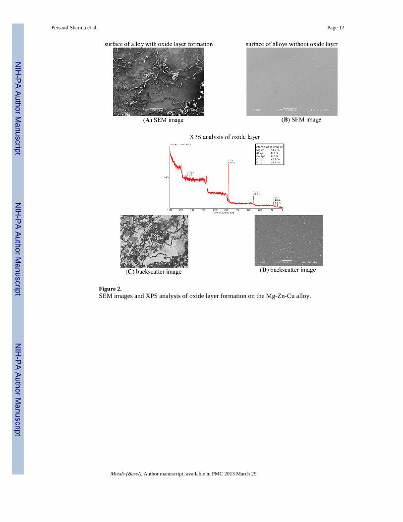
Figure 2.SEM images and XPS analysis of oxide layer formation on the Mg-Zn-Cu alloy.
Persaud-Sharma et al. Page 12
Metals (Basel). Author manuscript; available in PMC 2013 March 29.
NIH
-PA Author Manuscript
NIH
-PA Author Manuscript
NIH
-PA Author Manuscript
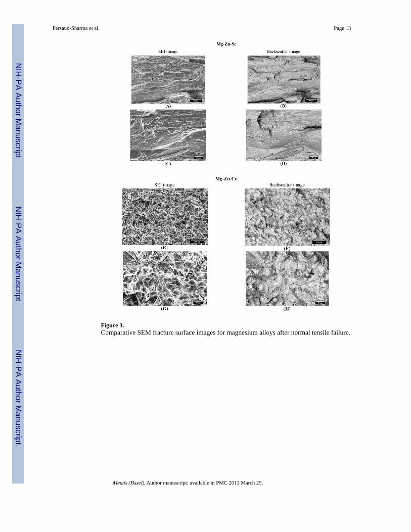
Figure 3.Comparative SEM fracture surface images for magnesium alloys after normal tensile failure.
Persaud-Sharma et al. Page 13
Metals (Basel). Author manuscript; available in PMC 2013 March 29.
NIH
-PA Author Manuscript
NIH
-PA Author Manuscript
NIH
-PA Author Manuscript
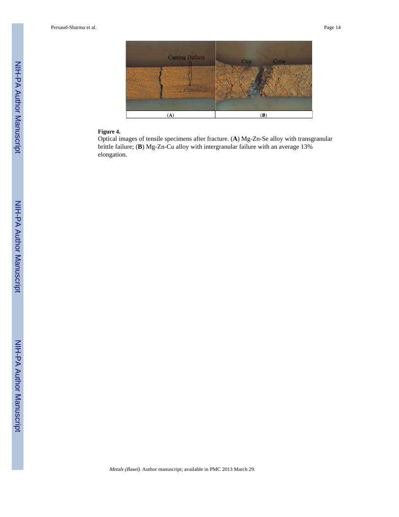
Figure 4.Optical images of tensile specimens after fracture. (A) Mg-Zn-Se alloy with transgranularbrittle failure; (B) Mg-Zn-Cu alloy with intergranular failure with an average 13%elongation.
Persaud-Sharma et al. Page 14
Metals (Basel). Author manuscript; available in PMC 2013 March 29.
NIH
-PA Author Manuscript
NIH
-PA Author Manuscript
NIH
-PA Author Manuscript
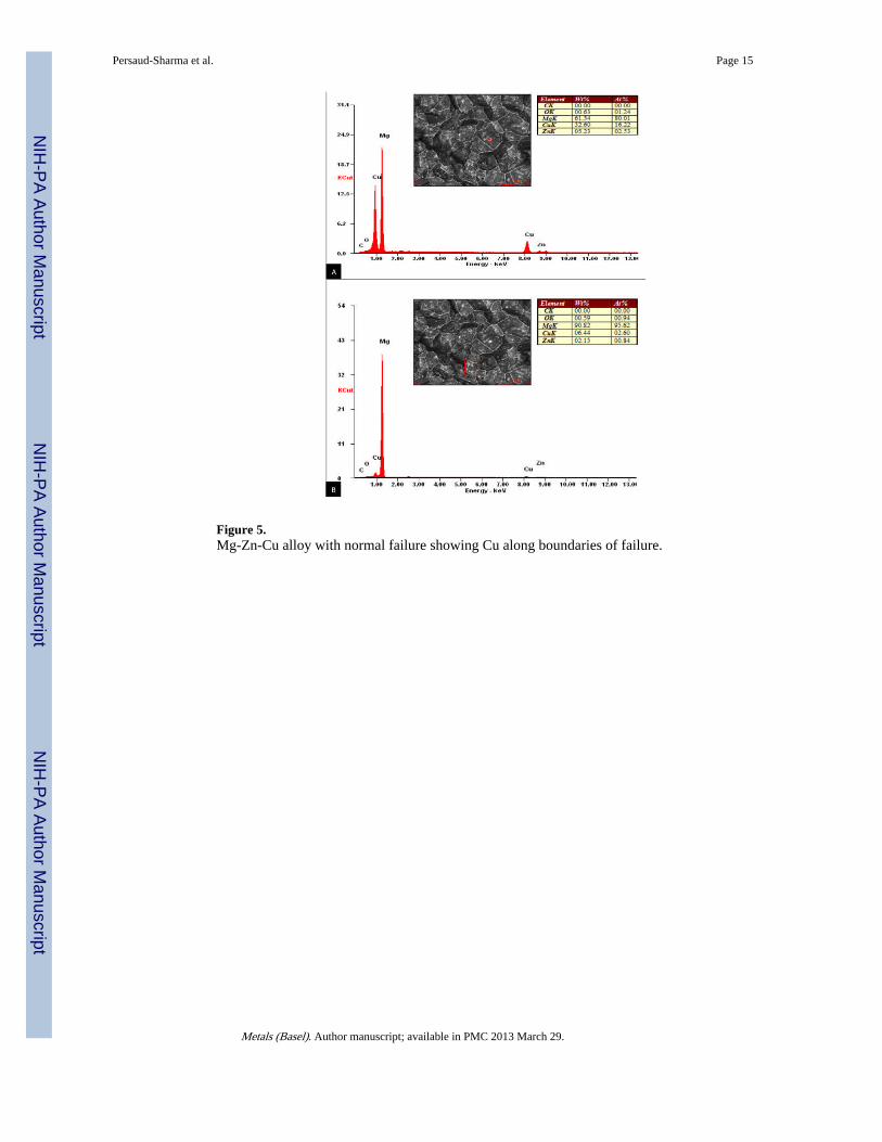
Figure 5.Mg-Zn-Cu alloy with normal failure showing Cu along boundaries of failure.
Persaud-Sharma et al. Page 15
Metals (Basel). Author manuscript; available in PMC 2013 March 29.
NIH
-PA Author Manuscript
NIH
-PA Author Manuscript
NIH
-PA Author Manuscript
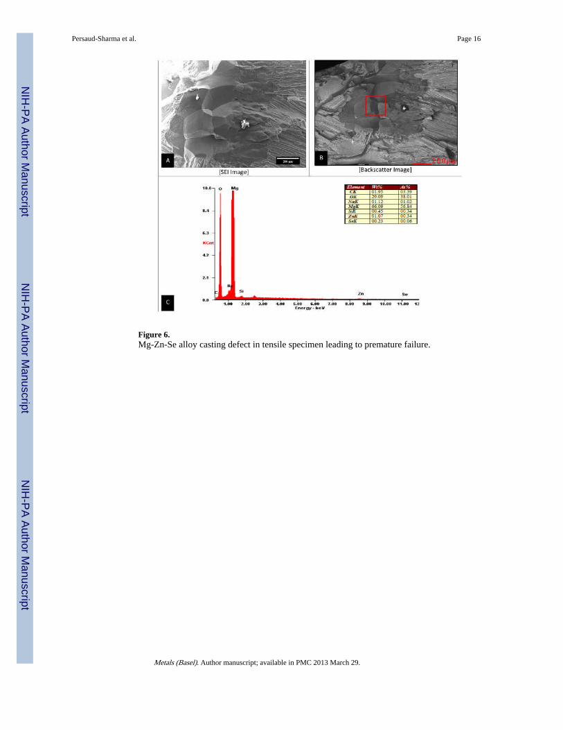
Figure 6.Mg-Zn-Se alloy casting defect in tensile specimen leading to premature failure.
Persaud-Sharma et al. Page 16
Metals (Basel). Author manuscript; available in PMC 2013 March 29.
NIH
-PA Author Manuscript
NIH
-PA Author Manuscript
NIH
-PA Author Manuscript
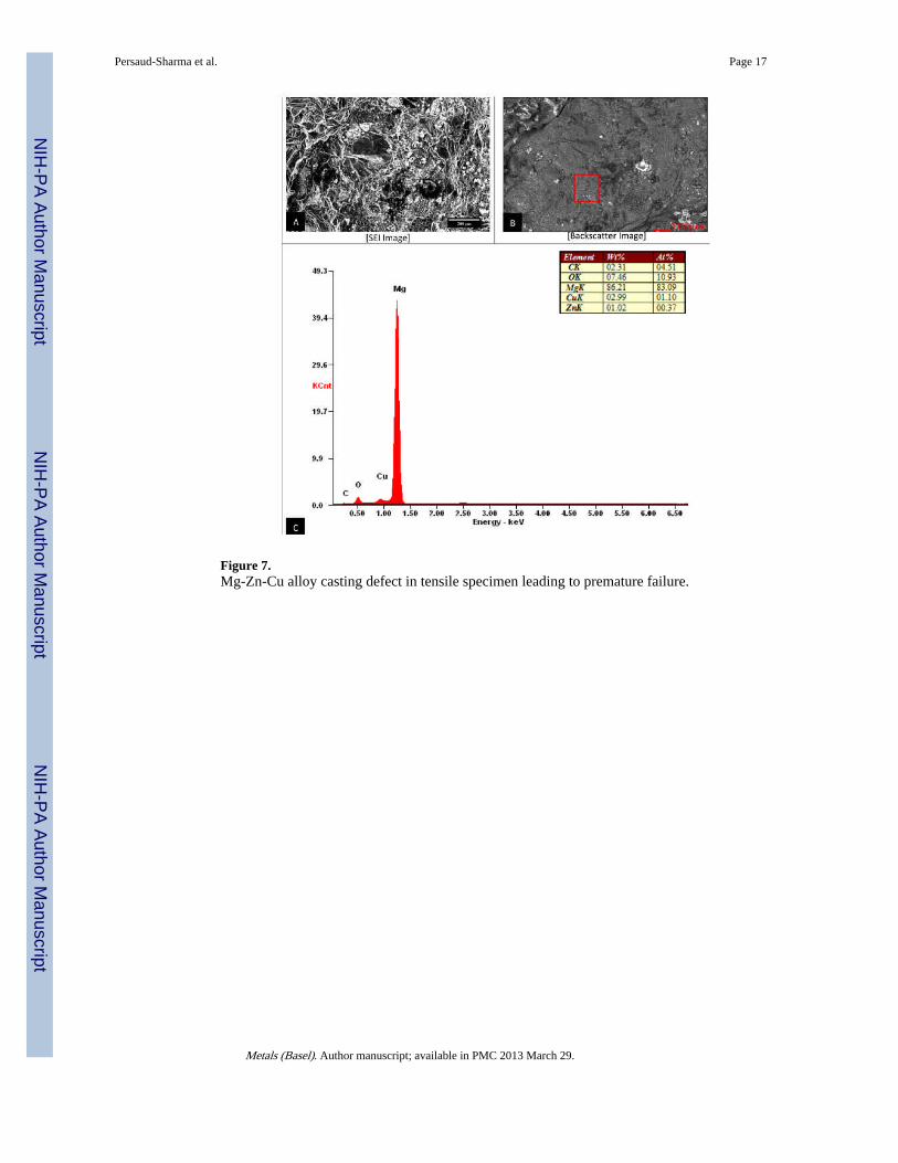
Figure 7.Mg-Zn-Cu alloy casting defect in tensile specimen leading to premature failure.
Persaud-Sharma et al. Page 17
Metals (Basel). Author manuscript; available in PMC 2013 March 29.
NIH
-PA Author Manuscript
NIH
-PA Author Manuscript
NIH
-PA Author Manuscript
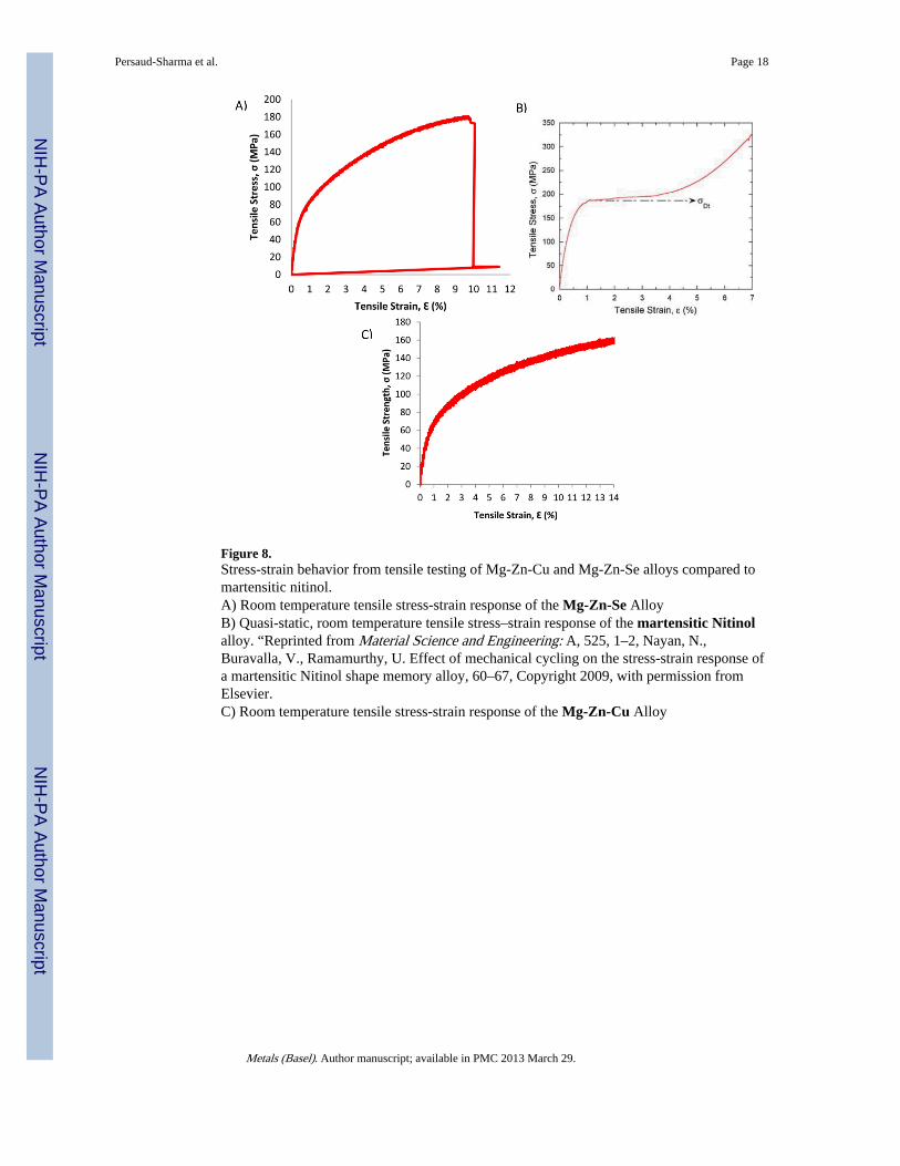
Figure 8.Stress-strain behavior from tensile testing of Mg-Zn-Cu and Mg-Zn-Se alloys compared tomartensitic nitinol.A) Room temperature tensile stress-strain response of the Mg-Zn-Se AlloyB) Quasi-static, room temperature tensile stress–strain response of the martensitic Nitinolalloy. “Reprinted from Material Science and Engineering: A, 525, 1–2, Nayan, N.,Buravalla, V., Ramamurthy, U. Effect of mechanical cycling on the stress-strain response ofa martensitic Nitinol shape memory alloy, 60–67, Copyright 2009, with permission fromElsevier.C) Room temperature tensile stress-strain response of the Mg-Zn-Cu Alloy
Persaud-Sharma et al. Page 18
Metals (Basel). Author manuscript; available in PMC 2013 March 29.
NIH
-PA Author Manuscript
NIH
-PA Author Manuscript
NIH
-PA Author Manuscript
NIH
-PA Author Manuscript
NIH
-PA Author Manuscript
NIH
-PA Author Manuscript
Persaud-Sharma et al. Page 19
Table 1
Composition of ternary magnesium alloys.
Alloy composition Wt.% Mg Wt.% Zn Wt.% Cu (X*) Wt.% Se (X*)
Mg-Zn-Se (98/1/1) 98 1 - 1
Mg-Zn-Cu (98/1/1) 98 1 1 -
Metals (Basel). Author manuscript; available in PMC 2013 March 29.
NIH
-PA Author Manuscript
NIH
-PA Author Manuscript
NIH
-PA Author Manuscript
Persaud-Sharma et al. Page 20
Tabl
e 2
Mec
hani
cal p
rope
rtie
s of
tern
ary
mag
nesi
um a
lloys
.
Allo
y co
mpo
siti
onM
etho
d of
ana
lysi
sE
last
ic m
odul
us (
E)
(Gpa
)*Su
rfac
e ha
rdne
ss (
H)
(Gpa
)*E
last
ic m
odul
us o
f ox
ide
laye
r(E
) (G
pa)*
Surf
ace
hard
ness
of
oxid
ela
yer
(H)
(Gpa
)*D
ensi
ty (ρ)
(g·
cm−3
)
Mg-
Zn-
SeN
anoi
nden
tatio
n38
± 4
a0.
69 ±
0.0
7a55
± 9
a1.
2 ±
0.3
a1.
75b
Mg-
Zn-
Cu
Nan
oind
enta
tion
41 ±
3a
0.62
± 0
.07a
60 ±
13a
1.3
± 0
.3a
1.76
b
Mg-
Zn-
SeSt
ress
-Str
ain
Cur
ve11
± 6
b-
--
-
Mg-
Zn-
Cu
Stre
ss-S
trai
n C
urve
11 ±
1b
--
--
* Dat
a is
rep
rese
nted
as
mea
n ±
sta
ndar
d de
viat
ion;
a n =
25,
b n =
5
Metals (Basel). Author manuscript; available in PMC 2013 March 29.
NIH
-PA Author Manuscript
NIH
-PA Author Manuscript
NIH
-PA Author Manuscript
Persaud-Sharma et al. Page 21
Table 3
Tensile properties of ternary magnesium alloys at room temperature (25 °C).
Alloy composition 0.2% Offset yield strength(Mpa)
Tensile strength (Mpa)(σmax) Elongation at failure (%) (ε) Poisson ratio (ν)
Mg-Zn-Se 51 ± 23* 159 ± 29* 12 ± 4* 0.39 ± 0.04*
Mg-Zn-Cu 59 ± 12* 152 ± 8* 13 ± 1* 0.27 ± 0.09*
Martensitic nitinol 200–300a 70–140b 5–10 (work-hardened)b 0.33b
Platinum-10Ir200a 380c 20c 0.38d
Annealed
Mg-3A1-1Z 162a 255a 10–25a -
aReference [8].
bNitinol: http://jmmedical.com/resources/221/Nitinol-Technical-Properties.htmlasof12/18/12.
cSandvik Bioline Pt/Ir:http://www2.sandvik.com/sandvik/0140/internet/se01598.nsf/cdatas/
AA573A7633EECC13C12574BE0046E612asof12/18/12.
dReference [25].
*Data is represented as Mean ± Standard deviation.
Metals (Basel). Author manuscript; available in PMC 2013 March 29.