Phylogenetic Analysis of Morphological Data from Extinct and ...
Upload
independentCategory
view
1download
0
Mechanical Analysis of Feeding Behavior in the Extinct‘‘Terror Bird’’ Andalgalornis steulleti (Gruiformes:Phorusrhacidae)Federico J. Degrange1*, Claudia P. Tambussi1, Karen Moreno2,3, Lawrence M. Witmer4, Stephen Wroe3
1 CONICET - Division Paleontologıa Vertebrados, Facultad de Ciencias Naturales y Museo, Museo de La Plata, Universidad Nacional de La Plata, La Plata, Argentina,
2 Laboratorio de Paleontologıa, Instituto de Geociencias, Universidad Austral de Chile, Valdivia, Chile, 3 Evolution and Ecology Research Centre, School of Biological, Earth
and Environmental Sciences, University of New South Wales, Sydney, Australia, 4 Department of Biomedical Sciences, College of Osteopathic Medicine, Ohio University,
Athens, Ohio, United States of America
Abstract
The South American phorusrhacid bird radiation comprised at least 18 species of small to gigantic terrestrial predators forwhich there are no close modern analogs. Here we perform functional analyses of the skull of the medium-sized (,40 kg)patagornithine phorusrhacid Andalgalornis steulleti (upper Miocene–lower Pliocene, Andalgala Formation, Catamarca,Argentina) to assess its mechanical performance in a comparative context. Based on computed tomographic (CT) scanningand morphological analysis, the skull of Andalgalornis steulleti is interpreted as showing features reflecting loss ofintracranial immobility. Discrete anatomical attributes permitting such cranial kinesis are widespread phorusrhacidsoutgroups, but this is the first clear evidence of loss of cranial kinesis in a gruiform bird and may be among the bestdocumented cases among all birds. This apomorphic loss is interpreted as an adaptation for enhanced craniofacial rigidity,particularly with regard to sagittal loading. We apply a Finite Element approach to a three-dimensional (3D) model of theskull. Based on regression analysis we estimate the bite force of Andalgalornis at the bill tip to be 133 N. Relative to resultsobtained from Finite Element Analysis of one of its closest living relatives (seriema) and a large predatory bird (eagle), thephorusrhacid’s skull shows relatively high stress under lateral loadings, but low stress where force is applied dorsoventrally(sagittally) and in ‘‘pullback’’ simulations. Given the relative weakness of the skull mediolaterally, it seems unlikely thatAndalgalornis engaged in potentially risky behaviors that involved subduing large, struggling prey with its beak. We suggestthat it either consumed smaller prey that could be killed and consumed more safely (e.g., swallowed whole) or that it usedmultiple well-targeted sagittal strikes with the beak in a repetitive attack-and-retreat strategy.
Citation: Degrange FJ, Tambussi CP, Moreno K, Witmer LM, Wroe S (2010) Mechanical Analysis of Feeding Behavior in the Extinct ‘‘Terror Bird’’ Andalgalornissteulleti (Gruiformes: Phorusrhacidae). PLoS ONE 5(8): e11856. doi:10.1371/journal.pone.0011856
Editor: Samuel T. Turvey, Zoological Society of London, United Kingdom
Received June 8, 2009; Accepted June 28, 2010; Published August 18, 2010
Copyright: � 2010 Degrange et al. This is an open-access article distributed under the terms of the Creative Commons Attribution License, which permitsunrestricted use, distribution, and reproduction in any medium, provided the original author and source are credited.
Funding: Funding was provided by Australian Research Council (ARC) Discovery Grants (DP0666374, DP098785), an Australian Pacific Science Foundation (APSF)and a University of New South Wales Internal Strategic Grant (to S.W.), Agencia Nacional de Investigaciones Cientıficas y Tecnicas PICT 32617 (to C.P.T.), andNational Science Foundation (NSF) IBN-0343744 and NSF IBN-0517257 to L.M.W.). The funders had no role in study design, data collection and analysis, decision topublish, or preparation of the manuscript.
Competing Interests: The authors have declared that no competing interests exist.
* E-mail: [email protected]
Introduction
Phorusrhacids were a predominantly South American radiation
of gruiform birds known from middle Paleocene to lower
Pleistocene deposits [1,2] and most closely related to seriemas
(Cariamidae) among extant birds [3-6]. Their often gigantic body
sizes, large skulls, and carnivorous lifestyles have resulted in
phorusrhacids being popularly termed ‘‘terror birds.’’ Andalgalornis
steulleti [7], from the upper Miocene–lower Pliocene (<6 million
years ago) of Argentina, was a medium-sized patagornithine
phorusrhacid of about 40 kg body mass, 1.4 m height, and
370 mm total skull length. All clades of phorusrhacids (Patagor-
nithinae, Phorusrhacinae, Brontornithinae, Psilopterinae, Mesem-
briornithinae; [5]) are considered to be flightless ground predators
or scavengers and ranged from 0.9 to 3.0 m in height [1,8]. They
are often regarded as apex predators that dominated South
American Tertiary environments in the absence of carnivorous
placental mammals [1]. However, phorusrhacids were largely
contemporaneous with the carnivorous borhyaenid marsupial
radiation, which also included some large to gigantic species [9].
To date, hypotheses of feeding ecology in terror birds have been
based mostly on the presence of large skulls with hooked beaks and
have yet to be supported by appropriate biomechanical studies.
Here we perform a biomechanical analysis of the skull of
Andalgalornis steulleti (FMNH P14357) in a comparative context,
using comparative anatomy and a powerful engineering tool,
Finite Element Analysis (FEA), to predict the mechanical behavior
of its skull.
Results
Divergent skull morphology and loss of cranial kinesisThe nature of the connections between the skull bones is
important to the analysis of mechanical behavior, because these
connections influence the relative movement of areas of the skull
(i.e., cranial kinesis) [10] and hence the distribution of stress and
PLoS ONE | www.plosone.org 1 August 2010 | Volume 5 | Issue 8 | e11856
strain [11,12]. Holliday and Witmer [10] recently proposed four
criteria for inferences of intracranial mobility in extinct taxa.
Extant neornithine birds (including seriemas) generally meet all
four criteria, and in the vast majority of extant birds part or all of
upper bill can be elevated and depressed relative to the braincase,
although these movements have been analyzed mechanically in
only a few taxa [13,14,15]. The fact that phorusrhacids are
phylogenetically embedded within Neornithes indicates that they
evolved from fully kinetic ancestors. The skull of Andalgalornis,
however, shows some features suggesting loss of intracranial
immobility. We assessed the detailed morphology of these features
in Andalgalornis and some other phorusrhacids using CT scanning,
supplementing these findings with observations of other taxa.
A key element of extant avian kinesis is the transformation of
bony connections that are immobile in nonavian outgroups into
thinned ‘‘flexion zones’’ that permit bending movements [10,13–
17]. These flexion zones, satisfying the criterion of ‘‘permissive
kinematic linkages’’ [10], typically are found in three locations in
extant birds where the bones become strongly dorsoventrally
flattened, permitting elevation/depression movements of the
upper jaw segment: (1) the craniofacial hinge (within or between
the premaxillae, nasals, and frontal), (2) the palatine bones, and (3),
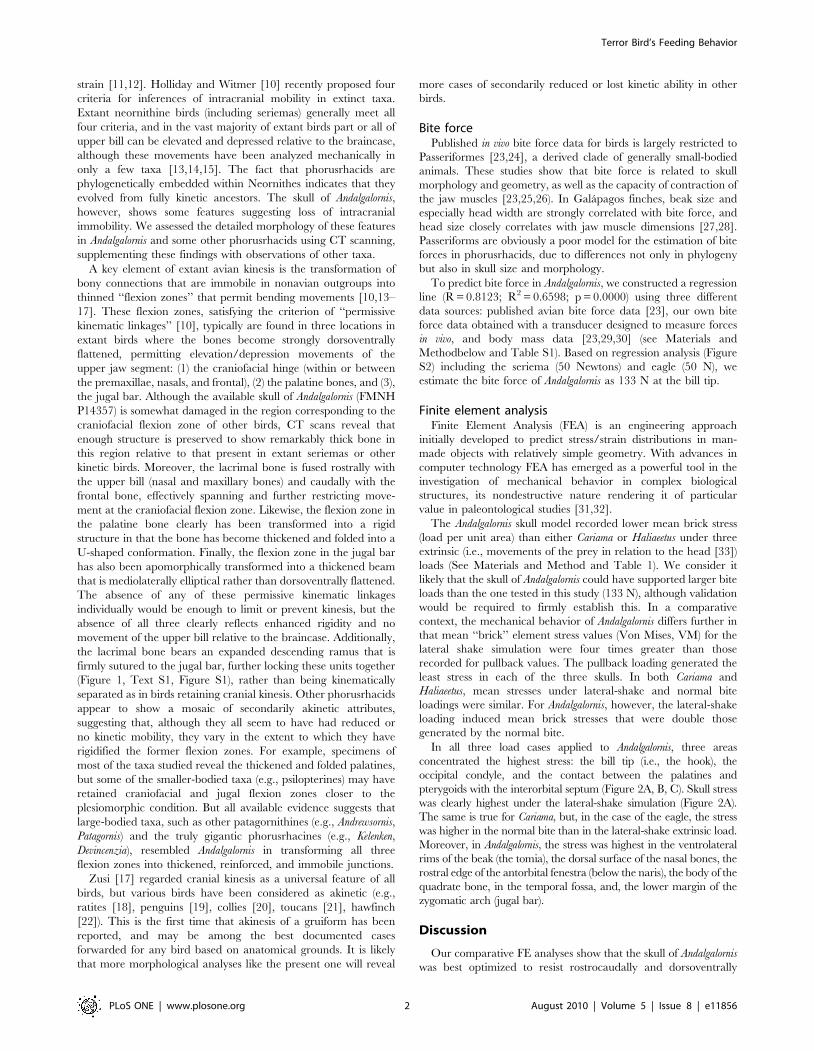
the jugal bar. Although the available skull of Andalgalornis (FMNH
P14357) is somewhat damaged in the region corresponding to the
craniofacial flexion zone of other birds, CT scans reveal that
enough structure is preserved to show remarkably thick bone in
this region relative to that present in extant seriemas or other
kinetic birds. Moreover, the lacrimal bone is fused rostrally with
the upper bill (nasal and maxillary bones) and caudally with the
frontal bone, effectively spanning and further restricting move-
ment at the craniofacial flexion zone. Likewise, the flexion zone in
the palatine bone clearly has been transformed into a rigid
structure in that the bone has become thickened and folded into a
U-shaped conformation. Finally, the flexion zone in the jugal bar
has also been apomorphically transformed into a thickened beam
that is mediolaterally elliptical rather than dorsoventrally flattened.
The absence of any of these permissive kinematic linkages
individually would be enough to limit or prevent kinesis, but the
absence of all three clearly reflects enhanced rigidity and no
movement of the upper bill relative to the braincase. Additionally,
the lacrimal bone bears an expanded descending ramus that is
firmly sutured to the jugal bar, further locking these units together
(Figure 1, Text S1, Figure S1), rather than being kinematically
separated as in birds retaining cranial kinesis. Other phorusrhacids
appear to show a mosaic of secondarily akinetic attributes,
suggesting that, although they all seem to have had reduced or
no kinetic mobility, they vary in the extent to which they have
rigidified the former flexion zones. For example, specimens of
most of the taxa studied reveal the thickened and folded palatines,
but some of the smaller-bodied taxa (e.g., psilopterines) may have
retained craniofacial and jugal flexion zones closer to the
plesiomorphic condition. But all available evidence suggests that
large-bodied taxa, such as other patagornithines (e.g., Andrewsornis,
Patagornis) and the truly gigantic phorusrhacines (e.g., Kelenken,
Devincenzia), resembled Andalgalornis in transforming all three
flexion zones into thickened, reinforced, and immobile junctions.
Zusi [17] regarded cranial kinesis as a universal feature of all
birds, but various birds have been considered as akinetic (e.g.,
ratites [18], penguins [19], collies [20], toucans [21], hawfinch
[22]). This is the first time that akinesis of a gruiform has been
reported, and may be among the best documented cases
forwarded for any bird based on anatomical grounds. It is likely
that more morphological analyses like the present one will reveal
more cases of secondarily reduced or lost kinetic ability in other
birds.
Bite forcePublished in vivo bite force data for birds is largely restricted to
Passeriformes [23,24], a derived clade of generally small-bodied
animals. These studies show that bite force is related to skull
morphology and geometry, as well as the capacity of contraction of
the jaw muscles [23,25,26). In Galapagos finches, beak size and
especially head width are strongly correlated with bite force, and
head size closely correlates with jaw muscle dimensions [27,28].
Passeriforms are obviously a poor model for the estimation of bite
forces in phorusrhacids, due to differences not only in phylogeny
but also in skull size and morphology.
To predict bite force in Andalgalornis, we constructed a regression
line (R = 0.8123; R2 = 0.6598; p = 0.0000) using three different
data sources: published avian bite force data [23], our own bite
force data obtained with a transducer designed to measure forces
in vivo, and body mass data [23,29,30] (see Materials and
Methodbelow and Table S1). Based on regression analysis (Figure
S2) including the seriema (50 Newtons) and eagle (50 N), we
estimate the bite force of Andalgalornis as 133 N at the bill tip.
Finite element analysisFinite Element Analysis (FEA) is an engineering approach
initially developed to predict stress/strain distributions in man-
made objects with relatively simple geometry. With advances in
computer technology FEA has emerged as a powerful tool in the
investigation of mechanical behavior in complex biological
structures, its nondestructive nature rendering it of particular
value in paleontological studies [31,32].
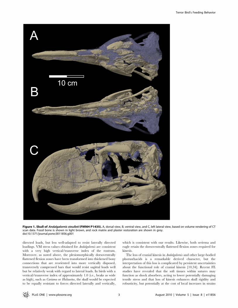
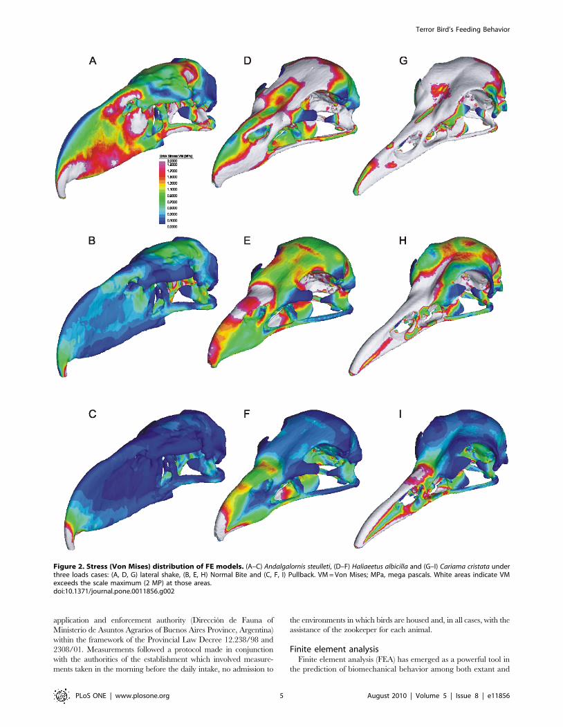
The Andalgalornis skull model recorded lower mean brick stress
(load per unit area) than either Cariama or Haliaeetus under three
extrinsic (i.e., movements of the prey in relation to the head [33])
loads (See Materials and Method and Table 1). We consider it
likely that the skull of Andalgalornis could have supported larger bite
loads than the one tested in this study (133 N), although validation
would be required to firmly establish this. In a comparative
context, the mechanical behavior of Andalgalornis differs further in
that mean ‘‘brick’’ element stress values (Von Mises, VM) for the
lateral shake simulation were four times greater than those
recorded for pullback values. The pullback loading generated the
least stress in each of the three skulls. In both Cariama and
Haliaeetus, mean stresses under lateral-shake and normal bite
loadings were similar. For Andalgalornis, however, the lateral-shake
loading induced mean brick stresses that were double those
generated by the normal bite.
In all three load cases applied to Andalgalornis, three areas
concentrated the highest stress: the bill tip (i.e., the hook), the
occipital condyle, and the contact between the palatines and
pterygoids with the interorbital septum (Figure 2A, B, C). Skull stress
was clearly highest under the lateral-shake simulation (Figure 2A).
The same is true for Cariama, but, in the case of the eagle, the stress
was higher in the normal bite than in the lateral-shake extrinsic load.
Moreover, in Andalgalornis, the stress was highest in the ventrolateral
rims of the beak (the tomia), the dorsal surface of the nasal bones, the
rostral edge of the antorbital fenestra (below the naris), the body of the
quadrate bone, in the temporal fossa, and, the lower margin of the
zygomatic arch (jugal bar).
Discussion
Our comparative FE analyses show that the skull of Andalgalornis
was best optimized to resist rostrocaudally and dorsoventrally
Terror Bird’s Feeding Behavior
PLoS ONE | www.plosone.org 2 August 2010 | Volume 5 | Issue 8 | e11856
directed loads, but less well-adapted to resist laterally directed
loadings. VM stress values obtained for Andalgalornis are consistent
with a very high vertical/transverse index of the rostrum.
Moreover, as noted above, the plesiomorphically dorsoventrally
flattened flexion zones have been transformed into thickened bony
connections that are reoriented into more vertically disposed,
transversely compressed bars that would resist sagittal loads well
but be relatively weak with regard to lateral loads. In birds with a
vertical/transverse index of approximately 1.0 (i.e., beaks as wide
as high), such as Cariama or Haliaeetus, the skull would be expected
to be equally resistant to forces directed laterally and vertically,
which is consistent with our results. Likewise, both seriema and
eagle retain the dorsoventrally flattened flexion zones required for
kinesis.
The loss of cranial kinesis in Andalgalornis and other large-bodied
phorusrhacids is a remarkable derived character, but the
interpretation of this loss is complicated by persistent uncertainties
about the functional role of cranial kinesis [10,34]. Recent FE
studies have revealed that the soft tissues within sutures may
function as shock absorbers, acting to lower potentially damaging
tensile stress and that loss of kinesis enhances skull rigidity and
robusticity, but potentially at the cost of local increases in strains
Figure 1. Skull of Andalgalornis steulleti (FMNH P1435). A, dorsal view, B, ventral view, and C, left lateral view, based on volume rendering of CTscan data. Fossil bone is shown in light brown, and rock matrix and plaster restoration are shown in grey.doi:10.1371/journal.pone.0011856.g001
Terror Bird’s Feeding Behavior
PLoS ONE | www.plosone.org 3 August 2010 | Volume 5 | Issue 8 | e11856
[11,12,35]. Thus, we consider it probable that the high stresses
recorded in the interorbital septum in our FE model of
Andalgalornis are artifactual, because the model did not incorporate
sutures between the palatines, pterygoids, and the septum. This
region may act as a stress releaser, compensating at least in part for
loss of the kinetic system.
A carnivorous lifestyle for phorusrhacids has been widely
assumed [1,3,5,36], but more detailed hypotheses of predatory
behavior have never been adequately tested. Previous reconstruc-
tion and biomechanical analysis of the jaw adductor musculature
of Andalgalornis [37] shows that the jaw apparatus is optimized for
strength at the expense of speed, and again, our discovery of loss of
cranial kinesis is consistent with increased skull rigidity. Any
adaptation that results in increased stiffness of the jaw apparatus,
such as transformation of mobile flexion zones into rigid struts, will
result in some increase in maximum theoretical bite force [31,38].
On the other hand, there may be complex trade-offs between
safety factors, the presence or absence of kinesis, and the bone and
muscle masses required to achieve any given performance limits.
For example, a stiffer and more brittle structure (as in akinesis)
may result in higher yield points and some reduction in the muscle
mass required to achieve a given bite force, but it may also limit
the response time available to modify behavior and avoid
catastrophic failure where an organism bites into unexpectedly
resistant materials [31]. Regarding the loss of cranial kinesis, a net
consequence might be that more bone or bone of greater density
(i.e., greater bone mass), might be needed to maintain effective
safety margins. In clearly akinetic phorusrhacids such as
patagornithines and phorusrhacines, the requisite increased bone
mass was presumably tolerated because their large body masses
precluded flight and hence the premium on ultra-light structures.
Future FE based analyses could aid in the quantification of such
interrelated factors.
Because most carnivorous birds use the hook on the tip of their
beak to hold and kill the prey (e.g. falcons [39]), all bite loads were
applied at the beak tip in our FE models. The functional
significance of a 133 N bite force for a 40 kg bird is difficult to
interpret because of the paucity of comparative bite-force data.
The recording of actual bite forces from living birds is restricted to
a few passeriforms, and there are no comparable extant
carnivorous bipeds. Certainly for its size, our estimate of bite
force in Andalgalornis is relatively weak compared to predictions
generated for anterior bites in carnivorous mammals. For
example, the 7.1 kg jaguarondi, Felis yagouaroundi, bites at 127 N
[40], the fox Pseudalopex griseus (5.9 kg) bites at 131 N, and the
mustelid Lutra longicauda (7.8 kg) bites at 129 N [41]. This is at least
in part explained by the comparatively longer distance between
the bite point and the jaw joint (outlever) in the terror bird.
However, we also note that mean VM stress computed in our
modeling of Andalgalornis is much lower than that calculated for
Cariama or Haliaeetus. This may indicate safety factors are unusually
high in Andalgalornis or that our predicted bite force for the
phorusrhacid is an underestimate, but differences in size between
the specimens makes these questions difficult to evaluate.
Moreover, our modeling does not attempt to simulate the
influence of jaw head flexor muscles, which may act to amplify
total recorded bite forces [32].
Alvarenga and Hofling [5] suggested that the beak morphology
of phorusrhacids allowed them to hunt in regions with high
vegetation and to catch small animals hiding under stones or fallen
tree trunks. Additionally, biomechanical analyses of locomotor
capabilities [42,43] suggest that speed and maneuverability in a
phorusrhacid of the size and proportions of Andalgalornis would be
comparable to those of an ostrich. Our biomechanical analyses
reveal that if Andalgalornis used its beak in the dispatch of relatively
large prey, then it must have been applied with considerable
precision in order to avoid sustaining high lateral loads. We
suggest that Andalgalornis consumed relatively small prey (i.e.,
smaller than itself) that could be killed and consumed more safely.
If Andalgalornis did take large prey, then it most likely applied
multiple well-targeted strikes in a repetitive attack-and-retreat
strategy. Restraining struggling prey with their feet also was
potentially an option, despite the absence of sharp talons.
Materials and Methods
CT scanningThe skulls of the patagornithines Andalgalornis steulleti ( = A. ferox
[44]; FMNH P14357) and Andrewsornis abbotti (FMNH P13417) and of
the psilopterine Psilopterus lemoinei (FMNH P13257) were scanned at
O’Bleness Memorial Hospital in Athens, Ohio, using a General
Electric (GE) LightSpeed Ultra Multislice CT scanner equipped with
the Extended Hounsfield option. Specimens were scanned helically at
a slice thickness of 625 mm, 120 kV, and 200–300 mA. Moreover,
another skull of Psilopterus lemoinei (AMNH 9257) and a skull of the
extant red-legged seriema (Cariama cristata, FMNH 105635) were
scanned at the Ohio University MicroCT Scanning Facility
(OUmCT) on a GE eXplore Locus MicroCT Scanner at a slice
thicknesses of 92 mm and 45 mm, 80 kV, and 500 mA. Data were
output from the scanner in DICOM format and then imported into
4.1.2 (Mercury-TGS, Chelmsford, MA) for viewing, analysis, and
visualization. The CT scan data were analyzed to assess the internal
architecture of bony regions relevant to assessments of cranial kinesis,
which were supplemented with gross observation of the skulls of these
specimens and others.
Body mass estimationThe body mass estimate of 40 kg for Andalgalornis was made
following previously published methodology [45] and submersion
of scale plastic models [43, unpublished data].
Bite forceBite force data were obtained in vivo from captive birds: an adult
Cariama cristata at the Zoological Garden of La Plata, Argentina, and
an adult black-chested eagle (Geranoaetus melanoleucus) from the Horco
Molle Reserve, Argentina. G. melanoleucus is similar in size to H.
albicilla, and its bite force was used as a proxy in the FEA of H. albicilla.
For each animal, multiple bites at the bill tip were recorded with the
transducer, and we use the maximum bite force in the analysis. The
safety standards related to health, hygienic, and dietary plans were
followed for the proper management of wildlife, issued by the
Table 1. Mean brick element stresses (von Misses, VM) insolved FE models under three load cases; L = lateral shake,N = normal bite, P = pullback.
Andalgalornis Haliaeetus Cariama
Element number 1,080,137 860,757 775,698
Lateral shake 1.028 2.272 3.285
Normal Bite 0.570 2.412 3.235
Pullback 0.234 0.678 0.833
L/N ratio 1.803 0.941 1.015
L/P ratio 4.393 3.351 3.943
doi:10.1371/journal.pone.0011856.t001
Terror Bird’s Feeding Behavior
PLoS ONE | www.plosone.org 4 August 2010 | Volume 5 | Issue 8 | e11856
application and enforcement authority (Direccion de Fauna of
Ministerio de Asuntos Agrarios of Buenos Aires Province, Argentina)
within the framework of the Provincial Law Decree 12.238/98 and
2308/01. Measurements followed a protocol made in conjunction
with the authorities of the establishment which involved measure-
ments taken in the morning before the daily intake, no admission to
the environments in which birds are housed and, in all cases, with the
assistance of the zookeeper for each animal.
Finite element analysisFinite element analysis (FEA) has emerged as a powerful tool in
the prediction of biomechanical behavior among both extant and
Figure 2. Stress (Von Mises) distribution of FE models. (A–C) Andalgalornis steulleti, (D–F) Haliaeetus albicilla and (G–I) Cariama cristata underthree loads cases: (A, D, G) lateral shake, (B, E, H) Normal Bite and (C, F, I) Pullback. VM = Von Mises; MPa, mega pascals. White areas indicate VMexceeds the scale maximum (2 MP) at those areas.doi:10.1371/journal.pone.0011856.g002
Terror Bird’s Feeding Behavior
PLoS ONE | www.plosone.org 5 August 2010 | Volume 5 | Issue 8 | e11856
extinct animals [38,46-52]. The protocols implemented in the
present study largely follow recently developed methodologies
[31,38,51,52].
Homogeneous models (assuming constant material properties
throughout) were constructed using the CT data for Andalgalornis
(FMNH P14357) with segmentation performed using Mimics (v.
11.01) software (see Figure 3A). A similar procedure was used for
the red-legged seriema (Cariama cristata, FMNH 105635) and the
White-tailed Eagle (Haliaeetus albicilla, from the University of
Austin Digital Morphology Web site: http://www.digimorph.org).
Cariama cristata was chosen for comparison because of its close
phylogenetic relationships with phorusrhacids and Haliaeetus
albicilla because of its predatory lifestyle.
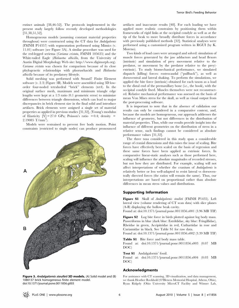
Solid meshing was performed with Strand7 Finite Element
software (v. 2.3) (Figure 3B). Models were assembled using 3D low-
order four-noded tetrahedral ‘‘brick’’ elements (tet4). In the
original surface mesh, maximum and minimum triangle edge
lengths were kept at a 1:3 ratio (0.1 geometric error) to minimize
differences between triangle dimensions, which can lead to major
discrepancies in brick element size in the final solid and introduce
artifacts. Brick elements were assigned a single set of material
properties as applied in previous studies [31,32], (Young’s modulus
of Elasticity [Y] = 27.0 GPa; Poisson’s ratio = 0.4; density =
2.19085 T/mm3).
Models were restrained to prevent free body motion. Point
constraints (restricted to single nodes) can produce pronounced
artifacts and inaccurate results [48]. For each loading we have
applied more realistic constraints by positioning them within
frameworks of rigid links at the occipital condyle as well as at the
tip of the beak to more broadly distribute forces in accordance
with previously published methods [32]. Statistical analyses were
performed using a customized program written in RGUI (by K.
Moreno).
Two kinds of load cases were arranged and solved: simulation of
muscle forces generated by the jaw adductors and head flexors
(intrinsic) and simulation of prey movement relative to the
predator, or movement by the predator relative to the prey(-
extrinsic). To study biomechanical performance, we simulated
dispatch (killing) forces: rostrocaudal (‘‘pullback’’), as well as
dorsoventral and lateral shaking. To perform the simulations, we
applied the bite force (intrinsic) obtained for each taxon as a load
to the distal end of the premaxillary bone (i.e., the hook), with the
occipital condyle fixed. Muscles themselves were not reconstruct-
ed. Relative mechanical performance was assessed on the basis of
mean Von Mises stress for the skull, as well as visual output from
the post-processing software.
It is important to note that in the absence of validation our
results can only be considered in a comparative context, and,
because the models are homogeneous, our approach addresses the
influence of geometry, but not differences in the distribution of
material properties. Thus, while our results provide insight into the
influence of different geometries on the distribution of stress in a
relative sense, such findings cannot be considered as absolute
performance values [31,32].
The three taxa considered in this study span a considerable
range of cranial dimensions and this raises the issue of scaling. Bite
forces have effectively been scaled on the basis of regression and
these same forces have been applied as extrinsic forces. In
comparative linear-static analyses such as those performed here,
scaling will influence the absolute magnitudes of recorded stresses,
but not how they are distributed. For example, scaling will not
affect interpretations of whether the cranium of Andalgalornis is
relatively better or less well-adapted to resist lateral vs dorsoven-
trally directed forces (the ratios will remain the same). Thus, our
interpretations are based on proportional rather than absolute
differences in mean stress values and distributions.
Supporting Information
Figure S1 Skull of Andalgalornis steulleti (FMNH P1435). Left
lateral view (volume rendering of CT scan data) with slice planes
(A-B) displaying the hollow beak cavity.
Found at: doi:10.1371/journal.pone.0011856.s001 (3.96 MB TIF)
Figure S2 Log bite force in birds plotted against log body mass.
Passeriforms in blue (dark blue: Estrildidae, sky blue: Fringillidae),
Rheidae in green, Accipitridae in red, Cathartidae in rose and
Cariamidae in black. See Table S1 for raw data.
Found at: doi:10.1371/journal.pone.0011856.s002 (3.39 MB TIF)
Table S1 Bite force and body mass table.
Found at: doi:10.1371/journal.pone.0011856.s003 (0.07 MB
DOC)
Text S1 Andalgalornis’ fossil.
Found at: doi:10.1371/journal.pone.0011856.s004 (0.03 MB
DOC)
Acknowledgments
For assistance with CT scanning, 3D visualization, and data management,
we thank Heather Rockhold (O’Bleness Memorial Hospital, Athens, Ohio),
Ryan Ridgely (Ohio University MicroCT Facility and Witmer Lab,
Figure 3. Andalgalornis steulleti 3D models. (A) Solid model and (B)1080137 brick homogeneous finite element model.doi:10.1371/journal.pone.0011856.g003
Terror Bird’s Feeding Behavior
PLoS ONE | www.plosone.org 6 August 2010 | Volume 5 | Issue 8 | e11856
Athens, Ohio), the Ohio Supercomputer Center (Columbus, Ohio), and
Marcelo Kaufman, Miriam Risso, Nicolas Nissan, Sebastian Salvarregui,
Nevenka Klinar and Daniel Cagnolo (Investigaciones Medicas, Buenos
Aires). For loan of specimens for CT scanning we thank the Field Museum
of Natural History (Chicago), the American Museum of Natural History
(New York City), Robert M. Chandler (Georgia College and State
University), and Museo de La Plata (Facultad de Ciencias Naturales y
Museo, Universidad Nacional de La Plata).
Author Contributions
Conceived and designed the experiments: FJD CPT KM SW. Performed
the experiments: FJD CPT. Analyzed the data: FJD KM SW. Contributed
reagents/materials/analysis tools: LMW SW. Wrote the paper: FJD CPT
KM LMW SW.
References
1. Tambussi CP, Noriega JI (1996) Summary of the Avian Fossil Record from
Southern South America. In: Arratia G, ed. Contributions of the southern southAmerica to vertebrate paleontology, Muncher Geowissenschaftliche Abhandlun-
gen. pp 245–264.
2. Tambussi C, Ubilla M, Perea D (1999) The youngest large carnassial bird(Phorusrhacidae, Phorusrhacinae) from South America (Pliocene-Early Pleisto-
cene of Uruguay). J Vert Paleontol 19: 404–406.
3. Andrews C (1899) On the extinct birds of Patagonia. Trans Zool Soc London15: 55–86.
4. Livezey BC (1998) A phylogenetic analysis of the Gruiformes (Aves) based on
morphological characters, with an emphasis on the rails (Rallidae). Tran R SocLondon 353: 2077–2151.
5. Alvarenga HMF, Hofling E (2003) Systematic revision of the Phorusrhacidae
(Aves: Ralliformes). Papeis Avulsos de Zoologia 43 (4): 55–91.
6. Mayr G (2002) A new specimen of Salmila robusta (Aves: Gruiformes: Salmilidaen. fam.) from the Middle Eocene of Messel. Palaontologische Zeitschrift 76 (2):
305–316.
7. Kraglievich L (1931) Contribucion al conocimiento de las aves fosiles de la epocaaraucoentrerriana. Physis 10: 304–315.
8. Chiappe LM, Bertelli S (2006) Skull morphology of giant terror birds. Nature
443: 929.
9. Wroe S, Argot C, Dickman C (2004) On the rarity of big fierce carnivores andprimacy of isolation and area: tracking large mammalian carnivore diversity on
two isolated continents. Proc R Soc London B 271: 1203–1211.
10. Holiday CM, Witmer LM (2008) Cranial kinesis in dinosaurs: intracranial joints,protractor muscles, and their significance for cranial evolution and function in
diapsids. J Ver Paleontol 28 (4): 1073–1088.
11. Rayfield EJ (2005) Using finite-element analysis to investigate suture morphol-
ogy: a case study using large carnivorous dinosaurs. Anat Rec 283A: 349–365.
12. Moazen M, Curtis N, O’Higgins P, Jones MEH, Evans SE, et al. (2009)
Assessment of the role of sutures in a lizard skull: a computer modelling study.
Proc R Soc B 276: 39–46.
13. Bout RG, Zweers GA (2001) The role of cranial kinesis in birds. Comp Biochem
Physiol A 131(1): 197–205.
14. Gussekloo WS, Bout RG (2005) Cranial kinesis in palaeognathus birds. J Exp
Biol 208: 3409–3419.
15. Gussekloo WS, Vosselman MG, Bout RG (2001) Three-dimensional kinematics
of skeletal elements in avian prokinetic and rhynchokinetic skulls determined by
roentgen stereophotogrammetry. J Exp Biol 204: 1735–1744.
16. Baumel JJ, Raikow RJ (1993) Arthrologia. In: Baumel J, King A, Breazile J,
Evans H, Vanden Berge J, eds. Handbook of avian anatomy: Nomina
Anatomica Avium Publications of the Nuttall Ornithological Club. pp 133–187.
17. Zusi RL (1993) Patterns of diversity in the Avian Skull. In: Hanken J, Hall BK,
eds. The Skull. Volume 2: Patterns of structural and systematic diversity
University of Chicago Press. pp 391–437.
18. McDowell S (1948) The bony palate of birds: Part I the Palaeognathae. Auk 65(4): 520–549.
19. Reid J (1835) Anatomical description of the Patagonian penguin. Proc Zool Soc
London 3: 132–148.
20. Schoonees J (1963) Some aspects of the cranial morphology of Colius indicus.
Annale Universiteit van Stellenbosch 38, 7 (7): 215–246.
21. Hofling E, Gasc JP (1984) Biomecanique du crane et du bec chez Ramphastos
(Ramphastidae, Aves). Gegenbaurs morphologisches Jahrbuch 130 (2): 235–262.
22. Sims SW (1955) The morphology f the head of the hawfinch (Coccothraustes
coccothraustes). Bull Brit Museum, Zool 2 (13): 371–393.
23. van der Meij MAA, Bout RG (2004) Scaling of jaw muscle size and maximal biteforce in finches. J Exp Biol 207: 2745–2753.
24. van der Meij MAA, Bout RG (2006) Seed husking time and maximal bite forces
in finches. J Exp Biol 209: 3329–3335.
25. Herrel A, Podos J, Huber SK, Hendry AP (2005a) Bite performance andmorphology in a population of Darwin’s finches: implications for the evolution of
beak shape. Funct Ecol 19: 43–48.
26. Herrel A, Podos J, Huber SK, Hendry AP (2005b) Evolution of bite force inDarwin’s finches: a key role for head width. J Evol Biol18: 669–675.
27. Herrel A, Van Damme R, Vanhooydonck B, De Vree F (2001) The implications
of bite performance for diet in two species of lacertid lizards. Can J Zool 79:
662–670.
28. Herrel A, O’Reilly JC, Richmond AM (2002) Evolution of bite force in turtles.
J Evol Biol 15: 1083–1094.
29. Jimenez J, Jaksic F (1990) Historia natural del aguila Geranoaetus melanoleucus: unarevision. El Hornero 13: 97–110.
30. Abourachid A, Hofling E, Renous S (2005) Walking kinematics parameters insome paleognathous and neognathous neotropical birds. Ornitologıa Neotrop-
ical 16: 471–479.
31. Wroe S, Moreno K, Clausen PD, McHenry CR (2007) High-resolutioncomputer simulation of hominid cranial mechanics. The Anatomical Record
290: 1248–1255.32. McHenry CR, Wroe S, Clausen PD, Moreno K, Cunningham E (2007) Super-
modeled sabercat, predatory behaviour in Smilodon fatalis revealed by high-resolution 3-D computer simulation. PNAS 104: 16010–16015.
33. Preuschoft H, Witzel U (2002) Biomechanical investigations on the skulls of
reptiles and mammals. Senckenbergiana Lethaea 82 (1): 207–222.34. Metzger K (2002) Cranial kinesis in lepidosaurs: skulls in motion. In: Aerts P,
D’Aout K, Herrel A, Van Damme R, eds. Topics in Functional and EcologicalVertebrate Morphology Shaker Publishing, Maastricht. pp 15–46.
35. Moazen M, Curtis N, O’Higgins P, Evans SE, Fagan MJ (2009) Biomechanical
assessment of evolutionary changes in the lepidosaurian skull. PNAS 106:8273–8277.
36. Sinclair W, Farr M (1932) Aves of the Santa Cruz beds. In: Scott W, ed. Reportsof the Princeton University expeditions to Patagonia (1896–1899) Princeton
University. pp 157–191.
37. Degrange FJ (2008) M. adductor mandibulae externus of Andalgalornis steulleti
(Aves, Phorusrhacidae): Reconstruction and biomechanics (in Spanish). III
Congreso Latinoamericano de Paleontologıa de Vertebrados, SimposioMorfologıa y Adaptacion en Aves: Nuevas Herramientas y Conceptos: 76.
38. Wroe S, Huber DR, Lowry M, McHenry C, Moreno K, et al. (2008) Three-dimensional computer analysis of white shark jaw mechanics: how hard can a
great white bite? J Zool 276: 336–342.
39. Sustaita D (2008) Musculoskeletal underpinnings to differences in killingbehavior between North American Accipiters (Falconiformes: Accipitridae)
and Falcons (Falconidae). J Morphol 269: 283–301.40. Wroe S, McHenry C, Thomason J (2005) Bite club: comparative bite force in big
biting mammals and the prediction of predatory behaviour in fossil taxa. Proc
Biol Sci 272: 619–625.41. Christiansen P, Wroe S (2007) Bite forces and evolutionary adaptations to
feeding ecology in carnivores. Ecology 88 (2): 347–358.42. Blanco RE, Jones WW (2005) Terror birds on the run: A mechanical model to
estimate its maximum running speed. Proc R Soc B 272: 1769–1773.43. Tambussi CP (1997) Some biomechanical aspects of the phorusrhacid
locomotion (Aves, Gruiformes) (in Spanish). Ameghiniana 34 (4): 541.
44. Patterson B, Kraglievich L (1960) Sistematica y nomenclatura de las avesfororracoideas del Plioceno Argentino. Publicacion del Museo Municipal
Ciencias Naturales y Tradicionales de Mar del Plata 1: 1–51.45. Campbell KE, Marcus L (1992) The relationship of hindlimb bone dimensions
to body weight in birds. In: Campbell KE editors. Papers in avian paleontology
honoring Pierce Brodkorb. Natural History Museum of Los Angeles County,Science Series 36: 395–412.
46. Rayfield EJ (2004) Cranial mechanics and feeding in Tyrannosaurus rex. Proc R SocLondon B 271: 1451–1459.
47. Rayfield EJ, Norman DB, Horner CC, Horner JR, Smith PM, et al. (2001)Cranial design and function in a large theropod dinosaur. Nature 409:
1033–1037.
48. Richmond BG, Wright BW, Grosse L, Dechow PC, Ross CF, et al. (2005) Finiteelement analysis in functional morphology. Anat Rec A 283: 259–274.
49. Tizzard A, Horesh L, Yerworth RJ, Holder DS, Bayford RH (2005) Generatingaccurate finite element meshes for the forward model of the human head in EIT.
Physiol Meas 26: 251–253.
50. McHenry CR, Clausen PD, Daniel WJT, Meers MB, Pendharkar A (2006) Thebiomechanics of the rostrum in crocodilians: a comparative analysis using finite
element modelling. Anat Rec 288: 827–849.51. Wroe S (2008) Cranial mechanics compared in extinct marsupial and extant
African lions using a finite-element approach. J Zool 274: 332–339.
52. Moreno K, Wroe S, Clausen PD, McHenry CR, D’Amore DC, et al. (2008)Cranial performance in the Komodo dragon (Varanus komodoensis) as revealed by
high-resolution 3-D finite element analysis. J Anat 212: 736–746.
Terror Bird’s Feeding Behavior
PLoS ONE | www.plosone.org 7 August 2010 | Volume 5 | Issue 8 | e11856
Copyright © 2022 FDOKUMEN