Mathematical modeling of sub-cellular asymmetry of fat-dachsous heterodimer for generation of planar...
10
Mathematical Modeling of Sub-Cellular Asymmetry of Fat-Dachsous Heterodimer for Generation of Planar Cell Polarity Mohit Kumar Jolly* . ¤ , Mohd Suhail Rizvi , Amit Kumar, Pradip Sinha . * Department of Biological Sciences and Bioengineering, Indian Institute of Technology Kanpur, Kanpur, India Abstract Planar Cell Polarity (PCP) is an evolutionarily conserved characteristic of animal tissues marked by coordinated polarization of cells or structures in the plane of a tissue. In insect wing epithelium, for instance, PCP is characterized by en masse orientation of hairs orthogonal to its apical-basal axis and pointing along the proximal-distal axis of the organ. Directional cue for PCP has been proposed to be generated by complex sets of interactions amongst three proteins - Fat (Ft), Dachsous (Ds) and Four-jointed (Fj). Ft and Ds are two atypical cadherins, which are phosphorylated by Fj, a Golgi kinase. Ft and Ds from adjacent cells bind heterophilically via their tandem cadherin repeats, and their binding affinities are regulated by Fj. Further, in the wing epithelium, sub-cellular levels of Ft-Ds heterodimers are seen to be elevated at the distal edges of individual cells, prefiguring their PCP. Mechanisms generating this sub-cellular asymmetry of Ft-Ds heterodimer in proximal and distal edges of cells, however, have not been resolved yet. Using a mathematical modeling approach, here we provide a framework for generation of this sub-cellular asymmetry of Ft-Ds heterodimer. First, we explain how the known interactions within Ft-Ds-Fj system translate into sub-cellular asymmetry of Ft-Ds heterodimer. Second, we show that this asymmetric localization of Ft-Ds heterodimer is lost when tissue-level gradient of Fj is flattened, or when phosphorylation of Ft by Fj is abolished, but not when tissue-level gradient of Ds is flattened or when phosphorylation of Ds is abrogated. Finally, we show that distal enrichment of Ds also amplifies Ft-Ds asymmetry. These observations reveal that gradient of Fj expression, phosphorylation of Ft by Fj and sub-cellular distal accumulation of Ds are three critical elements required for generating sub-cellular asymmetry of Ft-Ds heterodimer. Our model integrates the known experimental data and presents testable predictions for future studies. Citation: Jolly MK, Rizvi MS, Kumar A, Sinha P (2014) Mathematical Modeling of Sub-Cellular Asymmetry of Fat-Dachsous Heterodimer for Generation of Planar Cell Polarity. PLoS ONE 9(5): e97641. doi:10.1371/journal.pone.0097641 Editor: Eshel Ben-Jacob, Tel Aviv University, Israel Received January 16, 2014; Accepted April 23, 2014; Published May 19, 2014 Copyright: ß 2014 Jolly et al. This is an open-access article distributed under the terms of the Creative Commons Attribution License, which permits unrestricted use, distribution, and reproduction in any medium, provided the original author and source are credited. Funding: This work was supported by a project entitled ’Epithelial Morphogenesis and wound healing during animal development: Role of Fat cadherin in fruit fly Drosophila’ sponsored by the Department of Science and Technology (DST), New Delhi to Pradip Sinha No. SR/SO/AS-24/2008. Date: February 27, 2009. The funders had no role in study design, data collection and analysis, decision to publish, or preparation of the manuscript. Competing Interests: The authors have declared that no competing interests exist. * E-mail: [email protected] (MKJ); [email protected] (PS) . These authors contributed equally to this work. ¤ Current address: Center for Theoretical Biological Physics and Department of Bioengineering, Rice University, Houston, Texas, United States of America Introduction Planar Cell Polarity (PCP) represents coordinated orientation of cells or structures in an axis within the tissue plane. Most animal organs display PCP, which when perturbed lead to developmental anomalies such as open neural tubes, polycystic kidneys, and even can cause deafness due to loss of planar polarization of the cilia in the inner ear [1]. PCP has been extensively studied in Drosophila wing, eye, and in the adult abdomen [2]. Drosophila wing, in particular, is an ideal model system to study PCP, which is mirrored by projection of actin-rich trichomes (hairs) from the distal edges of its individual cells (Figure 1D) [3,4]. Three evolutionarily conserved proteins, namely, Fat (Ft), Dachsous (Ds) and Four-jointed (Fj), the so called Ft-Ds-Fj system or global regulators of PCP, generate a tissue-level directional cue thereby linking the axis of polarization of individual cells with that of the tissue/organ: for instance, projection of the wing hairs along the proximal-distal axis in Drosophila wing [5]. A second class of proteins consisting of Frizzled (Fz), Flamingo (Fmi), Van Gogh (Vang), Prickled (Pk) and Dishevelled (Dsh), referred to as core PCP proteins, propagate PCP signals from cell-to-cell [2,6]. Finally, several down-stream effectors, including cytoskeletal proteins, respond to these signals from global regulators and core proteins to establish PCP [7]. Upon loss of global signal, as in ft, ds, fj mutants, PCP could be seen to be coordinated amongst neighboring cells; however, the direction of their polarity is often not aligned with the tissue/organ axis thereby giving rise to swirling patterns (Figure 1D)[8–10]. Loss of activities of the core proteins, in contrast, abolish PCP altogether in individual cells; for instance, wing hairs in fz mutant may grow out of the centers of the cells, instead of their distal edges [6]. Ft, Ds and Fj display intricate interactions with each other. Ft and Ds, for instance, are large atypical cadherins that bind heterophilically between adjacent cells via their tandem cadherin repeats [5]. Fj, a golgi kinase, phosphorylates both Ft and Ds [11,12], which in turn increases affinity of Ft for Ds and decreases that of Ds for Ft [13,14]. Also, expression levels of Ds and Fj vary PLOS ONE | www.plosone.org 1 May 2014 | Volume 9 | Issue 5 | e97641
-
Upload
independent -
Category
Documents
-
view
0 -
download
0
Transcript of Mathematical modeling of sub-cellular asymmetry of fat-dachsous heterodimer for generation of planar...
Mathematical Modeling of Sub-Cellular Asymmetry ofFat-Dachsous Heterodimer for Generation of Planar CellPolarityMohit Kumar Jolly*.
¤
, Mohd Suhail Rizvi , Amit Kumar, Pradip Sinha. *
Department of Biological Sciences and Bioengineering, Indian Institute of Technology Kanpur, Kanpur, India
Abstract
Planar Cell Polarity (PCP) is an evolutionarily conserved characteristic of animal tissues marked by coordinated polarizationof cells or structures in the plane of a tissue. In insect wing epithelium, for instance, PCP is characterized by en masseorientation of hairs orthogonal to its apical-basal axis and pointing along the proximal-distal axis of the organ. Directionalcue for PCP has been proposed to be generated by complex sets of interactions amongst three proteins - Fat (Ft), Dachsous(Ds) and Four-jointed (Fj). Ft and Ds are two atypical cadherins, which are phosphorylated by Fj, a Golgi kinase. Ft and Dsfrom adjacent cells bind heterophilically via their tandem cadherin repeats, and their binding affinities are regulated by Fj.Further, in the wing epithelium, sub-cellular levels of Ft-Ds heterodimers are seen to be elevated at the distal edges ofindividual cells, prefiguring their PCP. Mechanisms generating this sub-cellular asymmetry of Ft-Ds heterodimer in proximaland distal edges of cells, however, have not been resolved yet. Using a mathematical modeling approach, here we provide aframework for generation of this sub-cellular asymmetry of Ft-Ds heterodimer. First, we explain how the known interactionswithin Ft-Ds-Fj system translate into sub-cellular asymmetry of Ft-Ds heterodimer. Second, we show that this asymmetriclocalization of Ft-Ds heterodimer is lost when tissue-level gradient of Fj is flattened, or when phosphorylation of Ft by Fj isabolished, but not when tissue-level gradient of Ds is flattened or when phosphorylation of Ds is abrogated. Finally, weshow that distal enrichment of Ds also amplifies Ft-Ds asymmetry. These observations reveal that gradient of Fj expression,phosphorylation of Ft by Fj and sub-cellular distal accumulation of Ds are three critical elements required for generatingsub-cellular asymmetry of Ft-Ds heterodimer. Our model integrates the known experimental data and presents testablepredictions for future studies.
Citation: Jolly MK, Rizvi MS, Kumar A, Sinha P (2014) Mathematical Modeling of Sub-Cellular Asymmetry of Fat-Dachsous Heterodimer for Generation of Planar CellPolarity. PLoS ONE 9(5): e97641. doi:10.1371/journal.pone.0097641
Editor: Eshel Ben-Jacob, Tel Aviv University, Israel
Received January 16, 2014; Accepted April 23, 2014; Published May 19, 2014
Copyright: � 2014 Jolly et al. This is an open-access article distributed under the terms of the Creative Commons Attribution License, which permits unrestricteduse, distribution, and reproduction in any medium, provided the original author and source are credited.
Funding: This work was supported by a project entitled ’Epithelial Morphogenesis and wound healing during animal development: Role of Fat cadherin in fruitfly Drosophila’ sponsored by the Department of Science and Technology (DST), New Delhi to Pradip Sinha No. SR/SO/AS-24/2008. Date: February 27, 2009. Thefunders had no role in study design, data collection and analysis, decision to publish, or preparation of the manuscript.
Competing Interests: The authors have declared that no competing interests exist.
* E-mail: [email protected] (MKJ); [email protected] (PS)
. These authors contributed equally to this work.
¤ Current address: Center for Theoretical Biological Physics and Department of Bioengineering, Rice University, Houston, Texas, United States of America
Introduction
Planar Cell Polarity (PCP) represents coordinated orientation of
cells or structures in an axis within the tissue plane. Most animal
organs display PCP, which when perturbed lead to developmental
anomalies such as open neural tubes, polycystic kidneys, and even
can cause deafness due to loss of planar polarization of the cilia in
the inner ear [1]. PCP has been extensively studied in Drosophila
wing, eye, and in the adult abdomen [2]. Drosophila wing, in
particular, is an ideal model system to study PCP, which is
mirrored by projection of actin-rich trichomes (hairs) from the
distal edges of its individual cells (Figure 1D) [3,4].
Three evolutionarily conserved proteins, namely, Fat (Ft),
Dachsous (Ds) and Four-jointed (Fj), the so called Ft-Ds-Fj system
or global regulators of PCP, generate a tissue-level directional cue
thereby linking the axis of polarization of individual cells with that
of the tissue/organ: for instance, projection of the wing hairs along
the proximal-distal axis in Drosophila wing [5]. A second class of
proteins consisting of Frizzled (Fz), Flamingo (Fmi), Van Gogh
(Vang), Prickled (Pk) and Dishevelled (Dsh), referred to as core
PCP proteins, propagate PCP signals from cell-to-cell [2,6].
Finally, several down-stream effectors, including cytoskeletal
proteins, respond to these signals from global regulators and core
proteins to establish PCP [7]. Upon loss of global signal, as in ft, ds,
fj mutants, PCP could be seen to be coordinated amongst
neighboring cells; however, the direction of their polarity is often
not aligned with the tissue/organ axis thereby giving rise to
swirling patterns (Figure 1D)[8–10]. Loss of activities of the core
proteins, in contrast, abolish PCP altogether in individual cells; for
instance, wing hairs in fz mutant may grow out of the centers of
the cells, instead of their distal edges [6].
Ft, Ds and Fj display intricate interactions with each other. Ft
and Ds, for instance, are large atypical cadherins that bind
heterophilically between adjacent cells via their tandem cadherin
repeats [5]. Fj, a golgi kinase, phosphorylates both Ft and Ds
[11,12], which in turn increases affinity of Ft for Ds and decreases
that of Ds for Ft [13,14]. Also, expression levels of Ds and Fj vary
PLOS ONE | www.plosone.org 1 May 2014 | Volume 9 | Issue 5 | e97641

across the tissue; in the wing primordium, for instance, these form
opposing gradients where Fj expression is higher in its distal than
that of its proximal domain, and vice-versa for Ds (Figure 1A)
[10,15,16]. These opposing gradients of Fj and Ds expressions
have been seen to be critical for establishing PCP [9,17]. Further,
Ft-Ds heterodimer also displays a sub-cellular distal enrichment,
thereby, resulting in its asymmetric localization (polarization) in
the proximal and distal membrane of the cells preceding their
overt display of PCP through asymmetric localization of the core
proteins such as Fz [18,19]. Ft-Ds heterodimer asymmetry thus
could be the earliest and coarse signals for cell polarization that are
eventually strengthened by sub-cellular asymmetry of the core
PCP proteins and their signaling. A critical question that has not
been answered yet is how the known interactions within these
three elements of the Ft-Ds-Fj system translate into a sub-cellular
asymmetry of the Ft-Ds heterodimer.
A mathematical modeling approach can offer insights into how
a set of biochemical interactions at a local (sub-cellular) scale could
impact a global pattern such as the coordinated PCP in an entire
organ like the insect wing. Several mathematical models have been
developed to understand various aspects of PCP [20]. The first
integrated modeling and experimental study in PCP [21] offered a
model for intercellular feedback regulations amongst core proteins
to explain ‘domineering effect’ [22] – a phenomenon wherein
perturbation in PCP in a group of cells lacking (mutant), for
instance, Fz protein activity was relayed to wild type neighboring
cells. This study proposed that domineering effect can simply
emerge from the dynamics of the known intercellular and
intracellular interactions among the core proteins (Fz, Vang, Pk
and Dsh). This model further suggested that a directional cue
provided by Ft-Ds-Fj system is crucial for explaining the observed
PCP patterns [21]; however, it did not elucidate how the Ft-Ds-Fj
system as such generates this cue. Further, attempts at mathemat-
ical modeling of PCP captured the mechanism of action of the
core proteins, and strengthened the idea that a weak tissue-level
directional cue is important for obtaining the experimentally
observed PCP patterns[23–30]. A recent phenomenological model
proposes how Ft-Ds heterodimer formation in a group of cells in
the tissue can induce collective polarization over the entire tissue;
however, this model did not factor the crucial question of the role
played by tissue-level expression gradients of Ds and Fj for the
regulation of PCP [31]. Since none of these models factored all
activities of the members of Ft-Ds-Fj system, our current
understanding of their contribution in PCP regulation remains
far from complete.
Here, using a mathematical modeling approach, we have
simulated the experimentally identified interactions amongst the
members of Ft-Ds-Fj system to reveal their impact on sub-cellular
asymmetry of Ft-Ds heterodimer. We have developed an Ordinary
Differential Equation (ODE)-based model by taking into consid-
eration the known interactions in Ft-Ds-Fj system. These elements
are: (a) Ft-Ds heterophilic binding, (b) opposing tissue-level
gradients of Ds and Fj and, finally, (c) phosphorylation of Ft and
Ds by Fj kinase. We show that Fj gradient, phosphorylation of Ft
by Fj, and, sub-cellular distal accumulations of Ds are three
essentials for establishing sub-cellular asymmetry of Ft-Ds hetero-
dimer in individual cells.
Results and Discussion
Model FormulationWe assumed that cells in Drosophila wing are arranged along
rows that are oriented in a single proximal-to-distal axis. We
modeled one such row of cells for deciphering the sub-cellular
Figure 1. Fat (Ft)-Dachsous (Ds)-Four-jointed (Fj) systemregulates PCP in Drosophila wing. (A) Schematic of Drosophila wingshowing opposing expression gradients of Ds (green) and Fj (grey)
Sub-Cellular Asymmetry of Ft-Ds Heterodimer for Planar Cell Polarity
PLOS ONE | www.plosone.org 2 May 2014 | Volume 9 | Issue 5 | e97641
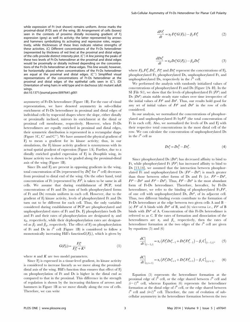
asymmetry of Ft-Ds heterodimer (Figure 1B). For the ease of visual
representation, we have denoted asymmetry in sub-cellular
enrichment of Ft-Ds heterodimer in proximal and distal edges of
individual cells by trapezoid shapes where the slope, either distally
or proximally inclined, mirrors its enrichment at the distal or
proximal cell membrane, respectively. However, when Ft-Ds
heterodimers are equally enriched in proximal and distal edges,
their symmetric distribution is represented in a rectangular shape
(Figure 1C, C9 and C99). We have assumed the physical gradient of
Fj to mean a gradient for its kinase activity; thus, in our
simulations, the Fj kinase activity gradient is synonymous with its
actual spatial gradient of expression (Figure 1A). Further, due to a
distally enriched graded expression of Fj in Drosophila wing, its
kinase activity too is shown to be graded along the proximal-distal
axis of the wing (Figure 1B).
Since Ds and Fj are present in opposing gradients in the wing,
total concentration of Ds (represented by Ds0i for ith cell) decreases
from proximal to distal end of the wing. On the other hand, total
concentration of Ft (represented by Ft0) is taken to be same in all
cells. We assume that during establishment of PCP, total
concentrations of Ft and Ds (sum of both phosphorylated forms
of Ft and Ds) remain uniform in each cell. However, due to the
gradient of Fj kinase activity, levels of phosphorylated Ft and Ds
turn out to be different for each cell. Thus, the only variables
considered during establishment of PCP are phosphorylated and
unphosphorylated states of Ft and Ds. Fj phosphorylates both Ds
and Ft and their rates of phosphorylation are designated af and
ad , respectively, while their dephosphorylation rates are designat-
ed as bf and bd , respectively. The effect of Fj on phosphorylation
of Ft and Ds in ith cell (Figure 1B) is considered to follow a
monotonically increasing Hill’s functionG Fjið Þ, which is given by
G(Fji)~Fjn
i
Fjni zKn
ð1Þ
where n and K are two model parameters.
Since Fj is expressed in a tissue-level gradient, its kinase activity
is considered to increase linearly as we move along the proximal-
distal axis of the wing. Hill’s function thus ensures that effect of Fj
on phosphorylation of Ft and Ds is higher in the distal end as
compared to that in the proximal. This difference in the strength
of regulation is shown by the increasing thickness of arrows and
hammers in Figure 1B as we move distally along the row of cells.
Therefore, we can write
dFtpi
dt~af Ftu
i G Fjið Þ{bf Ftpi ð2Þ
dDspi
dt~adDsu
i G Fjið Þ{bd Dspi ð3Þ
where Fji,Ftpi ,Ds
pi , Ftu
i and Dsui represent the concentrations of Fj,
phosphorylated Ft, phosphorylated Ds, unphosphorylated Ft, and
unphosphorylated Ds, respectively in the ith cell.
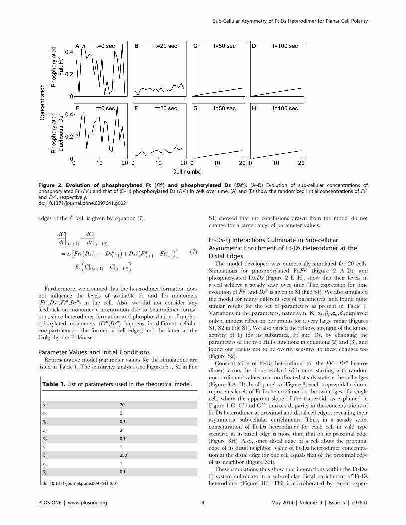
We performed the analysis with randomly initialized values of
concentrations of phosphorylated Ft and Ds (Figure 2A–H). In the
SI (File S1), we show that the levels of phosphorylated Ft (Ftp) and
Ds (Dsp) attain stable steady state values over time irrespective of
the initial values of Ftp and Dsp. Thus, our results hold good for
any set of initial values of Ftp and Dsp in the row of cells
considered.
In our analysis, we normalized the concentrations of phosphor-
ylated and unphosphorylated Ft byFt0 (the total concentration of
Ft in each cell). Also, we normalized the levels of Ds and Fj with
their respective total concentrations in the most distal cell of the
row. We can calculate the concentration of unphosphorylated Ds
in the ith cell as
Dsui ~Ds0
i {Dspi : ð4Þ
Since phosphorylated Ds (Dsp) has decreased affinity to bind to
Ft, while phosphorylated Ft (Ftp) has increased affinity to bind to
Ds [13,14], we assumed that the interaction between phosphor-
ylated Ft and unphosphorylated Ds (Ftp{Dsu) is much greater
than those between other forms of Ds and Ft (i.e. Ftp{Dsp,
Ftu{Dsu and Ftu{Dsp). Thus, Ftp{Dsu is the most abundant
form of Ft-Ds heterodimer. Therefore, hereafter, by Ft-Ds
heterodimer, we refer to the binding of phosphorylated Ft,Ftp,
of one cell with unphosphorylated Ds, Dsu, of its adjacent cell.
Thus, two different binding events contribute to the formation of
Ft-Ds heterodimer at the edge between two given cells A and B –
(a) Ftp of A binds with Dsu of B, and (b) vice-versa i.e., Ftp of B
binds with Dsu of A. Concentration of this Ft-Ds heterodimer is
referred to as C. If the rates of formation and dissociation of the
heterodimers are ac and bc, respectively, then the rates of
heterodimer formation at the two edges of the ith cell are given
by equations (5) and (6)
dC
dt Di{1,ið Þ
~ac Ftpi Dsu
i{1zDsui Ft
pi{1
� �{bcCD i{1,ið Þ, ð5Þ
dC
dt Di,iz1ð Þ
~ac Ftpi Dsu
iz1zDsui Ft
piz1
� �{bcCD i,iz1ð Þ: ð6Þ
Equation (5) represents the heterodimer formation at the
proximal edge of ith cell, or the edge shared between ith cell and
(i21)th cell, whereas Equation (6) represents the heterodimer
formation at the distal edge of ith cell, or the edge shared between
ith cell and (i+1)th cell. Therefore, the rate of evolution of sub-
cellular asymmetry in the heterodimer formation between the two
while expression of Ft (not shown) remains uniform. Arrow marks theproximal-distal (P/D) axis of the wing. (B) Arrangement of cells (boxes)seen in the contexts of proximo distally increasing gradient of Fjexpression (grey) as well its activity; the latter represented by arrowsand hammers symbolizing its activating and repressive roles, respec-tively, while thicknesses of these lines indicate relative strengths ofthese activities. (C) Different concentrations of the Ft-Ds heterodimer(represented by thickness of red line) at the proximal and distal edgesof the cells provide distinct intensity plot. (C9) A line joining the peaks ofthese two levels of Ft-Ds heterodimer at the proximal and distal edgeswould be proximally or distally inclined depending on the concentra-tions of the Ft-Ds heterodimer at these edges. This line would, however,be horizontally placed when concentrations of the Ft-Ds heterodimerare equal at the proximal and distal edges. (C99) Simplified visualrepresentations of the concentrations of Ft-Ds heterodimer at theproximal and distal edges of the epithelial cells seen in (C9). (D)Orientation of wing hairs in wild type and in dachsous (ds) mutant adultwings.doi:10.1371/journal.pone.0097641.g001
Sub-Cellular Asymmetry of Ft-Ds Heterodimer for Planar Cell Polarity
PLOS ONE | www.plosone.org 3 May 2014 | Volume 9 | Issue 5 | e97641
edges of the ith cell is given by equation (7).
dC
dt Di,iz1ð Þ
{dC
dt Di{1,ið Þ
~ac Ftpi Dsu
iz1{Dsui{1
� �zDsu
i Ftpiz1{Ft
pi{1
� �� �
{bc CD i,iz1ð Þ{CD i{1,ið Þ
� �ð7Þ
Furthermore, we assumed that the heterodimer formation does
not influence the levels of available Ft and Ds monomers
(Ftu,Dsu,Ftp,Dsp) in the cell. Also, we did not consider any
feedback on monomer concentration due to heterodimer forma-
tion, since heterodimer formation and phosphorylation of unpho-
sphorylated monomers (Ftu,Dsu) happens in different cellular
compartments – the former at cell edges, and the latter at the
Golgi by the Fj kinase.
Parameter Values and Initial ConditionsRepresentative model parameter values for the simulations are
listed in Table 1. The sensitivity analysis (see Figures S1, S2 in File
S1) showed that the conclusions drawn from the model do not
change for a large range of parameter values.
Ft-Ds-Fj Interactions Culminate in Sub-cellularAsymmetric Enrichment of Ft-Ds Heterodimer at theDistal Edges
The model developed was numerically simulated for 20 cells.
Simulations for phosphorylated Ft,Ftp (Figure 2 A–D), and
phosphorylated Ds,Dsp(Figure 2 E–H), show that their levels in
a cell achieve a steady state over time. The expression for time
evolution of Ftp and Dsp is given in SI (File S1). We also simulated
the model for many different sets of parameters, and found quite
similar results for the set of parameters as present in Table 1.
Variations in the parameters, namely, n, K, af ,bf ,ad ,bddisplayed
only a modest effect on our results for a very large range (Figures
S1, S2 in File S1). We also varied the relative strength of the kinase
activity of Fj for its substrates, Ft and Ds, by changing the
parameters of the two Hill’s functions in equations (2) and (3), and
found our results not to be overtly sensitive to these changes too
(Figure S2).
Concentration of Ft-Ds heterodimer (or the Ftp2Dsu hetero-
dimer) across the tissue evolved with time, starting with random
uncoordinated values to a coordinated steady state at the cell edges
(Figure 3 A–H). In all panels of Figure 3, each trapezoidal column
represents levels of Ft-Ds heterodimer on the two edges of a single
cell, where the apparent slope of the trapezoid, as explained in
Figure 1 C, C’ and C99, mirrors disparity in the concentrations of
Ft-Ds heterodimer at proximal and distal cell edges, revealing their
asymmetric sub-cellular enrichments. Thus, in a steady state,
concentration of Ft-Ds heterodimer for each cell in wild type
scenario at its distal edge is more than that on its proximal edge
(Figure 3H). Also, since distal edge of a cell abuts the proximal
edge of its distal neighbor, value of Ft-Ds heterodimer concentra-
tion at the distal edge for one cell equals that of the proximal edge
of its neighbor (Figure 3H).
These simulations thus show that interactions within the Ft-Ds-
Fj system culminate in a sub-cellular distal enrichment of Ft-Ds
heterodimer (Figure 3H). This is corroborated by recent exper-
Figure 2. Evolution of phosphorylated Ft (Ftp) and phosphorylated Ds (Dsp). (A–D) Evolution of sub-cellular concentrations ofphosphorylated Ft (Ftp) and that of (E–H) phosphorylated Ds (Dsp) in cells over time. (A) and (E) show the randomized initial concentrations of Ftp
and Dsp , respectively.doi:10.1371/journal.pone.0097641.g002
Table 1. List of parameters used in the theoretical model.
N 20
af 2
bf 0.1
ad 2
bd 0.1
N 1
K 250
ac 1
bc 0.1
doi:10.1371/journal.pone.0097641.t001
Sub-Cellular Asymmetry of Ft-Ds Heterodimer for Planar Cell Polarity
PLOS ONE | www.plosone.org 4 May 2014 | Volume 9 | Issue 5 | e97641
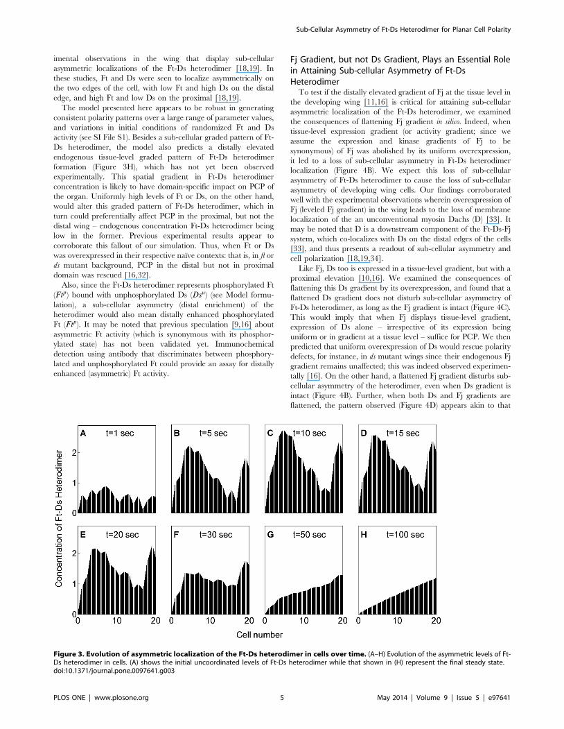
imental observations in the wing that display sub-cellular
asymmetric localizations of the Ft-Ds heterodimer [18,19]. In
these studies, Ft and Ds were seen to localize asymmetrically on
the two edges of the cell, with low Ft and high Ds on the distal
edge, and high Ft and low Ds on the proximal [18,19].
The model presented here appears to be robust in generating
consistent polarity patterns over a large range of parameter values,
and variations in initial conditions of randomized Ft and Ds
activity (see SI File S1). Besides a sub-cellular graded pattern of Ft-
Ds heterodimer, the model also predicts a distally elevated
endogenous tissue-level graded pattern of Ft-Ds heterodimer
formation (Figure 3H), which has not yet been observed
experimentally. This spatial gradient in Ft-Ds heterodimer
concentration is likely to have domain-specific impact on PCP of
the organ. Uniformly high levels of Ft or Ds, on the other hand,
would alter this graded pattern of Ft-Ds heterodimer, which in
turn could preferentially affect PCP in the proximal, but not the
distal wing – endogenous concentration Ft-Ds heterodimer being
low in the former. Previous experimental results appear to
corroborate this fallout of our simulation. Thus, when Ft or Ds
was overexpressed in their respective naı̈ve contexts: that is, in ft or
ds mutant background, PCP in the distal but not in proximal
domain was rescued [16,32].
Also, since the Ft-Ds heterodimer represents phosphorylated Ft
(Ftp) bound with unphosphorylated Ds (Dsu) (see Model formu-
lation), a sub-cellular asymmetry (distal enrichment) of the
heterodimer would also mean distally enhanced phosphorylated
Ft (Ftp). It may be noted that previous speculation [9,16] about
asymmetric Ft activity (which is synonymous with its phosphor-
ylated state) has not been validated yet. Immunochemical
detection using antibody that discriminates between phosphory-
lated and unphosphorylated Ft could provide an assay for distally
enhanced (asymmetric) Ft activity.
Fj Gradient, but not Ds Gradient, Plays an Essential Rolein Attaining Sub-cellular Asymmetry of Ft-DsHeterodimer
To test if the distally elevated gradient of Fj at the tissue level in
the developing wing [11,16] is critical for attaining sub-cellular
asymmetric localization of the Ft-Ds heterodimer, we examined
the consequences of flattening Fj gradient in silico. Indeed, when
tissue-level expression gradient (or activity gradient; since we
assume the expression and kinase gradients of Fj to be
synonymous) of Fj was abolished by its uniform overexpression,
it led to a loss of sub-cellular asymmetry in Ft-Ds heterodimer
localization (Figure 4B). We expect this loss of sub-cellular
asymmetry of Ft-Ds heterodimer to cause the loss of sub-cellular
asymmetry of developing wing cells. Our findings corroborated
well with the experimental observations wherein overexpression of
Fj (leveled Fj gradient) in the wing leads to the loss of membrane
localization of the an unconventional myosin Dachs (D) [33]. It
may be noted that D is a downstream component of the Ft-Ds-Fj
system, which co-localizes with Ds on the distal edges of the cells
[33], and thus presents a readout of sub-cellular asymmetry and
cell polarization [18,19,34].
Like Fj, Ds too is expressed in a tissue-level gradient, but with a
proximal elevation [10,16]. We examined the consequences of
flattening this Ds gradient by its overexpression, and found that a
flattened Ds gradient does not disturb sub-cellular asymmetry of
Ft-Ds heterodimer, as long as the Fj gradient is intact (Figure 4C).
This would imply that when Fj displays tissue-level gradient,
expression of Ds alone – irrespective of its expression being
uniform or in gradient at a tissue level – suffice for PCP. We then
predicted that uniform overexpression of Ds would rescue polarity
defects, for instance, in ds mutant wings since their endogenous Fj
gradient remains unaffected; this was indeed observed experimen-
tally [16]. On the other hand, a flattened Fj gradient disturbs sub-
cellular asymmetry of the heterodimer, even when Ds gradient is
intact (Figure 4B). Further, when both Ds and Fj gradients are
flattened, the pattern observed (Figure 4D) appears akin to that
Figure 3. Evolution of asymmetric localization of the Ft-Ds heterodimer in cells over time. (A–H) Evolution of the asymmetric levels of Ft-Ds heterodimer in cells. (A) shows the initial uncoordinated levels of Ft-Ds heterodimer while that shown in (H) represent the final steady state.doi:10.1371/journal.pone.0097641.g003
Sub-Cellular Asymmetry of Ft-Ds Heterodimer for Planar Cell Polarity
PLOS ONE | www.plosone.org 5 May 2014 | Volume 9 | Issue 5 | e97641

caused by flattening of Fj (Figure 4 B) but not the one presented by
flattened Ds gradient (Figure 4C). Therefore, Fj gradient, but not
that of Ds, is essential for attaining sub-cellular asymmetry of Ft-
Ds heterodimer. In other words, Ds gradient by itself (without the
Fj gradient) is not enough to give correct patterning. Together,
these findings suggest that while Ds is required for generation of
Ft-Ds asymmetry and cell polarization, its tissue-level gradient per
se may not be critical for PCP.
Phosphorylation of Ft by Fj is Essential in Achieving Sub-cellular Asymmetry of Ft-Ds
Fj kinase phosphorylates both Ft and Ds and regulates binding
of Ft to Ds in two ways – by inhibiting binding of Ds with Ft on
one hand and by promoting binding of Ft with Ds on the other
(Figure 5A) [13,14]. This opposing action of Fj on Ds and Ft has
been proposed to establish sub-cellular polarity with high fidelity
[6,14]. We asked whether the phosphorylation of both Ft and Ds
by Fj kinase is necessary for producing sub-cellular Ft-Ds
asymmetry. In silico mutation, which retained Fj kinase activity
(phosphorylation) against Ds but lacked that against Ft (Fj(Ft)
mutant) disrupted PCP across the tissue (Figure 5B). The converse,
meaning intact Fj kinase activity against Ft but loss of that against
Ds (Fj(Ds) mutant), however, did not disrupt PCP (Figure 5C).
Thus, while phosphorylation of Ft by Fj kinase appears to be an
essential for achieving Ft-Ds sub-cellular asymmetry, redundancy
of phosphorylation of Ds by Fj as seen in our simulation may
confer robustness to polarization process across the tissue.
Sub-cellular Polarity of Unphosphorylated Ds (Dsu)Amplifies Sub-cellular Asymmetry of Ft-Ds Heterodimer
Distal edge of a cell abuts proximal edge of its distal neighbor
or, in other words, the edge shared between ith cell and (i+1)th cell
is distal for ith cell but proximal for (i+1)th cell. At this edge, Ft-Ds
heterodimer can be formed in two different orientations–Dsu
(unphosphorylated Ds) of the ith cell binds with Ftp (phosphory-
lated Ft) of the (i+1)th cell (denoted by Ftpi Dsu
iz1), orFtpof the ith
cell binds to Dsuof (i+1)th cell (denoted by Dsui Ft
piz1), as shown in
Equation (6). Similarly, heterodimers formed at proximal edge of
the ith cell, or the edge shared between ith and (i21)th cell, can be
formed in two possible orientations - Dsuof the ith cell binds with
Ftp of the (i21)th cell (denoted by Dsui Ft
pi{1) or Ftp of the ith cell
binds to Dsu of the (i21)th cell (denoted by Ftpi Dsu
i{1), as shown in
Equation (5).
We calculate the difference in heterodimer concentration
between distal and proximal edges of the ith cell at steady state,
denoted by DCeqi and given by the equation given below (For
details, see equation (S4) in File S1)-
Figure 4. Fj gradient, but not that of Ds, is essential for generating the asymmetric localization of the Ft-Ds heterodimer. (A)Asymmetric localization of Ft-Ds heterodimer (distally heightened top of trapezoid) is seen when both- Fj and Ds are expressed in gradient. Datashown here is the same as that shown in Figure 3H. (B) Uniform over-expression of Fj results in a loss of asymmetry of Ft-Ds heterodimer (flattenedtop) in all cells, although Ds expression is maintained in a gradient. (C) Conversely, uniform overexpression of Ds does not affect Ft-Ds heterodimerasymmetry when Fj expression remains in a gradient. (D) When both Fj and Ds are uniformly overexpressed, asymmetry of Ft-Ds heterodimer is lost.doi:10.1371/journal.pone.0097641.g004
Sub-Cellular Asymmetry of Ft-Ds Heterodimer for Planar Cell Polarity
PLOS ONE | www.plosone.org 6 May 2014 | Volume 9 | Issue 5 | e97641
DCeqi ~
ac
bc
Ftpi Dsu
iz1{Dsui{1
� �zDsu
i Ftpiz1{Ft
pi{1
� �� �ð8Þ
The term Ftpi Dsu
iz1{Dsui{1
� �represents the difference be-
tween the distal and proximal sub-cellular concentrations of the
heterodimer formed by Ftpof the ith cell (with Dsu of (i+1)th cell and
with Dsu of (i21)th cell, respectively). Therefore, the term
Ftpi Dsu
iz1{Dsui{1
� �denotes sub-cellular polarity of Ft
pi or
phosphorylated Ft in the ith cell. By similar reasoning, the other
term Dsui Ft
piz1{Ft
pi{1
� �, denotes sub-cellular polarity of Dsuin ith
cell. Hence, it is evident that overall sub-cellular asymmetry of
heterodimer in ith cell, DCeqi , are contributed by sub-cellular
polarity of phosphorylated Ft, Ftpi Dsu
iz1{Dsui{1
� �, and that of
unphosphorylated Ds, Dsui Ft
piz1{Ft
pi{1
� �.
While sub-cellular asymmetry of the Ft-Ds heterodimer has
been shown experimentally [18,19], previous experiments did not
resolve if sub-cellular polarity of Dsu and that of Ftpcontribute
equally to sub-cellular asymmetry of the heterodimer. We thus
used in silico mutations to investigate this crucial question. In all
simulations till now, we have considered contributions of these two
mechanisms – sub-cellular polarity of Dsu and that of Ftp– to be
implicitly equal. However, in order to investigate whether they
contribute equally in generating the sub-cellular asymmetry of Ft-
Ds heterodimer, we introduced a weight factor min the equation as
DCeqi ~
ac
bc
Ftpi Dsu
iz1{Dsui{1
� �zmDsu
i Ftpiz1{Ft
pi{1
� �� �ð9Þ
When contribution of sub-cellular polarity of Dsu in determin-
ing overall sub-cellular polarity of the cell was taken to be much
higher (mww1 in the in silico mutations) than that of Ftp, we found
sub-cellular asymmetry of heterodimers to be much more
pronounced (Figure 6B) as compared to a condition when their
contributions were considered to be equal – that is m~1(Figure 6A). This suggests that sub-cellular polarity of Dsu
amplifies sub-cellular asymmetry of Ft-Ds heterodimer. On the
contrary, when contribution of sub-cellular polarity of Ft was
taken to be much higher, or that of sub-cellular polarity of Dsu was
taken to be much smaller (mvv1), asymmetric localization of
heterodimer was lost, or even reversed in some distal most cells
(Figure 6C). This reinforces the interpretation that sub-cellular
Figure 5. Phosphorylation of Ft by Fj kinase is necessary for Ft-Ds asymmetry. (A) Wild type Fj activity displays characteristic distalenrichment of Ft-Ds heterodimers. Data shown here is same as that shown in Figure 3H. Inset displays the ordered projection of Drosophila wing hairin proximal-distal axis. Arrow and hammer indicate the activating and repressing roles of kinase Fj against Ft and Ds, respectively. (B) Asymmetry ofFt-Ds heterodimer is disrupted when phosphorylation of Ft by Fj kinase is lost in Fj(Ft) mutant. This would result in swirling of wing hairs (inset). (C)Loss of kinase activity of Fj against Ds in Fj(Ds) mutant, however, does not influence asymmetric localization of Ft-Ds heterodimer. Orientations of winghairs now remain intact (inset) like that of wild type wing (A).doi:10.1371/journal.pone.0097641.g005
Sub-Cellular Asymmetry of Ft-Ds Heterodimer for Planar Cell Polarity
PLOS ONE | www.plosone.org 7 May 2014 | Volume 9 | Issue 5 | e97641
polarity or asymmetric localization of Dsu largely contributes to
sub-cellular distal enrichment of the Ft-Ds heterodimer.
As explained above, Ft-Ds heterodimer at the distal edge of ith
cell can be formed in two alternative orientations. However, it has
not been resolved experimentally which of these two orientations
(Dsui Ft
piz1 or Ft
pi Dsu
iz1) is the preferred orientation of the Ft-Ds
heterodimers at the all cell edges within a tissue. It is likely that
these two alternative orientations of the heterodimer are linked to
sub-cellular polarity of phosphorylated Ft and also that of
unphosphorylated Ds. As mentioned above, sub-cellular polarity
of unphosphorylated Ds, or Dsu in a cell is given by
Dsui Ft
piz1{Ft
pi{1
� �. Similarly, sub-cellular polarity of Ftpin a cell
is given by Ftpi Dsu
iz1{Dsui{1
� �. We found that overall sub-cellular
asymmetry of the Ft-Ds heterodimer is amplified by sub-cellular
polarity of unphosphorylated Ds (Figure 6B); therefore, the
preferred orientation of the heterodimer at all cell edges in the
tissue would be the one that corresponds to sub-cellular polarity of
unphosphorylated Ds, i.e. Dsui Ft
piz1 and not to that of phosphor-
ylated Ft, i.e. Ftpi Dsu
iz1. Thus, our results indicate that heterodi-
mer formed at the distal edge of a cell is between its unpho-
sphorylated Ds and phosphorylated Ft of its distal neighbor (Dsu of
ithcell with Ftp of (i+1)th cell, or Dsui Ft
piz1). This presents an
interesting possibility, namely, that within a cell, unphosphorylated
Ds is asymmetrically localized towards its distal edge, while
phosphorylated Ft is localized towards its proximal edge.
Immunochemical detection using an antibody that discriminates
between phosphorylated and unphosphorylated forms of Ft and
Ds can help validate this prediction.
In a recent phenomenological model of PCP [31], subcellular
asymmetry of Ft-Ds heterodimer and a consequent tissue–scale
collective cell polarization were achieved without considering the
Fj gradient. Rather, Ft-Ds heterodimer asymmetry at the cell
edges was obtained by assuming mutual inhibition between the
two alternative orientations of the heterodimers while promoting
their respective orientations. In other words, Ftpi Dsu
iz1 promotes
formation of more of its kind while inhibiting formation of
Dsui Ft
piz1; likewise, Dsu
i Ftpiz1 does the converse [31]. While this
remains an attractive possibility, available experimental data do
not offer support for such mutual inhibitory and self-promoting
roles of Ft-Ds heterodimers. In contrast, our model does not
assume such a mutually inhibitory and self-promoting role of Ft-
Ds heterodimers and yet achieves their sub-cellular asymmetry at
the cell edges by factoring Fj gradient (Figure 4A, C). A resolution
of the question as to which one of these two models approximates
an actual cellular event would demand experimental investigation.
It may be further noted that this model proposed that all cell edges
in a tissue would prefer any one out of two orientations of the
heterodimers (Ftpi Dsu
iz1 and Dsui Ft
piz1) without a specific prefer-
ence of one over the other [31]. Given that Fj kinase modulates the
Figure 6. Sub-cellular asymmetry of Dsuenriches asymmetry of Ft-Ds heterodimer. (A) Equal contributions of sub-cellular asymmetries ofFtp and Dsu result in asymmetric localization of Ft-Ds heterodimer (m = 1). Inset shows the enlarged view with inclined tops of the trapezoids. Datashown here is same as that shown in Figure 3H. (B) Higher contribution of sub-cellular asymmetry of Dsu augments the asymmetric localization of Ft-Ds heterodimer (steeper top of the trapezoids) in cells (m = 10). (C) Higher contribution of Ftp sub-cellular asymmetry, however, results in diminishedor even reversed asymmetry of Ft-Ds heterodimer in cells (m = 0.1). Inset shows the enlarged view with flatter tops of the trapezoids indicating weakasymmetry of Ft-Ds heterodimer.doi:10.1371/journal.pone.0097641.g006
Sub-Cellular Asymmetry of Ft-Ds Heterodimer for Planar Cell Polarity
PLOS ONE | www.plosone.org 8 May 2014 | Volume 9 | Issue 5 | e97641
affinities of Ds and Ft for each other, a consideration of Fj gradient
made in our model shows that Dsui Ft
piz1, and not Ft
pi Dsu
iz1, is the
preferred orientation of Ft-Ds heterodimer at the cell edges
(Figure 6B, C).
Caveats and Concluding RemarksA caveat of our model is its unidimensionality: interactions
within the Ft-Ds-Fj system have been captured in only one
dimension within the plane of tissue. Adopting this model in two
dimensions presents challenges in terms of defining the direction of
polarity. Further, our analysis explains sub-cellular asymmetry of
Ft-Ds heterodimer; deciphering interactions of the global regula-
tors (Ft-Ds-Fj system) with the core proteins (Fz, Vang, Pk, Dsh)
remains outside the scope of our model. These caveats notwith-
standing, our analysis shows that a mathematical model of PCP at
an appropriate level of abstraction can provide novel and non-
intuitive insights about the dynamics of the system [35] and
testable predictions. For instance, our simulations predict a tissue-
level spatial gradient of the Ft-Ds heterodimer concentration
(Figure 3H). We also predict that sub-cellular polarity of Ds
amplifies sub-cellular asymmetry of Ft-Ds heterodimer (Figure 6B,
6C), thereby revealing a fundamental feature of a planar polarized
cell. Similarly, we predict that phosphorylation of Ft by Fj is
essential for establishing PCP, and suggest specific mutational
studies to validate the same (Figure 5B, 5C).
In conclusion, our mathematical model reveals that a set of
known interactions within the Ft-Ds-Fj system, namely, gradient of
Fj expression, phosphorylation of Ft by Fj kinase, and sub-cellular
Ds localization generate an asymmetric enrichment of the Ft-Ds
heterodimer in distal cell membrane. This could in turn provide
PCP cue across the tissue. Our results are in agreement with
previous experimental findings which reveal that Fj gradient, but
not Ds gradient, is required for establishing PCP [16,33], and that
PCP is more resistant to overexpression of Ds or Ft in the distal
region of the wing as compared to the proximal [16,32]. It may be
further noted that global regulators and core proteins display
tissue-specific interactions. In Drosophila wing and eye, for instance,
global regulators acts upstream of the core module [6] – the latter
in turn act on tissue-specific effectors for PCP. In the abdomen, on
the other hand, Ft-Ds-Fj system is proposed to signal directly to
the tissue-specific PCP effectors [36], ‘bypassing’ the core proteins.
It is likely that the role of Ft-Ds-Fj system modeled here holds true
for PCP regulation in both these scenarios: namely, its down-
stream signaling directly via tissue-specific PCP effectors [6], or
indirectly through the core regulators [36].
Future iterative experimental and computational efforts in
multi-scale modeling of PCP by linking the models of the Ft-Ds-Fj
system with those of the core proteins can help resolve their
individual contributions in establishing PCP, and gain compre-
hensive insights about the regulation of this evolutionarily
conserved phenomenon of coordinated cell orientation [20].
Methods
We discretized the difference-differential equations using
forward difference scheme in the time domain. Time evolution
of Ftp,Dsp and Ft-Ds heterodimer concentrations were obtained
from the discretized equations using MATLAB (Mathworks,
USA). For random initialization of Ftp and Dsp, uniformly
distributed random numbers were used using MATLAB’s in-built
random number generator.
Supporting Information
File S1 Supplementary Information file that shows theexpression for Ftp and Dsu as a function of time, includesSI figures 1 and 2, and analyzes the robustness of themathematical model.
(DOCX)
Acknowledgments
We thank Dr. Malay Banerjee of the Department of Mathematics and
Statistics, Indian Institute of Technology Kanpur for discussion and
criticism.
Author Contributions
Conceived and designed the experiments: MKJ PS. Performed the
experiments: MKJ MSR. Analyzed the data: AK. Contributed reagents/
materials/analysis tools: MKJ MSR AK. Wrote the paper: MKJ PS.
Discussed the manuscript: MKJ MSR AK PS.
References
1. Wallingford JB (2012) Planar Cell Polarity and the Developmental Control of
Cell Behavior in Vertebrate Embryos. Annu Rev Cell Dev Biol 28: 627–653.
doi:10.1146/annurev-cellbio-092910-154208.
2. Goodrich LV, Strutt D (2011) Principles of planar polarity in animal.
Development 138: 1877–1892. doi:10.1242/dev.054080.
3. Wong LL, Adler PN (1993) Tissue polarity genes of Drosophila regulate the
subcellular location for prehair initiation in pupal wing cells. J Cell Biol 123:
209–221. doi:10.1083/jcb.123.1.209.
4. Turner CM, Adler PN (1998) Distinct roles for the actin and microtubule
cytoskeletons in the morphogenesis of epidermal hairs during wing development
in Drosophila. Mech Dev 70: 181–192. doi:10.1016/S0925-4773(97)00194-9.
5. Thomas C, Strutt D (2012) The roles of the cadherins Fat and Dachsous in
planar polarity specification in Drosophila. Dev Dyn 241: 27–39. doi:10.1002/
dvdy.22736.
6. Axelrod JD (2009) Progress and challenges in understanding planar cell polarity
signaling. Semin Cell Dev Biol 20: 964–971. doi:10.1016/j.semcdb.2009.08.001.
7. Tree DRP, Ma D, Axelrod JD (2002) A three-tiered mechanism for regulation of
planar cell polarity. Semin Cell Dev Biol 13: 217–224.
8. Adler PN, Charlton J, Liu J (1998) Mutations in the cadherin superfamily
member gene dachsous cause a tissue polarity phenotype by altering frizzled
signaling. Dev Camb Engl 125: 959–968.
9. Simon MA (2004) Planar cell polarity in the Drosophila eye is directed by graded
Four-jointed and Dachsous expression. Development 131: 6175–6184.
doi:10.1242/dev.01550.
10. Ma D, Yang C, McNeill H, Simon MA, Axelrod JD (2003) Fidelity in planar cell
polarity signalling. Nature 421: 543–547. doi:10.1038/nature01366.
11. Strutt H, Mundy J, Hofstra K, Strutt D (2004) Cleavage and secretion is not
required for Four-jointed function in Drosophila patterning. Development 131:
881–890. doi:10.1242/dev.00996.
12. Ishikawa HO, Takeuchi H, Haltiwanger RS, Irvine KD (2008) Four-jointed Is a
Golgi Kinase That Phosphorylates a Subset of Cadherin Domains. Science 321:401–404. doi:10.1126/science.1158159.
13. Brittle AL, Repiso A, Casal J, Lawrence PA, Strutt D (2010) Four-JointedModulates Growth and Planar Polarity by Reducing the Affinity of Dachsous for
Fat. Curr Biol 20: 803–810. doi:10.1016/j.cub.2010.03.056.
14. Simon MA, Xu A, Ishikawa HO, Irvine KD (2010) Modulation of Fat-Dachsous
binding by the cadherin domain kinase Four-jointed. Curr Biol 20: 811–817.
doi:10.1016/j.cub.2010.04.016.
15. Villano JL, Katz FN (1995) four-jointed is required for intermediate growth in
the proximal-distal axis in Drosophila. Development 121: 2767–2777.
16. Matakatsu H, Blair SS (2004) Interactions between Fat and Dachsous and the
regulation of planar cell polarity in the Drosophila wing. Development 131:3785–3794. doi:10.1242/dev.01254.
17. Casal J, Struhl G, Lawrence PA (2002) Developmental Compartments and
Planar Polarity in Drosophila. Curr Biol 12: 1189–1198. doi:10.1016/S0960-9822(02)00974-0.
18. Ambegaonkar AA, Pan G, Mani M, Feng Y, Irvine KD (2012) Propagation ofDachsous-Fat planar cell polarity. Curr Biol 22: 1302–1308. doi:10.1016/
j.cub.2012.05.049.
19. Brittle A, Thomas C, Strutt D (2012) Planar Polarity Specification through
Asymmetric Subcellular Localization of Fat and Dachsous. Curr Biol 22: 907–914. doi:10.1016/j.cub.2012.03.053.
20. Axelrod JD, Tomlin CJ (2011) Modeling the control of planar cell polarity.
Wiley Interdiscip Rev Syst Biol Med 3: 588–605. doi:10.1002/wsbm.138.
Sub-Cellular Asymmetry of Ft-Ds Heterodimer for Planar Cell Polarity
PLOS ONE | www.plosone.org 9 May 2014 | Volume 9 | Issue 5 | e97641

21. Amonlirdviman K, Khare NA, Tree DRP, Chen W-S, Axelrod JD, et al. (2005)
Mathematical Modeling of Planar Cell Polarity to Understand DomineeringNonautonomy. Science 307: 423–426. doi:10.1126/science.1105471.
22. Adler PN, Taylor J, Charlton J (2000) The domineering non-autonomy of
frizzled and Van Gogh clones in the Drosophila wing is a consequence of adisruption in local signaling. Mech Dev 96: 197–207. doi:10.1016/S0925-
4773(00)00392-0.23. Schamberg S, Houston P, Monk N a M, Owen MR (2010) Modelling and
Analysis of Planar Cell Polarity. Bull Math Biol 72: 645–680. doi:10.1007/
s11538–009–9464–0.24. Burak Y, Shraiman BI (2009) Order and Stochastic Dynamics in Drosophila
Planar Cell Polarity. PLoS Comput Biol 5. 12: e1000628. doi: 10.1371/journal.pcbi.1000628
25. Le Garrec J-F, Lopez P, Kerszberg M (2006) Establishment and maintenance ofplanar epithelial cell polarity by asymmetric cadherin bridges: A computer
model. Dev Dyn 235: 235–246. doi:10.1002/dvdy.20617.
26. Fischer S, Houston P, Monk NAM, Owen MR (2013) Is a Persistent Global BiasNecessary for the Establishment of Planar Cell Polarity? PLoS ONE 8: e60064.
doi:10.1371/journal.pone.0060064.27. Zhu H (2009) Is anisotropic propagation of polarized molecular distribution the
common mechanism of swirling patterns of planar cell polarization? J Theor Biol
256: 315–325. doi:10.1016/j.jtbi.2008.08.029.28. Hazelwood LD, Hancock JM (2013) Functional modelling of planar cell polarity:
an approach for identifying molecular function. BMC Dev Biol 13: 20.doi:10.1186/1471–213X-13–20.
29. Wang Y, Badea T, Nathans J (2006) Order from disorder: Self-organization in
mammalian hair patterning. Proc Natl Acad Sci 103: 19800–19805.
doi:10.1073/pnas.0609712104.
30. Zhu H, Owen MR (2013) Damped propagation of cell polarization explains
distinct PCP phenotypes of epithelial patterning. Sci Rep 3. doi: 10.1038/
srep02528
31. Mani M, Goyal S, Irvine KD, Shraiman BI (2013) Collective polarization model
for gradient sensing via Dachsous-Fat intercellular signaling. Proc Natl Acad Sci:
201307459. doi:10.1073/pnas.1307459110.
32. Matakatsu H, Blair SS (2012) Separating planar cell polarity and Hippo pathway
activities of the protocadherins Fat and Dachsous. Development 139: 1498–
1508. doi:10.1242/dev.070367.
33. Mao Y, Rauskolb C, Cho E, Hu W-L, Hayter H, et al. (2006) Dachs: an
unconventional myosin that functions downstream of Fat to regulate growth,
affinity and gene expression in Drosophila. Development 133: 2539–2551.
doi:10.1242/dev.02427.
34. Bosveld F, Bonnet I, Guirao B, Tlili S, Wang Z, et al. (2012) Mechanical Control
of Morphogenesis by Fat/Dachsous/Four-Jointed Planar Cell Polarity Pathway.
Science 336: 724–727. doi:10.1126/science.1221071.
35. Tomlin CJ, Axelrod JD (2007) Biology by numbers: mathematical modelling in
developmental biology. Nat Rev Genet 8: 331–340. doi:10.1038/nrg2098.
36. Lawrence PA, Struhl G, Casal J (2007) Planar cell polarity: one or two
pathways? Nat Rev Genet 8: 555–563. doi:10.1038/nrg2125.
Sub-Cellular Asymmetry of Ft-Ds Heterodimer for Planar Cell Polarity
PLOS ONE | www.plosone.org 10 May 2014 | Volume 9 | Issue 5 | e97641