Manipulation of isolated brain nerve terminals by an external magnetic field using D-mannose-coated...
11
778 Manipulation of isolated brain nerve terminals by an external magnetic field using D-mannose-coated γ-Fe 2 O 3 nano-sized particles and assessment of their effects on glutamate transport Tatiana Borisova *1,§ , Natalia Krisanova 1 , Arsenii Borуsov 1,2 , Roman Sivko 1 , Ludmila Ostapchenko 2 , Michal Babic 3 and Daniel Horak 3 Full Research Paper Open Access Address: 1 The Department of Neurochemistry, Palladin Institute of Biochemistry, NAS of Ukraine, 9 Leontovicha Street, Kiev, 01601, Ukraine, 2 The Biological Faculty, Taras Shevchenko National University of Kyiv, 64 Volodymyrska Str, Kiev, Ukraine and 3 The Department of Polymer Particles, Institute of Macromolecular Chemistry AS CR, Heyrovsky Sq. 2, 162 06 Prague 6, Czech Republic Email: Tatiana Borisova * - [email protected] * Corresponding author § Tel: +380 44 2343254; Fax: +38 044 279-6365 Keywords: extracellular level; γ-Fe 2 O 3 ; glutamate uptake and release; manipulation by an external magnetic field; D-mannose; membrane potential; nanoparticles; rat brain nerve terminals; synaptic vesicle acidification Beilstein J. Nanotechnol. 2014, 5, 778–788. doi:10.3762/bjnano.5.90 Received: 20 January 2014 Accepted: 12 May 2014 Published: 04 June 2014 Associate Editor: P. Ziemann © 2014 Borisova et al; licensee Beilstein-Institut. License and terms: see end of document. Abstract The manipulation of brain nerve terminals by an external magnetic field promises breakthroughs in nano-neurotechnology. D-Mannose-coated superparamagnetic nanoparticles were synthesized by coprecipitation of Fe(II) and Fe(III) salts followed by oxi- dation with sodium hypochlorite and addition of D-mannose. Effects of D-mannose-coated superparamagnetic maghemite (γ-Fe 2 O 3 ) nanoparticles on key characteristics of the glutamatergic neurotransmission were analysed. Using radiolabeled L-[ 14 C]glutamate, it was shown that D-mannose-coated γ-Fe 2 O 3 nanoparticles did not affect high-affinity Na + -dependent uptake, tonic release and the extracellular level of L-[ 14 C]glutamate in isolated rat brain nerve terminals (synaptosomes). Also, the membrane potential of synaptosomes and acidification of synaptic vesicles was not changed as a result of the application of D-mannose-coated γ-Fe 2 O 3 nanoparticles. This was demonstrated with the potential-sensitive fluorescent dye rhodamine 6G and the pH-sensitive dye acridine orange. The study also focused on the analysis of the potential use of these nanoparticles for manipu- lation of nerve terminals by an external magnetic field. It was shown that more than 84.3 ± 5.0% of L-[ 14 C]glutamate-loaded synap- tosomes (1 mg of protein/mL) incubated for 5 min with D-mannose-coated γ-Fe 2 O 3 nanoparticles (250 μg/mL) moved to an area, in
-
Upload
independent -
Category
Documents
-
view
1 -
download
0
Transcript of Manipulation of isolated brain nerve terminals by an external magnetic field using D-mannose-coated...
778
Manipulation of isolated brain nerve terminalsby an external magnetic field using
D-mannose-coated γ-Fe2O3 nano-sized particles andassessment of their effects on glutamate transport
Tatiana Borisova*1,§, Natalia Krisanova1, Arsenii Borуsov1,2, Roman Sivko1,Ludmila Ostapchenko2, Michal Babic3 and Daniel Horak3
Full Research Paper Open Access
Address:1The Department of Neurochemistry, Palladin Institute ofBiochemistry, NAS of Ukraine, 9 Leontovicha Street, Kiev, 01601,Ukraine, 2The Biological Faculty, Taras Shevchenko NationalUniversity of Kyiv, 64 Volodymyrska Str, Kiev, Ukraine and 3TheDepartment of Polymer Particles, Institute of MacromolecularChemistry AS CR, Heyrovsky Sq. 2, 162 06 Prague 6, CzechRepublic
Email:Tatiana Borisova* - [email protected]
* Corresponding author§ Tel: +380 44 2343254; Fax: +38 044 279-6365
Keywords:extracellular level; γ-Fe2O3; glutamate uptake and release;manipulation by an external magnetic field; D-mannose; membranepotential; nanoparticles; rat brain nerve terminals; synaptic vesicleacidification
Beilstein J. Nanotechnol. 2014, 5, 778–788.doi:10.3762/bjnano.5.90
Received: 20 January 2014Accepted: 12 May 2014Published: 04 June 2014
Associate Editor: P. Ziemann
© 2014 Borisova et al; licensee Beilstein-Institut.License and terms: see end of document.
AbstractThe manipulation of brain nerve terminals by an external magnetic field promises breakthroughs in nano-neurotechnology.
D-Mannose-coated superparamagnetic nanoparticles were synthesized by coprecipitation of Fe(II) and Fe(III) salts followed by oxi-
dation with sodium hypochlorite and addition of D-mannose. Effects of D-mannose-coated superparamagnetic maghemite
(γ-Fe2O3) nanoparticles on key characteristics of the glutamatergic neurotransmission were analysed. Using radiolabeled
L-[14C]glutamate, it was shown that D-mannose-coated γ-Fe2O3 nanoparticles did not affect high-affinity Na+-dependent uptake,
tonic release and the extracellular level of L-[14C]glutamate in isolated rat brain nerve terminals (synaptosomes). Also, the
membrane potential of synaptosomes and acidification of synaptic vesicles was not changed as a result of the application of
D-mannose-coated γ-Fe2O3 nanoparticles. This was demonstrated with the potential-sensitive fluorescent dye rhodamine 6G and
the pH-sensitive dye acridine orange. The study also focused on the analysis of the potential use of these nanoparticles for manipu-
lation of nerve terminals by an external magnetic field. It was shown that more than 84.3 ± 5.0% of L-[14C]glutamate-loaded synap-
tosomes (1 mg of protein/mL) incubated for 5 min with D-mannose-coated γ-Fe2O3 nanoparticles (250 µg/mL) moved to an area, in
Beilstein J. Nanotechnol. 2014, 5, 778–788.
779
which the magnet (250 mT, gradient 5.5 Т/m) was applied compared to 33.5 ± 3.0% of the control and 48.6 ± 3.0% of samples that
were treated with uncoated nanoparticles. Therefore, isolated brain nerve terminals can be easily manipulated by an external
magnetic field using D-mannose-coated γ-Fe2O3 nanoparticles, while the key characteristics of glutamatergic neurotransmission are
not affected. In other words, functionally active synaptosomes labeled with D-mannose-coated γ-Fe2O3 nanoparticles were
obtained.
Beilstein J. Nanotechnol. 2014, 5, 778–788.
779
IntroductionNanoparticles have great biotechnological potential opening a
wide range of new applications. Properties of nanomaterials
often differ from those in bulk forms providing unexpected
physical and chemical properties. Therefore, a detailed under-
standing of principles of nanoparticle interaction with the cells
is critical. Regarding the central nervous system, the investi-
gation of the interaction of nanoparticles with neurons showed
both negative and positive effects [1]. One of the concerns is
that nanoparticles can potentially harm the function of or have
toxic effects on human nerve cells owing to their ability to pass
through biological membranes [2].
Superparamagnetic iron oxide nanoparticles are considered as
promising candidates to increase the efficiency of targeted drug
delivery not because of the possibility to attach antibodies to
their surfaces, but also because of the possibly to use external
magnetic guidance [3]. Superparamagnetism is an important
characteristic because when the external magnetic field is taken
away, the inner magnetization of nanoparticles disappears, and
therefore their agglomeration, which carries the risk of
embolization of the capillary vessels, can be avoided [3]. A key
issue for enhancing of permeability of iron oxide nanoparticles
through the cell membrane is the modification of their surface.
In this context, biocompatible polymers can be attached to the
surface of the nanoparticles to avoid their agglomeration and
enhance their non-specific intracellular uptake [4]. Magnetic
resonance imaging could be used for tracking labeled cells in
vivo by using iron oxide nanoparticles coated by dextran [5,6].
Recently, immortalized cells of the MHP36 hippocampal cell
line labeled with gadolinium rhodamine dextran in vitro were
tracked in ischemia-damaged rat hippocampus in perfused
brains ex vivo [7]. Contrast agents based on dextran-coated iron
oxides are commercially available as blood pool agents for
human use (Endorem, Guerbet, France; Resovist, Bayer
Schering Pharma AG, Germany). Until the discontinuation of
their manufacture, they had been used for study of migration of
Endorem-labeled cells to a cortical photochemical lesion or
compression of a spinal cord lesion in rats [8,9].
Some cells possess receptors for D-mannose on their
membranes, that is, MMR on dendritic cells subsets,
macrophages, lymphatic and hepatic endothelium, Endo 180 on
subsets of endothelial cells, DC-SIGNR on hepatic and
lymphatic endothelial cells as well as serum contains mannose
binding lectines (MBL) [10]. In addition, D-mannose-bound
poly(2-hydroxyethyl methacrylate) hydrogels support the adhe-
sion of keratinocytes (epidermal cells) and their subsequent
cultivation in the absence of feeder cells [11]. Therefore,
D-mannose was selected to coat the iron oxide nanoparticles.
Recently, it was demonstrated that D-mannose-modified iron
oxide nanoparticles could cross the cell membranes and be
internalized by rat bone marrow stromal cells [12].
In the mammalian central nervous system, amino acid gluta-
mate plays a primary role as a key excitatory neurotransmitter.
Glutamate participates in many aspects of normal brain func-
tioning. Impaired glutamate homeostasis causes neuronal
dysfunction and contributes to the pathogenesis of major neuro-
logical disorders. For normal brain functioning, a low extracel-
lular level of glutamate should be maintained between episodes
of exocytotic release, thereby preventing continual activation of
glutamate receptors and protecting neurons from excitotoxic
injury [13]. A certain glutamate concentration in the synaptic
cleft is kept by high-affinity Na+-dependent glutamate trans-
porters located in the plasma membrane of neurons and glial
cells. For the uptake of neurotransmitters, glutamate trans-
porters use Na+/K+ electrochemical gradients across the
membrane as a driving force. As glutamate transporters are inte-
gral membrane proteins, it is clear that their functioning is
closely associated with the plasma membrane, and so conse-
quently can be influenced by changes in its properties. Synaptic
vesicles, which are acidic compartments of nerve terminals,
store neurotransmitters and release their contents by exocytosis
upon stimulation. Acidification of synaptic vesicles accompa-
nied by loading of the neurotransmitters appears to be their
common property [14]. The active transport of not only gluta-
mate, but also acetylcholine, monoamines, and γ-aminobutyric
acid/glycine to synaptic vesicles is accomplished by vesicular
transporters of the neurotransmitters, whose function depends
on the proton electrochemical gradient ΔμH+ generated by
V-ATPase that pumps protons into the vesicle interior.
The aim of the study was to assess the ability of new
D-mannose-coated γ-Fe2O3 nanoparticles to move isolated
Beilstein J. Nanotechnol. 2014, 5, 778–788.
780
Table 1: Properties of superparamagnetic nanoparticles.a
sample Dn (nm) PDI Dh (nm) PI
neat γ-Fe2O3 6.6 1.38 88.6 ± 0.7 0.162 ± 0.006D-mannose-coated γ-Fe2O3 6.8 1.42 88.3 ± 0.04 0.17 ± 0.01
aDn: number-average diameter; PDI: polydispersity index, Dh: hydrodynamic diameter, PI: polydipersity from DLS.
brain nerve terminals in response to application of an external
magnetic field and to analyze their effect on key characteristics
of glutamatergic neurotransmission: (1) the uptake of glutamate
by rat brain nerve terminals via specific high-affinity Na+-
dependent plasma membrane transporters by using radiolabeled
L-[14C]glutamate; (2) the membrane potential (Em) of the
plasma membrane of nerve terminals by using potential sensi-
tive fluorescent dye rhodamine 6G; and (3) the acidification of
synaptic vesicles in nerve terminals by using pH-sensitive fluo-
rescent dye acridine orange.
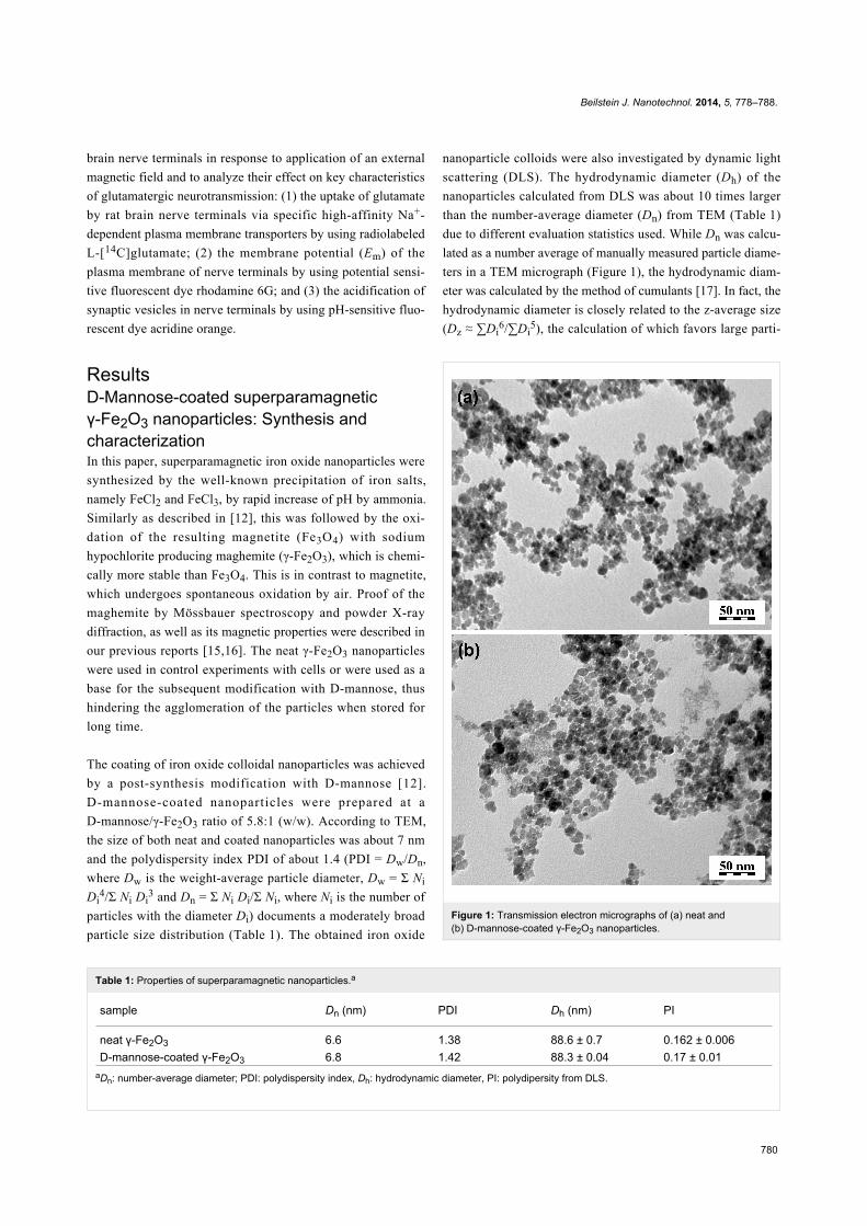
ResultsD-Mannose-coated superparamagneticγ-Fe2O3 nanoparticles: Synthesis andcharacterizationIn this paper, superparamagnetic iron oxide nanoparticles were
synthesized by the well-known precipitation of iron salts,
namely FeCl2 and FeCl3, by rapid increase of pH by ammonia.
Similarly as described in [12], this was followed by the oxi-
dation of the resulting magnetite (Fe3O4) with sodium
hypochlorite producing maghemite (γ-Fe2O3), which is chemi-
cally more stable than Fe3O4. This is in contrast to magnetite,
which undergoes spontaneous oxidation by air. Proof of the
maghemite by Mössbauer spectroscopy and powder X-ray
diffraction, as well as its magnetic properties were described in
our previous reports [15,16]. The neat γ-Fe2O3 nanoparticles
were used in control experiments with cells or were used as a
base for the subsequent modification with D-mannose, thus
hindering the agglomeration of the particles when stored for
long time.
The coating of iron oxide colloidal nanoparticles was achieved
by a post-synthesis modification with D-mannose [12].
D-mannose-coated nanoparticles were prepared at a
D-mannose/γ-Fe2O3 ratio of 5.8:1 (w/w). According to TEM,
the size of both neat and coated nanoparticles was about 7 nm
and the polydispersity index PDI of about 1.4 (PDI = Dw/Dn,
where Dw is the weight-average particle diameter, Dw = Σ Ni
Di4/Σ Ni Di
3 and Dn = Σ Ni Di/Σ Ni, where Ni is the number of
particles with the diameter Di) documents a moderately broad
particle size distribution (Table 1). The obtained iron oxide
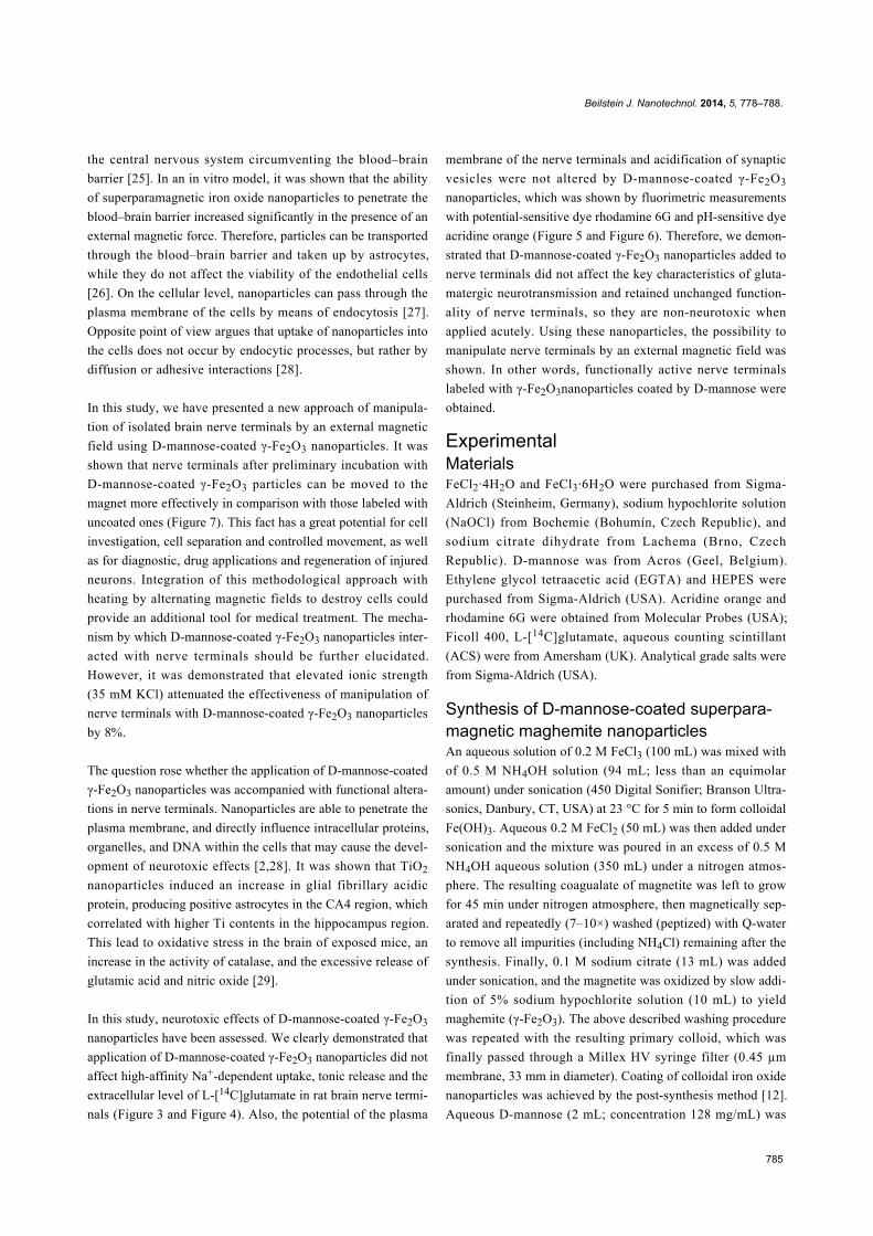
Figure 1: Transmission electron micrographs of (a) neat and(b) D-mannose-coated γ-Fe2O3 nanoparticles.
nanoparticle colloids were also investigated by dynamic light
scattering (DLS). The hydrodynamic diameter (Dh) of the
nanoparticles calculated from DLS was about 10 times larger
than the number-average diameter (Dn) from TEM (Table 1)
due to different evaluation statistics used. While Dn was calcu-
lated as a number average of manually measured particle diame-
ters in a TEM micrograph (Figure 1), the hydrodynamic diam-
eter was calculated by the method of cumulants [17]. In fact, the
hydrodynamic diameter is closely related to the z-average size
(Dz ≈ ∑Di6/∑Di
5), the calculation of which favors large parti-
Beilstein J. Nanotechnol. 2014, 5, 778–788.
781
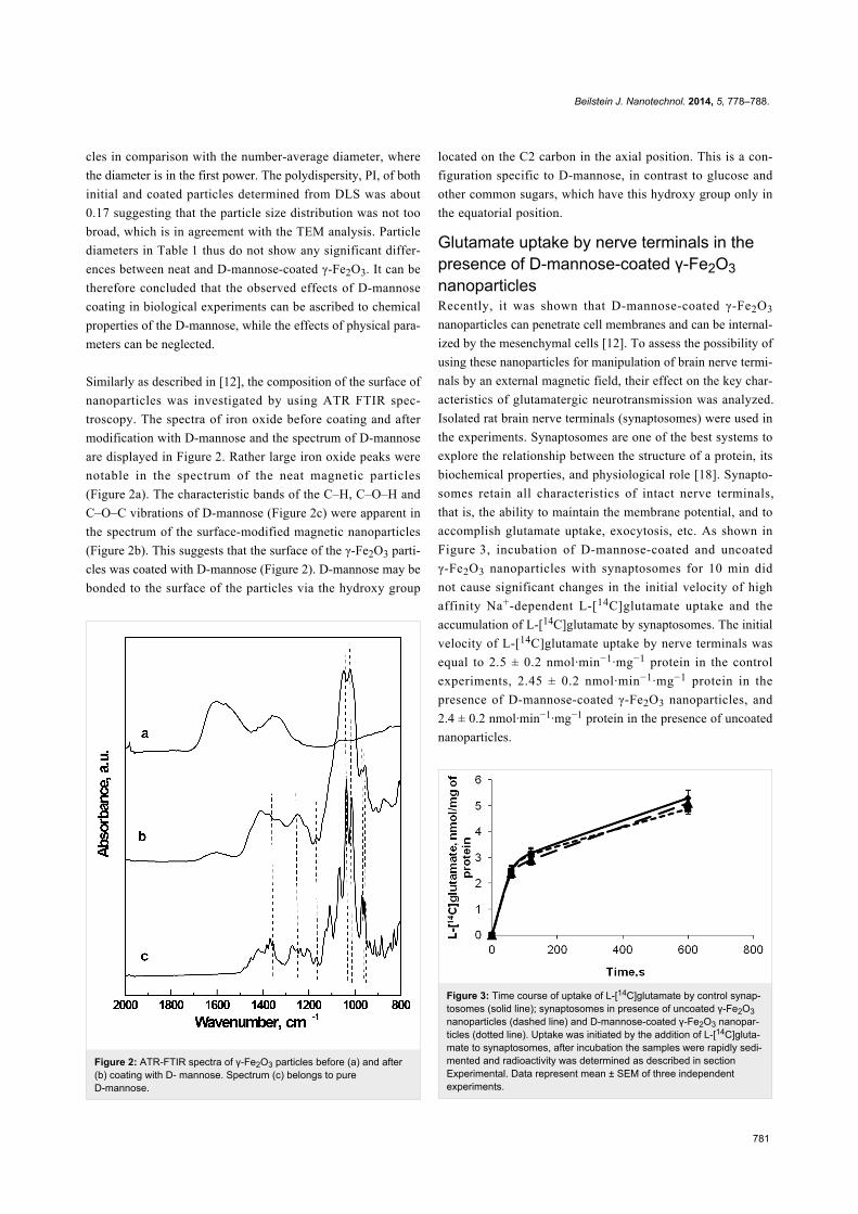
Figure 2: ATR-FTIR spectra of γ-Fe2O3 particles before (a) and after(b) coating with D- mannose. Spectrum (c) belongs to pureD-mannose.
cles in comparison with the number-average diameter, where
the diameter is in the first power. The polydispersity, PI, of both
initial and coated particles determined from DLS was about
0.17 suggesting that the particle size distribution was not too
broad, which is in agreement with the TEM analysis. Particle
diameters in Table 1 thus do not show any significant differ-
ences between neat and D-mannose-coated γ-Fe2O3. It can be
therefore concluded that the observed effects of D-mannose
coating in biological experiments can be ascribed to chemical
properties of the D-mannose, while the effects of physical para-
meters can be neglected.
Similarly as described in [12], the composition of the surface of
nanoparticles was investigated by using ATR FTIR spec-
troscopy. The spectra of iron oxide before coating and after
modification with D-mannose and the spectrum of D-mannose
are displayed in Figure 2. Rather large iron oxide peaks were
notable in the spectrum of the neat magnetic particles
(Figure 2a). The characteristic bands of the C–H, C–O–H and
C–O–C vibrations of D-mannose (Figure 2c) were apparent in
the spectrum of the surface-modified magnetic nanoparticles
(Figure 2b). This suggests that the surface of the γ-Fe2O3 parti-
cles was coated with D-mannose (Figure 2). D-mannose may be
bonded to the surface of the particles via the hydroxy group
located on the C2 carbon in the axial position. This is a con-
figuration specific to D-mannose, in contrast to glucose and
other common sugars, which have this hydroxy group only in
the equatorial position.
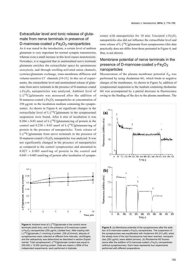
Glutamate uptake by nerve terminals in thepresence of D-mannose-coated γ-Fe2O3nanoparticlesRecently, it was shown that D-mannose-coated γ-Fe2O3
nanoparticles can penetrate cell membranes and can be internal-
ized by the mesenchymal cells [12]. To assess the possibility of
using these nanoparticles for manipulation of brain nerve termi-
nals by an external magnetic field, their effect on the key char-
acteristics of glutamatergic neurotransmission was analyzed.
Isolated rat brain nerve terminals (synaptosomes) were used in
the experiments. Synaptosomes are one of the best systems to
explore the relationship between the structure of a protein, its
biochemical properties, and physiological role [18]. Synapto-
somes retain all characteristics of intact nerve terminals,
that is, the ability to maintain the membrane potential, and to
accomplish glutamate uptake, exocytosis, etc. As shown in
Figure 3, incubation of D-mannose-coated and uncoated
γ-Fe2O3 nanoparticles with synaptosomes for 10 min did
not cause significant changes in the initial velocity of high
affinity Na+-dependent L-[14C]glutamate uptake and the
accumulation of L-[14C]glutamate by synaptosomes. The initial
velocity of L-[14C]glutamate uptake by nerve terminals was
equal to 2.5 ± 0.2 nmol·min−1·mg−1 protein in the control
experiments, 2.45 ± 0.2 nmol·min−1·mg−1 protein in the
presence of D-mannose-coated γ-Fe2O3 nanoparticles, and
2.4 ± 0.2 nmol·min−1·mg−1 protein in the presence of uncoated
nanoparticles.
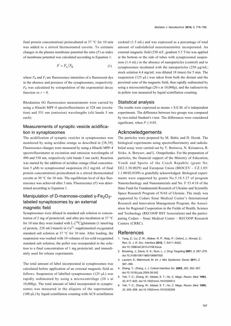
Figure 3: Time course of uptake of L-[14C]glutamate by control synap-tosomes (solid line); synaptosomes in presence of uncoated γ-Fe2O3nanoparticles (dashed line) and D-mannose-coated γ-Fe2O3 nanopar-ticles (dotted line). Uptake was initiated by the addition of L-[14C]gluta-mate to synaptosomes, after incubation the samples were rapidly sedi-mented and radioactivity was determined as described in sectionExperimental. Data represent mean ± SEM of three independentexperiments.
Beilstein J. Nanotechnol. 2014, 5, 778–788.
782
Figure 4: Ambient level of L-[14C]glutamate in the control nerveterminals (solid line), and in the presence of D-mannose-coatedγ-Fe2O3 nanoparticles (250 µg/mL) (dotted line). After loading withL-[14C]glutamate (1 nmol/mg of protein, 238 µCi/mmol), aliquots ofsynaptosomes were collected at different time intervals, centrifuged,and the radioactivity was determined as described in section Experi-mental. Total synaptosomal L-[14C]glutamate content was equal to200,000 ± 15,000 cpm/mg protein. Data are means ± SEM of fiveindependent experiments, each performed in triplicate.
Extracellular level and tonic release of gluta-mate from nerve terminals in presence ofD-mannose-coated γ-Fe2O3 nanoparticlesAs it was stated in the introduction, a certain level of ambient
glutamate is very important for normal synaptic transmission,
whereas even a small increase in this level causes neurotoxicity.
Nowadays, it is suggested that in unstimulated nerve terminals
glutamate enriches the extracellular space by spontaneous
exocytosis, and through swelling-activated anion channels,
cysteine/glutamate exchange, trans-membrane diffusion and
volume-sensitive Cl− channels [19-21]. In this set of experi-
ments, the extracellular level and unstimulated release of gluta-
mate from nerve terminals in the presence of D-mannose-coated
γ-Fe2O3 nanoparticles was analyzed. Ambient level of
L-[14C]glutamate was measured after the addition of
D-mannose-coated γ-Fe2O3 nanoparticles at concentration of
250 µg/mL to the incubation medium containing the synapto-
somes. As shown in Figure 4, no significant changes in the
extracellular level of L-[14C]glutamate in the synaptosomal
suspension were found. After 6 min of incubation it was
0.204 ± 0.03 nmol of L-[14C]glutamate/mg of protein in the
control and 0.230 ± 0.03 nmol of L-[14C]glutamate/mg of
protein in the presence of nanoparticles. Tonic release of
L-[14C]glutamate from nerve terminals in the presence of
D-mannose-coated γ-Fe2O3 nanoparticles was analyzed. It was
not significantly changed in the presence of nanoparticles
as compared to the control synaptosomes and amounted to
0.027 ± 0.005 nmol/mg of protein in the control and
0.045 ± 0.005 nmol/mg of protein after incubation of synapto-
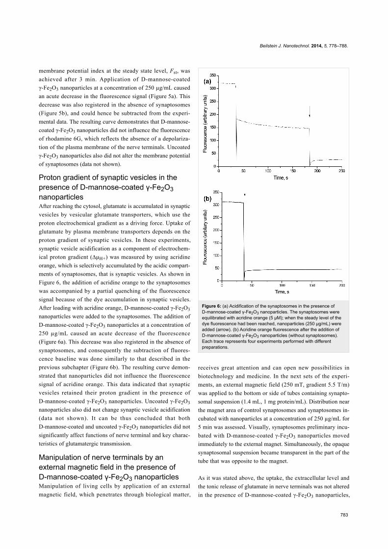
Figure 5: (a) Membrane potential of the synaptosomes after the addi-tion of D-mannose-coated γ-Fe2O3 nanoparticles. The suspension ofthe synaptosomes was equilibrated with rhodamine 6G (0.5 µM); whenthe steady level of the dye fluorescence had been reached, nanoparti-cles (250 µg/mL) were added (arrows). (b) Rhodamine 6G fluores-cence after the addition of D-mannose-coated γ-Fe2O3 nanoparticles(without synaptosomes). Each trace represents four experimentsperformed with different preparations.
somes with nanoparticles for 10 min. Uncoated γ-Fe2O3
nanoparticles also did not influence the extracellular level and
tonic release of L-[14C]glutamate from synaptosomes (this data
practically does not differ from those presented in Figure 4, and
thus, is not shown).
Membrane potential of nerve terminals in thepresence of D-mannose-coated γ-Fe2O3nanoparticlesMeasurement of the plasma membrane potential Em was
performed by using rhodamine 6G, which binds to negative
charges of the membranes. As shown in Figure 5a, addition of
synaptosomal suspension to the medium containing rhodamine
6G was accompanied by a partial decrease in fluorescence
owing to the binding of the dye to the plasma membrane. The
Beilstein J. Nanotechnol. 2014, 5, 778–788.
783
membrane potential index at the steady state level, Fst, was
achieved after 3 min. Application of D-mannose-coated
γ-Fe2O3 nanoparticles at a concentration of 250 µg/mL caused
an acute decrease in the fluorescence signal (Figure 5a). This
decrease was also registered in the absence of synaptosomes
(Figure 5b), and could hence be subtracted from the experi-
mental data. The resulting curve demonstrates that D-mannose-
coated γ-Fe2O3 nanoparticles did not influence the fluorescence
of rhodamine 6G, which reflects the absence of a depolariza-
tion of the plasma membrane of the nerve terminals. Uncoated
γ-Fe2O3 nanoparticles also did not alter the membrane potential
of synaptosomes (data not shown).
Proton gradient of synaptic vesicles in thepresence of D-mannose-coated γ-Fe2O3nanoparticlesAfter reaching the cytosol, glutamate is accumulated in synaptic
vesicles by vesicular glutamate transporters, which use the
proton electrochemical gradient as a driving force. Uptake of
glutamate by plasma membrane transporters depends on the
proton gradient of synaptic vesicles. In these experiments,
synaptic vesicle acidification as a component of electrochem-
ical proton gradient (∆μH+) was measured by using acridine
orange, which is selectively accumulated by the acidic compart-
ments of synaptosomes, that is synaptic vesicles. As shown in
Figure 6, the addition of acridine orange to the synaptosomes
was accompanied by a partial quenching of the fluorescence
signal because of the dye accumulation in synaptic vesicles.
After loading with acridine orange, D-mannose-coated γ-Fe2O3
nanoparticles were added to the synaptosomes. The addition of
D-mannose-coated γ-Fe2O3 nanoparticles at a concentration of
250 µg/mL caused an acute decrease of the fluorescence
(Figure 6a). This decrease was also registered in the absence of
synaptosomes, and consequently the subtraction of fluores-
cence baseline was done similarly to that described in the
previous subchapter (Figure 6b). The resulting curve demon-
strated that nanoparticles did not influence the fluorescence
signal of acridine orange. This data indicated that synaptic
vesicles retained their proton gradient in the presence of
D-mannose-coated γ-Fe2O3 nanoparticles. Uncoated γ-Fe2O3
nanoparticles also did not change synaptic vesicle acidification
(data not shown). It can be thus concluded that both
D-mannose-coated and uncoated γ-Fe2O3 nanoparticles did not
significantly affect functions of nerve terminal and key charac-
teristics of glutamatergic transmission.
Manipulation of nerve terminals by anexternal magnetic field in the presence ofD-mannose-coated γ-Fe2O3 nanoparticlesManipulation of living cells by application of an external
magnetic field, which penetrates through biological matter,
Figure 6: (a) Acidification of the synaptosomes in the presence ofD-mannose-coated γ-Fe2O3 nanoparticles. The synaptosomes wereequilibrated with acridine orange (5 µM); when the steady level of thedye fluorescence had been reached, nanoparticles (250 µg/mL) wereadded (arrow). (b) Acridine orange fluorescence after the addition ofD-mannose-coated γ-Fe2O3 nanoparticles (without synaptosomes).Each trace represents four experiments performed with differentpreparations.
receives great attention and can open new possibilities in
biotechnology and medicine. In the next sets of the experi-
ments, an external magnetic field (250 mT, gradient 5.5 Т/m)
was applied to the bottom or side of tubes containing synapto-
somal suspension (1.4 mL, 1 mg protein/mL). Distribution near
the magnet area of control synaptosomes and synaptosomes in-
cubated with nanoparticles at a concentration of 250 µg/mL for
5 min was assessed. Visually, synaptosomes preliminary incu-
bated with D-mannose-coated γ-Fe2O3 nanoparticles moved
immediately to the external magnet. Simultaneously, the opaque
synaptosomal suspension became transparent in the part of the
tube that was opposite to the magnet.
As it was stated above, the uptake, the extracellular level and
the tonic release of glutamate in nerve terminals was not altered
in the presence of D-mannose-coated γ-Fe2O3 nanoparticles,
Beilstein J. Nanotechnol. 2014, 5, 778–788.
784
therefore L-[14C]glutamate-loaded nerve terminals can be used
for quantitative assessment of their movement in response to the
application of an external magnetic field. The amount of the
radioactive label incorporated in the synaptosomes was calcu-
lated as percentage of the total amount of the label in the
suspensions before and after the application of an external
magnetic field. The synaptosomes were taken both from distant
and proximal zone to the magnet, and then rapidly sedimented
using a microcentrifuge and radioactivity in pellets was
measured by liquid scintillation counting. The extracellular
level of L-[14C]glutamate, which was similar for all probes, was
subtracted.
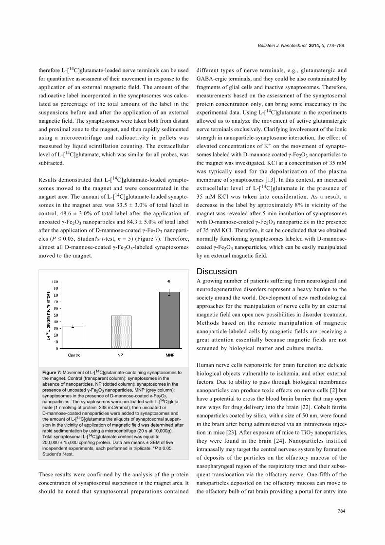
Results demonstrated that L-[14C]glutamate-loaded synapto-
somes moved to the magnet and were concentrated in the
magnet area. The amount of L-[14C]glutamate-loaded synapto-
somes in the magnet area was 33.5 ± 3.0% of total label in
control, 48.6 ± 3.0% of total label after the application of
uncoated γ-Fe2O3 nanoparticles and 84.3 ± 5.0% of total label
after the application of D-mannose-coated γ-Fe2O3 nanoparti-
cles (Р ≤ 0.05, Student's t-test, n = 5) (Figure 7). Therefore,
almost all D-mannose-coated γ-Fe2O3-labeled synaptosomes
moved to the magnet.
Figure 7: Movement of L-[14C]glutamate-containing synaptosomes tothe magnet. Control (transparent column): synaptosomes in theabsence of nanoparticles, NP (dotted column): synaptosomes in thepresence of uncoated γ-Fe2O3 nanoparticles, MNP (grey column):synaptosomes in the presence of D-mannose-coated γ-Fe2O3nanoparticles. The synaptosomes were pre-loaded with L-[14C]gluta-mate (1 nmol/mg of protein, 238 mCi/mmol), then uncoated orD-mannose-coated nanoparticles were added to synaptosomes andthe amount of L-[14C]glutamate the aliquots of synaptosomal suspen-sion in the vicinity of application of magnetic field was determined afterrapid sedimentation by using a microcentrifuge (20 s at 10,000g).Total synaptosomal L-[14C]glutamate content was equal to200,000 ± 15,000 cpm/mg protein. Data are means ± SEM of fiveindependent experiments, each performed in triplicate. *Р ≤ 0.05,Student's t-test.
These results were confirmed by the analysis of the protein
concentration of synaptosomal suspension in the magnet area. It
should be noted that synaptosomal preparations contained
different types of nerve terminals, e.g., glutamatergic and
GABA-ergic terminals, and they could be also contaminated by
fragments of glial cells and inactive synaptosomes. Therefore,
measurements based on the assessment of the synaptosomal
protein concentration only, can bring some inaccuracy in the
experimental data. Using L-[14C]glutamate in the experiments
allowed us to analyze the movement of active glutamatergic
nerve terminals exclusively. Clarifying involvement of the ionic
strength in nanoparticle-synaptosome interaction, the effect of
elevated concentrations of K+ on the movement of synapto-
somes labeled with D-mannose coated γ-Fe2O3 nanoparticles to
the magnet was investigated. KCl at a concentration of 35 mM
was typically used for the depolarization of the plasma
membrane of synaptosomes [13]. In this context, an increased
extracellular level of L-[14C]glutamate in the presence of
35 mM KCl was taken into consideration. As a result, a
decrease in the label by approximately 8% in vicinity of the
magnet was revealed after 5 min incubation of synaptosomes
with D-mannose-coated γ-Fe2O3 nanoparticles in the presence
of 35 mM KCl. Therefore, it can be concluded that we obtained
normally functioning synaptosomes labeled with D-mannose-
coated γ-Fe2O3 nanoparticles, which can be easily manipulated
by an external magnetic field.
DiscussionA growing number of patients suffering from neurological and
neurodegenerative disorders represent a heavy burden to the
society around the world. Development of new methodological
approaches for the manipulation of nerve cells by an external
magnetic field can open new possibilities in disorder treatment.
Methods based on the remote manipulation of magnetic
nanoparticle-labeled cells by magnetic fields are receiving a
great attention essentially because magnetic fields are not
screened by biological matter and culture media.
Human nerve cells responsible for brain function are delicate
biological objects vulnerable to ischemia, and other external
factors. Due to ability to pass through biological membranes
nanoparticles can produce toxic effects on nerve cells [2] but
have a potential to cross the blood brain barrier that may open
new ways for drug delivery into the brain [22]. Cobalt ferrite
nanoparticles coated by silica, with a size of 50 nm, were found
in the brain after being administered via an intravenous injec-
tion in mice [23]. After exposure of mice to TiO2 nanoparticles,
they were found in the brain [24]. Nanoparticles instilled
intranasally may target the central nervous system by formation
of deposits of the particles on the olfactory mucosa of the
nasopharyngeal region of the respiratory tract and their subse-
quent translocation via the olfactory nerve. One-fifth of the
nanoparticles deposited on the olfactory mucosa can move to
the olfactory bulb of rat brain providing a portal for entry into
Beilstein J. Nanotechnol. 2014, 5, 778–788.
785
the central nervous system circumventing the blood–brain
barrier [25]. In an in vitro model, it was shown that the ability
of superparamagnetic iron oxide nanoparticles to penetrate the
blood–brain barrier increased significantly in the presence of an
external magnetic force. Therefore, particles can be transported
through the blood–brain barrier and taken up by astrocytes,
while they do not affect the viability of the endothelial cells
[26]. On the cellular level, nanoparticles can pass through the
plasma membrane of the cells by means of endocytosis [27].
Opposite point of view argues that uptake of nanoparticles into
the cells does not occur by endocytic processes, but rather by
diffusion or adhesive interactions [28].
In this study, we have presented a new approach of manipula-
tion of isolated brain nerve terminals by an external magnetic
field using D-mannose-coated γ-Fe2O3 nanoparticles. It was
shown that nerve terminals after preliminary incubation with
D-mannose-coated γ-Fe2O3 particles can be moved to the
magnet more effectively in comparison with those labeled with
uncoated ones (Figure 7). This fact has a great potential for cell
investigation, cell separation and controlled movement, as well
as for diagnostic, drug applications and regeneration of injured
neurons. Integration of this methodological approach with
heating by alternating magnetic fields to destroy cells could
provide an additional tool for medical treatment. The mecha-
nism by which D-mannose-coated γ-Fe2O3 nanoparticles inter-
acted with nerve terminals should be further elucidated.
However, it was demonstrated that elevated ionic strength
(35 mM KCl) attenuated the effectiveness of manipulation of
nerve terminals with D-mannose-coated γ-Fe2O3 nanoparticles
by 8%.
The question rose whether the application of D-mannose-coated
γ-Fe2O3 nanoparticles was accompanied with functional altera-
tions in nerve terminals. Nanoparticles are able to penetrate the
plasma membrane, and directly influence intracellular proteins,
organelles, and DNA within the cells that may cause the devel-
opment of neurotoxic effects [2,28]. It was shown that TiO2
nanoparticles induced an increase in glial fibrillary acidic
protein, producing positive astrocytes in the CA4 region, which
correlated with higher Ti contents in the hippocampus region.
This lead to oxidative stress in the brain of exposed mice, an
increase in the activity of catalase, and the excessive release of
glutamic acid and nitric oxide [29].
In this study, neurotoxic effects of D-mannose-coated γ-Fe2O3
nanoparticles have been assessed. We clearly demonstrated that
application of D-mannose-coated γ-Fe2O3 nanoparticles did not
affect high-affinity Na+-dependent uptake, tonic release and the
extracellular level of L-[14C]glutamate in rat brain nerve termi-
nals (Figure 3 and Figure 4). Also, the potential of the plasma
membrane of the nerve terminals and acidification of synaptic
vesicles were not altered by D-mannose-coated γ-Fe2O3
nanoparticles, which was shown by fluorimetric measurements
with potential-sensitive dye rhodamine 6G and pH-sensitive dye
acridine orange (Figure 5 and Figure 6). Therefore, we demon-
strated that D-mannose-coated γ-Fe2O3 nanoparticles added to
nerve terminals did not affect the key characteristics of gluta-
matergic neurotransmission and retained unchanged function-
ality of nerve terminals, so they are non-neurotoxic when
applied acutely. Using these nanoparticles, the possibility to
manipulate nerve terminals by an external magnetic field was
shown. In other words, functionally active nerve terminals
labeled with γ-Fe2O3nanoparticles coated by D-mannose were
obtained.
ExperimentalMaterialsFeCl2·4H2O and FeCl3·6H2O were purchased from Sigma-
Aldrich (Steinheim, Germany), sodium hypochlorite solution
(NaOCl) from Bochemie (Bohumín, Czech Republic), and
sodium citrate dihydrate from Lachema (Brno, Czech
Republic). D-mannose was from Acros (Geel, Belgium).
Ethylene glycol tetraacetic acid (EGTA) and HEPES were
purchased from Sigma-Aldrich (USA). Acridine orange and
rhodamine 6G were obtained from Molecular Probes (USA);
Ficoll 400, L-[14C]glutamate, aqueous counting scintillant
(ACS) were from Amersham (UK). Analytical grade salts were
from Sigma-Aldrich (USA).
Synthesis of D-mannose-coated superpara-magnetic maghemite nanoparticlesAn aqueous solution of 0.2 M FeCl3 (100 mL) was mixed with
of 0.5 M NH4OH solution (94 mL; less than an equimolar
amount) under sonication (450 Digital Sonifier; Branson Ultra-
sonics, Danbury, CT, USA) at 23 °C for 5 min to form colloidal
Fe(OH)3. Aqueous 0.2 M FeCl2 (50 mL) was then added under
sonication and the mixture was poured in an excess of 0.5 M
NH4OH aqueous solution (350 mL) under a nitrogen atmos-
phere. The resulting coagualate of magnetite was left to grow
for 45 min under nitrogen atmosphere, then magnetically sep-
arated and repeatedly (7–10×) washed (peptized) with Q-water
to remove all impurities (including NH4Cl) remaining after the
synthesis. Finally, 0.1 M sodium citrate (13 mL) was added
under sonication, and the magnetite was oxidized by slow addi-
tion of 5% sodium hypochlorite solution (10 mL) to yield
maghemite (γ-Fe2O3). The above described washing procedure
was repeated with the resulting primary colloid, which was
finally passed through a Millex HV syringe filter (0.45 µm
membrane, 33 mm in diameter). Coating of colloidal iron oxide
nanoparticles was achieved by the post-synthesis method [12].
Aqueous D-mannose (2 mL; concentration 128 mg/mL) was
Beilstein J. Nanotechnol. 2014, 5, 778–788.
786
added dropwise under sonication to a portion of primary colloid
containing 44 mg of iron oxide and finally diluted to a volume
of 10 mL with ultrapure water. The obtained mixture was soni-
cated for 5 min and resulting D-mannose coated γ-Fe2O3 parti-
cles were used in biological experiments.
Properties of particlesThe particle shape, diameter and size distribution were recorded
by a JEOL JEM 200 CX transmission electron microscope
(TEM). The size was calculated by using the Atlas program
(Tescan, Digital Microscopy Imaging, Brno, Czech Republic).
The hydrodynamic diameter Dh (z-average) and polydispersity
as a measure of the particle size distribution were determined by
dynamic light scattering (DLS) with an Autosizer Lo-C
(Malvern Instruments, UK). The modification of the nanopar-
ticle surface with D-mannose was analyzed using a Nicolet
Impact 400 Fourier transformation infrared (FTIR) spectrom-
eter in water-purged surrounding with a DTGS detector. The
spectra were measured by ATR spectroscopy with the Golden
GateTM single reflection system (Specac Ltd., Orpington, UK)
at 400–4000 cm−1.
Ethics statementWistar male rats, 100–120 g body weight, were obtained from
the vivarium of M. D. Strazhesko Institute of Cardiology,
Medical Academy of Sciences of Ukraine. Animals were kept
in animal facilities of the Palladin Institute of Biochemistry in
accordance with the European guidelines and international laws
and policies. They were housed in a quiet, temperature-
controlled room (22–23 °C) and were provided with water and
dry food pellets ad libitum. Before removing the brain, rats
were decapitated. Experimental protocols were approved by the
Animal Care and Use Committee of the Palladin Institute of
Biochemistry (Protocol from 19/09-2011).
Isolation of rat brain nerve terminals (synap-tosomes)Synaptosomes were prepared from rat brain as described in
[30]. Cerebral hemispheres of decapitated animals were
removed and homogenized rapidly in ice-cold 0.32 M sucrose,
5 mM HEPES–NaOH, pH 7.4, and 0.2 mM EDTA. The synap-
tosomes were prepared by differential and Ficoll-400 density
gradient centrifugation of rat brain homogenate [31] with slight
modifications [30]. All manipulations were performed at 4 °C.
The synaptosomal suspensions were used in experiments during
2–4 h after isolation. The standard salt solution was oxygenated
and contained (in mM): NaCl 126; KCl 5; MgCl2 2.0; NaH2PO4
1.0; HEPES 20, pH 7.4; and D-glucose 10. Ca2+-supplemented
medium contained 2 mM CaCl2. The Ca2+-free medium
contained 1 mM EGTA and no added Ca2+. Protein concentra-
tion was measured as described by Larson et al. [32].
L-[14C]glutamate uptake by nerve terminalsThe uptake of L-[14C]glutamate by synaptosomes was
measured as described in [33]. The synaptosomal suspension
(125 μL of the suspension, 0.2 mg of protein/mL) was pre-incu-
bated in standard salt solution at 37 °C for 8 min, then magnetic
nanoparticles (250 µg/mL; stock solution 4.4 mg/mL was
diluted 18 times) were added to the synaptosomal suspension
and incubated for 10 min. Uptake was initiated by the addition
of 10 µM L-glutamate supplemented with 420 nM
L-[14C]glutamate (0.1 µCi/mL), incubated at 37 °C during
different time intervals (1, 2, 10 min) and then rapidly sedi-
mented by using a microcentrifuge (20 s at 10,000g).
L-[14C]glutamate uptake was determined as a decrease in
radioactivity in aliquots of the supernatant (100 μL) and an
increase in radioactivity of the pellet (SDS-treated) measured by
liquid scintillation counting with ACS scintillation cocktail
(1.5 mL).
L-[14C]glutamate release from nerve termi-nalsThe release of L-[14C]glutamate from the synaptosomes was
measured as described in [34,35]. The synaptosomes were
diluted in standard salt solution to reach concentration of 2 mg
of protein/mL and after pre-incubation at 37 °C for 10 min they
were loaded with L-[14C]glutamate (1 nmol/mg of protein,
238 mCi/mmol) in Ca2+-supplemented oxygenated standard salt
solution at 37 °C for 10 min. After loading, suspension was
washed with 10 volumes of ice-cold oxygenated standard salt
solution; the pellet was resuspended in a solution to a final
concentration of 1 mg protein/mL and immediately used for
release experiments. Release of L-[14C]glutamate from the
synaptosomes was performed in Ca2+-free incubation medium
according to following method. Synaptosomal suspension
(125 μL; 0.5 mg of protein/mL) was pre-incubated for 10 min,
then the nanoparticles (250 µg/mL; stock solution 4.4 mg/mL
was diluted 18 times) were added at 37 °C and incubated for
different time intervals (0, 3, 6, 10 min) and rapidly sedimented
by using a microcentrifuge (20 s at 10,000g). Release was
measured in the aliquots of the supernatants (100 μL) by liquid
scintillation counting with scintillation cocktail ACS (1.5 mL)
and was expressed a percentage of total amount of radiolabeled
neurotransmitter incorporated. Release of the neurotransmitter
from the synaptosomes incubated in Ca2+-free media without
stimulating agents was used for assay of unstimulated (tonic)
release.
Measurement of synaptosomal plasmamembrane potential (Em)The membrane potential was measured as described in [36,37]
by using rhodamine 6G (0.5 µM) which binds to the plasma
membrane. The suspension of synaptosomes (0.2 mg/mL of
Beilstein J. Nanotechnol. 2014, 5, 778–788.
787
final protein concentration) preincubated at 37 °C for 10 min
was added to a stirred thermostatted cuvette. To estimate
changes in the plasma membrane potential the ratio (F) as index
of membrane potential was calculated according to Equation 1:
(1)
where F0 and Ft are fluorescence intensities of a fluorescent dye
in the absence and presence of the synaptosomes, respectively.
F0 was calculated by extrapolation of the exponential decay
function to t = 0.
Rhodamine 6G fluorescence measurements were carried by
using a Hitachi MPF-4 spectrofluorimeter at 528 nm (excita-
tion) and 551 nm (emission) wavelengths (slit bands 5 nm
each).
Measurements of synaptic vesicle acidifica-tion in synaptosomesThe acidification of synaptic vesicles in synaptosomes was
monitored by using acridine orange as described in [38,39].
Fluorescence changes were measured by using a Hitachi MPF-4
spectrofluorimeter at excitation and emission wavelengths of
490 and 530 nm, respectively (slit bands 5 nm each). Reaction
was started by the addition of acridine orange (final concentra-
tion 5 μM) to synaptosomal suspension (0.2 mg/mL of final
protein concentration) preincubated in a stirred thermostatted
cuvette at 30 °C for 10 min. The equilibrium level of dye fluo-
rescence was achieved after 3 min. Fluorescence (F) was deter-
mined according to Equation 1.
Manipulation of D-mannose-coated γ-Fe2O3-labeled synaptosomes by an externalmagnetic fieldSynaptosomes were diluted in standard salt solution to concen-
tration of 2 mg of protein/mL and after pre-incubation at 37 °C
for 10 min they were loaded with L-[14C]glutamate (1 nmol/mg
of protein, 238 mCi/mmol) in Ca2+-supplemented oxygenated
standard salt solution at 37 °C for 10 min. After loading, the
suspension was washed with 10 volumes of ice-cold oxygenated
standard salt solution; the pellet was resuspended in the solu-
tion to a final concentration of 1 mg protein/mL and immedi-
ately used for release experiments.
The total amount of label incorporated in synaptosomes was
calculated before application of an external magnetic field as
follows. Suspension of labelled synaptosomes (125 μL) was
rapidly sedimented by using a microcentrifuge (20 s at
10,000g). The total amount of label incorporated in synapto-
somes was measured in the aliquots of the supernatants
(100 μL) by liquid scintillation counting with ACS scintillation
cocktail (1.5 mL) and was expressed as a percentage of total
amount of radiolabeled neurotransmitter incorporated. An
external magnetic field (250 mT, gradient 5.5 Т/m) was applied
at the bottom or the side of tubes with synaptosomal suspen-
sion (1.4 mL) in the absence of nanoparticles (control) and to
synaptosomes incubated with the nanoparticles (250 µg/mL;
stock solution 4.4 mg/mL was diluted 18 times) for 5 min. The
suspension (125 μL) was taken from both the distant and the
proximal zone of the magnetic field, then rapidly sedimented by
using a microcentrifuge (20 s at 10,000g), and the radioactivity
in pellets was measured by liquid scintillation counting.
Statistical analysisThe results were expressed as means ± S.E.M. of n independent
experiments. The difference between two groups was compared
by two-tailed Student's t-test. The differences were considered
significant, when Р ≤ 0.05.
AcknowledgementsThe particles were prepared by M. Babic and D. Horak. The
biological experiments using spectrofluorimetry and radiola-
beled assay were carried out by T. Borisova, N. Krisanova, R.
Sivko, A. Borуsov, and L. Ostapchenko. For the preparation of
particles, the financial support of the Ministry of Education,
Youth and Sports of the Czech Republic (grant No.
EE2.3.30.0029) and European Union (BIOCEV – CZ.1.05/
1.1.00/02.0109) is gratefully acknowledged. Biological experi-
ments were supported by grants No.5.18.5.27 of program
Nanotechnology and Nanomaterials and No. F 53.4/18 of the
State Fund for Fundamental Research of Ukraine and Scientific
Space Research Program of NAS of Ukraine. The study was
supported by Cedars Sinai Medical Center’s International
Research and Innovation Management Program, the Associ-
ation for Regional Cooperation in the Fields of Health, Science
and Technology (RECOOP HST Association) and the partici-
pating Cedars – Sinai Medical Center – RECOOP Research
Centers (CRRC).
References1. Yang, Z.; Liu, Z. W.; Allaker, R. P.; Reip, P.; Oxford, J.; Ahmad, Z.;
Ren, G. J. R. Soc. Interface 2010, 7, S411–S422.doi:10.1098/rsif.2010.0158.focus
2. Brooking, J.; Davis, S. S.; Illum, L. J. Drug Targeting 2001, 9, 267–279.doi:10.3109/10611860108997935
3. Laurent, S.; Mahmoudi, M. Int. J. Mol. Epidemiol. Genet. 2011, 2,367–390.
4. Zhang, Y.; Zhang, J. J. Colloid Interface Sci. 2005, 283, 352–357.doi:10.1016/j.jcis.2004.09.042
5. Yeh, T.-C.; Zhang, W.; Ildstad, S. T.; Ho, C. Magn. Reson. Med. 1993,30, 617–625. doi:10.1002/mrm.1910300513
6. Yeh, T.-C.; Zhang, W.; Ildstad, S. T.; Ho, C. Magn. Reson. Med. 1995,33, 200–208. doi:10.1002/mrm.1910330209
Beilstein J. Nanotechnol. 2014, 5, 778–788.
788
7. Modo, M.; Cash, D.; Mellodew, K.; Williams, S. C. R.; Fraser, S. E.;Meade, T. J.; Price, J.; Hodges, H. NeuroImage 2002, 17, 803–811.doi:10.1006/nimg.2002.1194
8. Jendelová, P.; Herynek, V.; De Croos, J.; Glogarova, K.;Andersson, B.; Hajek, M.; Sykova, E. Magn. Reson. Med. 2003, 50,767–776. doi:10.1002/mrm.10585
9. Jendelová, P.; Herynek, V.; Urdzíková, L.; Glogarová, K.; Kroupová, J.;Andersson, B.; Bryja, V.; Burian, M.; Hájek, M.; Syková, E.J. Neurosci. Res. 2004, 76, 232–243. doi:10.1002/jnr.20041
10. Irache, J. M.; Salman, H. H.; Gamazo, C.; Espuelas, S.Expert Opin. Drug Delivery 2008, 5, 703–724.doi:10.1517/17425247.5.6.703
11. Labský, J. Biomaterials 2003, 24, 4031–4036.doi:10.1016/S0142-9612(03)00313-2
12. Horák, D.; Babič, M.; Jendelová, P.; Herynek, V.; Trchová, M.;Pientka, Z.; Pollert, E.; Hájek, M.; Syková, E. Bioconjugate Chem.2007, 18, 635–644. doi:10.1021/bc060186c
13. Borisova, T.; Sivko, R.; Borysov, A.; Krisanova, N. Cell. Mol. Neurobiol.2010, 30, 1013–1023. doi:10.1007/s10571-010-9532-x
14. Zoccarato, F.; Cavallini, L.; Alexandre, A. J. Neurochem. 1999, 72,625–633. doi:10.1046/j.1471-4159.1999.0720625.x
15. Pollert, E.; Knížek, K.; Maryško, M.; Závěta, K.; Lančok, A.;Boháček, J.; Horák, D.; Babič, M. J. Magn. Magn. Mater. 2006, 306,241–247. doi:10.1016/j.jmmm.2006.03.069
16. Tocchio, A.; Horák, D.; Babic, M.; Trchová, M.; Veverka, M.;Beneš, M. J.; Šlouf, M.; Fojtík, A. J. Polym. Sci., Part A: Polym. Chem.2009, 47, 4982–4994. doi:10.1002/pola.23551
17. Koppel, D. E. J. Chem. Phys. 1972, 57, 4814–4820.doi:10.1063/1.1678153
18. Südhof, T. C. Annu. Rev. Neurosci. 2004, 27, 509–547.doi:10.1146/annurev.neuro.26.041002.131412
19. Cavelier, P.; Attwell, D. J. Physiol. 2005, 564, 397–410.doi:10.1113/jphysiol.2004.082131
20. Jabaudon, D.; Shimamoto, K.; Yasuda-Kamatani, Y.; Gähwiler, B. H.;Gerber, U. Proc. Natl. Acad. Sci. U. S. A. 1999, 96, 8733–8738.doi:10.1073/pnas.96.15.8733
21. Rutledge, E. M.; Aschner, M.; Kimelberg, H. K. Am. J. Physiol. 1998,274, C1511–C1520.
22. De Jong, W. H.; Borm, P. J. A. Int. J. Nanomed. 2008, 3, 133–149.doi:10.2147/IJN.S596
23. Kim, J. S.; Yoon, T. J.; Yu, K. N.; Kim, B. G.; Park, S. J.; Kim, H. W.;Lee, K. H.; Park, S. B.; Lee, J. K.; Cho, M. H. Toxicol. Sci. 2006, 89,338–347. doi:10.1093/toxsci/kfj027
24. Takeda, K.; Suzuki, K.; Ishihara, A.; Kubo-Irie, M.; Fujimoto, R.;Tabata, M.; Oshio, S.; Nihei, Y.; Ihara, T.; Sugamata, M. J. Health Sci.2009, 55, 95–102. doi:10.1248/jhs.55.95
25. Oberdörster, G.; Sharp, Z.; Atudorei, V.; Elder, A.; Gelein, R.;Kreyling, W.; Cox, C. Inhalation Toxicol. 2004, 16, 437–445.doi:10.1080/08958370490439597
26. Thomsen, L. B.; Linemann, T.; Pondman, K. M.; Lichota, J.; Kim, K. S.;Pieters, R. J.; Visser, G. M.; Moos, T. ACS Chem. Neurosci. 2013, 4,1352–1360. doi:10.1021/cn400093z
27. Xia, T.; Kovochich, M.; Liong, M.; Zink, J. I.; Nel, A. E. ACS Nano 2008,2, 85–96. doi:10.1021/nn700256c
28. Geiser, M.; Rothen-Rutishauser, B.; Kapp, N.; Schürch, S.;Kreyling, W.; Schulz, H.; Semmler, M.; Im Hof, V.; Heyder, J.; Gehr, P.Environ. Health Perspect. 2005, 113, 1555–1560.doi:10.1289/ehp.8006
29. Wang, J.; Chen, C.; Liu, Y.; Jiao, F.; Li, W.; Lao, F.; Li, Y.; Li, B.;Ge, C.; Zhou, G.; Gao, Y.; Zhao, Y.; Chai, Z. Toxicol. Lett. 2008, 183,72–80. doi:10.1016/j.toxlet.2008.10.001
30. Borisova, T. A.; Krisanova, N. V. Adv. Space Res. 2008, 42,1971–1979. doi:10.1016/j.asr.2008.04.012
31. Cotman, C. W. Methods Enzymol. 1974, 31, 445–452.doi:10.1016/0076-6879(74)31050-6
32. Larson, E.; Howlett, B.; Jagendorf, A. Anal. Biochem. 1986, 155,243–248. doi:10.1016/0003-2697(86)90432-X
33. Borisova, T. Cholesterol and presynaptic glutamate transport in thebrain; Springer: New York, 2013. doi:10.1007/978-1-4614-7759-4
34. Krisanova, N. V.; Trikash, I. O.; Borisova, T. A. Neurochem. Int. 2009,55, 724–731. doi:10.1016/j.neuint.2009.07.003
35. Krisanova, N.; Sivko, R.; Kasatkina, L.; Borisova, T.Biochim. Biophys. Acta, Mol. Basis Dis. 2012, 1822, 1553–1561.doi:10.1016/j.bbadis.2012.06.005
36. Kasatkina, L.; Borisova, T. Neurochem. Int. 2010, 56, 711–719.doi:10.1016/j.neuint.2010.02.008
37. Kasatkina, L.; Borisova, T. A. Int. J. Biochem. Cell Biol. 2013, 45,2585–2595. doi:10.1016/j.biocel.2013.08.004
38. Borisova, T.; Kasatkina, L.; Ostapchenko, L. Neurochem. Int. 2011, 59,965–975. doi:10.1016/j.neuint.2011.07.007
39. Borisova, T.; Krisanova, N.; Sivko, R.; Kasatkina, L.; Borysov, A.;Griffin, S.; Wireman, M. Neurochem. Int. 2011, 59, 272–279.doi:10.1016/j.neuint.2011.05.014
License and TermsThis is an Open Access article under the terms of the
Creative Commons Attribution License
(http://creativecommons.org/licenses/by/2.0), which
permits unrestricted use, distribution, and reproduction in
any medium, provided the original work is properly cited.
The license is subject to the Beilstein Journal of
Nanotechnology terms and conditions:
(http://www.beilstein-journals.org/bjnano)
The definitive version of this article is the electronic one
which can be found at:
doi:10.3762/bjnano.5.90