Malassezia Baillon, emerging clinical yeasts
13
Malassezia Baillon, emerging clinical yeasts Roma Batra a,1 , Teun Boekhout b, * , Eveline Gue ´ho c , F. Javier Caban ˜es d , Thomas L. Dawson Jr. e , Aditya K. Gupta a,f a Mediprobe Research, London, Ont., Canada b Centraalbureau voor Schimmelcultures, Uppsalalaan 8, 85167 Utrecht, The Netherlands c 5 rue de la Huchette, F-61400 Mauves sur Huisne, France d Departament de Sanitat i dÕ Anatomia Animals, Universitat Auto ` noma de Barcelona, Bellaterra, Barcelona E-08193, Spain e Beauty Care Technology Division, Procter & Gamble Company, Cincinnati, USA f Division of Dermatology, Department of Medicine, Sunnybrook and WomenÕs College Health Science Center (Sunnybrook site) and the University of Toronto, Toronto, Ont., Canada Abstract The human and animal pathogenic yeast genus Malassezia has received considerable attention in recent years from dermatolo- gists, other clinicians, veterinarians and mycologists. Some points highlighted in this review include recent advances in the techno- logical developments related to detection, identification, and classification of Malassezia species. The clinical association of Malassezia species with a number of mammalian dermatological diseases including dandruff, seborrhoeic dermatitis, pityriasis ver- sicolor, psoriasis, folliculitis and otitis is also discussed. Keywords: Malassezia; Yeast; Identification; Animals; Disease 1. Introduction Members of the genus Malassezia are opportunistic yeasts of increasing importance, due in large part to ad- vances in detection and culture methodology which have both allowed their investigation and revealed their importance in human and animal disease [1,2]. The genus Malassezia belongs to the basidiomycetous yeasts and is classified in the Malasseziales (Ustilaginomycetes, Basidiomycota) [3–5]. The cells show a multilayered cell wall, enteroblastic budding (Fig. 1), urease activity, and a positive staining reaction with Diazonium Blue B (DBB) [3]. The genus was named in 1889 by Baillon [6] with the species M. furfur, to accommodate the fila- mentous fungus observed in scales of the human skin disease pityriasis versicolor (PV). Pityrosporum [7] has been proposed as an alternative generic name, but be- cause Malassezia had been published earlier this name has nomenclatural priority. The genus remained limited to M. furfur and M. pachydermatis for a long time. M. pachydermatis is lipophilic but not lipid-dependent, and usually occurs on animals [8]. For many years all pathologies caused by M. furfur sensu lato, particularly disorders of the skin such as dandruff, seborrhoeic dermatitis (D/SD), pityriasis versicolor (PV), and follic- ulitis, were ascribed to a single species [9]. Only the re- cent recognition of a number of new species and the development of methods to differentiate them has * Corresponding author. Tel.: +31 30 212 2671; fax: +31 30 251 2097. E-mail address: [email protected] (T. Boekhout). 1 Present address: W281N4904, Theodores Cove, Pewaukee, WI, USA.
-
Upload
independent -
Category
Documents
-
view
0 -
download
0
Transcript of Malassezia Baillon, emerging clinical yeasts

Malassezia Baillon, emerging clinical yeasts
Roma Batra a,1, Teun Boekhout b,*, Eveline Gueho c, F. Javier Cabanes d,Thomas L. Dawson Jr. e, Aditya K. Gupta a,f
a Mediprobe Research, London, Ont., Canadab Centraalbureau voor Schimmelcultures, Uppsalalaan 8, 85167 Utrecht, The Netherlands
c 5 rue de la Huchette, F-61400 Mauves sur Huisne, Franced Departament de Sanitat i d� Anatomia Animals, Universitat Autonoma de Barcelona, Bellaterra, Barcelona E-08193, Spain
e Beauty Care Technology Division, Procter & Gamble Company, Cincinnati, USAf Division of Dermatology, Department of Medicine, Sunnybrook and Women�s College Health Science Center (Sunnybrook site)
and the University of Toronto, Toronto, Ont., Canada
Abstract
The human and animal pathogenic yeast genus Malassezia has received considerable attention in recent years from dermatolo-gists, other clinicians, veterinarians and mycologists. Some points highlighted in this review include recent advances in the techno-logical developments related to detection, identification, and classification of Malassezia species. The clinical association ofMalassezia species with a number of mammalian dermatological diseases including dandruff, seborrhoeic dermatitis, pityriasis ver-sicolor, psoriasis, folliculitis and otitis is also discussed.
Keywords: Malassezia; Yeast; Identification; Animals; Disease
1. Introduction
Members of the genus Malassezia are opportunisticyeasts of increasing importance, due in large part to ad-vances in detection and culture methodology which haveboth allowed their investigation and revealed theirimportance in human and animal disease [1,2]. Thegenus Malassezia belongs to the basidiomycetous yeastsand is classified in the Malasseziales (Ustilaginomycetes,Basidiomycota) [3–5]. The cells show a multilayered cellwall, enteroblastic budding (Fig. 1), urease activity, and
* Corresponding author. Tel.: +31 30 212 2671;fax: +31 30 251 2097.
E-mail address: [email protected] (T. Boekhout).1 Present address: W281N4904, Theodores Cove, Pewaukee, WI,
USA.
a positive staining reaction with Diazonium Blue B(DBB) [3]. The genus was named in 1889 by Baillon[6] with the species M. furfur, to accommodate the fila-mentous fungus observed in scales of the human skindisease pityriasis versicolor (PV). Pityrosporum [7] hasbeen proposed as an alternative generic name, but be-cause Malassezia had been published earlier this namehas nomenclatural priority. The genus remained limitedto M. furfur and M. pachydermatis for a long time. M.
pachydermatis is lipophilic but not lipid-dependent,and usually occurs on animals [8]. For many years allpathologies caused by M. furfur sensu lato, particularlydisorders of the skin such as dandruff, seborrhoeicdermatitis (D/SD), pityriasis versicolor (PV), and follic-ulitis, were ascribed to a single species [9]. Only the re-cent recognition of a number of new species and thedevelopment of methods to differentiate them has
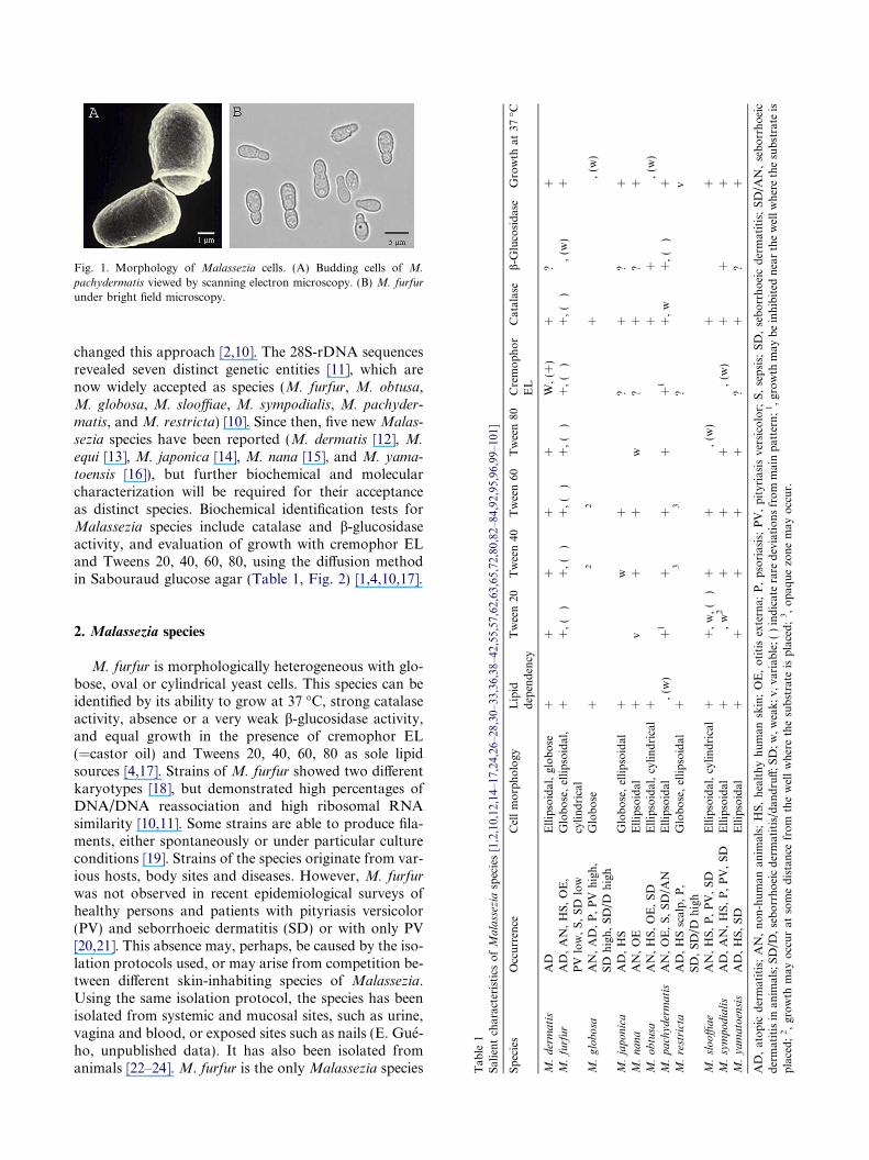
Fig. 1. Morphology of Malassezia cells. (A) Budding cells of M.
pachydermatis viewed by scanning electron microscopy. (B) M. furfur
under bright field microscopy.
,65,72,80,82–84,92,95,96,99–101]
Tween40
Tween60
Tween80
Cremophor
EL
Catalase
b-G
lucosidase
Growth
at37
�C
++
+W,(+
)+
?+
+,(�
)+,(�
)+,(�
)+,(�
)+,(�
)�,(w
)+
�2
�2
��
+�
�,(w
)
w+
�?
+?
++
+w
?+
?+
��
��
++
�,(w
)+
++
+1
+,w
+,(�
)+
�3
�3
�?
��
v
++
�,(w
)�
+�
++
++
�,(w
)+
++
++
+?
+?
+
,psoriasis;PV,pityriasisversicolor;
S,sepsis;
SD,seborrhoeicdermatitis;SD/A
N,seborrhoeic
erare
deviationsfrom
mainpattern;1,growth
may
beinhibited
nearthewellwherethesubstrate
isquezonemay
occur.
changed this approach [2,10]. The 28S-rDNA sequencesrevealed seven distinct genetic entities [11], which arenow widely accepted as species (M. furfur, M. obtusa,M. globosa, M. slooffiae, M. sympodialis, M. pachyder-matis, and M. restricta) [10]. Since then, five new Malas-
sezia species have been reported (M. dermatis [12], M.
equi [13], M. japonica [14], M. nana [15], and M. yama-
toensis [16]), but further biochemical and molecularcharacterization will be required for their acceptanceas distinct species. Biochemical identification tests forMalassezia species include catalase and b-glucosidaseactivity, and evaluation of growth with cremophor ELand Tweens 20, 40, 60, 80, using the diffusion methodin Sabouraud glucose agar (Table 1, Fig. 2) [1,4,10,17].
Tab
le1
SalientcharacteristicsofMalassezia
species[1,2,10,12,14–17,24,26–28,30–33,36,38–42,55,57,62,63
Species
Occurrence
Cellmorphology
Lipid
dependency
Tween20
M.dermatis
AD
Ellipsoidal,globose
++
M.furfur
AD,AN,HS,OE,
PV
low,S,SD
low
Globose,ellipsoidal,
cylindrical
++,(�
)
M.globosa
AN,AD,P,PV
high,
SD
high,SD/D
high
Globose
+�
M.japonica
AD,HS
Globose,ellipsoidal
+�
M.nana
AN,OE
Ellipsoidal
+v
M.obtusa
AN,HS,OE,SD
Ellipsoidal,cylindrical
+�
M.pachydermatis
AN,OE,S,SD/A
NEllipsoidal
�,(w
)+1
M.restricta
AD,HSscalp,P,
SD,SD/D
high
Globose,ellipsoidal
+�
M.slooffiae
AN,HS,P,PV,SD
Ellipsoidal,cylindrical
++,w,(�
)M.sympodialis
AD,AN,HS,P,PV,SD
Ellipsoidal
+�,w2
M.yamatoensis
AD,HS,SD
Ellipsoidal
++
AD,atopic
dermatitis;AN,non-human
anim
als;
HS,healthyhuman
skin;OE,otitisexterna;
Pdermatitisin
anim
als;SD/D
,seborrhoeicdermatitis/dan
druff;SD;w,weak;v,
variab
le;()indicat
placed;2,growth
may
occurat
somedistance
from
thewellwherethesubstrate
isplaced;3,opa
2. Malassezia species
M. furfur is morphologically heterogeneous with glo-bose, oval or cylindrical yeast cells. This species can beidentified by its ability to grow at 37 �C, strong catalaseactivity, absence or a very weak b-glucosidase activity,and equal growth in the presence of cremophor EL(=castor oil) and Tweens 20, 40, 60, 80 as sole lipidsources [4,17]. Strains of M. furfur showed two differentkaryotypes [18], but demonstrated high percentages ofDNA/DNA reassociation and high ribosomal RNAsimilarity [10,11]. Some strains are able to produce fila-ments, either spontaneously or under particular cultureconditions [19]. Strains of the species originate from var-ious hosts, body sites and diseases. However, M. furfur
was not observed in recent epidemiological surveys ofhealthy persons and patients with pityriasis versicolor(PV) and seborrhoeic dermatitis (SD) or with only PV[20,21]. This absence may, perhaps, be caused by the iso-lation protocols used, or may arise from competition be-tween different skin-inhabiting species of Malassezia.Using the same isolation protocol, the species has beenisolated from systemic and mucosal sites, such as urine,vagina and blood, or exposed sites such as nails (E. Gue-ho, unpublished data). It has also been isolated fromanimals [22–24]. M. furfur is the only Malassezia species
Fig. 2. Assimilation patterns of Tween 20, 40, 60 and 80 (Clockwisefrom bar) and Cremophor EL (center). (A) M. sympodialis; (B) M.
restricta.
to be isolated from non-mammalian sources, in one casefrom a hospital room floor [25].
M. slooffiae may be misidentified as M. furfur, but itcan be differentiated by its absence of growth with crem-ophor EL [4,10]. M. slooffiae is regularly isolated fromhuman skin and is mostly found in association withM. sympodialis or M. globosa. It may be a weak humanpathogen, and seems better adapted to animals, espe-cially pigs [22,23].
M. obtusa resembles M. furfur morphologically butdiffers physiologically as it does not grow at 37 �C,and cannot utilize any of the five lipids used in the tests.However, it darkens esculin medium [4,10]. It is a rarespecies that was known only from healthy human skin.Recently it has also been isolated from goats, horsesand dogs [26,27].
M. pachydermatis, a lipophilic species, is able to growwithout supplementation of long-chain fatty acids or
their esters. All isolates grow well at 37 �C, and someprimary cultures show a certain lipid-dependence[28,29]. These weakly-lipid-dependent isolates may havesmaller colonies than those growing on regular Sabou-raud dextrose agar. Differences in catalase and b-gluco-sidase expression, that can be absent, weak or positivedepending on the strain, and differences in reactivity tocremophor EL and Tweens 20, 40, 60, 80 occur in allrDNA genotypes [4,17]. These different compounds,particularly cremophor EL and Tween 20, may be moreor less inhibitory. In this case, growth may occur only atsome distance from the compound-containing wellwhere the compound is more diluted, or it may occurwithin the inhibitory area as secondary growth [30].M. pachydermatis occurs rarely on humans, althoughit has been found to cause septic epidemics, usually inneonates receiving intravenous lipid supplementation[31,32]. M. pachydermatis is well-known as a normalcutaneous inhabitant of numerous warm-blooded ani-mals. Seborrhoeic dermatitis and otitis associated withthis lipophilic yeast are now commonly recognized,especially in dogs [33].
M. sympodialis corresponds with the former serovarA of M. furfur [10,34] and is characterized by a strongb-glucosidase activity and growth at 37 �C, but cremo-phor EL as a lipid supplement does not allow goodgrowth. The yeast cells are small and ovoid. The speciesis commonly isolated from healthy as well as diseasedskin [20]. Its role as a pathogen has not yet been eluci-dated. Indeed, M. sympodialis is often present in skin le-sions, but usually associated with the more abundantlyoccurring M. globosa [35]. M. sympodialis has also beenisolated from healthy feline skin [36].
M. globosa corresponds with serovar B of M. furfur
[10,34] but has spherical yeast cells only [10]. Buds arealso spherical and emerge from the mother yeastthrough a narrow site, contrary to the patterns seen inother Malassezia spp. The species corresponds to theoriginal description of P. orbiculare obtained from aPV case [37]. M. globosa is able to produce filaments,in particular in primary cultures. The yeast does notgrow at 37 �C or does so very poorly, does not growon the five lipid substrates, and does not split esculin.M. globosa, with M restricta, has been implicated asthe causal organism in dandruff and seborrhoeic derma-titis [38]. M. globosa is also the most important speciesin PV, either alone or associated with other species, par-ticularly M. sympodialis [35]. M. globosa occurs mainlyon humans, but is also known from a cat [36].
M. restricta lacks catalase and b-glucosidase activity,does not grow at 37 �C, and is strongly lipid-dependent[10]. Growth of the colonies is very restricted. The spe-cies is isolated almost exclusively from the head, includ-ing scalp, neck and face [21], and it corresponds toserovar C [34]. M. restricta does not produce any fila-ments. Although M. restricta is very fastidious, new

Table 2Fragment lengths of ITS 1 and ITS 2 of strains belonging to differentMalassezia species as determined by terminal fragment lengthpolymorphism (tFLP)
Species CBS Designation ITS 1 (bp)c ITS 2 (bp)c
M. furfur 7982 286 5501878a 286 5507981 286 5506000 286 5507019 286 5507860 286 5507865 286 5507984 286 5504171 286 5505332 286 5505333 286 5505334 286 5507970 286 550
M. globosa 7966b 331 4697874 314 4577990 344 472
M. obtusa 7968 290 5467876b 290 546
M. restricta 7991 339 4557877b 289 4558747 289 455
M. slooffiae 7875 274 4927956 273 4887971 273 488
M. sympodialis 7977b 239 4127979 239 4137222 239 413
M. japonica 9431 285 5209432b 285 520
M. nana 9557b 258 4159558 262 4169559 263 4169560 262 4179561 263 417
M. dermatis 9145 235 4079169b 236 4079170 236 408
M. pachydermatis 74522 (ATCC) 262 527
a Neotype.b Type.c +/�1 base pair.
DNA-based methodologies will allow for more robustanalysis in situ. More studies are needed to understandits implication in Malassezia-associated diseases, in par-ticular dandruff and seborrhoeic dermatitis. This speciesis not known to occur in animals [22,23].
Recently a few other species have been reported. M.dermatis has been isolated from the skin of atopic der-matitis patients [12]. Analysis of the 26S ribosomalDNA (rDNA) and ITS sequences showed this speciesto be closely related to M. sympodialis. The two speciesdiffer by only 1.2% rDNA base divergence. M. dermatis
and M. sympodialis have a strong catalase activity andassimilate the four Tweens ([10,12]; R. Batra andT. Boekhout, unpubl. observ.). These two species differin growth on esculin and cremophor EL. In M. sympo-
dialis, growth on esculin was positive, whereas inM. der-
matis this was negative. Growth of M. sympodialis oncremophor EL was absent, but this was weakly positivefor M. dermatis (R. Batra and T. Boekhout, unpubl.observ.).
Nell et al. [13] have reported the presence of a newspecies from normal equine skin, which they tentativelynamed M. equi. This species has not formally been de-scribed. Like M. dermatis this species is also closely re-lated to M. sympodialis. Unfortunately, data ongrowth on the various Tweens, esculin and cremophorEL are missing in the description ofM. equi, as no strainhas been preserved.
Hirai et al. [15] reported a new species from the eardischarge of animals and proposed the name M. nana.It is possible that this species is the same as that pre-sented by Duarte et al. [39] as atypical strains ofM. sym-
podialis [40]. Previously, Crespo et al. [41] described thepresence of M. sympodialis from otitis externa in cats,which based on D1/D2 and ITS sequences turned outto be identical to M. nana [40] (F.J. Cabanes, unpubl.observ.). It is likely that more species are involved, aswas suggested by a D1/D2 and ITS sequencing studyof M. sympodialis-like isolates from animals [40].
Sugita et al. [14] recently have described another newspecies, M. japonica. This species can be distinguishedfrom the seven known lipophilic species by the abilityto assimilate Tween 40 and Tween 60, the inability toassimilate Tween 20 and Tween 80, and lack of growthat 40 �C. M. japonica seems to represent a separate spe-cies as the D1/D2 26S sequence is significantly divergent(4.6% with M. furfur and 6.9% with M. obtusa).
M. yamatoensis was described from a Japanese pa-tient with seborrhoeic dermatitis (SD) [16]. The speciesis physiologically close to M. furfur and M. dermatis,and sequence analysis of the D1/D2 domain of the26S rDNA placed the species in a cluster with M. furfur,M. Obtusa andM. japonica with 93% bootstrap support,and the ITS sequences were 40% different. Moleculardetection using M. yamatoensis-specific primers detectedthe species in only 9.7% of the SD patients, approxi-
mately 14% of the atopic dermatitis patients andapproximately 55% of the healthy people sampled.Therefore, the species was considered a minor compo-nent of the skin mycobiota [16].
More studies are needed to better characterize andpossibly validate the different lipid-dependent Malas-sezia spp., but data on fragment lengths of the ITS 1and 2 (Table 2) suggest that different Malassezia spp.may be involved indeed.

3. Detection and identification
Assigning the role of individual species in the clinicalcontext has been hampered by the difficulties involved inthe isolation, cultivation and identification of Malas-
sezia spp. [1,2,41]. Cultivation requirements vary by spe-cies [10]. M. furfur is by far the most robust of theMalassezia spp. in culture, and therefore is the organismmost frequently isolated when using culture-based tech-niques. M. restricta and M. obtusa are the most difficultto grow. Even under the most-favourable conditions,M.
restricta particularly grows much more slowly than M.
furfur and M. sympodialis and would be quickly over-grown by any of these two species even if initially pres-ent in much smaller numbers. Indeed M. globosa, as aprimary culture on Dixon or mDixon agar at 30–35 �C, is growing very well, giving colonies smaller thanthose of M. sympodialis but having about the samediameter as those of M. furfur. However, M. furfur
may outcompete M. globosa when the experiments areperformed at 37 �C.
Multiple approaches have been used to differentiateMalassezia species. Culture-based methods includemol% guanine + cytosine, DNA reassociation values,cell morphology, growth with different Tween non-ionicdetergents (Fig. 2), the presence of catalase, temperaturerequirements, the presence of b-glucosidase as revealedby the splitting of esculin, and selective growth withcremophor EL [4,10,43,44]. These culture-based meth-ods are effective but time consuming and technicallydemanding. Specific molecular methods have also beendeveloped for the identification of Malassezia isolates,such as pulsed-field gel electrophoresis (PFGE), randomamplification of polymorphic DNA (RAPD), DNA se-quence analysis, restriction analysis of PCR ampliconsof ribosomal sequences, amplified fragment length poly-morphism (AFLP�), denaturing gradient gel electro-phoresis (DGGE), and terminal fragment lengthpolymorphism (tFLP) [11,18,29,31,38,45–55]. Of themolecular methods, PFGE and DGGE have met withlimited success due to the need for specialized equipmentand training. AFLP, however, has been successfully ap-plied to the identification of Malassezia isolates andyields highly-specific genotypic information about eachstrain. The detailed ‘‘fingerprint’’ achieved from carefulAFLP analysis has revealed multiple genetic subgroupsin each Malassezia species, and may lead to the differen-tiation of clinically-important subgroups or even newspecies. [46,47]. The numerous bands resolved in theAFLP fingerprints are providing detailed informationrelevant to the identification of strains, but the methodrequires clonal isolates from culture. AFLP is, therefore,the tool of choice for analyses where detailed informa-tion is necessary and many isolates are available, suchas strain identification, epidemiology, phylogeny, andinvestigation of novel species. Multiple methods have
been reported to identify species in complex Malassezia
communities from skin without cultivation [38,48,50–52,55], but most of these methods require either separateamplification with specific primer sets or restrictiondigestion of the amplification products.
A new method, named terminal fragment lengthpolymorphism (tFLP) [38], can be used on non-inva-sively acquired swab samples. This method uses onlythree primer sets, and therefore minimizes the potentialbias related to amplification efficiency. It is sensitive en-ough to detect Malassezia with as few as 100 cells persample, either as spiked directly onto the swab to con-trol the extraction procedure, or from samples collectedfrom 1 cm2 of skin surface sample using a swab. As isthe case with any amplification reaction, target speciesfound at less than 1% of the total community may beunder-represented in the final products. Therefore, caremust be taken in the interpretation of results. This meth-od involves isolation of fungal DNA, followed by nestedPCR of the ribosomal gene cluster, followed by amplifi-cation with ITS 1- and ITS 2-specific fluorescently-labeled primers. The resulting terminally-labeledproducts are analyzed for fragment length on a fluores-cent-DNA sequencer. The amplifications are carried outwith universal-fungal primers [56], and therefore themethodology should be broadly applicable to other fun-gal species. The ITS 1 and ITS 2 amplifications are car-ried out individually and combined for final analysis.The primary advantage of this method is that the resul-tant sample contains only two labeled fragments perspecies, allowing analysis of complex communities. AllMalassezia species can be differentiated by length poly-morphisms, including multiple genotypes per knownspecies (Fig. 3, Table 2). Results obtained for standardsand mixtures of standards show that all known Malas-
sezia genotypes can be identified in a single amplificationreaction based on unique fragment lengths, eliminatingthe need for restriction analysis [38]. Results from thisstudy also have shown that tFLP is capable of reproduc-ibly assessing the Malassezia species present in complexmixtures and clinical samples. Importantly, it is specificfor fungi, and sufficiently sensitive to allow direct assess-ment of clinical samples without the need for prior cul-tivation. The tFLP method will become a powerful toolwhen used in conjunction with new fungal ITS regiondatabases that are becoming available. These includea.o. databases by Chen et al. [57] and by Boekhoutet al. [58], the latter of which is available as a CD-ROM.
4. General lipid requirements
As mentioned previously, all Malassezia species, ex-ceptM. pachydermatis, are lipid-dependent. The compli-cations with nomenclature are compounded in the studyof Malassezia biochemistry and metabolism. Elegant

Fig. 3. Fragments of ITS 1 (green peaks) and ITS 2 (blue peaks) showing differences among all known Malassezia species. The different peakscorrespond to the ITS 1 and 2 fragments given in Table 2.
early work in the 1960s and 1970s indicated that M. fur-
fur (then referred to as Pitrysporum ovale, CBS 1878) re-quired saturated fatty acids greater than twelve but lessthan 20 carbons [59]. In this study, unsaturated fattyacids stimulated growth, but only when saturated fattyacids were also present. In a later study, it was reportedthat both saturated and unsaturated fatty acids couldsupport growth of P. ovale, but it is unclear which cur-rent Malassezia species was investigated [60]. To furthercomplicate interpretation of Malassezia lipid metabolicdata, the concentrations of saturated fatty acids re-quired to support growth are extremely low, in thenanomolar range. It is very difficult and expensive to ob-tain fatty acids of sufficient purity to fully elucidate theirrole in Malassezia metabolism. For example, food-gradeoleic acid, which is frequently used in routineMalasseziaculture [61,62], contains approximately 74% oleic andapproximately 8% stearic acid, with the remaining beinga complex mixture of primarily C14, C16, and C18 sat-urated and mono-unsaturated acids (T.L. Dawson, un-publ. observ.). Cremophor-EL presents the samedifficulty, being a partially-purified plant extract con-taining primarily, but not exclusively, ricinoleic acid.Tween esters are likewise produced from enriched butnot purified fatty acid sources. While complex fatty acidsupplements are excellent tools for defining the speciesof Malassezia present in a culture, it will take a very de-tailed approach with analytically-pure standards to fullyestablish the fatty-acid requirements of each Malassezia
species. This type of detailed biochemical informationwill be necessary to understand the individual role ofeach species in human pathogenesis, to unravel theirphylogenic relationships, and to decide whether or notnew genetic entities will become accepted species.
5. Malassezia species as human pathogens
Malassezia species have been associated with a num-ber of diseases of the human skin, such as seborrhoeicdermatitis and dandruff, pityriasis versicolor, Malas-
sezia (Pityrosporum) folliculitis, atopic dermatitis, pso-riasis and, less commonly, with other dermatologicaldisorders such as confluent and reticulated papilloma-tosis, onychomycosis, and transient acantholytic der-matosis [1,2,42,63–65]. Although Malassezia speciesare a part of the microbial community on normal skin,under certain conditions they can cause a superficialskin infection. In general, due to their dependence onlipids for survival, Malassezia species are most oftenfound in sebum-rich areas of the skin, such as thetrunk, back, face and scalp. The Malassezia speciesare well-adapted to the human skin, and possibly occu-py well-defined niches on the human body. Studies havebeen carried out to answer the clinical question ofwhether there is a relationship between particularMalassezia species and various dermatological disor-ders [1,2,35,42,43,52,65].
6. Seborrhoeic dermatitis and dandruff
Seborrhoeic dermatitis and dandruff (SD/D) are per-haps the most common diseases associated with Malas-
sezia species, with SD occurring in 1–3% and dandruffin greater than 50% of the general population [20,66].The incidence of SD is much higher in immunocompro-mised patients, especially AIDS patients, ranging from30% to 33% [67,68]. The vast majority of more recentdata supports a direct causal link between Malassezia
and SD/D. The factors that support this hypothesis are:(i) antifungal treatment is found to be effective in treatingthese diseases, and (ii) improvement in SD/D is accompa-nied by a reduction inMalassezia levels on the scalp [69].
It has been reported that the proportion ofMalassezia
cells on the scalp is higher in patients with SD/D than innormal controls [70]. There are conflicting data regard-ing the number of yeasts in lesional versus non-lesionalskin. Some have reported a decrease in the density ofMalassezia cells recovered from lesional skin [43], whileothers have shown a greater number of yeasts in lesionalskin [55] or a greater detectable incidence in affected sub-jects [38]. It has been suggested that seborrhoeic derma-titis is not caused by an overgrowth of Malassezia cells,but by an abnormal host response to the yeasts on theskin [71]. It is likely that SD/D is caused by M. restrictaand M. globosa, in conjunction with increased host re-sponse to the fungi or their metabolites.
The species that have been shown to be most closelyassociated with seborrhoeic dermatitis and dandruff todate are M. restricta and M. globosa [1,38,42], with theformer occurring more frequently than the latter. How-ever, some authors have also reported the presence ofM. furfur, M. sympodialis, M. obtusa and M. slooffiae
[72]. These authors found that M. globosa was isolatedwith the same frequency from both lesional and non-lesional skin. Gupta et al. [43] noted that significantlymore Malassezia yeasts could be cultured from non-lesional skin. Two other species, M. sympodialis andM. obtusa, were often cultured from both lesional andnon-lesional skin of seborrhoeic dermatitis patients.Gemmer et al. [38], using DNA-based detection, re-ported a significantly higher detection rate for bothM.globosa andM. restricta in subjects with SD/D.
7. Atopic dermatitis
Malassezia species have been cultured from 83% ofadult patients, in whom the disease was localized tothe head and neck [73]. Because the yeasts are also fre-quently isolated from normal controls, it has beenhypothesized that they act as allergens in susceptible pa-tients, rather than as infectious agents [74,75]. Recently,molecular work has indeed elucidated the structure ofsome allergens derived from Malassezia species [76–
78]. Three major allergen components have been identi-fied, namely two protein components of 67 and 37 kDaeach, and one carbohydrate component of 14 kDa [78].These allergens are designated as Mala s 1, and Mala f 2to Mala f 4 for the protein components, and Mala s 5 toMala s 9 for the carbohydrate moieties. Genomic-DNAamplification by PCR and sequencing demonstratedthat M. globosa, M. obtusa and M. sympodialis con-tained DNA sequences corresponding to all the aller-gens except Mala f 2 and Mala f 3. M. pachydermatis
contained Mala s 1, Mala f 4, and Mala s 5 to Mala s8. M. restricta and M. slooffiae possessed Mala f 4 andMala s 6.M. furfur was seen to possess Mala f 2 to Malaf 4 as well as Mala s 5 to Mala s 7. Ashbee and Evans[79] and Faergemann [63] have elegantly described theimmunological role of Malassezia in atopic dermatitis.
Some studies have examined the prevalence and thespecies composition of Malassezia strains in atopic der-matitis patients. Sandstrom et al. [80] sampled skin onthe upper back, and found that M. sympodialis was thespecies most commonly isolated from both atopic der-matitis patients and healthy controls. These investigatorswere able to sample both lesional and non-lesional skinand found a significant difference, the yeasts being morecommon in non-lesional skin. Gupta et al. [43] demon-strated that the mean number of colony-forming unitsgrown from samples taken from atopic dermatitis pa-tients was significantly lower than that obtained fromsampling healthy controls. In both groups, however,the dominant species was M. sympodialis. Sugita andNishikawa [55] reported that M. restricta, M. globosa
and M. furfur are present at significantly higher frequen-cies in atopic dermatitis patients than in healthy subjects.Nakabayashi et al. [72] found thatM. furfur was isolatedmore frequently from lesional skin (21%) than from non-lesional skin (11%) of atopic dermatitis patients. Guptaet al. [43] sampled lesional skin and found that M. sym-
podialis was the species most commonly isolated fromboth atopic dermatitis patients and controls. Sandstromet al. [80] found a difference in species distribution on le-sional versus non-lesional skin in atopic dermatitis pa-tients. Non-lesional skin was most frequently colonizedbyM. globosa, whileM. sympodialiswas most commonlyfound on lesional skin. The differences in sampling andidentification methods used by various researchers mayhave contributed to the differences observed in the prev-alence and species composition of Malassezia species.
8. Pityriasis versicolor
Pityriasis versicolor is a chronic superficial fungal dis-ease that is characterized by the appearance of round tooval lesions, most commonly found on the trunk andupper arms. It is postulated that this disease occurswhen the Malassezia species that normally colonize the
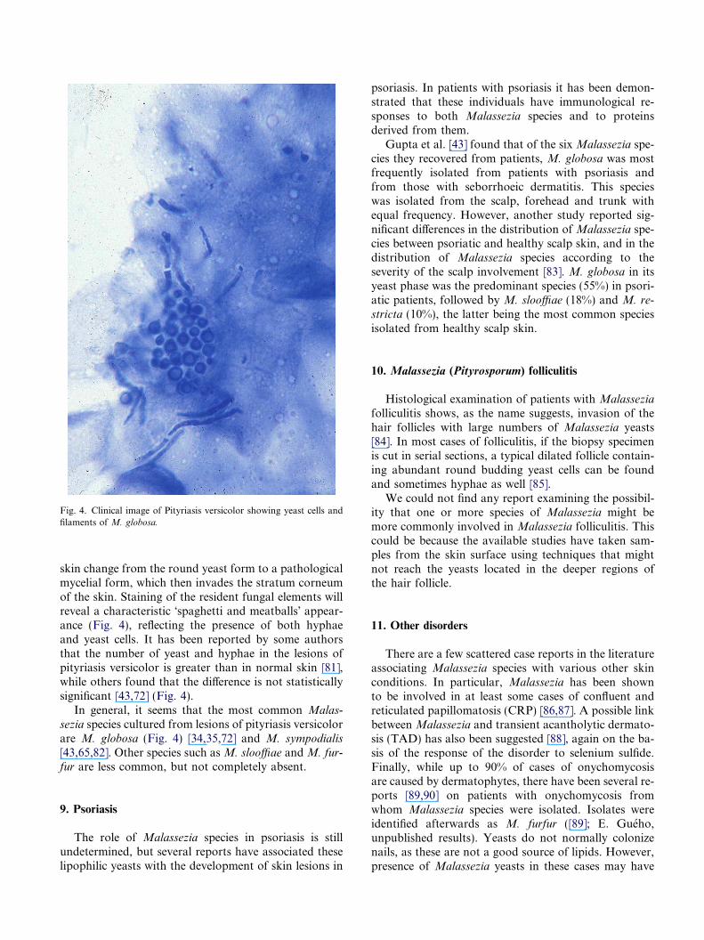
Fig. 4. Clinical image of Pityriasis versicolor showing yeast cells andfilaments of M. globosa.
skin change from the round yeast form to a pathologicalmycelial form, which then invades the stratum corneumof the skin. Staining of the resident fungal elements willreveal a characteristic �spaghetti and meatballs� appear-ance (Fig. 4), reflecting the presence of both hyphaeand yeast cells. It has been reported by some authorsthat the number of yeast and hyphae in the lesions ofpityriasis versicolor is greater than in normal skin [81],while others found that the difference is not statisticallysignificant [43,72] (Fig. 4).
In general, it seems that the most common Malas-
sezia species cultured from lesions of pityriasis versicolorare M. globosa (Fig. 4) [34,35,72] and M. sympodialis
[43,65,82]. Other species such as M. slooffiae and M. fur-fur are less common, but not completely absent.
9. Psoriasis
The role of Malassezia species in psoriasis is stillundetermined, but several reports have associated theselipophilic yeasts with the development of skin lesions in
psoriasis. In patients with psoriasis it has been demon-strated that these individuals have immunological re-sponses to both Malassezia species and to proteinsderived from them.
Gupta et al. [43] found that of the six Malassezia spe-cies they recovered from patients, M. globosa was mostfrequently isolated from patients with psoriasis andfrom those with seborrhoeic dermatitis. This specieswas isolated from the scalp, forehead and trunk withequal frequency. However, another study reported sig-nificant differences in the distribution of Malassezia spe-cies between psoriatic and healthy scalp skin, and in thedistribution of Malassezia species according to theseverity of the scalp involvement [83]. M. globosa in itsyeast phase was the predominant species (55%) in psori-atic patients, followed by M. slooffiae (18%) and M. re-
stricta (10%), the latter being the most common speciesisolated from healthy scalp skin.
10. Malassezia (Pityrosporum) folliculitis
Histological examination of patients with Malassezia
folliculitis shows, as the name suggests, invasion of thehair follicles with large numbers of Malassezia yeasts[84]. In most cases of folliculitis, if the biopsy specimenis cut in serial sections, a typical dilated follicle contain-ing abundant round budding yeast cells can be foundand sometimes hyphae as well [85].
We could not find any report examining the possibil-ity that one or more species of Malassezia might bemore commonly involved in Malassezia folliculitis. Thiscould be because the available studies have taken sam-ples from the skin surface using techniques that mightnot reach the yeasts located in the deeper regions ofthe hair follicle.
11. Other disorders
There are a few scattered case reports in the literatureassociating Malassezia species with various other skinconditions. In particular, Malassezia has been shownto be involved in at least some cases of confluent andreticulated papillomatosis (CRP) [86,87]. A possible linkbetween Malassezia and transient acantholytic dermato-sis (TAD) has also been suggested [88], again on the ba-sis of the response of the disorder to selenium sulfide.Finally, while up to 90% of cases of onychomycosisare caused by dermatophytes, there have been several re-ports [89,90] on patients with onychomycosis fromwhom Malassezia species were isolated. Isolates wereidentified afterwards as M. furfur ([89]; E. Gueho,unpublished results). Yeasts do not normally colonizenails, as these are not a good source of lipids. However,presence of Malassezia yeasts in these cases may have

represented a secondary infection in patients withonychomycosis.
Malassezia species have been associated with deep-seated infections such as catheter-related fungemia, espe-cially in neonates [31,32,91–93].M. pachydermatis, earlierexclusively considered to be associated with animals, hasbeen reported to cause intravascular catheter-acquiredinfections in humans [31,32]. The association of intravas-cular catheter-acquired nosocomial infection associatedwith colonization of health care workers� pet dogs hasalso been mentioned [91].
12. Malassezia species on animals
Malassezia species inhabit the skin of a variety ofmammals and birds [94]. So far, few studies have beenpublished regarding the distribution of the differentMalassezia spp. on animals following the last taxonomicrevision of the genus [10]. Traditionally, the lipid-depen-dent species were thought to occur only on human skin,while M. pachydermatis was assumed to be restricted toanimal skin and in particular to carnivores.
M. pachydermatis, the only species in the genus thatdoes not require lipid supplementation for developmentin culture medium, has been traditionally considered tobe zoophilic, and is frequently found on wild anddomestic carnivores including dogs, cats, bears, pinne-peds, ferrets and foxes [22,26,28]. M. pachydermatis isusually associated with otitis externa and different kindsof dermatitis in domestic animals (Fig. 5), but especiallyin dogs [23]. This species is more frequently isolatedfrom dogs than from cats and appears to be a relativelyinfrequent pathogen in other animals [23,26].
Several authors have mentioned the high incidence ofthis yeast in canine otitis externa, especially in chronicotitis. Although the pathogenic role of M. pachyderma-
tis in otitis externa has been a matter of controversy, itwas demonstrated that the yeast could induce inflamma-tory changes in the normal canine external ear canal in
Fig. 5. Clinical images of M. pachydermatis infections in animals. (A) Dermastain of a smear from an ear swab of a dog with otitis externa showing the
the presence of moisture [95], and the disease was exper-imentally induced with M. pachydermatis when a largeinoculum was used [96]. This yeast seems to have anopportunistic nature and may become pathogenic withany alteration in the microclimate of the skin surfaceor in the host defense. In some canine breeds, hypersen-sitivity conditions such as flea allergy dermatitis, foodhypersensitivity or atopy, and antimicrobial or cortico-steroid therapy may be factors favouring proliferationof these yeasts [97]. Primary diseases that cause inflam-mation and increased sebum production provide a cuta-neous microenvironment that encourages overgrowth ofthis yeast. M. pachydermatis is the most common yeastthat contributes to otitis externa as a perpetuating factorin dogs [98].
Recently lipid-dependent species have been isolatedfrom the skin of different animals, such as cats, dogs,cows and horses (e.g. [13,15,26,27]). Carnivores can becolonised by lipid-dependent species such as M. furfur,M. obtusa, M. sympodialis and M. globosa, in additionto M. pachydermatis [24,26,36,99]. Lipid-dependent spe-cies seem to be more frequent in cats than in dogs [26].However, very little is known about their pathogenicrole in animal skin. M. sympodialis was isolated fromcats with otitis externa [41] and M. furfur and M. obtusa
from dogs with otitis externa [100]. It is possible thatthese species are common in this microenvironmentand play a similar role as M. pachydermatis in canineotitis externa.
On the other hand, the lipid-dependent species seemto constitute the major population of the lipophilic mic-robiota in herbivores such as horses, goats, sheep andcows [27], and they have been associated with otitis ex-terna in bovines [101]. Otitis in cattle, among other fac-tors, is commonly attributed to infestation caused byrhabditiform nematodes [102]. Recently, soil nematodeshave been associated with Malassezia spp. by PCR tech-niques, and their role as possible vectors for species ofMalassezia has been suggested [102]. M. sympodialis
has also been isolated from affected skin of a horse [29].
titis of a cat associated with M. pachydermatis (arrow). (B) Diff-Quicktypical monopolar budding on a broad base of M. pachydermatis.

13. Conclusion
Recent research and technological developments havecontributed greatly to elucidating the role of Malassezia
spp. in skin diseases. It is now much easier to detect andidentify individual Malassezia spp. from complex clini-cal samples, and specific information can be gleanedfrom detailed genetic analyses. These, coupled with theincreasing interest and, therefore, increasing researchcommitment of the medical and scientific communities,will undoubtedly lead to rapid and significant advancesin the field. However, difficulties remain in obtaining ahigh level of certainty in the identification of some li-pid-dependent strains by means of physiological testsand further comparison of recent work to older data willremain difficult, due to changes in nomenclature andgrowth conditions. In addition, isolation and identifica-tion of these strains continues to be difficult due to thelow viability associated with some isolate types and lackof suitable methods for their isolation and preservation[103].
New molecular approaches to the identification ofdifferent species will certainly contribute to improvedmanagement of diseases associated with Malassezia
spp. As further detailed DNA-based identification tech-niques and biochemical analyses will be more broadlyapplied to the genus, almost certainly more species willbe identified. The elaboration of molecular genetic dif-ferences and detailed biochemisty will also be necessaryto define true species differences and to assess geneticvariation between strains within species.
Future work will hopefully answer many of the out-standing questions associated with Malassezia spp. andtheir role in human and animal pathophysiology. Theidentification of specific metabolic requirements andby-products in situ will also be necessary to understandhow the Malassezia spp. interact with human and ani-mal skin. Furthermore, the organisms occupy an impor-tant and poorly-defined section of the phylogenetic ‘‘treeof life’’. Application of whole-genome sequencing wouldallow a deeper understanding of the physiology, phylog-eny, and medical importance of the Malasseziales.
References
[1] Crespo-Erchiga, V. and Gueho, E. (2005) Superficial diseasescaused by Malassezia species. In: Topley and Wilson�s Micro-biology and Microbial Infections, Mycology (Hay, R. and Merz,W., Eds.), Vol. 5, 10th edn. Arnold, London, UK (in press).
[2] Midgley, G., Gueho, E. and Guillot, J. (1998) Diseases caused byMalassezia In: Topley and Wilson�s Microbiology and MicrobialInfections (Ajello, L. and Hay, R.J., Eds.), Vol. 4, pp. 201–211.Arnold, London, UK.
[3] Kurtzman, C.P. and Fell, J.W., Eds., (1998). The Yeasts, ATaxonomic Study, 4th edn. Elsevier, Amsterdam, TheNetherlands.
[4] Boekhout, T. and Gueho, E. (2003) Basidiomycetous yeasts. In:Pathogenic Fungi in Humans and Animals (Howard, D.H., Ed.),2nd edn, pp. 537–542. Marcel Dekker, Inc., New York, USA.
[5] Begerow, D., Bauer, R. and Boekhout, T. (2000) Phylogeneticplacement of ustilaginomycetes anamorphs as deduced fromnuclear LSU rDNA sequences. Mycol. Res. 104, 53–60.
[6] Baillon, H. (1889) Traite de botanique medicale cryptogamique.Octave Douin Paris, France..
[7] Sabouraud, R. (1904) Maladies du cuir chevelu II. Les maladiesdesquamatives. Masson, Paris, France..
[8] Ahearn, D. and Yarrow, D. (1984) Malassezia Baillon In: TheYeasts: A Taxonomic Study (Kreger-van Rij, N.J.W., Ed.), 3rdedn, pp. 882–885. Elsevier, Amsterdam, The Netherlands.
[9] Marcon, M.J. and Powell, D.A.. (1992) Human infections due toMalassezia spp. Clin. Microbiol. Rev. 5, 101–119.
[10] Gueho, E., Midgley, G. and Guillot, J. (1996) The genusMalassezia with the description of four new species. Antonie vanLeeuwenhoek 69, 337–355.
[11] Guillot, J. and Gueho, E. (1995) The diversity of Malassezia
yeasts confirmed by rRNA sequence and nuclear DNA compar-isons. Antonie van Leeuwenhoek 67, 297–314.
[12] Sugita, T., Takashima, M., Shinoda, T., Suto, H., Unno, T.,Tsuboi, R., Ogawa, H. and Nishikawa, A. (2002) New yeastspecies Malassezia dermatis isolated from patients with atopicdermatitis. J. Clin. Microbiol. 40, 1363–1367.
[13] Nell, A., James, S.A., Bond, C.J., Hunt, B. and Herrtage, M.E.(2002) Identification and distribution of a novelMalassezia yeastspecies on normal equine skin. Veter. Rec. 150, 395–398.
[14] Sugita, T., Takashima, M., Kodama, M., Tsuboi, R. andNishikawa, A. (2003) Description of a new yeast species, Malas-
sezia japonica, and its detection in patients with atopic dermatitisand healthy subjects. J. Clin. Microbiol. 41, 4695–4699.
[15] Hirai, A., Kano, R., Makimura, K., Duarte, E.R., Hamdan, J.S.,Lachance, M.A., Yamaguchi, A. and Hasegawa, A. (2004)Malassezia nana sp. nov., a novel lipid-dependent yeast speciesisolated from animals. Int. J. Syst. Evol. Microbiol. 54, 623–627.
[16] Sugita, T., Tajima, M., Takashima, M., Amaya, M., Saito, M.,Tsuboi, R. and Nishikawa, A. (2004) A new yeast, Malassezia
yamatoensis, isolated from a patient with seborrheic dermatitis,and its distribution in patients and healthy subjects. Microbiol.Immunol. 48, 579–583.
[17] Gueho, E., Boekhout, T., Ashbee, H.R., Guillot, J., van Belkum,A. and Faergemann, J. (1998) The role of Malassezia species inthe ecology of human skin and as pathogens. Med. Mycol. 36,220–229.
[18] Boekhout, T. and Bosboom, R.W. (1994) Karyotyping ofMalassezia yeasts: taxonomic and epidemiological implications.Syst. Appl. Microbiol. 17, 146–153.
[19] Guillot, J., Gueho, E. and Prevost, M.C. (1995) Ultrastructuralfeatures of the dimorphic yeast Malassezia furfur. J. Mycol.Med. 5, 86–91.
[20] Ashbee, H.R., Ingham, E., Holland, K.T. and Cunliffe, W.J.(1993) The carriage of Malassezia furfur serovar A, B and C inpatients with pityriasis versicolor, seborrheic dermatitis andcontrols. Br. J. Dermatol. 129, 533–540.
[21] Aspiroz, C., Moreno, L.A., Rezusta, A. and Rubio, C. (1999)Differentiation of three biotypes of Malassezia species on humannormal skin. Correspondence with M. globosa, M. sympodialis
and M. restricta. Mycopathologia 145, 69–74.[22] Guillot, J., Gueho, E., Mialot, M. and Chermette, R. (1998)
Importance des levures du genre Malassezia en pratique veteri-naire. Point-Veter. 29, 21–31.
[23] Guillot, J., Gueho, E. and Chermette, R. (1999) Infectionsanimales a Malassezia. Rev. Prat. 49, 1840–1843.
[24] Crespo, M.J., Abarca, M.L. and Cabanes, F.J. (1999) Isolationof Malassezia furfur from a cat. J. Clin. Microbiol. 37, 1573–1574.

[25] Tanaka, R., Nishimura, K., Kamei, K. and Murayama, S.Y.(2001) Assimilation test of Malassezia furfur isolated fromthe environment. Nippon Ishinkin Gakkai Zasshi 42, 123–126.
[26] Crespo, M.J., Abarca, M.L. and Cabanes, F.J. (2002)Occurrence of Malassezia spp. in the external ear canals ofdogs and cats with and without otitis externa. Med. Mycol.40, 115–121.
[27] Crespo, M.J., Abarca, M.L. and Cabanes, F.J. (2002) Occur-rence of Malassezia spp. in horses and domestic ruminants.Mycoses 45, 333–337.
[28] Bond, R. and Anthony, R.M. (1995) Characterization ofmarkedly lipid-dependent Malassezia pachydermatis isolatesfrom healthy dogs. J. Appl. Bacteriol. 78, 537–542.
[29] Senczek, D., Siesenop, U. and Bohm, K. (1999) Characterizationof Malassezia species by means of phenotypic characteristics anddetection of electrophoretic karyotypes by pulsed field gelelectrophoresis (PFGE). Mycoses 42, 409–414.
[30] Gueho, E. and Guillot, J. (1999) Comments on Malassezia
species from dogs and cats. Mycoses 42, 673–674.[31] Van Belkum, A., Boekhout, T. and Bosboom, R. (1994)
Monitoring spread of Malassezia infections in a neonatalintensive care unit by PCR-mediated genetic typing. J. Clin.Microbiol. 32, 2528–2532.
[32] Mickelsen, P.A., Viano-Paulson, M.C., Stevens, D.A. and Diaz,P.S. (1988) Clinical and microbiological features of infectionwithMalassezia pachydermatis in high-risk infants. J. Infect. Dis.157, 1163–1168.
[33] Guillot, J. and Bond, R. (1999) Malassezia pachydermatis: areview. Med. Mycol. 37, 295–306.
[34] Cunningham, A.C., Leeming, J.P., Ingham, E. and Gowland, G.(1990) Differentiation of three serovars of Malassezia furfur. J.Appl. Bacteriol. 68, 439–446.
[35] Crespo-Erchiga, V., Ojeda Martos, A., Vera Casano, A., Crespo-Erchiga, A., Sanchez Fajardo, F. and Gueho, E. (1999)Mycology of pityriasis versicolor. J. Mycol. Med. 9, 143–148.
[36] Bond, R., Howell, S.A., Haywood, P.J. and Lloyd, D.H. (1997)Isolation of Malassezia sympodialis and Malassezia globosa fromhealthy pet cats. Vet. Rec. 141, 200–201.
[37] Gordon, M.A. (1951) The lipophilic mycoflora of the skin. I. Invitro culture of Pityrosporum orbiculare n. sp. Mycologia 43,524–535.
[38] Gemmer, C.M., DeAngelis, Y.M., Theelen, B., Boekhout, T. andDawson, T.L. (2002) Fast, noninvasive method for moleculardetection and differentiation of Malassezia yeast species onhuman skin and application of the method to dandruff micro-biology. J. Clin. Microbiol. 40, 3350–3357.
[39] Duarte, E.R., Lachance, M.A. and Hamdan, J.S. (2002) Iden-tification of atypical strains of Malassezia spp. from cattle anddog. Can. J. Microbiol. 48, 749–752.
[40] Cabanes, F.J., Hernandez, J.J. and Castella, G. (2005) Molecularanalysis of Malassezia sympodialis related strains from domesticanimals. J. Clin. Microbiol. 43, 277–283.
[41] Crespo, M.J., Abarca, M.L. and Cabanes, F.J. (2000) Otitisexterna associated with Malassezia sympodialis in two cats. J.Clin. Microbiol. 38, 1263–1266.
[42] Gupta, A.K., Batra, R., Bluhm, R., Boekhout, T. and Dawson,T.L. (2004) Skin diseases associated with Malassezia species. J.Am. Acad. Dermatol. 51, 785–798.
[43] Gupta, A.K., Kohli, Y., Summerbell, R.C. and Faergemann, J.(2001) Quantitative culture of Malassezia species from differentbody sites of individuals with or without dermatoses. Med.Mycol. 39, 243–251.
[44] Mayser, P., Haze, P., Papavassilis, C., Pickel, M., Gruender, K.and Gueho, E. (1997) Differentiation of Malassezia species:selectivity of cremophor EL, castor oil and ricinoleic acid for M.
furfur. Br. J. Dermatol. 137, 208–213.
[45] Boekhout, T., Kamp, M. and Gueho, E. (1998) Molecular typingof Malassezia species with PFGE and RAPD. Med. Mycol. 36,365–372.
[46] Theelen, B., Silvestri, M., Gueho, E., van Belkum, A. andBoekhout, T. (2001) Identification and typing of Malassezia
yeasts using amplified fragment length polymorphism (AFLP),random amplified polymorphic DNA (RAPD) and denaturinggradient gel electrophoresis (DGGE). FEMS Yeast Res. 1, 79–86.
[47] Gupta, A.K., Boekhout, T., Theelen, B., Summerbell, R.C. andBatra, R. (2004) Identification and typing of Malassezia speciesby amplified fragment length polymorphism (AFLP) andsequence analyses of the internal transcribed spacer (ITS) andlarge subunit (LSU) regions of ribosomal DNA. J. Clin.Microbiol. 42, 4253–4260.
[48] Gaitanis, G., Velegraki, A., Frangoulis, E., Mitroussia, A.,Tsigonia, A., Tzimogianni, A., Katsambas, A. and Legakis, N.J.(2002) Identification of Malassezia species from patient skinscales by PCR-RFLP. Clin. Microbiol. Infect. 8, 162–173.
[49] Makimura, K., Tamura, Y., Kudo, M., Uchida, K., Saito,H. and Yamaguchi, H. (2000) Species identification andstrain typing of Malassezia species stock strains and clinicalisolates based on the DNA sequences of nuclear ribosomalinternal trancribed spacer 1 regions. J. Med. Microbiol. 49,29–35.
[50] Gupta, A.K., Kohli, Y. and Summerbell, R.C. (2000) Moleculardifferentiation of sevenMalassezia species. J. Clin. Microbiol. 38,1869–1875.
[51] Guillot, J., Deville, M., Berthelemy, F., Provost, F. and Gueho,E. (2000) A single PCR-restriction endonuclease analysis forrapid identification of Malassezia species. Lett. Appl. Microbiol.31, 400–403.
[52] Sugita, T., Suti, H., Unno, R., Tsuboi, H., Ogawa, T., Shinoda,T. and Nishikawa, A. (2001) Molecular analysis of Malassezia
microflora on the skin of atopic dermatitis patients and healthysubjects. J. Clin. Microbiol. 39, 3486–3490.
[53] Sugita, T., Kodama, M., Saito, M., Tomonobu, I., Kato, Y.,Tsuboi, R. and Nishikawa, A. (2003) Sequence diversity of theintergenic spacer region of the rRNA gene of Malassezia globosa
colonizing the skin of patients with atopic dermatitis and healthyindividuals. J. Clin. Microbiol. 41, 3022–3027.
[54] Yamada, Y., Osumi, M., Makimura, K. and Yamaguchi, H.(2002) Overview of lipophilic yeastMalassezia: the current statusof the molecular diagnosis. Jpn. J. Antibiot. 55, 493–499.
[55] Sugita, T. and Nishikawa, A. (2003) Molecular and quantitativeanalysis ofMalasseziamicroflora on the skin of atopic dermatitisand genotyping ofM. globosaDNA. Jpn. J. Med. Mycol. 44, 61–64.
[56] White, T.J., Bruns, T., Lee, S. and Taylor, J. (1990) Amplifica-tion and direct sequencing of fungal ribosomal RNA genes forphylogenetics In: PCR Protocols: A Guide to Methods andApplications (Innis, M.A., Gelfand, D.H., Sninsky, J.J. andWhite, T.J., Eds.), pp. 314–322. Academic Press, San Diego,USA.
[57] Chen, Y.-C., Eisner, J.D., Kattar, M.M., Rassoulian-Barret,S.L., LaFe, K., Yarfitz, S.L., Limaye, A.P. and Cookson, B.T.(2001) Polymorphic internal transcribed spacer region 1 DNAsequences identify medically important yeasts. J. Clin. Micro-biol. 39, 4042–4051.
[58] Boekhout, T., Robert, V., Smith, M.Th., Stalpers, J., Yarrow,D., Boer, P., Gijswijt, G., Kurtzman, C.P., Fell, J.W., Gueho, E.,Guillot, J. and Roberts, I. (2002) Yeasts of the world 2.0.Expertcenter Taxonomic Identification. Amsterdam, TheNetherlands.
[59] Wilde, P.F. and Stewart, P.S. (1968) A study of the fatty acidmetabolism of the yeast Pityrosporum ovale. Biochem. J. 108,225–231.
[60] Porro, M.N., Passi, S., Caprill, F., Nazzaro, P. and Morpurgo,G. (1976) Growth requirements and lipid metabolism of Pity-
rosporum orbiculare. J. Invest. Dermatol. 66, 178–182.[61] Leeming, J.P. and Notman, F.H. (1987) Improved methods for
isolation and enumeration of Malassezia furfur from humanskin. J. Clin. Microbiol. 25, 2017–2019.
[62] Guillot, J., Gueho, E., Lesourd, M., Midgley, G., Chevrier, G.and Dupont, B. (1996) Identification of Malassezia species, apractical approach. J. Mycol. Med. 6, 103–110.
[63] Faergemann, J. (2002) Atopic dermatitis and fungi. Clin.Microbiol. Rev. 15, 545–563.
[64] Van Abbe, N.J. (1964) The investigation of dandruff. J. Soc.Cosmetic Chem. 15, 609–630.
[65] Gupta, A.K., Kohli, Y., Faergemann, J. and Summerbell, R.C.(2001) Epidemiology of Malassezia yeasts associated withpityriasis versicolor in Ontario, Canada. Med. Mycol. 39, 199–206.
[66] Warner, R.R., Schwartz, J.R., Boissy, Y. and Dawson Jr, T.L.(2001) Dandruff has an altered stratum corneum ultrastructurethat is improved with zinc pyrithione shampoo. J. Am. Acad.Dermatol. 45, 897–903.
[67] Smith, K.J., Skelton, H.G., Yeager, J., Ledsky, R., McCarthy,W., Baxter, D., Turiansky, G.W., Wagner, K.F. and Turianski,G. (1994) Cutaneous findings in HIV-1-positive patients: a 42-month prospective study. Military Medical Consortium for theAdvancement of Retroviral Research (MMCARR). J. Am.Acad. Dermatol. 31, 746–754.
[68] Farthing, C.F., Staughton, R.C. and Rowland Payne, C.M.(1985) Skin disease in homosexual patients with acquiredimmune deficiency syndrome (AIDS) and lesser forms of humanT cell leukaemia virus (HTLV III) disease. Clin. Exp. Dermatol.10, 3–12.
[69] Pierard, G.E., Arrese, J.E. and Pierard-Franchimont, C. (1997)Prolonged effects of antidandruff shampoos. Time to recurrenceof Malassezia ovalis colonization of skin. Int. J. Cosm. Sci. 19,111–117.
[70] McGinley, K.J., Leyden, J.J., Marples, R.R. and Kligman, A.M.(1975) Quantitative microbiology of the scalp in non-dandruff,dandruff, and seborrheic dermatitis. J. Invest. Dermatol. 64,401–405.
[71] Bergbrant, I.M. and Faergemann, J. (1989) Seborrhoeic derma-titis and Pityrosporum ovale: a cultural and immunologicalstudy. Acta Derm. Venereol. 69, 332–335.
[72] Nakabayashi, A., Sei, Y. and Guillot, J. (2000) Identification ofMalassezia species isolated from patients with seborrheicdermatitis, atopic dermatitis, pityriasis versicolor and normalsubjects. Med. Mycol. 38, 337–341.
[73] Broberg, A., Faergemann, J., Johansson, S., Johansson, S.G.,Strannegard, I.L. and Svejgaard, E. (1992) Pityrosporum ovale
and atopic dermatitis in children and young adults. Acta Derm.Venereol. 72, 187–192.
[74] Kieffer, M., Bergbrant, I.M., Faergemann, J., Jemec, G.B.,Ottevanger, V., Stahl Skov, P. and Svejgaard, E. (1990) Immunereactions to Pityrosporum ovale in adult patients with atopic andseborrheic dermatitis. J. Am. Acad. Dermatol. 22, 739–742.
[75] Zargari, A., Midgley, G., Back, O., Johansson, S.G. andScheynius, A. (2003) IgE-reactivity to seven Malassezia species.Allergy 58, 306–311.
[76] Schmidt, M., Zargari, A., Holt, P., Lindbom, L., Hellman, U.,Whitley, P., van der Ploeg, I., Harfast, B. and Scheynius, A.(1997) The complete cDNA sequence and expression of the firstmajor allergenic protein of Malassezia furfur, Mal f 1. Eur. J.Biochem. 246, 181–185.
[77] Yasueda, H., Hashida-Okado, T., Saito, A., Uchida, K.,Kuroda, M., Onishi, Y., Takahashi, K., Yamaguchi, H.,Takesako, K. and Akiyama, K. (1998) Identification and cloning
of two novel allergens from the lipophilic yeast, Malassezia
furfur. Biochem. Biophys. Res. Commun. 248, 240–244.[78] Andersson, A., Scheynius, A. and Rasool, O. (2003) Detection of
Mala f and Mala s allergen sequences within the genusMalassezia. Med. Mycol. 41, 479–485.
[79] Ashbee, H.R. and Evans, E.G.V. (2002) Immunology of diseasesassociated with Malassezia species. Clin. Microbiol. Rev. 15, 21–57.
[80] Sandstrom, M.H., Bartosik, J., Back, O., Scheynius, A., Sarnh-ult, T. and Tengvall Linder, M. (2001) The prevalence of theMalassezia yeasts in patients with atopic dermatitis, seborrheicdermatitis and healthy controls. J. Eur. Acad. Dermatol.Venereol. 15, 104–274.
[81] McGinley, K.J., Lantis, L.R. and Marples, R.R. (1970) Micro-biology of tinea versicolor. Arch. Dermatol. 102, 168–171.
[82] Aspiroz, C., Ara, M., Varea, M., Rezusta, A. and Rubio, C.(2002) Isolation of Malassezia globosa and M. sympodialis frompatients with pityriasis versicolor in Spain. Mycopathologia 154,111–117.
[83] Prohic, A. (2003) Identification of Malassezia species isolatedfrom scalp skin of patients with psoriasis and healthy subjects.Acta Dermatovenerol. Croat. 11, 10–16.
[84] Back, O., Faergemann, J. and Hornqvist, R. (1985) Pityrospo-rum folliculitis: a common disease of the young and middle-aged.J. Am. Acad. Dermatol. 12, 56–61.
[85] Faergemann, J. (1999) Pityrosporum species as a cause of allergyand infection. Allergy 54, 413–419.
[86] Nordby, C.A. and Mitchell, A.J. (1986) Confluent and reticu-lated papillomatosis responsive to selenium sulfide. Int. J.Dermatol. 25, 194–199.
[87] Yesudian, P., Kamalam, S. and Razack, A. (1973) Confluent andreticulated papillomatosis (Gougerot-Carteaud). An abnormalhost reaction to Malassezia furfur. Acta Derm. Venereol. 53,381–384.
[88] Segal, R., Alteras, I. and Sandbank, M. (1987) Rapid response oftransient acantholytic dermatosis to selenium sulfide treatmentfor pityriasis versicolor. Dermatologica 175, 205–207.
[89] Silva, V., Moreno, G.A., Zaror, L., de Oliveira, E. andFischman, O. (1997) Isolation of Malassezia furfur from patientswith onychomycosis. J. Med. Vet. Mycol. 35, 73–74.
[90] Crozier, W.J. and Wise, K.A. (1993) Onychomycosis due toPityrosporum. Austral. J. Dermatol. 34, 109–112.
[91] Chang, H., Miller, H., Watkins, N., Arduino, M., Ashford, D.,Midgley, G., Aguero, R., Pinto-Powell, R., von Reyn, C.F.,Edwards, W., McNeil, M. and Jarvis, W. (1998) An epidemic ofMalassezia pachydermatis in an intensive care nursery associatedwith colonization of health care workers� pet dogs. N. Engl. J.Med. 338, 706–711.
[92] Gueho, E., Simmons, R., Pruitt, W., Meyer, S. and Ahearn, D.(1987) Association of Malassezia pachydermatis with systemicinfections of humans. J. Clin. Microbiol. 25, 1789–1790.
[93] Jahagirdar, B.N. and Morrison, V.A. (2002) Emerging fungalpathogens in patients with hematologic malignancies and mar-row/stem-cell transplant recipients. Semin. Respir. Infect. 17,113–120.
[94] Dufait, R. (1985) Presence de Malassezia pachydermatis (syn. P.canis) sur les poils et les plumes d�animaux domestiques. Bull.Soc. Fr. Mycol. Med. 14, 19–22.
[95] Mansfield, P.D., Boosinger, T.R. and Attleberger, M.H. (1990)Infectivity of Malassezia pachydermatis in the external ear canalof dogs. J. Am. Anim. Hosp. Assoc. 26, 97–100.
[96] Uchida, Y., Mizutani, M., Kubo, T., Nakade, T. and Otomo, K.(1992) Otitis externa induced with Malassezia pachydermatis indogs and the efficacy of pimaricin. J. Vet. Med. Sci. 54, 611–614.
[97] Pier, A.C., Cabanes, F.J., Chermette, R., Ferreiro, L., Guillot,J., Jensen, H.E. and Santurio, J.M. (2000) Prominent animal
mycoses from various regions of the world. Med. Mycol. 38 (1),47–58.
[98] Scott, D.W., Miller, W.H. and Griffin, C.E. (1995) Muller�s andKirk�s Small Animal Dermatology, 5th edn. W.B. Saunders,Philadelphia, USA.
[99] Raabe, P., Mayser, P. and Weiss, R. (1998) Demonstration ofMalassezia furfur and M. sympodialis together with M. pachy-
dermatis in veterinary specimens. Mycoses 41, 493–500.[100] Crespo, M.J., Abarca, M.L. and Cabanes, F.J. (2000) Atypical
lipid-dependent Malassezia species isolated from dogs with otitisexterna. J. Clin. Microbiol. 38, 2383–2385.
[101] Duarte, E.R., Batista, R.D., Hahn, R.C. and Hamdan, J.S.(2003) Factors associated with the prevalence of Malassezia
species in the external ears of cattle from the state of MinasGerais, Brazil. Med. Mycol. 41, 137–142.
[102] Renker, C., Alphei, J. and Buscot, F. (2003) Soil nematodesassociated with the mammal pathogenic fungal genusMalassezia
(Basidiomycota: Ustilaginomycetes) in Central European for-ests. Biol. Fertil. Soils 37, 70–72.
[103] Crespo, M.J., Abarca, M.L. and Cabanes, F.J. (2000) Evalua-tion of different preservation and storage methods for Malas-
sezia spp. J. Clin. Microbiol. 38, 3872–3875.