Magnetic Resonance Tracking of Human CD34+ Progenitor Cells Separated by Means of Immunomagnetic...
10
Cell Transplantation, Vol. 14, pp. 173–182, 2005 0963-6897/05 $20.00 + .00 Printed in the USA. All rights reserved. Copyright 2005 Cognizant Comm. Corp. www.cognizantcommunication.com Magnetic Resonance Tracking of Human CD34 + Progenitor Cells Separated by Means of Immunomagnetic Selection and Transplanted Into Injured Rat Brain Pavla Jendelova ´,*†§ Vı ´t Herynek,†‡ Lucia Urdzikova ´,* Kater ˇina Glogarova ´,*† S ˇ a ´rka Rahmatova ´,** Ivan Fales,** Benita Andersson,* Pavel Procha ´zka,¶ Josef Zamec ˇnı ´k,# Toma ´s ˇ Eckschlager,¶ Petr Kobylka,†** Milan Ha ´jek,†‡ and Eva Sykova ´*†§ *Institute of Experimental Medicine ASCR, Prague, Czech Republic †Center for Cell Therapy and Tissue Repair, Charles University, Prague, Czech Republic ‡MR-Unit, Department of Diagnostic and Interventional Radiology, Institute for Clinical and Experimental Medicine, Prague, Czech Republic §Department of Neuroscience, Charles University, Second Medical Faculty, Prague, Czech Republic ¶Department of Paediatric Haematology and Oncology and #Department of Pathology and Molecular Medicine, Charles University, Second Medical Faculty, Prague, Czech Republic **Institute of Hematology and Blood Transfusion, Prague, Czech Republic Magnetic resonance imaging (MRI) provides a noninvasive method for studying the fate of transplanted cells in vivo. We studied whether superparamagnetic nanoparticles (CD34 microbeads), used clinically for specific magnetic sorting, can be used as a magnetic cell label for in vivo cell visualization. Human cells from peripheral blood were selected by CliniMACS CD34 Selection Technology (Miltenyi). Purified CD34 + cells were implanted into rats with a cortical photochemical lesion, contralaterally to the lesion. Twenty-four hours after grafting, the implanted cells were detected in the contralateral hemisphere as a hypointense spot on T 2 weighted images; the hypointensity of the implant decreased during the first week. At the lesion site we observed a hypointensive signal 10 days after grafting that persisted for the next 3 weeks, until the end of the experiment. Prussian blue and anti-human nuclei staining confirmed the presence of magnetically labeled human cells in the corpus callosum and in the lesion 4 weeks after grafting. CD34 + cells were also found in the subventricular zone (SVZ). Human DNA (a human-specific 850 base pair fragment of α- satellite DNA from human chromosome 17) was detected in brain tissue sections from the lesion using PCR, confirming the presence of human cells. Our results show that CD34 microbeads superparamagnetic nanoparticles can be used as a magnetic cell label for in vivo cell visualization. The fact that microbeads coated with different commercially available antibodies can bind to specific cell types opens extensive possi- bilities for cell tracking in vivo. Key words: Cell transplantation; Magnetic resonance; Contrast agents; Hematopoietic stem cells; Photochemical lesion; Migration INTRODUCTION tion in the host organism. Methods have now been de- veloped to incorporate sufficient amounts of superpara- magnetic contrast agents into cells, enabling their detection During the past two decades, dramatic progress in cellular transplantation and in vivo and ex vivo gene in vivo using magnetic resonance (MR) imaging. Mag- netically labeled cells are then visible on T 2 weighted therapy has heightened optimism about future cures for injuries and diseases of the central nervous system (CNS). images as a hypointensive signal. For the synthesis of the contrast agents, small crystals of magnetite Fe 3 O 4 Some successful therapeutic approaches in animal mod- els (rodents and primates) have already been transferred are predominantly used. The crystals are covered by a macromolecular shell, which can be chemically or bio- to the clinical area (11,13). The implantation of thera- peutic cells in patients will require techniques that can chemically modified (40,42,44,45). When specific anti- bodies are attached to the shell, the contrast agent can noninvasively monitor their fate, behavior, and migra- Accepted February 3, 2005. Address correspondence to Pavla Jendelova ´, Ph.D., Institute of Experimental Medicine ASCR, Vı ´den ˇska ´ 1083, 142 20 Prague 4, Czech Republic. Tel: +420-224436781; Fax: + 420-224436799; E-mail: [email protected] 173
Transcript of Magnetic Resonance Tracking of Human CD34+ Progenitor Cells Separated by Means of Immunomagnetic...

Cell Transplantation, Vol. 14, pp. 173–182, 2005 0963-6897/05 $20.00 + .00Printed in the USA. All rights reserved. Copyright 2005 Cognizant Comm. Corp.
www.cognizantcommunication.com
Magnetic Resonance Tracking of Human CD34+ Progenitor CellsSeparated by Means of Immunomagnetic Selection and Transplanted
Into Injured Rat Brain
Pavla Jendelova,*†§ Vıt Herynek,†‡ Lucia Urdzikova,* Katerina Glogarova,*† Sarka Rahmatova,**Ivan Fales,** Benita Andersson,* Pavel Prochazka,¶ Josef Zamecnık,# Tomas Eckschlager,¶
Petr Kobylka,†** Milan Hajek,†‡ and Eva Sykova*†§
*Institute of Experimental Medicine ASCR, Prague, Czech Republic†Center for Cell Therapy and Tissue Repair, Charles University, Prague, Czech Republic
‡MR-Unit, Department of Diagnostic and Interventional Radiology, Institute for Clinical and Experimental Medicine,Prague, Czech Republic
§Department of Neuroscience, Charles University, Second Medical Faculty, Prague, Czech Republic¶Department of Paediatric Haematology and Oncology and #Department of Pathology and Molecular Medicine,
Charles University, Second Medical Faculty, Prague, Czech Republic**Institute of Hematology and Blood Transfusion, Prague, Czech Republic
Magnetic resonance imaging (MRI) provides a noninvasive method for studying the fate of transplantedcells in vivo. We studied whether superparamagnetic nanoparticles (CD34 microbeads), used clinically forspecific magnetic sorting, can be used as a magnetic cell label for in vivo cell visualization. Human cellsfrom peripheral blood were selected by CliniMACS CD34 Selection Technology (Miltenyi). Purified CD34+
cells were implanted into rats with a cortical photochemical lesion, contralaterally to the lesion. Twenty-fourhours after grafting, the implanted cells were detected in the contralateral hemisphere as a hypointense spoton T2 weighted images; the hypointensity of the implant decreased during the first week. At the lesion sitewe observed a hypointensive signal 10 days after grafting that persisted for the next 3 weeks, until the endof the experiment. Prussian blue and anti-human nuclei staining confirmed the presence of magneticallylabeled human cells in the corpus callosum and in the lesion 4 weeks after grafting. CD34+ cells were alsofound in the subventricular zone (SVZ). Human DNA (a human-specific 850 base pair fragment of α-satellite DNA from human chromosome 17) was detected in brain tissue sections from the lesion usingPCR, confirming the presence of human cells. Our results show that CD34 microbeads superparamagneticnanoparticles can be used as a magnetic cell label for in vivo cell visualization. The fact that microbeadscoated with different commercially available antibodies can bind to specific cell types opens extensive possi-bilities for cell tracking in vivo.
Key words: Cell transplantation; Magnetic resonance; Contrast agents; Hematopoietic stem cells;Photochemical lesion; Migration
INTRODUCTION tion in the host organism. Methods have now been de-veloped to incorporate sufficient amounts of superpara-magnetic contrast agents into cells, enabling their detectionDuring the past two decades, dramatic progress in
cellular transplantation and in vivo and ex vivo gene in vivo using magnetic resonance (MR) imaging. Mag-netically labeled cells are then visible on T2 weightedtherapy has heightened optimism about future cures for
injuries and diseases of the central nervous system (CNS). images as a hypointensive signal. For the synthesis ofthe contrast agents, small crystals of magnetite Fe3O4Some successful therapeutic approaches in animal mod-
els (rodents and primates) have already been transferred are predominantly used. The crystals are covered by amacromolecular shell, which can be chemically or bio-to the clinical area (11,13). The implantation of thera-
peutic cells in patients will require techniques that can chemically modified (40,42,44,45). When specific anti-bodies are attached to the shell, the contrast agent cannoninvasively monitor their fate, behavior, and migra-
Accepted February 3, 2005.Address correspondence to Pavla Jendelova, Ph.D., Institute of Experimental Medicine ASCR, Vıdenska 1083, 142 20 Prague 4, Czech Republic.Tel: +420-224436781; Fax: + 420-224436799; E-mail: [email protected]
173
174 JENDELOVA ET AL.
be specifically bound to tissue [for review see (4)]. Up agent. After washing away the excess unbound reagent,the automated selection was started. The CliniMACSto now, several types of contrast agents with a nanopar-
ticle diameter in the range of 20 to 150 nm have been system passed the antibody-labeled suspension througha column in which strong magnetic gradients were gen-described, some of which are commercially available (40).
Bone marrow (BM) cells were reported recently to erated. The column retained the magnetically labeledCD34+ cells, while unwanted cells passed through andselectively target injured brain and spinal cord tissue and
promote functional recovery (1,7,21,22,27,28). Some were collected in the negative fraction bag. The systemperformed several washing steps, disposing of most ofBM cells give rise to cells with a neural phenotype (22,
24). CD34+ cells—a subset of bone marrow cells—are the liquid into the buffer waste bag. The selected CD34+
cells were released from the column by removing theknown as hematopoietic progenitor cells (HPCs) (8).These cells, purified using a Miltenyi MACS sorter, are column from the magnetic field and eluting the cells into
the cell collection bag. These highly purified cells wereclinically used for hematopoiesis restoration (17,18,25).They are an important tool in clinical medicine, because cryopreserved. The viability of cells after thawing was
demonstrated indirectly by means of the cultivation ofthey are an integral part of the BM transplant protocolsused in leukemia therapy. In addition, HPCs can express CFU-GM and CFU-mix using the MethoCult system
(StemCell Technologies, Canada). Cryopreservation isneural genes (15) and may be a potential resource forgenerating neural stem cells in the injured CNS. routinely performed in our laboratory in the presence of
dimethyl sulfoxide (DMSO, Sigma, St. Louis, MO) at aWe studied if microbead superparamagnetic nanopar-ticles, normally used for specific magnetic sorting, can final concentration of 7.5% w/v, using an IceCube 1810
(Sy-Lab, Austria) programmable liquid nitrogen freezer.also be used as a magnetic cell label for in vivo cellvisualization. Generally, HPCs can be separated from Photochemical LesionBM or granulocyte-colony stimulating factor (G-CSF)
We used a photochemical lesion as a model of throm-mobilized peripheral blood by means of immunomag-
botic stroke (41). The lesion model uses a photochemi-netic selection using anti-CD34 antibodies. For sorting,
cal reaction in vivo to induce a thrombosis leading to amicrobeads with a superparamagnetic iron-oxide core
cerebral infarction. The method is virtually noninvasivecoated with a polysaccharide that is linked to an anti-
because the skull is translucent for light at 560 nm,body are bound to the respective cell. The selected
which is the wavelength used for inducing the photo-CD34+ cells retain the magnetic label attached to their
chemical reaction. As a result of the light exposure,surface, and because the size of the microbeads’ super-
Rose Bengal is excited and subsequently generates sin-paramagnetic core is comparable to the size of the super-
glet molecular oxygen, thus it has the ability to induceparamagnetic core of MR contrast agents, microbeads
photoperoxidative reactions. Damage to lipid membranesmay provide sufficient contrast on MR images. The la-
may provide the initial stimulus for platelet adhesionbeled cells may therefore be visualized as a hypointense
and subsequent aggregation, leading to thrombosis andsignal on T2 weighted MR images.
infarction within 1 h (41). The formation of thromboticplugs and adjacent red blood cell stasis within pial andMATERIALS AND METHODSparenchymal vessels is thus observed within the area ex-Cell Selectionposed to the light.
CD34+ cells were prepared from human G-CSF mobi-Wistar male rats, 6–8 weeks old, were used through-
lized peripheral blood progenitor cell concentrates usingout the study. Animals were divided into two groups: 1)
a CliniMACS (Miltenyi Biotech GmbH; Bergisch Glad-rats with a cortical photochemical lesion and with contra-
bach, Germany) system and cryopreserved. The CD34+
laterally grafted CD34+ cells (n = 8), and 2) rats with acells were obtained from parental donors for patients
cortical photochemical lesion and with contralaterallywho died before their planned haploidentical transplan-
injected phosphate-buffered saline (PBS) (n = 4).tation. The use of the cells for experimental purposes
The rats were anesthetized by isoflurane (2% isoflur-was based on the informed consent of the donors.
ane in air). Rose Bengal, a potent photosensitizing dye,The first peripheral blood progenitor cell (PBPC) har-
was injected intravenously into the femoral vein (1 mg/vest was performed on the fifth day of daily G-CSF ad-
100 g). A 1 × 2-mm area of the skull above the rightministration and the second harvest on the sixth day.
cortex was exposed to the light from a halogen lamp forThe sorting of CD34+ cells was performed on the sixth
10 min, while the rest of the skull was shielded withday from the pooled PBPCs. The viability of the cells
aluminum foil. The skin on the head was sutured, andafter cell selection was 99.3%, compared with the viabil-
the rats were left to recover and returned to their cages.ity before selection, as measured by flow cytometry. The
Grafting of CD34+ Progenitor Cellsyield of the selection procedure was 40%.For labeling of the CD34+ cells, the leukapheresis Rats were anesthetized with isoflurane (Isofluran,
Rhodia Organique Fine Limited, Bristol, UK) 7 daysproduct was incubated with the CliniMACS CD34 Re-
CD34+ PROGENITOR CELLS AND MR IMAGING 175
after the photochemical lesion and mounted in a stereo- by mouse monoclonal anti-human nuclei antibody di-luted 1:40 (Chemicon, Tenecula), and by a mousetactic frame. Using aseptic technique, a burr hole (1
mm) was made on the left side of the skull to expose monoclonal antibody directed against CD34 (cloneQBEND10, Immunotech, Co.), diluted 1:100. As a sec-the dura overlying the left cortex (22). Thawed CD34+
progenitor cells (7.5 × 10⁶) were suspended in 5 µl of ondary antibody goat anti-mouse IgG Alexa 594 (Mo-lecular Probes) was used for the anti-human nucleiPBS and slowly injected over a 10-min period using a
Hamilton syringe into the contralateral hemisphere AP assay, and the CD34 antigen–antibody complexes werevisualized using a biotin-streptavidin detection system−1 mm, ML 2.5 mm, and DV 3.5 mm from bregma
based on the atlas of Paxinos and Watson (30). The (LSAB2 System, HRP, DakoCytomation,) with 3,3′-diaminobenzidine as a chromogen (Fluka Chemie,opening was closed by bone wax and the skin was su-
tured. For immunosuppression, cyclosporin (Novartis) GmbH.). To visualize the possible colocalization of hu-man nuclei and cell type-specific markers in the same10 mg/kg IP was injected daily and Depo-Medrol (Up-
John) was administered weekly. cells, double staining was employed. Sections were in-cubated with cell type-specific antibodies directed against
Magnetic Resonance Imaging neuronal nuclear antigen (NeuN), dilution 1:100 (Chem-icon), or against glial fibrilary acidic protein (GFAP) forMR images of the brain were obtained using a 4.7 T
Bruker spectrometer equipped with a homemade surface identifying astrocytes, dilution 1:2500 (DAKO), as wellas with anti human-nuclei as described above. As a sec-coil. The rats were anesthetized by passive inhalation of
1.5–2% isoflurane in air. Breathing was monitored dur- ondary antibody, goat anti-mouse IgG Alexa 488 (Mo-lecular Probes) was used. Host macrophages were de-ing the measurements. Single sagittal, coronal, and trans-
versal images were obtained by a fast gradient echo tected by ED1 (mouse monoclonal anti-rat monocytesand macrophages FITC conjugate; Biosource interna-sequence for localizing the subsequent T2 weighted
transversal images measured by a standard turbospin tional, Camarillo, CA), diluted 1:100.To confirm that after cryopreservation microbeads re-echo sequence. Sequence parameters were: repetition
time TR = 2000 ms, effective echo time TE = 42.5 ms, main attached to the cell surface, electron microscopywas performed. For electron microscopic examination,turbo factor = 4, number of acquisitions AC = 16, field
of view FOV = 3.5 cm, matrix 256 × 256, slice thickness thawed CD34+ progenitor cells were fixed at 4°C in2.5% buffered glutaraldehyde for 1 h, followed by 1%0.5 mm, slice separation 1 mm. Two sets of interleaved
transversal images were measured to cover the whole osmium tetroxide for 2 h. The cells were dehydrated inascending concentrations of ethanol, immersed in pro-brain (22). All animals were scanned once 24 h after
lesion induction, then 1, 3, 10, 17, and 28 days after cell pylene oxide, and embedded in Epon 812 resin (AgarScientific Ltd, Standsted England). The samples weregrafting.
To check the sensitivity of the MRI technique, we cut in ultrathin sections (�60 nm). These sections werecontrasted with 4% uranyl acetate and Reynold’s leadperformed in vitro experiments in gelatin. Cell suspen-
sions were suspended in 1.7% gelatin at final concentra- citrate and examined in a Philips Morgagni 268 trans-mission electron microscope.tions of 97,000, 48,500, 24,250, 12,100, 6050, 3000,
1500, or 750 cells/µl. Samples, together with a gelatin-Iron Content Quantitative Analysisonly control, were scanned using a 4.7 T Bruker spec-
trometer. Phantoms containing labeled cells were mea- The amount of iron present in the cells was deter-mined after mineralization by spectrophotometry. Sam-sured by a similar T2 weighted sequence with a different
geometry: field of view FOV = 6 cm, matrix 256 × 256, ples (2 ml) containing 6 × 10⁷ cells were mineralizedafter the addition of 5 ml HNO3 and 1 ml H2O2 in anslice thickness 1 mm. Only one slice was measured in
the case of phantoms. ETHOS 900 microwave mineralizator (Millestone ETHOS900, Sydney, Australia). Deionized water was added to
Histology and Immunohistochemistry reach a total volume of 100 ml. The iron content wasdetermined using a Spectroflame M120S (Spectro Inc.,Rats were sacrificed 4 weeks after transplantation.
The animals were deeply anaesthetized with chloral hy- Littleton, MA) calibrated with a standard solution of As-tasol (Analytika Ltd., Prague, Czech Republic). Thedrate; their chest was opened and transcardially perfused
with physiological saline followed by 4% paraformalde- measurements were repeated four times, and the averagevalue was determined.hyde in phosphate buffer (pH 7.4). Fixed brains were
dissected and immersed in PBS with 30% sucrose. Fro-DNA Polymerase Chain Reactionzen coronal sections (40 µm) were cut through the whole
brain. Genomic DNA was extracted from rat brains usingthe Wizard Genomic DNA Purification Kit (PromegaTransplanted CD34+ cells were detected by staining
for iron to produce ferric ferrocyanide (prussian blue), Corporation, USA) according to the manufacturer’s in-
176 JENDELOVA ET AL.
structions. The presence of human-specific DNA within concentration in a 100-ml sample containing 7.28 × 10⁷
cells was 0.2 µg/ml, which corresponds to an averagethe rat brains was confirmed by polymerase chain reac-tion amplification of a 850-bp fragment of the α-satellite value of 0.275 pg of iron per cell. This value is lower
by an order of magnitude than in the case of cell labelingregion of human chromosome 17 using primers corre-sponding to the primer pair 17a1/17a2 as described by using other contrast agents that enter the cell (17 pg of
iron per cell in the case of labeling with the contrastBecker et al. (3). For PCR, AmpliTaq-Gold polymerase(Applied Biosystems, Foster City, CA) was used. The agent Endorem) (22); nevertheless, it still provides suffi-
cient MR contrast.PCR reaction mixture contained 200 µM each of therespective nucleotides, 250 nM of each primer, 2 mM
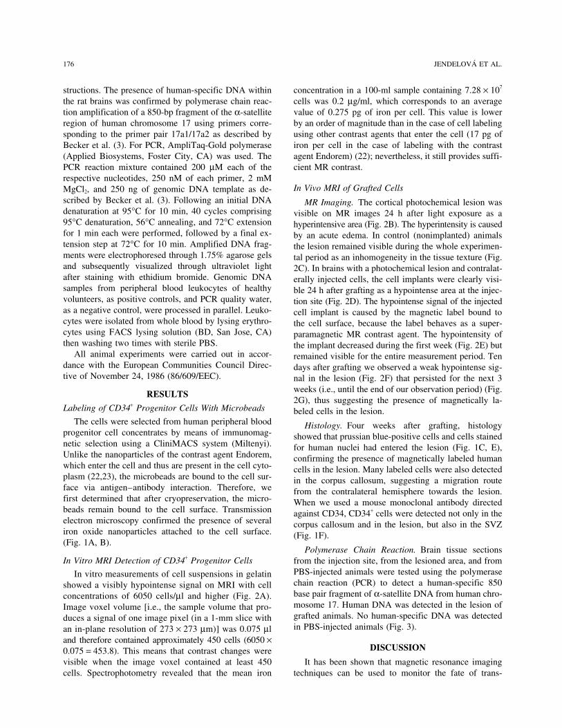
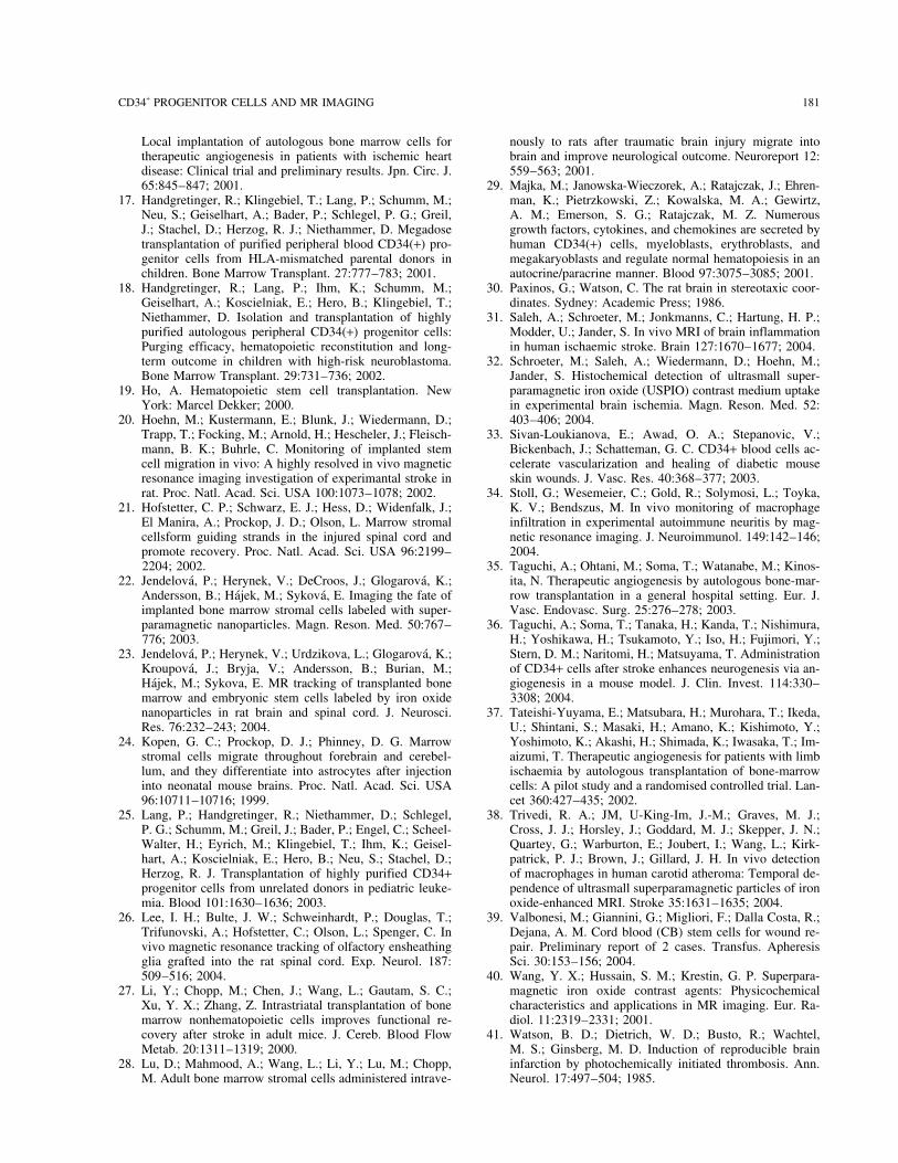
In Vivo MRI of Grafted CellsMgCl2, and 250 ng of genomic DNA template as de-scribed by Becker et al. (3). Following an initial DNA MR Imaging. The cortical photochemical lesion wasdenaturation at 95°C for 10 min, 40 cycles comprising visible on MR images 24 h after light exposure as a95°C denaturation, 56°C annealing, and 72°C extension hyperintensive area (Fig. 2B). The hyperintensity is causedfor 1 min each were performed, followed by a final ex- by an acute edema. In control (nonimplanted) animalstension step at 72°C for 10 min. Amplified DNA frag- the lesion remained visible during the whole experimen-ments were electrophoresed through 1.75% agarose gels tal period as an inhomogeneity in the tissue texture (Fig.and subsequently visualized through ultraviolet light 2C). In brains with a photochemical lesion and contralat-after staining with ethidium bromide. Genomic DNA erally injected cells, the cell implants were clearly visi-samples from peripheral blood leukocytes of healthy ble 24 h after grafting as a hypointense area at the injec-volunteers, as positive controls, and PCR quality water, tion site (Fig. 2D). The hypointense signal of the injectedas a negative control, were processed in parallel. Leuko- cell implant is caused by the magnetic label bound tocytes were isolated from whole blood by lysing erythro- the cell surface, because the label behaves as a super-cytes using FACS lysing solution (BD, San Jose, CA) paramagnetic MR contrast agent. The hypointensity ofthen washing two times with sterile PBS. the implant decreased during the first week (Fig. 2E) but
All animal experiments were carried out in accor- remained visible for the entire measurement period. Tendance with the European Communities Council Direc- days after grafting we observed a weak hypointense sig-tive of November 24, 1986 (86/609/EEC). nal in the lesion (Fig. 2F) that persisted for the next 3
weeks (i.e., until the end of our observation period) (Fig.RESULTS2G), thus suggesting the presence of magnetically la-
Labeling of CD34+ Progenitor Cells With Microbeads beled cells in the lesion.The cells were selected from human peripheral blood Histology. Four weeks after grafting, histology
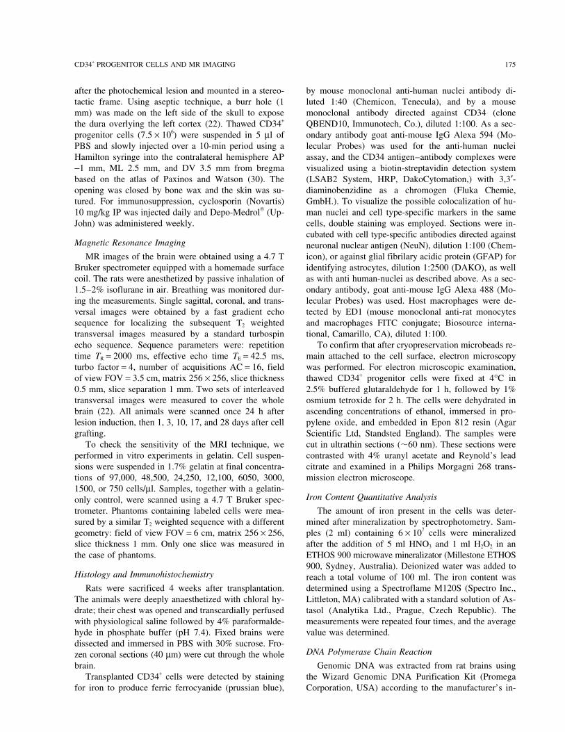
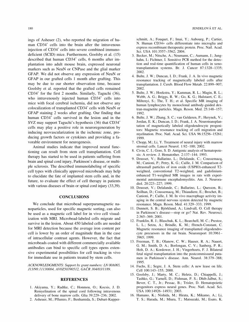
progenitor cell concentrates by means of immunomag- showed that prussian blue-positive cells and cells stainednetic selection using a CliniMACS system (Miltenyi). for human nuclei had entered the lesion (Fig. 1C, E),Unlike the nanoparticles of the contrast agent Endorem, confirming the presence of magnetically labeled humanwhich enter the cell and thus are present in the cell cyto- cells in the lesion. Many labeled cells were also detectedplasm (22,23), the microbeads are bound to the cell sur- in the corpus callosum, suggesting a migration routeface via antigen–antibody interaction. Therefore, we from the contralateral hemisphere towards the lesion.first determined that after cryopreservation, the micro- When we used a mouse monoclonal antibody directedbeads remain bound to the cell surface. Transmission against CD34, CD34+ cells were detected not only in theelectron microscopy confirmed the presence of several corpus callosum and in the lesion, but also in the SVZiron oxide nanoparticles attached to the cell surface. (Fig. 1F).(Fig. 1A, B).
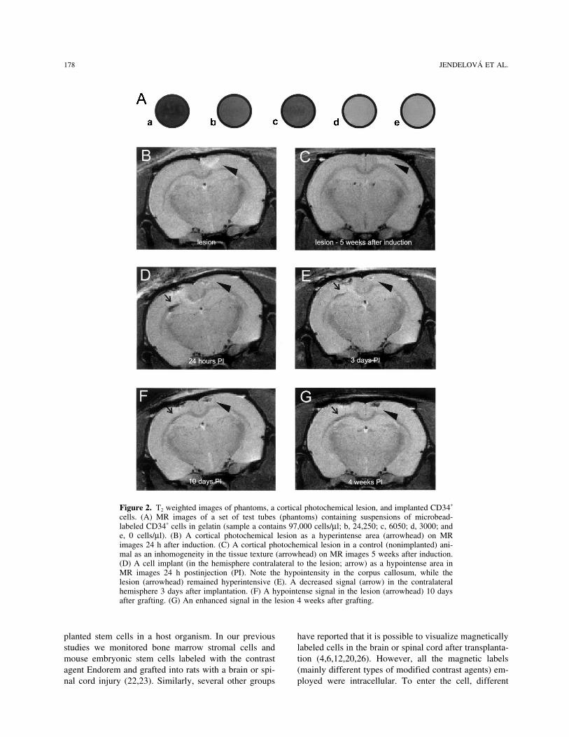
Polymerase Chain Reaction. Brain tissue sectionsIn Vitro MRI Detection of CD34+ Progenitor Cells from the injection site, from the lesioned area, and from
PBS-injected animals were tested using the polymeraseIn vitro measurements of cell suspensions in gelatinchain reaction (PCR) to detect a human-specific 850showed a visibly hypointense signal on MRI with cellbase pair fragment of α-satellite DNA from human chro-concentrations of 6050 cells/µl and higher (Fig. 2A).mosome 17. Human DNA was detected in the lesion ofImage voxel volume [i.e., the sample volume that pro-grafted animals. No human-specific DNA was detectedduces a signal of one image pixel (in a 1-mm slice within PBS-injected animals (Fig. 3).an in-plane resolution of 273 × 273 µm)] was 0.075 µl
and therefore contained approximately 450 cells (6050 ×DISCUSSION0.075 = 453.8). This means that contrast changes were
visible when the image voxel contained at least 450 It has been shown that magnetic resonance imagingtechniques can be used to monitor the fate of trans-cells. Spectrophotometry revealed that the mean iron

CD34+ PROGENITOR CELLS AND MR IMAGING 177
Figure 1. Labeling of CD34+ progenitor cells and their implantation into a rat brain with a photochemical lesion. (A) Transmissionelectron photomicrograph of a CD34+ cell showing the microbeads attached to the cell surface. Inset area shown at higher magnifica-tion in (B). (C) Histology performed 4 weeks postimplantation confirmed a large number of nanoparticle-labeled cells in the lesion(stained for prussian blue). (D) Serial section immunostained for ED1, showing that the presence of macrophages does not colocal-ize with staining for iron. (E) Human cells (positive staining for human nuclei) found in the lesion 4 weeks after grafting. (F)Staining with anti-CD34 revealed CD34+ cells in the subventricular zone.
178 JENDELOVA ET AL.
Figure 2. T2 weighted images of phantoms, a cortical photochemical lesion, and implanted CD34+
cells. (A) MR images of a set of test tubes (phantoms) containing suspensions of microbead-labeled CD34+ cells in gelatin (sample a contains 97,000 cells/µl; b, 24,250; c, 6050; d, 3000; ande, 0 cells/µl). (B) A cortical photochemical lesion as a hyperintense area (arrowhead) on MRimages 24 h after induction. (C) A cortical photochemical lesion in a control (nonimplanted) ani-mal as an inhomogeneity in the tissue texture (arrowhead) on MR images 5 weeks after induction.(D) A cell implant (in the hemisphere contralateral to the lesion; arrow) as a hypointense area inMR images 24 h postinjection (PI). Note the hypointensity in the corpus callosum, while thelesion (arrowhead) remained hyperintensive (E). A decreased signal (arrow) in the contralateralhemisphere 3 days after implantation. (F) A hypointense signal in the lesion (arrowhead) 10 daysafter grafting. (G) An enhanced signal in the lesion 4 weeks after grafting.
planted stem cells in a host organism. In our previous have reported that it is possible to visualize magneticallylabeled cells in the brain or spinal cord after transplanta-studies we monitored bone marrow stromal cells and
mouse embryonic stem cells labeled with the contrast tion (4,6,12,20,26). However, all the magnetic labels(mainly different types of modified contrast agents) em-agent Endorem and grafted into rats with a brain or spi-
nal cord injury (22,23). Similarly, several other groups ployed were intracellular. To enter the cell, different
CD34+ PROGENITOR CELLS AND MR IMAGING 179
agent); they provide sufficient MR contrast although theaverage iron content per cell is an order of magnitudelower than in the case of cell labeling with Endorem andother contrast agents that enter the cell (22).
The data obtained from MR correlated with histologi-cal findings. PCR confirmed the presence of human cellsin the lesion. To exclude that loose microbeads weretaken up by macrophages, we performed ED1 stainingfor activated microglia/macrophages. Although we foundstrong ED1 positivity in the lesions, prussian blue stain-ing did not colocalize with ED1 staining (Fig. 1C, D).In studies in which macrophage infiltration in differentcentral nervous system disorders has been visualizedwith ultrasmall superparamagnetic iron oxide particles(USPIO) (9,10,32,34), the time course of loading macro-phages with USPIO and their subsequent visualizationon MR images did not correspond with the changes inMRI signal observed in our experiments. In rats (9,10,32,34), as well as in humans (31,38), a low intensitysignal in the lesion due to infiltrating macrophagesloaded with USPIO was already visible on T2 weightedimages at 24–72 h after the injection of USPIO into theblood stream, while in our experiments we observed ahypointensive signal in the lesion 10 days after grafting.Therefore, if the label were released in vivo and taken
Figure 3. Polymerase chain reaction detection of a human- up by macrophages, the hypointensive signal would ap-specific 850-bp fragment of α-satelite DNA on human chro-pear in the lesion during the first 72 h after cell injectionmosome 17 in brain sections 4 weeks after grafting. m: marker,and not 10 days after grafting. The macrophages ob-a: negative control, b: lesion of PBS-injected animal, c: lesion
of grafted animal, d: injection site of grafted animal, e: empty served in the lesion most likely infiltrated the lesion dur-lane, f: positive control. ing the 7 days prior to cell grafting.
Bone marrow as an alternative source of adult stemcells may have practical advantages over embryonic
methods to facilitate entry are often required (e.g., lipo- stem cells and has been proposed for use as a source offection, transfection agents, antibody–iron oxide particle autologous grafts. HPCs (characterized by the presenceconstructs). Moreover, while commercially available of the CD34 antigen) represent a major focus of currentcontrast agents are clinically approved as blood pool research (14,43). These cells have the capacity for ex-agents, to be used as markers of implanted cells for clin- tensive self-renewal and pluripotent differentiation andical use in stem cell therapies further testing for cell are used therapeutically to provide long-term bone mar-toxicity and verification of CNS tolerance has to be row reconstitution in patients suffering from some leu-done. In contrast, the microbeads used for immunomag- kemias, lymphomas, solid tumors, aplastic anemias, andnetic selection remain attached to the surface of a spe- metabolic disorders (19). Furthermore, human CD34+
cific cell after sorting and have been safely transplanted cell have been shown to secrete numerous angiogenicinto patients with hematological diseases (17,18). Bulte factors, including VEGF, HGF, and IGF-1 (29). On theet al. (5) described experiments with human lympho- basis of these observations, clinical trials of cell trans-cytes labeled with biotinylated antilymphocyte-directed plantation in hindlimb (35,37) and cardiac ischemia (16)monoclonal antibodies, to which streptavidin and subse- have been initiated with promising results. Humanquently biotinylated dextran-magnetite particles were CD34+ cells can be collected from BM, cord blood, orcoupled. Similar to our results, labeled lymphocyte sus- from peripheral blood after cytokine mobilization. In ourpensions enhanced negative contrast, though the experi- study we grafted human CD34+ progenitor cells, ob-ments were done in a weaker magnetic field (2.0 Tesla). tained from peripheral blood after G-CSF stimulation,However, the study was done only in vitro, and the la- into the lesioned rat brain. The human cells migratedbeling kit used is not commercially available. In our from the injection site to the lesion and to the SVZ,study we confirmed that CD34 microbeads can be used where they survived throughout our observation periodas a magnetic label (a highly specific MR contrast of 1 month. Our results are in agreement with the find-
180 JENDELOVA ET AL.
schmitt, A.; Fouquet, F.; Imai, Y.; Aubourg, P.; Cartier,ings of Asheuer (2), who reported the migration of hu-N. Human CD34+ cells differentiate into microglia andman CD34+ cells into the brain after the intravenousexpress recombinant therapeutic protein. Proc. Natl. Acad.
injection of CD34+ cells into severe combined immuno- Sci. USA 101:3557–3562; 2004.deficient (SCID) mice. Furthermore, Goolsby et al. (15) 3. Becker, M.; Nitsche, A.; Neumann, C.; Aumann, J.; Jung-described that human CD34+ cells, 6 months after im- hahn, I.; Fichtner, I. Sensitive PCR method for the detec-
tion and real-time quantification of human cells in xeno-plantation into adult mouse brain, expressed neuronaltransplantation systems. Br. J. Cancer 87:1328–1335;markers such as NeuN or CNPase and the glial marker2002.GFAP. We did not observe any expression of NeuN or 4. Bulte, J. W.; Duncan, I. D.; Frank, J. A. In vivo magnetic
GFAP in our grafted cells 1 month after grafting. This resonance tracking of magnetically labeled cells aftermay be due to our shorter observation time, because transplantation. J. Cereb. Blood Flow Metab. 22:899–907;
2002.Goolsby et al. reported that the grafted cells remained5. Bulte, J. W.; Hoekstra, Y.; Kamman, R. L.; Magin, R. L.;CD34+ for the first 2 months. Similarly, Taguchi (36),
Webb, A. G.; Briggs, R. W.; Go, K. G.; Hulstaert, C. E.;who intravenously injected human CD34+ cells intoMiltenyi, S.; The, T. H.; et al. Specific MR imaging of
mice with focal cerebral ischemia, did not observe any human lymphocytes by monoclonal antibody-guided dex-colocalization of transplanted CD34+ cells with NeuN or tran-magnetite particles. Magn. Reson. Med. 25:148–157;GFAP staining 2 weeks after grafting. Our finding that 1992.
6. Bulte, J. W.; Zhang, S. C.; van Gelderen, P.; Herynek, V.;human CD34+ cells survived in the lesion and in theJordan, E. K.; Duncan, I. D.; Frank, J. A. Neurotransplan-SVZ may support Taguchi’s hypothesis (36) that CD34+
tation of magnetically labeled oligodendrocyte progeni-cells may play a positive role in neuroregeneration bytors: Magnetic resonance tracking of cell migration and
inducing neovascularization in the ischemic zone, pro- myelination. Proc. Natl. Acad. Sci. USA 96:15256–15261;ducing growth factors or cytokines and providing a fa- 1999.vorable environment for neurogenesis. 7. Chopp, M.; Li, Y. Treatment of neural injury with marrow
stromal cells. Lancet Neurol. 1:92–100; 2002.Animal studies indicate that improved neural func-8. Civin, C. I.; Gore, S. D. Antigenic analysis of hematopoie-tioning can result from stem cell transplantation. Cell
sis: A review. J. Hematother. 2:137–144; 1993.therapy has started to be used in patients suffering from 9. Dousset, V.; Ballarino, L.; Delalande, C.; Coussemacq,brain and spinal cord injury, Parkinson’s disease, or multi- M.; Canioni, P.; Petry, K. G.; Caille, J. M. Comparison ofple sclerosis. The described immunolabeling of specific ultrasmall particles of iron oxide (USPIO)-enhanced T2-
weighted, conventional T2-weighted, and gadolinium-cell types with clinically approved microbeads may helpenhanced T1-weighted MR images in rats with experi-to elucidate the fate of implanted stem cells and, in themental autoimmune encephalomyelitis. Am. J. Neurora-future, to evaluate the effect of cell therapy in patientsdiol. 20:223–227; 1999.
with various diseases of brain or spinal cord injury (33,39). 10. Dousset, V.; Delalande, C.; Ballarino, L.; Quesson, B.;Seilhan, D.; Coussemacq, M.; Thiaudiere, E.; Brochet, B.;Canioni, P.; Caille, J. M. In vivo macrophage activity im-CONCLUSIONSaging in the central nervous system detected by magnetic
We conclude that microbead superparamagnetic na- resonance. Magn. Reson. Med. 41:329–333; 1999.11. Dunnett, S. B.; Bjorklund, A.; Lindvall, O. Cell therapynoparticles, used for specific magnetic sorting, can also
in Parkinson’s disease—stop or go? Nat. Rev. Neurosci.be used as a magnetic cell label for in vivo cell visual-2:365–369; 2001.ization with MRI. Microbead-labeled cells migrate and
12. Franklin, R. J.; Blaschuk, K. L.; Bearchell, M. C.; Prestoz,survive in the lesion. About 450 labeled cells are needed L. L.; Setzu, A.; Brindle, K. M.; ffrench-Constant, C.for MRI detection because the average iron content per Magnetic resonance imaging of transplanted oligodendro-
cyte precursors in the rat brain. Neuroreport 10:3961–cell is lower by an order of magnitude than in the case3965; 1999.of intracellular contrast agents. However, the fact that
13. Freeman, T. B.; Olanow, C. W.; Hauser, R. A.; Nauert,microbeads coated with different commercially availableG. M.; Smith, D. A.; Borlongan, C. V.; Sanberg, P. R.;
antibodies can bind to specific cell types opens exten- Holt, D. A.; Kordower, J. H.; Vingerhoets, F. J. Bilateralsive experimental possibilities for cell tracking in vivo fetal nigral transplantation into the postcommissural puta-for immediate use in patients treated by stem cells. men in Parkinson’s disease. Ann. Neurol. 38:379–388;
1995.ACKNOWLEDGMENTS: Support by grant numbers: LN 00A065, 14. Fuchs, E.; Segre, J. A. Stem cells: A new lease on life.J13/98:11130004, AV0Z50390512, GACR 304/03/1189. Cell 100:143–155; 2000.
15. Goolsby, J.; Marty, M. C.; Heletz, D.; Chiappelli, J.;Tashko, G.; Yarnell, D.; Fishman, P. S.; Dhib-Jalbut, S.;REFERENCESBever, C. T., Jr.; Pessac, B.; Trisler, D. Hematopoieticprogenitors express neural genes. Proc. Natl. Acad. Sci.1. Akiyama, Y.; Radtke, C.; Honmou, O.; Kocsis, J. D.USA 100:14926–14931; 2003.Remyelination of the spinal cord following intravenous
16. Hamano, K.; Nishida, M.; Hirata, K.; Mikamo, A.; Li,delivery of bone marrow cells. Glia 39:229–236; 2002.T. S.; Harada, M.; Miura, T.; Matsuzaki, M.; Esato, K.2. Asheuer, M.; Pflumio, F.; Benhamida, S.; Dubart-Kupper-
CD34+ PROGENITOR CELLS AND MR IMAGING 181
Local implantation of autologous bone marrow cells for nously to rats after traumatic brain injury migrate intobrain and improve neurological outcome. Neuroreport 12:therapeutic angiogenesis in patients with ischemic heart
disease: Clinical trial and preliminary results. Jpn. Circ. J. 559–563; 2001.29. Majka, M.; Janowska-Wieczorek, A.; Ratajczak, J.; Ehren-65:845–847; 2001.
17. Handgretinger, R.; Klingebiel, T.; Lang, P.; Schumm, M.; man, K.; Pietrzkowski, Z.; Kowalska, M. A.; Gewirtz,A. M.; Emerson, S. G.; Ratajczak, M. Z. NumerousNeu, S.; Geiselhart, A.; Bader, P.; Schlegel, P. G.; Greil,
J.; Stachel, D.; Herzog, R. J.; Niethammer, D. Megadose growth factors, cytokines, and chemokines are secreted byhuman CD34(+) cells, myeloblasts, erythroblasts, andtransplantation of purified peripheral blood CD34(+) pro-
genitor cells from HLA-mismatched parental donors in megakaryoblasts and regulate normal hematopoiesis in anautocrine/paracrine manner. Blood 97:3075–3085; 2001.children. Bone Marrow Transplant. 27:777–783; 2001.
18. Handgretinger, R.; Lang, P.; Ihm, K.; Schumm, M.; 30. Paxinos, G.; Watson, C. The rat brain in stereotaxic coor-dinates. Sydney: Academic Press; 1986.Geiselhart, A.; Koscielniak, E.; Hero, B.; Klingebiel, T.;
Niethammer, D. Isolation and transplantation of highly 31. Saleh, A.; Schroeter, M.; Jonkmanns, C.; Hartung, H. P.;Modder, U.; Jander, S. In vivo MRI of brain inflammationpurified autologous peripheral CD34(+) progenitor cells:
Purging efficacy, hematopoietic reconstitution and long- in human ischaemic stroke. Brain 127:1670–1677; 2004.32. Schroeter, M.; Saleh, A.; Wiedermann, D.; Hoehn, M.;term outcome in children with high-risk neuroblastoma.
Bone Marrow Transplant. 29:731–736; 2002. Jander, S. Histochemical detection of ultrasmall super-paramagnetic iron oxide (USPIO) contrast medium uptake19. Ho, A. Hematopoietic stem cell transplantation. New
York: Marcel Dekker; 2000. in experimental brain ischemia. Magn. Reson. Med. 52:403–406; 2004.20. Hoehn, M.; Kustermann, E.; Blunk, J.; Wiedermann, D.;
Trapp, T.; Focking, M.; Arnold, H.; Hescheler, J.; Fleisch- 33. Sivan-Loukianova, E.; Awad, O. A.; Stepanovic, V.;Bickenbach, J.; Schatteman, G. C. CD34+ blood cells ac-mann, B. K.; Buhrle, C. Monitoring of implanted stem
cell migration in vivo: A highly resolved in vivo magnetic celerate vascularization and healing of diabetic mouseskin wounds. J. Vasc. Res. 40:368–377; 2003.resonance imaging investigation of experimantal stroke in
rat. Proc. Natl. Acad. Sci. USA 100:1073–1078; 2002. 34. Stoll, G.; Wesemeier, C.; Gold, R.; Solymosi, L.; Toyka,K. V.; Bendszus, M. In vivo monitoring of macrophage21. Hofstetter, C. P.; Schwarz, E. J.; Hess, D.; Widenfalk, J.;
El Manira, A.; Prockop, J. D.; Olson, L. Marrow stromal infiltration in experimental autoimmune neuritis by mag-netic resonance imaging. J. Neuroimmunol. 149:142–146;cellsform guiding strands in the injured spinal cord and
promote recovery. Proc. Natl. Acad. Sci. USA 96:2199– 2004.35. Taguchi, A.; Ohtani, M.; Soma, T.; Watanabe, M.; Kinos-2204; 2002.
22. Jendelova, P.; Herynek, V.; DeCroos, J.; Glogarova, K.; ita, N. Therapeutic angiogenesis by autologous bone-mar-row transplantation in a general hospital setting. Eur. J.Andersson, B.; Hajek, M.; Sykova, E. Imaging the fate of
implanted bone marrow stromal cells labeled with super- Vasc. Endovasc. Surg. 25:276–278; 2003.36. Taguchi, A.; Soma, T.; Tanaka, H.; Kanda, T.; Nishimura,paramagnetic nanoparticles. Magn. Reson. Med. 50:767–
776; 2003. H.; Yoshikawa, H.; Tsukamoto, Y.; Iso, H.; Fujimori, Y.;Stern, D. M.; Naritomi, H.; Matsuyama, T. Administration23. Jendelova, P.; Herynek, V.; Urdzikova, L.; Glogarova, K.;
Kroupova, J.; Bryja, V.; Andersson, B.; Burian, M.; of CD34+ cells after stroke enhances neurogenesis via an-giogenesis in a mouse model. J. Clin. Invest. 114:330–Hajek, M.; Sykova, E. MR tracking of transplanted bone
marrow and embryonic stem cells labeled by iron oxide 3308; 2004.37. Tateishi-Yuyama, E.; Matsubara, H.; Murohara, T.; Ikeda,nanoparticles in rat brain and spinal cord. J. Neurosci.
Res. 76:232–243; 2004. U.; Shintani, S.; Masaki, H.; Amano, K.; Kishimoto, Y.;Yoshimoto, K.; Akashi, H.; Shimada, K.; Iwasaka, T.; Im-24. Kopen, G. C.; Prockop, D. J.; Phinney, D. G. Marrow
stromal cells migrate throughout forebrain and cerebel- aizumi, T. Therapeutic angiogenesis for patients with limbischaemia by autologous transplantation of bone-marrowlum, and they differentiate into astrocytes after injection
into neonatal mouse brains. Proc. Natl. Acad. Sci. USA cells: A pilot study and a randomised controlled trial. Lan-cet 360:427–435; 2002.96:10711–10716; 1999.
25. Lang, P.; Handgretinger, R.; Niethammer, D.; Schlegel, 38. Trivedi, R. A.; JM, U-King-Im, J.-M.; Graves, M. J.;Cross, J. J.; Horsley, J.; Goddard, M. J.; Skepper, J. N.;P. G.; Schumm, M.; Greil, J.; Bader, P.; Engel, C.; Scheel-
Walter, H.; Eyrich, M.; Klingebiel, T.; Ihm, K.; Geisel- Quartey, G.; Warburton, E.; Joubert, I.; Wang, L.; Kirk-patrick, P. J.; Brown, J.; Gillard, J. H. In vivo detectionhart, A.; Koscielniak, E.; Hero, B.; Neu, S.; Stachel, D.;
Herzog, R. J. Transplantation of highly purified CD34+ of macrophages in human carotid atheroma: Temporal de-pendence of ultrasmall superparamagnetic particles of ironprogenitor cells from unrelated donors in pediatric leuke-
mia. Blood 101:1630–1636; 2003. oxide-enhanced MRI. Stroke 35:1631–1635; 2004.39. Valbonesi, M.; Giannini, G.; Migliori, F.; Dalla Costa, R.;26. Lee, I. H.; Bulte, J. W.; Schweinhardt, P.; Douglas, T.;
Trifunovski, A.; Hofstetter, C.; Olson, L.; Spenger, C. In Dejana, A. M. Cord blood (CB) stem cells for wound re-pair. Preliminary report of 2 cases. Transfus. Apheresisvivo magnetic resonance tracking of olfactory ensheathing
glia grafted into the rat spinal cord. Exp. Neurol. 187: Sci. 30:153–156; 2004.40. Wang, Y. X.; Hussain, S. M.; Krestin, G. P. Superpara-509–516; 2004.
27. Li, Y.; Chopp, M.; Chen, J.; Wang, L.; Gautam, S. C.; magnetic iron oxide contrast agents: Physicochemicalcharacteristics and applications in MR imaging. Eur. Ra-Xu, Y. X.; Zhang, Z. Intrastriatal transplantation of bone
marrow nonhematopoietic cells improves functional re- diol. 11:2319–2331; 2001.41. Watson, B. D.; Dietrich, W. D.; Busto, R.; Wachtel,covery after stroke in adult mice. J. Cereb. Blood Flow
Metab. 20:1311–1319; 2000. M. S.; Ginsberg, M. D. Induction of reproducible braininfarction by photochemically initiated thrombosis. Ann.28. Lu, D.; Mahmood, A.; Wang, L.; Li, Y.; Lu, M.; Chopp,
M. Adult bone marrow stromal cells administered intrave- Neurol. 17:497–504; 1985.

182 JENDELOVA ET AL.
42. Weissleder, R.; Cheng, H. C.; Bogdanova, A.; Bog- 44. Yeh, T.; Zhang, W.; Ildstad, S. T.; Ho, C. In vivo dynamicMRI tracking of rat T-cells labeled with superparamag-danov, A., Jr. Magnetically labeled cells can be de-
tected by MR imaging. J. Magn. Reson. Imaging 7:258– netic iron-oxide particles. Magn. Reson. Med. 33:200–208; 1995.263; 1997.
43. Weissman, I. L. Translating stem and progenitor cell biol- 45. Yeh, T.; Zhang, W.; Ildstad, S. T.; Ho, C. Intracellularlabeling of T-cells with superparamagnetic contrastogy to the clinic: Barriers and opportunities. Science 287:
1442–1446; 2000. agents. Magn. Reson. Med. 30:617–625; 1993.


![Ionic dimers in He droplets: Interaction potentials for Li[sub 2][sup +]–He,Na[sub 2][sup +]–He, and K[sub 2][sup +]–He and stability of the smaller clusters](https://static.fdokumen.com/doc/165x107/634156f0c38f655be209fcfa/ionic-dimers-in-he-droplets-interaction-potentials-for-lisub-2sup-henasub.jpg)