LUNG-RADS 1.1- A case based review: - piper
36
LUNG-RADS 1.1- A case based review: ǯ ǫ Shambo Guha Roy, MD Justin Mackey, MD Department of Radiology Mercy Catholic Medical Center, Darby PA
-
Upload
khangminh22 -
Category
Documents
-
view
0 -
download
0
Transcript of LUNG-RADS 1.1- A case based review: - piper
LUNG-RADS 1.1- A case based review: ����ǯ�����ǫ
Shambo Guha Roy, MDJustin Mackey, MD
Department of RadiologyMercy Catholic Medical Center, Darby PA
Neither I, my co-authors nor my immediate family members have a
financial relationship with a commercial organization that may have a
direct or indirect interest in the contents of this presentation.
Disclosure of Commercial Interest

1. Lung cancer screening rationale 2. Eligibility 3. LUNG-RADS scoring system4. Illustrative cases5. Highlight of the changes in 1.1
1. General & Thoracic Radiologists 2. Radiology Residents & Fellows
Target Audience
Goals and Objectives
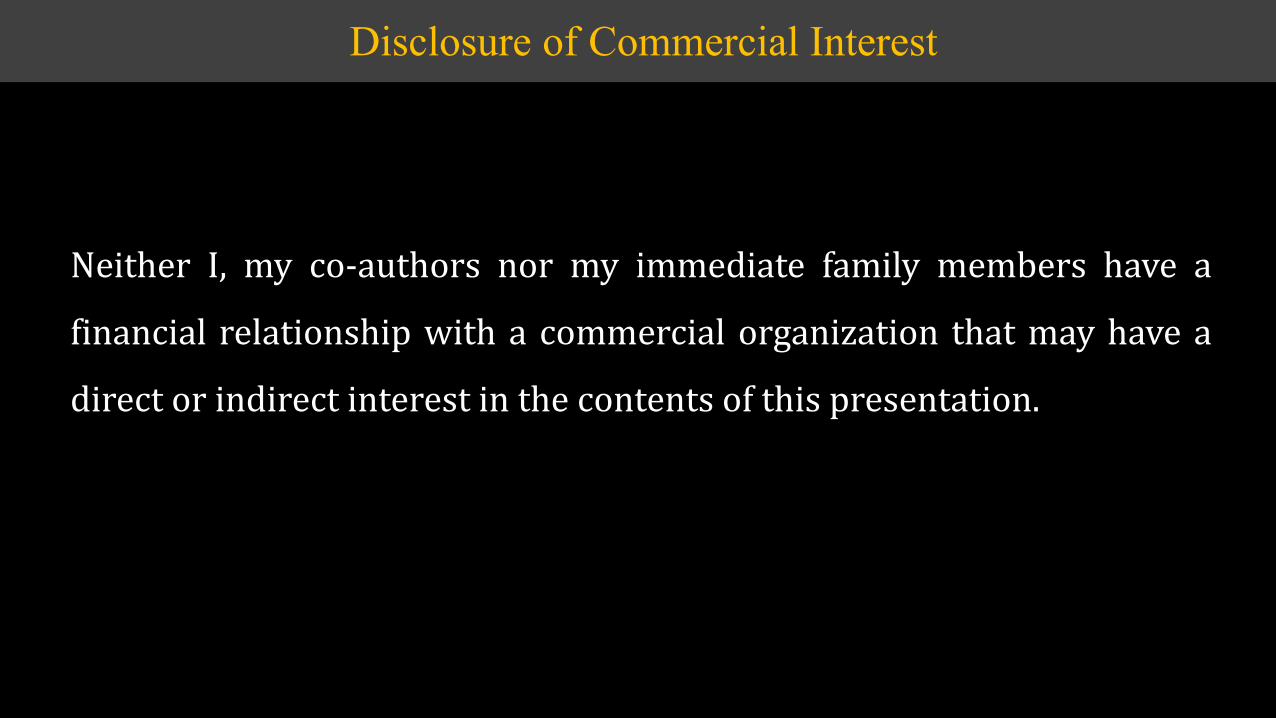
Introduction
de Groot PM, Wu CC, Carter BW, Munden RF. The epidemiology of lung cancer. Transl Lung Cancer Res. 2018;7(3):220Ȃ233
o Lung cancer is the 2nd most common cancer in both sex
o Approximately quarter million new cases and 150,000 deaths (2018)
o Approximately 14% of all cancers but 25% of all cancer mortality
o Overall 5 year survival 18% (diagnosed between 2003-2009)
o Median age: 70 years
o 88% of all lung cancers occur at age >55 years
o Approximately 90% of lung cancers are attributable to smoking
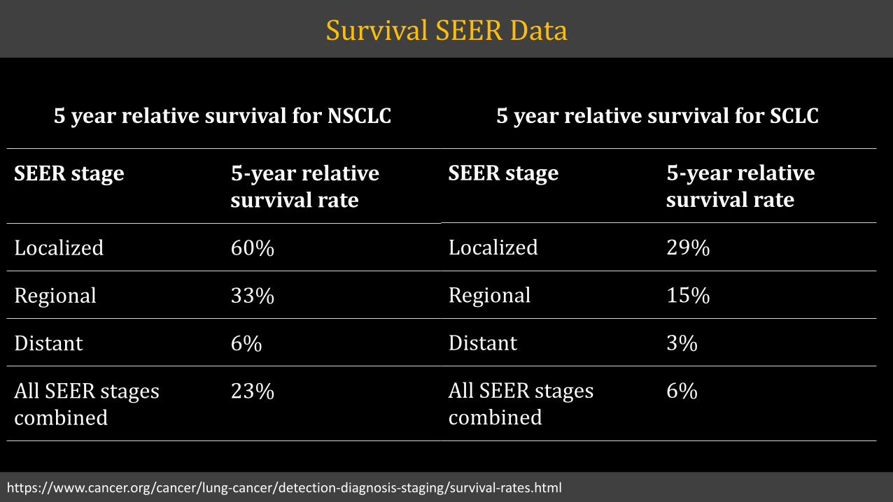
SEER stage 5-year relative survival rate
Localized 60%
Regional 33%
Distant 6%
All SEER stages combined
23%
SEER stage 5-year relative survival rate
Localized 29%
Regional 15%
Distant 3%
All SEER stages combined
6%
5 year relative survival for NSCLC 5 year relative survival for SCLC
https://www.cancer.org/cancer/lung-cancer/detection-diagnosis-staging/survival-rates.html
Survival SEER Data
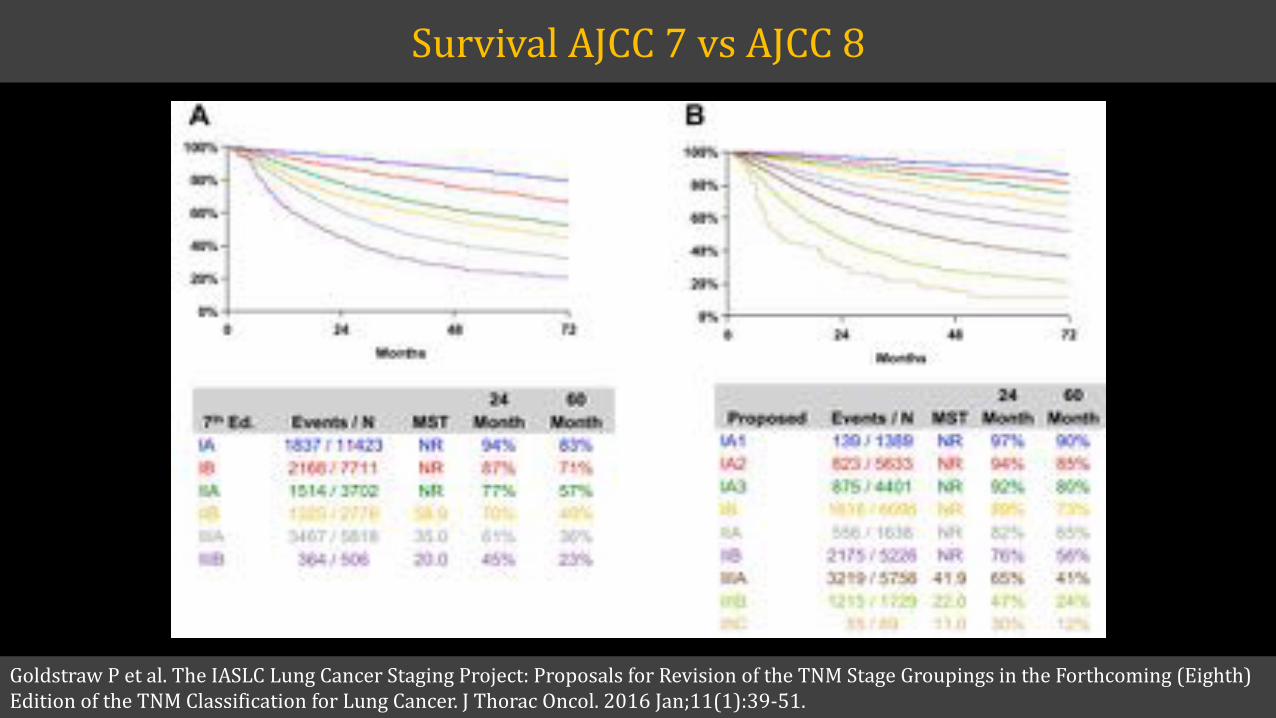
Goldstraw P et al. The IASLC Lung Cancer Staging Project: Proposals for Revision of the TNM Stage Groupings in the Forthcoming (Eighth)Edition of the TNM Classification for Lung Cancer. J Thorac Oncol. 2016 Jan;11(1):39-51.
Survival AJCC 7 vs AJCC 8
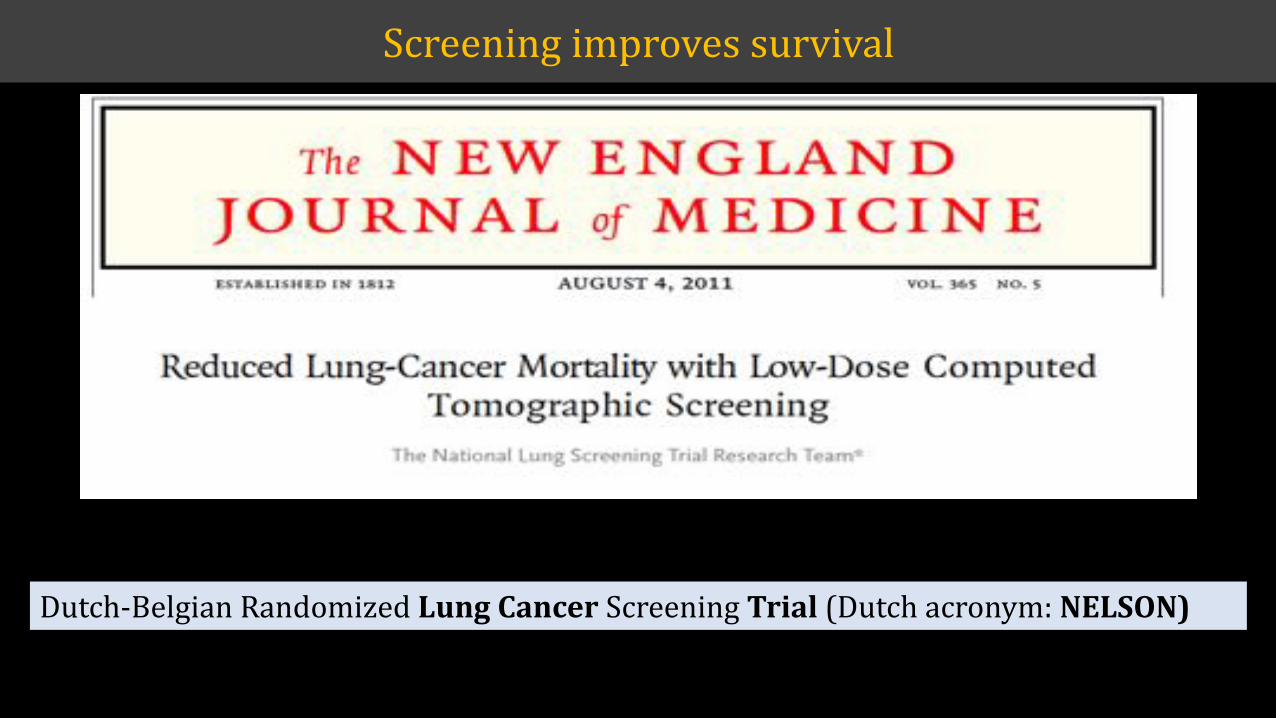
Screening improves survival
Dutch-Belgian Randomized Lung Cancer Screening Trial (Dutch acronym: NELSON)
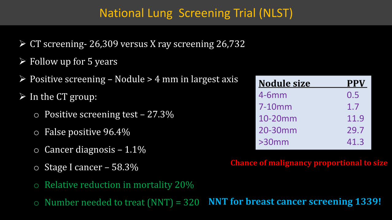
¾ CT screening- 26,309 versus X ray screening 26,732
¾ Follow up for 5 years
¾ Positive screening Ȃ Nodule > 4 mm in largest axis
¾ In the CT group:
o Positive screening test Ȃ 27.3%
o False positive 96.4%
o Cancer diagnosis Ȃ 1.1%
o Stage I cancer Ȃ 58.3%
o Relative reduction in mortality 20%
o Number needed to treat (NNT) = 320
Nodule size PPV 4-6mm 0.57-10mm 1.710-20mm 11.920-30mm 29.7>30mm 41.3
National Lung Screening Trial (NLST)
Chance of malignancy proportional to size
NNT for breast cancer screening 1339!
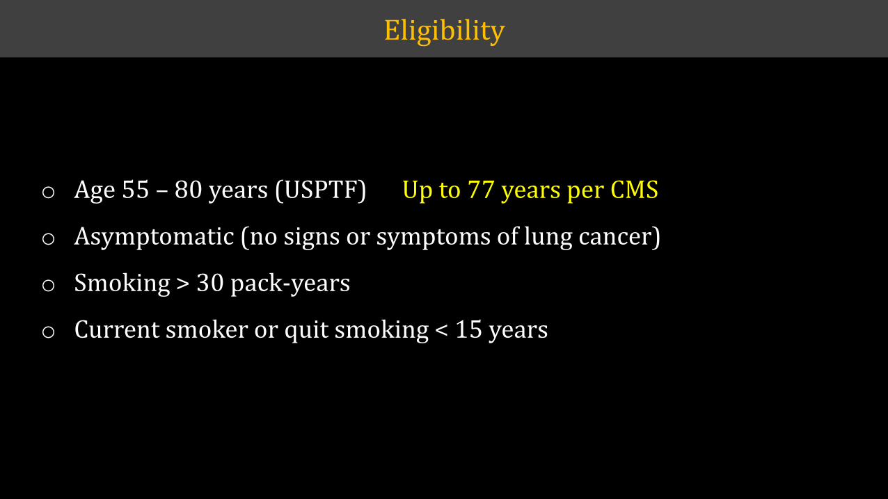
Eligibility
o Age 55 Ȃ 80 years (USPTF)
o Asymptomatic (no signs or symptoms of lung cancer)
o Smoking > 30 pack-years
o Current smoker or quit smoking < 15 years
Up to 77 years per CMS
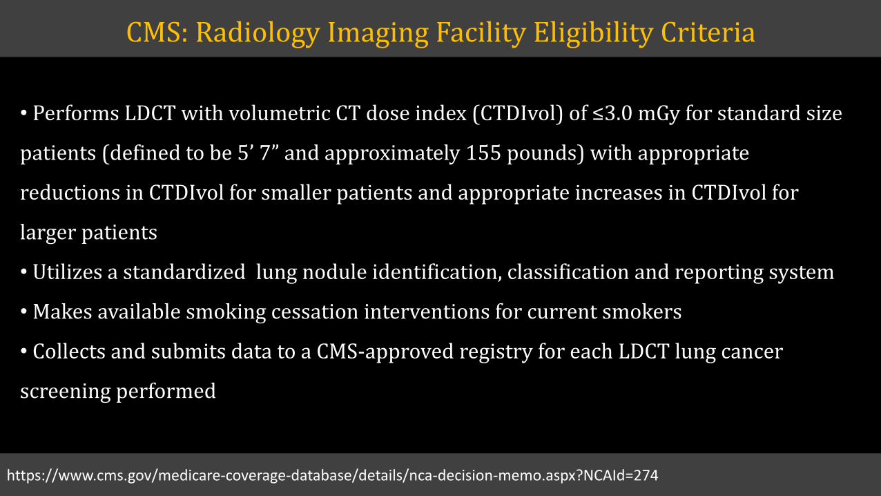
� Performs LDCT with volumetric CT dose index (CTDIvolȌ����ζ͵ǤͲ�mGy for standard size
���������ȋ��������������ͷǯ�dz�������������������ͳͷͷ�������Ȍ������������������
reductions in CTDIvol for smaller patients and appropriate increases in CTDIvol for
larger patients
� Utilizes a standardized lung nodule identification, classification and reporting system
� Makes available smoking cessation interventions for current smokers
� Collects and submits data to a CMS-approved registry for each LDCT lung cancer
screening performed
CMS: Radiology Imaging Facility Eligibility Criteria
https://www.cms.gov/medicare-coverage-database/details/nca-decision-memo.aspx?NCAId=274
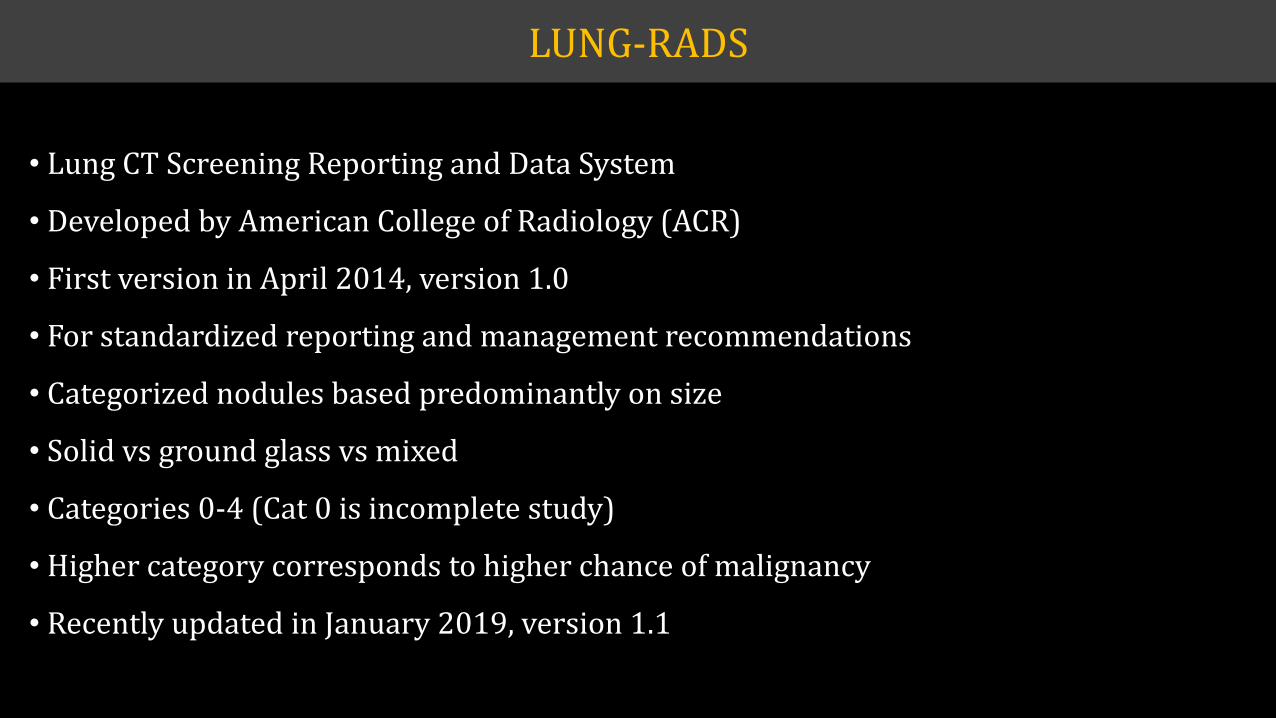
LUNG-RADS
� Lung CT Screening Reporting and Data System
� Developed by American College of Radiology (ACR)
� First version in April 2014, version 1.0
� For standardized reporting and management recommendations
� Categorized nodules based predominantly on size
� Solid vs ground glass vs mixed
� Categories 0-4 (Cat 0 is incomplete study)
� Higher category corresponds to higher chance of malignancy
� Recently updated in January 2019, version 1.1
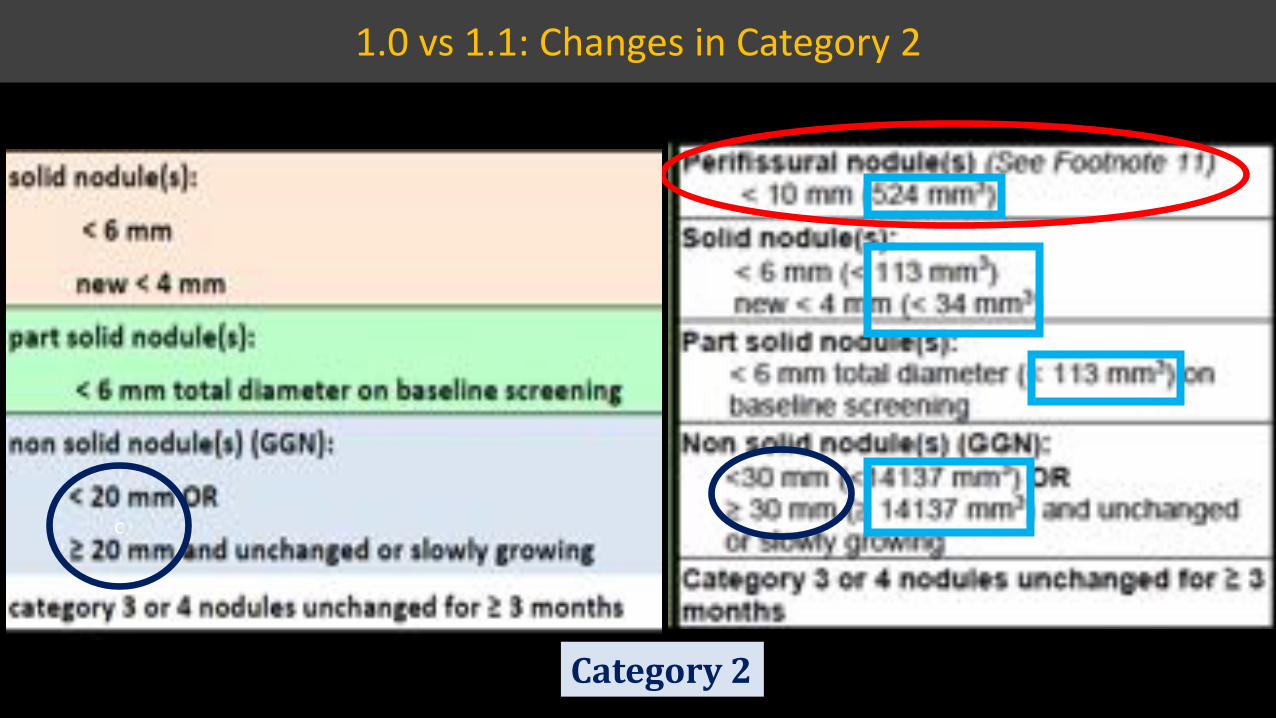
1.0 vs 1.1: Changes in Category 2
c
Category 2
c
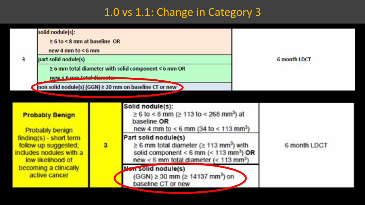
1.0 vs 1.1: Change in Category 3
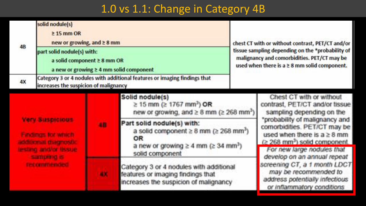
1.0 vs 1.1: Change in Category 4B

Version 1.1¾ Perifissural nodule measuring 6.8 mm¾ Volume 164 cc¾ LUNG RADS 2¾ Continue annual CT screening
Case 1
Version 1.0¾ Right middle lobe solid 7 mm nodule¾ LUNG RADS 3¾ CT in 6 months
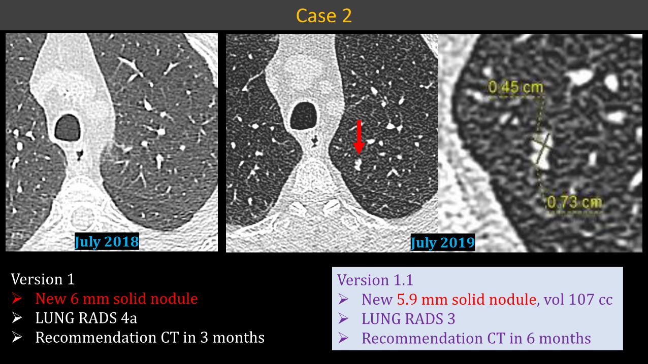
Version 1¾ New 6 mm solid nodule¾ LUNG RADS 4a¾ Recommendation CT in 3 months
Version 1.1¾ New 5.9 mm solid nodule, vol 107 cc¾ LUNG RADS 3¾ Recommendation CT in 6 months
July 2018 July 2019
Case 2
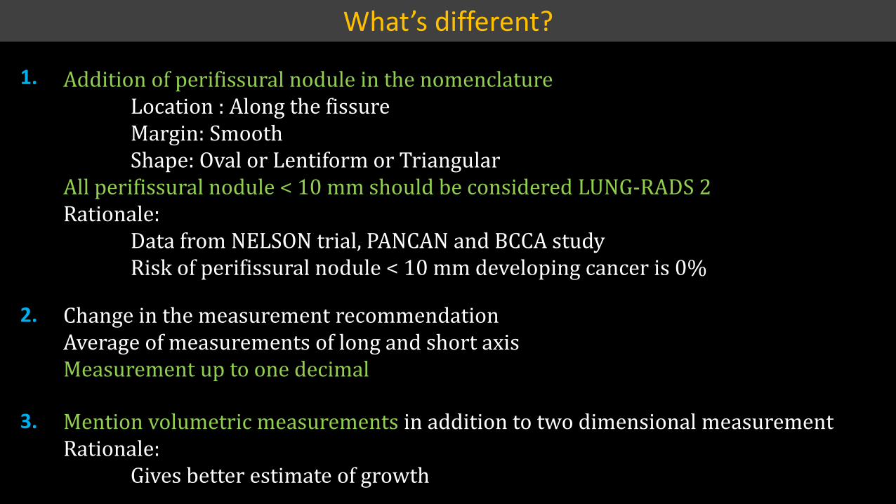
tŚĂƚ Ɛ͛�ĚŝĨĨĞƌĞŶƚ͍
Addition of perifissural nodule in the nomenclatureLocation : Along the fissureMargin: SmoothShape: Oval or Lentiform or Triangular
All perifissural nodule < 10 mm should be considered LUNG-RADS 2Rationale:
Data from NELSON trial, PANCAN and BCCA studyRisk of perifissural nodule < 10 mm developing cancer is 0%
Change in the measurement recommendationAverage of measurements of long and short axisMeasurement up to one decimal
Mention volumetric measurements in addition to two dimensional measurementRationale:
Gives better estimate of growth
1.
2.
3.
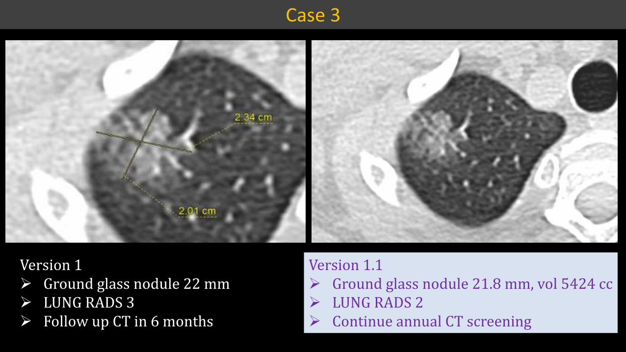
Version 1¾ Ground glass nodule 22 mm¾ LUNG RADS 3¾ Follow up CT in 6 months
Version 1.1¾ Ground glass nodule 21.8 mm, vol 5424 cc¾ LUNG RADS 2¾ Continue annual CT screening
Case 3
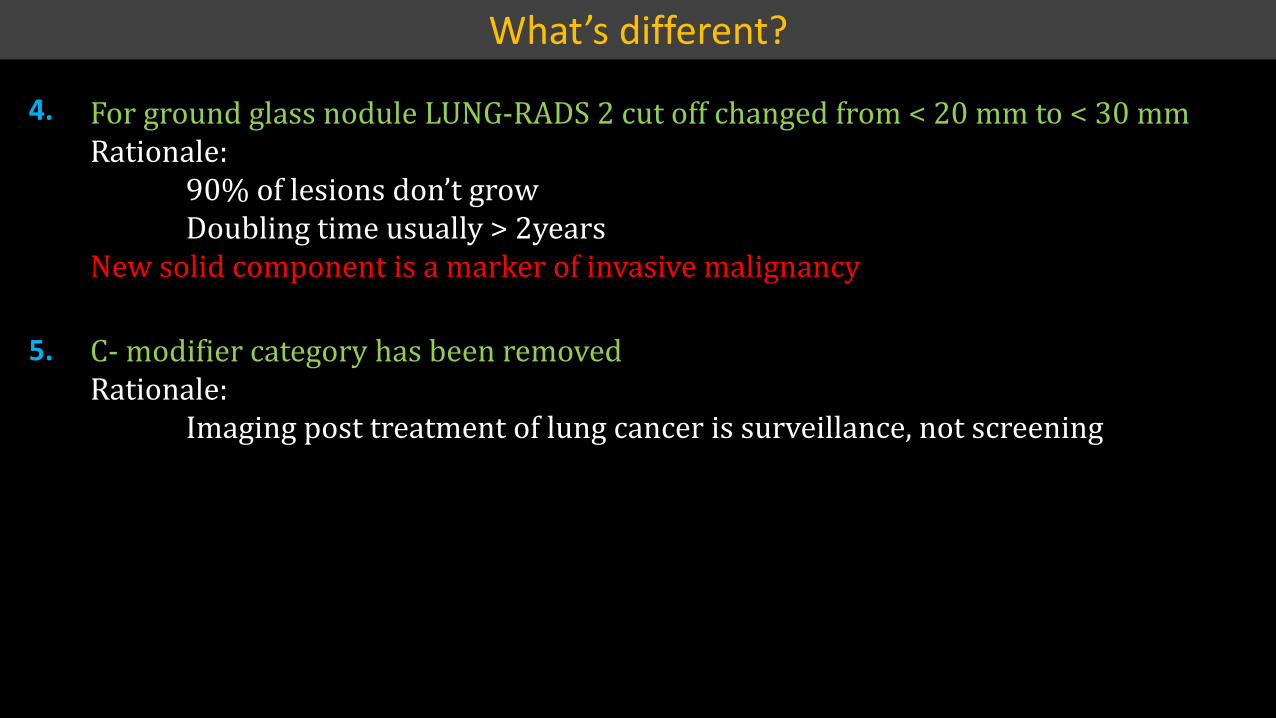
tŚĂƚ Ɛ͛�ĚŝĨĨĞƌĞŶƚ͍
For ground glass nodule LUNG-RADS 2 cut off changed from < 20 mm to < 30 mmRationale:
ͻͲΨ���������������ǯ������Doubling time usually > 2years
New solid component is a marker of invasive malignancy
4.
C- modifier category has been removedRationale:
Imaging post treatment of lung cancer is surveillance, not screening
5.
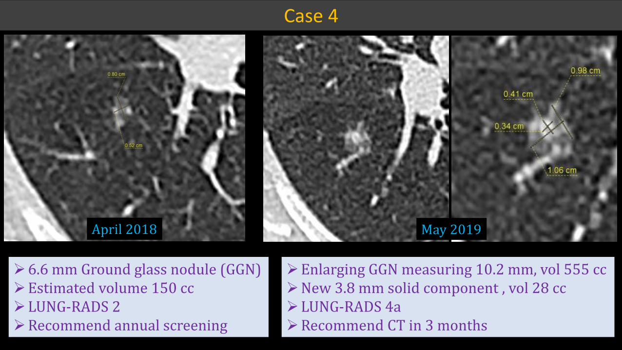
¾6.6 mm Ground glass nodule (GGN)¾Estimated volume 150 cc¾LUNG-RADS 2¾Recommend annual screening
¾Enlarging GGN measuring 10.2 mm, vol 555 cc¾New 3.8 mm solid component , vol 28 cc¾LUNG-RADS 4a¾Recommend CT in 3 months
April 2018 May 2019
Case 4

¾Part solid nodule: Overall 22.9 mm with 10 mm solid component ¾LUNG-RADS 4b¾Recommend PET-CT and biopsy
Case 5
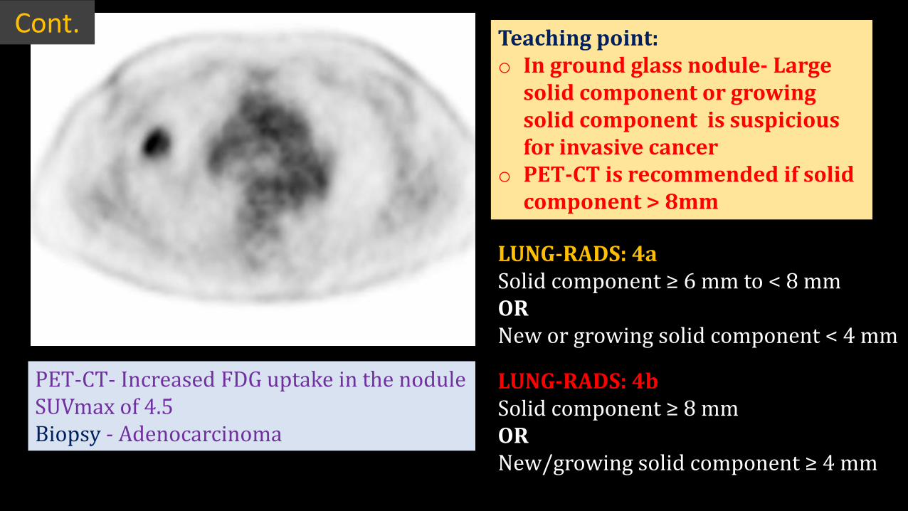
PET-CT- Increased FDG uptake in the nodule SUVmax of 4.5Biopsy - Adenocarcinoma
Teaching point:o In ground glass nodule- Large
solid component or growing solid component is suspicious for invasive cancer
o PET-CT is recommended if solid component > 8mm
LUNG-RADS: 4a����������������η��������δ�ͺ����OR New or growing solid component < 4 mm
LUNG-RADS: 4b����������������η�ͺ�����OR ���Ȁ������������������������η�Ͷ���
Cont.
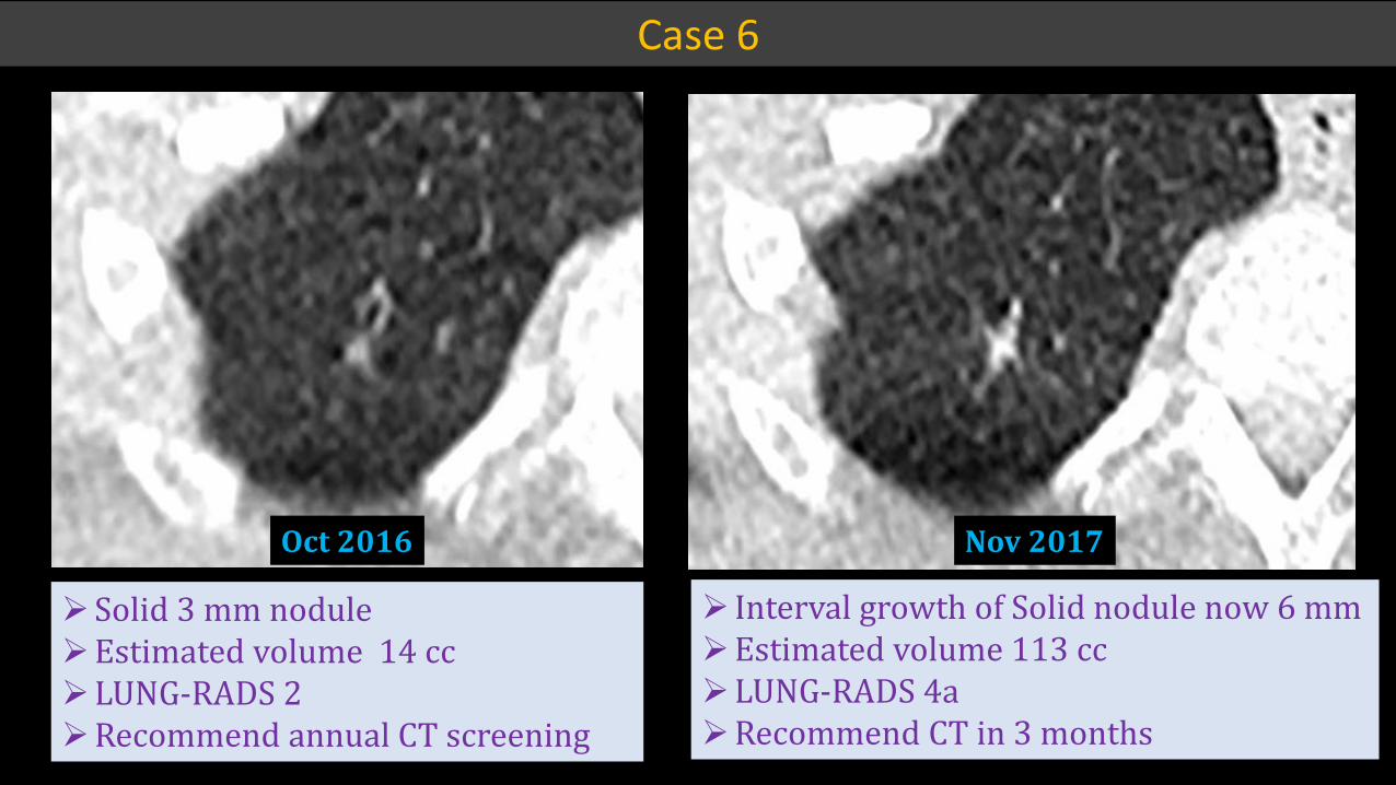
Oct 2016
¾Solid 3 mm nodule¾Estimated volume 14 cc¾LUNG-RADS 2¾Recommend annual CT screening
¾ Interval growth of Solid nodule now 6 mm¾Estimated volume 113 cc¾LUNG-RADS 4a¾Recommend CT in 3 months
Case 6
Nov 2017
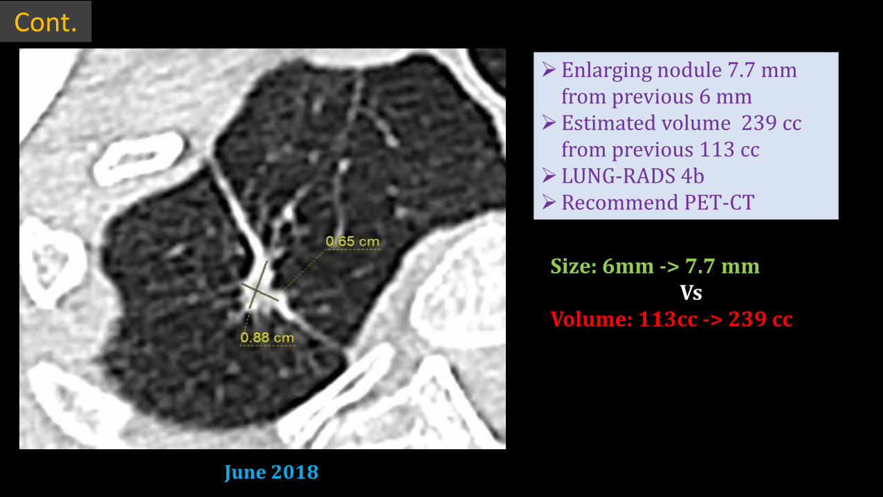
¾Enlarging nodule 7.7 mm from previous 6 mm
¾Estimated volume 239 cc from previous 113 cc
¾LUNG-RADS 4b¾Recommend PET-CT
June 2018
Size: 6mm -> 7.7 mmVs
Volume: 113cc -> 239 cc
Cont.
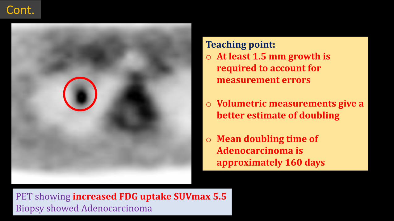
PET showing increased FDG uptake SUVmax 5.5Biopsy showed Adenocarcinoma
Teaching point:o At least 1.5 mm growth is
required to account for measurement errors
o Volumetric measurements give a better estimate of doubling
o Mean doubling time of Adenocarcinoma is approximately 160 days
Cont.
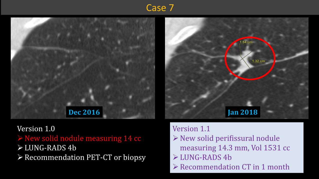
Dec 2016
Jan 2018Version 1.0¾New solid nodule measuring 14 cc¾LUNG-RADS 4b¾Recommendation PET-CT or biopsy
Version 1.1¾New solid perifissural nodule
measuring 14.3 mm, Vol 1531 cc¾LUNG-RADS 4b¾Recommendation CT in 1 month
Case 7
Jan 2018
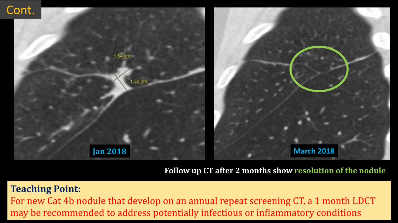
March 2018Jan 2018
Cont.
Teaching Point: For new Cat 4b nodule that develop on an annual repeat screening CT, a 1 month LDCT may be recommended to address potentially infectious or inflammatory conditions
Follow up CT after 2 months show resolution of the nodule
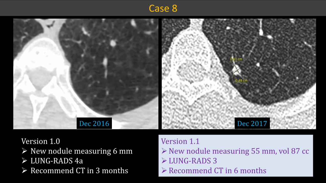
Dec 2016 Dec 2017
Version 1.1¾New nodule measuring 55 mm, vol 87 cc¾LUNG-RADS 3¾Recommend CT in 6 months
Version 1.0¾ New nodule measuring 6 mm¾ LUNG-RADS 4a¾ Recommend CT in 3 months
Case 8
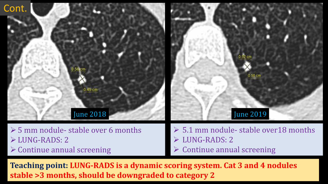
June 2018 June 2019
¾5 mm nodule- stable over 6 months¾LUNG-RADS: 2¾Continue annual screening
¾ 5.1 mm nodule- stable over18 months¾ LUNG-RADS: 2¾ Continue annual screening
Teaching point: LUNG-RADS is a dynamic scoring system. Cat 3 and 4 nodules stable >3 months, should be downgraded to category 2
Cont.
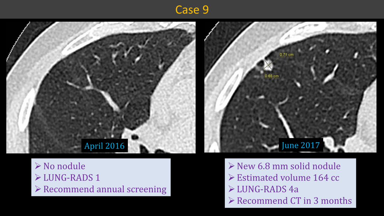
¾New 6.8 mm solid nodule¾Estimated volume 164 cc¾LUNG-RADS 4a¾Recommend CT in 3 months
April 2016 June 2017
Case 9
¾No nodule¾LUNG-RADS 1¾Recommend annual screening
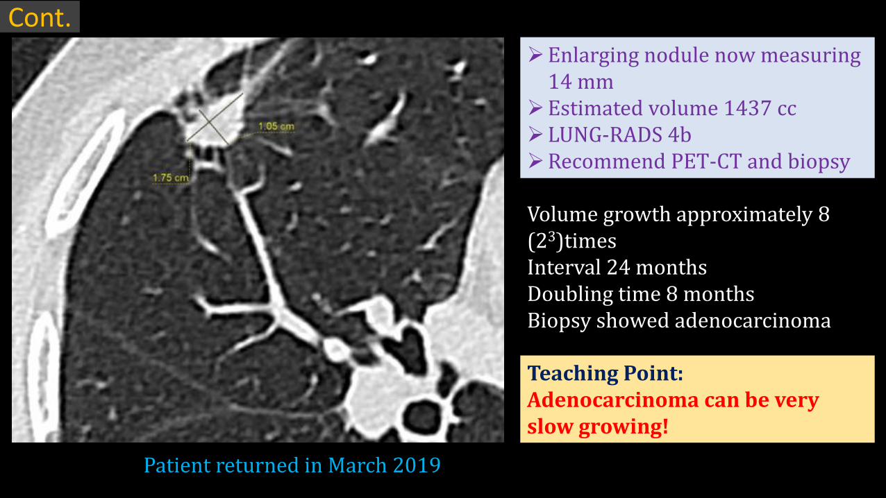
Patient returned in March 2019
¾Enlarging nodule now measuring 14 mm
¾Estimated volume 1437 cc¾LUNG-RADS 4b¾Recommend PET-CT and biopsy
Volume growth approximately 8 (23)timesInterval 24 monthsDoubling time 8 monthsBiopsy showed adenocarcinoma
Teaching Point:Adenocarcinoma can be very slow growing!
Cont.
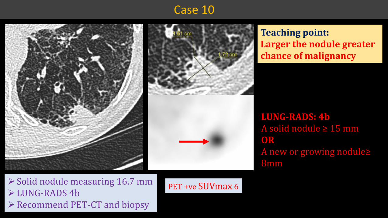
¾Solid nodule measuring 16.7 mm¾LUNG-RADS 4b¾Recommend PET-CT and biopsy
PET +ve SUVmax 6
LUNG-RADS: 4b���������������η�ͳͷ�����OR �����������������������η�8mm
Teaching point:Larger the nodule greater chance of malignancy
Case 10
Case 11
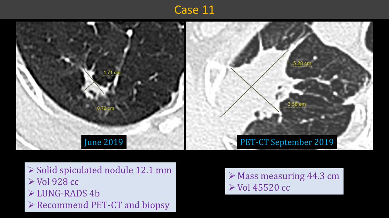
June 2019
¾Solid spiculated nodule 12.1 mm¾Vol 928 cc¾LUNG-RADS 4b¾Recommend PET-CT and biopsy
¾Mass measuring 44.3 cm¾Vol 45520 cc
PET-CT September 2019
Cont.
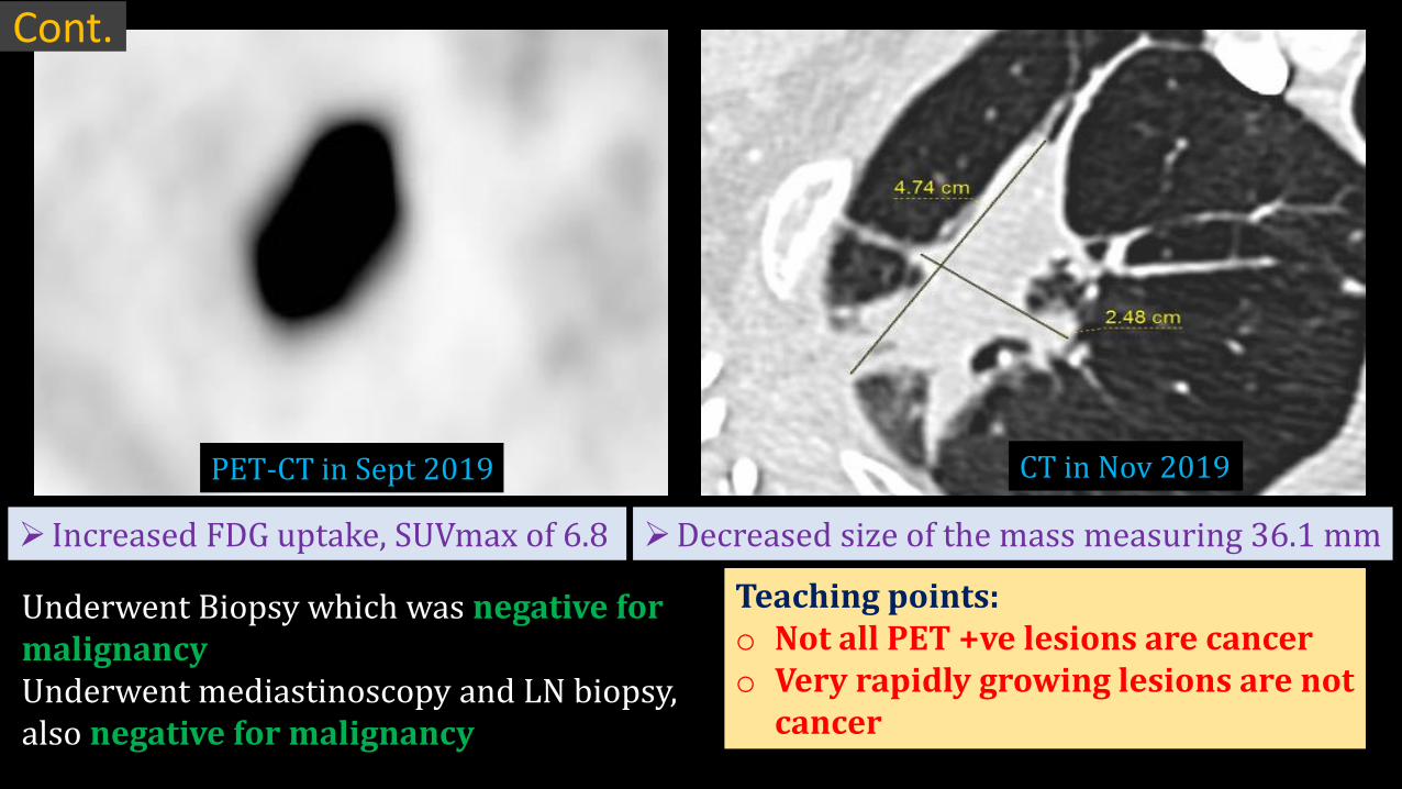
¾ Increased FDG uptake, SUVmax of 6.8
Underwent Biopsy which was negative for malignancyUnderwent mediastinoscopy and LN biopsy, also negative for malignancy
¾Decreased size of the mass measuring 36.1 mm
PET-CT in Sept 2019 CT in Nov 2019
Teaching points:o Not all PET +ve lesions are cancero Very rapidly growing lesions are not
cancer
Summary
o CT Lung cancer screening improves mortality
o Multi disciplinary team approach is essential
o LUNG-RADS to be followed for reporting and recommendation
o Radiologists should be aware of the changes in version 1.1