215 Про проведення І,ІІ етапів Всеукраїнських учнівських ...
RESEARCH Open Access
Low RBM3 protein expression correlates withtumour progression and poor prognosis inmalignant melanoma: An analysis of 215 casesfrom the Malmö Diet and Cancer StudyLiv Jonsson1†, Julia Bergman1†, Björn Nodin1, Jonas Manjer2,3, Fredrik Pontén4, Mathias Uhlén5,6 and Karin Jirström1*
Abstract
Background: We have previously reported that expression of the RNA- and DNA-binding protein RBM3 isassociated with a good prognosis in breast cancer and ovarian cancer. In this study, the prognostic value ofimmunohistochemical RBM3 expression was assessed in incident cases of malignant melanoma from a prospectivepopulation-based cohort study.
Methods: Until Dec 31st 2008, 264 incident cases of primary invasive melanoma had been registered in the MalmöDiet and Cancer Study. Histopathological and clinical information was obtained for available cases and tissuemicroarrays (TMAs) constructed from 226 (85.6%) suitable paraffin-embedded tumours and 31 metastases. RBM3expression was analysed by immunohistochemistry on the TMAs and a subset of full-face sections. Chi-square andMann-Whitney U tests were used for comparison of RBM3 expression and relevant clinicopathologicalcharacteristics. Kaplan Meier analysis and Cox proportional hazards modelling were used to assess the relationshipbetween RBM3 and recurrence free survival (RFS) and overall survival (OS).
Results: RBM3 could be assessed in 215/226 (95.1%) of primary tumours and all metastases. Longitudinal analysisrevealed that 16/31 (51.6%) of metastases lacked RBM3 expression, in contrast to the primary tumours in which RBM3 wasabsent in 3/215 (1.4%) cases and strongly expressed in 120/215 (55.8%) cases. Strong nuclear RBM3 expression in theprimary tumour was significantly associated with favourable clinicopathological parameters; i.e. non-ulcerated tumours,lower depth of invasion, lower Clark level, less advanced clinical stage, low mitotic activity and non-nodular histologicaltype, and a prolonged RFS (RR = 0.50; 95% CI = 0.27-0.91) and OS (RR = 0.36, 95%CI = 0.20-0.64). Multivariate analysisdemonstrated that the beneficial prognostic value of RBM3 remained significant for OS (RR = 0.33; 95%CI = 0.18-0.61).
Conclusions: In line with previous in vitro data, we here show that RBM3 is down-regulated in metastatic melanomaand high nuclear RBM3 expression in the primary tumour is an independent marker of a prolonged OS. The potentialutility of RBM3 in treatment stratification of patients with melanoma should be pursued in future studies.
BackgroundMalignant melanoma is an aggressive form of cancerwith a variable clinical course even in patients with thinmelanomas and localized disease [1-4]. Despite increas-ing insights into melanoma biology and the discovery ofgene- and protein-signatures that supplement
established prognostic clinicopathological parameters[5-7], no biomarkers have yet been incorporated intoclinical protocols.The RNA-binding motif protein 3, RBM3, was initially
identified in a human fetal brain tissue cDNA library[8]. The RBM3 gene maps to Xp11.23 and encodes twoalternatively spliced RNA transcripts. RBM3 transcriptshave been found in various human tissues [8] and invitro, RBM3 is one of the earliest proteins synthesizedin response to cold shock [9]. RBM3 contains one RNA-
* Correspondence: [email protected]† Contributed equally1Department of Clinical Sciences, Pathology, Lund University, SkåneUniversity Hospital, 221 85 Lund, SwedenFull list of author information is available at the end of the article
Jonsson et al. Journal of Translational Medicine 2011, 9:114http://www.translational-medicine.com/content/9/1/114
© 2011 Jonsson et al; licensee BioMed Central Ltd. This is an Open Access article distributed under the terms of the Creative CommonsAttribution License (http://creativecommons.org/licenses/by/2.0), which permits unrestricted use, distribution, and reproduction inany medium, provided the original work is properly cited.

recognition motif (RRM) and is able to bind to bothDNA and RNA, whereby a glycine rich region adjacentto the RNA binding motif is thought to enhance theprotein-RNA or protein-DNA interaction [8,10].Based on an initial discovery in the Human Protein
Atlas (HPA) http://www.proteinatlas.org[11-13], we haverecently demonstrated that tumour-specific expressionof RBM3, in particular its nuclear localization, is asso-ciated with a significantly improved survival in breastcancer [14] and ovarian cancer [15], and that RBM3confers cisplatin sensitivity in ovarian cancer cells [15].Apart from these studies, we are not aware of any otherpublications related to the prognostic or treatment pre-dictive impact of the tumour-specific expression ofRBM3 in human cancer, and the biological processesunderlying these observations have not yet been unra-veled. It is evident that RBM3 is up-regulated in varioustypes of human malignancies [14,16,17] and in vitro stu-dies in a wide range of different model systems havedemonstrated that RBM3 is involved in multiple pro-cesses central to cancer biology, like proliferation[15-17], apoptosis [18,19] and angiogenesis [16].The prognostic value of RBM3 expression has, to our
knowledge, not yet been investigated in malignant mela-noma. However, down-regulation of RBM3 at the geneexpression level has been demonstrated in an in vitromodel of melanoma progression [20].In the present study, we investigated the prognostic
impact of immunohistochemical (IHC) RBM3 expres-sion in 215 incident malignant melanomas in the pro-spective, population-based cohort Malmö Diet andCancer Study (MDCS) [21]. For this purpose, tissuemicroarrays (TMAs) were constructed from suitabletumours (n = 226) and a subset of metastases (n = 31).It is demonstrated that strong nuclear expression ofRBM3 correlates with favourable clinicopathologicalparameters and independently predicts a significantlyprolonged overall survival. In addition, a markedlyreduced expression of RBM3 was observed in metastasescompared to primary tumours, which is quite in linewith previous in vitro data [20].
MethodsThe Malmö Diet and Cancer StudyThe Malmö Diet and Cancer Study (MDCS) is a popula-tion-based prospective cohort study with the main aimto examine whether a Western diet rich in fat and lowin fruit and vegetables increases the risk of certainforms of cancer. Between 1991-1996, a total number of28 098 individuals; 11 063 (39,4%) men and 17 035(60,6%) women between 44-74 years where enrolled(from a background population of 74 138). All partici-pants completed the baseline examination, whichincluded a questionnaire, measures of anthropometric/
body compositions and a dietary assessment. The ques-tionnaire covered questions on physical activity, use oftobacco and alcohol, heredity, socio-economic factors,education, occupation, previous and current disease andcurrent medication. In addition, blood samples were col-lected and stored in -80°C. Follow up is done annuallyby record-linkage to national registries for cancer andcause of death [22].Ethical permissions for the MDCS (Ref. 51/90) and the
present study (Ref. 530/2008) were obtained from theEthical Committee at Lund University.
Incident malignant melanomas until Dec 31st 2008Until end of follow-up 31 December 2008, 264 incidentinvasive malignant melanomas had been registered inthe study population. Cases were identified from theSwedish Cancer Registry up until 31 Dec 2007, andfrom The Southern Swedish Regional Tumour Registryfor the period of 1 Jan-31 Dec 2008. All tumours withavailable slides and/or paraffin blocks were histopatholo-gically re-evaluated on haematoxylin and eosin stainedslides whereby information on lymphocytic infiltration(none, mild, moderate or high), ulceration (absent orpresent), mitotic count and vascular invasion wasobtained. Data on location, Clark level and Breslowdepth of invasion was obtained from the clinical- and/orpathology records.Information on recurrence (local, regional or distant)
was obtained in 2010 from patient records and pathol-ogy reports. Information on vital status and cause ofdeath was obtained from the Swedish Cause of DeathRegistry up until 31 Dec 2009.
Tissue microarray constructionParaffin-embedded tumour specimens were collectedfrom the archives of the pathology departments in theregion of Skåne in Southern Sweden. Tumours with aninsufficient amount of material were excluded. Areasrepresentative of cancer were then marked on haema-toxylin & eosin stained slides and TMAs constructed aspreviously described [23]. In brief, three 0,6 mm coreswere taken from each tumour and mounted in a newrecipient block using semi-automated arraying device(TMArrayer, Pathology Devices, Westminster, MD,USA). In addition, metastases (representing both regio-nal and distant metastases in various organs) weresampled from 31 cases. Thin melanomas (< 1 mm) weresubjected to TMA construction if the diameter was > 1cm. To check for heterogeneity, IHC staining was alsoperformed on additional full-face sections from 25 cases.
Immunohistochemistry and evaluation of RBM3 stainingFor immunohistochemical analysis, 4 μm TMA-sectionswere automatically pre-treated using the PT-link system
Jonsson et al. Journal of Translational Medicine 2011, 9:114http://www.translational-medicine.com/content/9/1/114
Page 2 of 9
(DAKO, Glostrup, Denmark) and then stained in aAutostainer Plus (DAKO, Glostrup, Denmark) with themouse monoclonal anti-RBM3 antibody (AAb030038;Atlas Antibodies AB, Stockholm, Sweden, diluted1:5000). The specificity of the antibody has been vali-dated previously [15].As RBM3, when present, was expressed in > 75% of
the cells, predominantly in the nuclei and in varyingintensities, only the intensity of the staining wasaccounted for and denoted a score from 0 (negative), 1(mild), 2 (moderate) and 3 (strong). The staining wasevaluated by three independent observers (LJ, JB, andBN) who were blinded to clinical and outcome data.Scoring differences were discussed in order to reachconsensus.
Statistical analysisChi-square and Mann-Whitney U tests were used forcomparison of RBM3 expression and relevant clinico-pathological characteristics. Recurrence was defined aslocal, regional or distant recurrence or death frommalignant melanoma and risk of recurrent disease wasreferred to as recurrence free survival (RFS). Follow-upstarted at date of diagnosis and ended at recurrent dis-ease, death, lost to follow-up (emigration) or last date offollow- up with regard to recurrent disease. No recur-rences were recorded following the last date of follow-up regarding death, i.e. 31 Dec 2009. Overall survival(OS) was assessed by calculating the risk of death fromall causes, overall mortality. Follow-up started at date ofdiagnosis and ended at death, emigration or 31 Dec2009, whichever came first.Kaplan-Meier analysis and log rank test were used to
illustrate differences in RFS and OS. Cox regression pro-portional hazards models were used to estimate theimpact of the investigated parameters on RFS and OS inboth uni- and multivariate analysis. Some subjects hadno information on one or several markers and missingvalues were coded as a separate category for categoricalvariables and as the mean of all observations for contin-uous variables. Missing values for categorical variablesco-varied and the multivariate model did not convergedue to many constant values. In order to avoid this, themultivariate analysis only included patients with infor-mation on RBM3. In addition, the patient with missinginformation on lymphocytic infiltration had to beexcluded. Co-variates were entered into the multivariateanalysis using backward selection were a p-value of 0.05decided entry and a p-value of 0.20 was used forremoval. RBM3 was included in all models irrespectiveof the backward selection procedure.All tests were two sided. A p-value of 0.05 was consid-
ered significant. All statistical analyses were performedusing SPSS version 17 (SPSS Inc, Chicago, IL).
REMARK criteriaA description of the fulfilment of REMARK [24] criteriafor biomarker studies is provided in Additional file 1,Table S1.
ResultsDistribution of clinicopathological parameters in thecohortThe distribution of patient- and tumour characteristicsin the full cohort is shown in Table 1. In line with therelatively high median age of cases (69 years, range 44-85), the frequency of lentigo maligna melanomas wasslightly higher (11.7%) than the average expected around7% in Sweden [2]. The proportion of thinner melanomas(< = 1 mm, Stages 1A-B) was also higher than expected(86.5% compared to ~55.1%) as well as the proportionof non-ulcerated tumours (14.1% compared to ~24.1%)[2].
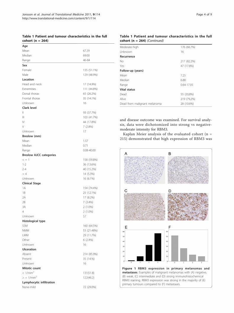
Immunohistochemical expression of RBM3 in primarytumours and metastasesOf the 226 cases in the TMA cohort it was possible toevaluate the expression of RBM3 protein in 215 cases(95,1%). There was no obvious heterogeneity in thestaining pattern between the tissue cores. There was anexcellent concordance between RBM3 scores assessedon full-face sections and TMAs (kappa-value 0.85).Examples of immunohistochemical staining are shownin Figure 1A-D and the staining distribution in primarytumours vs metastases in Figure 1E-F. Interestingly, andin line with previous in vitro data [20], RBM3 expres-sion was strong in the majority of primary tumours, butweak or absent in the metastases (Figure 1E-F). Notably,similar associations were seen when comparing primarytumours and metastases in the 31 cases, for which bothlocations had been sampled (data not shown).
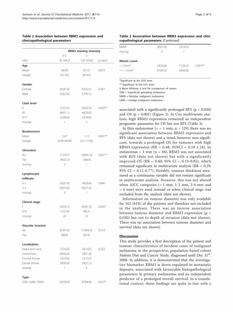
Association between RBM3 expression andclinicopathological parametersAs shown in Table 2, there was a strong associationbetween low RBM3 expression and depth of invasion,Clark level, clinical stage, mitotic count, nodular vs non-nodular type and ulceration. However, localization, age,lymphocytic infiltration and melanoma type were notassociated with RBM3 expression. In some cases withstrong RBM3 expression, cytoplasmic staining was pre-sent in various intensities, but this did not add anyprognostic value (data not shown).
Impact of high RBM3 expression on recurrence freesurvival and overall survivalHaving demonstrated that RBM3 is associated with lessadvanced disease and favourable clinicopathologicalparameters, the relationship between RBM3 expression
Jonsson et al. Journal of Translational Medicine 2011, 9:114http://www.translational-medicine.com/content/9/1/114
Page 3 of 9
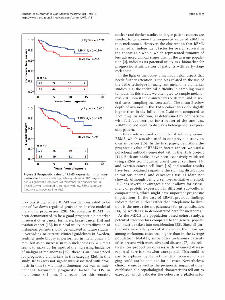
and disease outcome was examined. For survival analy-sis, data were dichotomized into strong vs negative-moderate intensity for RBM3.Kaplan Meier analysis of the evaluated cohort (n =
215) demonstrated that high expression of RBM3 was
Table 1 Patient and tumour characteristics in the fullcohort (n = 264)
Age
Mean 67.29
Median 69.00
Range 46-84
Sex
Female 135 (51.1%)
Male 129 (48.9%)
Location
Head and neck 17 (14.9%)
Extremities 111 (44.8%)
Dorsal thorax 65 (26.2%)
Frontal thorax 35 (14.1%)
Unknown 16
Clark level
II 93 (37.7%)
III 103 (41.7%)
IV 44 (17.8%)
V 7 (2.8%)
Unknown 17
Breslow (mm)
Mean 1.57
Median 0.71
Range 0.08-40.00
Breslow AJCC categories
< = 1 158 (59.8%)
1-2 36 (13.6%)
2-4 40 (15.2%)
> 4 14 (5.3%)
Unknown 16 (6.1%)
Clinical Stage
1A 154 (74.4%)
1B 25 (12.1%)
2A 17 (8.2%)
2B 7 (3.4%)
3A 2 (1.0%)
4 2 (1.0%)
Unknown 57
Histological type
SSM 160 (64.5%)
NMM 53 (21.48%)
LMM 29 (11.7%)
Other 6 (2.4%)
Unknown 16
Ulceration
Absent 214 (85.9%)
Present 35 (14.%)
Unknown 16
Mitotic count
< 1/mm2 131(51.8)
> = 1/mm2 122(46.2)
Lymphocytic infiltration
None-mild 72 (29.0%)
Figure 1 RBM3 expression in primary melanomas andmetastases. Examples of malignant melanomas with (A) negative,(B) weak, (C) intermediate and (D) strong immunohistochemicalRBM3 staining. RBM3 expression was strong in the majority of (E)primary tumours compared to (F) metastases.
Table 1 Patient and tumour characteristics in the fullcohort (n = 264) (Continued)
Moderate-high 176 (66.7%)
Unknown 16
Recurrence
No 217 (82.2%)
Yes 47 (17.8%)
Follow-up (years)
Mean 7.25
Median 6.88
Range 0.64-17.05
Vital status
Dead 55 (20,8%)
Alive 219 (79,2%)
Dead from malignant melanoma 28 (10.6%)
Jonsson et al. Journal of Translational Medicine 2011, 9:114http://www.translational-medicine.com/content/9/1/114
Page 4 of 9
associated with a significantly prolonged RFS (p = 0.020)and OS (p < 0.001) (Figure 2). In Cox multivariate ana-lysis, high RBM3 expression remained an independentprognostic parameter for OS but not RFS (Table 3).In thin melanomas (< = 1 mm; n = 129) there was no
significant association between RBM3 expression andRFS (data not shown) and a trend, however non-signifi-cant, towards a prolonged OS for tumours with highRBM3 expression (RR = 0.48; 95%CI = 0.18-1.24). Inmelanomas > 1 mm (n = 84), RBM3 was not associatedwith RFS (data not shown) but with a significantlyimproved OS (RR = 0.40; 95% CI = 0.19-0.85), whichremained significant in multivariate analysis (RR = 0.29;95% CI = 0.11-0.77). Notably, tumour thickness mea-sured as a continuous variable did not remain significantin multivariate analysis. However, this was not alteredwhen AJCC categories (< 1 mm, 1-2 mm, 2-4 mm and> 4 mm) were used instead or when clinical stage wasexcluded from the analysis (data not shown).Information on tumour diameter was only available
for 162 (61%) of the patients and therefore not includedin the analyses. There was an inverse associationbetween tumour diameter and RBM3 expression (p =0.030) but not to depth of invasion (data not shown).There was no association between tumour diameter andsurvival (data not shown)
DiscussionThis study provides a first description of the patient andtumour characteristics of incident cases of malignantmelanoma in the prospective, population-based cohortMalmö Diet and Cancer Study, diagnosed until Dec 31st,2008. In addition, it is demonstrated that the investiga-tive biomarker RBM3 is down-regulated in metastaticdeposits, associated with favourable histopathologicalparameters in primary melanomas and an independentpredictor of a prolonged overall survival. In a transla-tional context, these findings are quite in line with a
Table 2 Association between RBM3 expression andclinicopathological parameters
RBM3 staining intensity
0-2 3
n(%) 95 (44.2) 120 (55.8) p-value
Age
Mean 68.00 67.15 0.673
(range) (47-83) (46-81)
Gender
Female 45(47.4) 63(52.5) 0.457
Male 50(52.6) 57(47.5)
Clark level
II 21(22.3) 50(42.4) 0.005**
IIII 48(51.1) 46(39.0)
IV-V 25(26.6) 22(18.6)
missing 1 2
Breslow(mm)
Mean 2.47 1.12 0.001**
(range) (0.08-40.00) (0.11-7.00)
Ulceration
No 71(74.7) 109(91.6) 0.001**
Yes 24(25.3) 10(8.4)
missing 0 1
Lymphocytic
infiltrate
0-1 26(27.4) 34(28.6) 0.846
2-3 69(72.6) 85(71.4)
missing 0 1
Clinical stage
I 54(76.1) 95(91.3) 0.005*
II-IV 17(23.9) 9(8.7)
missing 24 16
Vascular invasion
No 87(91.6) 114(95.0) 0.314
Yes 8(8.4) 6(5.0)
Localization
Head and neck 15(16.0) 16(14.0) 0.352
Extremities 40(42.6) 59(51.8)
Frontal thorax 10(10.6) 15(13.2)
Dorsal thorax 29(30.9) 24(21.1)
missing 1 6
Type
SSM, LMM, Other 65(58.9) 97(68.9) 0.027*
Table 2 Association between RBM3 expression and clini-copathological parameters (Continued)
NMM 30(31.6) 22(18.5)
missing 0 1
Mitotic count
< 1/mm2 34(35.8) 71(59.2) 0.001**
> = 1/mm2 61(64.2) 49(40.8)
*Significant at the 0.05 level
** Significant at the 0.01 level
§ Mann Whitney U test for comparison of means
SSM = Superficial spreading melanoma
NMM = Nodular malignant melanoma
LMM = Lentigo malignant melanoma
Jonsson et al. Journal of Translational Medicine 2011, 9:114http://www.translational-medicine.com/content/9/1/114
Page 5 of 9
previous study, where RBM3 was demonstrated to beone of five down-regulated genes in an in vitro model ofmelanoma progression [20]. Moreover, as RBM3 hasbeen demonstrated to be a good prognostic biomarkerin several other cancer forms, e.g. breast cancer [14] andovarian cancer [15], its clinical utility in stratification ofmelanoma patients should be validated in future studies.According to current clinical guidelines in Sweden,
sentinel node biopsy is performed in melanomas > 1mm, but as an increase in thin melanomas (< = 1 mm)seems to make up for most of the increasing incidenceof malignant melanomas [25], there is an unmet needfor prognostic biomarkers in this category [26]. In thisstudy, RBM3 was not significantly associated with prog-nosis in thin (< = 1 mm) melanomas but was an inde-pendent favourable prognostic factor for OS inmelanomas > 1 mm. The reason for this remains
unclear and further studies in larger patient cohorts areneeded to determine the prognostic value of RBM3 inthin melanomas. However, the observation that RBM3remained an independent factor for overall survival inthe cohort as a whole, which represented tumours ofless advanced clinical stages than in the average popula-tion [2], indicates its potential utility as a biomarker forprognostic stratification of patients with early-stagemelanoma.In the light of the above, a methodological aspect that
needs further attention is the bias related to the use ofthe TMA technique in malignant melanoma biomarkerstudies, e.g. the technical difficulty in sampling smalltumours. In this study, we attempted to sample melano-mas < 0.5 mm if the diameter was > 10 mm, and in sev-eral cases, sampling was successful. The mean Breslowdepth of invasion in the TMA cohort was only slightlyhigher than in the full cohort (1.66 mm compared to1.57 mm). In addition, as determined by comparisonwith full-face sections for a subset of the tumours,RBM3 did not seem to display a heterogeneous expres-sion pattern.In this study we used a monoclonal antibody against
RBM3, which was also used in our previous study onovarian cancer [15]. In the first paper, describing theprognostic value of RBM3 in breast cancer, we used apolyclonal antibody generated within the HPA project[14]. Both antibodies have been extensively validatedusing siRNA techniques in breast cancer cell lines [14]and ovarian cancer cell lines [15] and similar resultshave been obtained regarding the staining distributionin various normal and cancerous tissues (data notshown). Although being a semi-quantitative method,IHC has several advantages since it allows for assess-ment of protein expression in different sub-cellularcompartments, which might have important prognosticimplications. In the case of RBM3, previous findingsindicate that its nuclear rather than cytoplasmic localiza-tion is the most relevant parameter for prognostication[14,15], which is also demonstrated here for melanoma.As the MDCS is a population-based cohort study, a
potential selection bias compared to the general popula-tion must be taken into consideration [22]. Since all par-ticipants were > 40 years at study entry, the mean ageamong melanoma cases was higher than in the averagepopulation. Notably, since older melanoma patientsoften present with more advanced disease [27], the rela-tively low proportion of cases with advanced diseasereported here is somewhat unexpected. This could inpart be explained by the fact that data necessary for sta-ging could not be obtained for all cases. Nevertheless,clinical stage, as well as the prognostic impact of otherestablished clinicopathological characteristics fell out asexpected, which validates the cohort as a platform for
Figure 2 Prognostic value of RBM3 expression in primarymelanoma. Tumours with high (strong intensity) RBM3 expressionhad a significantly improved (A) recurrence free survival and (B)overall survival compared to tumours with low RBM3 expression(negative to moderate intensity).
Jonsson et al. Journal of Translational Medicine 2011, 9:114http://www.translational-medicine.com/content/9/1/114
Page 6 of 9
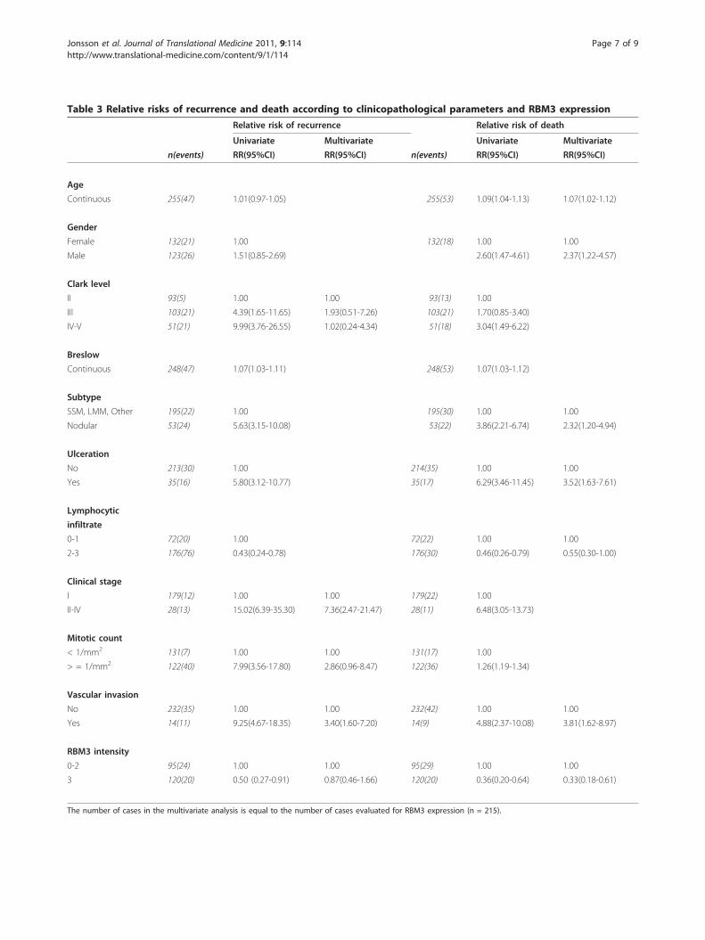
Table 3 Relative risks of recurrence and death according to clinicopathological parameters and RBM3 expression
Relative risk of recurrence Relative risk of death
Univariate Multivariate Univariate Multivariate
n(events) RR(95%CI) RR(95%CI) n(events) RR(95%CI) RR(95%CI)
Age
Continuous 255(47) 1.01(0.97-1.05) 255(53) 1.09(1.04-1.13) 1.07(1.02-1.12)
Gender
Female 132(21) 1.00 132(18) 1.00 1.00
Male 123(26) 1.51(0.85-2.69) 2.60(1.47-4.61) 2.37(1.22-4.57)
Clark level
II 93(5) 1.00 1.00 93(13) 1.00
III 103(21) 4.39(1.65-11.65) 1.93(0.51-7.26) 103(21) 1.70(0.85-3.40)
IV-V 51(21) 9.99(3.76-26.55) 1.02(0.24-4.34) 51(18) 3.04(1.49-6.22)
Breslow
Continuous 248(47) 1.07(1.03-1.11) 248(53) 1.07(1.03-1.12)
Subtype
SSM, LMM, Other 195(22) 1.00 195(30) 1.00 1.00
Nodular 53(24) 5.63(3.15-10.08) 53(22) 3.86(2.21-6.74) 2.32(1.20-4.94)
Ulceration
No 213(30) 1.00 214(35) 1.00 1.00
Yes 35(16) 5.80(3.12-10.77) 35(17) 6.29(3.46-11.45) 3.52(1.63-7.61)
Lymphocytic
infiltrate
0-1 72(20) 1.00 72(22) 1.00 1.00
2-3 176(76) 0.43(0.24-0.78) 176(30) 0.46(0.26-0.79) 0.55(0.30-1.00)
Clinical stage
I 179(12) 1.00 1.00 179(22) 1.00
II-IV 28(13) 15.02(6.39-35.30) 7.36(2.47-21.47) 28(11) 6.48(3.05-13.73)
Mitotic count
< 1/mm2 131(7) 1.00 1.00 131(17) 1.00
> = 1/mm2 122(40) 7.99(3.56-17.80) 2.86(0.96-8.47) 122(36) 1.26(1.19-1.34)
Vascular invasion
No 232(35) 1.00 1.00 232(42) 1.00 1.00
Yes 14(11) 9.25(4.67-18.35) 3.40(1.60-7.20) 14(9) 4.88(2.37-10.08) 3.81(1.62-8.97)
RBM3 intensity
0-2 95(24) 1.00 1.00 95(29) 1.00 1.00
3 120(20) 0.50 (0.27-0.91) 0.87(0.46-1.66) 120(20) 0.36(0.20-0.64) 0.33(0.18-0.61)
The number of cases in the multivariate analysis is equal to the number of cases evaluated for RBM3 expression (n = 215).
Jonsson et al. Journal of Translational Medicine 2011, 9:114http://www.translational-medicine.com/content/9/1/114
Page 7 of 9
future studies of lifestyle and tumour biology in relationto melanoma risk and prognosis.Given the previously demonstrated association
between RBM3 and cisplatin sensitivity in ovarian can-cer cell lines [15], the potential value of RBM3 as a pre-dictor of response to platinum-based chemotherapy inpatients with metastatic malignant melanoma could beof interest to investigate in future studies. However, incontrast to the situation in ovarian cancer, where RBM3showed a consistent expression pattern in primarytumours and omental deposits [15], the data presentedhere, and previous in vitro data [20], show that RBM3 isdown-regulated in the majority of metastatic melano-mas. Hence, in the predictive setting in melanomapatients, thorough sampling and immunohistochemicalanalysis of metastatic deposits would be required inorder to identify a comparatively small number ofpatients with RBM3 positive metastases.
ConclusionsWe have demonstrated that the RNA- and DNA-bind-ing protein RBM3 is an independent biomarker of aprolonged OS in patients with primary malignant mela-noma and that RBM3 expression is lost during progres-sion of the disease. The potential utility of RBM3 in riskstratification of patients with melanoma should be pur-sued in future studies.
Additional material
Additional file 1: Supplementary Table 1. Fulfilment of REMARKcriteria [24].
AcknowledgementsThis study was supported by grants from the Knut and Alice WallenbergFoundation, the Swedish Cancer Society, Gunnar Nilsson’s CancerFoundation, the Crafoord Foundation, and the Research Funds of SkåneUniversity Hospital.We thank Elise Nilsson for excellent technical assistance.
Author details1Department of Clinical Sciences, Pathology, Lund University, SkåneUniversity Hospital, 221 85 Lund, Sweden. 2Department Clinical Sciences,Surgery, Lund University, Skåne University Hospital, 205 02 Malmö, Sweden.3The Malmö Diet and Cancer Study, Lund University, 205 02 Malmö,Sweden. 4Department of Genetics and Pathology, Rudbeck Laboratory,Uppsala University, 251 87 Uppsala, Sweden. 5Department of Proteomics,AlbaNova University Center, Royal Institute of Technology, 106 91 Stockholm,Sweden. 6Science for Life Laboratory, Royal Institute of Technology, 106 91Stockholm, Sweden.
Authors’ contributionsLJ and JB participated in the data collection, performed the statisticalanalysis and drafted the manuscript. BN assisted with the data collection,constructed the tissue microarrays and helped draft the manuscript. JM, FPand MU participated in the design of the study and helped draft themanuscript. KJ conceived of the study, participated in its design andcoordination and helped to draft the manuscript. All authors read andapproved the final manuscript.
Competing interestsThe authors declare that they have no competing interests.
Received: 31 March 2011 Accepted: 21 July 2011Published: 21 July 2011
References1. Gimotty PA, Elder DE, Fraker DL, Botbyl J, Sellers K, Elenitsas R, Ming ME,
Schuchter L, Spitz FR, Czerniecki BJ, Guerry D: Identification of high-riskpatients among those diagnosed with thin cutaneous melanomas. J ClinOncol 2007, 25:1129-1134.
2. Lindholm C, Andersson R, Dufmats M, Hansson J, Ingvar C, Moller T,Sjodin H, Stierner U, Wagenius G: Invasive cutaneous malignantmelanoma in Sweden, 1990-1999. A prospective, population-basedstudy of survival and prognostic factors. Cancer 2004, 101:2067-2078.
3. McKinnon JG, Yu XQ, McCarthy WH, Thompson JF: Prognosis for patientswith thin cutaneous melanoma: long-term survival data from New SouthWales Central Cancer Registry and the Sydney Melanoma Unit. Cancer2003, 98:1223-1231.
4. Sondak VK, Messina JL: Current status of biomarkers for melanomametastasis. IDrugs 2006, 9:627-631.
5. Bittner M, Meltzer P, Chen Y, Jiang Y, Seftor E, Hendrix M, Radmacher M,Simon R, Yakhini Z, Ben-Dor A, Sampas N, Dougherty E, Wang E,Marincola F, Gooden C, Lueders J, Glatfelter A, Pollock P, Carpten J,Gillanders E, Leja D, Dietrich K, Beaudry C, Berens M, Alberts D, Sondak V:Molecular classification of cutaneous malignant melanoma by geneexpression profiling. Nature 2000, 406:536-540.
6. Winnepenninckx V, Lazar V, Michiels S, Dessen P, Stas M, Alonso SR,Avril MF, Ortiz Romero PL, Robert T, Balacescu O, Eggermont AM, Lenoir G,Sarasin A, Tursz T, van den Oord JJ, Spatz A: Gene expression profiling ofprimary cutaneous melanoma and clinical outcome. J Natl Cancer Inst2006, 98:472-482.
7. Jonsson G, Busch C, Knappskog S, Geisler J, Miletic H, Ringner M,Lillehaug JR, Borg A, Lonning PE: Gene expression profiling-basedidentification of molecular subtypes in stage IV melanomas withdifferent clinical outcome. Clin Cancer Res 16:3356-3367.
8. Derry JM, Kerns JA, Francke U: RBM3, a novel human gene in Xp11.23with a putative RNA-binding domain. Hum Mol Genet 1995,4:2307-2311.
9. Danno S, Nishiyama H, Higashitsuji H, Yokoi H, Xue JH, Itoh K, Matsuda T,Fujita J: Increased transcript level of RBM3, a member of the glycine-richRNA-binding protein family, in human cells in response to cold stress.Biochem Biophys Res Commun 1997, 236:804-807.
10. Lleonart ME: A new generation of proto-oncogenes: cold-inducible RNAbinding proteins. Biochim Biophys Acta 1805:43-52.
11. Uhlen M, Bjorling E, Agaton C, Szigyarto CA, Amini B, Andersen E,Andersson AC, Angelidou P, Asplund A, Asplund C, Berglund L,Bergstrom K, Brumer H, Cerjan D, Ekstrom M, Elobeid A, Eriksson C,Fagerberg L, Falk R, Fall J, Forsberg M, Bjorklund MG, Gumbel K, Halimi A,Hallin I, Hamsten C, Hansson M, Hedhammar M, Herkules G, Kampf C,Larsson K, Lindskog M, Lodewyckx W, Lund J, Lundeberg J, Magnusson K,Malm E, Nilsson P, Odling J, Oksvold P, Olsson I, Oster E, Ottosson J,Paavilainen L, Persson A, Rimini R, Rockberg J, Runeson M, Sivertsson A,Sköllermo A, Steen J, Stenvall M, Sterky F, Strömberg S, Sundberg M,Tegel H, Tourle S, Wahlund E, Waldén A, Wan J, Wernérus H, Westberg J,Wester K, Wrethagen U, Xu LL, Hober S, Pontén F: A human protein atlasfor normal and cancer tissues based on antibody proteomics. Mol CellProteomics 2005, 4:1920-1932.
12. Bjorling E, Lindskog C, Oksvold P, Linne J, Kampf C, Hober S, Uhlen M,Ponten F: A web-based tool for in silico biomarker discovery based ontissue-specific protein profiles in normal and cancer tissues. Mol CellProteomics 2008, 7:825-844.
13. Ponten F, Jirstrom K, Uhlen M: The Human Protein Atlas–a tool forpathology. J Pathol 2008, 216:387-393.
14. Jogi A, Brennan DJ, Ryden L, Magnusson K, Ferno M, Stal O, Borgquist S,Uhlen M, Landberg G, Pahlman S, Jirstrom K: Nuclear expression of theRNA-binding protein RBM3 is associated with an improved clinicaloutcome in breast cancer. Mod Pathol 2009, 22:1564-1574.
15. Ehlen A, Brennan DJ, Nodin B, O’Connor DP, Eberhard J, Alvarado-Kristensson M, Jeffrey IB, Manjer J, Brandstedt J, Uhlen M, Ponten F,Jirstrom K: Expression of the RNA-binding protein RBM3 is associated
Jonsson et al. Journal of Translational Medicine 2011, 9:114http://www.translational-medicine.com/content/9/1/114
Page 8 of 9

with a favourable prognosis and cisplatin sensitivity in epithelial ovariancancer. J Transl Med 8:78.
16. Sureban SM, Ramalingam S, Natarajan G, May R, Subramaniam D,Bishnupuri KS, Morrison AR, Dieckgraefe BK, Brackett DJ, Postier RG,Houchen CW, Anant S: Translation regulatory factor RBM3 is a proto-oncogene that prevents mitotic catastrophe. Oncogene 2008,27:4544-4556.
17. Wellmann S, Truss M, Bruder E, Tornillo L, Zelmer A, Seeger K, Buhrer C: TheRNA-binding protein RBM3 is required for cell proliferation and protectsagainst serum deprivation-induced cell death. Pediatr Res 67:35-41.
18. Martinez-Arribas F, Agudo D, Pollan M, Gomez-Esquer F, Diaz-Gil G, Lucas R,Schneider J: Positive correlation between the expression of X-chromosome RBM genes (RBMX, RBM3, RBM10) and the proapoptoticBax gene in human breast cancer. J Cell Biochem 2006, 97:1275-1282.
19. Sutherland LC, Rintala-Maki ND, White RD, Morin CD: RNA binding motif(RBM) proteins: a novel family of apoptosis modulators? J Cell Biochem2005, 94:5-24.
20. Baldi A, Battista T, De Luca A, Santini D, Rossiello L, Baldi F, Natali PG,Lombardi D, Picardo M, Felsani A, Paggi MG: Identification of genes down-regulated during melanoma progression: a cDNA array study. ExpDermatol 2003, 12:213-218.
21. Berglund G, Elmstahl S, Janzon L, Larsson SA: The Malmo Diet and CancerStudy. Design and feasibility. J Intern Med 1993, 233:45-51.
22. Manjer J, Carlsson S, Elmstahl S, Gullberg B, Janzon L, Lindstrom M,Mattisson I, Berglund G: The Malmo Diet and Cancer Study:representativity, cancer incidence and mortality in participants and non-participants. Eur J Cancer Prev 2001, 10:489-499.
23. Kononen J, Bubendorf L, Kallioniemi A, Barlund M, Schraml P, Leighton S,Torhorst J, Mihatsch MJ, Sauter G, Kallioniemi OP: Tissue microarrays forhigh-throughput molecular profiling of tumour specimens. Nat Med1998, 4:844-847.
24. McShane LM, Altman DG, Sauerbrei W, Taube SE, Gion M, Clark GM:Reporting recommendations for tumour marker prognostic studies. JClin Oncol 2005, 23:9067-9072.
25. Jemal A, Siegel R, Ward E, Hao Y, Xu J, Thun MJ: Cancer statistics, 2009. CACancer J Clin 2009, 59:225-249.
26. Gimotty PA, Guerry D: Prognostication in thin cutaneous melanomas.Arch Pathol Lab Med 134:1758-1763.
27. Lasithiotakis KG, Leiter U, Eigentler T, Breuninger H, Metzler G, Meier F,Garbe C: Improvement of overall survival of patients with cutaneousmelanoma in Germany, 1976-2001: which factors contributed? Cancer2007, 109:1174-1182.
doi:10.1186/1479-5876-9-114Cite this article as: Jonsson et al.: Low RBM3 protein expressioncorrelates with tumour progression and poor prognosis in malignantmelanoma: An analysis of 215 cases from the Malmö Diet and CancerStudy. Journal of Translational Medicine 2011 9:114.
Submit your next manuscript to BioMed Centraland take full advantage of:
• Convenient online submission
• Thorough peer review
• No space constraints or color figure charges
• Immediate publication on acceptance
• Inclusion in PubMed, CAS, Scopus and Google Scholar
• Research which is freely available for redistribution
Submit your manuscript at www.biomedcentral.com/submit
Jonsson et al. Journal of Translational Medicine 2011, 9:114http://www.translational-medicine.com/content/9/1/114
Page 9 of 9
Copyright © 2022 FDOKUMEN