Inspiration Meditation & Personal Wellbeing - The Awakening ...
ORIGINAL RESEARCH ARTICLEpublished: 10 June 2014
doi: 10.3389/fnhum.2014.00197
Low and then high frequency oscillations of distinct rightcortical networks are progressively enhanced by mediumand long term Satyananda Yoga meditation practiceJohn Thomas1*, Graham Jamieson2 and Marc Cohen1
1 School of Health Sciences, RMIT University, Bundoora, VIC, Australia2 School of Behavioural, Cognitive and Social Sciences, University of New England, Armidale, NSW, Australia
Edited by:
Etzel Cardeña, Lund University,Sweden
Reviewed by:
Thilo Hinterberger,Universitätsklinikum Regensburg,GermanyVilfredo De Pascalis, La SapienzaUniversity of Rome, Italy
*Correspondence:
John Thomas, School of HealthSciences, RMIT University, 8/393Cambridge St., Wembley, WA 6014,Australiae-mail: [email protected]
Meditation proficiency is related to trait-like (learned) effects on brain function, developedover time. Previous studies show increases in EEG power in lower frequency bands(theta, alpha) in experienced meditators in both meditation states and baseline conditions.Higher gamma band power has been found in advanced Buddhist meditators, yet it is notknown if this occurs in Yoga meditation practices. This study used eLORETA to comparedifferences in cortical source activity underlying scalp EEG from intermediate (meanexperience 4 years) and advanced (mean experience 30 years) Australian meditators fromthe Satyananda Yoga tradition during a body-steadiness meditation, mantra meditation,and non-meditation mental calculation condition. Intermediate Yoga meditators showedgreater source activity in low frequencies (particularly theta and alpha1) during mentalcalculation, body-steadiness and mantra meditation. A similar spatial pattern of significantdifferences was found in all conditions but the number of significant voxels was doubleduring body-steadiness and mantra meditation than in the non-meditation (calculation)condition. These differences were greatest in right (R) superior frontal and R precentral gyriand extended back to include the R parietal and occipital lobes. Advanced Yoga meditatorsshowed greater activity in high frequencies (beta and especially gamma) in all conditionsbut greatly expanded during meditation practice. Across all conditions (meditation andnon-meditation) differences were greatest in the same regions: R insula, R inferiorfrontal gyrus and R anterior temporal lobe. Distinct R core networks were identified inalpha1 (8–10 Hz) and gamma (25–42 Hz) bands, respectively. The voxels recruited to thesenetworks greatly expanded during meditation practice to include homologous regions ofthe left hemisphere. Functional interpretation parallels traditionally described stages ofdevelopment in Yoga proficiency.
Keywords: meditation, Yoga, EEG, eLORETA, neural networks, default mode network
INTRODUCTIONThe introduction of Yoga and Buddhist meditation practices intoWestern countries has extended their application beyond theiroriginal spiritual goals (Shapiro, 2003) to include a range ofmind-body interventions for health-related problems (Ospinaet al., 2007). Research has explored the neurophysiology ofmeditation during the actual practice (as examples of specificaltered states of consciousness) and changes persisting into non-meditation conditions (as examples of neuroplasticity). A centralquestion in this investigation is how the level of meditator profi-ciency contributes to the development of these effects.
As yet there is no accepted objective measure of meditatorproficiency. Most studies report proficiency in years, although amore recent trend uses the more accurately calculated “hours ofpractice,” “based on daily practice and time spent in meditativeretreats” (Brefczynski-Lewis et al., 2007). The term “advanced”has usually been reserved for meditators with more than 20years’ experience (Arambula et al., 2001). Numerous studies
conducted over the last 40 years with Western meditators, usu-ally with less than 10 years’ experience, have reported increasedpower and coherence in the alpha and theta frequency bandsduring meditation practice (Cahn and Polich, 2006). Strikingincreases in gamma band power have also been reported in stud-ies with “advanced” Buddhist monks as meditators (Lehmannet al., 2001; Lutz et al., 2004). A recent study also found sig-nificantly increased gamma power at parieto-occipital electrodesin a group of “advanced” Western Vipassana meditators (withmean 20 years’ meditation experience—although the nature ofthat experience is undefined) engaged in a “mindfulness” body-scan meditation when compared to a deliberate mind-wanderinginstruction condition (Cahn et al., 2010).
Fell and colleagues proposed that meditators from differenttraditions progress through similar developmental stages, whichare marked by changing EEG frequency patterns (Fell et al.,2010). Initial expertise is reflected in changes in slower (specif-ically theta and alpha) frequency bands, but these effects are
Frontiers in Human Neuroscience www.frontiersin.org June 2014 | Volume 8 | Article 197 | 1
HUMAN NEUROSCIENCE
Thomas et al. Satyananda Yoga meditation
not considered to be meditation-specific. The authors proposethat an “advanced” stage, only reached by experts, is markedby increased synchronized (fast) gamma band activity, related to“processes of cortical restructuring and learning” which facilitate“specific meditation-related states of consciousness,” with uniqueelectrophysiological signatures.
A recent focus in EEG and brain imaging meditation studieshas been on the pivotal role of the modulation of the “defaultmode network” (DMN) in the understanding of meditationtraining effects (Brewer et al., 2011; Berkovich-Ohana et al., 2012;Malinowski, 2013). This neural network is considered to be activewhen effortful attention is not required to direct responses tothe external environment. Anatomically the DMN consists of twomajor interacting subsystems defined by the cluster of activa-tions and functional connections around medial prefrontal andmedial parietal hubs, respectively (Buckner et al., 2008). A seriesof both EEG and fMRI studies have converged on the finding ofreduced activity in these major hubs of the DMN during Buddhistand/or mindfulness meditation practice (Farb et al., 2007; Hölzelet al., 2007; Berkovich-Ohana et al., 2012). The DMN is knownto be activated by spontaneous self-related mentation, directedto either the future or the past (Schooler et al., 2011), character-istic of distraction or mind wandering. Both Yoga and Buddhistmeditation traditions1 note the emergence and progressive reso-lution of distraction and mind wandering during the early phasesof mediation training irrespective of content. Rather than belong-ing to the essence of states cultivated by respective meditationpractices, current DMN findings may only reveal the expectablepsychological effects of early steps in the process of learning tomeditate.
Recent brain imaging techniques have begun to reveal train-ing effects of a variety of meditations, across a range of practiceperiods, on the neuroplastic response of the brain in both graymatter and white matter structures (Lazar et al., 2005; Pagnoniand Cekic, 2007; Holzel et al., 2008, 2011; Luders et al., 2009,2011, 2012a,b; Vestergaard-Poulsen et al., 2009; Grant et al., 2010;Tang et al., 2010; Murakami et al., 2012; Kang et al., 2013; Leunget al., 2013). In EEG studies Berkovich-Ohana et al. (2012) foundsignificantly greater gamma-band activity at rest in mindfulnessmeditators than controls in a cluster of electrodes in the rightparieto-occipital region and the reverse in a cluster of electrodesin the right frontal region, however no significant relationship wasreported for these measures with meditation experience withinthe mindfulness meditation group. Cahn et al. (2010) foundgamma power at occipital electrodes increased significantly fromrest to “vipassana meditation” in those with greater than 10 yearsof daily practice but only marginally in those with less than 10years of daily practice. Although their findings vary widely (asmight be expected from their methodological differences) thesestudies point to the importance of evaluating both the long-termimpact of specific meditation practices and the way in whichthat impact unfolds over time. Understanding these medium and
1In the present study Buddhist and Yoga meditation practices are neitherassumed to be equivalent nor different at a neurophysiological and/or psy-chological level, rather this is something to be established by empiricalinvestigation.
longer term changes may require a focus beyond the role ofthe DMN.
Within the Yoga tradition sequential stages in the develop-ment of meditation proficiency are delineated in the “eight-fold”path of Patanjali’s Yoga Sutras (Radhakrishnan, 1999). Followingfour “external” stages, the practitioner progresses through the“internal” stages of “pratyahara” (sense withdrawal), “dharana”(concentration), “dhyana” (absorption), and finally “samadhi”(self-realization). Repeated experience of these states during med-itation practice leads to a long-term progressive refinement inthe “sense of self” (asmita). One’s self-identity becomes pro-gressively detached from identification with externally-orientedperceptions, then from identification with the body, then fromidentification with thoughts, to absorption in the object of med-itation in samadhi. The process has been likened to peelingthe skins from an onion (Maheshwarananda, 2005). However,the electrophysiological changes corresponding to such a devel-opmental sequence will not be evident within current cross-sectional designs comparing only short-term and non-meditatorsor short-term and long-term meditators. While longitudinalstudies are ideal, as a first step it is necessary to track the transitionbetween medium term and long term effects of Yoga meditationtraining.
The present study made use of the long period of estab-lishment of Satyananda Yoga in Australia for the availabilityof “advanced” Western practitioners with over 30 years’ expe-rience. The study compared “advanced” Australian SatyanandaYoga teachers (SYT) with students studying to become SYT(having an intermediate level of experience). Two meditationpractices were used for the study. The first “kaya sthairyam”is a preparatory body-steadiness practice designed to take thepractitioner into the “pratyahara” stage (withdrawal of the mindfrom the external world). The second practice “japa” uses men-tal repetition of a personal mantra to move from “pratya-hara” to the “dharana” stage (a focussed internal awarenessof the mantra). Advanced practitioners may progress furtherto “dhyana” (absorption in the mantra) or even “samadhi”in this practice (Saraswati, 1983). Based on this tradition, wehypothesized that student meditators are more likely to expe-rience sense withdrawal; the advanced practitioners are morelikely to progress to the later stages when engaged in thesepractices.
In keeping with results from moderately experienced Westernmeditators and the demands of sensory inhibition, we hypothe-sized firstly that the Satyananda students (with an intermediatelevel of meditation practice) would show more EEG activity inthe lower frequency bands (theta, alpha1, and alpha2). It wasexpected that this activity would be higher in the students thanin the SYT group as a consequence of the teachers’ developmentbeyond this stage of practice. Secondly, it was expected that theSYT would show greater activity in the higher frequency bands(beta and gamma) compared to the Satyananda Yoga studentgroup in both meditation conditions. Thirdly, extended prac-tice (spanning years) in both student and teacher groups wasexpected to result in enhanced neural connectivity and thus traitactivation in networks habitually activated in each group duringmeditation outside the context of meditation itself. Fourth and
Frontiers in Human Neuroscience www.frontiersin.org June 2014 | Volume 8 | Article 197 | 2

Thomas et al. Satyananda Yoga meditation
finally, to the extent that Yoga meditation practices engender sim-ilar DMN changes to the Buddhist-mindfulness practices recentlyinvestigated by meditation researchers, it was expected that thecortical foci of these length of practice differences would lie in theanterior and posterior midline hubs of the DMN at least duringactive meditation practice2.
MATERIALS AND METHODSPARTICIPANTSTwelve Satyananda Yoga practitioners (teachers and students)were recruited for the study from the Satyananda Yoga Academyin Mangrove Creek, NSW, Australia by the first author. At the timeof the study, he was a resident lecturer at the Academy. In hissixties, he is a karma sannyasin disciple of Swam Satyananda ofMunger, India. Participants were recruited initially by direct con-tact for a qualitative study of meditation. Following that study,participants for the EEG study were selected from suitable volun-teers. All the teachers had received initiation as sannyasin disciplesof Swami Satyananda and had been regular meditators for over20 years. The student group was studying an accredited course toqualify as SYT. All participants had received a personal mantrafrom their guru (Swami Satyananda or his successor, SwamiNiranjanananda), which they used in this study. In pre-studyinterviews, all reported they were free from medical, psychiatric,or drug usage issues that might alter their brain functioning.
The participants were divided into two groups—SYT, threemale and three female, age range from 44 to 63 years (mean =54 years, SD = 6.5 years) and Satyananda Yoga students (SYS),three male and three female trainee Yoga teachers, age range 30–51 years (mean = 42 years, SD = 8.0 years). The teacher groupwas significantly older [t(11) = 2.90, one-tailed, p < 0.05]. TheSYT group had a mean of 30 years regular practice (range: 24–37years) and the SYS group a mean of 4 years (range: 3–5 years).Based on an average of 1 h regular practice a day, this wouldequate to a mean of 11,000 h for the SYT group and 1500 h forthe SYS group.
2The research reported here formed part of the Ph.D. thesis of the first author,supervised by the second and third authors. Following the framework of Cahnand Polich (2006) the thesis sought to determine expected trait (length ofpractice) effects, state (meditation vs. non-meditation) effects and state bytrait interaction effects of Yoga meditation practice in EEG cortical sourceactivity. Specific trait expectations (see above) were principally based uponthe work of Farb et al. (2007) (for cortical regions) and Fell et al. (2010) (forfrequency bands) and it is the results pertaining to these hypotheses that arereported here. It was expected that state effects would be found in higher high(i.e. gamma) frequency source activity in cortical regions specialized in pro-cessing the different contents of specific meditations (e.g., bilateral parietaland somatosensory cortex for body scan and left inferior frontal gyrus formantra) during those specific meditations as compared to the non-meditationcondition. Following the person by situation interactionist approach in per-sonality psychology it was expected that state by trait interaction (as distinctfrom additive) effects would be highly significant (and informative) althoughthe specific form of those effects was not predicted. Neither the expectedstate nor state by trait interaction effects were found. We consider these nullfindings to be highly informative (although beyond the scope of the currentreport) and they have reorientated our perspective to focus on the under-standing of trait changes arising over the long term time course of meditationpractice.
SETTINGThe study was conducted in a small meditation room at theSatyananda Yoga Academy to provide an “ecologically valid” sit-uation conducive to the attainment of deep meditation states.After being fitted with the Compumedics 32 channel EEG “QuikCap,” participants sat in their usual cross-legged meditation posi-tion on the floor, supported by cushions, with the room dimlylit by a candle on a meditation table. The small battery-operatedCompumedics “Siesta 802” recording unit attached to the “QuikCap” was the only electronic device in the meditation room. Thisunit transmitted signals by radio to a laptop computer in anadjacent room.
PROCEDURETo obtain the most authentic meditation experience possible, thesequence of conditions was selected to resemble the practitioners’usual meditation practices. We considered that a design incor-porating a counterbalanced order of conditions would introducea conflict with traditional practice, as kaya stairyam would usu-ally precede but not follow japa (Saraswati, 1981). Following themental calculation condition, the participants performed fourmeditation practices in the same order (for brevity only the firsttwo of these are reported here).
The experimental conditions were:
• Non-meditation condition—“Calculation”—mentally count-ing backwards from 200 by 4 s—(5 min).
• Meditation 1—“Body-steadiness”—Satyananda Yoga practice“kaya stairyam”—awareness focused on body-steadiness andawareness of flow of the natural breath—(5 min).
• Meditation 2—“Mantra”—Satyananda Yoga practice “japa”—mental repetition of personal mantra, using mala (beads).The mantra consisted of a short Sanskrit phrase—(10 min)(Saraswati, 1981).
All conditions were performed, sitting in cross-legged medita-tion posture, with eyes closed. Self-report ratings of “meditationdepth” for each condition were obtained immediately followingthe EEG recording. These ratings were made on a ten-point visualanalog scale, based on the “Meditation Depth Questionnaire” ofUlrich Ott (2001). The zero point was “no meditation state” andten was “deepest meditation I have experienced.”
The study was approved by the RMIT University HumanResearch Ethics Committee.
EEG DATA COLLECTIONEEG signals were obtained using a Compumedics “Quik Cap”from 25 scalp electrodes, based on the International 10/20 system,referenced to left mastoid. Electrodes were placed at FP1, FP2, F3,F4, C3, C4, P3, P4, O1, O2, F7, F8, T3, T4, T5, T6, Fz, Cz, Pz, Oz,TP7, TP8, CP3, CP4, FC3. Four additional channels were allo-cated to eye movement detection, with electrodes positioned onthe outer canthi of each eye and above and below the left eye.The sampling rate was 256 Hz. Data was acquired via radio sig-nal to a laptop computer running Compumedics Profusion EEGsoftware.
Frontiers in Human Neuroscience www.frontiersin.org June 2014 | Volume 8 | Article 197 | 3

Thomas et al. Satyananda Yoga meditation
EEG DATA PRE-PROCESSINGFollowing the recording session, the data was exported fromthe Profusion EEG in EDF format for input into the EEGlab(Delorme and Makeig, 2004) program running in Matlab. Datawas preprocessed through the FASTER algorithm (Nolan et al.,2010) which employs independent components analysis (ICA) toidentify and remove both physiological (eye movements/blinks,muscle movement, skin potentials) and non-physiological (elec-tromagnetic interference, electrode pop offs and drift, shiftingelectrodes and residual white noise) sources of artifact from therecorded EEG. FASTER interpolated missing or bad channels, re-referenced to the common average and applied a bandpass filterof 1–45 Hz to remove drift and further high frequency artifacts.FASTER detects and removes ICA components with propertiesuncharacteristic of cortical signals or conversely with propertiescharacteristic of specific artifact sources. Z-score thresholds forrejecting artifactual components were set at 3.0 (except for eyemovement, which was set at a threshold of 1.8). EEGLAB version9 (Delorme and Makeig, 2004) was employed for criterion-basedartifact rejection of epochs with values greater than ±75 mV. Therecorded EEG was then subject to visual inspection as a final checkof artifact removal.
Ten sample epochs, each of 5 s duration, were extracted for thefirst 5 min of each condition for each participant from the cleaneddatasets, commencing 100 s from the start of the practice andthen at 20 s intervals. If a selected epoch showed residual artifactson visual inspection, the subsequent 5 s epoch was selected. Theepochs were analyzed using a user defined frequency allocationinto bands of: delta (1–4 Hz), theta (4–8 Hz), alpha1 (8–10 Hz),alpha2 (10–12 Hz), beta (12–25 Hz), and gamma (25–42 Hz).
Precautions were taken to ensure the gamma band analysis wasnot confounded by electromyographic (EMG) muscle activity oreye-saccades artifacts. All conditions were conducted with eyes-closed. Each epoch was visually inspected for artifacts and the cut-off frequency for gamma was set well below the EMG frequencyrange, which peaks at 70–80 Hz (Lutz et al., 2004).
EEG SOURCE ANALYSISBased on the scalp-recorded electric potential distribution,the exact low resolution brain electromagnetic tomography(eLORETA) software (publicly available free academic software athttp://www.uzh.ch/keyinst/loreta.htm) was used to compute thecortical three-dimensional distribution of current source density(CSD). The eLORETA method is a discrete, three-dimensional(3D) distributed, linear, weighted minimum norm inverse solu-tion. The particular weights used in eLORETA endow the tomog-raphy with the property of exact localization to test point sources,yielding images of current density with exact localization but lowspatial resolution (neighboring neuronal sources will be highlycorrelated). The description of the method together with theproof of its exact zero-error localization property, are describedin two papers by Pascual-Marqui (2007, 2009). It is important tonote that eLORETA has no localization bias even in the presenceof structured noise which constitutes an improvement over theprevious tomographies of LORETA (Pascual-Marqui et al., 1994)and the standardized version sLORETA (Pascual-Marqui, 2002).It is important in the context of assessing length of practice related
differences in anterior and posterior hubs of the DMN that activ-ity in these deep structures can be correctly localized with thesemethods (Pizzagalli et al., 2001; Zumsteg et al., 2006).
Current eLORETA computations were made using a realis-tic head model (Fuchs et al., 2002), using the MNI152 template(Mazziotta et al., 2001), with the three-dimensional solutionspace restricted to cortical gray matter, as determined by the prob-abilistic Talairach atlas (Lancaster et al., 2000). Standard electrodepositions on the MNI152 scalp were taken from Jurcak et al.(2007) and from Oostenveld and Praamstra (2001). The intrac-erebral volume is partitioned in 6239 voxels of 5 × 5 × 5 mm3
spatial resolution. These eLORETA images represent the electricactivity at each voxel in Montreal Neurological Institute (MNI)space as the exact magnitude of the estimated current density.Anatomical labels and/or Brodmann areas are reported usingMNI space, with correction to Talairach space (Brett et al., 2002).
The KEY Institute eLORETA software package was used toperform these statistical analyses. The methodology used is non-parametric. It is based on estimating, via randomized permuta-tion testing, the empirical probability distribution for the valueof the maximum statistic across all voxels under the null hypoth-esis. This methodology corrects for multiple testing (i.e. for thecollection of tests performed for all voxels, and for all discretefrequencies). Due to the non-parametric nature of the method,its validity need not rely on any assumption of Gaussianity. Thereader is referred to Nichols and Holmes (2002) for a detailedoverview of this methodology.
RESULTSSUBJECTIVE RATINGSThe subjective reports of “meditation depth” are shown inFigure 1. A low level of “meditation depth” was reported in thenon-meditation (Calculation) condition, with increasing depthreported with progression through the meditation conditions.The highest rating for meditation depth was for Meditation2 (Mantra), although this may include a duration effect. Thereported levels of ratings were similar between the groups, butwith slightly higher levels for the student group (SYS) than theteacher group (SYT).
FIGURE 1 | Subjective ratings of meditation “depth.”
Frontiers in Human Neuroscience www.frontiersin.org June 2014 | Volume 8 | Article 197 | 4

Thomas et al. Satyananda Yoga meditation
COMPARISON OF GROUPS ACROSS CONDITIONSWe conducted a single test for CSD differences between SYS andSYT groups including all frequency bands (delta, theta, alpha1,alpha2, beta, and gamma). Voxel by voxel the between group logF ratio (equivalent to a t-test statistic) was calculated for eachfrequency band considered simultaneously, resulting in 6239 × 6comparisons. Following the procedure of Nichols and Holmes(2002) implemented in the KEY Institute eLORETA softwarepackage, we applied 5000 random permutations to calculate thedistribution of the F-max voxel statistic (the maximum F-valuefor the full voxel set). The resulting distribution establishes thethreshold for significance of the F statistic obtained at individ-ual voxels in a way which controls for the family-wise error ratedue to multiple testing. The p-values presented below representthe probability of the obtained F statistic at each voxel underthe null hypothesis while simultaneously correcting for multipletesting.
Tables 1–3 show for each frequency band the region,Brodmann area (BA), Tailairach coordinates and the (absolute)
maximum voxel F statistic or statistical difference between SYTand SYS groups and total number of significant voxels (twotailed threshold) for Meditation 1 (Body-steadiness), Meditation2 (Mantra), and the non-meditation condition (Calculation),respectively.
DIFFERENCES BETWEEN GROUPS ON SOURCE ACTIVATIONFREQUENCY AND LOCATION IN MEDITATION CONDITIONSThe two meditation conditions showed a similar pattern, the SYS(intermediate group) having voxels with significantly higher CSDin the lower frequencies (theta, alpha1) and the SYT (advancedgroup) having voxels with significantly higher CSD in the higherfrequencies (beta, gamma). The SYS group had significantlyhigher CSD voxels in the alpha2 band in Meditation 1 (Body-steadiness) but not Meditation 2 (Mantra). No significant voxeldifferences were found in either meditation condition in the deltaband.
For the SYS group compared to the SYT group the great-est number of voxels with significantly higher CSD values were
Table 1 | SYT > SYS comparison for Meditation 1—(Body-steadiness).
Band Max/min region Lobe Max/min BA Tailairach coordinates Max/min F -value No. of voxels p < 0.05
Delta Inf parietal lobule Parietal R40 40, −40, 60 −0.729 ns
Theta Inf parietal lobule Parietal R40 45, −45, 60 −1.32** 490
Alpha1 Precentral gyrus Frontal R4 55, −10, 45 −2.01** 1827
Alpha2 Inf parietal lobule Parietal R1 45, −30, 65 −1.08* 30
Beta Sub−gyral Temporal R20 40, −10, −25 1.03* 33
Gamma Fusiform gyrus Temporal R20 40, −15, −30 1.65** 1631
*p < 0.05, **p < 0.01.
Table 2 | SYT > SYS comparison for Meditation 2—(Mantra).
Band Max/min region Lobe Max/min BA Tailairach coordinates Max/min F -value No. of voxels p < 0.05
Delta Ant cingulate Limbic L32 −10, 35, −5 0.822 ns
Theta Inf parietal lobule Parietal R40 50, −50, 55 −1.52** 613
Alpha1 Precentral gyrus Frontal R4 50, −10, 50 −1.97** 1676
Alpha2 Sup temp gyrus Temporal R22 45, −20, 0 1.10 ns
Beta Rectal gyrus Frontal L11 −10, 40, −25 1.30* 917
Gamma Insula Sub−lobar R13 30, 20, 15 1.92** 2092
*p < 0.05, **p < 0.01.
Table 3 | SYT > SYS comparison for control condition (Mental calculation).
Band Max/min region Lobe Max/min BA Tailairach coordinates Max/min F -value No. of voxels p < 0.05
Delta Sup temp gyrus Temporal R41 55, −25, 5 −1.72** 390
Theta Sup temp gyrus Temporal R22 65, −20, 0 −1.07 ns
Alpha1 Precentral gyrus Frontal R6 45, −10, 40 −1.96** 856
Alpha2 Inf parietal lobule Parietal R40 50, −50, 55 −1.43* 123
Beta Sub−gyral Temporal L20 −45, −10, −25 1.07 ns
Gamma Fusiform gyrus Temporal R20 40, −15, −30 1.52* 85
*p < 0.05, **p < 0.01.
Positive value for F indicates SYT shows higher CSD than STS, negative value for F indicates SYS has higher CSD than SYT.
Frontiers in Human Neuroscience www.frontiersin.org June 2014 | Volume 8 | Article 197 | 5
Thomas et al. Satyananda Yoga meditation
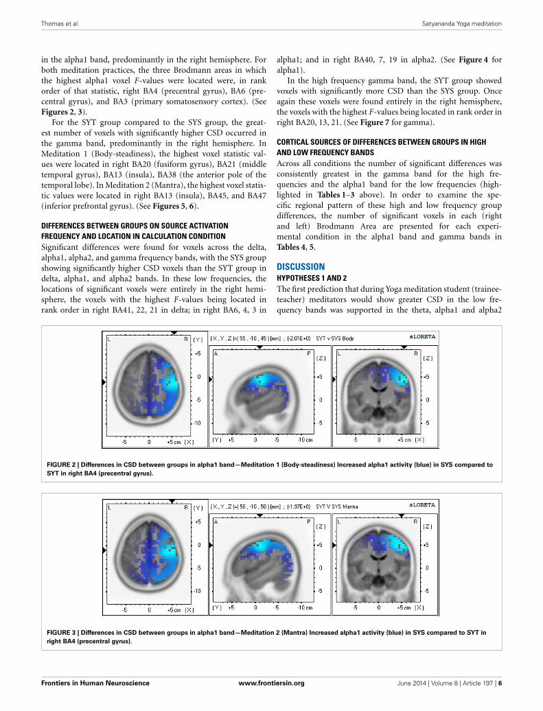
in the alpha1 band, predominantly in the right hemisphere. Forboth meditation practices, the three Brodmann areas in whichthe highest alpha1 voxel F-values were located were, in rankorder of that statistic, right BA4 (precentral gyrus), BA6 (pre-central gyrus), and BA3 (primary somatosensory cortex). (SeeFigures 2, 3).
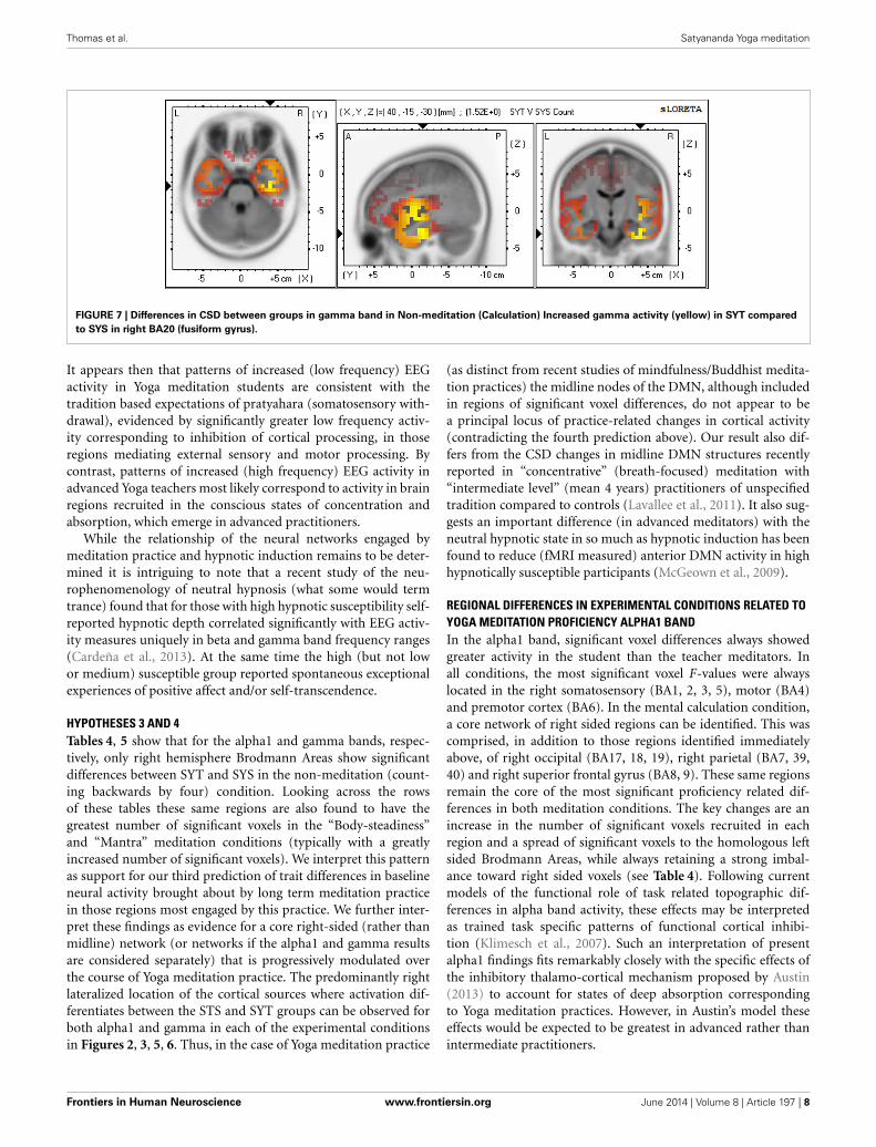
For the SYT group compared to the SYS group, the great-est number of voxels with significantly higher CSD occurred inthe gamma band, predominantly in the right hemisphere. InMeditation 1 (Body-steadiness), the highest voxel statistic val-ues were located in right BA20 (fusiform gyrus), BA21 (middletemporal gyrus), BA13 (insula), BA38 (the anterior pole of thetemporal lobe). In Meditation 2 (Mantra), the highest voxel statis-tic values were located in right BA13 (insula), BA45, and BA47(inferior prefrontal gyrus). (See Figures 5, 6).
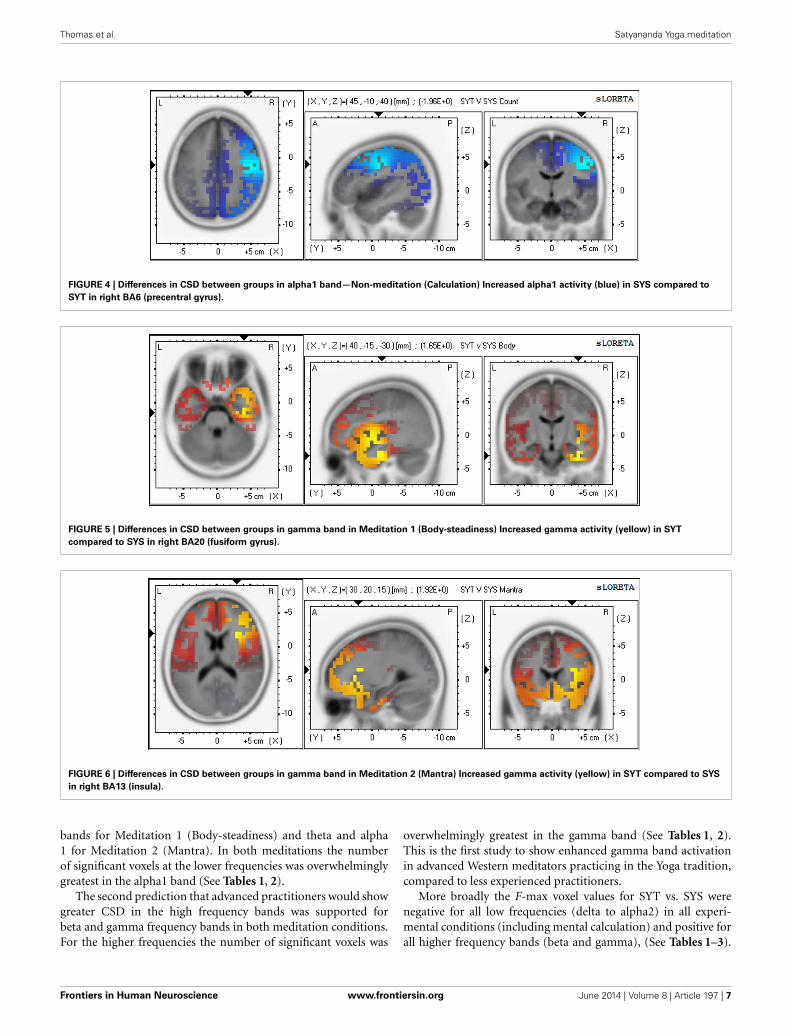
DIFFERENCES BETWEEN GROUPS ON SOURCE ACTIVATIONFREQUENCY AND LOCATION IN CALCULATION CONDITIONSignificant differences were found for voxels across the delta,alpha1, alpha2, and gamma frequency bands, with the SYS groupshowing significantly higher CSD voxels than the SYT group indelta, alpha1, and alpha2 bands. In these low frequencies, thelocations of significant voxels were entirely in the right hemi-sphere, the voxels with the highest F-values being located inrank order in right BA41, 22, 21 in delta; in right BA6, 4, 3 in
alpha1; and in right BA40, 7, 19 in alpha2. (See Figure 4 foralpha1).
In the high frequency gamma band, the SYT group showedvoxels with significantly more CSD than the SYS group. Onceagain these voxels were found entirely in the right hemisphere,the voxels with the highest F-values being located in rank order inright BA20, 13, 21. (See Figure 7 for gamma).
CORTICAL SOURCES OF DIFFERENCES BETWEEN GROUPS IN HIGHAND LOW FREQUENCY BANDSAcross all conditions the number of significant differences wasconsistently greatest in the gamma band for the high fre-quencies and the alpha1 band for the low frequencies (high-lighted in Tables 1–3 above). In order to examine the spe-cific regional pattern of these high and low frequency groupdifferences, the number of significant voxels in each (rightand left) Brodmann Area are presented for each experi-mental condition in the alpha1 band and gamma bands inTables 4, 5.
DISCUSSIONHYPOTHESES 1 AND 2The first prediction that during Yoga meditation student (trainee-teacher) meditators would show greater CSD in the low fre-quency bands was supported in the theta, alpha1 and alpha2
FIGURE 2 | Differences in CSD between groups in alpha1 band—Meditation 1 (Body-steadiness) Increased alpha1 activity (blue) in SYS compared to
SYT in right BA4 (precentral gyrus).
FIGURE 3 | Differences in CSD between groups in alpha1 band—Meditation 2 (Mantra) Increased alpha1 activity (blue) in SYS compared to SYT in
right BA4 (precentral gyrus).
Frontiers in Human Neuroscience www.frontiersin.org June 2014 | Volume 8 | Article 197 | 6
Thomas et al. Satyananda Yoga meditation
FIGURE 4 | Differences in CSD between groups in alpha1 band—Non-meditation (Calculation) Increased alpha1 activity (blue) in SYS compared to
SYT in right BA6 (precentral gyrus).
FIGURE 5 | Differences in CSD between groups in gamma band in Meditation 1 (Body-steadiness) Increased gamma activity (yellow) in SYT
compared to SYS in right BA20 (fusiform gyrus).
FIGURE 6 | Differences in CSD between groups in gamma band in Meditation 2 (Mantra) Increased gamma activity (yellow) in SYT compared to SYS
in right BA13 (insula).
bands for Meditation 1 (Body-steadiness) and theta and alpha1 for Meditation 2 (Mantra). In both meditations the numberof significant voxels at the lower frequencies was overwhelminglygreatest in the alpha1 band (See Tables 1, 2).
The second prediction that advanced practitioners would showgreater CSD in the high frequency bands was supported forbeta and gamma frequency bands in both meditation conditions.For the higher frequencies the number of significant voxels was
overwhelmingly greatest in the gamma band (See Tables 1, 2).This is the first study to show enhanced gamma band activationin advanced Western meditators practicing in the Yoga tradition,compared to less experienced practitioners.
More broadly the F-max voxel values for SYT vs. SYS werenegative for all low frequencies (delta to alpha2) in all experi-mental conditions (including mental calculation) and positive forall higher frequency bands (beta and gamma), (See Tables 1–3).
Frontiers in Human Neuroscience www.frontiersin.org June 2014 | Volume 8 | Article 197 | 7
Thomas et al. Satyananda Yoga meditation
FIGURE 7 | Differences in CSD between groups in gamma band in Non-meditation (Calculation) Increased gamma activity (yellow) in SYT compared
to SYS in right BA20 (fusiform gyrus).
It appears then that patterns of increased (low frequency) EEGactivity in Yoga meditation students are consistent with thetradition based expectations of pratyahara (somatosensory with-drawal), evidenced by significantly greater low frequency activ-ity corresponding to inhibition of cortical processing, in thoseregions mediating external sensory and motor processing. Bycontrast, patterns of increased (high frequency) EEG activity inadvanced Yoga teachers most likely correspond to activity in brainregions recruited in the conscious states of concentration andabsorption, which emerge in advanced practitioners.
While the relationship of the neural networks engaged bymeditation practice and hypnotic induction remains to be deter-mined it is intriguing to note that a recent study of the neu-rophenomenology of neutral hypnosis (what some would termtrance) found that for those with high hypnotic susceptibility self-reported hypnotic depth correlated significantly with EEG activ-ity measures uniquely in beta and gamma band frequency ranges(Cardeña et al., 2013). At the same time the high (but not lowor medium) susceptible group reported spontaneous exceptionalexperiences of positive affect and/or self-transcendence.
HYPOTHESES 3 AND 4Tables 4, 5 show that for the alpha1 and gamma bands, respec-tively, only right hemisphere Brodmann Areas show significantdifferences between SYT and SYS in the non-meditation (count-ing backwards by four) condition. Looking across the rowsof these tables these same regions are also found to have thegreatest number of significant voxels in the “Body-steadiness”and “Mantra” meditation conditions (typically with a greatlyincreased number of significant voxels). We interpret this patternas support for our third prediction of trait differences in baselineneural activity brought about by long term meditation practicein those regions most engaged by this practice. We further inter-pret these findings as evidence for a core right-sided (rather thanmidline) network (or networks if the alpha1 and gamma resultsare considered separately) that is progressively modulated overthe course of Yoga meditation practice. The predominantly rightlateralized location of the cortical sources where activation dif-ferentiates between the STS and SYT groups can be observed forboth alpha1 and gamma in each of the experimental conditionsin Figures 2, 3, 5, 6. Thus, in the case of Yoga meditation practice
(as distinct from recent studies of mindfulness/Buddhist medita-tion practices) the midline nodes of the DMN, although includedin regions of significant voxel differences, do not appear to bea principal locus of practice-related changes in cortical activity(contradicting the fourth prediction above). Our result also dif-fers from the CSD changes in midline DMN structures recentlyreported in “concentrative” (breath-focused) meditation with“intermediate level” (mean 4 years) practitioners of unspecifiedtradition compared to controls (Lavallee et al., 2011). It also sug-gests an important difference (in advanced meditators) with theneutral hypnotic state in so much as hypnotic induction has beenfound to reduce (fMRI measured) anterior DMN activity in highhypnotically susceptible participants (McGeown et al., 2009).
REGIONAL DIFFERENCES IN EXPERIMENTAL CONDITIONS RELATED TOYOGA MEDITATION PROFICIENCY ALPHA1 BANDIn the alpha1 band, significant voxel differences always showedgreater activity in the student than the teacher meditators. Inall conditions, the most significant voxel F-values were alwayslocated in the right somatosensory (BA1, 2, 3, 5), motor (BA4)and premotor cortex (BA6). In the mental calculation condition,a core network of right sided regions can be identified. This wascomprised, in addition to those regions identified immediatelyabove, of right occipital (BA17, 18, 19), right parietal (BA7, 39,40) and right superior frontal gyrus (BA8, 9). These same regionsremain the core of the most significant proficiency related dif-ferences in both meditation conditions. The key changes are anincrease in the number of significant voxels recruited in eachregion and a spread of significant voxels to the homologous leftsided Brodmann Areas, while always retaining a strong imbal-ance toward right sided voxels (see Table 4). Following currentmodels of the functional role of task related topographic dif-ferences in alpha band activity, these effects may be interpretedas trained task specific patterns of functional cortical inhibi-tion (Klimesch et al., 2007). Such an interpretation of presentalpha1 findings fits remarkably closely with the specific effects ofthe inhibitory thalamo-cortical mechanism proposed by Austin(2013) to account for states of deep absorption correspondingto Yoga meditation practices. However, in Austin’s model theseeffects would be expected to be greatest in advanced rather thanintermediate practitioners.
Frontiers in Human Neuroscience www.frontiersin.org June 2014 | Volume 8 | Article 197 | 8
Thomas et al. Satyananda Yoga meditation
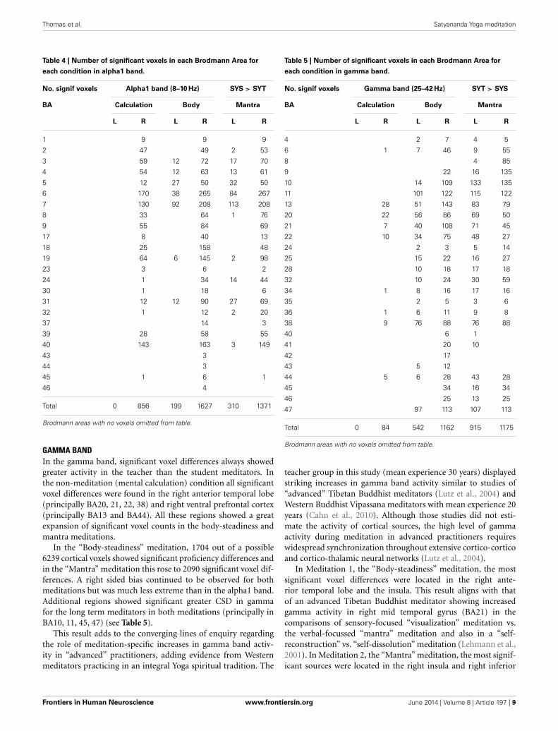
Table 4 | Number of significant voxels in each Brodmann Area for
each condition in alpha1 band.
No. signif voxels Alpha1 band (8–10 Hz) SYS > SYT
BA Calculation Body Mantra
L R L R L R
1 9 9 9
2 47 49 2 53
3 59 12 72 17 70
4 54 12 63 13 61
5 12 27 50 32 50
6 170 38 265 84 267
7 130 92 208 113 208
8 33 64 1 76
9 55 84 69
17 8 40 13
18 25 158 48
19 64 6 145 2 98
23 3 6 2
24 1 34 14 44
30 1 18 6
31 12 12 90 27 69
32 1 12 2 20
37 14 3
39 28 58 55
40 143 163 3 149
43 3
44 3
45 1 6 1
46 4
Total 0 856 199 1627 310 1371
Brodmann areas with no voxels omitted from table.
GAMMA BANDIn the gamma band, significant voxel differences always showedgreater activity in the teacher than the student meditators. Inthe non-meditation (mental calculation) condition all significantvoxel differences were found in the right anterior temporal lobe(principally BA20, 21, 22, 38) and right ventral prefrontal cortex(principally BA13 and BA44). All these regions showed a greatexpansion of significant voxel counts in the body-steadiness andmantra meditations.
In the “Body-steadiness” meditation, 1704 out of a possible6239 cortical voxels showed significant proficiency differences andin the “Mantra” meditation this rose to 2090 significant voxel dif-ferences. A right sided bias continued to be observed for bothmeditations but was much less extreme than in the alpha1 band.Additional regions showed significant greater CSD in gammafor the long term meditators in both meditations (principally inBA10, 11, 45, 47) (see Table 5).
This result adds to the converging lines of enquiry regardingthe role of meditation-specific increases in gamma band activ-ity in “advanced” practitioners, adding evidence from Westernmeditators practicing in an integral Yoga spiritual tradition. The
Table 5 | Number of significant voxels in each Brodmann Area for
each condition in gamma band.
No. signif voxels Gamma band (25–42 Hz) SYT > SYS
BA Calculation Body Mantra
L R L R L R
4 2 7 4 5
6 1 7 46 9 55
8 4 85
9 22 16 135
10 14 109 133 135
11 101 122 115 122
13 28 51 143 83 79
20 22 56 86 69 50
21 7 40 108 71 45
22 10 34 75 48 27
24 2 3 5 14
25 15 22 16 27
28 10 18 17 18
32 10 24 30 59
34 1 8 16 17 16
35 2 5 3 6
36 1 6 11 9 8
38 9 76 88 76 88
40 6 1
41 20 10
42 17
43 5 12
44 5 6 28 43 28
45 34 16 34
46 25 13 25
47 97 113 107 113
Total 0 84 542 1162 915 1175
Brodmann areas with no voxels omitted from table.
teacher group in this study (mean experience 30 years) displayedstriking increases in gamma band activity similar to studies of“advanced” Tibetan Buddhist meditators (Lutz et al., 2004) andWestern Buddhist Vipassana meditators with mean experience 20years (Cahn et al., 2010). Although those studies did not esti-mate the activity of cortical sources, the high level of gammaactivity during meditation in advanced practitioners requireswidespread synchronization throughout extensive cortico-corticoand cortico-thalamic neural networks (Lutz et al., 2004).
In Meditation 1, the “Body-steadiness” meditation, the mostsignificant voxel differences were located in the right ante-rior temporal lobe and the insula. This result aligns with thatof an advanced Tibetan Buddhist meditator showing increasedgamma activity in right mid temporal gyrus (BA21) in thecomparisons of sensory-focused “visualization” meditation vs.the verbal-focussed “mantra” meditation and also in a “self-reconstruction” vs. “self-dissolution” meditation (Lehmann et al.,2001). In Meditation 2, the “Mantra” meditation, the most signif-icant sources were located in the right insula and right inferior
Frontiers in Human Neuroscience www.frontiersin.org June 2014 | Volume 8 | Article 197 | 9
Thomas et al. Satyananda Yoga meditation
frontal gyrus. The right insula has been linked with a moredetached and objective awareness of interoceptive sensory events(Farb et al., 2007) involved in the shift from “narrative” to “expe-riential” self-awareness. Deen et al. (2011) identify three distinctclusters of functional connectivity with network hubs in pos-terior, mid, and anterior insula, respectively, suggesting that afurther parcelation of anatomical and function subregions withinthe insula will be required to fully understand the role (or roles)it plays in Yoga and Buddhist tradition meditative states. It maybe of note that a major white fiber tract, the uncinated fasciculis,mediates bidirectional connections between the high level associ-ation cortex of the anterior temporal lobe and the inferior frontalgyrus (Von der Heide et al., 2013), the only such structure whichcontinues to develop beyond the age of 30 (Lebel et al., 2008).
STRENGTHS OF THIS STUDYA strength of this study is the advanced level of meditation expe-rience in a sample of Western Yoga practitioners following anidentical spiritual tradition. All of the “advanced” group were san-nyasin (initiated) disciples of Swami Satyananda who had spentconsiderable time in ashrams, both in Australia and India. Theywere all Yoga teachers. Studies reporting results from large groupsof advanced meditation practitioners often combine meditatorsfrom different traditions into a single experimental group. Suchdifferences are not considered equivalent by the members of thesetraditions themselves and any such equivalence must be demon-strated rather than assumed. We have chosen not to make thisassumption even at the cost of a lower sample size in order tomaintain the validity of our analysis. A further strength was theattention given to preserving the ecological validity of the medi-tation states attained, the study being conducted in a meditationsetting conducive to the participants’ usual practice. The intru-sion of equipment into this setting was minimized, with theEEG cap and small radio transmitter being the only items in themeditation room.
As with all EEG source analysis methods, eLORETA resultsare highly susceptible to distortion by artifacts (Pizzagalli, 2007),therefore the quality of data preprocessing is essential to thevalidity of the results obtained. We adopted a recently devel-oped (Nolan et al., 2010) ICA component rejection procedure forartifact cleaning developed by the Neural Engineering Group atTrinity College Dublin. This is one of a new generation of ICAartifact correction procedures which is guided by a series of objec-tive component parameters rather than the momentary choiceof the individual experimenter. We believe the effective utiliza-tion of this method for preparing large volumes of clean data wasan essential factor contributing to the validity of this research.We strongly recommend the future adoption of this or similarmethod by related research programs.
LIMITATIONS OF THIS STUDYLimitations of the study include the small sample size (althoughthat did not prevent significant findings) and the potentialconfound due to significant age difference between the sub-ject groups. A larger sample size is desirable to enhance thepower and generalizability of the current results and would allowthe additional possibility of parametric testing (Thatcher et al.,
2005). However, the non-parametric statistical mapping methodadopted here is well suited to the limitations of the current designand sample size. In fact, one of the worked examples offered byNichols and Holmes (2002, p. 15) is a PET study with 12 sub-jects equally divided amongst 2 testing conditions which directlyreflects the constraints of the present study. In addition the ran-domization distribution is known to be overly conservative whenthe effect is distributed, as occurs in the present results (Troendle,1995). Therefore, we believe that there is a strong justification forreporting the group effects identified here.
EEG spectral power density changes dramatically with ageuntil early adulthood. From that point changes are slow in healthyaging. Power in lower frequency bands (delta through alpha)decreases throughout this time period (Rossini et al., 2007). Theage range of our subjects extends from 30 to 63, so it could be thatsignificantly greater activity in the younger group in these lowerbands is due to normal aging rather than years of meditationpractice.
A comparison of the effects of normal aging to the presentlyobserved differences between the SYS (younger) and SYT (older)studies of EEG spectral source changes across this part of the lifes-pan shows that delta decreases in occipital sources (Babiloni et al.,2006), but we only found significant delta differences in one con-dition (counting) and this was maximal in right BA41 (primaryauditory cortex). In normal aging alpha1 and alpha2 decreases inoccipital, parietal, temporal and limbic cortices (Babiloni et al.,2006). We found significantly lower alpha1 in occipital and pari-etal cortex but not in temporal or limbic cortex. Alpha alsoincreases with age in frontal cortex (Basar, 2012) but we foundsignificant alpha decreases in superior and middle frontal gyri inthe older group and no significant differences in inferior frontalgyri. Once again, the pattern of our findings does not fit with whatwould be expected if they were due to normal aging.
Despite available data there is no clear evidence for changesdue to normal aging through middle age in the high frequencybands. Therefore, we do not consider age differences to be a plau-sible alternative explanation for those high frequency effects. Aconfound due to the age difference between the groups fails toaccount for why the set of BA containing significantly greaterlow frequency voxel activation in the SYS group forms the com-plement to the set of BA containing significantly greater highfrequency voxel activation in the SYT group. Nor does it explainwhy a core pattern found in the non-meditation Calculationcondition is maintained but greatly expanded in the Body Scanand Mantra meditation conditions. Lutz et al. (2004), in a studywith advanced and novice meditators with age means of 49 yearsand 21 years, found hours of practice, but not age, significantlypredicted gamma in their baseline condition.
The additional analysis of age matched subgroups goes someway toward addressing this issue with significant F-max voxelfindings in both alpha1 and gamma bands located in the sameregions as the original group analysis. In this case the furtherreduced sample size points to the robustness of these effects.Notwithstanding these considerations practice related differencesin age will be intrinsic to group membership whenever truly“advanced” (such as the 30 year group in the present study)are compared with medium term practice groups. Now that
Frontiers in Human Neuroscience www.frontiersin.org June 2014 | Volume 8 | Article 197 | 10
Thomas et al. Satyananda Yoga meditation
specific regions of interest have been identified parametric meth-ods which enable statistical control of age effects present a viableoption to address this issue in future (larger n) studies.
The density (number) of electrodes in the recording arrayalso places limitations on the resolution of source localization(Pizzagalli, 2007). Dense array recordings are so far limited to lab-oratory settings, unlike the present study. However, successful val-idation studies of LORETA, sLORETA and eLORETA use only theoriginal 19 electrodes of the 10–20 system (Pascual-Marqui et al.,2002). Particularly for non-evoked recording conditions, 19 chan-nel recordings are considered adequate and are widely reportedfor the LORETA family of source analysis methods (Babiloniet al., 2014). By comparison, the current study employed 25 activerecording electrodes. A further possible limitation was the fixedsequence of conditions rather than a systematically counterbal-anced design. For investigation of deeper meditation states, thebenefits of experimental manipulation of conditions and intrusivemeasuring instruments must be balanced by the need to obtain anauthentic meditation experience.
CONCLUSIONThis study is the first to report enhanced gamma band activationin advanced Western meditators practicing in the (Satyananda)Yoga tradition, compared to less experienced (intermediate) prac-titioners. It found significant differences in EEG frequency bandsources between “experienced” (mean 4 years) and “advanced”(mean 30 years) meditators in two meditation conditions (Body-steadiness and Mantra) and a mental calculation condition.The findings, strongest in alpha1 and gamma bands, consis-tently support the frequency band hypothesis proposed by Fellet al. (2010) of enhanced low and high frequency band effects,respectively, for intermediate and advanced levels of medita-tion experience. It further adds a finding of increased gammaband activity in advanced western Yoga meditators to similarfindings in advanced Buddhist meditators. In addition, the Body-steadiness meditation condition in the present study appears tohave much in common with Buddhist mindfulness of breath-ing practice. Both practices direct awareness to the felt quality ofbodily sensations. The present findings then call for much closerscrutiny of the distinction, at least in the long term, betweenthe functional networks trained by concentrative and mind-fulness meditation methods. Those regions which consistentlydifferentiated between the groups in the low and high frequencybands across all meditation conditions showed similar (butreduced) differences in the non-meditation (counting backwards)condition.
This study extends previous EEG findings by estimating cor-tical gray matter sources for frequency band specific meditationtraining effects. The cortical loci of high and low frequencyband meditation training effects are both specific and distinct.Approximately one third of available cortical gray matter vox-els showed significantly greater gamma band activity in mantrameditation in advanced (SYT) than intermediate (SYS) med-itators. Despite this, there is little overlap with those regionswhich show increased alpha1 in the student meditators. In factthe Brodmann areas in which significant voxel differences arefound during meditation in the alpha1 (suggesting functional
inhibition) and gamma bands (suggesting integration into con-sciousness) form almost absolute complement subsets of the fullset of cytoarchitecturally defined gray matter regions. We proposethat selective inhibition of a right lateralized network comprisingvisual, somatosensory and body-world self-representations cor-responds to the earlier stages of sensory withdrawal and strippingaway of the “outer” onion-like layers of self as described in tra-ditional Yoga literature. The subsequent emergence of consciousstates specific to advanced practitioners requires both the dis-engagement from these self-world representational systems andthe development of widespread gamma synchronization through-out anterior temporal and ventral prefrontal cortical regionsextending from a right sided core network incorporating anteriortemporal lobe and insula.
ACKNOWLEDGMENTSThis study was assisted by a grant from Swan Research Institute,a non-profit organization promoting research into Yoga inAustralia. The authors are grateful to Mr. Ian Evans and Dr. PeterGrimbeek for their assistance and advice in the analysis of thisstudy.
REFERENCESArambula, P., Peper, E., Kawakami, M., and Gibney, K. H. (2001). The physiolog-
ical correlates of Kundalini Yoga meditation: a study of a yoga master. Appl.Psychophysiol. Biofeedback 26, 147–153. doi: 10.1023/A:1011343307783
Austin, J. (2013). Zen and the brain: mutually illuminating topics. Front. Psychol.4:784. doi: 10.3389/fpsyg.2013.00784
Babiloni, C., Binetti, G., Cassarino, A., Dal Forno, G., Del Percio, C., Ferreri,F., et al. (2006). Sources of cortical rhythms in adults during physiologi-cal aging: a multicentric EEG study. Hum. Brain Mapp. 27, 162–172. doi:10.1002/hbm.20175
Babiloni, C., Del Percio, C., Lizio, R., Marzano, N., Infarinato, F., Soricelli, A., et al.(2014). Cortical sources of resting state electroencephalographic alpha rhythmsdeteriorate across time in subjects with amnesic mild cognitive impairment.Neurobiol. Aging 35, 130–142. doi: 10.1016/j.neurobiolaging.2013.06.019
Basar, E. (2012). A review of alpha activity in integrative brain function: fundamen-tal physiology, sensory coding, cognition and pathology. Int. J. Psychophysiol. 86,1–24. doi: 10.101016/j.ijpsycho.2012.07.002
Berkovich-Ohana, A., Glicksohn, J., and Goldstein, A. (2012). Mindfulness-induced changes in gamma band activity - Implications for the default modenetwork, self-reference and attention. Clin. Neurophysiol. 123, 700–710. doi:10.1016/j.clinph.2011.07.048
Brefczynski-Lewis, J., Lutz, A., Schaefer, H. S., Levinson, D., and Davidson,R. (2007). Neural correlates of attentional expertise in long-term medi-tation practitioners. Proc. Natl. Acad. Sci. U.S.A. 104, 11483–11488. doi:10.1073/pnas.0606552104
Brett, M., Johnsrude, I., and Owen, A. (2002). The problem of functional localiza-tion in the human brain. Nat. Rev. Neurosci. 3, 243–249. doi: 10.1038/nrn756
Brewer, J., Worhunsky, P., Gray, J., Tang, Y., Weber, J., and Kober, H. (2011).Meditation experience is associated with differences in default mode networkactivity and connectivity. Proc. Natl. Acad. Sci. U.S.A. 108, 20254–20259. doi:10.1073/pnas.1112029108
Buckner, R., Andrews-Hanna, J., and Schacter, D. L. (2008). The brain’s defaultnetwork. Ann. N.Y. Acad. Sci. 1124, 1–38. doi: 10.1196/annals.1440.011
Cahn, B., Delorme, A., and Polich, J. (2010). Occipital gamma activation dur-ing Vipassana meditation. Cogn. Process. 11, 39–56. doi: 10.1007/s10339-009-0352-1
Cahn, B., and Polich, J. (2006). Meditation states and traits: EEG, ERP,and Neuroimaging Studies. Psychol. Bull. 132, 180–211. doi: 10.1037/0033-2909.132.2.180
Cardeña, E., Jonsson, P., Terhune, D. B., and Marcusson-Clavertz, D. (2013).The neurophenomenology of neural hypnosis. Cortex 49, 375–385. doi:10.1016/j.cortex.2013.04.001
Frontiers in Human Neuroscience www.frontiersin.org June 2014 | Volume 8 | Article 197 | 11
Thomas et al. Satyananda Yoga meditation
Deen, B., Pitskel, N., and Pelphrey, K. (2011). Three systems of insular functionalconnectivity identified with cluster analysis. Cereb. Cortex 21, 1498–1506. doi:10.1093/cercor/bhq186
Delorme, A., and Makeig, S. (2004). EEGLab: an open source toolbox foranalysis of single-trial EEG dynamics. J. Neurosci. Methods 134, 9–21. doi:10.1016/j.jneumeth.2003.10.009
Farb, N., Segal, Z., Mayberg, H., Bean, J., McKeon, D., Fatima, Z., et al.(2007). Attending to the present: mindfulness meditation reveals distinctneural modes of self-reference. Soc. Cogn. Affect. Neurosci. 2, 313–322. doi:10.1093/scan/nsm030
Fell, J., Axmacher, N., and Haupt, S. (2010). From alpha to gamma: electrophysio-logical correlates of meditation-related states of consciousness. Med. Hypotheses75, 218–224. doi: 10.1016/j.mehy.2010.02.025
Fuchs, M., Kastner, J., Wagner, M., and Hawes, S. (2002). A standardized boundaryelement method volume conductor model. Clin. Neurophysiol. 113, 702–712.doi: 10.1016/S1388-2457(02)00030-5
Grant, J., Courtemanche, J., Duerden, E., Duncan, G., and Rainville, P. (2010).Cortical thickness and pain sensitivity in zen meditators. Emotion 10, 43–53.doi: 10.1037/a0018334
Holzel, B., Carmody, J., Vangel, M., Congleton, C., Yerramsetti, S., Gard, T., et al.(2011). Mindfulness practice leads to increases in regional brain gray matterdensity. Psychiatry Res. 191, 36–43. doi: 10.1016/j.pscychresns.2010.08.006
Holzel, B., Ott, U., Gard, T., Hempel, H., Weygandt, M., Morgen, K., et al.(2008). Investigation of mindfulness meditation practitioners with voxel-basedmorphometry. Soc. Cogn. Affect. Neurosci. 3, 55–61. doi: 10.1093/scan/nsm038
Hölzel, B., Ott, U., Hempel, H., Hackl, A., Wolf, K., Stark, R., et al. (2007).Differential engagement of anterior cingulate and adjacent medial frontal cor-tex in adept meditators and non-meditators. Neurosci. Lett. 421, 16–21. doi:10.1016/j.neulet.2007.04.074
Jurcak, V., Tsuzuki, D., and Dan, I. (2007). 10/20, 10/10, and 10/5 systems revisited:their validity as relative head-surface-based positioning systems. Neuroimage 34,1600–1611. doi: 10.1016/j.neuroimage.2006.09.024
Kang, D., Jo, H., Jung, W., Kim, S., Jung, Y., Choi, C., et al. (2013). The effect ofmeditation on brain structure: cortical thickness mapping and diffusion tensorimaging. Soc. Cogn. Affect. Neurosci. 8, 27–33. doi: 10.1093/scan/nss056
Klimesch, W., Sauseng, P., and Hanslmayr, S. (2007). EEG alpha oscillations:the inhibition–timing hypothesis. Brain Res. Brain Res. Rev. 53, 63–88. doi:10.1016/j.brainresrev.2006.06.003
Lancaster, J., Woldorff, M., Parsons, L., and Liotti, M. (2000). Automatedtalairach atlas labels for functional brain mapping. Hum. Brain Mapp. 10,120–131. doi: 10.1002/1097-0193(200007)10:3%3C120::AID-HBM30%3E3.0.CO;2-8
Lavallee, C., Hunter, M., and Persinger, M. (2011). Intracerebral source genera-tors characterizing concentrative meditation. Cogn. Process. 12, 141–150. doi:10.1007/s10339-011-0394-z
Lazar, S., Kerr, C., Wasserman, R., Gray, J., Greve, D., Treadway, M., et al.(2005). Meditation experience is associated with increased cortical thickness.Neuroreport 16, 1893–1897. doi: 10.1097/ 01.wnr.0000186598.66243.19
Lebel, C., Walker, L., Leemans, A., Phillips, L., and Beaulieu, C. (2008).Microstructural maturation of the human brain from childhood to adulthood.Neuroimage 40, 1044–1055. doi: 10.1016/j.neuroimage.2007.12.053
Lehmann, D., Faber, P., Achermann, P., Jeanmonod, D., Gianotti, L., andPizzagalli, D. (2001). Brain sources of EEG gamma frequency during volition-ally meditation-induced, altered states of consciousness, and experience of theself. Psychiatry Res. 108, 111–121. doi: 10.1016/S0925-4927(01)00116-0
Leung, M., Chan, C., Yin, J., Lee, C., So, K., and Lee, T. (2013). Increased gray mattervolume in the right angular and posterior parahippocampal gyri in loving-kindness meditators. Soc. Cogn. Affect. Neurosci. 8, 34–39. doi: 10.1093/scan/nss076
Luders, E., Clark, K., Narr, K., and Toga, A. (2011). Enhanced brain connectiv-ity in long- term meditation practitioners. Neuroimage 57, 1308–1316. doi:10.1016/j.neuroimage.2011.05.075
Luders, E., Kurth, F., Mayer, E., Toga, A., Narr, K., and Gaser, C. (2012a). Theunique brain anatomy of meditation practitioners: alterations in cortical gyrifi-cation. Front. Hum. Neurosci. 6:34. doi: 10.3389/fnhum.2012.00034
Luders, E., Phillips, O., Clark, K., Kurth, F., Toga, A., and Narr, K. (2012b). Bridgingthe hemispheres in meditation: thicker callosal regions and enhanced frac-tional anisotropy (FA) inlong-term practitioners. Neuroimage 61, 181–187. doi:10.1016/j.neuroimage.2012.02.026
Luders, E., Toga, A., Lepore, N., and Gaser, C. (2009). The underlying anatomi-cal correlates of long- term meditation:larger hippocampal and frontal volumesof graymatter. Neuroimage 45, 672–678. doi: 10.1016/j.neuroimage.2008.12.061
Lutz, A., Greischar, L. L., Rawlings, N. B., Ricard, M., and Davidson, R. J.(2004). Long-term meditators self-induce high-amplitude gamma synchronyduring mental practice. Proc. Natl. Acad. Sci. U.S.A. 101, 16369–16373. doi:10.1073/pnas.0407401101
Maheshwarananda, P. (2005). The System Yoga in Daily Life. Vienna: EuropeanUniversity Press.
Malinowski, P. (2013). Neural mechanisms of attentional control in mindfulnessmeditation. Front. Neurosci. 7:8. doi: 10.3389/fnins.2013.00008
Mazziotta, J., Toga, A., Evans, A., and Fox, P. (2001). A probabilistic atlas andreference system for the human brain: international Consortium for BrainMapping (ICBM). Philos. Trans. R. Soc. Lond. B Biol. Sci. 356, 1293–1322. doi:10.1098/rstb.2001.0915
McGeown, W., Mazzoni, G., Venneri, A., and Kirsch, I. (2009). Hypnotic induc-tion decreases anterior default mode activity. Conscious. Cogn. 18, 848–855. doi:10.1016/j.concog.2009.09.001
Murakami, H., Nakao, T., Matsunaga, M., Kasuya, Y., Shinoda, J., Yamada, J., et al.(2012). The structure of mindful brain. PLoSONE 7:e46377. doi: 10.1371/jour-nal.pone.0046377
Nichols, T., and Holmes, A. (2002). Nonparametric permutation tests for func-tional neuroimaging: a primer with examples. Hum. Brain Mapp. 15, 1–25. doi:10.1002/hbm.1058
Nolan, H., Whelan, R., and Reilly, R. (2010). FASTER: fully automated statisticalthresholding for EEG artifact rejection. Neurosci. Methods 192, 152–162. doi:10.1016/j.jneumeth.2010.07.015
Oostenveld, R., and Praamstra, P. (2001). The five percent electrode system forhigh-resolution EEG and ERP measurements. Clin. Neurophysiol. 112, 713–719.doi: 10.1016/S1388-2457(00)00527-7
Ospina, M., Bond, K., Karkhaneh, M., Tjosvold, L., Vandermeer, B., Liang, Y., et al.(2007). Meditation practices for health: state of the research. Evid. Rep. Technol.Assess. (Full Rep.) 155, 1–263.
Ott, U. (2001). The EEG and the depth of meditation. J. Med. Med. Res. 1, 55–68.Pagnoni, G., and Cekic, M. (2007). Age effects on gray matter volume and atten-
tional performance in Zen meditation. Neurobiol. Aging 28, 1623–1627. doi:10.1016/ j.neurobiolaging.2007.06.008
Pascual-Marqui, R. (2002). Standardised low-resolution brain electromeagnetictomography (sLORETA). Methods Find. Exp. Clin. Pharmacol. 24(Suppl.D), 5–12.
Pascual-Marqui, R. (2007). Discrete, 3D distributed, linear imaging methods ofelectric neuronal activity. Part 1: exact, zero error localization. arXiv:0710.3341[math-ph].
Pascual-Marqui, R. (2009). “Theory of the EEG inverse problem,” in QuantitativeEEG Analysis: Methods and Clinical Applications, eds N. V. Thakor and S. Tong(Boston: Artech House), 121–140.
Pascual-Marqui, R., Esslen, M., Kochi, K., and Lehmann, D. (2002). Functionalimaging with low resolution brain electromagnetic tomography (LORETA):review, new comparisons, and new validation. Jap. J. Clin. Neurophysiol.30, 81–94.
Pascual-Marqui, R., Michel, C., and Lehmann, D. (1994). Low resolution elec-tromagnetic tomography: a new method for localizing electrical activityin the brain. Int. J. Psychophysiol. 18, 49–65. doi: 10.1016/0167-8760(84)90014-X
Pizzagalli, D. (2007). “Electroencephalography and high-density electrophysiolog-ical source localization,” in Handbook of Psychophysiology, eds J. T. Cacioppo,L. G. Tassinaru, and G. G. Berntson (Cambridge: Cambridge UniversityPress), 56–84.
Pizzagalli, D., Pascual-Marqui, R., Nitschke, J., and Oakes, T. (2001).Anterior cingulate activity as a predictor of degree of treatmentresponse in major depression: evidence from brain electrical tomogra-phy analysis. Am. J. Psychiatry 158, 405–415. doi: 10.1176/appi.ajp.158.3.405
Radhakrishnan, S. (1999). Indian Philosophy. Oxford: Oxford University Press.Rossini, P., Rossi, S., Babiloni, C., and Polich, J. (2007). Clinical neurophysiology
of aging brain: from normal aging to neurodegeneration. Prog. Neurobiol. 83,375–400. doi: 10.1016/j.pneurobio.2007.07.010
Saraswati, S. (1981). Yoga and Kriya. Munger: Yoga Publications Trust.
Frontiers in Human Neuroscience www.frontiersin.org June 2014 | Volume 8 | Article 197 | 12
Thomas et al. Satyananda Yoga meditation
Saraswati, S. (1983). Meditations from the Tantras. Munger: Yoga Publications Trust.Schooler, J., Smallwood, J., Christoff, K., Handy, T., Reichle, E., and Sayette,
M. (2011). Meta-awareness, perceptual decoupling and the wandering mind.Trends Cogn. Sci. 15, 319–326. doi: 10.1016/j.tics.2011.05.006
Shapiro, S. (2003). An analysis of recent meditation research. J. Med. Res. 3, 69–90.doi: 10.1080/0887367.2003.9986927
Tang, Y., Lu, Q., Geng, X., Stein, E., Yang, Y., and Posner, M. (2010). Short-termmeditation induces white matter changes in the anterior cingulate. Proc. Natl.Acad. Sci. U.S.A. 107, 15649–15652. doi: 10.1073/pnas.1011043107
Thatcher, R., North, D., and Biver, C. (2005). Parametric vs. non-parametricstatistics of low resolution electromagnetic tomography (LORETA). Clin. EEGNeurosci. 36, 1–8. doi: 10.1177/155005940503600103
Troendle, J. (1995). A stepwise resampling method of multiple hypothesis testing.J. Amer. Stat. Assoc. 90, 370–378. doi: 10.1080/01621459.1995.10476522
Vestergaard-Poulsen, P., van Beek, M., Skewes, J., Bjarkam, C., Stubberup,M, Bertelsen, J., et al. (2009). Long-term meditation is associated withincreased gray matter density in the brain stem. Neuroreport 20, 170–174. doi:10.1097/WNR.0b013e3283 20012a
Von der Heide, R., Skipper, L., Klobusicky, E., and Olsen, I. (2013). Dissectingthe uncinate fasciculus: disorders, controversies and a hypothesis. Brain 136,1692–1707. doi: 10.1093/brain/awt094
Zumsteg, D., Friedman, A., Wieser, H., and Wennberg, R. (2006). Propagationof interictal discharges in temporal lobe epilepsy: correlation of spatiotem-poral mapping with intracranial foramen ovale electrode recordings. Clin.Neurophysiol. 117, 2615–2626. doi: 10.1016/j.clinph.2006.07.319
Conflict of Interest Statement: The authors declare that the research was con-ducted in the absence of any commercial or financial relationships that could beconstrued as a potential conflict of interest.
Received: 31 December 2013; accepted: 20 March 2014; published online: 10 June 2014.Citation: Thomas J, Jamieson G and Cohen M (2014) Low and then high frequencyoscillations of distinct right cortical networks are progressively enhanced by mediumand long term Satyananda Yoga meditation practice. Front. Hum. Neurosci. 8:197.doi: 10.3389/fnhum.2014.00197This article was submitted to the journal Frontiers in Human Neuroscience.Copyright © 2014 Thomas, Jamieson and Cohen. This is an open-access article dis-tributed under the terms of the Creative Commons Attribution License (CC BY). Theuse, distribution or reproduction in other forums is permitted, provided the originalauthor(s) or licensor are credited and that the original publication in this jour-nal is cited, in accordance with accepted academic practice. No use, distribution orreproduction is permitted which does not comply with these terms.
Frontiers in Human Neuroscience www.frontiersin.org June 2014 | Volume 8 | Article 197 | 13
Copyright © 2022 FDOKUMEN