Long-term benefits, risks and adverse events after intended ...
161
Long-term benefits, risks and adverse events after intended curative rectal cancer treatment in Denmar k PhD dissertation Jesper Beck Jørgensen Health Aarhus University Department of Surgery, Aarhus University Hospital 2020
-
Upload
khangminh22 -
Category
Documents
-
view
1 -
download
0
Transcript of Long-term benefits, risks and adverse events after intended ...
Long-term benefits, risks and adverse events after
intended curative rectal cancer treatment in Denmark
PhD dissertation
Jesper Beck Jørgensen
Health
Aarhus University
Department of Surgery,
Aarhus University Hospital
2020

2
Supervisors
Professor, Lene Hjerrild Iversen, MD, PhD, DMSc (main supervisor)
Department of Surgery, Aarhus University Hospital
Aarhus, Denmark
Bodil Ginnerup Pedersen, MD, PhD
Department of Radiology, Aarhus University Hospital
Aarhus, Denmark
Professor, Søren Laurberg, MD, DMSc
Department of Surgery, Aarhus University Hospital
Aarhus, Denmark
Collaborators
Rune Erichsen, MD, PhD
Department of Clinical Epidemiology, Aarhus University Hospital
Department of Surgery, Regional Hospital Randers
Aarhus Denmark
Peter Bondeven, MD, PhD
Department of Surgery, Aarhus University Hospital
Department of Surgery, Regional Hospital Randers
Aarhus, Denmark

3
Evaluation committee
Brendan Moran, MD, MCh, FRCS, FRCSI
Department of Surgery
Basingstoke and North Hampshire Hospital
Basingstoke, England
Professor Torbjörn Holm, MD, PhD
Department of Surgery
Södersjukhuset
Stockholm, Sweden
Professor Kari Tanderup, Cand.scient., PhD (chairman)
Department of Oncology
Aarhus University Hospital
Aarhus, Denmark

4
Preface
This thesis is based on studies carried out during my employment at Department of
Surgery, Aarhus University Hospital and Regional Hospital Randers. The work is
funded by the Danish Cancer Society and performed between 2014 and 2020. I wish
to express my sincere gratitude to all those who have helped directly or indirectly
during this work.
First of all, I wish to express my deepest gratitude to Lene Hjerrild Iversen. I am truly
grateful for your commitment and devotion to this project. You have invested heart
and soul, and have catalyzed my development and continued learning with a never
failing enthusiasm. You own an ability to make the best of things and have shown
incredible loyalty and patience in all contexts during this journey. I really appreciate
our friendship and confidentiality.
To Bodil Ginnerup Pedersen. I have the utmost respect for your professional and
personal dedication. You have shown wise and caring support, and have been bedrock
throughout the realization of this work. I thank you for your always-constructive
criticism and unquestionable integrity.
To Søren Laurberg. You have provided truly inspiring mentoring sessions and have
brought huge dedication in to this project. Throughout the process you have been
offering great hospitality and provided constructive criticism delivered in a personal
and subtle way. I admire your passion for research and sense of humour.
To my estimated colleagues, Rune Erichsen and Peter Bondeven, who have offered
excellent professional and personal guidance throughout the process. I thank you both
for your availability and cheerful support.
Finally, I am especially thankful to my best friend and wife, Ann Olivia and our
beautiful children Sia, Anton, Sigurd, Alma, and Otto for your patience and loving
support. I am infinitely grateful for your strength and wise guidance in all matters of
life. You are truly inspiring and the best companion through unprecedented paths in
life. I would be lost without you.

5
Table of contents
List of publications ....................................................................................................................... 10
Introduction .................................................................................................................................... 11
Background ..................................................................................................................................... 13 Rectal cancer ..................................................................................................................................................... 13 Survival and Recurrence .............................................................................................................................. 13 Staging of rectal cancer ................................................................................................................................ 16 Magnetic Resonance Imaging .................................................................................................................... 17 Multidisciplinary team directed treatment ......................................................................................... 19 Neoadjuvant treatment ................................................................................................................................ 20 Surgical treatment .......................................................................................................................................... 24 Total mesorectal excision .................................................................................................................. 25 Partial mesorectal excision ............................................................................................................... 25 Abdominoperineal resection ............................................................................................................ 26 Use of diverting stoma ........................................................................................................................ 27
Postoperative surveillance .......................................................................................................................... 28 Long-‐term adverse events following intended curative rectal cancer treatment .............. 30 Adverse events ....................................................................................................................................... 30 Non-‐reversal of diverting stoma ..................................................................................................... 30 Pelvic insufficiency fractures ........................................................................................................... 33
Aims of dissertation ..................................................................................................................... 35
Hypotheses ...................................................................................................................................... 36
Methods ............................................................................................................................................ 37 Study designs and settings .......................................................................................................................... 37 Data sources ...................................................................................................................................................... 37 Civil Registration System (CRS) ...................................................................................................... 37 Danish Colorectal Cancer Group (DCCG) Database ................................................................ 37 The Danish National Patient Registry (DNRP) ......................................................................... 38
Study cohorts ..................................................................................................................................................... 38 Oncological and surgical treatment ....................................................................................................... 41 Histopathology ................................................................................................................................................. 42 3-‐year postoperative pelvic MRI ............................................................................................................... 42

6
Recurrent disease ............................................................................................................................................ 45 Outcomes ............................................................................................................................................................. 46 Overall survival and recurrent disease (Paper I) .................................................................... 46 Stoma reversal (Paper II) ................................................................................................................... 46 Pelvic insufficiency fractures (Paper III) .................................................................................... 46
Statistical analysis .......................................................................................................................................... 47 Characteristics ........................................................................................................................................ 47 Overall survival, cumulative incidence proportion, and competing risk analysis
(Paper I and II) ....................................................................................................................................... 47 Cox proportional Hazards (Paper I and II) ................................................................................. 47 Multiple logistic regression (Paper III) ........................................................................................ 48
Methodological considerations ................................................................................................................. 49 Strengths ................................................................................................................................................... 49 Limitations ............................................................................................................................................... 49
Results .............................................................................................................................................. 51 Survival ................................................................................................................................................................ 51 Recurrence .......................................................................................................................................................... 55 Stoma reversal .................................................................................................................................................. 59 Pelvic insufficiency fractures ...................................................................................................................... 62
Discussion ........................................................................................................................................ 65 Survival ................................................................................................................................................................ 65 Recurrence .......................................................................................................................................................... 66 Stoma reversal .................................................................................................................................................. 68 Pelvic insufficiency fractures ...................................................................................................................... 69
Conclusions ..................................................................................................................................... 72
Perspective ...................................................................................................................................... 73
Summary .......................................................................................................................................... 76
Summary in Danish (Dansk resumé) ..................................................................................... 77
References ....................................................................................................................................... 78
Paper I ............................................................................................................................................... 98
Paper II .......................................................................................................................................... 127
Paper III ......................................................................................................................................... 148

7
List of tables and figures
Table 1: Cumulative incidence proportions of recurrence and survival 5 years after rectal
cancer treatment according to randomised controlled trials (p15)
Table 2: Rectal cancer treatment with curative intent according to Danish guidelines (p23)
Table 3: Previous studies on diverting stoma reversal after restorative rectal cancer resection
(p32)
Table 4: Studies on pelvic insufficiency fractures following intended curative rectal cancer
treatment (p34)
Table 5: Rates of 3-year overall survival, and cumulative incidence proportions of distant
recurrence and local recurrence in stage I-IV rectal cancer patients, Denmark 2011-2012
(particular low survival rates and CIPs are presented only) (p53)
Table 6: Significant predictors of overall survival, distant recurrence and local recurrence for
stage I-IV patients undergoing intended curative rectal cancer resection, Denmark 2011-2012
(p54)
Table 7: Characteristics of 6,859 patients undergoing intended restorative rectal resection or
Hartmann’s operation, Denmark 2001-2012 (p58)
Table 8: One and three-year cumulative incidence proportions of stoma reversal in patients
undergoing intended restorative rectal cancer resection with diverting stoma, Denmark 2001-
2012 (particular low CIPs are presented only) (p60)
Table 9: Significant hazard ratios (0-3 years), of predictive characteristics associated with
delay in stoma reversal for patients with intended restorative rectal cancer resection with
diverting stoma, Denmark 2001-2012 (p61)
Table 10: Risk factors significantly associated with PIF (p63)
Figure 1: Flow chart on diagnostics and intended curative treatment of rectal cancer in
Denmark during 3 years of follow-up (p29)
Figure 2: Study populations in Paper I and III (p40)
Figure 3: Study population in Paper II (p41)
Figure 4: Sagittal STIR sequence showing bone marrow oedema in Alae Ossis Sacri (p43)
Figure 5: Axial T2 was planned from sagittal T2 (p44)
Figure 6: Axial T2-weighted MRI showing sclerosis and fracture in Alae Ossis Sacri (p44)
Figure 7: Planning of coronal T2 including the anterior edge of sacrum (p45)
Figure 8: Overall survival rates of stage I-IV rectal cancer after intended curative resectional
surgery during 3 years of follow-up (p52)
Figure 9: Cumulative incidence proportion of local and/or distant recurrence in stage I-IV
rectal cancer patients during 3 years of follow-up (p55)

8
Figure 10: Cumulative incidence proportion of distant recurrence in stage I-IV rectal cancer
patients during 3 years of follow-up (p56)
Figure 11: Cumulative incidence proportion of local recurrence in stage I-IV rectal cancer
patients during 3 years of follow-up (p56)
Figure 12: Cumulative incidence proportion of stoma reversal during 3 years of follow up
(p59)
Figure 13: Anatomical distribution and frequency of pelvic insufficiency fractures (p64)

9
Abbreviations
APE Abdominoperineal excision
AR Anterior resection
CRC Colon and rectal cancer
CRM Circumferential resection margin
CRT (Chemo) radiotherapy
CT Computed Tomography
DCCG Danish Colorectal Cancer Group
DRM Distal resection margin
DS Diverting stoma
ELAPE Extralevator abdominoperineal excision
EMVI Extramural venous invasion
ENTD Extra nodal tumour deposits
LR Local recurrence
MDT Multidisciplinary team
MRF Mesorectal fascia
MRI Magnetic resonance imaging
mrT Preoperative T-category on MRI
PIF Pelvic insufficiency fractures
PME Partial mesorectal excision
pT Pathological T-category
QoL Quality of life
STIR Short T2 inversion recovery sequence
TME Total mesorectal excision
TNM Tumor, nodes, metastasis

10
List of publications
This PhD dissertation is based on the following papers referred to in the text by
Roman numerals:
#
I. Jørgensen JB, Bondeven P, Laurberg S, MRI Study Group, Pedersen BG and
Iversen LH. Comorbidity and UICC stage IV disease are main risk factors for
decreased 3-year survival and recurrence after intended curative surgery for
rectal cancer – A population-based study. In preparation for submission,
August 2020.
II. Jørgensen JB, Erichsen R, Pedersen BG, Laurberg S, Iversen LH. Stoma
reversal after intended restorative rectal cancer resection in Denmark. A
nationwide population-based study. BJS Open 2020: In press.
III. Jørgensen JB, Bondeven P, Iversen LH, Laurberg S, Pedersen BG. Pelvic
insufficiency fractures are frequent after preoperative chemo-radiotherapy for
rectal cancer – A nationwide MRI study. Colorectal Disease. 2018; 20(10):
873-80
#

11
Introduction
There have been major advances in management of rectal cancer over the past
decades. Standardization of rectal cancer surgery, involving the concepts of
mesorectal excision surgery and rectal resection with tumour free margins, are the
main reasons for these improvements. In addition to surgery, all of pre-operative
rectal cancer staging by magnetic resonance imaging (MRI), multidisciplinary team
directed treatment planning, neoadjuvant (chemo) radiotherapy (CRT), and quality
assurance by pathological assessment, contribute to improved outcome for rectal
cancer patients. Consequently, survival rates in rectal cancer are now superior to
colon cancer [1].
Despite a thorough optimization in management of rectal cancer in Denmark, with
significantly increased survival rates [1-4], we still need more knowledge about the
oncological implications of current treatment strategies in the management of rectal
cancer, including rates of local (LR) and distant recurrence (DR). Accordingly, we
have conducted a population-based cohort study to evaluate overall survival (OS), and
particularly LR and DR rates in Denmark.
In rectal cancer surgery, diverting stomas (DS) are created during intended restorative
resection for mid and distal rectal cancer (i.e. tumour located 0-10 cm from the anal
verge) primarily to reduce the consequences of a possible anastomotic leakage [5-11].
Unfortunately, not all patients have their DS reversed after surgery [12-22], resulting
in significant restrictions to activities of everyday life and requiring resources to adapt
both physically, socially and mentally [13, 18, 23, 24]. In order to provide detailed
and concise information to patients prior to surgery, we have conducted a nationwide
study concerning implications of the widespread DS use during restorative rectal
cancer surgery.
Over the last three decades, there has been a considerable effort to define the role of
radiotherapy. Although neoadjuvant chemo-radiotherapy (CRT) reduces the risk of
LR, there is no evidence of improved survival [25-28]. Successful CRT induces
downsizing and downstaging of the rectal tumor and increases the chance of clear
circumferential resection margin (R0 resection). However, the possible benefits must

12
be weighed against the risks of both early and late toxicities [28-35]. Pelvic
insufficiency fractures (PIF) are a frequent complication to CRT in the treatment of
pelvic malignancies, and detection rates between 3% and 11% following rectal cancer
treatment have been reported in previous trials [36-39]. In order to provide the best
possible conditions for detection and evaluation of PIF, we conducted a study with
consequent use of MRI including highly sensitive sequences for this the specific
purpose.

13
Background
Rectal cancer Colorectal cancer (CRC) is currently the third leading cause of cancer globally with
1.8 million new cases in 2018 [40]. Cancer is the second leading cause of death
worldwide and was responsible for estimated 9.6 million deaths in 2018,
corresponding to about 1 in 6 deaths globally [40]. However, public access to
treatment services and availability of data on cancer treatment is still far from the
desired level globally. More than 90% of high-income countries reported available
treatment services compared to less than 30% of low-income countries, and only 1 in
5 low- and middle-income countries have the necessary data to drive cancer policy
[40]. In Denmark, 4,433 patients had the diagnosis of colon or rectal cancer in 2018,
with rectal cancer accounting for about one third of the patients (n=1,369) [41].
European data on CRC from 2014 reveals similar results with rectal cancer incidence
of 20 per 100,000 inhabitants annually, constituting about one third of all colorectal
cancer cases across Europe [42].
The definition of rectal cancer in Denmark is adenocarcinoma arsing from 0 to 15 cm
from the anal verge, as measured by rigid proctoscopy [11]. The rectum is subdivided
into thirds: upper (>10-15 cm), mid (>5-10 cm), and lower (0-5 cm), since prognosis
and surgical management is affected by location of the tumour.
In March 2014, CRC screening was implemented in Denmark and the incidence of
CRC went from 4,138 new cases in 2013 to 5,186 new cases in 2014 [43, 44]. Since
2015 the incidence has been declining and is now almost at a level similar to the pre-
screening era [41, 43-49].
Survival and Recurrence
Distant recurrence (DR) and/or local recurrence (LR) of rectal cancer occurs in up to
40% of patients undergoing curatively intended surgery, and 60%-80% of recurrences
present within 2 years of surgery and 90% within 5 years of surgery, with liver and
lung metastasis as the most frequent locations [50, 51]. This opens for a discussion of
when the terms of synchronic or metachronous disease can be used in a temporal
context (i.e. at 120 or 180 days, or something else, following completed rectal cancer
treatment). DR of disease is generally associated with increased mortality and may
represent progression of disease not detected initially, rather than inadequate

14
treatment. Contrary, LR, which often is responsible for severe morbidity, is
increasingly recognized as failure of complete tumour resection, and thus, associated
with failure of surgical technique. However, the definition of LR varies. Following
the introduction of TME surgery, the risk of LR after rectal resection has decreased
below 10% and an additional risk reduction has been achieved by implementation of
neoadjuvant CRT [27]. These findings are commonly reported together with
improved survival rates [52, 53], but the effect of these initiatives on DR remains
unclear. Reported outcome from major rectal cancer trials during the last two decades
are presented in Table 1. The advances in surgery evolving from a comprehensive
understanding of the local spread of disease, and the adoption of neoadjuvant CRT
along with optimized preoperative staging with MRI have all contributed to improved
outcome in rectal cancer treatment nationally and internationally [1, 54]. The effect of
optimal surgery and neoadjuvant CRT on OS and recurrence will be discussed in
specific sections below.

15
Table 1: Cumulative incidence proportions of recurrence and survival 5 years
after rectal cancer treatment according to randomised controlled trials
Trials Inclusion
period
Number
of
patients
Treatment 5-year
CIP of
overall
recurrence
5-year
CIP of
distant
recurrence
5-year
CIP of
local
recurrence
5-year
overall
survival
Swedish
Rectal
Cancer Trial,
1997 [55]
1987-
1990
n=1168 Preop. RT/
surgery vs.
surgery
28% vs.
38%
(p<0.001)
11% vs.
27%
(p<0.001)
58% vs.
48%
(p=0.004)
German
Rectal
Cancer Trial,
2004 [26]
1995-
2002
n=823
UICC
stage II-
III
Preop.
CRT/
TME vs.
TME/
postop.
CRT
36% vs.
38%
(p=0.840)
6% vs.
13%
(p=0.006)
76% vs.
74%
(p=0.800)
FFCD 9203
Trial, 2006
[56]
1993-
2003
n=733
UICC
stage II-
III
Preop.
CRT/
surgery
vs.
Preop. RT/
surgery
8.1% vs.
16.5%
(p=0.004)
67.9% vs.
67.4%
(p=0.684)
EORTC
Radiotherapy
Group Trial,
2006 [57]
1993-
2003
n=1011
UICC
stage II
1) Preop.
RT/
surgery
2) Preop.
CRT/
surgery
3) Preop.
RT/
surgery/
Postop. CT
4) Preop.
CRT/
surgery/
Postop. CT
No
difference
between
groups
(p=0.620)
1) 17.1%
vs.
2) 8.7%
3) 9.6%
4) 7.6%
(p=0.002)
No
diference
between
groups
(p=0.430)

16
Dutch TME
Trial, 2007
[58]
1996-
1999
n=1861 Preop. RT/
TME vs.
TME alone
25.8% vs.
28.3%
(p=0.387)
5.6% vs.
10.9%
(p<0.001)
64% vs.
64%
(p=0.902)
MRC-CR07
Trial, 2009
[25]
1998-
2005
n=1350 Preop. RT/
TME vs.
TME/
postop. RT
19% vs.
21%*
4.7% vs.
11.5%
(p<0.001)
70% vs.
68%
(p=0.400)
*Distant recurrence rates (no CIP / p-value indicated in study)
Staging of rectal cancer The Union for International Cancer Controls (UICC) 8th edition of the Tumour, Node,
Metastasis (TNM) classification from 2017 is an anatomical tumour classification,
which is based on the depth of tumour infiltration through the intestinal wall and
whether the tumour infiltrates neighbouring organs or structures (T category),
involvement of regional lymph nodes (N category) and occurrence of disseminated
disease (M category) [59]. The classification includes additional categories on venous
invasion (V category), nervous infiltration (Pn category), lymphatic invasion (L
category) and incomplete resection with residual tumour (R category). Two
classifications are applied for each patient: (1) Clinical TNM (cTNM) is based on
clinical examinations, endoscopic and radiological findings before decision and
evaluation of treatment. (2) Pathological TNM (pTNM) is based on examination of
the excised specimen, including the tumour and associated regional lymph nodes.
Determination of the pM category often relies on preoperative staging alone;
however, the pathoanatomical findings may supplement or modify preoperative
clinical findings.
The UICC stage indicates the anatomical spread of cancer disease and both a clinical
and a pathological UICC stage may be registered. The clinical UICC stage, which
determines the decision on treatment strategy, might be registered in the medical
records at primary multidisciplinary teams (MDT) conference. The pathological
UICC stage can be determined when the definitive surgical procedure has been
performed and requires the presence of both the pT and pN category.

17
Magnetic Resonance Imaging Magnetic resonance imaging (MRI) has an important role in the preoperative
multidisciplinary assessment of rectal cancer and is primarily used for local tumour
staging (T and N categories), selection of patients for neoadjuvant treatment, and
planning of surgical strategy. Given the improved ability to produce high-quality
images of the primary tumour, preoperative staging with pelvic MRI has been
mandatory in Denmark throughout the last two decades.
MRI with high-resolution T2 weighted sequences and thin sections with slices
perpendicular to the axis of the tumour is a mandatory requirement in the efforts to
achieve necessary and sufficient image quality [60, 61]. This provides the opportunity
to assess both the depth of tumour infiltration (T category), distance to the mesorectal
fascia (MRF) (representing the anticipated circumferential resection margin (CRM) in
successful surgery) and pelvic floor, lymph node involvement, tumour deposits, and
presence of extramural vascular invasion.
MRI has been particularly useful in the ability to evaluate the MRF and predict either
involved, threatened or clear margins and thus direct a treatment strategy [60, 62].
Supplemental sagittal and coronal images will add important additional information
on tumour height in the rectum and the relation between advanced stage tumours and
adjacent pelvic structures. These parameters underlie the potential decision for
neoadjuvant treatment and planning of the surgical treatment strategy [63, 64].
Furthermore, MRI is useful in re-assessment of rectal cancer with evaluation of
tumour response to neoadjuvant treatment and planning of additional intervention [65,
66].
Tumour infiltration depth has been validated as a significant prognostic factor in
rectal cancer [67-70]. In patients with an extramural tumour penetration depth ≤5 mm
(T3a) and ≥5 mm (T3b), respectively, a corresponding cancer specific 5-year survival
of 85% and 54% has been found regardless of lymph node status [67]. In a British
study, 94% weighted agreement between MRI and pathology assessment of T
category was demonstrated [60]. Moreover, a European multicentre study comparing
the tumour infiltration depth estimated by pathological evaluation and preoperative

18
MRI, respectively, found a mean difference of 0.05 mm, and thus concluded that
extramural depth of infiltration was accurately predicted to within 0.5 mm in 95% of
295 patients who had surgery without neoadjuvant CRT [71]. An additional Danish
multicentre study, conducted in the Central and Northern Region of Jutland,
demonstrated good inter-observer agreement between radiologists in assessment of
the tumour infiltration depth by MRI [72].
The MRF is clearly identified on axial T2 weighted images as a narrow hypo intense
line and can be reliably assessed preoperatively [73]. Patients with threatened MRF
on MRI should be considered for neoadjuvant CRT (including the majority of very
low tumours) to reduce the risk of CRM involvement at subsequent surgery by
downstaging of disease or possible downsizing of the tumour [74, 75]. A threatened
MRF on preoperative MRI is accurately correlated with pathological evaluation of the
resected specimen [60, 62, 76]. The MERCURY study, conducted using state-of-the-
art MRI protocols and with histopathological evaluation of the resected specimen as
golden standard, found that a free resection margin within a 1 mm limit could be
predicted with a high specificity using MRI [64, 77, 78]. When the MRF was
predicted free of tumour on MRI and the patient had surgery without neoadjuvant
CRT, a histologically clear CRM was achieved in 92% of patients.
Studies on survival has found that a histopathological free resection margin of ≥2 mm
is associated with improved survival in relation to a 1 mm cut off value [79], and an
exponential increase in LR rate with decreasing distance to CRM [80]. None of these
studies were correlated with findings on MRI. However, a retrospective Dutch study
from 2001 found that a pathological tumour free margin of ≥1 mm is predictable if the
measured distance on MRI is ≥5 mm. Further, a Danish study anticipating the
reproducibility of MRI measurements on minimal distance from tumour to the MRF,
found a moderate to good inter-observer agreement concerning CRM status at the 1-
mm level, but less acceptable at 5 mm distance [72]. Based on these findings, a 0-2
mm distance from tumour to the MRF evaluated by MRI underlies the current Danish
recommendations for allocation to preoperative CRT [64, 72].
In a number of countries, the presence of metastatic regional lymph nodes, extra nodal
tumour deposits (ENTD), and extramural venous invasion (EMVI), demonstrated by

19
preoperative MRI, serve as prognostic markers in the allocation of patients to
neoadjuvant therapy [81-83]. ENTD serve as an independent prognostic factor [83]
and is used as an indication for preoperative treatment with short distance to the MRF
[11]. Tumour deposits may be perceived as either lymph node metastasis or focus of
EMVI. However, the correlation between MRI and pathology is not unequivocal
when it comes to predicting lymph node status, and metastatic lymph nodes, as
evaluated by MRI (mrLN), is not recognised as an independent criterion for referral to
neoadjuvant treatment nationally, unless the distance of a metastatic lymph node is <
2 mm to the MRF [11, 73, 84-86].
EMVI with direct tumour infiltration of mesorectal vessels leads to significantly
impaired prognosis [87] and MRI evaluation of EMVI (mrEMVI) is therefore
incorporated in recent recommendations from The European Society of
Gastrointestinal and Abdominal Radiology (ESGAR) 2016 [88, 89]. mrEMVI < 2 mm
to MRF is implemented as an independent criterion for referral to neoadjuvant
treatment nationally, and guidelines from European Society for Medical Oncology
(ESMO) recommends re-staging within 3 months to exclude metastatic disease [90].
Tumour response to neoadjuvant CRT may be predicted by MRI performed (weeks)
after completed oncologic treatment [78]. It is particularly important to ensure that
tumour does not progress on the given treatment urging the previously established
surgical strategy to change.
Multidisciplinary team directed treatment Regular and structured conferences in multidisciplinary teams (MDT) must be
established to ensure optimal diagnosis and treatment of rectal cancer patients, in
accordance with National and international recommendations [91-93]. Dedicated
specialists in oncology, radiology, pathology, and colorectal surgery are reviewing
incoming results from preoperative clinical and radiological examinations. The
structured discussion of each patient aims at an optimized and individual treatment
strategy to improve prognosis [94-96].
MDT conference can be held (1) as decision-making (preoperative) conference on
treatment of newly diagnosed rectal cancer patients; (2) to assess optional treatment
strategies in newly diagnosed patients with metastatic disease and in re-evaluation of
patient response to neoadjuvant treatment; and (3) as post-operative follow-up

20
conference, advantageously held for auditing and teaching purposes, aiming to
strengthen and develop the MDT concept. Post-operative MDT offers the opportunity
to assess the quality of radiological staging, effect of neoadjuvant treatment, tumour
characteristics, and the quality of surgery, in order to schedule an individualized
surveillance program and decide for content and frequency of follow-up.
Neoadjuvant treatment Neoadjuvant treatment in rectal cancer is characterized by preoperative treatment with
either radiotherapy (RT) alone or concomitant chemo and radiotherapy (CRT).
Indications for neoadjuvant treatment are both to reduce the risk of LR and to achieve
downstaging of primary non-resectable tumours, in order to facilitate R0 resection.
In randomized controlled trials, use of neoadjuvant CRT in addition to mesorectal
excisional surgery has been found to significantly decrease LR rates, but no difference
was found in survival (Table 1) [25-28]. Only a meta-analysis from 2001 based on
data from 22 studies and completed before the introduction of mesorectal excisional
surgery found that preoperative radiotherapy of rectal tumours gave rise to a survival
benefit [97]. The Swedish rectal cancer trial of 1168 patients, the largest single study
contributor to the meta-analysis, found significant improvements in OS following pre-
operative short course radiotherapy (25 Gy / 5 fractions) with increased 5-year
survival (58% vs. 48%) [55].
In a German, randomized controlled study of UICC stage II and III rectal cancer
patients, a 5-year CIP of LR at 6% was found in patients treated with pre-operative
chemo-radiotherapy versus 13% after postoperative chemo-radiotherapy. No
difference in the 5-year survival was found (76% versus 74% respectively). The study
primarily included patients with mid and lower rectal cancer [26].
In the Dutch TME trial, including 1,861 patients with resectable rectal cancer, patients
were randomised to TME surgery either with or without pre-operative radiotherapy
(25 Gy / 5 fractions). A significant reduction in the frequency of LR among patients
treated with pre-operative radiotherapy was found (5% versus 11%) at 10 years of
follow-up. For patients with a negative CRM, the effect of radiotherapy was
independent of tumour height [27, 28]
In the English / Canadian MRC CR07 NCIC CO16 study, 1350 patients were
randomised to either preoperative short course radiotherapy or selective postoperative
CRT. A significant reduction in the LR rate of 6.2% in preoperatively irradiated

21
patients was found independent of tumour location in the rectum. There was no
difference in survival [25].
Apart from improved local control, neoadjuvant CRT provides various potential
advantages to rectal cancer patients. It allows for early re-assessment of disease by
MDT, and could potentially enable the consideration of organ preservation by
allowing for more effective local excision and even non-operative management
strategies.
Patients treated with neoadjuvant CRT exhibit a pathologic complete response (pCR)
at the time of surgical resection in up to 33% and have improved oncological outcome
with reported LR rates of less than 1% and 5-year survival above 90%, leading to
question the added benefit of surgery in these patients [98-101]. The rate of pCR is
higher after treatment with long-course CRT than short-course radiotherapy (SCRT)
at re-evaluation 6-8 weeks after completed oncological treatment [102], and meta-
analysis has shown increased pCR rate with increasing interval to surgery [103].
Recent studies, evaluating non-operative management of UICC stage I disease
following neoadjuvant CRT, have been conducted in patients undergoing various
watchful waiting protocols [104-110]. A meta-analysis from 2017, found that 16% of
patients undergoing a watchful waiting strategy had intraluminal recurrence following
clinical complete response (cCR) [111]. Further, analysis from an international
watchful waiting database reported a DR rate of 8%, a LR rate of 25%, and a 5-year
OS of 85% within 2 years, and curative treatment was achieved in 95% of patients
who developed LR. Pelvic MRI may play an increasingly important future role in
monitoring of treatment effect and presence of local recurrence [104, 106, 109].
Delivering systemic chemotherapy before surgery in patients at risk for distant
metastatic disease has the potential to improve survival by addressing micrometastatic
disease earlier [112, 113]. The NEOLAR study [113] and the RAPIDO Trial [112] are
both ongoing randomised controlled trials from Denmark and The Netherlands,
respectively, investigating the effect of preoperative chemotherapy on distant
recurrence(s) of rectal cancer. It is anticipated that intense systemic combination
chemotherapy reduces the risk of distant recurrence and increases survival by
eradication of potential micrometastatic disease. Previous trials studying the effect of
postoperative chemotherapy in combination with preoperative radiotherapy did not

22
result in an improved survival [56, 114]. The hypothesis is that traditional rectal
cancer treatment is associated with high complication rates due to primarily
preoperative radiotherapy, leading to a substantial proportion of patients unable to
receive chemotherapy postoperatively. Further, early surgical and medical
complications, the functional outcome, toxicity and quality of life (QoL) may all be
improved if radiotherapy can be avoided, adding significant perspectives to future
rectal cancer treatment.
National guidelines on rectal cancer rectal treatment with curative intent are
summarized in Table 2.

23
Table 2: Rectal cancer treatment with curative intent according to Danish
guidelines*
Distance of primary
tumour (lower
edge) from anal
verge (cm)
Rectal cancer
surgery without
neoadjuvant CRT
Rectal cancer
surgery and
neoadjuvant long-
course CRT
Post operative
chemotherapy**
>10-15
cT1, cT2, cT3 and
resectable cT4
Non-resectable cT4 UICC stage III and
none neoadjuvant
CRT
or
UICC stage II with at
least one risk
factor*** and none
neoadjuvant CRT
>5-10
cT1, cT2,
cT3 with >2 mm* to
mesorectal fascia or
transmural
infiltration 0-5 mm
cT4,
cT3 with 0-2 mm* to
mesorectal fascia,
including mesorectal
nodal and extra nodal
tumour deposits or
transmural
infiltration >5 mm
d.o.
0-5
cT1, cT2 cT3, cT4 d.o.
* Guidelines revised October 2018 with a previous limit of 0-5 mm distance from tumour to mesorectal
fascia
** Offered to patients aged ≤75 years with WHO performance status ≤2, and without microsatellite
instability.
*** Emergency surgery, anastomotic leakage, pT4 category, <12 lymph nodes in the excised specimen
as detected at histopathological evaluation.

24
Surgical treatment Surgical resection is considered a cornerstone in intended curative treatment of rectal
cancer.
However, new approaches to rectal cancer management have developed in recent
years with non-surgical management of selected patients and refinement of
endoscopic microsurgical techniques. Early in the 20th century, rectal cancer was
considered a non-curable disease and patients underwent different perineal procedures
with palliative intent to address primarily symptomatic and typically advanced
disease. Patients had near 100% recurrence rates and very high perioperative mortality
rates.
In The Lancet in 1908, Ernest W. Miles published “A method of performing
abdominoperineal excision for carcinoma of the rectum and of the terminal portion of
the pelvic colon” presenting the successful results of radical treatment of 12 patients
with a perioperative mortality rate of 41.6% [115]. This description of a curatively
intended two-part procedure introduced a new era in rectal cancer surgery and became
the gold standard throughout the following decades.
In a national report from 1942 on 1444 rectal cancer patients undergoing APE in 121
different hospitals in Denmark in the time period from 1931 to 1935, only 27% of
patients had radical surgery with perioperative mortality rate of 60% to 70% [116].
In 1940’s and 1950’s, sphincter-sparing rectal cancer surgery with re-establishment of
intestinal continuity became a much-debated issue. Scepticism towards oncological
safety of the approach and mortality rates related to anastomotic failure was main the
main causes of concern. However, in 1948 restorative rectal cancer resection was
introduced by Claude F. Dixon in the surgical treatment of mid and upper rectal
cancer, with the concept of anterior resection (AR) of the rectum [117]. His results
justified the surgical concept and AR became the standard surgical approach to upper
rectal cancer in the following years. During the next decades, technical advances
made restorative surgery possible even in patients with low rectal tumours and
without compromising oncological results [118-120].
25
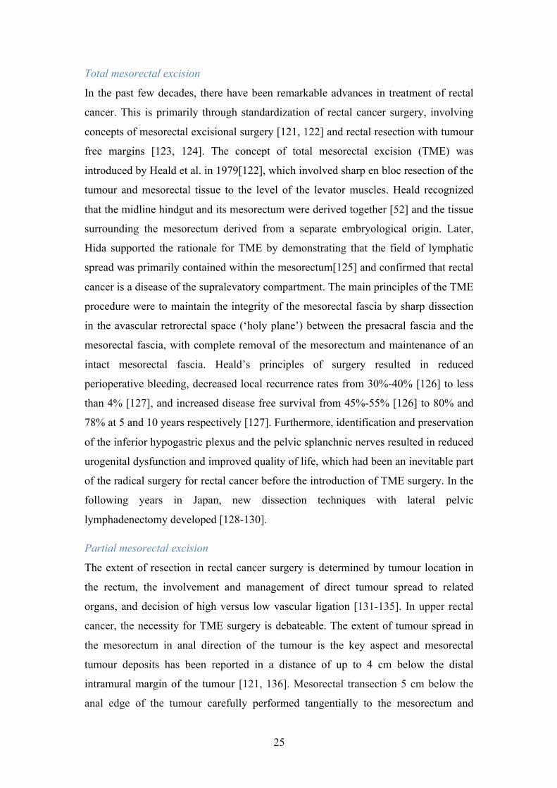
Total mesorectal excision
In the past few decades, there have been remarkable advances in treatment of rectal
cancer. This is primarily through standardization of rectal cancer surgery, involving
concepts of mesorectal excisional surgery [121, 122] and rectal resection with tumour
free margins [123, 124]. The concept of total mesorectal excision (TME) was
introduced by Heald et al. in 1979[122], which involved sharp en bloc resection of the
tumour and mesorectal tissue to the level of the levator muscles. Heald recognized
that the midline hindgut and its mesorectum were derived together [52] and the tissue
surrounding the mesorectum derived from a separate embryological origin. Later,
Hida supported the rationale for TME by demonstrating that the field of lymphatic
spread was primarily contained within the mesorectum[125] and confirmed that rectal
cancer is a disease of the supralevatory compartment. The main principles of the TME
procedure were to maintain the integrity of the mesorectal fascia by sharp dissection
in the avascular retrorectal space (‘holy plane’) between the presacral fascia and the
mesorectal fascia, with complete removal of the mesorectum and maintenance of an
intact mesorectal fascia. Heald’s principles of surgery resulted in reduced
perioperative bleeding, decreased local recurrence rates from 30%-40% [126] to less
than 4% [127], and increased disease free survival from 45%-55% [126] to 80% and
78% at 5 and 10 years respectively [127]. Furthermore, identification and preservation
of the inferior hypogastric plexus and the pelvic splanchnic nerves resulted in reduced
urogenital dysfunction and improved quality of life, which had been an inevitable part
of the radical surgery for rectal cancer before the introduction of TME surgery. In the
following years in Japan, new dissection techniques with lateral pelvic
lymphadenectomy developed [128-130].
Partial mesorectal excision
The extent of resection in rectal cancer surgery is determined by tumour location in
the rectum, the involvement and management of direct tumour spread to related
organs, and decision of high versus low vascular ligation [131-135]. In upper rectal
cancer, the necessity for TME surgery is debateable. The extent of tumour spread in
the mesorectum in anal direction of the tumour is the key aspect and mesorectal
tumour deposits has been reported in a distance of up to 4 cm below the distal
intramural margin of the tumour [121, 136]. Mesorectal transection 5 cm below the
anal edge of the tumour carefully performed tangentially to the mesorectum and

26
muscle tube seems adequate and TME surgery for upper rectal cancer is often
considered unnecessary for oncological reasons [137-139].
The concept of partial mesorectal excision (PME) evolved on basis of these findings
and the rationale is that this procedure is less extensive, results in a better long-term
functional outcome, decreases post-operative complication rates, and is an
oncological safe method that reflects outcome after TME surgery [35, 138, 140].
However, in Swedish series of resectional rectal cancer surgery a LR rate of 9% was
found in tumours of the upper rectum following PME surgery despite concomitant
treatment with neoadjuvant radiotherapy [141]. Interestingly the Swedish study
described radiological evidence of residual mesorectum (RM) in 86% of patients with
LR [141, 142]. Later, a Danish cohort study reported a 3-year LR rate of 7%
following resectional rectal cancer surgery performed between 2007 and 2010 in a
specialized colorectal referral centre [143]. The 3-year LR rate after PME surgery,
however, was found to be significantly increased compared to TME or APE surgery
(14% vs. 3% or 6%). In parallel to the Swedish observations, all patients with LR
after PME surgery had evidence of either RM or insufficient distal resection margin
(DRM) as evaluated by MRI [143].
Abdominoperineal resection
Abdominoperineal excision (APE) is performed in rectal cancer if sphincter-
preserving surgery is not an option, either for oncological or functional reasons, or in
frail and elderly patients where potential complications of attempts to restore
intestinal continuity are prohibitive [58, 144-146].
It is therefore of great importance preoperatively to determine the precise location of
the tumour in relation to the anal verge, the levator ani muscles, and the sphincter
complex, both radiologically and clinically [77]. In clinical practice, a distinction is
made between intersphincteric APE, conventional APE, extralevatory APE (ELAPE),
and ischioanal APE [11]. Intersphincteric APE is used in patients with increased risk
of functional problems, severe comorbidity or high age, and in patients with cT1 and
cT2 tumours if an anastomosis is not an option. Conventional APE is the traditional
method of rectal resection, as described by Miles, and indicated in selected patients
with cT1, cT2 or early cT3 tumours if intersphincteric APE is oncologically unsafe.
ELAPE can be used if tumour is inseparable from the puborectal/levator muscle(s) or
27
the internal sphincter. The method is recommended to avoid a waist at the specimen
in order to increase the chances for R0-resection [147]. ELAPE was introduced in
Denmark in 2007 with systematic implementation of the surgical technique at selected
centres and training with assistance from international experts. Ischioanal APE is
recommended in tumours involving the ischioanal compartment, either at the levator
or sphincter level. Reconstruction of the pelvic floor should be considered in patients
undergoing ELAPE and is always required after ischioanal APE.
In recent years, several authors have shown that oncologic outcomes after APE have
not improved to the same degree as those seen after implementation of TME
surgery. Compared with patients undergoing TME during the same time period,
patients undergoing APE have higher rates of local recurrence and poorer survival
[146, 148]. The difference in oncologic outcomes may be explained to a substantial
degree by the increased risk of tumour-involved margins (CRM) and inadvertent
bowel perforations associated with APE, as both of these factors are significantly
related to local control and survival [149].
Use of diverting stoma
A diverting stoma (DS) is created during restorative resection to reduce the
consequences of a possible anastomotic leakage and is recommended according to
Danish guidelines along with TME as part in the surgical treatment of mid and low
rectal cancer [11]. A DS is not routinely created during PME surgery for upper rectal
cancer [150]. In randomized trials it has been reported that anastomotic leakage rates
decline in patients receiving a DS after TME surgery with low anastomosis [5, 151-
155] while other trials found no effect of diversion [6, 8, 9, 156, 157]. The value of a
DS has been a subject for much debate as several trials are clearly indicating a
protective function of a DS as it reduces the consequences of an eventual leakage
(faecal peritonitis, lower rates of reoperation, reduced long-term morbidity, decreased
permanent stoma rates and reduced mortality rates) [5, 6, 8-10]. On contrary, stoma-
related complications are common, not to mention the risk of a permanent stoma.
Accordingly, many centres have adopted a highly selective approach to diversion
depending on both patient-related risk factors and anastomotic height with promising
results [157, 158].

28
Postoperative surveillance Postoperative surveillance is indicated for early detection of local and distant
recurrence after intended curative colorectal cancer treatment, for early diagnosis of
metachronous cancer, for prevention of rectal cancer by removal of adenomas in the
rectum, for psychosocial support, in assessment of the quality of treatment, to identify
long-term adverse events, and to initiate treatment of these. However, established
scientific committees have published guidelines with widely differing
recommendations for postoperative surveillance [159-162] and studies of clinical
practice in member countries of the European Society of Coloproctology (ESCP)
reveal great disparity at international level [162].
The standard surveillance programme according to the Danish Colorectal Cancer
Group (DCCG) guidelines is a computed tomography (CT) scan of thorax, abdomen,
and pelvis after 12 and 36 months, in addition to outpatient visits with rigid
proctoscopy after 6, 12, 18, 24, and 36 months [11]. Pelvic MRI is not a part of the
standard surveillance programme in Denmark.
An intensive surveillance program in patients undergoing intended curative colorectal
cancer surgery has previously been shown to increase survival by 7-8% [163-165].
However, a recent meta-analysis [166] and a systematic review [162] have questioned
these findings, as underlying studies are highly heterogeneous on follow-up regimens
[162, 166]. Asymptomatic recurrence of disease is more frequently detected when
patients undergo follow-up with short intervals, and may be surgically treated more
often than symptomatic LR [167, 168]. However, intensive surveillance programmes
has high costs for both patients and society, as it will result in a high degree of mental
distress and large economical expenses. Intended curative surgery for local recurrent
rectal cancer is achieved in less than 40% of patients as they are often diagnosed with
advanced disease [169, 170].
With the existing postoperative surveillance programmes, patients with recurrence of
disease have often exceeded possibilities of curative treatment due to delayed
diagnostics. The IMPROVE IT2 trial is a National Danish interdisciplinary initiative
to facilitate potential early detection of recurrence by cancer DNA testing in blood.
Preliminary results are promising, and it is expected that this program will increase
29
the proportion of patients eligible for intended curative surgery due to early diagnosis
[171, 172].
In currently available European guidelines, no consensus has been reached on
surveillance regimen after treatment of colorectal cancer (CRC) with curative intent.
The evidence for alternating international follow-up programs is limited, as current
multimodal follow-up methods has failed to show any impact on survival. Results
from current trials, investigating new biomarkers [171] and individualized ‘patient-
driven’ follow-up [173] are awaited and may potentially lead to international
consensus between countries being member of the European Society of
Coloproctology on a joint CRC surveillance program.
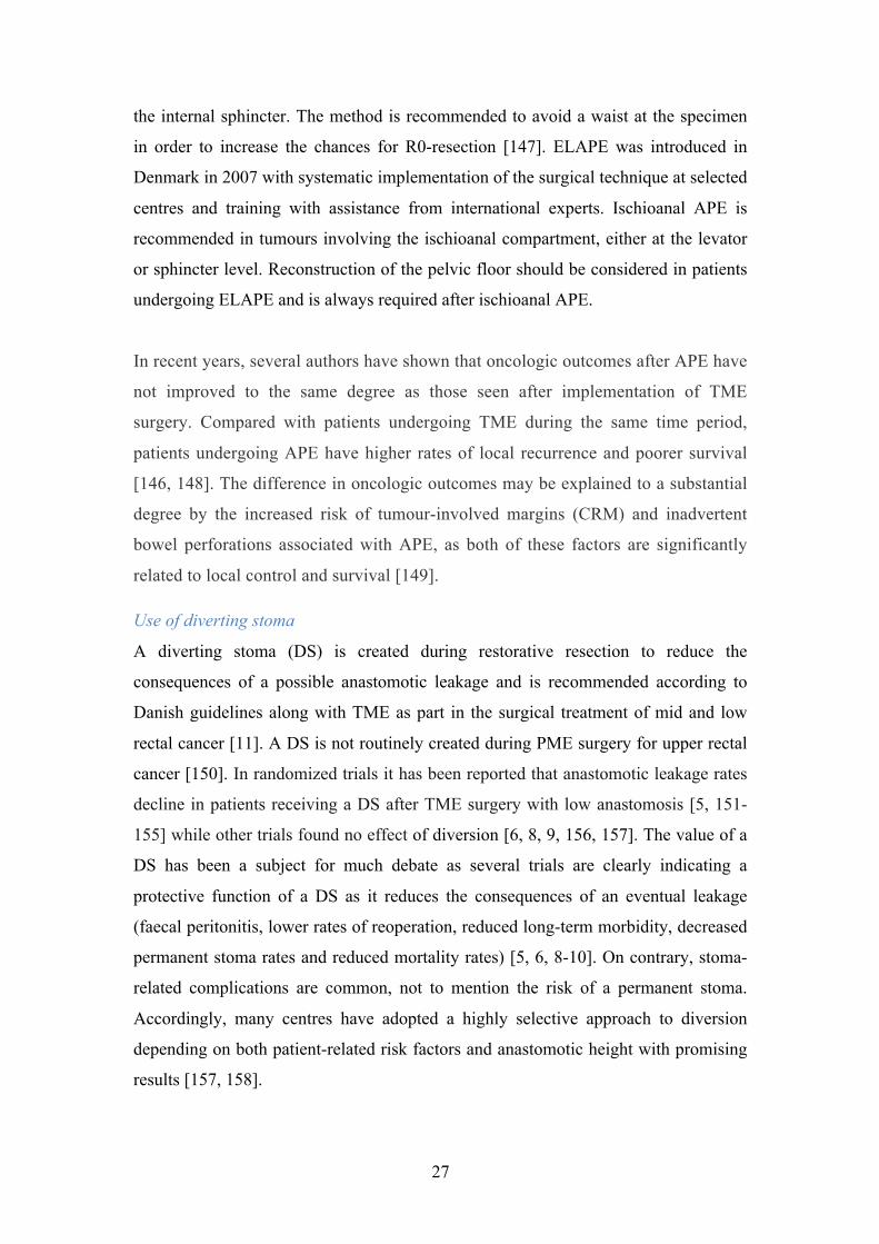
Figure 1 summarizes the timely relation between diagnostics and intended curative
treatment of rectal cancer in Denmark during 3-years follow-up.
Figure 1: Flow chart on diagnostics and intended curative treatment of rectal
cancer in Denmark during 3 years of follow-up.
* PIF may occur at any point of time during 3-year follow-up.
** Three-year postoperative MRI is not a part of the standard surveillance programme in Denmark.
*** Excluding a) low cT1-cT2, b) mid cT1-cT2 and cT3 with >2 mm to MRF and transmural
infiltration 0-5 mm, and c) upper cT1-cT3 and resectable cT4.
28
Start of follow-up
3-year follow-up incl. MRI**
36 month rectal cancer surgery!
12 month rectal cancer surgery
Reversal of diverting stoma
3-6 month rectal cancer surgery
PIF* Post-op MDT
Rectal cancer surgery
2. Pre-op MDT (6-8 weeks after CRT)
Neo- adjuvant CRT*** (6 weeks duration)
1. Pre-op MDT
Diagnosis of rectal cancer

30
Long-term adverse events following intended curative rectal cancer
treatment
Adverse events
Understanding the consequences of treatment is important to optimise patient support
and minimise impact on daily life. Adverse effects of long-course CRT combined
with rectal resection are well documented and include a broad variety of clinical
manifestations. Frequent and well-described complications with a substantial impact
on QoL are bowel dysfunction, sexual dysfunction, urinary problems, occasional
rectal bleeding, impaired wound healing, and mental distress among others [29, 31-
35]. Following restorative rectal cancer surgery, the long-term functional outcome is
poor in approximately half of the patients with frequent and fragmented stools,
urgency, and incontinence described as ‘low anterior resection syndrome’ (LARS)
[35, 174, 175]. A symptom scoring system assessing the severity of LARS (grouped
in three categories: no LARS, minor LARS and major LARS) has been developed and
validated in Denmark in 2012 [176]. The risk of severe bowel dysfunction is
increased in patients undergoing neoadjuvant CRT [35, 145, 177], in patients with
low anastomoses [178, 179], in patients undergoing TME vs. PME [35], following
anastomotic leakage [180, 181], in patients with more than two than anastomoses
[182-184], and the use of a diverting ileostomy is associated with twice the risk of
suffering from LARS [185, 186]. Thorough preoperative counselling emphasizing the
expected postoperative changes in bowel function and the possible of improvement
over time enables the patient to better assist in the choice of surgical method and to
better adapt to the situation [174].
Non-reversal of diverting stoma
Although stoma closure is considered a simple surgical procedure, the interval
between stoma construction and reversal is often prolonged, and some patients, DS
may never be reversed [18]. In recent years, much attention has been given to
perioperative management, morbidity, oncological outcome, and QoL analysis in
rectal cancer patients. Less attention has been given to long-term risk of permanent
stoma. Non-reversal of a DS or secondary construction of a permanent colostomy, are
the two main conditions responsible for a permanent stoma after intended restorative
rectal cancer resection. The prevalence of non-closure of intended temporary stomas

31
after rectal resection is reported as 3% to 32% after 1.5 to 7.1 years [12-22], and in
multicentre studies on rectal cancer patients only [12, 13, 18, 20], a risk of 17% [12]
to 25% [13] of a permanent stoma after intended restorative rectal resection is
reported, Table 3. Previously reported risk factors associated with non-reversal of an
intended temporary stoma include advanced age [13, 21], anastomotic leak [14, 17,
18], and metastatic disease [14, 20].
Recent studies from The Netherlands found no difference in the short-term
postoperative complication rates between patients undergoing rectal cancer resection
with DS by routine or if DS only was performed in highly selective patients [157,
158]. It seems that the ability to select patients for stoma construction is the key
towards preferable outcomes, not a risk adverse strategy [157].

32
Table 3: Previous studies on diverting stoma reversal after restorative rectal
cancer resection
Studies Setting Number
of
patients
Type of stoma Median
time to
reversal,
days
Follow-up,
years
(range)
Permanent
stoma rate
Bailey et al. 2003 [19] Local
(Reading, England)
n=59 End stoma/
Diverting stoma
- 4.0 (1.5-6.5) 9%
Lordan et al.
2006 [16]
Local
(Camberley,
England)
n=50 Diverting stoma 142 - ( - ) 32%
den Dulk et al. 2007
[18]
(Prospective design)
National
(Netherlands)
n=523 End stoma/
Diverting stoma
123 7.1 (2.5-9.8) 19%
David et al. 2010 [13] National (England) n=964 Diverting stoma 207 3.0 ( - ) 25%
Lindgren et al. 2011
[20]
(Prospective design)
National
(Sweden)
n=234 End stoma/
Diverting stoma
570 6.0 (3.5-9.0) 19%
Gessler et al. 2012 [14] Regional
(Västra Götaland,
Sweden)
n=262 Diverting stoma 178 2.8 (0-4.8) 23%
Dinnewitser et al. 2013
[17]
Local
(Salzburg, Austria)
n=98 Diverting stoma 240 5.1 (1.8-9.4)
Chiu et al. 2014 [21] Local
(Vancouver,
Canada)
n=162 End stoma/
Diverting stoma
347 - ( - ) 15%
Pan et al. 2016 [15] Local
(Beijing, China)
n=296 Diverting stoma 192 2.4 (1.8-8.3) 17%
Gustafsson et al. 2018
[12]
National
(Sweden)
n=3564 Diverting stoma 191 1.5 ( - ) 17%

33
Pelvic insufficiency fractures
Neoadjuvant CRT in the treatment of rectal cancer may result in a number of acute
and late toxicities [29, 31-35], and the widespread use of RT in rectal cancer patients
accentuates the importance of understanding these toxicities. PIF is considered an
uncommon late complication to pelvic radiotherapy and is acknowledged as a stress
fracture in structurally weakened bone [187, 188]. It can cause significant morbidity
[189-195] with reported symptoms in 16% to 58% of patients and chronic pain as the
most frequent clinical manifestation [196-198]. Unfortunately, patients with PIF often
face missed or delayed diagnosis and may mimic local recurrence of rectal cancer in
their presentation. However, PIF present characteristic patterns on modern modalities
of MRI, thus obviating the need for biopsies and other diagnostic interventions [36-
39].
Although many studies have investigated insufficiency fractures after radiotherapy for
gynaecologic cancers with PIF rates from 8% to 45% and other pelvic malignancies
(i.e. prostate and anal cancer) with PIF rates between 6% and 14% [191, 196-200], the
incidence and clinical course of insufficiency fractures in rectal cancer patients have
not been well characterized [190, 201, 202]. In previous small retrospective studies of
rectal cancer patients only, PIF rates between 3% and 11% have been reported, Table
4. During the course of the present study, we were increasingly aware of PIF as a
common radiological presentation on MRI in patients undergoing preoperative CRT.

34
Table 4: Studies on pelvic insufficiency fractures following intended curative
rectal cancer treatment
Authors
Inclusion
period
Patients and
treatment
Number
of
patients
Imaging
modality
Incidence
of PIF
Time to
follow-up
(years)
Fracture
site
Risk factors
Baxter et al.
2005 [190]
1986-
1999
Women ≥65
Surgery +/-
neoadjuvant
CRT
1,317
-
11%
5
Femur neck
(90%)
Pelvic ring
(10%)
Neoadjuvant
CRT
Herman et al.
2009 [202]
1989-
2004
Surgery +
neoadjuvant
CRT
562 CT 3% 3 Sacrum Female
gender
Kim et al.
2012 [201]
1998-
2007
Surgery +
neoadjuvant
CRT
582 CT, MRI 9% 4 Sacrum Age >60
Female
gender
Osteoporosis

35
Aims of dissertation
This dissertation aims to discuss aspects of the outcome of intended curative rectal
cancer treatment in Denmark with regard to long-term benefits and with a particular
focus on risk factors, but also on two less well-described adverse events.
The specific aims were as follows:
I. To estimate the 3-year overall survival rate and risk of recurrence in a well-
defined Danish cohort with a particular focus on risk factors.
II. To examine the use of diverting stoma and the stoma reversal rate 3 years
after intended restorative rectal cancer resection in Denmark.
III. To determine the prevalence and localization of pelvic insufficiency
fractures detected on 3-year postoperative MRI after mesorectal excision
surgery with or without neoadjuvant CRT.

36
Hypotheses
The specific hypotheses were as follows:
I. Several national initiatives during the last two decades has generally improved
rectal cancer outcome, however, recurrence rates following rectal cancer
resection with curative intent are underestimated due to inadequate
surveillance programmes with anachronistic and insufficient diagnostic
methods of follow-up.
II. A substantial part of patients undergoing intended restorative rectal cancer
resection with a diverting stoma would eventually end up with a permanent
stoma or significant delay in time to reversal and the risk may be extensively
underestimated in previous studies mainly due to varying inclusion criteria.
III. State of the art MRI detected pelvic insufficiency fractures are common in a
consecutive population undergoing comparable preoperative oncological
treatment across Denmark.

37
Methods
Study designs and settings The studies I and III were conducted on a well-defined Danish cohort of rectal cancer
patients 3 years after intended curative surgery with prospective sampling of data.
Study II, a population-based nationwide cohort study, was conducted in the setting of
the entire Danish population, a country with 5.8 million citizens [203]. The studies
were performed in accordance with the regulations of the National Board of Health
(ref.: 3-3013-1272/1/), the Scientific Committee of the Danish Colorectal Cancer
Group (DCCG.dk), and approved by the Danish Data Protection Agency (ref.:
2007-58-0010) pursuant to the Danish act on storage and processing of personal data.
The National Health Service in Denmark provides universal, tax-supported health
care to all citizens [204], guaranteeing free access to general practitioners and public
hospitals treating rectal cancer.
The following is a supplement and discussion of some of the applied methods in
Papers I to III.
Data sources
Civil Registration System (CRS)
Since April 1968, the Danish CRS has assigned a unique 10-digit personal
identification number (Civil Personal Register number) to every resident in Denmark
(1) born alive of a mother already registered in the CRS; (2) have their birth or
baptism registered in a Danish electronic church register; or (3) reside legally in
Denmark for 3 months or more [205]. The registry provides data on date of birth,
gender, residence, vital status (updated daily), and death. The CPR number permits
data linkage among registries in Denmark and within the healthcare system [206].
Danish Colorectal Cancer Group (DCCG) Database
Since May 2001, perioperative details on Danish colorectal cancer patients have been
consecutively reported to the DCCG database, including >60.000 registered patients
with approximately one-third rectal cancer and an overrepresentation of men [207].
The purpose of the database is to monitor the compliance and ensure uniform quality
in the treatment of colorectal cancer in Denmark with defined quality standards set by

38
the DCCG to improve the prognosis in this patient group. All surgical departments
across the country prospectively report data to the registry on patient performance and
comorbidity, diagnostic staging, treatment and postoperative complications (occurring
within 30 days after surgery) [207]. Pathological departments provide data on tumour
type, number of lymph nodes / metastatic lymph nodes, surgical margin status, and
other pathological risk factors. The database does not provide data on recurrences
after primary surgery. The estimated completeness in the DCCG Database was 99%
in the study period [207]. The DCCG database links to the CRS and variables of
patient demographics, tumour location, surgical type, and pathological T category
were retrieved from the database along with relevant data from the CRS.
The Danish National Patient Registry (DNRP)
Since 1977, the DNRP has expanded as a key Danish health register, maintaining
records on all hospitalizations including information on hospital diagnoses and
procedures [208]. Data were originally collected for administrative purposes only,
unrelated to research objectives. Since 2003, with the introduction of a private
healthcare sector in Denmark, it has additionally served as the basis for tax-funded
payment of both public and private hospitals via the Diagnostic Related Group
(DRG)-system [209]. Data include the CPR number, dates of admission and discharge
and up to 20 discharge diagnoses, among others. Diagnostic coding were performed
by physicians according to the 10th revision of the International Classification of
Diseases (ICD-10) from 1993 and onwards [209]. Registration of surgical procedures
in Denmark has been classified according to the Nordic Medico-Statistical Committee
(NOMESCO) Classification of Surgical Procedures since 1996. DCCG and DNRP
data were linked to obtain information on surgical events during follow-up.
Study cohorts
In the studies I and III, patients registered in the DCCG database with rectal
carcinoma (located ≤ 15 cm from the anal verge) and undergoing TME, PME, APE or
Hartmann’s operation with curative intent from April 2011 through August 2012,
were invited to participate in a national MRI study aiming to evaluate rectal cancer
outcome and specifically to detect LR 3 years after surgery. Approximately 1100
patients underwent treatment in 15 surgical centres in Denmark during the study
period [46]. Ten departments (Aarhus University Hospital, Odense University
Hospital, Regional Hospital Randers, Regional Hospital West Jutland, Hvidovre
39
Hospital, Zealand University Hospital, Slagelse Hospital, Svendborg Hospital, Vejle
Hospital, and Esbjerg Hospital), providing for approximately 65% of the entire
Danish population, were able to include patients. The remaining 5 departments
abstained from participation mainly due to deficient capacity.
Patients were examined for vital status through the DNRP based on CPR number and
eligibility was determined from a thorough review of medical records by the author.
All data on patient demographics including comorbidity, tumour characteristics,
cancer treatment, and follow-up data on recurrence of disease, were obtained from
medical records and retrieved for further analysis. Patients with disseminated disease
or local recurrence, or who had left the country, or terminated their surveillance
programme due to old age and/or advanced comorbidity, were exempt from invitation
to further examination. Included patients were offered a pelvic MRI at their 36-month
follow-up visit additional to standard examinations with proctoscopy and CT, in order
to obtain a more precise estimate of the incidence of recurrence at 3 years rectal
cancer surgery. Study populations in Paper I and Paper III are depicted in Figure 2.

40
Figure 2: Study populations in Paper I and III
In study II, patients registered in the DCCG database from May 2001 through April
2012 who were diagnosed with rectal carcinoma (located ≤ 15 cm from the anal
verge) and undergoing restorative rectal resection (TME, PME) or Hartmann’s
operation with curative intent (index procedure), were included using existing
national registries. Patients undergoing surgical procedures other than the above
mentioned or surgery with palliative intent, or had registration of a surgical procedure
in the DCCG without confirmation of in the DNRP, were excluded.
• Ineligible n=324: !• Local recurrence n=45
• Distant recurrence n=79 • Death within 3 years after surgery
n=106 • Terminated medical surveillance
programme n=81 • Emigration n=9
• No preoperative MRI n=4
Study population in Paper I:
First-time rectal cancer diagnosis between 1 April 2011
and 31 August 2012 and undergoing rectal cancer
resection (TME, PME, Hartmann or
APE) with curative intent at 10 of 15 hospitals in Denmark
n=890!
Candidates for post-operative MRI
n=566!
Study population in Paper III:
MRI performed 3 years postoperatively
n=403!
• Non-participation n=163:!• Declined invitation n=78
• Contraindication to MRI n=16 • Non-respondents n=69

41
Patients with a DS were followed from the date of the index procedure until the date
of stoma reversal, death, emigration or end of 3-year follow-up. Data on stoma
reversal or non-reversal within 3 years of follow-up among patients undergoing
restorative rectal cancer resection, were retrieved from the DNRP records of surgical
procedures via the NOMESCO classification system. This information was validated
through a systematic review of medical records in 9% of the patients. Study
population in Paper II is depicted in Figure 3.
Figure 3: Study population in Paper II.
Oncological and surgical treatment Oncological and surgical treatment was performed according to recommendations in
Danish guidelines as described in sections above (see Table 2 and Figure 1). National
guidelines recommend neoadjuvant long-course CRT (50 Gy in 25-28 fractions
• Intended restorative rectal cancer resection with no diverting stoma
n=2,876!• Hartmann's operation with formation
of end-colostomy n=1,534
Study population in Paper II:
Intended restorative rectal cancer resection or Hartmann's operation in Denmark between 1 May 2001 and 30 April 2012
n=6,859!
Intended restorative rectal cancer resection with diverting
stoma n=2,449!

42
combined with 5-Fluorouracil (5-FU)) to patients with locally advanced rectal cancer
[11]. Intended restorative rectal resection and Hartmann’s operation performed as
mesorectal excision were done 8-10 weeks after the completion of CRT. All other
patients underwent direct intended restorative rectal resection or Hartmann’s
operation. Short-course radiotherapy with immediate surgery is not routinely
performed in Denmark. Selected UICC stage II rectal cancer patients were offered 6
months of adjuvant chemotherapy. The same applied for UICC stage III rectal cancer
patients who had not received neoadjuvant CRT [11].
Histopathology The quality of the excised specimen was determined by pathologists according to the
classification system by Quirke et al. [144, 210]. The pathological examination
includes information from a) macroscopic evaluation (extent and dimensions of the
specimen, location of the index tumour, diameter of the index tumour, occurrence of
tumour perforation, and the plane of surgery), b) combined microscopic / macroscopic
evaluation (the depth of infiltration of the index tumour through the intestinal wall
including the CRM (involved CRM defined as any tumour, ENTD, or involved lymph
nodes ≤1mm from the lateral resection margin), the distal resection margin (DRM),
and the microradicality of the resection), and c) microscopic evaluation (histology and
degree of differentiation of the index tumour, tumour response after preoperative
neoadjuvant oncological treatment, infiltration to free peritoneum, infiltration of
neighbouring organs or structures, occurrence of lymphatic invasion, venous invasion,
nervous invasion, tumour budding, tumour satellites, number of metastatic and non-
metastatic regional lymph nodes). Further, the evaluation includes a description
tumour characteristic according to the TNM Classification of Malignant Tumours, 8th
(pTNM and UICC stage) [59].
3-year postoperative pelvic MRI All patients fulfilling the eligibility criteria in study I and III were invited to a 3-year
postoperative MRI in addition to the standard 3-year postoperative surveillance
programme. Those, who did not respond to the primary request received a
supplemental inquiry by telephone.
Postoperative 3-year pelvic MRI examinations were performed using 1.5 Tesla
(preferred) or 3 Tesla platforms with a detailed scan-protocol established by the

43
research group. Sagittal, axial and coronal T2-weighted turbo spin echo images, field
of view (FOV) 240 mm, slice thickness 4-5 mm, were obtained.
Additionally, sagittal short tau inversion recovery (STIR) sequence of the bony pelvis
(Figure 4) and a sagittal T2 3D sequence of the smaller pelvis were obtained. The
sagittal sequences covered the pelvis from tuber ischii to tuber ischii with the L5
depicted cranially.
Figure 4: Sagittal STIR sequence showing bone marrow oedema in Alae Ossis
Sacri
The axial sequence covered the entire pelvis from the lower border of the external
sphincter to the promontory, Figure 5. Sequences were performed covering the entire
anal canal scanned including the subcutaneous part of the external sphincter,
representing the anal verge on MRI.

44
Figure 5: Axial T2 was planned from sagittal T2
STIR images indicating bone marrow oedema accompanied by subtle linear changes
of low signal intensity at T2 weighted images (Figure 6), were regarded as suggestive
of PIF.
Figure 6: Axial T2-weighted MRI showing sclerosis and fracture in Alae Ossis
Sacri

45
The coronal sequence covered the pubic bone anteriorly and the sacrum posteriorly,
Figure 7. The sag T2 3D sequence covered the smaller pelvis with a slice thickness of
1 mm.
Figure 7: Planning of coronal T2 including the anterior edge of sacrum
In patients undergoing perineal reconstruction with a vertical rectus abdominis
musculocutaneous (VRAM) flap or similar, MRI scans included 'open air' caudally
and reached the promontory cranially since recurrences in these patients are often
located at the edges of the VRAM flap. In patients with perineal herniation the
VRAM flap may be quiet declive.
A dedicated multidisciplinary team radiologist at Aarhus University Hospital with 8
years of sub-specialisation in pelvic MRI re-evaluated all examinations and was
blinded to all clinical data with the exception of the preoperative MRI examination.
Recurrent disease Recurrent tumour mass outside the pelvic cavity, documented in the medical record
by radiological, clinical or histological examination, was defined as DR.
Recurrent pelvic tumour mass detected by clinical or radiological examination or
histologically verified adenocarcinoma as documented in the medical record or by the
3-year postoperative MRI was defined as LR, independent of the presence or absence
of concomitant DR. The MDT at Aarhus University Hospital evaluated any pelvic

46
tumour mass detected at 3-year postoperative MRI and suspicious of LR.
Histopathological confirmation was obtained if clinically relevant.
Outcomes
Overall survival and recurrent disease (Paper I)
In study I, outcomes were 3-year OS and cumulative incidence proportion (CIP)
(named risk) of DR and/or LR in UICC stage I-IV rectal cancer patients treated with
intended curative resectional surgery. Secondary outcome measures were significant
predictors of OS, DR, and LR.
Stoma reversal (Paper II)
In study II, outcomes were 1 and 3-year stoma reversal rates in patients undergoing
intended restorative rectal resection with a DS. Secondary outcome measures were
significant predictors of stoma reversal during 3-years of follow-up.
Pelvic insufficiency fractures (Paper III)
In study III, outcome was the PIF rate as detected by 3-year postoperative MRI in
patients following rectal cancer resection with curative intent with or without
preoperative CRT. Secondary outcome measures were significant risk factors of PIF
and the anatomical distribution of PIF.

47
Statistical analysis
Characteristics
Characteristics and demographics were presented as categorical variables by counts
and percentages in studies I, II, and III.
In study I, characteristics were presented in groups as patient-related,
pathology/tumour-related, or treatment-related variables, including rectal resection
procedure (PME, TME, Hartmann’s procedure, or APE), neoadjuvant treatment, and
histopathological examination.
In study II, patients were grouped according to the surgical procedure and
characterized pursuant to year of index operation and demographics. Patients with a
DS, patients with no stoma, or patients undergoing Hartmann’s procedure were
presented in separate groups.
In study III, continuous data were categorised and categorical data were compared by
Fisher’s exact test (univariate analysis).
Overall survival, cumulative incidence proportion, and competing risk analysis
(Paper I and II)
The OS rate and CIP of DR and/or LR at 3 years (Paper I) and stoma reversal at 1 and
3 years (Paper II) following rectal cancer surgery were calculated overall and for
various patient-, pathology/tumour-, and treatment-related factors treating death as a
competing risk. Kaplan-Meier survival curves were constructed. Disease-free survival
(DFS) was calculated as the rate of patients who were alive and had no sign of
recurrence 3 years after rectal cancer resectional surgery.
Cox proportional Hazards (Paper I and II)
Cox proportional Hazards regression analysis with computed Hazard ratios and
corresponding 95% confidence intervals were used to explore potential predictors for
OS, DR, and LR (study I) and stoma reversal (study II). Univariate and multivariate
analysis were performed. All predictors were entered into a multivariate Cox
regression model to adjust for potential confounders and to identify independent risk
factors.
In Paper I, potential predictive factors (risk factors) for OS, DR, LR were explored in
the following groups: patient-related risk factors: gender, age at surgery, Charlson

48
comorbidity index, ASA classification; pathology/tumour-related risk factors: tumour
height (low (0-5 cm) vs. mid/high (6-10 cm / 11-15 cm), (y)pT-category (according to
T category), UICC stage, and involved CRM (<1mm); and treatment-related risk
factors: use of pre-operative CRT, surgical approach, type of rectal resection,
intraoperative blood loss (0-300 ml vs. >300 ml), anastomotic leakage, and plane of
surgery.
In Paper II, potential predictive factors of stoma reversal included in the analysis
were: period of surgery, gender, age at surgery, body-mass index (BMI), Charlson
comorbidity index (CCI), ASA classification, distance of primary tumour (lower
edge) from anal verge (categorised as low (0 - 5 cm) vs. mid/ high (6 - 10 cm / 11 - 15
cm), use of neoadjuvant CRT, surgical approach, intraoperative blood loss,
anastomotic leakage, (y)pT category (according to T category), and UICC stage.
Multiple logistic regression (Paper III)
Factors found to have significance less than 0.1 in the univariate model were entered
into a multiple logistic regression model to identify independent predictors for PIF
Adjusted ORs for PIF were computed using multiple logistic regression to estimate
the impact of gender, age at surgery (categorised as <65 years or ≥65 years according
to the median age at time of surgery), use of neoadjuvant therapy, tumour height
(categorised as low vs. mid/high), surgical procedure (categorised as APE vs.
TME/PME), and pT/ypT-category (according to T-subcategory). A p-value of less
than 0.05 was considered significant.
Stata® version 12.0 (StataCorp, College Station, Texas, USA) was used for statistical
analysis.

Methodological considerations
Strengths
Studies I and III include a prospective design and well-defined study cohorts of 890 and 403
patients, respectively, undergoing treatment regimens that comply with modern standards and
a long follow-up period of 3 years. The data volume was of a considerable size and was
retrieved from a reliable register based on prospective national data sampling with high data
completeness [207]. The author reviewed medical records from all patients and information
from the data extraction were re-evaluated. In study III, the consequent use of MRI with STIR
sequences that is highly sensitive in detecting PIF strengthened the estimate significantly, and
further, blinding of the multidisciplinary team radiologist to clinical data reduced the risk of
information bias.
In study II, the nationwide prospective design and the large patient cohort constituted the
biggest strengths. Data were retrieved from three highly reliable registries with prospective
sampling of data and high data completeness [206-209, 211].
Limitations
Despite data being retrieved from validated databases, incorrect reporting of data from the
individual hospitals may occur. In study I, estimates of survival are based on data from the
Danish CRS and thereby very reliable. However, recurrence estimates are dependent on
methods of follow-up, including variations in quality of the clinical and radiological
examinations. Lack of follow-up according to the study algorithm and death between follow-
up may tend towards an underestimated rate of recurrence. The same applies for patients with
metastatic disease at various locations, whom will only rarely have all foci encoded.
However, recurrence of disease is not always histologically verified and an estimate of
recurrence on basis of information from medical records only, may, potentially, lead to an
overestimation of the actual recurrence rate.
In study II, incorrect reporting of data with coding errors related to operative procedures may
occur. However, data from DNPR regarding stoma status at 3-year follow-up were consistent
with data from medical records in a sample survey of 9% of patients who had rectal resection
with formation of a DS.
In study III, the primary limitation was the lack of information regarding clinical symptoms of
PIF (typically pelvic pain). Further, information on the exact radiotherapy regimen and
50
information on potentially predisposing conditions (osteoporosis, corticosteroid use etc.) for
development of PIF were not included.
Even though the DCCG Database has a high patient completeness, missing values are present
in some variables, like patient characteristics as height/weight, tobacco use, alcohol
consumption, ASA score, among other. Multiple imputation for missing data has not been
performed in any of the three studies.

51
Results
The following section intends to give a summary of the most important findings in Papers I to
III.
I: Comorbidity and UICC stage IV disease are main risk factors for decreased 3-year
survival and recurrence after intended curative surgery for rectal cancer – A population-
based study.
Jørgensen JB, Bondeven P, Laurberg S, MRI Study Group, Pedersen BG, Iversen LH.
In preparation for submission to Dis Colon & Rectum, August 2020.
In study I, survival and recurrence after intended curative resectional surgery for rectal cancer
was evaluated in a national trial by 3-year postoperative follow-up of 890 patients with pelvic
MRI, CT of the thorax and abdomen, and proctoscopy.
In total, 1,976 patients had rectal cancer diagnosed between April 2011 and August 2012.
Ninety-four patients undergoing local excision, 124 patients undergoing palliative surgery,
and 405 patients not undergoing surgery were excluded from the study. Accordingly,
resectional surgery with curative intend was performed on 1,353 patients. In total, 10 of 15
surgical departments treating rectal cancer were able to include patients for pelvic MRI at 3-
year postoperative follow-up, excluding an additional 463 patients, Figure 2.
Demographics, tumour characteristics, and treatment regimens of 890 patients included in the
study are summarised in Table 1, Paper I.
Survival In total, 169 patients died during 3 years of follow up after rectal cancer resection. Among the
deceased patients, 107 (63.3%) died without recurrence of disease. For UICC stage I-IV
patients, the 3-year overall survival rate was 80.6% and the 3-year disease-free survival was
73.0%. Restricted to UICC stage I-III patients, the 3-year overall survival rate and disease-
free survival rate was 83.6% and 77.1%, respectively. OS in stage I-IV rectal cancer patients
is depicted in Figure 8.

52
Figure 8: Overall survival rates of stage I-IV rectal cancer after intended curative
resectional surgery during 3 years of follow-up
Factors associated with particularly poor 3-year OS are summarized in Table 5. Statistical
significant factors predictive of decreased OS in multivariable analysis are presented in Table
6.
1.00
0.75
0.50
0.25
Survi
val ra
te
0 1 2 3Time since rectal resection (years)
Overall survival

53
Table 5: Rates of 3-year overall survival, and cumulative incidence proportions of
distant recurrence and local recurrence in stage I-IV rectal cancer patients, Denmark
2011-2012 (particular low survival rates and CIP’s are presented only)
* Three-year cumulative incidence proportion. Calculated treating death as a competing risk.
** UICC stage IV rectal cancer patients had curatively intended treatment of distant metastasis along with rectal
resection either during the index procedure or during an independent procedure in close timely relation to the
index procedure.
*** Circumferential resection margin
**** Includes intended laparoscopic surgery but converted to open surgery.
Characteristic 3-year overall
survival
% (95% CI)
Distant
recurrence
CIP3 *
% (95% CI)
Local
recurrence
CIP3 *
% (95% CI)
Total
80.6 (77.9; 83.1) 12.8 (10.6; 15.2) 6.5 (5.0; 8.4)
Age >65 years
75.9 (72.2; 79.4) - -
Charlson Comorbidity
Index ≥2
61.0 (52.6; 69.6)
- -
ASA-grade II
III-
IV
80.4 (76.8; 83.8)
61.2 (52.9; 69.6)
- -
pT/ypT-category
T3
T4
-
65.7 (55.5.;
75.6)
14.0 (11.0; 17.4)
17.9 (9.7; 28.2)
-
25.0 (16.1; 34.8)
UICC stage
III
IV**
-
58.7 (49.3; 68.5)
16.8 (12.4; 21.8)
46.3 (36.0; 56.1)
9.4 (6.2; 13.5)
12.6 (6.9; 20.1)
Involved CRM (<1 mm)***
63.2 (52.7; 73.8) 34.5 (23.9; 45.2) 19.7 (11.6; 29.3)
Laparotomy as surgical
approach****
73.7 (68.4; 78.7)
18.5 (14.1; 23.4)
11.6 (8.1; 15.8)
PME surgery
- 17.6 (13.1; 22.7) 11.0 (7.4; 15.3)
Blood loss >300 ml
(intraoperative)
69.8 (63.5; 76.0)
-
13.5 (9.1; 18.9)
Non-mesorectal plane of surgery 75.7 (71.0; 80.2)
- -

54
Table 6: Significant predictors of overall survival, distant recurrence and local
recurrence for stage I-IV patients undergoing intended curative rectal cancer resection,
Denmark 2011-2012.
Characteristic Overall survival
Distant recurrence
Local recurrence
Patient related Charlson Comorbidity
Index ≥2
(HR 2.72)
ASA grade III-IV
(HR 2.71)
Age >65 years
(HR 1.86)
Charlson Comorbidity
Index ≥2
(HR 2.15)
Charlson Comorbidity
Index ≥2
(HR 3.32)
Pathology/tumour
related
UICC stage IV
(HR 3.27)
UICC stage IV
(HR 8.86)
Involved CRM
(HR 2.27)
UICC stage IV
(HR 6.57)
Distance of primary
tumour (lower edge)
from anal verge 0-5 cm
(HR 4.60)
Involved CRM
(HR 2.72)
Treatment related Plane of surgery
Non-mesorectal
(HR 1.68)
PME surgery
(HR 4.70)

55
Recurrence For UICC stage I-IV patients, the overall CIP of rectal cancer recurrence following
resectional surgery with or without neoadjuvant treatment was 14.9%. The CIP of DR was
12.8% and the CIP of LR was 6.5%. Synchronous DR was found among 49% of the patients
with LR.
The majority of patients with DR were diagnosed between 0 and 12 months after surgery,
whereas the majority of patients with LR were diagnosed between 12 and 24 months after
surgery.
In 403 patients undergoing 3-year postoperative pelvic MRI, a previously undiagnosed LR
was found in 2% of the patients. The risk of LR after 3 years rectal cancer surgery was 5.6%
excluding LR detected by pelvic MRI. Accordingly, by including pelvic MRI in the follow-up
programme a 16.1% increase in the LR estimate was achieved.
Analysis of UICC stage I-III patients was performed in order to investigate if the outcome of
rectal cancer treatment Denmark is comparable with recently reported results from
neighbouring countries. Accordingly, an overall recurrence rate of 11.0%, a DR rate of 8.4%,
and a LR rate of 5.8% were found among UICC stage I-III patients.
CIPs of DR and/or LR in stage I-IV rectal cancer patients are shown in Figures 9-11.
Figure 9: Cumulative incidence proportion of local and/or distant recurrence in stage I-
IV rectal cancer patients during 3 years of follow-up
0.05
0.15
0.25
Cumu
lative
incid
ence
0 1 2 3Time since rectal resection (years)
Local and/or distant recurrence

56
Figure 10: Cumulative incidence proportion of distant recurrence in stage I-IV rectal
cancer patients during 3 years of follow-up
Figure 11: Cumulative incidence proportion of local recurrence in stage I-IV rectal
cancer patients during 3 years of follow-up
Factors associated with particularly low CIPs of DR and LR are presented in Table 5 (see
above). Statistical significant predictors of DR and LR in multivariable analysis are presented
in Table 6 (see above).
0.05
0.15
0.25
Cumu
lative
incid
ence
0 1 2 3Time since rectal resection (years)
Distant recurrence
0.05
0.15
0.25
Cumu
lative
incid
ence
0 1 2 3Time since rectal resection (years)
Local recurrence

57
II: Stoma reversal after intended restorative rectal cancer resection in Denmark. A
nationwide population-based study.
Jørgensen JB, Erichsen R, Pedersen BG, Laurberg S, Iversen LH.
BJS Open 2020: In press.
In total, 6,859 rectal cancer patients underwent intended restorative rectal resection or
Hartmann’s operation with curative intent in Denmark between May 1 2001 and April 2012
and comprised the study cohort (Figure 3). Excluded patients are shown in Figure 1, Paper II.
Interestingly, we found a general tendency towards increasing proportion of patients
undergoing restorative rectal resection with DS during the study period from 29.7% (2001 -
2004) to 42.2% (2009-2012). Accordingly, we found a generally declining trend for both
restorative rectal resection with no DS and Hartmann’s procedure, Table 7.
Comparing patients undergoing restorative rectal resection, the use of a DS was observed
more often among patients with low rectal tumours and in patients undergoing neoadjuvant
CRT. Demographics, tumour characteristics and treatment regimens are summarized in Table
7.
To ensure valid estimates for stoma reversal at 3-year follow-up, 225 medical records were
reviewed in a random audit by the author. Incorrect NOMESCO registration of stoma reversal
in medical records during follow-up or incorrect registration of the CPR number, caused
discrepancies for 7 patients (3.1%) as compared with the register-based status.

58
Table 7: Characteristics of 6,859 patients undergoing intended restorative rectal
resection or Hartmann’s operation, Denmark 2001-2012 Characteristic Rectal resection with
diverting stoma
n (%)
Rectal resection with no
stoma
n (%)
Hartmann’s
operation
n (%)
Number 2,449 (35.7) 2,876 (41.9) 1,534 (22.4)
Period of
surgery
May 2001–
Dec 2004
Jan 2005 –
Dec 2008
Jan 2009 –
Apr 2012
677 (29.7)
865 (35.4)
907 (42.4)
979 (43.0)
1,090 (44.6)
807 (37.7)
620 (27.2)
490 (20.0)
424 (19.8)
Gender Male
Female
1,551 (63.3)
898 (36.7)
1,571 (54.6)
1,305 (45.4)
943 (61.5)
591 (38.5)
Age (years),
median (range)
65 (20-91) 67 (29-97) 75 (28-94)
Charlson
Comorbidity
Index
0
1-2
+3
2,079 (84.9)
327 (13.4)
43 (1.8)
2,390 (83.1)
424 (14.7)
62 (2.2)
1,082 (70.5)
363 (23.7)
89 (5.8)
ASA grade I
II
III
IV
Missing
745 (30.4)
1,402 (57.2)
250 (10.2)
5 (0.2)
47 (1.9)
903 (31.4)
1,567 (54.5)
332 (11.5)
22 (0.8)
52 (1.8)
178 (11.6)
789 (51.4)
466 (30.4)
52 (3.4)
49 (3.2)
UICC stage I
II
III
IV*
Missing
655 (26.8)
763 (31.2)
839 (34.3)
175 (7.2)
17 (0.7)
613 (21.3)
1 013 (35.2)
943 (32.8)
277 (9.6)
30 (1.0)
283 (18.5)
526 (34.3)
430 (28.0)
270 (17.6)
25 (1.6)
Death within 3
years after
surgery
394 (16.1)
551 (19.2)
661 (43.1)
* UICC stage IV rectal cancer patients had curatively intended treatment of distant metastasis along with
intended restorative rectal resection or Hartmann’s operation either during the index procedure or during an
independent procedure in close timely relation to the index procedure.

59
Stoma reversal The cumulative incidence proportion of diverting stoma reversal after 1 and 3 years
restorative rectal cancer resection was 70.3% and 74.3%, respectively, (Figure 12) with a
median time from the index operation to stoma reversal of 173 days. The highest 3-year CIP
of stoma reversal at 80.0% was observed among patients with UICC stage I disease and the
lowest CIP at 45% in patients with anastomotic leakage.
Figure 12: Cumulative incidence proportion of stoma reversal during 3 years of follow
up*
*Cumulative incidence proportion was calculated treating death as competing risk
Particularly low 1 and 3 year CIPs of stoma reversal in patients undergoing intended
restorative rectal cancer resection with a DS are presented in Table 8. Independent predictors
of delay in time to stoma reversal in multivariable analysis are shown in Table 9.
0.25
.5.75
1Cu
mulat
ive In
ciden
ce of
stom
a rev
ersal
0 1 2 3Time (in years) since rectal resection with diverting stoma

60
Table 8: One and three-year cumulative incidence proportions of stoma reversal in
patients undergoing intended restorative rectal cancer resection with diverting stoma,
Denmark 2001-2012 (particular low CIP’s are presented only)
Characteristic CIP1*
(95% CI)
CIP3*
(95% CI)
Total
0.70 (0.68;0.72) 0.74 (0.73;0.76)
Age (y)
>65 - 0.72 (0.69;0.74)
ASA grade III-IV 0.60 (0.54;0.66) 0.64 (0.58;0.69)
-
Neoadjuvant (chemo-)
radiotherapy
0.64 (0.60;0.67) 0.68 (0.64;0.71)
Blood loss,
intraoperative (ml)
>300
0.66 (0.63;0.69)
0.70 (0.67;0.73)
Anastomotic leak 0.33 (0.29;0.38)
0.45 (0.39;0.51)
(y)pT-category T3
T4
0.68 (0.65;0.71)
0.61 (0.50;0.70)
0.73 (0.70;0.75)
0.66 (0.56;0.74)
UICC stage III
IV
0.66 (0.63;0.69)
0.50 (0.42;0.57)
0.71 (0.68;0.74)
0.57 (0.50;0.64)

61
Table 9: Significant hazard ratios (0-3 years), of predictive characteristics associated
with delay in stoma reversal for patients with intended restorative rectal cancer
resection with diverting stoma, Denmark 2001-2012 Characteristic Crude
Hazard-ratio*
(95%CI)
Adjusted
Hazard-ratio**
(95%CI)
Total
- -
ASA grade I
II
III-IV
1.0 (ref)
0.87 (0.79;0.96)
0.75 (0.63;0.89)
1.0 (ref)
-
-
Neoadjuvant (chemo-)
radiotherapy
Yes
No
0.76 (0.69;0.85)
1.0 (ref)
0.75 (0.66;0.85)
1.0 (ref)
Blood loss,
intraoperative (ml)
≤300
>300
1.0 (ref)
0.80 (0.73;0.88)
1.0 (ref)
0.86 (0.76;0.97)
Anastomotic leakage
Yes
No
0.42 (0.35;0.50)
1.0 (ref)
0.41 (0.33;0.50)
1.0 (ref)
(y)pT-category T1
T2
T3
T4
1.0 (ref)
0.83 (0.69;0.99)
0.63 (0.53;0.74)
0.51 (0.39;0.69)
1.0 (ref)
-
0.63 (0.47;0.83)
0.62 (0.42;0.90)
UICC stage I
II
III
IV
1.0 (ref)
0.86 (0.76;0.97)
0.65 (0.58;0.73)
0.42 (0.34;0.52)
1.0 (ref)
-
-
0.57 (0.41;0.80)
* COX regression analysis. Hazard ratio (HR) <1 describes reduced “risk” of stoma reversal (i.e. the “risk” of
early stoma closure is reduced when HR<1).
** Mutually adjusted

62
III: Pelvic insufficiency fractures are frequent after preoperative chemo-radiotherapy for
rectal cancer – A nationwide MRI study
Jørgensen JB, Bondeven P, Iversen LH, Laurberg S, Pedersen BG.
Colorectal Disease. 2018; 20(10):873-80
Between April 1, 2011 and August 31, 2012, 890 patients underwent rectal cancer resection
with curative intent at 10 hospitals in Denmark. In total, 403 patients had 3-year postoperative
pelvic MRI and comprised the study cohort, Figure 2. Of these, 116 patients underwent
neoadjuvant CRT and the frequency (28.8%) of neoadjuvant treatment was comparable to the
group of patients not included for postoperative MRI (33.3%). Details regarding the study
cohort are described in Results/Study 1 and illustrated in Figure 2. Patient demographics,
tumour characteristics, and treatment regimens are summarised in Table 1, Paper III.
Pelvic insufficiency fractures In total, 49 patients had PIF detected on 3-year postoperative pelvic MRI and the rate of PIF
was 12.2% overall. We found a significantly increased PIF rate of 33.6% among patients
undergoing preoperative radiotherapy. The rate of PIF in the non-irradiated group of patients
was 3.5% (p<0.001). Risk factors significantly associated with PIF in both univariate and
multivariate analysis are presented in Table 10.
The localization and anatomical distribution of pelvic insufficiency fractures as detected by 3-
year pelvic MRI is shown in Figure 13. The sacrum and the ileum (near the sacroiliac
articulations) were found to be the anatomical predilection sites of PIF observed in 95.9% and
91.8% of the patients, respectively.

63
Table 10: Risk factors significantly associated with PIF.
OR = odds ratio, CI = confidence interval.
* Measured by rigid proctoscopy at pre-treatment clinical evaluation.
** Based on histopathological evaluation of excised specimen. The pathological tumour category for the 115
patients who had neoadjuvant CRT was: ypT0, 15; ypT1, 9; ypT2, 32; ypT3, 51; ypT4, 8). One patient with T0
did not receive CRT. This patient underwent local excision of the tumour prior to definitive surgery and the T-
categorization here was T0.
*** Fisher’s exact test.
**** Multiple logistic regression adjusted for gender, age at surgery, tumour height, surgical procedure,
neoadjuvant therapy and pT/ypT-category.
Characteristic Patients with
PIF
n (%)
Unadjusted
p***
Adjusted OR
(95% CI)****
Number
49 (12.2) . .
Gender Male
Female
22 (8.7)
27 (17.9)
0.008 3.52 (1.7; 7.5)
Age (y) <65
≥65
16 (8.4)
33 (15.6)
0.032 3.20 (1.5; 6.9)
Distance of primary
tumour from
anal verge (cm)*
0-5
>5-10
>10-15
26 (26.8)
22 (12.8)
1 (0.8)
<0.001 1.75 (0.89; 3.42)
Neoadjuvant (chemo-)
radiotherapy
No
Yes
10 (3.5)
39 (33.6)
<0.001 14.2 (6.1; 33.1)
Surgical preocedure PME
TME
APE
5 (4.2)
14 (8.1)
30 (26.5)
<0.001 2.17 (0.71; 6.58)
pT/ypT-category** T0
T1
T2
T3
T4
8 (50.0)
3 (14.3)
15 (12.8)
20 (9.3)
3 (10.3)
0.001 0.81 (0.56; 1.17)

64
Figure 13: Anatomical distribution and frequency of pelvic insufficiency fractures

65
Discussion
This thesis presents long-term benefits and risks of intended curative rectal cancer treatment
in Denmark. Three different studies have been conducted to elucidate some common and
critical challenges in the long-term postoperative course after rectal cancer treatment.
Survival We found a 3-year OS rate of 81% and a DFS rate of 73% in UICC stage I-IV rectal cancer
patients treated with intended curative resectional surgery in a well-defined Danish cohort.
Excess mortality the first 12 months after diagnosis in Denmark as compared to other
countries, has repeatedly been demonstrated in cohorts diagnosed between 1960’s until the
end of 1990’s [212-214]. The present results indicate that survival rates in rectal cancer
patients in Denmark are now comparable to rates observed in other Scandinavian countries
with similar health systems [54] and more favorable as compared to survival estimates in
OECD cancer care 2013 [215]. Present estimates are based on data from the national clinical
DCCG database with a patient coverage of at least 99% since 2010 and histological
verifications of the great majority of patients.
Standardization and quality assurance of mesorectal excision by training and pathological
audit were implemented in previous trials to ensure that optimal surgery was performed [144,
210]. We have demonstrated favorable survival and recurrence rates for Danish patients
diagnosed with rectal cancer from 2011 through 2012, after implementation of several
initiatives aiming to improve outcome of rectal cancer therapy nationally. During the last two
decades, a national strategy has been initiated with centralisation of surgery from 47 to 15
surgical departments involved in rectal cancer treatment in Denmark. Additionally, the
implementation of mesorectal excision surgery and national guidelines for rectal cancer
treatment, repeated postgraduate multidisciplinary training and teaching programmes,
specialization of personnel involved in MDT (colorectal surgeons, oncologists, pathologists,
and dedicated radiologists), and establishment of continuous data reporting to a national
database, has markedly optimised outcome of rectal cancer [1, 3].
UICC stage IV disease was the most important predictor for decreased overall survival with a
three times higher risk of death during follow-up as compared to UICC stage I-III patients

66
(HR 3.27). Other independent patient-related predictors for decreased 3-year OS were age
>65 years (1 in 4 patients dying during follow-up), ASA grade III-IV (1 in 3 patients dying
during follow-up), and Charlson comorbidity index ≥2 (1 in 3 patients dying during follow-
up), all of which are not surprising findings in the view of their life-threatening character.
Recurrence The observed 3-year DR rate of 12.8% and LR rate of 6.5% are low rates [25, 54, 58]. With
inclusion of a 3-year pelvic MRI, the LR rate was increased by 16% (from 5.6% to 6.5%).
Reported LR rates in the modern era of rectal cancer treatment with mesorectal excisional
surgery and neoadjuvant CRT are highly variable ranging from 4% to 14% [54, 216, 217].
UICC stage IV disease was the most important predictor for recurrence with a DR rate of 46%
and LR rate of 13%. Charlson comorbidity index ≥2 was the only patient-related risk factor
predictive of recurrence in multivariate analysis and increased the risk of both DR and LR
significantly. The negative impact of comorbidity on recurrence may be due to suboptimal
oncological treatment in patients with excessive chronic illness due to either omission or
interruption of neoadjuvant CRT, and/or compromised surgery with less central ligation and
less extensive mesorectal excision (confounding-by-indication). Alternatively, comorbidity
may increase the risk of recurrent disease per se, and comorbidity obviously has negative
impact on survival.
Internationally, PME surgery and omission CRT for cancer in the upper rectum have resulted
in poor outcomes with LR rates between 9% and 16% [141, 143, 216, 218]. Results from the
present study confirm these findings with a significantly increased LR rate of 11.0% in
patients undergoing PME surgery. Data from the DCCG reveals a tendency to overestimate
tumour height in the rectum by rigid proctoscopy with preoperative MRI as reference [48].
The differentiation between mid or upper rectum has a significant impact on the treatment
regimen (see Table 2). Furthermore, radiological evidence of residual or inadvertent
mesorectum has been observed in 63% to 74% of patients undergoing PME surgery,
indicating suboptimal quality of the surgery performed [63, 141-143]. These findings may
partly explain the higher incidence of LR in patients undergoing PME surgery for rectal
cancer.

67
In previous studies, R1 resection with CRM involvement (margin <1mm) is negatively
associated with outcome of curatively intended rectal cancer treatment [79, 219-221]. In the
present study, a significantly increased DR rate of 35% and a LR rate of 20% were observed
after 3 years, including a markedly decreased OS rate of 63%.
Thus, present study suggests that the outcome of rectal cancer is hampered due to particularly
two specific determinants: the proportion of patients presenting with UICC stage IV disease
and the management of patients with severe comorbidity. Therefore, efforts aiming to
decrease their proportions should be addressed. Furthermore, R0 resection is critically
important both to reduce recurrence rates and improve survival.
Since implementation of a national colorectal cancer screening programme in March 2014,
data from the DCCG indicates a permanent reduction in the proportion of patients with UICC
stage IV disease [41, 222]. The proportion of UICC stage IV disease has decreased from 23%
in 2012 (study period in Paper I) [46] to 16% in 2018 [41], according to annual DCCG
reports. Additionally, reported data in study I, indicates that survival has markedly improved
following curatively intended treatment of UICC stage IV disease, with a 3-year OS of 59%
(Table 5). An additional gain may be achieved through reduction of patient delay by increased
symptom awareness, and equally important, developing an understanding of when to act on
these symptoms [223, 224].
The negative impact of comorbidity in an aging population may be addressed by optimization
of surgical decision-making, and efforts should be directed against evaluation of the
adequateness of pre-, intra-, and postoperative therapy. Traditional approaches have targeted
the postoperative period for rehabilitation and lifestyle changes. However, recent evidence
shows that the preoperative period might be the optimal moment for intervention with
smoking cessation, psychological coping, individualized training programmes, and nutritional
support [225, 226]. Multimodal prehabilitation may improve functional capacity and reduce
postoperative complication rates in extensively comorbid rectal cancer patients, resulting in
improved OS [225].

68
Surveillance in rectal cancer has repeatedly failed to prove any impact on rectal cancer
outcome and has primarily been focused on detection of recurrent disease [227-230].
However, in the present study, nearly two thirds of the patients dying within 3 years of
follow-up had no sign of recurrence. The effect of various surveillance programmes may
remain limited, as the majority of patients dying within 3 years of rectal cancer treatment will
not die from recurrence of disease.
Three-year postoperative pelvic MRI of 403 patients, revealed a previously undetected LR in
8 patients only. Accordingly, number needed to examine was 50 patients to detect 1 extra
patient with LR. Further, considering that only 40% of patients diagnosed with LR will be
potential candidates for curatively intended surgery, 3-year pelvic MRI may be a poor
investment. As the majority of patients with LR were diagnosed between 12 and 24 months
after surgery, the best possible timing of pelvic MRI may be approximately 12 months
postoperatively in order to detect non-symptomatic LR’s. Considering that it may be possible
to diagnose LR in an early stage, the rate of possible candidates for curatively intended
surgery may increase above 40%.
Stoma reversal Approximately 45% of patients undergoing intended restorative rectal resection in Denmark
between 2001 and 2012 had a DS and only 74% of these patients had their stoma reversed
during 3 years of follow-up. In previous multicenter studies investigating stoma reversal rates
in rectal cancer patients, the reported permanent stoma rate after intended restorative rectal
resection together with a DS is similar to our findings with a risk of 17-25% [12, 13, 18, 20].
However, these studies are few and obvious differences in baseline characteristics of the
patient cohorts may lead to non-comparable results [13, 18, 20]. In a recent Swedish
retrospective multicenter study 3,564 patients underwent restorative rectal cancer resection
with a DS. Stoma reversal was performed in 2,954 (82.9%) patients during 1.5 years of
follow-up. However, the follow-up was short and information on stoma use at index surgery
was unavailable, and the study included no validation on clinical information from Swedish
registries [12]. The total number of patients undergoing restorative rectal cancer resection is
unknown and, accordingly, the proportion of patients undergoing restorative rectal resection
unspecified.

69
Accordingly, results from previous studies cannot be extrapolated directly as the inclusion
period; baseline characteristics of the cohorts, type and use of diverting stomas at index
surgery, and period of follow-up are differing.
Risk factors for stoma non-reversal within 3 years in present study were all of anastomotic
leakage, advanced UICC stage IV, T category 4, T category 3, use of neoadjuvant CRT, and
perioperative blood loss > 300 ml. Consistently, previous studies have found that increasing
age and comorbidity [13, 15, 20, 21, 23], advanced T category and UICC stage [14, 15, 19,
20], any postoperative complications [14, 15, 18, 20] and anastomotic leakage [17, 19, 21]
increase the probability of a permanent stoma after rectal cancer resection.
Interestingly, our study, including patients undergoing restorative rectal cancer resection or
Hartmann’s operation, revealed an increasing proportion of patients with a DS from 2001-
2004 to 2009-2012, with a 30% rate in the early period to a 42% rate in the late period.
Similarly, the proportions of patients undergoing restorative rectal resection without a DS or
Hartmann’s operation decreased. Revision of national guidelines in 2009 and methodological
alignment between departments are the most obvious explanations for this general change in
surgical approach, and suggest that surgeons may be increasingly more cautious for
anastomotic leakage following restorative rectal cancer resection and thus, defunction with a
DS. However, according to DCCG data, anastomotic leakage rates following rectal cancer
resection in Denmark were increasing from 2001 (11.0%) to 2012 (15.6%) [46, 231].
Furthermore, recent studies from the Netherlands of patients undergoing restorative rectal
cancer resection with DS found no difference in the short-term postoperative complication
rates between routine use and highly selective use of a DS [157, 158]. Findings from present
study suggest that selection of specific patient groups for stoma construction may be a key
element in optimizing long-term patient outcomes. Thus, the future trend may likely change
toward a more selective approach to DS construction during index surgery.
Pelvic insufficiency fractures Study III reports the 3-year rate of pelvic insufficiency fractures in patients undergoing
curatively intended rectal cancer treatment in Denmark. The main finding was an
unexpectedly high PIF-rate of 33.6% in patients undergoing neoadjuvant CRT followed by

70
mesorectal excision surgery. In the group of patients treated with surgery alone, the
corresponding PIF rate was 3.5% only, which is equivalent to rates in the background
population [232]. Despite technological advances with development of modern intensity
modulated radiotherapy, we have demonstrated a risk of PIF following CRT that markedly
exceeds the expected level of 3-11% in literature [190, 201, 202]. As MRI constitutes a
particularly sensitive tool in detection of PIF, the consequent use of MRI in study III with
sequences (STIR) that are specifically suitable in detecting PIF (i.e. bone marrow oedema)
may likely explain the difference between detection rates of PIF in previous studies and the
present study [37].
In previous studies, reported predisposing conditions for development of PIF is primarily
osteoporosis and radiotherapy, including a number of medical conditions associated with
development of osteoporosis. Thus, elderly postmenopausal women constitute a group of
patients particularly vulnerable to PIF [189, 190, 233]. Accordingly, in present study,
independent risk factors for PIF were female gender (OR=3.52), age above 65 (OR=3.20),
and preoperative CRT (OR=14.20).
Consistent with observations in other studies of PIF, the anatomical predilection sites were
found in the sacrum and the medial part of os ileum near the sacroiliac articulations, which
are the weight-bearing parts of the pelvic ring. Performing specific MRI sequences with
identification of characteristic patterns in specific areas of the pelvic ring will assist in
differentiating PIF from LR in rectal cancer patients with pelvic pain. MRI examination in
patients who are examined on suspicion of recurrence should include bone-specific
sequences, as PIF may be the only (visible) explanation for their pain. Many patients will be
able to cope with their symptoms, if they are thoroughly examined for recurrence of disease.
Future MRI studies combined with QoL assessment are needed to evaluate pelvic pain in
relation to PIF in patients treated for rectal cancer.
New regimens for neoadjuvant therapy may have potential to reduce the risk of PIF.
Preoperative 3 arc volumetric arc therapy (intensity modulated radiotherapy) has
demonstrated pronounced bone sparing capacity and proton beam therapy have the potential

71
to further lower doses [234]. However, precise dose planning would lower the exposure of
pelvic bones regardless of the applied technique [234].

72
Conclusions
In summary, this thesis presents favorable three-year outcome of intended curative rectal
cancer treatment in Denmark with high OS and low recurrence rates for UICC stage I-IV
patients diagnosed from 2011 through 2012. However, less advantageous long-term outcome
has been demonstrated in patients undergoing intended restorative rectal cancer treatment
with a concerning high permanent stoma rate. Furthermore, neoadjuvant CRT in curatively
intended treatment of rectal cancer is associated with a surprisingly high risk of PIF during 3
years of follow-up. Potential risk factors for poor 3-year outcome of rectal cancer treatment in
all three studies were specifically related to patient age and comorbidity (low OS, high
recurrence rates, and high PIF rates), neoadjuvant CRT (non-reversal of DS and high PIF
rates) and UICC stage IV (low OS, high recurrence rates, and non-reversal of DS).
Specific conclusions:
• Intended curative treatment of rectal cancer including UICC stage IV conveys a high
3-year OS and low DR and LR rates. The main risk factors of poor outcome are UICC
stage IV and Charlson comorbidity index ≥2.
• One quarter of rectal cancer patients undergoing intended restorative resection had not
their diverting stoma reversed within 3 years after surgery. The permanent stoma rate
increased more than 100% if patients developed anastomotic leakage postoperatively.
• Neoadjuvant CRT in the treatment of rectal cancer was associated with a 14 times
higher risk of PIF after 3 years, whereas female gender and age above 65 years each
tripled the risk of PIF detected by MRI.

73
Perspective
Treatment of rectal cancer is constantly changing with optimized principles of surgery;
technical advances in radiotherapy, modified imaging modalities, and improved pathological
and immunohistochemical audits of the specimen. These changes are further driven forward
by the implementation of colorectal cancer screening in the general population, an increased
focus on non-surgical management of rectal caner in patients with pCR after neoadjuvant
treatment, increasing use of minimally invasive surgical techniques, investigation of the
potential of circulating tumour DNA in diagnosis and surveillance of rectal cancer, and trials
examining the role of preoperative chemotherapy as a potential successor to preoperative
radiotherapy.
The most optimal strategy to reduce the rectal cancer burden is primary prevention of disease
with a lowering of its incidence. The incidences may decline with continuous efforts to
improve unfavourable and characteristic lifestyle in the Western populations in addition to the
positive effects already seen after implementation of the national CRC screening programme.
Therapeutic delay has been reduced by several initiatives from the National Board of Health
since the introduction of ‘the National Cancer Plan’ in 2000, and the two-week waiting time
guarantee from diagnosis to treatment and fast track cancer packages introduced by the
Government (latest revision in 2012).
In rectal cancer surgery, patient age and comorbidity determines the number and severity of
complications and predicts poor oncological outcome as it is closely related to preoperative
functional capacity, nutritional state and psychological reserve. Recent studies show that the
preoperative period may be the optimal moment for intervention (prehabilitation) in comorbid
and elderly patients contrary to traditional approaches targeting the postoperative period for
rehabilitation and lifestyle changes. A randomised controlled trial is initiated to explore
possible benefits of multimodal prehablitation as determined by effect on complication rates
and survival[225, 235], among others.
On-going randomised controlled trials [112, 113] are investigating the effect of neoadjuvant
chemotherapy on recurrence of rectal cancer. The main hypothesis is that compared to

74
neoadjuvant CRT, neoadjuvant chemotherapy reduces the rate of distant recurrence without
increasing the rate of local recurrence. Furthermore, surgical and medical complications, the
functional outcome, toxicity and QoL may be improved if radiotherapy can be avoided.
Avoidance of radiotherapy, potentially leading to reduced complication rates, may
additionally increase the number of patients capable of receiving postoperative oncological
treatment. However, according to findings in the present study, the effect of chemotherapy
may be limited, as the majority of patients dying within 3 years of rectal cancer treatment will
not die from recurrence of disease. Further, among those dying within 3 years after surgery
the majority die from comorbidity, which will limit these patients from receiving
chemotherapy at all.
Moreover, patients undergoing preoperative RT are exposed to a particularly high risk of
postoperative complications, increasing both mortality and long-term adverse events.
Neoadjuvant preoperative chemotherapy with omission of RT, may potentially reduce
postoperative complications in these patients leading to increased survival and reduced rates
of long-term adverse events. Yet another concern may be that neoadjuvant CRT is a greater
burden for patients with advanced comorbidity, resulting in a higher mortality. Data are
biased by confounding by indication, restraining possibilities of interpretation of causal
relationships.
In selected groups of UICC stage I patients unable to undergo surgery (i.e. due to high age or
advanced comorbidity), however, neoadjuvant CRT provides various potential advantages. It
allows for early re-assessment of disease by MDT, and may potentially enable the
consideration of organ preservation by allowing for more effective local excision and non-
operative management strategies. In light of various watchful waiting protocols evaluating
deferral of surgery after radiation therapy of early rectal cancers, pelvic MRI may in future
not only be a useful tool to monitor the effect of treatment or the presence of local recurrence,
but may also be a useful tool for the evaluation of more gentle ways to irradiate rectal
tumours.
There is an urgent need in future studies to explore new methods of surveillance in rectal
cancer patients, as previous studies evaluating various surveillance programmes, has found no
impact on colorectal cancer outcome [227, 229, 230]. The COLOFOL trial[236], investigating

75
the impact of frequent CT scans in addition to the normal surveillance programme, found no
significant effect on DR. In the present study, we found that additional pelvic MRI had only
limited effect on detection of LR. The on-going FURCA trial [230], investigates the effect of
patient-led follow-up based on patient education and self-referral, with recurrence as primary
outcome. The IMPROVE-IT2 trial [171, 172] investigating circulating tumour DNA guided
post-operative surveillance, may result in earlier detection of recurrent disease and identify
more patients eligible for curative treatment. This method of surveillance may potentially
provide information about radicality of the primary resection, response to adjuvant therapy,
incipient recurrence and response to treatment performed in relation to diagnosis of
recurrence. However, the findings in the present study suggest that high attention should
primarily be paid to reduce comorbidity, as the majority of patients dying within 3 years have
no sign of recurrence.
Patients undergoing preoperative RT with the current modalities of intensity-modulated
radiotherapy are prone to develop PIF with multiple fracture sites, as detected by pelvic MRI
at 3-year postoperative follow-up. A more selective use of CRT, precise dose planning, and
new technologies, including proton beam therapy, have potential to increase the bone sparing
capacity with further lowering of doses. We await results to be validated in larger and
preferably prospective cohorts [234].

76
Summary
This dissertation is based on three original articles, two of which have been accepted and/or
published in international scientific journals and a third is in preparation for submission. The
papers have not previously been included in a dissertation. The work was funded by the
Danish Cancer Society and performed between 2014 and 2020 while the author was
employed at Department of Surgery, Aarhus University Hospital / Regional Hospital Randers.
This thesis presents long-term benefits, risks and adverse events 3 years after intended
curative rectal cancer treatment in Denmark. The papers I-III contain an evaluation of 3-year
survival and recurrence, and attempts to illuminate important adverse events related to rectal
cancer treatment, affecting the overall outcome.
Rectal cancer constitutes one third of colorectal cancers, and the incidence in Denmark have
been declining since the implementation of a national screening programme in 2014. We have
demonstrated favorable survival and recurrence rates for Danish patients diagnosed with
rectal cancer from 2011 through 2012, after implementation of several initiatives aiming to
improve outcome of rectal cancer therapy nationally. In study I, we found a high 3-year OS of
81% and a low risk of DR (12.8%) and/or LR (6.5%) in UICC stage I-IV rectal cancer
patients treated with intended curative resectional surgery in a well-defined Danish cohort.
In study II, we found that only 74% of UICC stage I-IV patients undergoing intended
restorative rectal cancer resection in Denmark between 2001 and 2012 had their stoma
reversed during 3 years of follow-up.
In study III, an unexpectedly high PIF-rate of 33.6% was detected by 3-year postoperative
MRI, in patients undergoing neoadjuvant CRT followed by curatively intended mesorectal
excision surgery, in a well-defined Danish cohort, between April 2011 and August 2012.

77
Summary in Danish (Dansk resumé)
Denne afhandling er baseret på tre originalarbejder, hvoraf to er blevet accepteret og / eller
publiceret i internationale videnskabelige tidsskrifter og en tredje klar til submission.
Arbejderne er ikke tidligere inkluderet i en afhandling. Arbejdet er finansieret af Kræftens
Bekæmpelse og udført mellem 2014 og 2020, mens forfatteren var ansat på Kirurgisk
Afdeling, Aarhus Universitetshospital / Regionshospitalet Randers.
Afhandlingen præsenterer langsigtede fordele, risici og bivirkninger 3 år efter intenderet
kurativ behandling af endetarmskræft i Danmark. Arbejderne I-III indeholder en evaluering af
3-års overlevelse og recidiv, og belyser desuden bivirkninger relateret til behandling af
endetarmskræft som har stor betydning for patienters livskvalitet og dermed afgørende
betydning for det samlede resultat af behandlingen.
Endetarmskræft udgør en tredjedel af det samlede antal tilfælde af tyk- og endetarmskræft, og
forekomsten i Danmark har været faldende siden implementeringen af et nationalt
screeningsprogram i 2014. Vi har påvist gunstige overlevelse- og recidiv rater for danske
patienter, der er diagnosticeret med endetarmskræft i 2011 og 2012, efter implementering af
flere initiativer, der sigter mod at forbedre resultaterne af endetarmskræft behandling
nationalt.
Studie I viser en høj 3-års overlevelse på 81% og en lav risiko for fjern recidiv (12,8%) og /
eller lokal recidiv (6,5%) i UICC stadie I-IV endetarmskræftpatienter behandlet med
intenderet kurativ resektion i en veldefineret dansk kohorte.
Studie II viser, at kun 74% af UICC stadie I-IV patienter, der gennemgår intenderet restorativ
resektion for endetarmskræft i Danmark mellem 2001 og 2012, fik deres stomi lagt tilbage i
løbet af 3 års opfølgning.
Studie III beskriver en uventet høj insufficiensfraktur rate på 33,6% påvist ved 3 års
postoperativ MR skanning af bækkenet hos patienter, der gennemgik neoadjuverende kemo-
stråleterapi efterfulgt af kurativt intenderet mesorektal excision, i en veldefineret dansk
kohorte, mellem april 2011 og august 2012.

78
References
1. Iversen, L.H., et al., Improved survival of colorectal cancer in Denmark during 2001-
2012 - The efforts of several national initiatives. Acta Oncol, 2016. 55 Suppl 2: p. 10-
23.
2. Bulow, S., et al., Improved survival after rectal cancer in Denmark. Colorectal Dis,
2010. 12(7 Online): p. e37-42.
3. Iversen, L.H., Aspects of survival from colorectal cancer in Denmark. Dan Med J
2012. 59: p. B4428.
4. Poulsen, L.O., et al., Local recurrence rate in a national Danish patient cohort after
curative treatment for rectal cancer. Acta Oncol, 2018. 57(12): p. 1639-1645.
5. Matthiessen, P., et al., Defunctioning stoma reduces symptomatic anastomotic leakage
after low anterior resection of the rectum for cancer: a randomized multicenter trial.
Ann Surg, 2007. 246(2): p. 207-14.
6. Gastinger, I., et al., Protective defunctioning stoma in low anterior resection for rectal
carcinoma. Br J Surg, 2005. 92(9): p. 1137-42.
7. Karanjia, N.D., et al., Risk of peritonitis and fatal septicaemia and the need to
defunction the low anastomosis. Br. J. Surg., 1991. 78: p. 196-1 98.
8. Marusch, F., et al., Value of a Protective Stoma in Low Anterior Resections for Rectal
Cancer. Dis Colon Rectum, 2002. 45: p. 1164-1171.
9. Dehni, N., et al., Influence of a defunctioning stoma on leakage rates after low
colorectal anastomosis and colonic J pouch–anal anastomosis. British Journal of
Surgery, 1998. 85: p. 1114–1117.
10. Chow, A., et al., The morbidity surrounding reversal of defunctioning ileostomies: a
systematic review of 48 studies including 6,107 cases. Int J Colorectal Dis, 2009.
24(6): p. 711-23.
11. (DCCG), D.C.C.G. Guidelines for the diagnosis and the management of colorectal
cancer (in Danish) 2009; 4:[Available from: https://dccg.dk/wp-
content/uploads/2017/08/Retningslinier2009revOKT2010.pdf.
12. Gustafsson, C.P., et al., Loop-ileostomy reversal-patient-related characteristics
influencing time to closure. Int J Colorectal Dis, 2018. 33(5): p. 593-600.

79
13. David, G.G., et al., Loop ileostomy following anterior resection: is it really
temporary? Colorectal Dis, 2010. 12(5): p. 428-32.
14. Gessler, B., E. Haglind, and E. Angenete, Loop ileostomies in colorectal cancer
patients--morbidity and risk factors for nonreversal. J Surg Res, 2012. 178(2): p. 708-
14.
15. Pan, H.D., et al., Risk Factors for Nonclosure of a Temporary Defunctioning
Ileostomy Following Anterior Resection of Rectal Cancer. Dis Colon Rectum, 2016.
59: p. 94–100.
16. Lordan, J.T., et al., Following anterior resection for rectal cancer, defunctioning
ileostomy closure may be significantly delayed by adjuvant chemotherapy: a
retrospective study. Colorectal Dis, 2007. 9(5): p. 420-2.
17. Dinnewitzer, A., et al., Cumulative Incidence of Permanent Stoma After Sphincter
Preserving Low Anterior Resection of Mid and Low Rectal Cancer. Dis Colon
Rectum, 2013. 56: p. 1134–1142.
18. den Dulk, M., et al., A multivariate analysis of limiting factors for stoma reversal in
patients with rectal cancer entered into the total mesorectal excision (TME) trial: a
retrospective study. The Lancet Oncology, 2007. 8(4): p. 297-303.
19. Bailey, C.M.H., et al., The incidence and causes of permanent stoma after anterior
resection. Colorectal Disease, 2003. 5: p. 331–334.
20. Lindgren, R., et al., What is the risk for a permanent stoma after low anterior
resection of the rectum for cancer? A six-year follow-up of a multicenter trial. Dis
Colon Rectum, 2011. 54(1): p. 41-7.
21. Chiu, A., et al., Failing to reverse a diverting stoma after lower anterior resection of
rectal cancer. Am J Surg, 2014. 207(5): p. 708-11; discussion 711.
22. Maggiori, L., et al., Conservative management is associated with a decreased risk of
definitive stoma after anastomotic leakage complicating sphincter-saving resection for
rectal cancer. Colorectal Dis, 2011. 13(6): p. 632-7.
23. Kairaluoma, M., et al., Outcome of Temporary Stomas. Dig Surg, 2002. 19: p. 45–51.
24. O'Leary, D.P., et al., Quality of life after low anterior resection with total mesorectal
excision and temporary loop ileostomy for rectal carcinoma. British Journal of
Surgery, 2001. 88: p. 1216-1220.
80
25. Sebag-Montefiore, D., et al., Preoperative radiotherapy versus selective postoperative
chemoradiotherapy in patients with rectal cancer (MRC CR07 and NCIC-CTG C016):
a multicentre, randomised trial. Lancet, 2009. 373: p. 811–20.
26. Sauer, R., et al., Preoperative versus Postoperative Chemoradiotherapy for Rectal
Cancer. N Engl J Med, 2004. 351(17): p. 1731-40.
27. Kapiteijn, E., C.A.M. Marijnen, and I.D. Nagtegaal, Preoperative radiotherapy
combined with total mesorectal excision for resectable rectal cancer. N Engl J Med,
2001. 345: p. 638-46.
28. van Gijn, W., et al., Preoperative radiotherapy combined with total mesorectal
excision for resectable rectal cancer: 12-year follow-up of the multicentre,
randomised controlled TME trial. The Lancet Oncology, 2011. 12(6): p. 575-582.
29. Marijnen, C.A., et al., Impact of short-term preoperative radiotherapy on health-
related quality of life and sexual functioning in primary rectal cancer: report of a
multicenter randomized trial. J Clin Oncol, 2005. 23(9): p. 1847-58.
30. Birgisson, H., et al., Late adverse effects of radiation therapy for rectal cancer - a
systematic overview. Acta Oncol, 2007. 46(4): p. 504-16.
31. Pollack, J., et al., Late adverse effects of short-course preoperative radiotherapy in
rectal cancer. Br J Surg, 2006. 93(12): p. 1519-25.
32. Dahlberg, M., et al., Preoperative Irradiation Affects Functional Results After Surgery
for Rectal Cancer. Dis Colon Rectum, 1998. 41(5): p. 543-49.
33. Peeters, K.C., et al., Risk factors for anastomotic failure after total mesorectal
excision of rectal cancer. Br J Surg, 2005. 92(2): p. 211-6.
34. Marijnen, C.A.M., et al., Acute Side Effects and Complications After Short-Term
Preoperative Radiotherapy Combined With Total Mesorectal Excision in Primary
Rectal Cancer: Report of a Multicenter Randomized Trial. Journal of Clinical
Oncology, 2002. 20(3): p. 817-25.
35. Bregendahl, S., et al., Bowel dysfunction after low anterior resection with and without
neoadjuvant therapy for rectal cancer: a population-based cross-sectional study.
Colorectal Dis, 2013. 15(9): p. 1130-9.
36. Mammone, J.E. and M.E. Schweitzer, MRI of occult sacral insufficiency fractures
following radiotherapy. Skeletal Radiol, 1995. 24: p. 101-104.

81
37. Blomlie, V., et al., Incidence of Radiation-Induced Insufficiency Fractures of the
Female Pelvis: Evaluation with MR Imaging. Am Journal of Radiology, 1996. 167: p.
1205–10.
38. Brahme, S.K., et al., Magnetic resonance appearance of sacral insufficiency fractures.
Skeletal Radiol, 1990. 19: p. 489-493.
39. Cabarrus, M.C., et al., MRI and CT of insufficiency fractures of the pelvis and the
proximal femur. AJR Am J Roentgenol, 2008. 191(4): p. 995-1001.
40. Cancer, I.A.f.R.o. Cancer today. World Health Organisation (WHO) 2018; Available
from: https://gco.iarc.fr/today/fact-sheets-cancers.
41. (DCCG), D.C.C.G. Annual report (in Danish). 2018; Available from:
https://dccg.dk/wp-content/uploads/2019/09/DCCG-%C3%85rsrapport-2018.pdf.
42. van de Velde, C.J., et al., EURECCA colorectal: multidisciplinary management:
European consensus conference colon & rectum. Eur J Cancer, 2014. 50(1): p. 1 e1-1
e34.
43. (DCCG), D.C.C.G., Annual report (in Danish). 2013.
44. (DCCG), D.C.C.G. Annual report (in Danish). 2014; Available from:
https://dccg.dk/wp-content/uploads/2017/10/Aarsrapport_2014.pdf.
45. (DCCG), D.C.C.G. Annual report (in Danish). 2011; Available from:
https://dccg.dk/wp-content/uploads/2017/10/Aarsrapport_2011.pdf.
46. (DCCG), D.C.C.G. Annual report (in Danish). 2012; Available from:
https://dccg.dk/wp-content/uploads/2017/10/Aarsrapport_2012.pdf.
47. (DCCG), D.C.C.G. Annual report (in Danish). 2015; Available from:
https://dccg.dk/wp-content/uploads/2017/10/Aarsrapport_2015.pdf.
48. (DCCG), D.C.C.G. Annual report (in Danish). 2016; Available from:
https://dccg.dk/wp-content/uploads/2017/10/Aarsrapport_2016.pdf.
49. (DCCG), D.C.C.G. Annual report (in Danish). 2017; Available from:
https://dccg.dk/wp-content/uploads/2019/03/DCCG-%C3%85rsrapport-2017v2.pdf.
50. Galandiuk, S., et al., Patterns of recurrence after curative resection of carcinoma of
the colon and rectum. Surg Gyn & Obstetrics
, 1992. 174: p. 27-32.
51. Anthony, T., et al., Practice parameters for the surveillance and follow-up of patients
with colon and rectal cancer. Dis Colon Rectum 2004. 47(6): p. 807-17.

82
52. Wibe, A., et al., A National Strategic Change in Treatment Policy for Rectal Cancer—
Implementation of Total Mesorectal Excision as Routine Treatment in Norway. A
National Audit. Dis Colon Rectum, 2002. 45(7): p. 857-66.
53. Martling, A., et al., Impact of a surgical training programme on rectal cancer
outcomes in Stockholm. Br J Surg, 2005. 45(7): p. 225-229.
54. Glimelius, B., et al., Two countries – Two treatment strategies for rectal cancer.
Radiotherapy and Oncology, 2016. 121(3): p. 357-363.
55. Cedermark, B., et al., Improved survival with preoperative radiotherapy in resectable
rectal cancer: Swedish rectal cancer trial. N Engl J Med, 1997. 336(14): p. 980-87.
56. Gerard, J.P., et al., Preoperative radiotherapy with or without concurrent fluorouracil
and leucovorin in T3-4 rectal cancers: results of FFCD 9203. J Clin Oncol, 2006.
24(28): p. 4620-5.
57. Bosset, J.F., et al., Chemotherapy with Preoperative Radiotherapy in Rectal Cancer.
N Engl J Med, 2006. 355: p. 1114-23.
58. Peeters, K.C., et al., The TME trial after a median follow-up of 6 years: increased
local control but no survival benefit in irradiated patients with resectable rectal
carcinoma. Ann Surg, 2007. 246(5): p. 693-701.
59. Brierley, J., M.K. Gospodarowicz, and C. Wittekind, TNM classification of malignant
tumours. Eighth edition. ed. Chichester, West Sussex, UK ; Hoboken, NJ: John Wiley
& Sons, Inc., 2017.
60. Brown, G., et al., Preoperative assessment of prognostic factors in rectal cancer using
high-resolution magnetic resonance imaging. Br J Surg 2003. 90(3): p. 355–64.
61. Brown, G., et al., High-resolution MRI of the anatomy important in total mesorectal
excision of the rectum. AJR Am J Roentgenol 2004. 182(2): p. 431-439.
62. Beets-Tan, R.G., MRI in rectal cancer: the T stage and circumferential resection
margin. Colorectal Dis, 2003. 5(5): p. 392–5.
63. Bondeven, P., et al., Impact of a multidisciplinary training programme on outcome of
upper rectal cancer by critical appraisal of the extent of mesorectal excision with
postoperative MRI. BJS Open, 2019.
64. Battersby, N.J., et al., Prospective Validation of a Low Rectal Cancer Magnetic
Resonance Imaging Staging System and Development of a Local Recurrence Risk
Stratification Model: The MERCURY II Study. Ann. Surg. , 2016. 263: p. 751–760.

83
65. Patel, U.B., et al., MRI after treatment of locally advanced rectal cancer: how to
report tumour response - the MERCURY experience. AJR Am J Roentgenol, 2012.
199(4): p. 486-495.
66. Battersby, N.J., et al., MR imaging for rectal cancer: The role in staging the primary
reponse to neoadjuvant therapy. Expert Rev Gastroenterol Hepatol, 2014. 8(6): p.
703-719.
67. Merkel, S., et al., The prognostic inhomogeneity in pT3 rectal carcinomas. Int J
Colorectal Dis 2001. 16(5): p. 298-304.
68. Willett, C.G., et al., Prognostic factors in stage T3N0 rectal cancer: do all patients
require postoperative pelvic irradiation and chemotherapy? . Dis Colon Rectum,
1999. 42(2): p. 167-173.
69. Steel, M.C., et al., Extent of mesorectal invasion is a prognostic indicator in T3 rectal
carcinoma. ANZ J Surg, 2002. 72(7): p. 483-487.
70. Miyoshi, M., et al., Extent of mesorectal tumor invasion as a prognostic factor after
curative surgery for T3 rectal cancer patients. Ann Surg, 2006 243(4): p. 492-498.
71. MERCURY, S.G., Extramural depth of tumor invasion at thin-section MR in patients
with rectal cancer: results of the MERCURY study. Radiology 2007. 243(1): p. 132-
139.
72. Pedersen, B.G., et al., Reproducibility of depth of extramural tumor spread and
distance to circumferential resection margin at rectal MRI: enhancement of clinical
guidelines for neoadjuvant therapy. AJR Am J Roentgenol, 2011. 197(6): p. 1360-6.
73. Lahaye, M.J., et al., Imaging for Predicting the Risk Factors—the Circumferential
Resection Margin and Nodal Disease—of Local Recurrence in Rectal Cancer: A
Meta-Analysis. Seminars in Ultrasound, CT and MRI, 2005. 26(4): p. 259-268.
74. Frykholm, G.J., L. Påhlman, and B. Glimelius, Combined chemo- and radiotherapy
vs. radiotherapy alone in the treatment of primary, nonresectable adenocarci- noma
of the rectum. Int J Radiat Oncol Biol Phys 2001. 50(2): p. 427–34.
75. Fernandez-Acenero, M.J., et al., Prognostic influence of histopathological regression
patterns in rectal adenocarcinoma receiving neoadjuvant therapy. J Gastrointest
Oncol 2017. 8(1): p. 49-54.

84
76. Blomqvist, L., et al., Rectal tumours – MR imaging with endorectal and/or phased-
array coils, and histopathological staging on giant sections. A comparative study. .
Acta Radiol 1997. 38(3): p. 437–44.
77. group, M.s., Diagnostic accuracy of preoperative magnetic resonance imagination in
predicting curative resection of rectal cancer: prospective observational study. BMJ
2006. 333: p. 770-84
78. Patel, U.B., et al., Magnetic resonance imaging-detected tumor response for locally
advanced rectal cancer predicts survival outcomes: MERCURY experience. J Clin
Oncol, 2011. 29(28): p. 3753-3760.
79. Nagtegaal, I.D., et al., Circumferential margin involvement is still an important
predictor of local recurrence in rectal carcinoma: not one millimeter but two
millimeters is the limit. . Am J Surg Path 2002. 26(3): p. 350–7.
80. Wibe, A., et al., Prognostic significance of the circumferential resection margin
following total mesorectal excision for rectal cancer. Br J Surg, 2002. 89: p. 327-34.
81. Smith, N. and G. Brown, Preoperative staging of rectal cancer. Acta Oncol, 2008.
47(1): p. 20-31.
82. Blomqvist, L. and B. Glimelius, The 'good', the 'bad', and the 'ugly' rectal cancers.
Acta Oncol 2008. 47(1): p. 5-8.
83. Lord, A.C., et al., Significance of extranodal tumour deposits in colorectal cancer: A
systematic review and meta-analysis. Eur. J. Cancer 2017. 82: p. 92–102.
84. Klerkx, W.M., et al., Detection of lymph node metastases by gadolinium-enhanced
magnetic resonance imaging: systematic review and meta-analysis. J Natl Cancer Inst
2010. 102(4): p. 244-253.
85. Bipat, S., et al., Rectal cancer: local staging and assessment of lymph node
involvement with endoluminal US, CT, and MR imaging--a metaanalysis. Radiology
2004. 232(3): p. 773-783.
86. Al Sukhni, E., et al., Diagnostic Accuracy of MRI for Assessment of T Category,
Lymph Node Metastases, and Circumferential Resection Margin Involvement in
Patients with Rectal Cancer: A Systematic Review and Metaanalysis. Ann Surg Oncol
2012. 19(7): p. 2212-23.
87. Harrison, J.C., et al., From dukes through jass: Pathological prognostic indicators in
rectal cancer. Hum. Pathol. , 1994. 25: p. 498-505.

85
88. Chand, M., et al., The prognostic significance of postchemoradiotherapy high-
resolution MRI and histopathology detected extramural venous invasion in rectal
cancer. Ann. Surg. , 2015. 261: p. 473–479.
89. Taylor, F.G.M., et al., Preoperative Magnetic Resonance Imaging Assessment of
Circumferential Resection Margin Predicts Disease-Free Survival and Local
Recurrence: 5-Year Follow-Up Results of the MERCURY Study. J. Clin. Oncol. ,
2014. 32: p. 34–43.
90. Glynne-Jones, R., L. Wyrwicz, and E.e.a. Tiret, Rectal cancer: ESMO clinical
practice guidelines for diagnosis, treatment and follow-up. Ann Oncol. , 2017. 28: p.
72–83.
91. Wille-Jorgensen, P. and S. Bulow, The multidisciplinary team conference in rectal
cancer - a step forward. Colorectal Dis, 2009. 11(3): p. 231-232.
92. Brown, G., Specialist multidisciplinary team working in the treatment of cancer BMJ,
2012. 344: p. e2780.
93. Augestad, K.M., et al., International preoperative rectal cancer management: staging,
neoadjuvant treatment, and impact of multidiscplinary teams. World J Surg 2010.
34(11): p. 2689-2700.
94. Burton, S., G. Brown, and I.R. Daniels, MRI directed multidisciplinary team
preoperative treatment strategy: the way to eliminate positive circumferential
margins? British Journal of Cancer, 2006. 94: p. 351 – 357.
95. Palmer, G., et al., Preoperative tumour staging with multidisciplinary team assessment
improves the outcome in locally advanced primary rectal cancer. Colorectal Dis,
2011. 13(12): p. 1361-9.
96. Engelen, S.M., et al., Modern multidisciplinary treatment of rectal cancer based on
staging with magnetic resonance imaging leads to excellent local control, but distant
control remains a challenge. Eur J Cancer, 2013. 49(10): p. 2311-20.
97. Group, C.C.C., Adjuvant radiotherapy for rectal cancer: a systematic overview of
8507 patients from 22 randomised trials. The Lancet, 2001. 358(9290): p. 1291-1304.
98. Francois, Y., C.J. Nemoz, and B. J., Influence of the interval between preoperative
radiation therapy and surgery on downstaging and on the rate of sphincter-sparing
surgery for rectal cancer: the Lyon R90-01 randomized trial. J Clin Oncol., 1999. 17:
p. 2396

86
99. Park, I.J., Y.N. You, and A. Agarwal, Neoadjuvant treatment response as an early
response indicator for patients with rectal cancer. . J Clin Oncol., 2012. 30: p. 1770–
6.
100. Martin, S.T., H.M. Heneghan, and D.C. Winter, Systematic review and meta-analysis
of outcomes following pathological complete response to neoadjuvant
chemoradiotherapy for rectal cancer. Br J Surg. , 2012. 99: p. 918–28.
101. Zorcolo, L., A.S. Rosman, and A. Restivo, Complete pathologic response after
combined modality treatment for rectal cancer and long-term survival: a meta-
analysis. Ann Surg Oncol., 2012. 19(9): p. 2822–32.
102. Glynne-Jones, R., M. Harrison, and R. Hughes, Challenges in the neoadjuvant
treatment of rectal cancer: balancing the risk of recurrence and quality of life. Cancer
Radiother. , 2013. 17(7): p. 675-85.
103. Petrelli, F., et al., Increasing the interval between neoadjuvant chemoradiation and
surgery in rectal cancer. A metaanalysis of published studies. Ann Surg, 2016 263(3):
p. 458-64.
104. Habr-Gama, A., et al., Local recurrence after complete clinical response and watch
and wait in rectal cancer after neoadjuvant chemoradiation: impact of salvage
therapy on local disease control. Int J Radiat Oncol Biol Phys, 2014. 88(4): p. 822-8.
105. Habr-Gama, A., G.P. Sao Juliao, and R.O. Perez, Nonoperative management of rectal
cancer: identifying the ideal patients. Hematol Oncol Clin North Am, 2015. 29(1): p.
135-51.
106. Appelt, A.L., et al., High-dose chemoradiotherapy and watchful waiting for distal
rectal cancer: a prospective observational study. The Lancet Oncology, 2015. 16(8):
p. 919-927.
107. Beets, G.L., et al., A new paradigm for rectal cancer: Organ preservation:
Introducing the International Watch & Wait Database (IWWD). Eur J Surg Oncol,
2015. 41(12): p. 1562-4.
108. Smith, J.D., et al., Nonoperative management of rectal cancer with complete clinical
response after neoadjuvant therapy. Ann Surg, 2012. 256(6): p. 965-72.
109. Smith, R.K., et al., Surveillance after neoadjuvant therapy in advanced rectal cancer
with complete clinical response can have comparable outcomes to total mesorectal
excision. Int J Colorectal Dis, 2015. 30(6): p. 769-74.

87
110. Maas, M., et al., Wait-and-see policy for clinical complete responders after
chemoradiation for rectal cancer. J Clin Oncol, 2011. 29(35): p. 4633-40.
111. Dossa, F., et al., A watch-and-wait approach for locally advanced rectal cancer after
a clinical complete response following neoadjuvant chemoradiation: a systematic
review and meta-analysis. Lancet Gastroenterol Hepatol., 2017. 2(7): p. 501-513.
112. van Etten, B. Rectal Cancer And Pre-operative Induction Therapy Followed by
Dedicated Operation. The RAPIDO Trial (RAPIDO). 2011; Available from:
https://clinicaltrials.gov/ct2/show/NCT01558921.
113. Gögenur, I. NEOadjuvant Chemotherapy Only Compared With Standard Treatment
for Locally Advanced Rectal Cancer: a Randomized Phase II Trial (NEOLAR). 2017;
Available from: https://clinicaltrials.gov/ct2/show/study/NCT03280407.
114. Bosset, J.F., et al., Enhanced tumorocidal effect of chemotherapy with preoperative
radiotherapy for rectal cancer: preliminary results--EORTC 22921. J Clin Oncol,
2005. 23(24): p. 5620-7.
115. Miles, W.E., A method of performing abdomino-perineal excision for carcinoma of
the rectum and of the terminal portion of the pelvic colon. Lancet 1908. 2 p. 1812-
1813.
116. Bekæmpelse, L.t.K., Skrivelse vedr. cancer recti. København, 1942.
117. Dixon, C.F., Anterior resection for malignant lesions of the upper part of the rectum
and lower part of the sigmoid. Ann Surg, 1948. 128(3): p. 425-442.
118. Pollett, W.G. and R.J. Nicholls, The relationship between the extent of distal
clearance and survival and local recurrence rates after curative anterior resection for
carcinoma of the rectum. . Annals of Surgery, 1983. 198(2): p. 159–63.
119. Parks, A.G., Transanal technique in low rectal anastomosis. Proc R Soc Med, 1972.
65(11): p. 975-976.
120. Parks, A.J. and J.P. Percy, Resection and sutured colo-anal anastomosis for rectal
carcinoma. Br J Surg, 1982. 69(6): p. 301-304.
121. Heald, R.J., E.M. Husband, and R.D.H. Ryall, The mesorectum in rectal cancer
surgery-the clue to pelvic recurrence? Br J Surg, 1982. 69: p. 613-616.
122. Heald, R.J., A new approach to rectal cancer. Br J Hosp Med, 1979. 22(3): p. 277-
281.

88
123. Quirke, P., et al., Local recurrence of rectal adenocarcinoma due to inadequate
surgical resection. Histopathological study of lateral tumour spread an surgical
excision. Lancet, 1986. 2(8514): p. 996-999.
124. Adam, I.J., et al., Role of circumferential margin involvement in the local recurrence
of rectal cancer. Lancet, 1994. 344(8924): p. 707-711.
125. Hida, J., et al., Lymphnode metastaes detected in the mesorectum distal to carcinoma
of the rectum by the clearing method: justification of total mesorectal excision. J Am
Coll Surg, 1997. 184: p. 584-588.
126. Enker, W.E., The natural history of rectal cancer 1908-2008: the evolving treatment
of rectal cancer into the twenty-first century. Semin Colon Rectal Surg, 2010. 21: p.
56-74.
127. Heald, R.J., et al., Rectal cancer: the Basingstoke experience of total mesorectal
excision, 1978-1997. Arch Surg, 1998. 133: p. 894-9.
128. Moriya, Y., et al., Nerve-sparing surgery with lateral node dissection for advanced
lower rectal cancer. Eur J Cancer 1995. 31A(7–8): p. 1229-32.
129. Sugihara, K., et al., Pelvic auto- nomic nerve preservation for patients with rectal
carcinoma. Oncologic and functional outcome. Cancer 1996. 78(9): p. 1871-80.
130. Mori, T., K. Takahashi, and M. Yasuno, Radical resection with autonomic nerve
preservation and lymph node dissection techniques in lower rectal cancer surgery and
its results: the impact of lateral lymph node dis- section. Langenbecks Arch Surg
1998. 383(6): p. 409-15.
131. Enker, W.E., S. Philipshen, and M.L. Heilwell, En bloc pelvic lymphadenectomy and
sphincter preservation in the surgical management of rectal cancer Ann Surg 1986.
293: p. 426–33.
132. Moriya, Y., K. Hojo, and T. Sawada, Significance of lateral node dissection for
advanced rectal car- cinoma at or below the peritoneal reflection. Dis Colon Rectum
1989. 32: p. 307–15.
133. Yano, H. and B.J. Moran, The incidence of lateral pelvic sidewall nodal involvement
in low rectal cancer may be sim- ilar in Japan and the West. Br J Surg 2008. 95(1): p.
33–49.
134. Surtees, P., J. Ritchie, and R.K.S. Phillips, High versus low ligation of the inferior
mesenteric artery in rectal cancer. Br J Surg 1990. 77: p. 618–21.

89
135. Corder, A.P., N.D. Karanjia, and J.D. Williams, Flush aortic tie versus selective
preservation of the ascend- ing left colic artery in low anterior resection for rec- tal
carcinoma. Br J Surg 1992. 79: p. 680–2.
136. Morikawa, E., et al., Distribution of metastatic lymph nodes in colorectal cancer by
the modified clearing method. Dis Colon Rectum, 1994. 37(3): p. 219–23.
137. Bokey, E.L., et al., Local recurrence after curative excision of the rectum for cancer
without adjuvant therapy: role of total anatomical dissection. British Journal of
Surgery, 1999. 86: p. 1164-70.
138. Lopez-Kostner, F., et al., Total mesorectal excision is not necessary for cancers of the
upper rectum. Surgery, 1998. 124(4): p. 612-18.
139. Nesbakken, A., et al., Local recurrence after mesorectal excision for rectal cancer.
Eur J Surg Oncol, 2002. 28(2): p. 126-34.
140. Law, W.L. and K.W. Chu, Anterior Resection for Rectal Cancer With Mesorectal
Excision. Annals of Surgery, 2004. 240(2): p. 260-268.
141. Syk, E., et al., Local recurrence in rectal cancer: anatomic localization and effect on
radiation target. Int J Radiat Oncol Biol Phys, 2008. 72(3): p. 658-64.
142. Syk, E., et al., Radiological findings do not support lateral residual tumour as a major
cause of local recurrence of rectal cancer. Br J Surg, 2006. 93(1): p. 113-9.
143. Bondeven, P., et al., Suboptimal surgery and omission of neoadjuvant therapy for
upper rectal cancer is associated with a high risk of local recurrence. Colorectal Dis,
2015. 17(3): p. 216-24.
144. Sebag-Montefiore, D., et al., Effect of the plane of surgery achieved on local
recurrence in patients with operable rectal cancer: a prospective study using data
from the MRC CR07 and NCIC-CTG CO16 randomised clinical trial. Lancet, 2009.
373: p. 821–28.
145. Pahlman, L., et al., The Swedish rectal cancer registry. Br J Surg, 2007. 94(10): p.
1285-92.
146. Marr, R., et al., The modern abdominoperineal excision: the next challenge after total
mesorectal excision. Ann Surg, 2005. 242(1): p. 74-82.
147. Holm, T., et al., Extended abdominoperineal resection with gluteus maximus flap
reconstruction of the pelvic floor for rectal cancer. Br J Surg, 2007. 94(2): p. 232-8.

90
148. den Dulk, M., et al., The abdominoperineal resection itself is associated with an
adverse outcome: the European experience based on a pooled analysis of five
European randomised clinical trials on rectal cancer. Eur J Cancer, 2009. 45(7): p.
1175–83.
149. Bülow, S., et al., Intra-operative perforation is an important predictor of local
recurrence and impaired survival after abdominoperineal resection for rectal cancer.
Colorectal Dis, 2011. 13(11): p. 1256-64.
150. Danish Society of Surgery, D.C.C.G., Guidelines for the diagnosis and the
management of colorectal cancer (in Danish). 2009(4).
151. Huser, N., et al., Systematic review and meta-analysis of the role of defunctioning
stoma in low rectal cancer surgery. Ann Surg 2008. 248(1): p. 52–60.
152. den Dulk, M., et al., Multicentre analysis of oncological and survival outcomes
following anastomotic leakage after rectal cancer surgery. Br J Surg 2009. 96(9): p.
1066–75.
153. Tan, W.S., et al., Meta-analysis of defunctioning stomas in low anterior resection for
rectal cancer. Br J Surg, 2009. 96(5): p. 462-72.
154. Gu, W.L. and S.W. Wu, Meta-analysis of defunctioning stoma in low anterior
resection with total mesorectal excision for rectal cancer: evidence based on thirteen
studies. World J Surg Oncol, 2015. 13(1): p. 9.
155. Matthiessen, P., et al., Population-based study of risk factors for postoperative death
after anterior resection of the rectum. Br J Surg, 2006. 93(4): p. 498-503.
156. Pakkastie, T.E., et al., A randomised study of colostomies in low colorectal
anastomoses. Eur J Surg, 1997 163(12): p. 929-33.
157. Snijders, H.S., et al., Optimal Treatment Strategy in Rectal Cancer Surgery: Should
We Be Cowboys or Chickens? Ann Surg Oncol, 2015. 22(11): p. 3582-9.
158. Blok, R.D., et al., Impact of an institutional change from routine to highly selective
diversion of a low anastomosis after TME for rectal cancer. Eur J Surg Oncol, 2018.
44(8): p. 1220-1225.
159. Figueredo, A., et al., Follow-up of patients with curatively resected colorectal cancer:
a practice guideline. BMC Cancer 2003 3(1): p. 26.
160. Desch, C.E., et al., Recommended colorectal cancer surveillance guidelines by the
American Society of Clinical Oncology. J Clin Oncol 1999. 17(4): p. 1312.

91
161. Anthony, T., et al., Postoperative colorectal cancer surveillance. J Am Coll Surg,
2000 190(6): p. 737-49.
162. Bastiaenen, V.P., et al., Consensus and controversies regarding follow-up after
treatment with curative intent of nonmetastatic colorectal cancer: a synopsis of
guidelines used in countries represented in the European Society of Coloproctology.
Colorectal Dis, 2019. 21(4): p. 392-416.
163. Tjandra, J.J. and M.K. Chan, Follow-up after curative resection of colorectal cancer:
a meta-analysis. Dis Colon Rectum 2007 50(11): p. 1783-99.
164. Jeffery, M., B.E. Hickey, and P.N. Hider, Follow-up strategies for patients treated for
non-metastatic colorectal cancer Cochrane Database Syst Rev 2007. 1: p. CD002200.
165. Renehan, A.G., et al., Impact on survival of intensive follow-up after curative
resection for colorectal cancer: systematic review and meta- analysis of randomised
trials. Br Med J 2002. 324: p. 813.
166. Baca, B., R.W. Beart, and D.A. Etzioni, Surveillance after colorectal cancer
resection: a systematic review. Dis Colon Rectum 2011. 54(8): p. 1036-48.
167. Kjeldsen, B.J., et al., A prospective randomized study of follow-up after radical
surgery for colorectal cancer. Br J Surg 1997 84(5): p. 666-9.
168. Bruinvels, D.J., et al., Follow-up of patients with colorectal cancer. A meta-analysis.
Ann Surg 1994 219(2): p. 174-82.
169. Barillari , P., et al., Surveillance of colorectal cancer: effectiveness of early detection
of intraluminal recurrences on prognosis and survival of patients treated for cure. Dis
Colon Rectum 1996 39(4): p. 388-93.
170. Nielsen, M.B., S. Laurberg, and T. Holm, Current management of locally recurrent
rectal cancer. . Colorectal Dis, 2011 13(7): p. 732-42.
171. Sunesen, K.G., et al. IMPROVE-IT2. 2020; Available from:
https://clinicaltrials.gov/ct2/show/NCT04084249.
172. Nors, J., et al., IMPROVE-IT2: implementing noninvasive circulating tumor DNA
analysis to optimize the operative and postoperative treatment for patients with
colorectal cancer–intervention trial 2. Study protocol. Acta Oncologica, 2020. 59(3):
p. 336-341.

92
173. Jakobsen, I.H., et al., Follow-up After Rectal Cancer: Developing and Testing a Novel
Patient-Led Follow-Up Program. Study Protocol. Acta Oncol, 2017 56(2): p. 307-
313.
174. Engel, J., et al., Quality of life in rectal cancer patients: a four-year prospective study.
Ann Surg, 2003 238(2): p. 203-13.
175. Bryant, C.L., et al., Anterior resection syndrome. Lancet Oncol 2012 13(9): p. 403-
408.
176. Emmertsen, K.J. and S. Laurberg, Low anterior resection syndrome score:
development and validation of a symptom-based scoring system for bowel dysfunction
after low anterior resection for rectal cancer. Ann Surg 2012. 255(5): p. 922-8.
177. Peeters, K.C., et al., Late side effects of short-course preoperative radiotherapy
combined with total mesorectal excision for rectal cancer: increased bowel
dysfunction in irradiated patients - a Dutch colorectal cancer group study. J Clin
Oncol 2005. 23(25): p. 6199-206.
178. Lee, T.G., et al., Risk factors for persistent anal incontinence after restorative
proctectomy in rectal cancer patients with anal incontinence: prospective cohort
study. World J Surg 2011. 35(8): p. 1918-24.
179. Mulsow, J. and D.C. Winter, Sphincter preservation for distal rectal cancer--a goal
worth achieving at all costs? . World J Gastroenterol, 2011. 17(7): p. 855-61.
180. Hallbook, O. and R. Sjodahl, Anastomotic leakage and functional outcome after
anterior resection of the rectum. Br J Surg 1996 83(1): p. 60-62.
181. Nesbakken, A., K. Nygaard, and O.C. Lunde, Outcome and late functional results
after anastomotic leakage following mesorectal excision for rectal cancer. Br J Surg,
2001. 88(3): p. 400-404.
182. Fazio, V.W., et al., A randomized multicenter trial to compare long-term functional
outcome, quality of life, and complications of surgical procedures for low rectal
cancers. Ann Surg, 2007. 246(3): p. 481-488.
183. Heriot, A.G., et al., Meta-analysis of colonic reservoirs versus straight coloanal
anastomosis after anterior resection. Br J Surg, 2006. 93(1): p. 19-32.
184. Brown, C.J., D.S. Fenech, and R.S. McLeod, Reconstructive techniques after rectal
resection for rectal cancer. Cochrane Database Syst Rev 2008. 2: p. CD006040.

93
185. Keane, C., et al., Impact of temporary ileostomy on long‐term quality of life and bowel
function: a systematic review and meta‐analysis. ANZ Journal of Surgery, 2020. 90:
p. 687-692.
186. Gadan, S., et al., Does a Defunctioning Stoma Impair Anorectal Function After Low
Anterior Resection of the Rectum for Cancer? A 12-Year Follow-up of a Randomized
Multicenter Trial. Dis Colon Rectum, 2017. 60(8): p. 800-806.
187. Pentecoast, R.L., R.A. Murray, and H.H. Brindley, Fatigue, insufficiency, and
pathologic fractures. JAMA, 1964: p. 1001-4.
188. Lourie, H., Spntaneous osteoporotic fracture of the sacrum. Spine, 1982. 248: p. 715-
17.
189. Ogino, I., et al., Pelvic insufficiency fractures in postmenopausal woman with
advanced cervical cancer treated by radiotherapy. Radiotherapy and Oncology, 2003.
68(1): p. 61-67.
190. Baxter, N.N., et al., Risk of Pelvic Fractures in Older Women Following Pelvic
Irradiation. JAMA, 2005. 294(2): p. 2587-93.
191. Oh, D. and S.J. Huh, Insufficiency fracture after radiation therapy. Radiat Oncol J,
2014. 32(4): p. 213-20.
192. Ikushima, H., et al., Pelvic bone complications following radiation therapy of
gynecologic malignancies: clinical evaluation of radiation-induced pelvic
insufficiency fractures. Gynecol Oncol, 2006. 103(3): p. 1100-4.
193. Saraux, A., I. Valls, and C. Guedes, Insufficiency fractures of the sacrum in elderly
subjects. Rev Rhum Engl Ed, 1995. 62: p. 582-86.
194. Gotis-Graham, I., L. McGuigan, and T. Diamond, Sacral insufficiency fractures in the
elderly. J Bone Joint Surg Br, 1994. 76: p. 882-886.
195. Rawlings, C.E., R.H. Wilkins, and S. Martinez, Osteoporotic sacral fractures: A
clinical study. Neurosurgery, 1988. 22: p. 72-76.
196. Oh, D., et al., Pelvic insufficiency fracture after pelvic radiotherapy for cervical
cancer: analysis of risk factors. Int J Radiat Oncol Biol Phys, 2008. 70(4): p. 1183-8.
197. Kwon, J.W., et al., Pelvic bone complications after radiation therapy of uterine
cervical cancer: evaluation with MRI. AJR Am J Roentgenol, 2008. 191(4): p. 987-
94.

94
198. Tokumaru, S., et al., Insufficiency fractures after pelvic radiation therapy for uterine
cervical cancer: an analysis of subjects in a prospective multi-institutional trial, and
cooperative study of the Japan Radiation Oncology Group (JAROG) and Japanese
Radiation Oncology Study Group (JROSG). Int J Radiat Oncol Biol Phys, 2012. 84(2):
p. e195-200.
199. Igdem, S., et al., Insufficiency fractures after pelvic radiotherapy in patients with
prostate cancer. Int J Radiat Oncol Biol Phys, 2010. 77(3): p. 818-23.
200. Ramlov, A., et al., Risk Factors for Pelvic Insufficiency Fractures in Locally
Advanced Cervical Cancer Following Intensity Modulated Radiation Therapy. Int J
Radiat Oncol Biol Phys, 2017. 97(5): p. 1032-1039.
201. Kim, H.J., et al., Fractures of the sacrum after chemoradiation for rectal carcinoma:
incidence, risk factors, and radiographic evaluation. Int J Radiat Oncol Biol Phys,
2012. 84(3): p. 694-9.
202. Herman, M.P., et al., Sacral insufficiency fractures after preoperative chemoradiation
for rectal cancer: incidence, risk factors, and clinical course. Int J Radiat Oncol Biol
Phys, 2009. 74(3): p. 818-23.
203. Frank, L., When an Entire Country Is a Cohort. Science, 2000. 287: p. 2398-99.
204. Statistics Denmark – StatBank Denmark – Population at the first day of the quater by
municipality, sex, age, marital status, ancestry, country of origin and citizenship Data
and statistics available from :
http://www.statistikbanken.dk/statbank5a/SelectVarVal/Define.asp, 2013.
205. The CPR law (in Danish: CPR-loven). 2013, The Ministry of Economic Affairs and
the Interior.
206. Schmidt, M., L. Pedersen, and H.T. Sorensen, The Danish Civil Registration System
as a tool in epidemiology. Eur J Epidemiol, 2014. 29(8): p. 541-9.
207. Ingeholm, P., I. Gogenur, and L.H. Iversen, Danish Colorectal Cancer Group
Database. Clin Epidemiol, 2016. 8: p. 465-468.
208. Schmidt, M., et al., The Danish National Patient Registry: a review of content, data
quality, and research potential. Clin Epidemiol, 2015. 7: p. 449-90.
209. Lynge, E., J.L. Sandegaard, and M. Rebolj, The Danish National Patient Register.
Scand J Public Health, 2011. 39(7 Suppl): p. 30-3.

95
210. Nagtegaal, I.D., et al., Macroscopic evaluation of rectal cancer resection specimen:
Clinical significance of the pathologist in quality control. Journal of Clinical
Oncology, 2002. 20(7): p. 1729-1734.
211. Schmidt, M., et al., The Danish health care system and epidemiological research:
from health care contacts to database records. Clin Epidemiol, 2019. 11: p. 563-591.
212. Klint, A., et al., Trends in survival of patients diagnosed with cancer of the digestive
organs in the Nordic countries 1964-2003 followed up to the end of 2006. Acta Oncol
2010. 49: p. 578–607.
213. Engholm, G., et al., Colorectal cancer survival in the Nordic countries and the United
Kingdom: excess mortality risk analysis of 5 year relative period survival in the
period 1999 to 2000. Int J Cancer, 2007. 121: p. 1115–22.
214. Engeland, A., et al., Relative survival of cancer patients-a comparison between
Denmark and the other Nordic countries. Acta Oncol 1998. 37: p. 49–59.
215. Publishing, O., Cancer Care: Assuring Quality to Improve Survival. OECD Health
Policy Studies, 2013.
216. Kodeda, K., et al., Regional differences in local recurrence rates after rectal cancer
surgery. Colorectal Dis, 2010. 12(10 Online): p. e206-15.
217. Guren, M.G., et al., Nationwide improvement of rectal cancer treatment outcomes in
Norway, 1993-2010. Acta Oncol, 2015. 54(10): p. 1714-22.
218. Rosenberg, R., et al., Does a rectal cancer of the upper third behave more like a colon
or a rectal cancer? Dis Colon Rectum, 2010. 53(5): p. 761-70.
219. Wibe, A., et al., Prognostic significance of the circumferential resection margin
following total mesorectal excision for rectal cancer. Br J Surg, 2002. 89: p. 327-34.
220. Quirke, P. and M. Dixon, The prediction of local recurrence in rectal
adenocarcinoma by histopathological examination. . International Journal of
Colorectal Disease, 1988: p. 127–131.
221. Tilly, C., et al., R1 rectal resection: look up and don't look down. Ann Surg, 2014
260(5): p. 794-800.
222. Larsen, M.B., et al., Potential for prevention: a cohort study of colonoscopies and
removal of adenomas in a FIT-based colorectal cancer screening programme.
Scandinavian Journal of Gastroenterology, 2019. 54(8): p. 1008-1014.

96
223. Austoker, J., et al., Interventions to promote cancer awareness and early presentation:
systematic review Br J Cancer, 2009. 101: p. 31-39.
224. Neal, R.D., Do diagnostic delays in cancer matter? Br J Cancer, 2009. 101: p. 9-12.
225. van Rooijen, S., et al., Multimodal Prehabilitation in Colorectal Cancer Patients to
Improve Functional Capacity and Reduce Postoperative Complications: The First
International Randomized Controlled Trial for Multimodal Prehabilitation. BMC
Cancer, 2019 19(1): p. 98.
226. Gillis, C., et al., Effects of Nutritional Prehabilitation, With and Without Exercise, on
Outcomes of Patients Who Undergo Colorectal Surgery: A Systematic Review and
Meta-analysis. Gastroenterology, 2018. 155(2): p. 391-410.
227. Wille-Jørgensen, P., et al., Effect of more vs less frequent follow-up testing on overall
and colorectal cancer-specific mortality in patients with Stage II or III colorectal
cancer: The COLOFOL randomized clinical trial. JAMA, 2018. 319(20): p. 2095-
2103.
228. Rosati, G., et al., randomized trial of intensive versus minimal surveillance of patients
with resected Dukes B2-C colorectal carcinoma. Ann Oncol. , 2016. 27(2): p. 274-
280.
229. Snyder, R., et al., Association between intensity of post-treatment surveillance testing
and detection of recurrence in patients with colorectal cancer. JAMA, 2018. 319(20):
p. 2104-2115.
230. Jakobsen, I.H. Individual Follow-up After Rectal Cancer (FURCA). 2018; Available
from:
https://clinicaltrials.gov/ct2/show/study/NCT03622437?cond=follow+up+after+rectal
+cancer&draw=2&rank=2.
231. (DCCG), D.C.C.G. Annual report (in Danish). 2001; Available from:
https://dccg.dk/wp-content/uploads/2017/10/Aarsrapport_20012002.pdf.
232. Melton, r.L.J., et al., Epidemiologic features of pelvic fractures. Clinical orthopaedics
and related research, 1981(155): p. 43-47.
233. Cooper, K.L., J.W. Beabout, and R.G. Swee, Pelvic insufficiency fractures of the
sacrum. Radiology, 1985. 156: p. 15-20.

97
234. Kronborg, C.J.S., et al., Pelvic insufficiency fractures, dose volume parameters and
plan optimization after radiotherapy for rectal cancer. Clin Transl Radiat Oncol. ,
2019 19: p. 72–76.
235. Fit for Surgery. Clinical Randomized Trial of Multimodal Prehabilitation Strategy in
Patients With Colorectal Cancer. 2019; Available from:
https://clinicaltrials.gov/ct2/show/study/NCT04167436?term=Multimodal+prehabilitat
ion+in+colorectal+cancer&draw=2&rank=1.
236. Andersson, P.H., et al., The COLOFOL trial: study design and comparison of the
study population with the source cancer population. Clin Epidemiol, 2016. 8: p. 15–
21.

98
Paper I
!"#$%&%'(%)&%
Comorbidity and UICC stage IV disease are main risk factors for decreased 3-
year survival and recurrence after intended curative surgery for rectal cancer –
A population-based study
J. B. Jørgensen1, 2, P. Bondeven1, 3, S. Laurberg1, MRI Study Group4 B. G. Pedersen3,
and L. H. Iversen1
1 Aarhus University Hospital, Department of Surgery 2 Regional Hospital Randers, Department of Surgery 3 Aarhus University Hospital, Department of Radiology 4 MRI Study Group:
Niels Qvist and Jon Thor Asmussen, Odense University Hospital. Safaa Al-Ubaidi
and Frédérique Johnsen, Regional Hospital West Jutland. Steffen Jais Rosenstock
and Annette Bøjer Jensen, Hvidovre Hospital. Ole Roikjær and Karina Vinum,
Zealand University Hospital. Sharaf Karim Perdawood and Odai Adnan Jalel,
Slagelse Hospital. Thomas Kolbro and Jacob Christian Bang, Svendborg Hospital.
Lars Bundgaard and Søren Rafaelsen, Vejle Hospital. Pernille Øhlenschlager Larsen,
Esbjerg Hospital.
Corresponding author
Jesper Beck Jørgensen, M.D.
Aarhus University Hospital, Department of Surgery
Palle Juul-Jensens Boulevard 99
8200 Aarhus N
Denmark
E-mail: [email protected]
Conflict of interest statement
None declared
Word count

99
!"#$%&%'(%&)%
Abstract
Background
In the last two decades, there have been marked improvements in the treatment of
rectal cancer due to optimization of both the surgical and oncological treatment. The
aim of this study was to evaluate 3-year overall survival (OS), and rates of distant
recurrence (DR) and local recurrence (LR), in a well-defined Danish cohort
undergoing intended curative rectal cancer resection.
Method
Patients registered with primary rectal cancer in the Danish Colorectal Cancer Group
database, who underwent rectal cancer resection with curative intent from April 2011
through August 2012 were included and followed 3 years after surgery. Patients being
disease-free up to the 3-year follow-up visit were offered a pelvic MRI in addition to
routine computed tomography of the lungs and abdomen. The 3-year OS, and 3-year
cumulative incidence proportions (CIP) of DR and LR were calculated treating death
as competing risk. Predictive factors of OS, DR, and LR were explored using multiple
Cox regression analysis.
Results
In total, 890 patients from 10 of 15 centres in Denmark treating rectal cancer were
included. Among these, 403 patients had a 3-year follow-up MRI. Three-year OS, and
3-year CIP of DR and LR was 80.6% (95% CI 77.9-83.1), 12.8% (95% CI 8.2-12.5),
and 6.5% (95% CI 5.0-8.4) respectively.
Conclusion
The 3-year OS after mesorectal excisional surgery in a well-defined cohort of patients
in Denmark was high and 3-year rates of DR and LR were low. Main risk factors of
both inferior survival and recurrence were UICC stage IV disease and advanced
patient comorbidity.

100
!"#$%&%'(%)*%
Introduction
In the past few decades, there have been remarkable advances in treatment of rectal
cancer. This is primarily through standardization of rectal cancer surgery, involving
concepts of mesorectal excisional surgery [1, 2] and resection with tumour free
margins [3, 4]. Furthermore, the management of rectal cancer has been fundamentally
revised with organized treatment planning directed by a multidisciplinary team
(MDT), following pre-operative staging by magnetic resonance imaging (MRI), use
of neoadjuvant (chemo) radiotherapy (CRT), and quality assurance by pathological
assessment of the excised specimen [5-7]. All these elements combined have resulted
in improved outcome for rectal cancer patients [8-14] and survival rates exceeding
those of colon cancer [15, 16].
Despite a thorough optimization in management of rectal cancer over the last 10 to 20
years in Denmark, with significantly increased survival rates [15-18], we still need
more knowledge about the oncological implications of current treatment strategies in
the management of rectal cancer[18-20].
Following results from the Swedish rectal cancer trial [21] implying that neoadjuvant
CRT could reduce the frequency of local recurrence (LR) with 50%, the approach to
rectal cancer treatment has changed. In general, the introduction of preoperative CRT
and an intensified focus on the quality of surgery performed have led to a decreased
LR rates internationally and nationally[18]. However, data on DR rates is limited
internationally, although this is the most frequent form of rectal cancer recurrence
[19, 22].
Standardized surveillance programmes for detection of recurrent disease in rectal
cancer with, for instance, computed tomography (CT) [23] and clinical examination
with rigid proctoscopy at regular intervals [23] most often fail to detect
recurrence[23]. Therefore, we hypothesized that pelvic MRI 3 years after rectal
cancer resection might be valuable in detecting LR assuming this modality has the
potential to detect yet asymptomatic LR. Accordingly, we conducted a prospective
population-based study in which rectal cancer patients were offered a pelvic MRI 3
years after rectal resection, besides the standard CT, to achieve a more comprehensive
estimate of the combined rate of LR and DR.

101
!"#$%&%'(%)*%
Thus, the aim of this study was to evaluate OS, the rates of DR and LR, including the
combined rate, in rectal cancer patients 3 years following mesorectal excisional
surgery in a well-defined cohort of patients in Denmark. Further, we aimed to
evaluate predictive factors of OS, DR, and LR.

102
!"#$%&%'(%)*%
Materials and methods
Study design and setting
A prospective population-based study was conducted on a well-defined Danish cohort
of rectal cancer patients 3 years after intended curative surgery.
The National Health Service in Denmark provides universal, tax-supported health
care to all citizens [24], guaranteeing free access to general practitioners and public
hospitals treating rectal cancer.
During the study period, patients undergoing surgery for rectal cancer were treated in
15 departments in Denmark. Screening for colorectal cancer was not implemented at
time of surgery. The standard surveillance programme according to the Danish
Colorectal Cancer Group (DCCG) guidelines was rigid proctoscopy after 6, 12, 18,
24, and 36 months, and a computed tomography (CT) scan of thorax, abdomen, and
pelvis after 12 and 36 months. To obtain a better estimate of the local recurrence risk
within 3 years after surgery, the participating departments were asked to offer a pelvic
MRI at 36-month follow-up visit in patients without confirmed and without symptoms
of distant and local recurrence. Ten out of 15 departments, serving approximately
65% of the Danish population, were able to include patients for MRI.
The study was approved by the National Board of Health (ref.: 3-3013-1272/1/), the
Scientific Committee of the Danish Colorectal Cancer Group (DCCG.dk), and the
Danish Data Protection Agency (ref.: 2007-58-0010). The Central Denmark Region
Committees on Health Research Ethics considered the study as a quality assurance
study.
Data sources
There has been a consecutive national reporting on patients with colorectal cancer to
the DCCG Database since May 2001 [25]. Data on diagnostic staging, treatment and
postoperative complications are prospectively provided to the registry. The DCCG
database obtain data from the Danish National Patient Registry (indsæt reference) to
summarize the Charlson comorbidity index (Indsæt reference J Chronic Dis 1987,
40:373-383). The estimated completeness in the DCCG Database was in the study
period 99% [25].
Data is linked between registries by the Danish Civil Registration System number, a
unique 10-digit personal identification number assigned to every citizen in Denmark

103
!"#$%&%'(%)*%
since 1968 allowing for data linkage and ensuring complete follow-up of patients. The
registry maintains information on date of birth and death, gender, residence and vital
status[26].
Cohort
Patients with rectal adenocarcinoma (located ! 15 cm from the anal verge) who
underwent rectal resection (partial mesorectal excision (PME), total mesorectal
excision (TME), Hartmann’s operation, or abdominoperineal excision (APE)), i.e. the
index operation, with curative intent from April 2011 to August 2012 were identified
through the DCCG Database. Patients, who according to medical records were
disease-free up to the planned 3-year follow-up visit, were offered a pelvic MRI scan
unless they had left the country, or terminated their surveillance programme due to
very old age or comorbidity. Medical records from all patients were reviewed by the
first author (surgical trainee), and information regarding cancer treatment,
comorbidity, and recurrence of disease was extracted.
Oncological and surgical treatment
Rectal cancer treatment was performed according to national guidelines [23].
Preoperative staging included CT scan of thorax, abdomen, and pelvis and pelvic
MRI. Treatment planning was discussed at MDT conference. Most patients
underwent mesorectal excision upfront without neoadjuvant treatment. Patients with
locally advanced rectal cancer (low cT3 and cT4 tumours, mid cT4 tumours, and mid
cT3 tumours with <5mm distance to mesorectal fascia at MRI) were offered
neoadjuvant long-course CRT. Short-course radiotherapy could be used in selected
cases. Restorative rectal resection, Hartmann’s operation, or abdominoperineal
resection, performed as mesorectal excision, was performed 8-10 weeks after
completion of neoadjuvant treatment.
UICC stage IV patients undergoing intended curative treatment for distant metastases
(resection or local treatment with radiofrequency ablation, microwave ablation, or
stereotactic radiotherapy) in the perioperative course of the rectal cancer resection
either during the index procedure or during an independent procedure in close timely
relation to the index procedure were eligible.
Postoperative chemotherapy was offered to patients aged !75 years with WHO
performance status !2, and without microsatellite instability if either UICC stage II

104
!"#$%&%'(%)*%
with at least one risk factor (emergency surgery, anastomotic leakage, pT4 category,
<12 lymph nodes in the excised specimen as detected at histopathological evaluation)
or UICC stage III and none neoadjuvant CRT [23].
Histopathology
The histopathological examination was performed in accordance to the principles
described in DCCG [23]. An involved circumferential resection margin (CRM) was
defined as residual tumour within 1 mm of the resection margin and classified as R1
resection. The quality of the specimen was evaluated and classified as mesorectal
(mesorectal defects !5 mm), intramesorectal (mesorectal defects >5 mm), or
muscularis plane of surgery (visible muscularis propria or perforation of the rectal
wall).
3-year postoperative pelvic MRI
All patients fulfilling the eligibility criteria were invited to a 3-year postoperative
MRI in addition to the standard 3-year postoperative surveillance programme. Those
who did not respond to the primary request received a supplemental inquiry by
telephone.
MRI examinations were performed on 1.5T or 3T platforms with a detailed scan-
protocol established by the research group. Sagittal, axial and coronal T2-weighted
turbo spin echo images, field of view (FOV) 240 mm, slice thickness 4-5 mm, were
obtained. Based on the sagittal T2 weighted sequence, the axial T2 and the coronal T2
weighted sequences were planned. The axial sequence covered the entire pelvis from
the lower border of the subcutaneous part of the external sphincter to the promontory.
The sag T2 3D sequence covered the smaller pelvis with a slab of 88 slices, slice
thickness 1 mm.
All MRI examinations were re-evaluated by a dedicated MDT radiologist at Aarhus
University Hospital with 8 years of sub-specialisation in pelvic MRI. This radiologist
was blinded to all data with the exception of the preoperative MRI examination.
Recurrent disease
DR was defined as radiological, clinical or histological evidence of a recurrent tumour
outside the pelvic cavity as documented in the medical record.
LR was defined as clinical or radiological evident tumour mass, or histologically

105
!"#$%&%'(%)*%
verified adenocarcinoma in the pelvis, regardless of the presence or absence of
simultaneous distant metastases as documented in the medical record or by the 3-year
post-operative MRI. The multidisciplinary team at Aarhus University Hospital
evaluated any radiological evident pelvic mass suspicious of LR. Histopathological
verification was only achieved if clinical relevant.
Outcome measures
The primary outcome was OS and the cumulative incidence (risk) proportion (CIP) of
DR, LR, and DR and/or LR 3 years after rectal cancer surgery. Secondary outcome
measures were significant predictors of OS, DR, and LR.
Statistical analysis
Patient characteristics, demographics, rectal resection procedure (PME, TME
(including Hartmann’s procedure), APE), neoadjuvant treatment, histopathological
examination are presented as categorical variables by counts and percentages.
We calculated the 3-year OS rate and constructed Kaplan-Meier survival curves.
Patients with UICC stages I-III were analysed separately
We estimated the CIP (risk) of DR and/or LR as well as DR and LR separately at 3
years following rectal cancer surgery and adjusted for various patient-related risk
factors (gender, age at surgery, Charlson comorbidity index, ASA classification),
pathology/tumour-related risk factors (tumour height (low (0-5 cm) vs. mid/high (6-
10 cm / 11-15 cm), (y)pT-category (according to T category), UICC stage, and
involved CRM (<1mm)), and treatment-related risk factors (use of pre-operative
CRT, surgical approach, type of rectal resection, intraoperative blood loss (0-300 ml
vs. >300 ml), anastomotic leakage, and plane of surgery) treating death as a
competing risk. Potential predictors for recurrence and survival were explored in a
Cox regression model. Stata® version 12.0 (StataCorp, College Station, Texas, USA)
was used for statistical analysis.

106
!"#$%&%'(%)*%
Results
In total, 1,353 rectal cancer patients with UICC stage I-IV had resectional surgery
(PME, TME or APE) with curative intent in Denmark from April 2011 through
August 2012 (excluding patients undergoing local excision (n=94), patients
undergoing palliative surgery (n=124), and patients not undergoing surgery (n=405)).
Of these, 890 patients (65.8% of the total national cohort) underwent surgery at one of
the 10 participating hospitals and comprised the study cohort, Figure 1.
Demographics, tumour characteristics, and treatment regimens are summarised in
Table 1.
Overall survival and disease-free survival at 3 years (Figure 2 and Table 2)
The 3-year OS rate was 80.6% (95% CI 77.9-83.1) and for UICC stage I-III patients it
was 83.6% (95% CI 80.9-86.2). The 3-year disease-free survival (DFS) was 73.0%
(95% CI 70.0-75.9) and restricted to UICC stage I-III patients it was 77.1% (95% CI
74.1-80.0). Among the 169 deceased patients (UICC stage I-IV), 62 patients (36.7%;
29.3-44.0) died with recurrent disease (LR and/or DR). OS stratified for Patient-
related, Pathology/tumour-related, and Treatment-related factors are presented in
Table 2.
Factors predictive of decreased OS in multivariable analysis (Table 3)
Statistical significant patient-related risk factors predictive of decreased OS rate were
Charlson comorbidity index !2, ASA grades III-IV, and age >65 years. UICC stage
IV was the only significant pathology/tumour-related risk factor and non-mesorectal
plane of surgery was the only significant treatment-related risk factor.
Recurrence of rectal cancer within 3 years (Figures 3-5)
The risk of DR and/or LR following rectal cancer resection was 14.9% (95% CI 12.6-
17.3) and for UICC stage I-III patients it was 11.0% (95% CI 8.9-13.3).
The risk of DR was 12.8% (95% CI 10.6-15.2) and for UICC stage I-III patients it was
8.4% (95% CI 6.5-10.5). The majority of patients with DR were diagnosed between 0
and 12 months after surgery with a CIP of 6.1% (95% CI 4.6-7.9).
The risk of LR was 6.5% (95% CI 5.0-8.4) and for UICC stage I-III patients it was
5.8% (95% CI 4.2-7.7). The majority of patients with LR were diagnosed between 12
and 24 months after surgery with a CIP of 2.5% (95% CI 2.0–2.9) from 12 to 24

107
!"#$%&'%()%*&%
months. The 3-year post-operative MRI revealed an undiagnosed LR in 8 of 403
(2.0%; 95% CI 0.6-3.4) patients. The risk of LR at 3-year excluding MRI detected LR
was 5.6% (95% CI 4.1-7.3). Among 53 patients with LR, 26 (49.1%; 95% CI 35.1-
63.0) had synchronous DR.
LR and DR stratified for Patient-related, Pathology/tumour-related, and Treatment-
related risk factors are presented in Table 2.
Factors predictive of distant recurrence in multivariable analysis (Table 3).
The only significant patient-related risk factor of DR was Charlson comorbidity index
!2. Significant pathology/tumour-related risk factors were UICC stage category IV
and involved CRM, while no treatment-related risk factors were found.
Factors predictive of local recurrence in multivariable analysis (Table 3)
The only significant patient-related risk factor of LR was Charlson comorbidity index
!2. Significant pathology/tumour-related risk factors were UICC stage IV, tumour
height 0-5 cm from anal verge, and involved CRM. PME surgery was the only
significant treatment-related risk factor.

108
!"#$%&&%'(%)&%
Discussion
This prospective study reports favourable 3-year outcome of UICC stage I-IV rectal
cancer patients treated with intended curative resectional surgery in a well-defined
Danish cohort. Advanced disease (UICC stage IV) and severe comorbidity (Charlson
comorbidity index !2) were the main risk factors for poor outcome. Other risk factors
had only minor impact on both survival and risk of recurrence. Nearly two thirds of
the patients died without sign of recurrence.
A very high 3-year OS rate of 81% and a relatively high 3-year DFS of 73% were
found in the present study [11, 27, 28]. Recent studies have reported less favourable
outcome after rectal cancer treatment, but direct comparison should be made with
caution, as patient inclusion, treatment strategies, and follow-up regimens differ[18,
19]. As expected UICC stage IV disease was the most important predictor for
decreased overall survival after intended curative treatment of rectal cancer with a 3-
year OS rate of 59% opposed to 84% among UICC stage I-III patients (HR 3.27).
Patient-related risk factors, including age >65 years, ASA grade III-IV, and Charlson
comorbidity index !2, were important predictors for decreased OS rate. The majority
of patients dying within 3 years had no sign of recurrence, which may underlie our
finding of a high DFS rate. Non-mesorectal plane of surgery was another independent
risk factor, which has not been found in previous studies [9, 30]
The observed DR rate of 13% was very low in relation to findings in previous
studies[8, 31]. UICC stage IV disease was the most important predictor for DR with a
very high 3-year DR rate of 46% opposed to 8% among UICC stages I-III patients
(HR 8.86). Among the analysed variables, Charlson comorbidity index !2 was the
only significant patient-related risk factor predictive of DR in multivariate analysis.
Present study found a low 3-year risk of LR at 6.5%[8, 31]. Reported LR rates in the
modern era of rectal cancer treatment with mesorectal excisional surgery and
neoadjuvant CRT are highly variable ranging from 4% to 14% [19, 22, 32]. This
relatively low risk of LR was found despite the added pelvic MRI at 3 years to obtain
a more accurate estimate. With inclusion of pelvic MRI, a relative increase in the LR
rate of 16% was achieved. However, as the LR rate was relatively low, only a very

109
!"#$%&'%()%'&%
modestly increased CIP from 5.6% to 6.5% was observed. UICC stage IV was the
most important predictor for development of LR (HR 6.57) with a significantly
increased LR rate of 13% opposed to 6% in UICC stage I-III patients. Among the
analysed variables, Charlson comorbidity index !2 was the only significant patient-
related risk factor predictive of LR.
PME surgery was an independent predictor for LR (HR 4.7) with TME surgery as
reference. Poor outcomes with LR rates between 9% and 15% have been reported
following PME surgery and no CRT for cancer in the upper rectum nationally and
internationally [32-35]. This is likely due to suboptimal PME surgery [34], since
optimizing surgery for upper rectal cancer with a more selective use of PME surgery
has led to substantially decreased LR rates [36].
CRM involvement (margin <1mm) significantly increased the risk of both DR (35%
at 3 years) and LR (20% at 3 years). However, CRM involvement did not affect OS
unlike findings in previous studies [37, 38]
Interestingly, increased Charlson comorbidity index !2 both have a negative impact
on OS and risk of DR and LR. We may speculate that the negative impact of
comorbidity on both DR and LR may be due to suboptimal oncological treatment or
that comorbidity per se increases the risk of recurrence.
In this population-based study, we found a very high OS and a low DR and LR rate
compared to a recent study from neighbouring Scandinavian countries [19]. This is in
contrast to data from the 1980’s, showing that the outcome of rectal cancer treatment
in Denmark was clearly inferior to results from both Norway and Sweden[39]. The
achievements documented here is most likely due to a national strategy to improve
outcome of rectal cancer by implementation of national guidelines, continuous data
reporting to a national database, centralisation of rectal cancer surgery from 47 to 15
surgical departments, specialization of surgeons (colorectal surgeons), Så sandelig
også radiologer (i hvert fald ’dedicated’), patologer og onkooger
and effectuation of postgraduate multidisciplinary training and teaching programmes
[16, 17].

110
!"#$%&'%()%*&%
This study suggests that we can improve outcome of rectal cancer by (1) reducing the
proportion of patients presenting with UICC stage IV disease and (2) improving the
management of patients with comorbidity. In March 2014, colorectal cancer screening
was implemented nationally and the national database has shown a substantial
reduction in patients with stage IV patients since the initiation of this programme [40,
41].
Generally, increased resources must be deployed into primary (preventive) health care
to reduce comorbidity and improve the general health status in an aging population.
Specifically, multimodal prehabilitation with smoking cessation, psychological
coping, individualized training programmes, and nutritional support may improve
functional capacity and reduce postoperative complication rates in extensively
comorbid rectal cancer patients, resulting in improved OS [42, 43].
Until now, surveillance has primarily been focused on detection of recurrence.
However, the majority of patients dying within 3 years have no sign of recurrence.
Furthermore, previous studies on the impact of various surveillance programmes, has
found no impact on colorectal cancer outcome [44-47]. In the present study, nearly
two thirds of the patients dying within 3 years had no sign of recurrence. Therefore,
there is an urgent need for studies to explore if specific symptom awareness
Symptomer på recidiv – eller anden sygdom (komorbidtiy) - specificer
and improved collaboration between general practitioners and internists, will improve
survival. Skal uddybes – fremstår lidt ’out of context’ for ikke -danskere
The NEOLAR study[48] and the RAPIDO Trial[49] are on-going randomised
controlled trials investigating the effect of preoperative chemotherapy on recurrence
of locally advanced rectal cancer. Chemotherapy may contribute to improved
outcome of rectal cancer treatment, however, the effect may be limited as the majority
of patients dying within 3 years of rectal cancer treatment will not die from recurrence
of disease.
The strength of present study includes the prospective design, a large and well-
defined patient cohort comprising 890 patients, and a long follow-up period of 3
years. Data were retrieved from a highly reliable registry with prospective data
sampling and high data completeness[15]. Further, one surgical trainee systematically

111
!"#$%&'%()%*&%
reviewed medical records from all 890 included patients. Unlike most previous
studies; present study is based on data from current time period, and the treatment
regimens comply with modern standards.
The study has some limitations, as recurrence estimates may depend on the method of
follow-up, including the frequency and quality of radiological and clinical patient
examinations. In a society with highly reliable registries, the OS rate will be an
accurate estimate, as opposed to estimates of DR and LR. Accordingly, some patients
died in the intervals between follow-up, and not all patients received follow-up
according to the study algorithm, both of which may underestimate the DR and LR
rates. Further, patients with disseminated disease will rarely have all recurrent foci
encoded. This may result in an underestimation of both DR and LR. On the other
hand, from review of medical records it is obvious that recurrence is not always
histologically verified, which potentially may lead to an overestimation of the
recurrence rate.
Intended curative treatment of rectal cancer including stage IV carries a high 3-year
OS and low DR and LR rates. The main risk factors are UICC stage IV and Charlson
comorbidity index !2.

112
!"#$%&'%()%*&%
Acknowledgements
The present study was funded by the Danish Cancer Society.
The Study Group of the project ”Mesorectal Excision for Rectal Cancer: Aspects of
Recurrence and Survival” is acknowledged for their contribution in patient
recruitment and MRI examinations.

113
!"#$%&'%()%*&%
References
1. Heald, R.J., E.M. Husband, and R.D.H. Ryall, The mesorectum in rectal
cancer surgery-the clue to pelvic recurrence? Br J Surg, 1982. 69: p. 613-616.
2. Heald, R.J., A new approach to rectal cancer. Br J Hosp Med, 1979. 22(3): p.
277-281.
3. Quirke, P., et al., Local recurrence of rectal adenocarcinoma due to
inadequate surgical resection. Histopathological study of lateral tumour
spread an surgical excision. Lancet, 1986. 2(8514): p. 996-999.
4. Adam, I.J., et al., Role of circumferential margin involvement in the local
recurrence of rectal cancer. Lancet, 1994. 344(8924): p. 707-711.
5. Burton, S., G. Brown, and I.R. Daniels, MRI directed multidisciplinary team
preoperative treatment strategy: the way to eliminate positive circumferential
margins? British Journal of Cancer, 2006. 94: p. 351 – 357.
6. Engelen, S.M., et al., Modern multidisciplinary treatment of rectal cancer
based on staging with magnetic resonance imaging leads to excellent local
control, but distant control remains a challenge. Eur J Cancer, 2013. 49(10):
p. 2311-20.
7. Palmer, G., et al., Preoperative tumour staging with multidisciplinary team
assessment improves the outcome in locally advanced primary rectal cancer.
Colorectal Dis, 2011. 13(12): p. 1361-9.
8. Peeters, K.C., et al., The TME trial after a median follow-up of 6 years:
increased local control but no survival benefit in irradiated patients with
resectable rectal carcinoma. Ann Surg, 2007. 246(5): p. 693-701.
9. Sebag-Montefiore, D., et al., Effect of the plane of surgery achieved on local
recurrence in patients with operable rectal cancer: a prospective study using
data from the MRC CR07 and NCIC-CTG CO16 randomised clinical trial.
Lancet, 2009. 373: p. 821–28.
10. Braendengen, M. and B. Glimelius, Preoperative radiotherapy or
chemoradiotherapy in rectal cancer - Is survival improved? An update of the
"Nordic" LARC study in non-resectable cancers. Radiother Oncol, 2018.
127(3): p. 392-395.
11. Bosset, J.F., et al., Chemotherapy with Preoperative Radiotherapy in Rectal
Cancer. N Engl J Med, 2006. 355: p. 1114-23.

114
!"#$%&'%()%*&%
12. MECURY, S.G., Diagnostic accuracy of preoperative magnetic resonance
imaging in predicting curative resection of rectal cancer: prospective
observational study. BMJ, 2006. 333(7572): p. 779.
13. Beets-Tan, R.G.H., et al., Accuracy of magnetic resonance imaging in
prediction of tumour-free resection margin in rectal cancer surgery. Lancet,
2001. 357: p. 497–504.
14. Nagtegaal, I.D., et al., Macroscopic evaluation of rectal cancer resection
specimen: Clinical significance of the pathologist in quality control. Journal of
Clinical Oncology, 2002. 20(7): p. 1729-1734.
15. Bulow, S., et al., Improved survival after rectal cancer in Denmark. Colorectal
Dis, 2010. 12(7 Online): p. e37-42.
16. Iversen, L.H., Aspects of survival from colorectal cancer in Denmark. Dan
Med J 2012. 59: p. B4428.
17. Iversen, L.H., et al., Improved survival of colorectal cancer in Denmark
during 2001-2012 - The efforts of several national initiatives. Acta Oncol,
2016. 55 Suppl 2: p. 10-23.
18. Poulsen, L.O., et al., Local recurrence rate in a national Danish patient
cohort after curative treatment for rectal cancer. Acta Oncol, 2018. 57(12): p.
1639-1645.
19. Glimelius, B., et al., Two countries – Two treatment strategies for rectal
cancer. Radiotherapy and Oncology, 2016. 121(3): p. 357-363.
20. Bernstein, T.E., et al., What is a safe distal resection margin in rectal cancer
patients treated by low anterior resection without preoperative radiotherapy?
Colorectal Disease, 2012 14(2): p. 48–55.
21. Cedermark, B., et al., Improved survival with preoperative radiotherapy in
resectable rectal cancer: Swedish rectal cancer trial. N Engl J Med, 1997.
336(14): p. 980-87.
22. Guren, M.G., et al., Nationwide improvement of rectal cancer treatment
outcomes in Norway, 1993-2010. Acta Oncol, 2015. 54(10): p. 1714-22.
23. (DCCG), D.C.C.G. Guidelines for the diagnosis and the management of
colorectal cancer (in Danish) 2009; 4:[Available from: https://dccg.dk/wp-
content/uploads/2017/08/Retningslinier2009revOKT2010.pdf.
24. Statistics Denmark – StatBank Denmark – Population at the first day of the
quater by municipality, sex, age, marital status, ancestry, country of origin
115
!"#$%&'%()%*&%
and citizenship Data and statistics available from :
http://www.statistikbanken.dk/statbank5a/SelectVarVal/Define.asp, 2013.
25. Ingeholm, P., I. Gogenur, and L.H. Iversen, Danish Colorectal Cancer Group
Database. Clin Epidemiol, 2016. 8: p. 465-468.
26. Schmidt, M., L. Pedersen, and H.T. Sorensen, The Danish Civil Registration
System as a tool in epidemiology. Eur J Epidemiol, 2014. 29(8): p. 541-9.
27. Sauer, R., et al., Preoperative versus Postoperative Chemoradiotherapy for
Rectal Cancer. N Engl J Med, 2004. 351(17): p. 1731-40.
28. Gerard, J.P., et al., Preoperative radiotherapy with or without concurrent
fluorouracil and leucovorin in T3-4 rectal cancers: results of FFCD 9203. J
Clin Oncol, 2006. 24(28): p. 4620-5.
29. Brouwer, N., et al., An overview of 25 years of incidence, treatment and
outcome of colorectal cancer patients. . International journal of cancer 2018.
143(11): p. 2758–2766.
30. Kitz, J., et al., Association of Plane of Total Mesorectal Excision With
Prognosis of Rectal Cancer: Secondary Analysis of the CAO/ARO/AIO-04
Phase 3 Randomized Clinical Trial. JAMA Surg, 2018. 153(8): p. e181607.
31. Sebag-Montefiore, D., et al., Preoperative radiotherapy versus selective
postoperative chemoradiotherapy in patients with rectal cancer (MRC CR07
and NCIC-CTG C016): a multicentre, randomised trial. Lancet, 2009. 373: p.
811–20.
32. Kodeda, K., et al., Regional differences in local recurrence rates after rectal
cancer surgery. Colorectal Dis, 2010. 12(10 Online): p. e206-15.
33. Syk, E., et al., Local recurrence in rectal cancer: anatomic localization and
effect on radiation target. Int J Radiat Oncol Biol Phys, 2008. 72(3): p. 658-
64.
34. Bondeven, P., et al., Suboptimal surgery and omission of neoadjuvant therapy
for upper rectal cancer is associated with a high risk of local recurrence.
Colorectal Dis, 2015. 17(3): p. 216-24.
35. Rosenberg, R., et al., Does a rectal cancer of the upper third behave more like
a colon or a rectal cancer? Dis Colon Rectum, 2010. 53(5): p. 761-70.
36. Bondeven, P., et al., Impact of a multidisciplinary training programme on
outcome of upper rectal cancer by critical appraisal of the extent of
mesorectal excision with postoperative MRI. BJS Open, 2019.

116
!"#$%&'%()%*&%
37. Nagtegaal, I.D., et al., Circumferential margin involvement is still an
important predictor of local recurrence in rectal carcinoma: not one
millimeter but two millimeters is the limit. . Am J Surg Path 2002. 26(3): p.
350–7.
38. Wibe, A., et al., Prognostic significance of the circumferential resection
margin following total mesorectal excision for rectal cancer. Br J Surg, 2002.
89: p. 327-34.
39. Engeland, A., et al., Relative survival of cancer patients-a comparison
between Denmark and the other Nordic countries. Acta Oncol 1998. 37: p.
49–59.
40. (DCCG), D.C.C.G. Annual report (in Danish). 2018; Available from:
https://dccg.dk/wp-content/uploads/2019/09/DCCG-%C3%85rsrapport-
2018.pdf.
41. Larsen, M.B., et al., Potential for prevention: a cohort study of colonoscopies
and removal of adenomas in a FIT-based colorectal cancer screening
programme. Scandinavian Journal of Gastroenterology, 2019. 54(8): p. 1008-
1014.
42. van Rooijen, S., et al., Multimodal Prehabilitation in Colorectal Cancer
Patients to Improve Functional Capacity and Reduce Postoperative
Complications: The First International Randomized Controlled Trial for
Multimodal Prehabilitation. BMC Cancer, 2019 19(1): p. 98.
43. Gillis, C., et al., Effects of Nutritional Prehabilitation, With and Without
Exercise, on Outcomes of Patients Who Undergo Colorectal Surgery: A
Systematic Review and Meta-analysis. Gastroenterology
, 2018. 155(2): p. 391-410.
44. Wille-Jørgensen, P., et al., Effect of more vs less frequent follow-up testing on
overall and colorectal cancer-specific mortality in patients with Stage II or III
colorectal cancer: The COLOFOL randomized clinical trial. JAMA, 2018.
319(20): p. 2095-2103.
45. Rosati, G., et al., randomized trial of intensive versus minimal surveillance of
patients with resected Dukes B2-C colorectal carcinoma. Ann Oncol. , 2016.
27(2): p. 274-280.

117
!"#$%&'%()%&*%
46. Snyder, R., et al., Association between intensity of post-treatment surveillance
testing and detection of recurrence in patients with colorectal cancer. JAMA,
2018. 319(20): p. 2104-2115.
47. Jakobsen, I.H. Individual Follow-up After Rectal Cancer (FURCA). 2018;
Available from:
https://clinicaltrials.gov/ct2/show/study/NCT03622437?cond=follow+up+afte
r+rectal+cancer&draw=2&rank=2.
48. Gögenur, I. NEOadjuvant Chemotherapy Only Compared With Standard
Treatment for Locally Advanced Rectal Cancer: a Randomized Phase II Trial
(NEOLAR). 2017; Available from:
https://clinicaltrials.gov/ct2/show/study/NCT03280407.
49. van Etten, B. Rectal Cancer And Pre-operative Induction Therapy Followed
by Dedicated Operation. The RAPIDO Trial (RAPIDO). 2011; Available
from: https://clinicaltrials.gov/ct2/show/NCT01558921.

118
Table 1: Characteristics of 890 patients undergoing rectal cancer resection with curative
intent, Denmark 2011-2012
No. of patients
n (%)
Number
890
Patient-related
Gender Male 557 (62.6)
Female 333 (37.4)
Age (years) !65
>65
331 (37.2)
559 (62.8)
Charlson comorbidity
index
0
1
" 2
633 (71.1)
130 (14.6)
127 (14.3)
ASA grade I 224 (25.2)
II 532 (59.8)
III-IV 129 (14.5)
Missing 5 (0.6)
Pathology/tumour-related
Distance of primary tumour (lower edge)
from anal verge (cm)*
0-5 218 (24.5)
>5-10 367 (41.2)
>10-15 265 (29.8)
Missing 40 (4.5)
pT/ypT category
T0-T2
T3
T4
Missing
296 (33.3)
490 (55.1)
89 (10.0)
15 (1.7)
UICC stage I
II
III
IV**
Missing
229 (25.7)
283 (31.8)
270 (30.3)
103 (11.6)
5 (0.6)
Involved CRM
(<1 mm)***
No
Yes
Missing
768 (86.3)
83 (9.3)
39 (4.4)

119
* Measured by rigid proctoscopy ** UICC stage IV rectal cancer patients had curatively intended treatment of distant metastasis along with rectal resection either during the index procedure or during an independent procedure in close timely relation to the index procedure. *** Circumferential resection margin **** Includes intended laparoscopic surgery but converted to open surgery.
Treatment-related
Neoadjuvant (chemo-) radiotherapy No 610 (68.5)
Yes 280 (31.5)
Surgical approach Open**** 301 (33.8)
Laparoscopy 589 (66.2)
Surgery TME
PME
APE
355 (39.9)
258 (29.0)
277 (31.1)
Blood loss,
intraoperative (ml)
0-300
>300
Missing
674 (75.7)
211 (23.7)
5 (0.6)
Anastomotic leakage Yes
No
92 (10.3)
798 (89.7)
Plane of surgery Mesorectal
Non-mesorectal
Missing
511 (57.4)
343 (38.5)
36 (4.0)

120
Table 2: Three-year cumulative incidence proportions of 3-year overall survival, distant recurrence
and local recurrence, Denmark 2011-2012
Characteristic
3-year overall
survival
% (95% CI)
Distant recurrence
CIP3 *
% (95% CI)
Local recurrence
CIP3 *
% (95% CI)
Total 80.6 (77.9; 83.1) 12.8 (10.6; 15.2) 6.5 (5.0; 8.4)
Patient-related
Gender Male 79.2 (75.7; 82.5) 13.0 (9.6; 17.0)
12.7 (10.0; 15.8)
6.7 (4.7; 9.1)
Female 83.0 (78.7; 86.9) 6.3 (4.0; 9.4)
Age (years) !65
>65
88.3 (84.5; 91.5)
75.9 (72.2; 79.4)
12.9 (9.5; 16.9)
12.7 (10.0; 15.8)
6.4 (4.0; 9.4)
6.6 (4.7; 9.0)
Charlson Comorbidity
Index
0
1
"2
84.6 (81.6; 87.3)
80.2 (72.8; 86.6)
61.0 (52.6; 69.6)
12.1 (9.7; 14.9)
11.9 (6.9; 18.5)
18.2 (11.3; 26.4)
6.2 (4.4; 8.3)
6.9 (3.2; 12.5)
8.2 (3.8; 14.7)
ASA-grade I
II
III-IV
91.8 (87.7; 94.9)
80.4 (76.8; 83.8)
61.2 (52.9; 69.6)
12.3 (8.4; 17.0)
12.2 (9.5; 15.3)
17.3 (10.7; 25.3)
6.8 (4.0; 10.7)
6.6 (4.6; 9.0)
5.9 (2.4; 11.6)
Pathology/tumour-
related
Distance of primary
tumour (lower edge)
from anal verge (cm)**
0-5
>5-10
>10-15
79.1 (73.4; 84.2)
81.1 (76.9; 85.0)
82.0 (77.1; 86.4)
10.4 (6.7; 15.1)
11.9 (8.7; 15.6)
15.7 (11.4; 20.5)
6.0 (3.3; 10.0)
5.7 (3.6; 8.5)
7.5 (4.6; 11.3)
pT/ypT-category
T0-T2
T3
T4
86.7 (82.6; 90.3)
79.5 (75.7; 83.0)
65.7 (55.5.; 75.6)
5.1 (2.9; 8.2)
14.0 (11.0; 17.4)
17.9 (9.7; 28.2)
2.9 (1.4; 5.5)
5.5 (3.6; 7.9)
25.0 (16.1; 34.8)
UICC stage
I
II
III
IV***
88.2 (83.6; 92.0)
83.8 (79.2; 87.8)
79.5 (74.3; 84.2)
58.7 (49.3; 68.5)
3.8 (1.8; 7.0)
4.1 (2.2; 7.0)
16.8 (12.4; 21.8)
46.3 (36.0; 56.1)
1.9 (0.6; 4.4)
5.5 (3.2; 8.8)
9.4 (6.2; 13.5)
12.6 (6.9; 20.1)
Involved CRM
(<1 mm)****
No
Yes
82.8 (80.1; 0.85)
63.2 (52.7; 73.8)
9.8 (7.7; 12.1)
34.5 (23.9; 45.2)
5.0 (3.6; 6.8)
19.7 (11.6; 29.3)
Treatment-related
Neoadjuvant (chemo)
radiotherapy
No 81.6 (78.4; 84.7) 11.1 (8.8; 14.0)
16.4 (12.1; 21.7)
6.7 (4.8; 8.9)
Yes 78.4 (73.4; 83.0) 6.3 (3.7; 9.7)
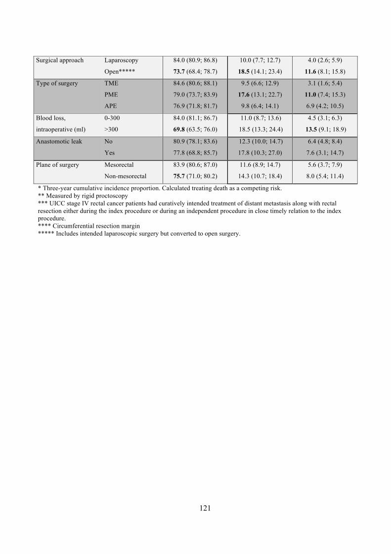
121
* Three-year cumulative incidence proportion. Calculated treating death as a competing risk. ** Measured by rigid proctoscopy *** UICC stage IV rectal cancer patients had curatively intended treatment of distant metastasis along with rectal resection either during the index procedure or during an independent procedure in close timely relation to the index procedure. **** Circumferential resection margin ***** Includes intended laparoscopic surgery but converted to open surgery.
Surgical approach Laparoscopy
Open*****
84.0 (80.9; 86.8)
73.7 (68.4; 78.7)
10.0 (7.7; 12.7)
18.5 (14.1; 23.4)
4.0 (2.6; 5.9)
11.6 (8.1; 15.8)
Type of surgery TME
PME
APE
84.6 (80.6; 88.1)
79.0 (73.7; 83.9)
76.9 (71.8; 81.7)
9.5 (6.6; 12.9)
17.6 (13.1; 22.7)
9.8 (6.4; 14.1)
3.1 (1.6; 5.4)
11.0 (7.4; 15.3)
6.9 (4.2; 10.5)
Blood loss,
intraoperative (ml)
0-300
>300
84.0 (81.1; 86.7)
69.8 (63.5; 76.0)
11.0 (8.7; 13.6)
18.5 (13.3; 24.4)
4.5 (3.1; 6.3)
13.5 (9.1; 18.9)
Anastomotic leak
No
Yes
80.9 (78.1; 83.6)
77.8 (68.8; 85.7)
12.3 (10.0; 14.7)
17.8 (10.3; 27.0)
6.4 (4.8; 8.4)
7.6 (3.1; 14.7)
Plane of surgery
Mesorectal
Non-mesorectal
83.9 (80.6; 87.0)
75.7 (71.0; 80.2)
11.6 (8.9; 14.7)
14.3 (10.7; 18.4)
5.6 (3.7; 7.9)
8.0 (5.4; 11.4)

122
Table 3: Adjusted Hazard Ratios (0-3 years) associating different characteristics with overall
survival, distant recurrence and local recurrence after intended curative rectal cancer resection Characteristic Adjusted
Hazard ratio
Overall survival*
(95%CI)
Adjusted
Hazard ratio
Distant recurrence*
(95%CI)
Adjusted
Hazard ratio
Local recurrence*
(95%CI)
Total - - -
Patient-related
Gender Male
Female
1.05 (0.72; 1.52)
1.0 (ref)
0.84 (0.52; 1.36)
1.0 (ref)
0.97 (0.50; 1.90)
1.0 (ref)
Age (years) !65
>65
1.0 (ref)
1.86 (1.19; 2.91)
1.0 (ref)
1.19 (0.73; 1.94)
1.0 (ref)
1.18 (0.58; 2.42)
Charlson Comorbidity
Index
0
1
"2
1.0 (ref)
1.11 (0.67; 1.84)
2.72 (1.77; 4.16)
1.0 (ref)
1.11 (0.53; 2.32)
2.15 (1.08; 4.29)
1.0 (ref)
1.27 (0.50; 3.24)
3.32 (1.37; 8.06)
ASA grade I
II
III-IV
1.0 (ref)
1.70 (0.97; 2.98)
2.71 (1.41; 5.21)
1.0 (ref)
0.67 (0.39; 1.16)
0.84 (0.37; 1.89)
1.0 (ref)
0.95 (0.43; 2.07)
0.54 (0.17; 1.75)
Pathology/tumour-
related
Distance of primary
tumour (lower edge)
from anal verge (cm)**
>10-15
>5-10
0-5
1.0 (ref)
1.06 (0.64; 1.79)
1.06 (0.54; 2.07)
1.0 (ref)
0.77 (0.39; 1.52)
0.73 (0.31; 1.70)
1.0 (ref)
2.55 (0.99; 6.55)
4.60 (1.28; 16.46)
pT/ypT category T0-T2
T3
T4
1.0 (ref)
1.24 (0.62; 2.46)
1.34 (0.58; 3.10)
1.0 (ref)
2.45 (0.94; 6.39)
2.06 (0.68; 6.24)
1.0 (ref)
0.56 (0.17; 1.85)
2.31 (0.65; 8.18)
UICC stage I
II
III
IV***
1.0 (ref)
0.82 (0.36; 1.90)
1.34 (0.64; 2.85)
3.27 (1.50; 7.11)
1.0 (ref)
0.58 (0.14; 2.37)
2.52 (0.70; 9.07)
8.86 (2.48; 31.74)
1.0 (ref)
1.76 (0.36; 8.73)
3.56 (0.84; 15.13)
6.57 (1.43; 30.16)
Involved CRM
(<1 mm)****
Yes
No
1.60 (0.95; 2.68)
1.0 (ref)
2.27 (1.26; 4.09)
1.0 (ref)
2.72 (1.18; 6.26)
1.0 (ref)
Treatment-related
Neoadjuvant CRT Yes 1.17 (0.77; 1.76) 1.53 (0.91; 2.59) 0.98 (0.42; 2.29)

123
No 1.0 (ref) 1.0 (ref) 1.0 (ref)
Surgical approach Laparoscopy
Open*****
0.73 (0.49; 1.10)
1.0 (ref)
0.64 (0.38; 1.09)
1.0 (ref)
0.68 (0.31; 1.46)
1.0 (ref)
Type of surgery TME
PME
APE
1.0 (ref)
1.53 (0.89; 2.63)
1.37 (0.85; 2.19)
1.0 (ref)
1.25 (0.62; 2.57)
1.09 (0.57; 2.08)
1.0 (ref)
4.70 (1.70; 13.03)
0.80 (0.30; 2.10)
Blood loss,
intraoperative (ml)
0-300
>300
1.0 (ref)
1.46 (0.96; 2.23)
1.0 (ref)
1.28 (0.73; 2.24)
1.0 (ref)
1.98 (0.94; 4.16)
Anastomotic leakage Yes
No
1.46 (0.87; 2.47)
1.0 (ref)
1.16 (0.60; 2.28)
1.0 (ref)
0.97 (0.37; 2.56)
1.0 (ref)
Plane of surgery
Mesorectal
Non-mesorectal
1.0 (ref)
1.68 (1.17; 2.38)
1.0 (ref)
1.29 (0.81; 2.07)
1.0 (ref)
1.24 (0.64; 2.41)
* COX regression analysis. Mutually adjusted. Hazard ratio (HR) >1 describes increased risk of local recurrence or distant recurrence, or reduced survival. ** Measured by rigid proctoscopy *** UICC stage IV rectal cancer patients had curatively intended treatment of distant metastasis along with rectal resection either during the index procedure or during an independent procedure in close timely relation to the index procedure. **** Circumferential resection margin ***** Includes intended laparoscopic surgery but converted to open surgery.

124
Figure 1 Flow-chart of 1,353 patients with first-time rectal cancer diagnosis between 1 April 2011 and 31 August 2012 and treated at 10 hospitals in Denmark
*Bispebjerg, n=78, Herlev, n=97, Hillerød, n=92, Aalborg, n=131, Sønderjylland, n=62, Horsens, n=1, Viborg, n=2
Underwent rectal cancer resection (TME, PME or APE) with curative
intent n=1,353
MRI performed 3 years postoperatively
n= 403
Non-participating departments* n=463
Ineligible n=324: Local recurrence n=45
Distant recurrence n=79 Death within 3 years after surgery
n=106 Terminated medical surveillance
programme n=81
Emigration=9 No preoperative MRI n=4
Non-participation n=163: Declined invitation n=78
Contraindication to MRI n=16 Non-respondents n=69
Study population n=890
Candidates for post-operative MRI
n=566

125
Figure 2 Overall survival of rectal cancer during 3 years of follow-up
Figure 3 Cumulative incidence proportion of local and/or distant recurrence during 3 years of follow-up
1.00
0.75
0.50
0.25
Surv
ival r
ate
0 1 2 3Time since rectal resection (years)
Overall survival
0.05
0.15
0.25
Cum
ulat
ive in
ciden
ce
0 1 2 3Time since rectal resection (years)
Local and/or distant recurrence

126
Figure 4 Cumulative incidence proportion of distant recurrence during 3 years of follow-up
Figure 5 Cumulative incidence proportion of local recurrence during 3 years of follow-up
0.05
0.15
0.25
Cum
ulat
ive
inci
denc
e
0 1 2 3Time since rectal resection (years)
Distant recurrence
0.05
0.15
0.25
Cum
ulat
ive
inci
denc
e
0 1 2 3Time since rectal resection (years)
Local recurrence

127
Paper II
1
!"#$%& '()('*%+& %,"('& -."(./(/& '(*"#'%"-)(& '(0"%+& 0%.0('& '(*(0"-#.& -.& 1(.$%'23& 4&
.%"-#.5-/(&6#67+%"-#.89%*(/&*"7/:3&
!
"#!$#!"%&'()*()+,-,!.#!/&012*()-,3,!$#!4#!5(6(&*()7,!8#!9:;&<(&'+,!:)6!9#!=#!>?(&*()+,@!!
+!A:&2;*!B)0?(&*0CD!=E*F0C:G,!H(F:&CI()C!EJ!8;&'(&D,!H()I:&K!-!.:)6(&*!.('0E):G!=E*F0C:G,!H(F:&CI()C!EJ!8;&'(&D,!H()I:&K!3!A:&2;*!B)0?(&*0CD!=E*F0C:G,!H(F:&CI()C!EJ!LG0)01:G!/F06(I0EGE'D,!H()I:&K!7!A:&2;*!B)0?(&*0CD!=E*F0C:G,!H(F:&CI()C!EJ!.:60EGE'D,!H()I:&K!@!H:)0*2!LEGE&(1C:G!L:)1(&!4&E;F,!LEF()2:'(),!H()I:&K!
!
&
&
&
!
&
&
&
&
&
;#''(*6#./-.<&%7"=#'&
"(*F(&!$(1K!"%&'()*(),!M#H#!
A:&2;*!B)0?(&*0CD!=E*F0C:G,!H(F:&CI()C!EJ!8;&'(&D!
5:GG(!";;GN"()*()*!$E;G(?:&6!OO!
P-@Q!A:&2;*!R!
H()I:&K!
/NI:0GS!T(*TE(U&I#6K!
&
&
&
&
&
& &

128
2
!
"#$%&'(%!
!
!"#$%&'()*+
,"-"+ ')+ .-'/"+ &010&."2+ 3456+ 7'22'89)%+ &0.-'&"-910+ &0#-"2+ &0.0#-9')+ 35556+ 89-:+ "+ *910&-9)%+
.-'/"+ 3,46+ "&0+ #')729#-9)%;+ <:9.+ .-(*=+ 9)10.-9%"-0*+ "+ ,")9.:+ >'>(2"-9')?@".0*+ #':'&-+ '7+
>"-90)-.+()*0&%'9)%+555+-'+01"2("-0+7"#-'&.+>&0*9#-910+'7+45+*(&9)%+A+=0"&.+'7+7'22'8?(>;++
+
B0-:'*+
C"-90)-.+ 7&'/+ D"-9')"2+ 50%9.-&90.+ 89-:+ &0#-"2+ #")#0&+ ()*0&%'9)%+ 555+ '&+ E"&-/"))F.+
'>0&"-9')+3EG6+89-:+#(&"-910+9)-0)-+7&'/+B"=+HIIJ+-:&'(%:+K>&92+HIJH+80&0+9)#2(*0*;+
555+>"-90)-.+89-:+"+,4+80&0+7'22'80*+7&'/+-:0+-9/0+'7+>&9/"&=+&0#-"2+#")#0&+&0.0#-9')+()-92+
*"-0+ '7+ 45L+ *0"-:L+ 0/9%&"-9')+ '&+ 0)*+ '7+ A+ =0"&.+ 7'22'8?(>;+ <:0+ #(/(2"-910+ 9)#9*0)#0+
>&'>'&-9')+ 3MNC6+'7+ 45+"-+J+")*+A+=0"&.+8".+ #"2#(2"-0*+ -&0"-9)%+*0"-:+".+"+ #'/>0-9)%+ &9.$;+
C&0*9#-910+7"#-'&.+'7+45+80&0+0O>2'&0*+(.9)%+/(2-9>20+M'O+&0%&0..9');++
+
50.(2-.+
G(-+'7+-:0+P+QRS+>"-90)-.+9)#2(*0*L+AR;TUL+VJ;SU+")*+HH;VU+:"*+555+89-:+,4L+555+89-:'(-+"+
.-'/"L+ ")*+ EG+89-:+ ")+ 0)*?#'2'.-'/=+ &0.>0#-9102=;+ N)+ >"-90)-.+ 89-:+ "+ ,4L+ MNC+ '7+ 45+ 8".+
TI;AU+ 3SRU+ MN+ PQ;V?TH;J6+ "7-0&+ J+ =0"&L+ ")*+ TV;AU+ 3SRU+ MN+ TH;R?TP;I6+ "7-0&+ A+ =0"&.;+
D0'"*W(1")-+ -&0"-/0)-+ 3E5+I;TRX+SRU+MN+I;PP?I;QR6L+@2''*+ 2'..+Y+AII+/2+ 3E5+I;QPX+SRU+MN+
I;TP?I;ST6L+")".-'/'-9#+ 20"$"%0+3E5+I;VJX+SRU+MN+I;AA?I;RI6L+<+#"-0%'&=+A+3E5+I;PAX+SRU+MN+
I;VT?I;QA6L+<+#"-0%'&=+V+3E5+I;PHX+SRU+MN+I;VH?I;SI6+")*+ZNMM+.-"%0+N[+3E5+I;RTX+SRU+MN+I;VJ?
I;QI6+80&0+>'..9@20+>&0*9#-'&.+7'&+*02"=0*+45;+
+
M')#2(.9')+
G)0+\("&-0&+'7+-:0+>"-90)-.+:"*+)'-+:"*+-:09&+*910&-9)%+.-'/"+&010&.0*+-:&00+=0"&.+"7-0&+-:0+
9)-0)*0*+&0.-'&"-910+&0#-"2+#")#0&+&0.0#-9');++
! +

129
3
!"#$%&'(#)%"**
*
!"# $%&'()# &("&%$# *+$,%$-.# /01%$'0",# *'23(*# 4567# ($%# &$%('%/# 8$03($0)-# '2# $%/+&%# '9%#
&2"*%:+%"&%*#2;#(#82**0<)%#("(*'232'0&# )%(=(,%# >?@ABC#5("0*9#,+0/%)0"%*# $%&233%"/#(#56#
()2",#D0'9#'2'()#3%*2$%&'()#%E&0*02"#4FGH7#0"#'9%#*+$,0&()#'$%('3%"'#2;#30/@("/#/0*'()#$%&'()#
&("&%$#40C%C.#'+32+$#)2&('%/#I#@#?I#&3#;$23#'9%#("()#1%$,%7C#J#56.#92D%1%$.#0*#"2'#$2+'0"%)-#
&$%('%/#(*#8($'#2;#8($'0()#3%*2$%&'()#%E&0*02"#4KGH7#;2$#+88%$#$%&'()#&("&%$#>LBC##
56*# ($%# +*+())-# $%1%$*%/# (;'%$# M# 32"'9*# ("/# 82**0<)-# %1%"# %($)0%$# 0"# *%)%&'%/# &(*%*# D0'9#
+"%1%"';+)#82*'28%$('01%#&2+$*%*#>NBC#O2$#8('0%"'*#0"#'9%#"%%/#2;#82*'28%$('01%#2"&2)2,0&()#
'$%('3%"'#92D%1%$.#*'23(#$%1%$*()#&("#<%#82*'82"%/#>NBC#
P";2$'+"('%)-.#"2'#())#8('0%"'*#+"/%$,2# $%1%$*02"#2;# '9%# *'23(#(;'%$# *+$,%$-C#F9%# $0*=#2;#(#
"2"@$%1%$*()#(;'%$#$%&'()#$%*%&'02"#D0'9#("#0"'%"/%/#'%382$($-#56#1($0%*#<%'D%%"#M#("/#MQR#
(;'%$#?CS#'2#LC?#-%($*#>T@?TBC#U+$$%"'#)0'%$('+$%#*+,,%*'*#(#3%/0("#'03%#'2#$%1%$*()#<%'D%%"#
?CS# ("/# SC?# -%($*# >T@?TBC# U2"*%:+%"&%*# 2;# "2"@$%1%$*()# 3(-# <%# ,$(1%C# 6'23(@$%)('%/#
&238)0&('02"*#$(",0",# ;$23#30"2$# 0"&2"1%"0%"&%# 4%C,C# )%(=(,%#;$23#'9%#(88)0("&%#("/#*=0"#
$(*97# '2#3(V2$#/0*(<0)0'0%*# 4%C,C#/%9-/$('02"#("/#%)%&'$2)-'%# 03<()("&%#/+%#'2#(#90,9#*'23(#
2+'8+'.#8($(*'23()#9%$"0(#("/#*'23(#8$2)(8*%7#($%#&2332"#>AB.#("/#3(-#<%#(**2&0('%/#D0'9#
$%*'$0&'02"#0"#*2&0()#(&'010'0%*#("/#$%/+&%/#:+()0'-#2;#)0;%#>?I.#?S.#QI.#Q?BC#
G("-# *'+/0%*# 9(1%# <%%"# &2"/+&'%/# 0"# (# $%,02"()# *%''0",.# >??@?NBW# '9%$%;2$%.# (# "('02"D0/%#
$%,0*'$-@<(*%/#&292$'#*'+/-#2;#$%&'()#&("&%$#8('0%"'*#'9('#($%#3("(,%/#(&&2$/0",#'2#32/%$"#
'$%('3%"'#$%,03%"*#&("#<%#0382$'("'#'2#9(1%#(#&)%($#80&'+$%#2;#'9%#&+$$%"'#&)0"0&()#8$(&'0&%C##
F9%# 8$03($-# (03# 2;# '90*# *'+/-# D(*# '2# %*'03('%# '9%# 56# $%1%$*()# $('%# (32",# 8('0%"'*#
+"/%$,20",#$%&'()#$%*%&'02"C#6%&2"/($-#(03*#0"&)+/%/#&9($(&'%$0*'0&*#2;#8('0%"'*#+"/%$,20",#
XXX#("/#Y($'3(""Z*#28%$('02".#%*'03('02"#2;#'9%#8$282$'02"#2;#8('0%"'*#$%&%010",#(#*'23(.#
()2",#D0'9#'9%#%E8)2$('02"#2;#8$%/0&'2$*#2;#56#$%1%$*()C*
#

130
4
!"#$%&"'(")*(+$#,-*.(
(
/#0*1(*$.&2)(")*(.$##&)23(!"#$%&'&()*+#',-.*$/0%,*+#',1#0/%2'"'3+%$+(04%1*$%*&&3'5/0%.4%
+"/%6*+#',*)%7'*30%'8%9/*)+"%:3/8;<%=-=>?=-?@A@B?BCD%+"/%E2#/,+#8#2%F'GG#++//%'8%+"/%H*,#$"%
F')'3/2+*)% F*,2/3% I3'(&% :HFFI;0JCD% *,0% +"/% H*,#$"% H*+*% K3'+/2+#',% LM/,24% :3/8;<%
@>>A-NO->>?>C;%!"/%$+(04%1*$%2',0(2+/0%#,%+"/%$/++#,M%'8%+"/%/,+#3/%H*,#$"%&'&()*+#',%P@@Q;%
!"/%6*+#',*)%9/*)+"%E/35#2/%#,%H/,G*3J%&3'5#0/$%(,#5/3$*)D%+*R-$(&&'3+/0%"/*)+"%2*3/%+'%*))%
2#+#S/,$D%M(*3*,+//#,M%83//%*22/$$%+'%M/,/3*)%&3*2+#+#',/3$%*,0%&(.)#2%"'$&#+*)$P@=Q;(
H*+*% 3/+3#/5/0% #,2)(0/0D% 2)#,#2*)D% 0/G'M3*&"#2% 0*+*D% 2'-G'3.#0#+#/$D% LEL% $2'3/D% +(G'(3%
2"*3*2+/3#$+#2$%:0#$+*,2/%83'G%+"/%*,*)%5/3M/CD%,/'*0T(5*,+%+3/*+G/,+D%&*+"')'M#2%$+*M/D%*,0%
$(3M#2*)%&3'2/0(3/$%*,0%*&&3'*2"D%.)''0%)'$$D%&'$+-'&/3*+#5/%*,*$+'G'+#2%)/*J*M/%*,0%0/*+"%
1#+"#,%=%4/*3$%83'G%+"/%$(3M#2*)%#,0/R%&3'2/0(3/;%%
%
4"#"( .-0%5$.3( H*+*% 1/3/% '.+*#,/0% 83'G% =% 3/M#$+3#/$<% +"/% H*,#$"% F')'3/2+*)% F*,2/3% I3'(&%
:HFFI;0JC%H*+*.*$/%:HFFICD%F#5#)%U/M#$+3*+#',%E4$+/G%:FUEC%*,0%+"/%H*,#$"%6*+#',*)%K*+#/,+%
U/M#$+34%:H6UKC;%(
E#,2/% V*4% @>>?D% HFFI% 0*+*.*$/% "*$% .//,% 3/2'30#,M% #,8'3G*+#',% ',% *))% &*+#/,+$% 1#+"%
2')'3/2+*)% 2*,2/3%1#+"% *% 2'G&)/+/,/$$% '8% WXYWWZ% P@[Q;% !"/% &(3&'$/% '8% +"/% 0*+*.*$/% #$% +'%
/,$(3/% (,#8'3G% \(*)#+4% #,% 0#*M,'$+#2$% *,0% +3/*+G/,+% '8% 2')'3/2+*)% 2*,2/3% #,% H/,G*3J;% L))%
$(3M#2*)% 0/&*3+G/,+$% *23'$$% H/,G*3J% 3/&'3+% ',% 0#*M,'$+#2% $+*M#,MD% +3/*+G/,+% *,0%
&'$+'&/3*+#5/%2'G&)#2*+#',$%:'22(33#,M%1#+"#,%=>%0*4$%*8+/3%$(3M/34C%&3'$&/2+#5/)4%P@[Q;%]'3%
+"#$%$+(04D%+"/%HFFI%0*+*.*$/%1*$%($/0%+'%#0/,+#84%+"/%$+(04%2'"'3+D%*$%0/$23#./0%./)'1;%
!"/%H*,#$"%FUE%"*$%*$$#M,/0%*%(,#\(/%?>-0#M#+%&/3$',*)%#0/,+#8#2*+#',%,(G./3%:FKU%,(G./3C%
+'%/5/34%3/$#0/,+% #,%H/,G*3J%*,0% #+% #$%($/0%$#,2/%?WXO;%!"/%3/M#$+34%G*#,+*#,$% #,8'3G*+#',%
',%0*+/%'8%.#3+"D%0/*+"D%$/RD%3/$#0/,2/%*,0%5#+*)%$+*+($;%!"/%2#5#)%3/M#$+3*+#',%,(G./3%&/3G#+$%
)#,J*M/%1#+"#,%+"/%"/*)+"2*3/%$4$+/G%*,0%*G',M%3/M#$+3#/$%#,%H/,G*3J%P@NQ;%
]#,*))4D%+"/%H6UK%"*$%G*#,+*#,/0%3/2'30$%',%*))%,',-&$42"#*+3#2%"'$&#+*)#$*+#',$%#,%H/,G*3J%
$#,2/%?WAA;%!"/$/%#,2)(0/%#,8'3G*+#',%',%"'$&#+*)%0#*M,'$/$%*,0%&3'2/0(3/$%P@XQ;%H*+*%*3/%
2'))/2+/0% 8'3%*0G#,#$+3*+#5/%&(3&'$/$%(,3/)*+/0% +'% 3/$/*32"%'.T/2+#5/$;% ]'3% #,$+*,2/D% +"/$/%
0*+*% #,2)(0/% +"/% FKU% ,(G./3D% 0*+/$% '8% *0G#$$#',% *,0% 0#$2"*3M/% *,0% (&% +'% @>% 0#$2"*3M/%
0#*M,'$/$D% 2'0/0% .4% &"4$#2#*,$% *22'30#,M% +'% +"/% ?>+"% 3/5#$#',% '8% +"/% ^,+/3,*+#',*)%
F)*$$#8#2*+#',% '8% H#$/*$/$% :^FH-?>C% 83'G% ?WW=% *,0% ',1*30$% P@AQ;% E#,2/% _*,(*34% ?D% ?WWXD%
3/M#$+3*+#',%'8% $(3M#2*)%&3'2/0(3/$% #,%H/,G*3J%"*$%.//,%2)*$$#8#/0%*22'30#,M% +'% +"/%6'30#2%
V/0#2'-%E+*+#$+#2*)%F'GG#++//%F)*$$#8#2*+#',%'8% E(3M#2*)%K3'2/0(3/$% :6`VaEF`C;%HFFI%*,0%

131
5
!"#$%&'('%)*+*%,-./*&%(0%01('-.%-.20+3'(-0.%0.%45+6-7',%*8*.(4%&5+-.6%20,,0)95:%;-<*<=%4(03'%
+*8*+4',><%%
%
!"#$%& '()(*"+&$'(-*.(4%&-'6.04*&%)-(?% +*7(',% '&*.07'+7-.03'% ;,07'(*&%@A% 73%0+% ,*44% 2+03%
(?*% '.',% 8*+6*>% -.% (?*% !BBC% &'('1'4*% 1*()**.% D'E% @=% FGG@% '.&% H:+-,% IG=% FG@F=% )?0%
5.&*+)*.(%-.(*.&*&%+*4(0+'(-8*%+*7(',%+*4*7(-0.%;JDK=%$DK>%0+%L'+(3'..M4%:+07*&5+*%)-(?%
75+'(-8*%-.(*.(=%)*+*%-&*.(-2-*&<%$'(-*.(4%)*+*%*N7,5&*&%-2%(?*E%?'&%45+6-7',%:+07*&5+*4%0(?*+%
(?'.% -.(*.&*&% +*4(0+'(-8*% +*7(',% +*4*7(-0.% 0+% L'+(3'..M4% 0:*+'(-0.=% (?04*%)?0% *3-6+'(*&%
1*20+*% 4('+(% 02% 20,,0)95:=% ?'&% :',,-'(-8*% 45+6*+E% 0+% ?'&% .0% +*6-4(+'(-0.% 02% '.E% 45+6-7',%
:+07*&5+*%-.%(?*%!"#$%&*4:-(*%+*6-4(+'(-0.%-.%(?*%!BBC%&'('1'4*%;O-65+*%@><%&
"'(-0.',% 65-&*,-.*4% +*7033*.&% .*0'&P58'.(% ,0.69705+4*% ;7?*30>9% +'&-0(?*+':E% ;B#J>% ;AG%
CE%-.%FA9FQ%2+'7(-0.4%7031-.*&%)-(?%A9O,50+05+'7-,%;A9OR>>%(0%:'(-*.(4%)-(?%,07',,E%'&8'.7*&%
+*7(',% 7'.7*+% SFQT<% H,(*+.'(-8*,E=% 4?0+(9705+4*% +'&-0(?*+':E% 7'.% 1*% 022*+*&% -.% 403*%
4-(5'(-0.4<% U.(*.&*&% +*4(0+'(-8*% +*7(',% +*4*7(-0.% '.&% L'+(3'..M4% 0:*+'(-0.% :*+20+3*&% '4%
3*40+*7(',% *N7-4-0.% )*+*% &0.*% Q9@G% )**/4% '2(*+% (?*% 703:,*(-0.% 02% B#J<% V?0+(9705+4*%
+'&-0(?*+':E% )-(?% -33*&-'(*% 45+6*+E% -4% .0(% +05(-.*,E% :*+20+3*&% -.% !*.3'+/<% H,,% 0(?*+%
:'(-*.(4% 5.&*+)*.(% &-+*7(% -.(*.&*&% +*4(0+'(-8*% +*7(',% +*4*7(-0.% 0+% L'+(3'..M4% 0:*+'(-0.<%
V*,*7(*&% RUBB% 4('6*% UU% +*7(',% 7'.7*+% :'(-*.(4% )*+*% 022*+*&% W% 30.(?4% 02% '&P58'.(%
7?*30(?*+':E<% J?*% 4'3*% '::,-*&% 20+% RUBB% 4('6*% UUU% +*7(',% 7'.7*+% :'(-*.(4% )?0% ?'&% .0(%
+*7*-8*&%.*0'&P58'.(%B#J%SFQT<%
%
!"(,-& *./.*0-1+& $'(-*.(4% )?0% 5.&*+)*.(% -.(*.&*&% +*4(0+'(-8*% +*7(',% +*4*7(-0.% )-(?% '% !V%
)*+*%20,,0)*&%2+03%(?*%(-3*%02%:+-3'+E%+*7(',%7'.7*+%+*4*7(-0.%;X%-.&*N%0:*+'(-0.>%5.(-,%(?*%
&'(*%02%4(03'%+*8*+4',%;4(5&E%*.&:0-.(>=%&*'(?=%*3-6+'(-0.=%0+%*.&%02%20,,0)95:%'2(*+%I%E*'+4<%
U.20+3'(-0.%0.%4(03'%4('(54%;-<*<=%+*8*+4',%0+%.0.9+*8*+4',%02%!V>%&5+-.6%I%E*'+4%02%20,,0)95:%
)'4% 01('-.*&% (?+056?% (?*% !"#$=% +*(+-*8-.6% 45+6-7',% :+07*&5+*% 70&*4% -.&-7'(-.6% 4(03'%
+*8*+4',%'770+&-.6%(0%(?*%"0+&-7%D*&-709V('(-4(-7',%B033-((**%;"YDKVBY>%B,'44-2-7'(-0.%02%
V5+6-7',%$+07*&5+*4<%&
D*&-7',%+*70+&4%2+03%Z[%02%:'(-*.(4%+*7*-8-.6%'%!V%'(%(?*%-.&*N%0:*+'(-0.%)*+*%4E4(*3-7',,E%
+*8-*)*&%-.%0+&*+%(0%8',-&'(*%(?*%4(03'%4('(54%'4%+*(+-*8*&%2+03%(?*%!"#$<%!*8-'(-0.4%2+03%
4(03'%4('(54%'4%+*(+-*8*&%2+03%!"#$%)*+*%+*6-4(*+*&<%%
%
!"-"20"2'-1&-3-1%020+&$'(-*.(4%)*+*%7?'+'7(*+-\*&%'770+&-.6%(0%+*7(',%+*4*7(-0.%:+07*&5+*%;!V=%
.0%4(03'=%'.&%L'+(3'..M4%:+07*&5+*>=%E*'+%02%-.&*N%0:*+'(-0.%'.&%&*306+':?-74<%&

132
6
!"#$%&'&()*+,#$+-%+.#-%#$/01/10*+1-$23456$17$8*1')$0#,#08)($)*$9$)-.$:$;#)08$71((1<+-=$0#%*)($
%)-%#0$8&0=#0;$<#0#$%)(%&()*#.$1,#0)((>$)-.$710$,)0+1&8$/)*+#-*?$)-.$.+8#)8#?0#()*#.$7)%*108$
*0#)*+-=$.#)*"$)8$)$%1'/#*+-=$0+8@A$$
51*#-*+)($ /0#.+%*+,#$ 7)%*108$ 710$ 8*1')$ 0#,#08)($ 2/#0+1.$ 17$ 8&0=#0;>$ =#-.#0>$ )=#$ )*$ 8&0=#0;>$
B1.;?')88$ +-.#C$ 2DE46>$ 3")0(81-$ %1'10B+.+*;$ +-.#C$ 23346>$ FGF$ %()88+7+%)*+1->$ .+8*)-%#$ 17$
/0+')0;$*&'1&0$2(1<#0$#.=#6$701'$)-)($,#0=#$2%)*#=10+8#.$)8$(1<$2H$?$I$%'6$,8A$'+.J$"+="$2K$?$
9H$%'$J$99$?$9I$%'6>$&8#$17$-#1).L&,)-*$3M!>$8&0=+%)($)//01)%">$ +-*0)1/#0)*+,#$B(11.$ (188>$
)-)8*1'1*+%$ (#)@)=#>$ 2;6/!$ %)*#=10;$ 2)%%10.+-=$ *1$ !$ %)*#=10;6>$ )-.$ N433$ 8*)=#6$ <#0#$
#C/(10#.$+-$)$31C$/01/10*+1-)($")O)0.8$0#=0#88+1-$'1.#(A$D1*"$&-+,)0+)B(#$)-.$'&(*+,)0+)B(#$
)-)(;8+8$ <)8$ /#0710'#.A$E&(*+,)0+)B(#$ )-)(;8+8$ +-%(&.#.$ )(($ /1*#-*+)($ /0#.+%*108$ 710$ 8*1')$
0#,#08)(A$$
G*)*)P$,#08+1-$9QAH$2G*)*)310/>$31((#=#$G*)*+1->$!#C)8>$NGF6$<)8$&8#.$710$8*)*+8*+%)($)-)(;8+8A$$

133
7
!"#$%&#'
!!
"#$%##&!'()!*+!,--*!(&.!/0123!4-+!,-*,+!*5!4-4!0($2#&$6!%2$7!8216$9$2:#!1#;$(3!;(&;#1!%#1#!
$1#($#.! ($! ,*! 7<602$(36! 2&! =#&:(1>?! /:<&@! $7#6#+! A! B5C! 0($2#&$6! D&.#1%#&$! 2&$#&.#.!
1#6$<1($2E#!1#;$(3!1#6#;$2<&!<1!F(1$:(&&G6!<0#1($2<&!%2$7!;D1($2E#!2&$#&$!(&.!;<:0126#.!$7#!
6$D.)!;<7<1$?!=#$(236!1#@(1.2&@!6#3#;$2<&!<8!$7#!6$D.)!;<7<1$!(1#!.#6;12H#.!2&!I2@D1#!*?!!
J7#! 01<0<1$2<&! <8! 0($2#&$6! D&.#1@<2&@! 2&$#&.#.! 1#6$<1($2E#! 1#;$(3! 1#6#;$2<&! %2$7! (! =K!
@#(33)!2&;1#(6#.!<E#1!$7#!6$D.)!0#12<.!81<:!,C?L!$<!M,?MN!H#$%##&!,--*!9!,--M!(&.!,--C!
9!,-*,!OJ(H3#!*P?!Q&!;<:0(126<&+!$7#!01<0<1$2<&!<8!0($2#&$6!%7<!7(.!#2$7#1!1#6$<1($2E#!1#;$(3!
1#6#;$2<&!%2$7!&<!=K!<1!F(1$:(&&G6!01<;#.D1#+!@1(.D(33)!.2:2&267#.!<E#1!$7#!6$D.)!0#12<.?!
R($2#&$6!D&.#1@<2&@!1#6$<1($2E#!1#;$(3!1#6#;$2<&!%2$7!8<1:($2<&!<8!(!=K+!(&.!0($2#&$6!%2$7<D$!
(! 6$<:(!%#1#! ;<:0(1(H3#!%2$7! 1#60#;$! $<! (@#+! "'Q+! ;<:<1H2.2$)+! J! ;($#@<1)+! SQTT! 6$(@#+!
6D1@2;(3!(001<(;7+!(&.!(&(6$<:<$2;! 3#(>(@#!1($#! OJ(H3#!*P?!F<%#E#1+!:(3#!@#&.#1+! $D:<D1!
3<;($2<&! 2&! $7#! 3<%#1!<1!:2.! 1#;$D:+!D6#!<8! &#<(.UDE(&$!TVJ+! (&.!72@7#1! H3<<.! 3<66!%#1#!
:<1#!;<::<&!(:<&@!0($2#&$6!%2$7!(!=K!;<:0(1#.!$<!$7<6#!%2$7<D$!(!=K?!=#:<@1(072;6+!
$D:<D1!;7(1(;$#126$2;6!(&.!$1#($:#&$!1#@2:#&6!(1#!6D::(126#.!2&!J(H3#!*?!
Q&! $<$(3+! ,,5!:#.2;(3! 1#;<1.6!%#1#! 1#E2#%#.! $<! E(32.($#! 6$<:(! 6$($D6! ($! 49)#(1! 8<33<%9D0?!
=26;1#0(&;2#6! %#1#! 8<D&.! 8<1! 6#E#&! 0($2#&$6! O4?*NW! C5N! TQ! -?B95?MP! (6! ;<:0(1#.! $<! $7#!
1#@26$#19H(6#.! 6$($D6?! =26;1#0(&;2#6! %#1#! ;(D6#.! H)! 2&;<11#;$! XY'ZKTY! 1#@26$1($2<&! <8!
6$<:(! 1#E#16(3! .D12&@! 8<33<%9D0! OH<$7! :2662&@! 1#@26$1($2<&! <8! 1#E#16(3! (&.! 1#@26$1($2<&! <8!
1#E#16(3!&#E#1!:#&$2<&#.!2&!:#.2;(3!1#;<1.6P!<1!2&;<11#;$!1#@26$1($2<&!<8!$7#!TRV!&D:H#1?!!
!
($)$%*&+,"'+-.+/"-."'012021&+2-'23'#&2)*'1","1#*%4'J7#!TQR!<8!=K!1#E#16(3!%(6!L-?4N!OC5N!
TQ!AB?M[L,?*P!(8$#1!*!)#(1+!(&.!LM?4N!OC5N!TQ!L,?59LA?-P!(8$#1!4!)#(16+!(6!67<%&!2&!I2@D1#!,!
(&.!J(H3#!,?!'#.2(&!$2:#!$<!6$<:(!1#E#16(3!%(6!*L4?-!.()6!O1(&@#\!*9*-L5P?!'
R($2#&$6!%2$7!/K/!6;<1#!]! QQ+!&#<(.UDE(&$!TVJ+!H3<<.!3<66!]!4--!:3+!(&(6$<:<$2;! 3#(>(@#+!J!
;($#@<1)!]!,+!(&.!SQTT!6$(@#!]!QQ!7(.!0(1$2;D3(13)!3<%!TQR!<8!6$<:(!1#E#16(3!($!*!)#(1!(8$#1!$7#!
2&.#^!<0#1($2<&!OJ(H3#!,P?!!K2:23(13)+!($!4!)#(16!(8$#1!$7#!2&.#^!<0#1($2<&+!0($2#&$6!(@#.!]!A5!
)#(16! OTQR! -?L,W! C5N! TQ! -?AC9-?LMP+! %2$7! /K/! @1(.#! ]! QQ! OTQR! -?AMW! C5N! TQ! -?5B9-?ACP+!
&#<(.UDE(&$!TVJ! OTQR!-?ABW!C5N!TQ!-?AM9-?L*P+!H3<<.! 3<66!]!4--!:3! OTQR!-?L-W!C5N!TQ!-?AL9
-?L4P+!(&(6$<:<$2;!3#(>(@#!OTQR!-?M5W!C5N!TQ!-?4C9-?5*P+!J!;($#@<1)!4!OTQR!-?L4W!C5N!TQ!-?L-9
-?L5P!(&.!M! OTQR!-?AAW!C5N!TQ!-?5A9-?LMP+!SQTT!6$(@#! QQQ! OTQR!-?L*W!C5N!TQ!-?AB9-?LMP!(&.! Q_!
OTQR!-?5LW!C5N!TQ!-?5-9-?AMP+!7(.!0(1$2;D3(13)!3<%!TQRG6!OJ(H3#!,P?!

134
8
!"#$%&#'()$#*(+,--('#".%(,(/$'%"'%(*"/(#*%(*$.*%'#(-,!(01('#02"(3%4%3'"5("#(6(7%"3'("1#%3(#*%(
$&/%8( 09%3"#$0&( :;<=<>?=( !"#$%&#'( )$#*( "&"'#020#$@( 5%"A".%( *"/( #*%( 50)%'#( -,!( 01( '#02"(
3%4%3'"5("#(6(7%"3'("1#%3(#*%($&/%8(09%3"#$0&(:BC=<>?=((
!"#$%&'( )&*+,#$,-*( %.( +*/"0( ,1( '$%2"( &*-*&'"/( ,1(23/$,-"&,"4/*( "1"/0','5(D$.*%3(EFE(.3"/%(
:EFE(,,("&/(EFE(,,,G,H?I(&%0"/JK4"&#(-LMI(N500/(50''(O(6<<(25I("&"'#020#$@(5%"A".%I("/4"&@%/(
M( @"#%.037( :M( @"#%.037( PI( M( @"#%.037( 6( "&/( M( @"#%.037( B?I( "&/(203%( "/4"&@%/(+,--( '#".%(
:+,--(,,I(+,--(,,,I("&/(+,--(,H?()%3%("''0@$"#%/()$#*("(3$'A(01(/%5"7($&(#$2%(#0('#02"(3%4%3'"5(
$&(K&$4"3$"N5%("&"57'$'=((
,&(2K5#$4"3$"N5%("&"57'$'I(&%0"/JK4"&#(-LM(:DL(<=QCR(SC>(-,(<=TTG<=;C?I(N500/(50''(O(6<<(25(
:DL(<=;TR(SC>(-,(<=QTG<=SQ?I("&"'#020#$@( 5%"A".%(:DL(<=BUR(SC>(-,(<=66G<=C<?I(M(@"#%.037(6(
:DL(<=T6R(SC>(-,(<=BQG<=;6?I(M(@"#%.037(B(:DL(<=TPR(SC>(-,(<=BPG<=S<?I("&/(+,--('#".%(,H(:DL(
<=CQR(SC>(-,(<=BUG<=;<?()%3%(10K&/(#0(N%($&/%9%&/%&#(93%/$@#03'(01(/%5"7($&(#$2%(#0('#02"(
3%4%3'"5(:M"N5%(6?=((

135
9
!"#$%##"&'((
!
"#$!%&'(! )'(*'(+! ,)! -#'.! (&-',(/'*$0! 1,123&-',(45&.$*! 6,#,7-! .-2*8! /&.! -#$! )&6-! -#&-! &(!
2($91$6-$*38!3,/!7&-$!,)!,(38!:;<!,)!7$6-&3!6&(6$7!1&-'$(-.!/'-#!&(!'(-$(*$*!-$%1,7&78!=>!
#&*!-#$'7!.-,%&!7$?$7.$*!*27'(+!@!8$&7.!,)!),33,/421A!>'(6$!%,7$!-#&(!,($!-#'7*!,)!1&-'$(-.!
2(*$7+,'(+!'(-$(*$*!7$.-,7&-'?$!7$6-&3!7$.$6-',(!#&*!&!=>0!&!.25.-&(-'&3!(2%5$7!,)!1&-'$(-.!
$(*!21!/'-#!&!1$7%&($(-!.-,%&A!
B7$?',2.!.-2*'$.!'(?$.-'+&-'(+!.-,%&!7$?$7.&3!7&-$.!),33,/'(+!&(-$7',7!7$.$6-',(!.#,/!&!+7$&-!
?&7'&-',(!'(!1$7%&($(-!.-,%&!7&-$.!CD@0!DEFA!G&H,7'-8!,)!-#$.$!.-2*'$.!&7$!5&.$*!,(!.%&33$7!
1&-'$(-!6,#,7-.!I7&(+'(+!)7,%!JK!-,!JL@!'(632*$*!1&-'$(-.!CDD4D;0!DM0!DNFO!&(*!,7'+'(&-$!)7,%!
.'(+3$!6$(-7$.!,7!)7,%!&!3,6&3!7$+',(A!!
G23-'6$(-7$!.-2*'$.!,(!7$6-&3!6&(6$7!1&-'$(-.!,(38!CE0!DK0!DJ0!D:F!7$1,7-!&!D:4LJ<!CEF!CDKF!7'.P!
,)! &! 1$7%&($(-! .-,%&! &)-$7! '(-$(*$*! 7$6-&3! 7$.$6-',(! -,+$-#$7!/'-#! &!=>0! .'%'3&7! -,!/#&-!
/&.!#$7$'(!*,62%$(-$*A!!
Q(!-#'.!.-2*80!-#$!'(*$1$(*$(-!17$*'6-'?$!)&6-,7.!),7!.-,%&!(,(47$?$7.&3!/'-#'(!@!8$&7.!/$7$!
&(&.-,%,-'6!3$&P&+$0!&*?&(6$*!RQSS!.-&+$!QT0!"!6&-$+,78!;0!"!6&-$+,78!@0!2.$!,)!($,&*H2?&(-!
SU"0!&(*!1$7',1$7&-'?$!53,,*!3,..!V!@KK!%3A!"#$.$!7$.23-.!&7$! '(! 3'($!/'-#!17$?',2.!.-2*'$.0!
/#'6#! &33! .#,/! -#&-! '(67$&.'(+! &+$0! 6,%,75'*'-8! CDK0! DL0! D:0! DN0! LKF0! 3,6&338! &*?&(6$*! ,7!
%$-&.-&-'6! *'.$&.$! CDD0! DL0! DM0! D:F0! &(8! 1,.-,1$7&-'?$! 6,%13'6&-',(.! CDD0! DL0! DJ0! D:F! &(*!
&(&.-,%,-'6! 3$&P&+$CD;0! DM0! DNF! '(67$&.$*! -#$! 17,5&5'3'-8! ,)! &! 1$7%&($(-! .-,%&! &)-$7!
&(-$7',7!7$.$6-',(A!
W!7$-7,.1$6-'?$0!%23-'6$(-7$!X(+3'.#!.-2*8!ILKKD4LKK@O!'(632*'(+!M!JNL!1&-'$(-.!IEM;!1&-'$(-.!
/'-#!&!=>O0!$9&%'($*!-#$!2.$!,)!3,,14'3$,.-,%8!),33,/'(+!3,/!&(-$7',7!7$.$6-',(A!"#$!.-2*8!
.#,/$*! -#&-! '(67$&.'(+! &+$! &(*! 6,%,75'*'-8! '(67$&.$*! -#$! 17,5&5'3'-8! ,)! &! 1$7%&($(-!
.-,%&A!Y,/$?$70! ,(38! &! 7$3&-'?$38! 3,/! 17,1,7-',(! ,)! 1&-'$(-.! ID;AM<O! #&*! &!=>! &-! -'%$! ,)!
17'%&78! .27+$78! &(*! (,($! ,)! -#$! 1&-'$(-.! #&*! ($,&*H2?&(-! SU"! CDKFA! Q(! 17$.$(-! 6,#,7-0!
($,&*H2?&(-! SU"! /&.! &! .'+(')'6&(-! 17$*'6-,7! ,)! .-,%&! (,(47$?$7.&3A! "#$! *'))$7$(6$.! '(!
5&.$3'($! 6#&7&6-$7'.-'6.! ,)! -#$! 1&-'$(-! 6,#,7-.! '(! -#$! -/,! .-2*'$.! %&8! 3$&*! -,! (,(4
6,%1&7&53$!7$.23-.A!
W!>/$*'.#!7$-7,.1$6-'?$!%23-'6$(-7$!.-2*8!'(632*'(+!@!JM;!1&-'$(-.!2(*$7+,'(+!7$6-&3!6&(6$7!
.27+$78! ,5.$7?$*! -#&-! #'+#$7! 3$?$3! ,)! $*26&-',(! '(67$&.$*! -#$! 17,5&5'3'-8! ,)! $&738! .-,%&!
7$?$7.&3A! B,.-,1$7&-'?$! 6,%13'6&-',(.0! &*H2?&(-! 6#$%,-#$7&180! &*?&(6$*! RQSS! .-&+$0! &(*!
&*?&(6$*!W>W!+7&*$!/$7$!&..,6'&-$*!/'-#!*$3&8!'(!-'%$!-,!.-,%&!7$?$7.&3A!"#'.!.-2*8!*'))$7.!
)7,%!17$.$(-!.-2*80!&.!'-!17,?'*$.!(,!'(),7%&-',(!,(!-#$!17,1,7-',(!,)!1&-'$(-.!/#,!7$6$'?$!

136
10
!"#$%&!"!$" '()*+"#,-.*-/0"1,-$2*-3" 4%55%67,8"6!#"#2%-$" 9$%$!5"%4":0;"/*!-#<"!()" '(=5,)*)"(%"
>!5')!$'%("%("=5'('=!5"'(4%-&!$'%("4-%&"?6*)'#2"-*.'#$-'*#"@AB0""
C6%"8-%#8*=$'>*"&,5$'=*($-*"#$,)'*#"*+!&'(*)"-'#D"4!=$%-#"%4"!"8*-&!(*($"#$%&!"4%55%6'(."
CEF"6'$2"4%-&!$'%("%4"*'$2*-"!")'>*-$'(."=%5%#$%&/"9GHI"!()"GI"-*#8*=$'>*5/<"%-"'5*%#$%&/"
9JGI" !()" AHI" -*#8*=$'>*5/<0" K%&%-L')'$/3" &*$!#$!$'=" )'#*!#*3" !(!#$%&%$'=" 5*!D!.*3"
)*$*-'%-!$*)" !(%7-*=$!5" 4,(=$'%(3" 8%#$%8*-!$'>*" =%&85'=!$'%(#" %4" !(/" D'()" !()" #*=%()!-/"
=%(#$-,=$*)" #$%&!#" 6*-*" #'.('4'=!($" -'#D" 4!=$%-#" @:;3" :HB0" M%6*>*-3" -*#,5$#" 4-%&" $2*#*"
#$,)'*#" =!((%$" L*" *+$-!8%5!$*)" )'-*=$5/3" !#" $2*" -'#D" %4" !" 8*-&!(*($" #$%&!" 4%55%6'(."
=%(#$-,=$'%("%4")'>*-$'(."=%5%#$%&/"&!/"L*")'44*-*($"4-%&"!")'>*-$'(."'5*%#$%&/0""
N($*-*#$'(.5/3" 8-*#*($" #$,)/" #2%6#" $2!$" !(" '(=-*!#'(." 8-%8%-$'%(" %4" 8!$'*($#" ,()*-6*($"
-*#$%-!$'>*" -*=$!5" =!(=*-" -*#*=$'%(" !#" $'&*" 8!##*)0" O2*-*!#" GPI" %4" 8!$'*($#" ,()*-6*($"
-*#$%-!$'>*"#,-.*-/"4-%&"QPP:"$%"QPPR3"!"62%5*"%4"RQI")')"$2*"#!&*"4-%&"QPPA"$%"QP:Q0"C2'#"
.*(*-!5"=2!(.*"'("#,-.'=!5"!88-%!=2"&!/"8!-$5/"L*"!"-*#,5$"%4"-*>'#'%("%4"(!$'%(!5".,')*5'(*#"
'("QPPA"!()"&*$2%)%5%.'=!5"!5'.(&*($"L*$6**(")*8!-$&*($#0""
K2!(.*#" '(" #,-.'=!5" !88-%!=2" %>*-" $'&*"6'$2" *+$*()*)" ,#*" %4" S?" 2!>*" (%$" L**(" 4%,()" '("
%$2*-" #$,)'*#3" !()" #,..*#$" $2!$" #,-.*%(#" &!/" L*" '(=-*!#'(.5/" &%-*" =!,$'%,#" 4%55%6'(."
-*#$%-!$'>*"-*=$!5"=!(=*-"-*#*=$'%(0"M%6*>*-3"-*=*($"#$,)'*#"4-%&"C2*"T*$2*-5!()#"4%,()"(%"
)'44*-*(=*"'("$2*"#2%-$7$*-&"8%#$%8*-!$'>*"=%&85'=!$'%("-!$*#"L*$6**("8!$'*($#",()*-.%'(."
-*=$!5" =!(=*-" -*#*=$'%("6'$2"S?"L/" -%,$'(*"%-" '4"S?"%(5/"6!#"8*-4%-&*)" '(" 2'.25/" #*5*=$'>*"
8!$'*($#"@QA3"GPB0"FU,!55/3"4'()'(.#"4-%&"8-*#*($"#$,)/"#,..*#$"$2!$"5%(.7$*-&"=%&85'=!$'%(#"
&'.2$" L*" !(%$2*-" '&8%-$!($" '##,*" $2!$" -*U,'-*#" !$$*($'%(" '(" $2*" 8-*7%8*-!$'>*" #*$$'(.0"
?*5*=$'(." #8*='4'=" 8!$'*($" .-%,8#" 4%-" #$%&!" =%(#$-,=$'%(" '#" 4%,()" $%" L*" !" D*/" *5*&*($" '("
%8$'&'#'(." 8!$'*($" %,$=%&*#0" C2*#*" 4'()'(.#" &!/" =2!(.*" $2*" $*()*(=/" $%6!-)#" !" &%-*"
#*5*=$'>*"!88-%!=2"$%"S?"=%(#$-,=$'%("'("4,$,-*0""
C2*"L'..*#$"#$-*(.$2#"%4"$2'#"#$,)/"!-*"$2*"(!$'%(6')*"8-%#8*=$'>*")*#'.(3"$2*"5!-.*"8!$'*($"
=%2%-$"!()"!"5%(."4%55%67,8"8*-'%)"%4"G"/*!-#0"S!$!"6*-*"-*$-'*>*)"4-%&"$2-**"2'.25/"-*5'!L5*"
-*.'#$-'*#"6'$2"8-%#8*=$'>*"#!&85'(."%4")!$!"!()"2'.2")!$!"=%&85*$*(*##"@Q;3"QJ3"G:B0"V(5'D*"
&%#$" 8-*>'%,#" #$,)'*#3" $2'#" #$,)/" '#" L!#*)" %(" )!$!" 4-%&" =,--*($" $'&*" 8*-'%)3" 62*-*"
$-*!$&*($"-*.'&*(#"=%&85/"6'$2"&%)*-("#$!()!-)#0""
W5$2%,.2" )!$!" 6*-*" -*$-'*>*)" 4-%&" >!5')!$*)" -*.'#$-'*#3" $2*" >!5')'$/" !()"&*$2%)#" %4" )!$!"
-*8%-$'(."&'.2$")'44*-"L*$6**("2%#8'$!5#0"C2'#" '#"%(*"%4" $2*" 5'&'$!$'%(#3"#'(=*"=%)'(."*--%-#"
-*5!$*)" $%" %8*-!$'>*" 8-%=*),-*#" &!/" %==,-0" M%6*>*-3" )'#=-*8!(='*#" L*$6**(" &*)'=!5"
-*=%-)#"!()")!$!L!#*#" -*.!-)'(." #$%&!"#$!$,#"!$"G7/*!-" 4%55%67,8"6*-*" 5%6" '(" $2*" #!&85*"
#,->*/"%4"AI"%4"8!$'*($#"62%"2!)"-*=$!5"-*#*=$'%("6'$2"4%-&!$'%("%4"!"S?0"

137
11
!"#$%&#'#()$')*+,$'"-.'$)"/)$/$0*/&)#&$-1$ )"#$%/)2#()'$"/+$(-)$*(+#&3-(#$')-4/$#&'/6$
)"&##$,#/&'$/1)#&$&#')-&/)25#$)/6$7/(7#&$&#'#7)2-($2($8#(4/&9:$!"2'$(*4;#&$./'$+-*;6#+<$
."#&#$)"#$%-')-%#&/)25#$4/(/3#4#()$./'$7-4%627/)#+$;,$/($/(/')-4-)27$6#/9:$!
$
"#$%&'()*+),)%-.!
!"#$%&#'#()$')*+,$./'$1*(+#+$;,$)"#$8/(2'"$=/(7#&$>-72#),:$$
!"#$ '72#()2127$ 7-442))##$ *(+#&$ )"#$ 8==?$ 2'$ /79(-.6#+3#+$ 1-&$ )"#2&$ 7-()&2;*)2-($ 2($
7-('*6)/(7,$'#&527#'$2($62(9/3#$;/'#+$-($ ')&2#'$/(+$762(27/6$+/)/;/'#':$
$
!

138
12
!"#"$"%&"'!
!
"#! $%&&'()**)+,! -#,! )&! %.#,! !"#$%&'()%(%*+ ,')-.+ /"0$&",+ ,1-2')-.'(&+ .%.,')-)'(&+3".4.*"+ .#'"/+ 3)5+ .%'"/()/+ /","&'()%+ )#+ '6"+ /"&'$-+ #)/+ &.%&"/7+ .+ /.%0)-(8"0+-$3'(&"%'"/+'/(.39!/++!0123,!4556#!()*7489!:#!456;"<#!
4#! =%*&(+3)2,! >#,! )&! %.#,! :/)'"&'(;"+ 0"#$%&'()%(%*+ ,')-.+ (%+ 3)5+ .%'"/()/+ /","&'()%+ #)/+/"&'.3+&./&(%)-.9!?2!@!0123,!455A#!+(7B89!:#!""C6;<4#!
C#! D%2%+E(%,! F#G#,! )&! %.#,! <(,4+ )#+ 2"/(')%('(,+ .%0+ #.'.3+ ,"2'(&."-(.+ .%0+ '6"+ %""0+ ')+0"#$%&'()%+'6"+3)5+.%.,')-),(,9!?2#!@#!0123#,!"BB"#!,-9!:#!"BH;"!BI#!
<#! $%21*J',!K#,!)&!%.#,!=.3$"+)#+.+:/)'"&'(;"+>')-.+(%+?)5+@%'"/()/+<","&'()%,+#)/+<"&'.3+A.%&"/9!G(*!LM.M+!N)J&1O,!4554#!).9!:#!""H<;""6"#!
A#! G)'+(,! F#,! )&! %.#,! B%#3$"%&"+ )#+ .+ 0"#$%&'()%(%*+ ,')-.+ )%+ 3".4.*"+ /.'",+ .#'"/+ 3)5+&)3)/"&'.3+ .%.,')-),(,+ .%0+ &)3)%(&+ C+ 2)$&6D.%.3+ .%.,')-),(,9! ?2(&(*'! @M12+%.! MP!0123)2Q,!"BBI#!-.9!:#!"""<R"""6#!
H#! L'MS,!/#,!)&!%.#,!E6"+-)/F(0('1+,$//)$%0(%*+/";"/,.3+)#+0"#$%&'()%(%*+ (3"),')-(",7+.+,1,'"-.'(&+ /";("5+ )#+ GH+ ,'$0(",+ (%&3$0(%*+ IJKLM+ &.,",9! >+&! @! LM.M2)J&%.! G(*,! 455B#!()7H89!:#!6"";4C#!
6#! G%+(*'! 0MJ()&Q! MP! 0123)2Q,! G#L#L#=#,! N$(0"3(%",+ #)/+ '6"+ 0(.*%),(,+ .%0+ '6"+-.%.*"-"%'+)#+&)3)/"&'.3+&.%&"/+O(%+!.%(,6P9!455B7<8#!
I#! /.T)*,!/#,! )&! %.#,!<.%0)-(8"0+ &3(%(&.3+ '/(.3+ )#+ "./31+ ;"/,$,+ 0"3.1"0+ '"-2)/./1+ ,')-.+&3),$/"+.#'"/+2/)&'"&')-19!?2!@!0123,!455I#!+.7H89!:#!HBC;I#!
B#! =1*&%P**M+,! L#-#,! )&! %.#,! ?))2Q(3"),')-1+ /";"/,.3Q2.'("%'Q/"3.'"0+ &6./.&'"/(,'(&,+(%#3$"%&(%*+'(-"+')+&3),$/"9!>+&!@!LM.M2)J&%.!G(*,!45"I#!//7A89!:#!ABC;H55#!
"5#! G%T(U,!=#=#,!)&!%.#,!?))2+(3"),')-1+#)33)5(%*+.%'"/()/+/","&'()%7+(,+('+/".331+'"-2)/./1R!LM.M2)J&%.!G(*,!45"5#!0(7A89!:#!<4I;C4#!
""#! =)**.)2,! ?#,! V#! W%3.(+U,! %+U! V#! /+3)+)&),! ?))2+ (3"),')-(",+ (%+ &)3)/"&'.3+ &.%&"/+2.'("%',QQ-)/F(0('1+.%0+/(,4+#.&')/,+#)/+%)%/";"/,.39!@!0123!N)*,!45"4#!0,-7489!:#!65I;"<#!
"4#! -%+,!W#G#,!)&!%.#,!<(,4+S.&')/,+#)/+T)%&3),$/"+)#+.+E"-2)/./1+!"#$%&'()%(%*+B3"),')-1+S)33)5(%*+@%'"/()/+ <","&'()%+)#+ <"&'.3+ A.%&"/9! G(*! LM.M+!N)J&1O,! 45"H#!.+9! :#! B<R"55#!
"C#! XM2U%+,! @#Y#,! )&! %.#,! S)33)5(%*+ .%'"/()/+ /","&'()%+ #)/+ /"&'.3+ &.%&"/J+ 0"#$%&'()%(%*+(3"),')-1+ &3),$/"+ -.1+ F"+ ,(*%(#(&.%'31+ 0"3.1"0+ F1+ .0U$;.%'+ &6"-)'6"/.217+ .+/"'/),2"&'(;"+,'$019!LM.M2)J&%.!G(*,!4556#!+7A89!:#!<45;4#!
"<#! G(++)S(&Z)2,! /#,! )&! %.#,! A$-$3.'(;"+ B%&(0"%&"+ )#+ :"/-.%"%'+ >')-.+ @#'"/+ >26(%&'"/+:/","/;(%*+?)5+@%'"/()/+<","&'()%+)#+V(0+.%0+?)5+<"&'.3+A.%&"/9!G(*!LM.M+!N)J&1O,!45"C#!.*9!:#!""C<R""<4#!
"A#! U)+!G1.[,!$#,!)&!%.#,!@+-$3'(;./(.'"+.%.31,(,+)#+ 3(-('(%*+#.&')/,+#)/+,')-.+/";"/,.3+ (%+2.'("%',+5('6+/"&'.3+&.%&"/+"%'"/"0+ (%')+ '6"+')'.3+-",)/"&'.3+"W&(,()%+ OEVXP+ '/(.37+.+/"'/),2"&'(;"+,'$019!Y')!X%+J)&!\+JM.M3Q,!4556#!-7<89!:#!4B6;C5C#!
"H#! ?%(.)Q,! L#$#W#,! )&! %.#,!E6"+ (%&(0"%&"+.%0+ &.$,",+ )#+ 2"/-.%"%'+ ,')-.+.#'"/+ .%'"/()/+/","&'()%9!LM.M2)J&%.!G(*)%*),!455C#!.9!:#!CC"RCC<#!
"6#! X(+U32)+,! N#,! )&! %.#,!Y6.'+ (,+ '6"+ /(,4+ #)/+ .+ 2"/-.%"%'+ ,')-.+ .#'"/+ 3)5+ .%'"/()/+/","&'()%+ )#+ '6"+ /"&'$-+ #)/+ &.%&"/R+ @+ ,(WQ1"./+ #)33)5Q$2+ )#+ .+-$3'(&"%'"/+ '/(.39! G(*!LM.M+!N)J&1O,!45""#!.)7"89!:#!<";6#!
"I#! L'(1,!/#,!)&!%.#,!S.(3(%*+')+/";"/,"+.+0(;"/'(%*+,')-.+.#'"/+3)5"/+.%'"/()/+/","&'()%+)#+/"&'.3+&.%&"/9!/O!@!0123,!45"<#!(1,7A89!:#!65I;""]!U(*J1**(M+!6""#!
"B#! $%33(M2(,!X#,!)&!%.#,!A)%,"/;.'(;"+-.%.*"-"%'+(,+.,,)&(.'"0+5('6+.+0"&/".,"0+/(,4+)#+0"#(%('(;"+ ,')-.+.#'"/+ .%.,')-)'(&+ 3".4.*"+ &)-23(&.'(%*+ ,26(%&'"/Q,.;(%*+ /","&'()%+#)/+/"&'.3+&.%&"/9!LM.M2)J&%.!G(*,!45""#!0/7H89!:#!HC4;6#!

139
13
!"#$ %&'(&)*+,&-$.#-$/0$&)#-$!"#$%&'(%)(*'&+%,-,.(/#%&-01$1'2$3*(2-$!""!#$!"4$5#$67879#$!9#$ :;</&(=-$1#>#-$/0$&)#-$2"-34#.(%)(34)'(-)#',(3%5(-6#',4%,(,'0'$#4%6(54#7(#%#-3(&'0%,'$#-3(
'8$404%6( -69( #'&+%,-,.( 3%%+( 43'%0#%&.( )%,( ,'$#-3( $-,$46%&-1$ ?('0'@A$ B+*(C&)$ +D$3*(2/(=-$!""9#$##4$5#$9!9EF9!!"#$
!!#$ G(&CH-$<#-$:7'6(-6(;6#4,'(<%"6#,.(=0(-(<%7%,#1$3I'/CI/-$!"""#$$#%4$5#$!JKLFKK#$!J#$ 3IA,'M0-$ .#-$ /0$ &)#-$ *7'( >-6407( 7'-3#7( $-,'( 0.0#'&( -69( '+49'&4%3%?4$-3( ,'0'-,$7@(
),%&(7'-3#7($-,'($%6#-$#0(#%(9-#-A-0'(,'$%,901$N)'C$O5'M/,'+)-$!"9K#$!!4$5#$7EJF7K9#$!6#$ PC2/A+),-$>#-$P#$Q+2/C*(-$&CM$<#R#$PS/(@/C-$>-6407(<%3%,'$#-3(<-6$',(B,%"+(>-#-A-0'1$
N)'C$O5'M/,'+)-$!"9E#$#4$5#$6E7F6EL#$!7#$ 3IA,'M0-$.#-$<#$>/M/(@/C-$&CM$R#T#$3+(/C@/C-$*7'(>-6407(<4C43(D'?40#,-#4%6(/.0#'&(-0(
-(#%%3(46('+49'&4%3%?.1$O*($B$O5'M/,'+)-$!"96#$$"ULV4$5#$769FK#$!E#$ 3IA,'M0-$.#-$/0$&)#-$*7'(>-6407(E-#4%6-3(F-#4'6#(D'?40#,.@(-(,'C4'5(%)($%6#'6#G(9-#-(
H"-34#.G(-69(,'0'-,$7(+%#'6#4-31$N)'C$O5'M/,'+)-$!"97#$%4$5#$66KFK"#$!W#$ <=C2/-$ O#-$ B#<#$ 3&CM/2&&(M-$ &CM$ .#$ X/Y+)Z-$ *7'( >-6407( E-#4%6-3( F-#4'6#( D'?40#',1$
3I&CM$B$>*Y)'I$R/&)0A-$!"99#$&"UW$3*55)V4$5#$J"FJ#$!L#$ U1NNQV-$ 1#N#N#Q#$ B"49'346'0( )%,( #7'( 94-?6%040( -69( #7'( &-6-?'&'6#( %)( $%3%,'$#-3(
$-6$',( I46( >-6407J( !""K[$ 64\]S&')&Y)/$ D(+,4$ A005@4^^MII2#MH^_5FI+C0/C0^*5)+&M@^!"9W^"L^X/0C'C2@)'C'/(!""K(/S:%T!"9"#5MD#$
!K#$ 3C'ZM/(@-$ R#3#-$ /0$ &)#-$!+#4&-3( *,'-#&'6#( /#,-#'?.( 46( D'$#-3( <-6$',( /",?',.@( /7%"39(:'(K'(<%5A%.0(%,(<74$L'60M$]CC$3*(2$:CI+)-$!"97#$$$U99V4$5#$J7L!FK#$
J"#$ ?)+H-$X#1#-$ /0$ &)#-$ =&+-$#(%)(-6( 460#4#"#4%6-3( $7-6?'( ),%&( ,%"#46'( #%(74?73.( 0'3'$#4C'(94C',04%6(%)(-( 3%5(-6-0#%&%040(-)#',(*N;()%,(,'$#-3($-6$',1$O*($B$3*(2$:CI+)-$!"9L#$''ULV4$5#$9!!"F9!!7#$
J9#$ ?*)+_-$ 3#-$ /0$ &)#-$ =&+,%C'9( 0",C4C-3( -)#',( ,'$#-3( $-6$',( 46( >'6&-,L1$ N+)+(/I0&)$ 1'@-$!"9"#$!$UW$:C)'C/V4$5#$/JWF6!#$

140
14
!
Table 1: Characteristics of 6 859 patients undergoing intended restorative rectal resection or Hartmann’s
operation, Denmark 2001-2012
Surgical procedure Rectal resection with
diverting stoma
n (%)
Rectal resection with
no stoma
n (%)
Hartmann’s
operation
n (%)
Number 2 449 (35.7) 2 876 (41.9) 1 534 (22.4)
Period of surgery May 2001 - Dec 2004
Jan 2005 - Dec 2008
Jan 2009 - Apr 2012
677 (29.7)
865 (35.4)
907 (42.4)
979 (43.0)
1,090 (44.6)
807 (37.7)
620 (27.2)
490 (20.0)
424 (19.8)
Gender Male
Female
1 551 (63.3)
898 (36.7)
1 571 (54.6)
1 305 (45.4)
943 (61.5)
591 (38.5)
Age (years),
median (range)
65 (20-91) 67 (29-97) 75 (28-94)
BMI (kg/m2) 0-19
20-24
25-29
+30
Missing
101 (4.1)
853 (34.8)
781 (31.9)
277 (11.3)
437 (17.8)
155 (5.4)
1 017 (35.4)
878 (30.5)
296 (10.3)
530 (18.4)
109 (7.1)
447 (29.4)
339 (22.1)
157 (10.2)
482 (31.4)
Charlson
Comorbidity Index
0
1-2
+3
2 079 (84.9)
327 (13.4)
43 (1.8)
2 390 (83.1)
424 (14.7)
62 (2.2)
1 082 (70.5)
363 (23.7)
89 (5.8)
ASA grade I
II
III
IV
Missing
745 (30.4)
1 402 (57.2)
250 (10.2)
5 (0.2)
47 (1.9)
903 (31.4)
1 567 (54.5)
332 (11.5)
22 (0.8)
52 (1.8)
178 (11.6)
789 (51.4)
466 (30.4)
52 (3.4)
49 (3.2)
Distance of primary
tumour (lower edge)
from anal verge (cm)
0-5
6-10
11-15
Missing
207 (8.5)
1 515 (61.9)
704 (28.8)
23 (0.9)
87 (3.0)
824 (28.7)
1 924 (66.9)
9 (0.3)
173 (11.3)
823 (53.7)
495 (32.3)
4 (0.3)
Neoadjuvant (chemo)
radiotherapy
719 (29.4) 274 (9.5) 373 (24.3)
Surgical approach Laparotomy*
Laparoscopy
Missing
1 887 (77.1)
562 (23.0)
0 (0.0)
2 133 (74.2)
743 (25.8)
0 (0.0)
1 277 (83.3)
256 (16.7)
1 (0.0)
Blood loss, 0-300 1 006 (41.1) 1 364 (47.4) 536 (34.9)

141
15
intraoperative (ml) >300
Missing
1 413 (57.7)
30 (1.2)
1 443 (50.2)
69 (2.4)
943 (61.5)
55 (3.6)
Anastomotic leakage 286 (11.7) 373 (13.0) NA
(y)pT-category T1
T2
T3
T4
Missing
176 (7.2)
528 (21.6)
1 316 (53.7)
97 (4.0)
332 (13.6)
187 (6.5)
467 (16.2)
1 571 (54.6)
193 (6.7)
458 (15.9)
46 (3.0)
259 (16.9)
734 (47.9)
208 (13.6)
287 (18.7)
UICC stage I
II
III
IV†
Missing
655 (26.8)
763 (31.2)
839 (34.3)
175 (7.2)
17 (0.7)
613 (21.3)
1 013 (35.2)
943 (32.8)
277 (9.6)
30 (1.0)
283 (18.5)
526 (34.3)
430 (28.0)
270 (17.6)
25 (1.6)
Death within 3 years
after surgery
394 (16.1)
551 (19.2)
661 (43.1)
*Includes intended laparoscopic surgery but converted to open surgery. †UICC stage IV rectal cancer patients had curatively intended treatment of distant metastasis along with intended restorative rectal resection or Hartmann’s operation either during the index procedure or during an independent procedure in close timely relation to the index procedure.

142
16
Table 2: One and three-year cumulative incidence proportions of stoma reversal in patients
undergoing intended restorative rectal cancer resection with diverting stoma, Denmark 2001-2012
Patients with
rectal resection
and DS
n (%)
CIP1*
(95% CI)
CIP3*
(95% CI)
Total 2 449 0.70 (0.68;0.72) 0.74 (0.73;0.76)
Period of surgery May 2001 - Dec 2004
Jan 2005 - Dec 2008
Jan 2009 - Apr 2012
677 (27.6)
865 (35.3)
907 (37.0)
0.68 (0.65;0.72)
0.69 (0.66;0.72)
0.73 (0.70;0.76)
0.71 (0.67;0.74)
0.74 (0.70;0.76)
0.78 (0.75;0.80)
Gender Male 1 551 (63.3) 0.72 (0.69;0.75) 0.75 (0.72;0.78)
0.74 (0.71;0.76) Female 898 (36.7) 0.69 (0.67;0.72)
Age (y) !65
>65
1 240 (50.6)
1 209 (49.4)
0.73 (0.70;0.75)
0.68 (0.65;0.71)
0.77 (0.74;0.79)
0.72 (0.69;0.74)
BMI 0-19
20-24
25-29
+30
Missing
101 (4.1)
853 (34.8)
781 (31.9)
277 (11.3)
437 (17.8)
0.64 (0.54;0.73)
0.72 (0.68;0.75)
0.74 (0.71;0.77)
0.70 (0.64;0.75)
-
0.65 (0.55;0.74)
0.75 (0.72;0.78)
0.78 (0.75;0.81)
0.73 (0.68;0.78)
-
Charlson Comorbidity
Index
0
1-2
+3
2 079 (84.9)
327 (13.4)
43 (1.8)
0.71 (0.69;0.73)
0.66 (0.61;0.71)
0.58 (0.42;0.71)
0.75 (0.73;0.77)
0.70 (0.64;0.73)
0.60 (0.44;0.73)
ASA grade I 745 (30.4) 0.75 (0.71;0.77) 0.78 (0.75;0.81)
0.74 (0.71;0.76)
0.64 (0.58;0.69)
-
II 1 402 (57.3) 0.70 (0.67;0.72)
III-IV 255 (10.4) 0.60 (0.54;0.66)
Missing 47 (1.9) -
Distance of primary
tumour (lower edge)
from anal verge (cm)†
0-5 207 (8.5) 0.63 (0.56;0.69) 0.68 (0.61;0.74)
0.74 (0.72;0.76)
0.76 (0.73;0.79)
-
>5-10 1 515 (61.9) 0.70 (0.68;0.73)
>10-15 704 (28.8) 0.72 (0.69;0.75)
Missing 23 (0.9) -
Neoadjuvant (chemo-)
radiotherapy
No 1 730 (70.6) 0.73 (0.71;0.75) 0.77 (0.75;0.79)
0.68 (0.64;0.71) Yes 719 (29.4) 0.64 (0.60;0.67)
Surgical approach Laparotomy 1 887 (77.1) 0.71 (0.67;0.73) 0.76 (0.72;0.79)
0.74 (0.72;0.76) Laparoscopy 562 (23.0) 0.70 (0.68;0.72)
Blood loss,
intraoperative (ml)
0-300
>300
Missing
1 006 (41.1)
1 413 (57.7)
30 (1.2)
0.75 (0.72;0.77)
0.66 (0.63;0.69)
-
0.79 (0.76;0.81)
0.70 (0.67;0.73)
-
Anastomotic leak No 2 162 (88.3) 0.75 (0.73;0.77) 0.78 (0.76;0.80)
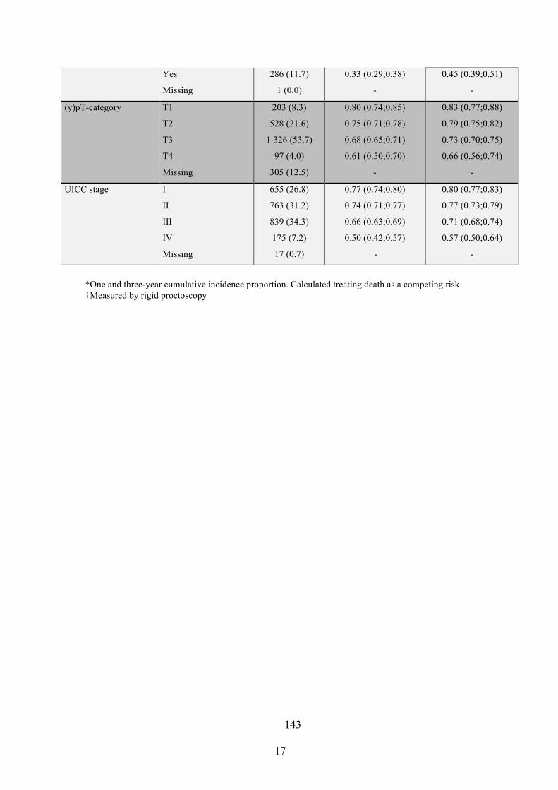
143
17
*One and three-year cumulative incidence proportion. Calculated treating death as a competing risk. †Measured by rigid proctoscopy
Yes
Missing
286 (11.7)
1 (0.0)
0.33 (0.29;0.38)
-
0.45 (0.39;0.51)
-
(y)pT-category T1
T2
T3
T4
Missing
203 (8.3)
528 (21.6)
1 326 (53.7)
97 (4.0)
305 (12.5)
0.80 (0.74;0.85)
0.75 (0.71;0.78)
0.68 (0.65;0.71)
0.61 (0.50;0.70)
-
0.83 (0.77;0.88)
0.79 (0.75;0.82)
0.73 (0.70;0.75)
0.66 (0.56;0.74)
-
UICC stage I
II
III
IV
Missing
655 (26.8)
763 (31.2)
839 (34.3)
175 (7.2)
17 (0.7)
0.77 (0.74;0.80)
0.74 (0.71;0.77)
0.66 (0.63;0.69)
0.50 (0.42;0.57)
-
0.80 (0.77;0.83)
0.77 (0.73;0.79)
0.71 (0.68;0.74)
0.57 (0.50;0.64)
-

144
18
Table 3: Crude and adjusted hazard ratios (0-3 years), associating different
characteristics with delay in stoma reversal for patients with intended restorative
rectal cancer resection with diverting stoma, Denmark 2001-2012
Characteristic No of subjects
n (%)
Crude
Hazard-ratio*
(95%CI)
Adjusted
Hazard-ratio†
(95%CI)
Total 2 449 - -
Period of surgery May 2001 - Dec
2004
Jan 2005 - Dec
2008
Jan 2009 - Apr
2012
677 (27.6)
865 (35.3)
907 (37.0)
1.0 (ref)
0.96 (0.85;1.08)
1.01 (0.90;1.13)
1.0 (ref)
1.08 (0.91;1.28)
1.07 (0.90;1.28)
Gender Male
Female
1 551 (63.3)
898 (36.7)
0.98 (0.89;1.08)
1.0 (ref)
1.05 (0.94;1.19)
1.0 (ref)
Age (y) !65
>65
1,240
1,209
1.0 (ref)
0.95 (0.87;1.04)
1.0 (ref)
0.92 (0.82;1.03)
BMI 0-19
20-24
25-29
+30
Missing
101 (4.1)
853 (34.8)
781 (31.9)
277 (11.3)
437 (17.8)
1.0 (ref)
1.13 (0.87;1.45)
1.17 (0.91;1.51)
1.06 (0.80;1.39)
-
1.0 (ref)
1.12 (0.84;1.49)
1.17 (0.87;1.56)
1.08 (0.79;1.48)
-
Charlson
Comorbidity
Index
0
1-2
+3
2 079 (84.9)
327 (13.4)
43 (1.8)
1.0 (ref)
0.93 (0.81;1.07)
0.82 (0.56;1.21)
1.0 (ref)
0.99 (0.84;1.17)
0.77 (0.49;1.20)
ASA grade I
II
III-IV
Missing
745 (30.4)
1 402 (57.3)
255 (10.4)
47 (1.9)
1.0 (ref)
0.87 (0.79;0.96)
0.75 (0.63;0.89)
-
1.0 (ref)
0.91 (0.80;1.03)
0.82 (0.65;1.02)
-
Distance of
primary tumour
(lower edge)
from anal verge
(cm)
0-5
>5-10
>10-15
Missing
207 (8.5)
1 515 (61.9)
704 (28.8)
23 (0.9)
0.84 (0.69;1.01)
1.03 (0.93;1.14)
1.0 (ref)
-
0.84 (0.66;1.06)
1.11 (0.97;1.26)
1.0 (ref)
-
Neoadjuvant
(chemo-)
radiotherapy
Yes
No
719 (29.4)
1 730 (70.6)
0.76 (0.69;0.85)
1.0 (ref)
0.75 (0.66;0.85)
1.0 (ref)
Surgical approach Laparoscopy 562 (23.0) 1.0 (ref) 1.0 (ref)

145
19
Laparotomy 1 887 (77.1) 0.96 (0.86;1.07) 1.00 (0.87;1.16)
Blood loss,
intraoperative
(ml)
!300
>300
Missing
1 212 (49.5)
1 207 (49.3)
30 (1.2)
1.0 (ref)
0.80 (0.73;0.88)
-
1.0 (ref)
0.86 (0.76;0.97)
-
Anastomotic
leakage
Yes
No
Missing
286 (11.7)
2 162 (88.3)
1 (0.0)
0.42 (0.35;0.50)
1.0 (ref)
-
0.41 (0.33;0.50)
1.0 (ref)
-
(y)pT-category T1
T2
T3
T4
Missing
203 (8.3)
528 (21.6)
1 316 (53.7)
97 (4.0)
305 (12.5)
1.0 (ref)
0.83 (0.69;0.99)
0.63 (0.53;0.74)
0.51 (0.39;0.69)
-
1.0 (ref)
0.86 (0.70;1.05)
0.63 (0.47;0.83)
0.62 (0.42;0.90)
-
UICC stage I
II
III
IV
Missing
655 (26.8)
763 (31.2)
839 (34.3)
175 (7.2)
17 (0.70)
1.0 (ref)
0.86 (0.76;0.97)
0.65 (0.58;0.73)
0.42 (0.34;0.52)
-
1.0 (ref)
1.23 (0.94;1.63)
0.83 (0.65;1.05)
0.57 (0.41;0.80)
-
*COX regression analysis. Hazard ratio (HR) <1 describes reduced “risk” of stoma reversal (i.e. the “risk” of early stoma closure is reduced when HR<1). †Mutually adjusted
!
!
!
! !

146
20
!"#$%&'('!"#$%&'()*%#+%,-./0/%1(*234*5%$2*'%+2)5*6*273%)3&*("%&(4&3)%82(94#525%:3*$334%,%;(<%
=00,%(48%/0%>1)2"%=0,=%(48%*)3(*38%(*%=,%'#512*("5%24%?347()@A%
Registrated rectal cancer patients in Denmark
between 1 May 2001 and 30 April 2012
n=15,303
Intended restorative rectal cancer resection
or Hartmann's operation n=6,859
Intended restorative rectal cancer resection
with no diverting stoma n=2,876
Hartmann's operation with formation of end-
colostomy
n=1,534
Intended restorative rectal cancer resection with diverting stoma
n=2,449
Excluded: No surgery
n=2,568 Surgical procedures other than intended
restorative rectal resection or Hartmann's operation n=3.753
Emigration before follow-up n=45
Surgery outside inclusion period n=567
Palliative surgery n=1,272
No procedure registrated in National Registry of Patients (NRP)
n=239

147
21
!"#$%&'(!"#$#%&'()*!(+,(-*+,*!./!0'.$&!1*)*10&%!-#1(+2!3!4*&10!./!/.%%.5!#67!
7"#$#%&'()*!(+,(-*+,*!61.6.1'(.+!5&0!,&%,#%&'*-!'1*&'(+2!-*&'8!&0!,.$6*'(+2!1(09!
0.2
5.5
.75
1C
umul
ativ
e In
cide
nce
of s
tom
a re
vers
al
0 1 2 3Time (in years) since rectal resection with diverting stoma

148
Paper III
Pelvic insufficiency fractures frequently occur followingpreoperative chemo-radiotherapy for rectal cancer – anationwide MRI study
J. B. Jørgensen*, P. Bondeven*, L. H. Iversen*, S. Laurberg* and B. G. Pedersen†*Department of Surgery, Aarhus University Hospital, Aarhus, Denmark, and †Department of Radiology, Aarhus University Hospital, Aarhus, Denmark
Received 5 December 2017; accepted 19 March 2018; Accepted Article Online 19 April 2018
Abstract
Aim The aim of this prospective case–control study wasto evaluate the rate of pelvic insufficiency fractures (PIFs)
in Denmark using MRI at the 3-year follow-up. All
patients had rectal cancer and had undergone surgery
with or without preoperative chemo-radiotherapy (CRT).
Method Patients registered with primary rectal cancer
in the Danish Colorectal Cancer Group database, who
underwent rectal cancer resection from April 2011
through August 2012, were invited to participate in a
national MRI study aiming to detect local recurrence
and evaluate quality of the surgical treatment. Pelvic
MRI including bone-specific sequences 3 years aftertreatment was obtained. The primary outcome was the
rate of PIFs; secondary outcome was risk factors of PIFs
evaluated in multivariate analysis.
Results During the study period, 890 patients under-went rectal cancer surgery. Of these, 403 patients were
included in the MRI study and had a 3-year follow-up
MRI. PIFs were detected in 49 (12.2%; 95% CI 9.0–15.4) patients by MRI. PIFs were detected in 39
patients (33.6%; 95% CI 24.9–42.3) treated with
preoperative CRT compared to 10 (3.5%; 95% CI 1.3–5.6) non-irradiated patients (P < 0.001). In a multivari-
ate analysis female gender (OR = 3.52; 95% CI 1.7–7.5), age above 65 years (OR = 3.20; 95% CI 1.5–6.9)and preoperative CRT (OR = 14.20; 95% CI 6.1–33.1)were significant risk factors for PIFs.
Conclusion Preoperative CRT in the treatment of rec-
tal cancer was associated with a 14-fold higher risk of
PIFs after 3 years, whereas female gender and age above65 years each tripled the risk of PIFs.
Keywords Rectal cancer, chemo-radiotherapy, pelvic
insufficiency fractures
What does this paper add to the existing litera-ture?
This paper demonstrates a risk of pelvic insufficiencyfractures following chemo-radiotherapy in the treatmentof rectal cancer that significantly exceeds the expectedlevels from the international literature. It contributes toa broader awareness of the potential adverse effects ofradiation therapy seen in the treatment of locallyadvanced rectal cancer.
Introduction
Neoadjuvant chemo-radiotherapy (CRT) is indicated for
patients with locally advanced rectal cancer in order toreduce the risk of local recurrence. Adverse effects of
short-course radiotherapy as well as long-course CRT,
combined with rectal resection, are well documented
and include a broad variety of clinical manifestations.
Frequent and well-described complications, with a sub-
stantial impact on quality of life, include bowel
dysfunction, sexual dysfunction, urinary problems, occa-
sional rectal bleeding and impaired wound healing
among others [1–6].Pelvic insufficiency fracture (PIF) is another known,
although not well described, complication of CRT in the
treatment of rectal cancer. PIF occurs as a stress fracture in
structurally weakened bone exposed to normal physiologi-
cal stress [7,8]. Decreased mineralization and deficient
elastic resistance are the underlying causes of bone atro-
phy. Development of radiation-induced osteopenia arises
partly from local ischaemia caused by damage to the
micro-vascular (Haversian) system in bone [9]. Anotherpart of the pathological pathway is probably direct impair-
ment of regeneration and resorption [10,11].
Correspondence to: Jesper Beck Jørgensen MD, Aarhus University Hospital,
THG, Department of Surgery, Tage-Hansens Gade 2, 8000 Aarhus C,
Denmark.
E-mail: [email protected]
Colorectal Disease ª 2018 The Association of Coloproctology of Great Britain and Ireland. 20, 873–880 873
Original article doi:10.1111/codi.14224

149
The most common clinical manifestation of PIF is
chronic pelvic pain [12–14]. Between 16% and 58% of
patients have been reported to be symptomatic in previ-
ous studies [10,15,16].
In the international literature, detection rates of PIF
between 3% and 11% are suggested following CRT andresection for rectal cancer. Only a few small, retrospec-
tive, non-blinded studies have been conducted until
now [17–19]. PIF rates between 8% and 45% are
reported in patients with cervical cancer, and rates from
6% to 14% are reported in other pelvic malignancies
(i.e. prostate and anal cancer) [10,15,16,20–22]. The
imaging modalities underlying these studies include CT,
MRI and bone scintigraphy in different combinations.PIF occurs in characteristic locations, and patterns
within the bony pelvis are best detected and evaluated
using MRI [23–26].The aim of this study was to evaluate the rate of PIF
at 3 years postoperatively among Danish patients trea-
ted with curative intent for rectal cancer with mesorectal
excisional surgery with or without preoperative CRT.
Methods
The prospective national study Mesorectal Excision forRectal Cancer: Aspects of Recurrence and Survival was
approved by the National Board of Health (ref.: 3-
3013-1272/1/), the Central Denmark Region Com-
mittees on Health Research Ethics, the Scientific Com-
mittee of the Danish Colorectal Cancer Group(DCCG.dk) and the Danish Data Protection Agency
(ref.: 2007!58!0010). The present study is a supple-
mental study to the MRI study.
Data sources
In Denmark there has been consecutive national report-
ing on patients with colorectal cancer to the DCCGdatabase since 1 May 2001 [27]. The purpose of the
database is to monitor the compliance and treatment of
colorectal cancer with defined quality standards set by
the DCCG.dk. Patients were identified in the DCCG
database and variables of patient demographics, tumour
location, surgical type and pathological T category were
retrieved from the database.
Setting
Between 2011 and 2012 approximately 1100 rectal can-
cer patients were treated surgically in 15 different surgical
departments in Denmark [28]. Ten surgical departments
(Aarhus University Hospital, Odense University Hospi-
tal, Regional Hospital Randers, Regional Hospital West
Jutland, Hvidovre Hospital, Zealand University Hospital,
Slagelse Hospital, Svendborg Hospital, Vejle Hospital
and Esbjerg Hospital) agreed to participate in the MRI
study, covering 65% of the patients treated for rectal can-
cer nationwide in 2011–2012. Reasons for non-participa-tion of the remaining five departments were mainly basedon lack of staff capacity.
Patients
Patients with rectal adenocarcinoma (located 15 cm or
less from the anal verge) who underwent partial
mesorectal excision (PME), total mesorectal excision
(TME) or abdominoperineal excision (APE) with cura-tive intent from April 2011 to August 2012 were iden-
tified through the DCCG database. Patients were
examined for eligibility and vital status by obtaining
information from the National Registry of Patients
based on Central Person Registry number. Patients who
developed disseminated disease or local recurrence of
rectal cancer within 3 years of follow-up were not
invited to participate. The same applied to patients whohad left the country, terminated their medical surveil-
lance programme due to very old age or comorbid dis-
ease, or who were deceased.
Consecutive patients fulfilling eligibility criteria were
invited for 3-year postoperative MRI of the pelvis.
Information on neoadjuvant oncological treatment was
retrieved from the medical records.
Oncological and surgical treatment
Patients were treated according to national guidelines
[29]. Neoadjuvant long-course CRT [50 Gy in 25–28fractions in combination with fluorouracil (5-FU)] was
offered to patients with locally advanced rectal cancer
(low T3 and T4 tumours, mid T4 tumours and mid T3
with < 5 mm to the mesorectal fascia at MRI). Short-course radiotherapy, with delay, could be chosen in those
rare situations where long-course therapy seemed to be
difficult to complete. Short-course radiotherapy with
immediate surgery is not practised in Denmark. Targeted
radiation treatment with 3D-conformal and intensity
modulated radiation therapy was standard in the partici-
pating units. Mesorectal excision was performed 8–10 weeks after completion of CRT regardless of whetherit was long-course CRT or short-course radiotherapy. All
other patients underwent direct mesorectal excision,
without preoperative oncological treatment.
Patients who completed radical surgery for Union
for International Cancer Control (UICC) Stage II (with
at least one risk factor) or III rectal cancer and had not
received neoadjuvant CRT were offered 6 months of
Colorectal Disease ª 2018 The Association of Coloproctology of Great Britain and Ireland. 20, 873–880874
Pelvic insufficiency fractures after chemo-radiotherapy J. B. Jørgensen et al.

150
adjuvant chemotherapy with 5-FU [29]. According to
national guidelines postoperative radiation therapy is
not provided in Denmark.
Magnetic resonance imaging
Patients underwent pelvic MRI as part of a national
study aiming to detect local recurrence. All MRI exami-
nations were performed on 1.5 T or 3 T platforms with
a detailed scan protocol established by the research
group. Sagittal, axial and coronal T2-weighted turbo
spin echo images, field of view (FOV) 240 mm, slice
thickness 4–5 mm, were obtained in addition to a sagit-
tal short tau inversion recovery (STIR) sequence of thebony pelvis and a sagittal T2 3D sequence of the smal-
ler pelvis. The sagittal sequences depicted the L5 cra-
nially and the buttocks caudally covering the pelvis
between the ischial tuberosities. Based on the sagittal
T2-weighted sequence, axial T2-weighted and coronal
T2-weighted sequences were planned. The axial
sequence covered the entire pelvis from the lower bor-
der of the subcutaneous part of the external sphincterto the promontory. The coronal sequence covered ante-
riorly the posterior part of the pubic bone and posteri-
orly the sacrum. The sagittal T2 3D sequence covered
the smaller pelvis with a slab of 88 slices and with a slice
thickness of 1 mm.
The MR examinations were all evaluated by a dedi-
cated multidisciplinary team radiologist at Aarhus
University Hospital with 8 years of sub-specialization inpelvic MRI. The multidisciplinary team radiologist was
blinded to all clinical data with the exception of the
preoperative MRI examination.
Detection of pelvic insufficiency fractures by MRI
All MRI examinations were prospectively evaluated for
the presence of changes compatible with PIFs. Highsignal intensity changes in the bone marrow on STIR
images (Fig. 1), indicative of bone marrow oedema,
accompanied by subtle linear changes of low signal
intensity on T2-weighted images were regarded as sug-
gestive of PIFs (Fig. 2). STIR is the most sensitive cur-
rent method to detect bone marrow oedema while T2
images are primarily used for anatomical mapping and
identification of low signal intensity changes [30]. Loca-tions of the areas of abnormal bone marrow signal were
reviewed.
Outcome measures
The primary outcome was the rate of PIFs as
detected by 3-year postoperative MRI examinations.
Secondary outcomes were the anatomical distribution
of PIFs and risk factors of PIF evaluated in multivari-
ate analysis, including sub-analysis of patients who
had APE.
Statistical analysis
The PIF rate was calculated and stratified for neoadju-
vant CRT. Other potential risk factors for PIF were
investigated. Continuous data were categorized. Demo-
graphics and categorical data were compared by Fisher’s
exact test (univariate analysis). Factors found to have a
significance less than 0.1 in the univariate model wereentered into a multiple logistic regression model to
identify independent predictors for PIF. Adjusted ORs
for PIF were computed using multiple logistic regres-
sion to estimate the impact of gender, age at surgery
(categorized as < 65 years or ! 65 years according to
SAG-STIR
Figure 1 Sagittal STIR sequence showing bone marrowoedema in the alar of the sacrum.
AXT2
Figure 2 Axial T2-weighted MRI showing sclerosis and frac-ture in the alar of the sacrum.
Colorectal Disease ª 2018 The Association of Coloproctology of Great Britain and Ireland. 20, 873–880 875
J. B. Jørgensen et al. Pelvic insufficiency fractures after chemo-radiotherapy

151
the median age at time of surgery), use of neoadjuvant
therapy, tumour height (categorized as low vs mid/
high), surgical procedure (categorized as APE vs TME/
PME) and pT/ypT category (according to T subcate-
gory). A P value of less than 0.05 was considered signif-
icant.Stata! version 12.0 (StataCorp, College Station,
Texas, USA) was used for statistical analysis.
Results
In total 890 patients underwent rectal cancer surgery
with curative intent at the 10 participating hospitals
during April 2011 through August 2012. Of these, 320patients did not fulfil the eligibility criteria, leaving 570
potentially eligible patients (Fig. 3). In total, 403
patients were included in the MRI study and had a 3-
year follow-up MRI. Among these, 116 (28.8%; 95%
CI 24.3–33.2) patients had been treated with neoadju-
vant CRT. Reasons for non-participation were con-
traindications to MRI, non-respondents, decline of
invitation or missing preoperative MRI (Fig. 3).
The frequency of neoadjuvant CRT in the group of
patients not included for MRI (n = 487) was 33.3%
(95% CI 29.1–37.5). No significant difference in CRT
rates between the included and non-included group of
patients was observed (P = 0.167). Patient demograph-
ics, tumour characteristics and treatment regimens aresummarized in Table 1.
Pelvic insufficiency fractures
During follow-up at 36 months, 49 (12.2%; 95% CI
9.0–15.4) patients had PIFs detected on MRI. PIFs
were detected in 39 of 116 (33.6%; 95% CI 24.9–42.3)patients treated with preoperative CRT. The rate ofPIFs in the non-irradiated group was 3.5% (95% CI
1.3–5.6) (P < 0.001).
In univariate analysis of risk factors, age above 65 years
(P = 0.032), female gender (P = 0.008), tumour in the
lower part (0–5 cm) of the rectum (P < 0.001), preopera-
tive CRT (P < 0.001), lower tumour category (P < 0.001)
and APE as surgical procedure (P < 0.001) were associated
with a higher risk of PIF (Table 1).
Registered patients
n = 890
Invited for post-operative MRI
n = 570
MRI performed 3 years postoperatively
n = 403
Preoperative radiotherapy
n = 116
No preoperative radiotherapy
n = 287
Excluded:
Local recurrence n = 46
Disseminated metastatic disease n = 75
Dead before time of inclusion n = 110
Co-morbid disease n = 52
Age n = 28
Emigration = 9
Declined invitation n = 78
Contraindication to MRI n = 16
Non-respondents n = 69
No preoperative MRI n = 4
Figure 3 Flowchart.
Colorectal Disease ª 2018 The Association of Coloproctology of Great Britain and Ireland. 20, 873–880876
Pelvic insufficiency fractures after chemo-radiotherapy J. B. Jørgensen et al.

152
Multivariate analysis of the risk factors associated with
PIF found that age above 65 years (P = 0.004), female
gender (P = 0.001) and preoperative CRT (P < 0.001)
were independent risk factors for PIF (Table 1).
In patients who underwent APE, excision of the coccyx
was associated with a higher risk of PIF (31.5% vs 11.9%,P < 0.001) but was not an independent risk factor in mul-
tivariate analysis (data not shown). Female gender, ageabove 65 years and preoperative radiotherapy, however,
remained significant risk factors of PIF in patients who
underwent APE as in the entire cohort (data not shown).
Localization and anatomical distribution of pelvic
insufficiency fractures
A fracture was observed in the sacrum in 47 of 49
(95.9%; 95% CI 86.0–99.5) patients with PIFs. Lesionsnear the sacroiliac joints were observed in 45 (91.8%;
Table 1 Patient demographics, treatment characteristics and risk factors associated with PIF.
Characteristic
All patients
n (%)
Patients with PIF
n (%) Unadjusted P*
Adjusted OR
(95% CI) Adjusted P†
Number 403 49 (12.2)
Gender Male 252 (62.5) 22 (8.7) 0.008 3.52 (1.7;7.5) 0.001
Female 151 (37.5) 27 (17.9)
Age (years) < 65 191 (47.4) 16 (8.4) 0.032 3.20 (1.5;6.9) 0.004
! 65 212 (52.6) 33 (15.6)
Body mass index (kg/m2) < 25 183 (46.3) 25 (13.7) 0.354
! 25 212 (53.7) 24 (11.3)
Smoker Yes 69 (17.2) 7 (10.1) 0.764
Former‡ 162 (40.3) 23 (14.2)
Never 132 (32.8) 14 (10.6)
Missing 40 (9.8) 5 (12.8)
ASA score I 134 (33.3) 14 (10.5) 0.227
II 236 (58.7) 28 (11.9)
III 29 (7.2) 7 (24.1)
Missing 3 (0.8) 0 (0.0)
Distance of primary tumour
from anal verge (cm)§0–5 97 (24.1) 26 (26.8) < 0.001 1.75 (0.89;3.42) 0.118
> 5–10 172 (42.7) 22 (12.8)
> 10–15 120 (29.8) 1 (0.8)
Missing 14 (3.5) 0 (0.0)
Neoadjuvant (chemo-)
radiotherapy
No 287 (71.2) 10 (3.5) < 0.001 14.2 (6.1;33.1) <0.001Yes 116 (28.8) 39 (33.6)
Surgical approach Laparotomy 133 (33.1) 18 (13.5) 0.627
Laparoscopy 269 (66.9) 31 (11.5)
Surgical preocedure PME 118 (29.3) 5 (4.2) < 0.001 2.17 (0.71;6.58) 0.172
TME 172 (42.7) 14 (8.1)
APE 113 (28.0) 30 (26.5)
pT/ypT category¶ T0 16 (4.0) 8 (50.0) 0.001 0.81 (0.56;1.17) 0.259
T1 21 (5.2) 3 (14.3)
T2 117 (29.0) 15 (12.8)
T3 216 (53.6) 20 (9.3)
T4 29 (7.2) 3 (10.3)
Missing 4 (1.0) 0 (0.0)
*Fisher’s exact test.
†Multiple logistic regression adjusted for gender, age at surgery, tumour height, surgical procedure, neoadjuvant therapy and
pT/ypT category.
‡No smoking within 8 weeks.
§Measured by rigid proctoscopy at pretreatment clinical evaluation.
¶Based on histopathological evaluation of excised specimen. The pathological tumour category for the 115 patients who had neo-
adjuvant CRT was ypT0, 15; ypT1, 9; ypT2, 32; ypT3, 51; ypT4, 8. One patient with T0 did not receive CRT. This patient under-
went local excision of the tumour prior to definitive surgery and the T categorization here was T0.
Colorectal Disease ª 2018 The Association of Coloproctology of Great Britain and Ireland. 20, 873–880 877
J. B. Jørgensen et al. Pelvic insufficiency fractures after chemo-radiotherapy

153
95% CI 83.9–99.8) patients, and 39 (79.6%; 95% CI
67.9–91.3) had bilateral lesions detected. In patients
with PIF in the sacrum 38 (80.9%; 95% CI 69.2–92.5)had additional fractures at another site in the pelvis, the
right posterior-medial part of ilium (close to the sacral
joint) being the most frequent, 68.1% (95% CI 54.3–81.9). The anatomical predilection sites of PIFs are
shown in Fig. 4.
Discussion and conclusions
This prospective, population-based, case–control study
reports the rate of PIF 3 years after rectal cancer treat-
ment with curative intent in the national setting ofDenmark.
The main finding was a surprisingly high PIF rate of
33.6% in patients who underwent neoadjuvant CRT in
combination with mesorectal excision surgery compared
to only 3.5% in the group of patients treated with surgery
alone. This 3-year rate is probably an underestimate as
some fractures may heal or partly heal within 3 years.
The lifetime risk of all pelvic fractures at the age of85 years in the USA has been estimated at 2% for white
women and 0.5% for white men, which corresponds
well to our findings of 3.5% in the control group [31].
Well known predisposing conditions for development
of PIFs are osteoporosis and radiation therapy. PIFs pri-
marily occur in postmenopausal, elderly women. Other
conditions associated with PIFs are rheumatoid arthritis,
prolonged corticosteroid therapy, renal failure and hipsurgery [18,32,33].
In the present study independent risk factors for
PIFs were female gender (OR = 3.52), age above 65
(OR = 3.20) and preoperative CRT (OR = 14.20).
In recent years there have been major technological
advances in the administration of radiation therapy.
Modern intensity modulated radiation therapy is specifi-
cally targeted to the malignant tissue. This is supposed
to decrease both acute and late-onset toxicity. Despite
these technological advances, we have demonstrated arisk of PIF following CRT that greatly exceeds the
expected level of 3%–11% reported in the literature.
The difference between our findings and those
reported in previous studies may be due to our consis-
tent use of a highly sensitive imaging modality. MRI is
superior in this field of interest compared to conven-
tional imaging modalities [23]. In addition, we used
MRI sequences (STIR) that are specifically suitable fordetecting PIFs (i.e. bone marrow oedema). The use of
MRI, with disease-specific sequences, is not seen in pre-
vious studies of PIFs in rectal cancer.
Internationally, three different strategies in the
neoadjuvant treatment of rectal cancer exist: short-
course radiotherapy (25 Gy in five fractions) followed
by immediate surgery or delayed surgery, or long-
course CRT (50 Gy in 25–28 fractions in combinationwith a cytostatic agent) and tumour resection 8–10 weeks after completion. Radiation induced damage
to bone is highly dependent on exposure.
Recent studies of non-operative management of low
rectal cancer following neoadjuvant CRT have been
conducted in selected patients with UICC Stage I dis-
ease [34–40]. However, none of these trials is assessing
the risk of PIF. In the light of various ‘watch and wait’protocols evaluating deferral of surgery after radiation
therapy of early rectal cancers, pelvic MRI may in future
not only be a useful tool to monitor the effect of treat-
ment or the presence of local recurrence, but also be a
0.4%
17% 12%
9%9%
18%14%
9%
2%4% 4%
0.9%
0.4%0.9%Figure 4 Frequency and anatomicaldistribution of PIF.
Colorectal Disease ª 2018 The Association of Coloproctology of Great Britain and Ireland. 20, 873–880878
Pelvic insufficiency fractures after chemo-radiotherapy J. B. Jørgensen et al.
154
useful tool for the evaluation of more gentle ways to
irradiate rectal tumours.
A sub-analysis of risk factors associated with PIFs in
patients who had an APE performed was completed to
verify if excision of the coccyx adds a further weakening
or an additional trauma to the pelvic ring. In the multi-variate model excision of the coccyx was not a predictor
of PIFs. This is not surprising, since the coccyx is not a
weight-bearing part of the pelvic ring.
The anatomical predilection sites of PIFs were found
to be the sacrum and the medial part of the os ilium
close to the sacroiliac joints. This is the weight-bearing
part of the pelvic ring and the high occurrence of PIFs
in this area is consistent with observations in otherstudies.
In a clinical setting, PIFs must be considered a dif-
ferential diagnosis in patients complaining of pelvic pain
following neoadjuvant CRT in the treatment of rectal
cancer. In the first instance it is critically important to
exclude local recurrence. Performing specific MRI
sequences with identification of characteristic patterns
of PIFs makes it possible to avoid further unnecessaryclinical examination. Assessing the prevalence of pain
was not an aim in this study and future MRI studies
combined with quality of life assessment are needed to
evaluate pelvic pain in relation to PIFs in patients trea-
ted for rectal cancer.
The strength of the present study includes the
nationwide, prospective design and the consequent use
of MRI with STIR sequences which are highly sensitivein detecting PIFs. In addition, blinding of the multidis-
ciplinary team radiologist to all clinical data of the
patients reduced the risk of information bias.
The study was initiated with the intent to detect
local recurrence of rectal cancer and visualize inadver-
tent residual mesorectal fat. Regarding the detection of
PIFs, it is a limitation that our cohort was not examined
with bone sequences (STIR and T1) covering the entirepelvis in at least two planes including the femoral head
and the proximal part of the femoral shaft. The latter
could potentially harbour insufficiency fractures, espe-
cially in patients treated with CRT for low rectal cancer.
Our protocol did not include contrast-enhanced
sequences either. However, these limitations may tend
to underestimate the true prevalence of PIFs in this
group of patients.Other limitations of the study are lack of information
on the clinical consequences of PIFs and the presence
of chronic pain. Furthermore, we did not have informa-
tion on the exact radiotherapy regimen as well as the
specific dose–volume relationships since the aim of the
present explorative study was to evaluate the PIF rate
only. In addition we had no information about
osteoporosis or corticosteroid use, both of which are
associated with increased risk of PIFs.
In conclusion, neoadjuvant CRT in the treatment of
rectal cancer is associated with a substantial risk of PIF.
One-third of patients who underwent mesorectal exci-
sion surgery for rectal cancer in combination with pre-operative CRT had PIFs 3 years after surgery detected
by MRI.
Acknowledgements
The present study was funded by the Danish Cancer
Society. The Study Group of the project MesorectalExcision for Rectal Cancer: Aspects of Recurrence andSurvival is acknowledged for their contribution in
patient recruitment and MRI examinations. The Study
Group consists of Niels Qvist and Jon Thor Asmussen,
Odense University Hospital; Safaa Al-Ubaidi and
Fr!ed!erique Johnsen, Regional Hospital West Jutland;
Steffen Jais Rosenstock and Annette Bøjer Jensen, Hvi-
dovre Hospital; Ole Roikjær and Karina Vinum, Zeal-
and University Hospital; Sharaf Karim Perdawood andOdai Adnan Jalel, Slagelse Hospital; Thomas Kolbro
and Jacob Christian Bang, Svendborg Hospital; Lars
Bundgaard and Søren Rafaelsen, Vejle Hospital; Pernille
Øhlenschlager Larsen, Esbjerg Hospital.
Conflict of interest
None declared.
References
1 Pollack J, Holm T, Cedermark B et al. Late adverse effects
of short-course preoperative radiotherapy in rectal cancer.
Br J Surg 2006; 93: 1519–25.2 Dahlberg M, Glimelius B, Graf W, P"ahlman L. Preopera-
tive irradiation affects functional results after surgery for
rectal cancer. Dis Colon Rectum 1998; 41: 543–9.3 Marijnen CA, van de Velde CJ, Putter H et al. Impact of
short-term preoperative radiotherapy on health-related
quality of life and sexual functioning in primary rectal can-
cer: report of a multicenter randomized trial. J Clin Oncol
2005; 23: 1847–58.4 Peeters KC, Tollenaar RA, Marijnen CA et al. Risk factors
for anastomotic failure after total mesorectal excision of
rectal cancer. Br J Surg 2005; 92: 211–6.5 Marijnen CAM, Kapiteijn E, van de Velde CJ et al. Acute
side effects and complications after short-term preoperative
radiotherapy combined with total mesorectal excision in
primary rectal cancer: report of a multicenter randomized
trial. J Clin Oncol 2002; 20: 817–25.6 Bregendahl S, Emmertsen KJ, Lous J, Laurberg S. Bowel
dysfunction after low anterior resection with and without
Colorectal Disease ª 2018 The Association of Coloproctology of Great Britain and Ireland. 20, 873–880 879
J. B. Jørgensen et al. Pelvic insufficiency fractures after chemo-radiotherapy

155
neoadjuvant therapy for rectal cancer: a population-based
cross-sectional study. Colorectal Dis 2013; 15: 1130–9.7 Pentecoast RL, Murray RA, Brindley HH. Fatigue, insuffi-
ciency, and pathologic fractures. JAMA 1964; 187: 1001–4.8 Lourie H. Spontaneous osteoporotic fracture of the
sacrum. Spine 1982; 248: 715–7.9 Hopewell JW. Radiation-therapy effects on bone density.
Med Pediatr Oncol 2003; 41: 208–11.10 Kwon JW, Huh SJ, Yoon YC et al. Pelvic bone complica-
tions after radiation therapy of uterine cervical cancer: evalu-
ation with MRI. AJR Am J Roentgenol 2008; 191: 987–94.11 Howland WJ, Loeffler RK, Starchman DE, Johnson RG.
Postirradiation atrophic changes of bone and related com-
plications. Radiology 1975; 117: 677–85.12 Saraux A, Valls I, Guedes C. Insufficiency fractures of the
sacrum in elderly subjects. Rev Rhum Engl Ed 1995; 62:
582–6.13 Gotis-Graham I, McGuigan L, Diamond T. Sacral insuffi-
ciency fractures in the elderly. J Bone Joint Surg Br 1994;
76: 882–6.14 Rawlings CE, Wilkins RH, Martinez S. Osteoporotic sacral
fractures: a clinical study. Neurosurgery 1988; 22: 72–6.15 Oh D, Huh SJ, Nam H et al. Pelvic insufficiency fracture
after pelvic radiotherapy for cervical cancer: analysis of
risk factors. Int J Radiat Oncol Biol Phys 2008; 70:
1183–8.16 Tokumaru S, Toita T, Oguchi M et al. Insufficiency frac-
tures after pelvic radiation therapy for uterine cervical can-
cer: an analysis of subjects in a prospective multi-
institutional trial, and cooperative study of the Japan Radia-
tion Oncology Group (JAROG) and Japanese Radiation
Oncology Study Group (JROSG). Int J Radiat Oncol Biol
Phys 2012; 84: e195–200.17 Kim HJ, Boland PJ, Meredith DS et al. Fractures of the
sacrum after chemoradiation for rectal carcinoma: inci-
dence, risk factors, and radiographic evaluation. Int J
Radiat Oncol Biol Phys 2012; 84: 694–9.18 Baxter NN, Habermann EB, Tepper JE, Durham SB, Vir-
nig BA. Risk of pelvic fractures in older women following
pelvic irradiation. JAMA 2005; 294: 2587–93.19 Herman MP, Kopetz S, Bhosale PR et al. Sacral insuffi-
ciency fractures after preoperative chemoradiation for rectal
cancer: incidence, risk factors, and clinical course. Int J
Radiat Oncol Biol Phys 2009; 74: 818–23.20 Oh D, Huh SJ. Insufficiency fracture after radiation ther-
apy. Radiat Oncol J 2014; 32: 213–20.21 Igdem S, Alc!o G, Ercan T et al. Insufficiency fractures after
pelvic radiotherapy in patients with prostate cancer. Int J
Radiat Oncol Biol Phys 2010; 77: 818–23.22 Ramlov A, Pedersen EM, Røhl L et al. Risk factors for pel-
vic insufficiency fractures in locally advanced cervical cancer
following intensity modulated radiation therapy. Int J
Radiat Oncol Biol Phys 2017; 97: 1032–9.23 Blomlie V, Rofstad EK, Talle K, Sundfør K, Winderen M,
Lien HH. Incidence of radiation-induced insufficiency frac-
tures of the female pelvis: evaluation with MR imaging.
Am J Radiol 1996; 167: 1205–10.
24 Mammone JE, Schweitzer ME. MRI of occult sacral insuf-
ficiency fractures following radiotherapy. Skeletal Radiol
1995; 24: 101–4.25 Brahme SK, Cervilla V, Vint V, Cooper K, Kortman K,
Resnick D. Magnetic resonance appearance of sacral insuffi-
ciency fractures. Skeletal Radiol 1990; 19: 489–93.26 Cabarrus MC, Ambekar A, Lu Y, Link TM. MRI and CT
of insufficiency fractures of the pelvis and the proximal
femur. AJR Am J Roentgenol 2008; 191: 995–1001.27 Ingeholm P, Gogenur I, Iversen LH. Danish colorectal
cancer group database. Clin Epidemiol 2016; 8: 465–8.28 Danish Colorectal Cancer Group. D.C.C.G. Annual Report
(in Danish). 2012.
29 Danish Colorectal Cancer Group. D.C.C.G. Guidelines for
the Diagnosis and the Management of Colorectal Cancer (in
Danish). 2009.
30 Matcuk GR, Mahanty SR, Skalski MR, Patel DB, White
EA, Gottsegen CJ. Stress fractures: pathophysiology, clini-
cal presentation, imaging features, and treatment options.
Emerg Radiol 2016; 23: 365–75.31 Melton RLJ, Sampson JM, Morrey BF, Ilstrup DM. Epi-
demiologic features of pelvic fractures. Clin Orthop Relat
Res 1981; 155: 43–7.32 Cooper KL, Beabout JW, Swee RG. Pelvic insufficiency
fractures of the sacrum. Radiology 1985; 156: 15–20.33 Ogino I, Okamoto N, Ono Y, Kitamura T, Nakayama H.
Pelvic insufficiency fractures in postmenopausal woman
with advanced cervical cancer treated by radiotherapy.
Radiother Oncol 2003; 68: 61–7.34 Habr-Gama A, Gama-Rodrigues J, S~ao Juli~ao GP et al.
Local recurrence after complete clinical response and watch
and wait in rectal cancer after neoadjuvant chemoradiation:
impact of salvage therapy on local disease control. Int J
Radiat Oncol Biol Phys 2014; 88: 822–8.35 Habr-Gama A, Sao Juliao GP, Perez RO. Nonoperative
management of rectal cancer: identifying the ideal patients.
Hematol Oncol Clin North Am 2015; 29: 135–51.36 Appelt AL, Pløen J, Harling H et al. High-dose chemora-
diotherapy and watchful waiting for distal rectal cancer: a
prospective observational study. Lancet Oncol 2015; 16:
919–27.37 Beets GL, Figueiredo NL, Habr-Gama A, van de Velde CJ.
A new paradigm for rectal cancer: organ preservation:
introducing the International Watch & Wait Database
(IWWD). Eur J Surg Oncol 2015; 41: 1562–4.38 Smith JD, Ruby JA, Goodman KA et al. Nonoperative
management of rectal cancer with complete clinical
response after neoadjuvant therapy. Ann Surg 2012; 256:
965–72.39 Smith RK, Fry RD, Mahmoud NN, Paulson EC. Surveil-
lance after neoadjuvant therapy in advanced rectal cancer
with complete clinical response can have comparable out-
comes to total mesorectal excision. Int J Colorectal Dis
2015; 30: 769–74.40 Maas M, Beets-Tan RG, Lambregts DM et al. Wait-and-
see policy for clinical complete responders after chemoradi-
ation for rectal cancer. J Clin Oncol 2011; 29: 4633–40.
Colorectal Disease ª 2018 The Association of Coloproctology of Great Britain and Ireland. 20, 873–880880
Pelvic insufficiency fractures after chemo-radiotherapy J. B. Jørgensen et al.
156

157

158

159

160

161