LITERATURE REVIEW OF RESEARCH TOPIC - UM Students ...
29
6 2.0 LITERATURE REVIEW 2.1 Medicinal mushrooms Since Greek and Roman antiquity, mushroom has been used as food and medicine (Anke, 1989). The ancient Roman believed mushrooms to be 'the food of the Gods', the Egyptians considered them to be 'a gift from Osiris' and the Chinese regarded them as 'the elixir of life' (Samorini, 2001). Many cultures worldwide, especially in the orient recognized that extracts from certain mushrooms could have profound promoting benefits, and consequently became essential components in many traditional Chinese medicines. In fact, mushrooms have seen a summation of more than 3000 years of use for the prevention and treatment of diseases in Chinese medicine (Chang, 2006). Mushrooms produce a large number of metabolites that show antibacterial, antifungal, antiviral, antitumor, hypoglycemic, anti allergenic, immunodulating, anti- inflammatory, hypolipidemic, and hepatoprotective activity (Yamac and Bilgili, 2006). There are at least 270 species of mushrooms that known to posses various therapeutic properties and the term „medicinal mushroom‟ is now increasingly gaining worldwide recognition (Ying et al., 1987). Medicinal mushrooms are macrofungi that contain pharmaceutical substances with potent and unique health enhancing properties. They posses enormous potential and should be included into the medical biotechnology industry for the benefits of humankind (Wasser and Weis, 1999). Historically, most medicinal mushrooms species were relatively scarce and were collected from the forest where they grew on dead or living trees and forest litter. They are
-
Upload
khangminh22 -
Category
Documents
-
view
1 -
download
0
Transcript of LITERATURE REVIEW OF RESEARCH TOPIC - UM Students ...

6
2.0 LITERATURE REVIEW
2.1 Medicinal mushrooms
Since Greek and Roman antiquity, mushroom has been used as food and medicine
(Anke, 1989). The ancient Roman believed mushrooms to be 'the food of the Gods', the
Egyptians considered them to be 'a gift from Osiris' and the Chinese regarded them as 'the
elixir of life' (Samorini, 2001). Many cultures worldwide, especially in the orient
recognized that extracts from certain mushrooms could have profound promoting benefits,
and consequently became essential components in many traditional Chinese medicines. In
fact, mushrooms have seen a summation of more than 3000 years of use for the prevention
and treatment of diseases in Chinese medicine (Chang, 2006).
Mushrooms produce a large number of metabolites that show antibacterial,
antifungal, antiviral, antitumor, hypoglycemic, anti allergenic, immunodulating, anti-
inflammatory, hypolipidemic, and hepatoprotective activity (Yamac and Bilgili, 2006).
There are at least 270 species of mushrooms that known to posses various therapeutic
properties and the term „medicinal mushroom‟ is now increasingly gaining worldwide
recognition (Ying et al., 1987).
Medicinal mushrooms are macrofungi that contain pharmaceutical substances with
potent and unique health enhancing properties. They posses enormous potential and should
be included into the medical biotechnology industry for the benefits of humankind (Wasser
and Weis, 1999).
Historically, most medicinal mushrooms species were relatively scarce and were
collected from the forest where they grew on dead or living trees and forest litter. They are

7
predominantly lignocelluloses degraders. For medicinal purposes, they were almost always
prepared either as hot water extracts, concentrates or in powdered. To date, almost all of the
important medicinal mushrooms have been subjected to large-scale artificial cultivation by
solid substrate or low moisture fermentation, thus removing the historical scarcity factor
and allowing large commercial operation to develop (Stamets, 2000).
2.2 Pharmacological potential of medicinal mushrooms
There have been a number of reviews published on the bioactive substances found
in mushrooms and their medicinal properties (Rai et al., 2005; Stamets, 2002; Lindequist et
al., 2005). Mushroom has many significant medicinal values. Experiences from Asian and
Eastern countries show that mushroom could play important role in cancer prevention and
treatment. Besides, in some East Asian countries, Japan, China and Korea, mushrooms are
used as „immunomodulator‟ (biological response modifier, immune potentiators and
immuno stimulants). They are the most important medicinal mushroom drugs used in these
countries (Lindequest et al., 2005). The used of mushroom I. obliquus as a folk medicine
for cancer and stomach have also been reported in Eastern Europe since the 16th
and 17th
century (Molitoris, 1994).
Besides the ability of many mushroom to stimulate the immune system, some
medicinal mushroom were demonstrated to suppress immune responses. This is very
attractive mechanism especially for the treatment of allergic diseases. Anti allergic affects
of few ethanol extracts have been investigated in-vivo (Sano et al., 2002). Furthermore,
eating of Tricholoma populinum J. E. Lange was proven to lead to the regression of severe

8
allergic symptoms in a patient with thrombongitis obliterans and in another patient with
urticaria (Kreisel et al., 1990).
The mushrooms also have the ability to lower blood pressure and free cholesterol in
plasma, as well as accelerate the accumulation of lipid in the liver by removing them from
circulation (Rai et al., 2005). The control of blood lipids especially the cholesterol is
important in reducing the risk of atherosclerosis development. A pronounced
hypocholesteremic effect of Pleurotus ostreatus with inhibition of lipid peroxidation was
demonstrated in rats and rabbits. Ten percent intake of dried fruiting bodies of this
mushroom has significantly reduced the incidence and size of atherosclerotic plaques in
rabbits (Bobek et al., 1998). Gunde-Cinerman (1999) has suggested that Pleurotus
mushroom could be recommended as a neutral cholesterol lowering substances in the
human diet. Biosynthesis of cholesterol was also inhibited by some of Triterpenes from
Ganoderma lucidum (Komoda et al., 1989).
In addition to medicinal potentials mention above, mushrooms also exhibit anti-
inflammatory effect. This is supported by studies conducted by Kim et al., in 2003 and
2004. In these studies, ethanolic extracts and a proteoglycan from P. linteus showed anti-
inflammatory effect in the collagen-induced arthritis and the croton oil-induced ear edema
test in mice and antinociceptic effect in the writhing test (Kim et al., 2003; Kim et al.,
2004).
2.3 Cultivation of medicinal mushrooms
Cultivation of mushroom is a global practice. To date, there are only 20 mushroom
species currently cultivated on an industrial scale out of about 35 mushroom species

9
cultivated commercially. World production of mushrooms is increasingly being dominated
by species that are both edible and have medicinal properties or only medicinal such as
Ganoderma and Trametes (Smith et al., 2002). According to Chang (1999), the overall
production of cultivated edible and/or medicinal mushrooms was recorded as 4909 x 103
tons in 1994, increasing to 6158 x 103 in 1997, with an estimated value in access of 14
billion US dollar.
Medicinal mushrooms can be cultivated through a variety of methods. Production of
the fleshy mushroom fruit-bodies utilizes various forms of solid substrate or low moisture
fermentations whereas for mycelial biomass production, liquid tank fermentations are now
becoming increasingly important particularly for nutriceutical and pharmaceutical
productions (Smith et al., 2002).
According to Stamets (2000), the rapid worldwide cultivation of medicinal
mushroom is due largely to the use of specially designed polypropylene bags or containers
with microfilter window for ion exchange. The bags contain the substrate of sawdust and
selected nutrients, and sterilized prior to inoculation with the mushroom mycelia. The entire
growing process is carried out under controlled environmental conditions over a reduced
time scale (1-3 months). The composition of basic substrate and supplementary ingredients
can vary considerably since the basic raw materials are derived from lignocellulosics from
agriculture or forestry, thus there will be some degree of variation in size and age which
will influence specific biochemical composition of mushrooms (Gunde-Cimerman, 1999;
Wasser et al., 2000). This is not critical for the production of fruit bodies for fresh market
but undoubtedly could create problems and preclude standardization of the extracted
products for nutriceuticals and pharmaceuticals purposes.

10
Submerged cultivation of mushroom mycelia in liquid media is a promising method
used due to the advance in biotechnology. As stated by Smith et al. (2002), submerged pure
culture fermentation techniques have been widely developed for most of the main
medicinal mushrooms and used in the propagation of mycelia for three main applications:
(1) liquid spawn for solid substrate fruit-body production; (2) biomass that can be used for
food and dietary supplements; and (3) biomass and/or extruded metabolites especially exo-
polysaccharides as raw materials for pharmaceutical studies. In all cases, the underlying
principle in each approach is to use mycelia in the active physiological state and of known
purity. Submerged cultivation in liquid media provides fast growth and high productivity of
desired specific compounds in optimum physical-chemical conditions thus enhance the
nutritional and nutraceutical values of the mycelia (Wasser et al., 2000).
2.4 Ganoderma
Ganoderma species belong to the kingdom of Fungi, the division of Basidiomycota,
the class of Homobasidiomycetes, the order of Aphllophorales, the family of polyporaceae
(Ganodermataceae) and the genus of Ganoderma (Chang, 1995; Wasser and Weis, 1999).
They are regarded as polypores because they possess tiny pores on the underside of their
pileus (cap) and contained reproductive spores. The caps are spongy when fresh, hardening
to a shiny, smooth woody structure when matured. The colour of the caps range from
brown, to yellowish, with reddish-brown being typical. The pore surface is cream in colour
and the spores are brown (Zoberi, 1972). Ganoderma species could not be eaten directly
and are not listed among the list of edible mushrooms because the fruiting bodies are
11
always thick, corky, and tough. They also do not have the fleshy texture characteristics of
true edible fungi (Jong and Birmingham, 1991; Jonathan et al., 2008).
2.4.1 Ganoderma australe
Ganoderma australe is non-laccate species and are often found in tropical and sub-
tropical regions. In Malaysia, G. australe has been found on mango (Mangifera indica)
stumps (Abdullah et al., 1997) and as wood degrading fungi in forest reserves and
plantation forests (Zakaria et al., 2009). According to Zakaria et al. (2009), there was no
proper record on the occurrence of G. australe on different types of forest trees in
Malaysia. Existence of biological species of G. australe was reported in Taiwan (Yeh et al.,
1995) and southern India (Kaliyaperumal and Kalaichelvan, 2008).
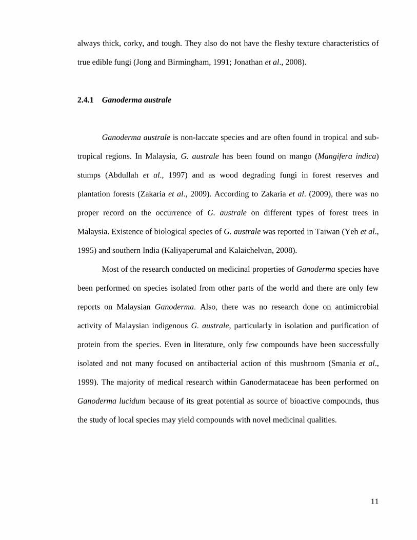
Most of the research conducted on medicinal properties of Ganoderma species have
been performed on species isolated from other parts of the world and there are only few
reports on Malaysian Ganoderma. Also, there was no research done on antimicrobial
activity of Malaysian indigenous G. australe, particularly in isolation and purification of
protein from the species. Even in literature, only few compounds have been successfully
isolated and not many focused on antibacterial action of this mushroom (Smania et al.,
1999). The majority of medical research within Ganodermataceae has been performed on
Ganoderma lucidum because of its great potential as source of bioactive compounds, thus
the study of local species may yield compounds with novel medicinal qualities.

12
2.5 Proteins from mushroom
Many molecules with interesting biological activities are produced by mushrooms.
One of the important components of mushroom is protein. Mushrooms have a high protein
content. In fact, qualitative analyses by Barbisan et al. (2003) demonstrated that
mushrooms have eight essential amino acids in addition to non-essential amino acids.
A variety of proteins with remarkable biological actions are elaborated by mushrooms and
many of these proteins have potential bioactivities. They include antifungal and
antibacterial proteins, ribosome inactivating proteins, proteins with nuclease activity,
ubiquitin-like proteins and enzymes (Ng, 2004).
2.5.1 Antimicrobial proteins
Proteins isolated from mushrooms have broad spectrum of antimicrobial activities
including antifungal, antibacterial and antiviral activity. Antifungal protein is an
extensively isolated protein from mushroom. According to Chu et al. (2005), antifungal
peptides and proteins have great economic implications because they are protective against
the destructive effects of pathogenic fungi. They can be classified according to their
structure or function into chitinases and chitinase-like proteins, glucanases, lipid transfer
proteins, protease inhibitors, ribonucleases, deoxyribonucleases, peroxidases, miraculin-
like proteins, cyclophilin-like proteins, allergen-like proteins, thaumatin-like proteins,
ribosome-inactivating proteins, antifungal peptides, glutamine-rich proteins, calcyon-like
protein, defensin-like protein, and pathogenesis-related protein.

13
Antifungal proteins have been isolated from only a few species of mushrooms,
despite the existence of numerous mushrooms species. Among the reported antifungal
proteins are from the fruiting bodies of Tricholoma giganteum (Guo et al., 2005),
Lyphyllum shimeji (Lam and Ng, 2001a), Lentinula edodes (Ngai and Ng, 2003), Polyporus
alveolaris (Wang et al., 2004), and Ganoderma lucidum (Wang and Ng, 2006b).
In contrast to antifungal proteins from mushroom, literature relating to antibacterial
proteins is only a handful. Antibacterial proteins have been isolated from the mycelia of
Cordyceps sinensis (Zheng et al., 2006) and the fruiting bodies of Clitocybe sinopica
(Zheng, 2010). Antibacterial protein from Clitocybe sinopica possessed potent antibacterial
activity against Agrobacterium rhizogenes, A. tumefaciens, A. vitis, Xanthomonas oryzae
and X. malvacearum whereas antibacterial protein from Cordyceps sinensis inhibit the
growth of both Gram negative and Gram positive bacteria.
Proteins, peptides and polysaccharopeptides from mushrooms have been reported to
be capable of inhibiting human immunodeficiency virus type 1 (HIV-1) reverse
transcriptase and protease, the two enzymes of paramount importance to the life cycle of
the HIV. Inhibitory effects on hepatitis B and Herpes simplex virus type I have also been
reported. The anti-viral effects of mushrooms do not seem to be related to viral adsorption
or virucidal effects (i.e. they do not kill the virus). However, a number of studies have
reported inhibitory effects at the initial stage of virus replication (Roupas et al., 2010).
Proteins with activity against HIV-1 reverse transcriptase have been isolated from
mushrooms such as Russula paludosa (Wang et al., 2007) and Pleurotus ostreatus (Wang
and Ng, 2000b). A novel anti-viral protein has been isolated from an extract of Grifola
frondosa (Maitake) fruiting bodies. The protein inhibited Herpes simplex virus type 1
(HSV-1) replication in vitro with an IC50 value of 4.1 mg/ml. It was reported that the

14
protein directly inactivated HSV-1 while simultaneously inhibiting HSV-1 penetration into
Vero cells (Gu et al., 2006).
2.5.2 Other proteins with biological activities from mushroom
Ribosome inactivating proteins (RIPs) is another protein isolated from mushroom.
This protein boasts a variety of activities including immunosuppressive, anti-proliferative
and antiviral activities (Ng et al., 1992).
Ribosome inactivating proteins have been isolated from few mushrooms species
including Volvariella volvacea (Yao et al., 1998), Flammulina velutipes (Ng and Wang,
2004c), Lyophyllum shemeiji (Lam and Ng, 2001a), Calvatia caelata (Ng et al., 2003) and
Hypsizigus marmoreus (Lam and Ng, 2001b). It is remarkable that, from the fruiting bodies
of Flammulina velutipes, four RIPs have been isolated designated flammulin (Wang and
Ng, 2000), velutin (Wang and Ng, 2001a), flammin and velin (Ng and Wang, 2004c). All
RIPs from Flammulina velutipes inhibit cell-free translational in the rabbit reticulocyte
lysate system. Velutin exhibited ability to inhibit β-glucosidase and β-glucoronidase which
are implicated in viral infection and also human immunodeficiency virus-1 (HIV-1) reverse
transcriptase (Wang and Ng, 2001a).
Ribonuclease is also another medicinally protein reported from several mushroom
species including Dictyophora indusiata, Ganoderma lucidum, Irpex lacteus, Lentinula
edodes, Pleurotus ostreatus, Pleurotus pulmonarius, Pleurotus sajor-caju, Pleurotus tuber-
regium, Russulus virescens, and Volvariella volvacea (Wang and Ng, 2004a). Ribonuclease
demonstrates an array of activities including antiviral, immunomodulatory and anti-
neoplastic activities.

15
Besides the previous mention proteins, there are significant amount of literature
regarding mushroom enzymes exist. These enzymes play an important role in the
saprophytic mode of life in mushroom. Together with protease, these enzymes find
important application in biotechnology and industry (Ng, 2004). Laccases which play an
important role in lignin degradation demonstrated to be very potential applications in
detoxification of polluted water, biosensor, pulping and textile dyes. These ligninolytic
enzymes have been isolated and characterize from some mushroom species including
Coriolus versicolor, Panus tigrinus, and Phlebia tremellosa (Ng, 2004). Besides, there are
numerous researches done on isolation of fungal cellulases and xylanase from mushroom.
2.6 Isolation, purification and characterization of proteins
Protein purification is a multi-step process exploiting a wide range of biochemical
and biophysical characteristics of the target protein, such as its source, relative
concentration, solubility, charge and hydrophobicity. Rosenberg (1996) stated few targets
to achieve in designing a purification protocol for those working with proteins: (1) high
recovery, (2) high purified end product, (3) reproducibility, within the lab, in other lab and
also when either scale up or down, (4) Economical use of reagent and convenience with
regard of time. Most successful isolation procedures involve only a few steps, chosen to
give the highest yields.

16
2.6.1 Extraction
The first step for purification of bioactive compounds is extraction process. There
are few factors affecting the type of compounds that can be extracted from fungi or any
living sources including the type of solvent used, the extraction process employed and the
age, and part or type of cultivation of the living tissue (Roberts, 2004). Many compounds
have been extracted from both the fruiting body and mycelia of Ganoderma. These
compounds found to exhibit remarkable biological activities (Paterson, 2006).
2.6.2 Ammonium sulfate precipitation of proteins
Ammonium sulfate precipitation is a highly effective method for proteins separation
and used as an early step in purification protocol (Rosenberg, 1996). In this method, protein
precipitation is achieved by dehydration in the microenvironment of the protein molecule.
Principally, a large number of water molecules are bound to the sulfate ion (SO4-2
) in
solution, thus reduce the amount of water available to interact with the protein molecules.
At a particular concentration of ammonium sulfate, an insufficient quantity of unbound
water will remain to keep a given protein species in solution, resulting in the precipitation
of that protein (Rosenberg, 1996).
As stated by Bonner (2007), practice ammonium sulfate is used for the routine
precipitation of proteins for the following reasons, (1) ammonium sulfate dissolves at high
concentrations (about 4 M at 0°C) generating little heat; (2) the density of saturated
ammonium sulfate solution (1.235 gml–1
) is less than the density of aggregated protein
(1.29 gml–1
) which allows collection of the precipitate by centrifugation; (3) ammonium

17
sulfate precipitation is a mild method of protein concentration giving very good recoveries
of activity; (4) proteins can be stored as an ammonium sulfate precipitate (covered in
saturated ammonium sulfate at –25°C) for long periods with little loss of activity when the
precipitate is redissolved.
2.6.3 Chromatographic techniques for protein purification
Chromatography refers to a group of separation techniques that involves a
retardation of molecules with respect to the solvent front that progresses through the
material. The name literally means “color drawing” and was originally used to describe the
separation of natural pigments on filter papers by differential retardation. The same
principle is now commonly used for protein separation. Column chromatography is the
most common physical configuration, in which the stationary phase is packed into a tube, a
column, through which the mobile phase, the eluent, is pumped. The degree to which the
molecule adsorbs or interacts with the stationary phase will determine how fast it will be
carried by the mobile phase. Chromatographic separation of protein mixtures has become
one of the most effective and widely used means of purifying individual proteins (Bonner,
2007).
Chromatography technique allowed biomolecules to be separated according to
differences in their specific properties such as size (gel filtration chromatography), charge
(ion exchange chromatography), hydrophobicity (hydrophobic interaction chromatography)
and biorecognition-ligand specificity (affinity chromatography).
In the present study, purification of protein was achieved through size exclusion
chromatography, which is also known as gel permeation chromatography and gel filtration

18
chromatography. According to Booner (2007), the separation in gel filtration
chromatography does not involve binding between the sample and the resin and it is
independent of the eluent used. These properties have made gel filtration chromatography a
technique that is useful for the separation of biological (proteins, nucleic acids and
oligosaccharides) and organic polymers.
Gel filtration can be used to isolate one or more components, to determine
molecular weight, or to analyze the molecular weight distribution in the sample. The best
result for high resolution fractionation can be achieved with sample that originally contain
few components or with samples that have been partially purified by other chromatography
techniques. This is different from ion exchange or affinity chromatography. Thus, a
significant advantage of gel filtration is that condition can be varied to suit the type of
sample or the requirement for further purification, analysis or storage without altering the
separation. Besides, it is also suitable for the final polishing step in the purification scheme
(Amersham, 2002).
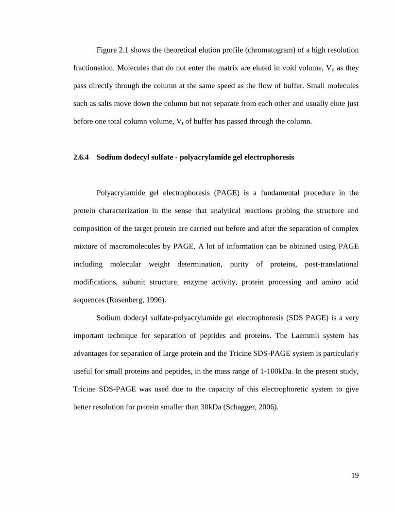
Figure 2.1 : Theoretical chromatogram of high resolution fractionation (UV absorbance).
Source : Amersham (2002)
19
Figure 2.1 shows the theoretical elution profile (chromatogram) of a high resolution
fractionation. Molecules that do not enter the matrix are eluted in void volume, Vo as they
pass directly through the column at the same speed as the flow of buffer. Small molecules
such as salts move down the column but not separate from each other and usually elute just
before one total column volume, Vt of buffer has passed through the column.
2.6.4 Sodium dodecyl sulfate - polyacrylamide gel electrophoresis
Polyacrylamide gel electrophoresis (PAGE) is a fundamental procedure in the
protein characterization in the sense that analytical reactions probing the structure and
composition of the target protein are carried out before and after the separation of complex
mixture of macromolecules by PAGE. A lot of information can be obtained using PAGE
including molecular weight determination, purity of proteins, post-translational
modifications, subunit structure, enzyme activity, protein processing and amino acid
sequences (Rosenberg, 1996).
Sodium dodecyl sulfate-polyacrylamide gel electrophoresis (SDS PAGE) is a very
important technique for separation of peptides and proteins. The Laemmli system has
advantages for separation of large protein and the Tricine SDS-PAGE system is particularly
useful for small proteins and peptides, in the mass range of 1-100kDa. In the present study,
Tricine SDS-PAGE was used due to the capacity of this electrophoretic system to give
better resolution for protein smaller than 30kDa (Schagger, 2006).

20
2.6.5 Protein sequencing and identification
Most analytical proteomics problem begin with a protein mixture. This mixture
contains intact protein of varying molecular weight, modifications, and solubilities. The
protein must be cleaved in order to obtain the peptides sequences. This is due to the
inability of mass spectrometers (MS) to perform measurement of peptide masses or obtain
peptide sequence directly on intact proteins. Although modern MS instruments can obtain a
tremendous amount of data even from relatively complete protein mixtures, simplification
of the mixtures allows data to be collected on the greatest number of components.
Therefore, to analyze protein mixture by MS, the complete mixtures of many components
must be separated into less complex mixtures containing fewer components. It is possible
to separate the intact proteins then cleave them into peptides or cleave the proteins into
peptides first then separate the peptides prior to analysis. The peptides are then analyzed by
mass spectrometer. The data from the mass spectrometer is then used, with the aid of
specialized software, to identify peptides and peptides sequences from database that match
the data from the MS data (Fig. 2.2) (Liebler, 2002).
The use of database searches is very important in protein sequence analysis. When
the partial or complete amino acids of the target protein is known, computer assisted
searches should perform to compare the new sequences to a database of known sequences.
This will reveal whether there is a protein of known structure or homologous sequence that
is sufficiently similar to the sequence of the target protein to suggest a familiar relationship
and a possible function. Two mathematical aspects to database searches are the algorithm
which used to find sequence similarities, and the method used to determine which
21
similarities are statistically significant and therefore, potentially interesting (Rosenberg,
1996).
The two popular algorithm used for rapid searches of databases are FASTA and
BLAST which look for intervals or segments of good matching between sequences.
FASTA is a multistep algorithm for sequence alignment (Wilbur and Lipman, 1983). Basic
Local Alignment Search Tool (BLAST) is the most popular computer program used to
perform database similarity searches. The program allows rapid identification of sequences
from large databases that are similar to the query and provides sound statistical evaluation
of the significant of the finding (Dong and Brendel, 2005).
In the present study, Profound is used for identification of the purified protein.
Profound is a tool for searching a protein sequence collections with peptide mass maps. A
Bayesian algorithm is used to identify proteins from protein databases using mass
spectrometric peptide mapping data. The algorithm ranks protein candidates by taking into
account individual properties of each protein in the database as well as other information
relevant to the peptide mapping experiment. The program consistently identifies the correct
protein(s) even when the data quality is relatively low or when the sample consists of a
simple mixture of proteins (Zang and Chait, 2000). This system of estimating the risk of a
random match versus a true match is used in most conventional sequence homology
matching systems, such as BLAST. It has the distinct advantage of being independent of
the scoring system: the expectation value is calculated from a distribution of scored
sequences, rather than on a particular result.
According to Rossenberg (1996), if database screening based on overall sequences
similarity does not identify the target protein as a family member, a short sequence of the
target protein may be recognized as a commonly occurring structural motif, which may be

22
helpful for assigning a function. The fundamental unit of protein structure, the domain, may
possess functions independently of whether they are present in isolation or a part of a larger
multidomain protein. The domain is defined as the region or regions of a polypeptide that
fold independently and possesses a hydrophobic core with a hydrophilic exterior (Ponting
and Birney, 2005).
Figure 2.2 : General flow scheme for proteomic analysis started from protein mixture to
identification of protein.
2.7 Antimicrobial activity
2.7.1 Pathogenic bacteria
Bacteria tested in this study are divided into 2 categories which are environmental
bacteria and skin opportunistic bacteria. E. coli (ATCC 29552), E. coli (O157:H7),
Salmonella spp (ATCC 13076), Salmonella typhi, Shigella spp., Bacillus cereus, and

23
Plesiomonas shigelloides can be found in environment and are common cause of food and
water poisoning. On the other hand, Staphylococcus aureus, Staphylococcus epidermidis,
Pseudomonas aeruginosa, and Bacillus subtilis are common opportunistic skin bacteria
which can cause variety of infections including skin infection, bacteremia, eyes and ears
infections. They are also common cause of hospital/healthcare associated infection
(HAI/HCAI) and often turn out being multiple resistant organisms.
Health care-associated infection (HCAI), which also referred to as “nosocomial” or
“hospital” infection, is defined as: “An infection occurring in a patient during the process of
care in a health-care facility which was not present or incubating at the time of admission.
This includes infections acquired in the hospital but appearing after discharge, and also
occupational infections among staff. HCAI is acknowledged as the most frequent adverse
event in health care. In Malaysia, the estimated incidence of HCAI from 1995 and 2008
was reported by World Health Organization (WHO) to be 13.9 percent
(http://www.who.int/gpsc/country_work/summary_20100430_en.pdf).
2.7.2 Pathogenic yeast
In the present study, antimicrobial activity of the protein from Ganoderma australe
strains and Ganoderma tsugae were tested against pathogenic yeast Candida albicans,
Candida parasilopsis and Schizosaccaromyces pombe.
Candida is the fourth most common cause of hospital related bloodstream infections
(Banerjee et al., 1991). Candida albicans is a dimorphic fungus lacks a sexual cycle and is
a diploid organism. It exhibits many different morphological forms under different
environmental conditions, including budding yeast cells, pseudohyphae, true hyphae and

24
clamydospores (Odds and Baillihre, 1988). The fungus which is the most isolated yeast,
exist as a commensal of mammalian including humans. According to Molero et al. (1998),
C. albicans colonize mucosal surfaces of the oral and vaginal cavities and the digestive
tract and is also able to cause various infections, depending on the nature of the underlying
host defect. This yeast cause infections known as candidacies which can be divided into
superficial and deep seated and represent a major clinical problem.
Candida parasilopsis is pathogenic yeast which infects hospitalized patients.
According to Levy et al. (1998), this fungus particularly affects critically ill neonates and
surgical intensive care unit (ICU) patients. The hands of the healthcare workers are
suggested as the predominant environmental source responsible for the spreading of
infections (Lupetti et al., 2002).
Schizosaccaromyces pombe which is called “fission yeast” is a unicellular
eukaryote, used as a model organism in molecular and cell biology. The cells are rod-
shaped and have approximately 14.1 million base pairs in its genome, which is estimated to
contain 4,970 protein-coding genes and at least 450 non-coding RNA (Wilhelm et al.
2008). This fungus is an important organism in studying the cellular responses to DNA
damage and process of DNA replication. Furthermore, the full genome sequence of this
yeast has been published in 2002, with many genes homologous to human disease genes
being identified (http://en.wikipedia.org/wiki/Schizosaccharomyces_pombe).

25
2.7.3 Pathogenic fungi
There are five phytopathogenic fungi tested in this study including Fusarium
oxysporium cubense I (FOC1), Fusarium oxysporium cubense II (FOC2), Fusarium
oxysporium cubense IV (FOC4), Ganoderma boninense and Colletotrichum sp.
Fusarium oxysporum f sp. cubense (FOC) is plant pathogenic fungus which causes
Fusarium wilt or Panama disease, the most destructive disease of banana worldwide. The
disease have been threat to banana industry since 1900s especially in the developing
countries where banana is one the important staple foods. In 1940s, almost 40,000 ha of
banana plantation were affected by the pathogen, which lead to the very severe loss to the
industry (Ploetz and Pegg, 2000).
There are four races of Fusarium were found to be virulent towards Cavendish
recognized as FOC 1, FOC 2, FOC 3 and FOC 4. According to Stover (1972), races 1 and 2
are virulent to Gros Michel and Bluggoe banana cultivar while race 3 is a pathogen of
Heliconia sp. and only have only mild effect on Musa sp. The most virulent race is race 4
which affects Cavendish and other banana cultivar susceptible to races 1 and 2. In
Malaysia, areas cultivated with banana have decreased in recent years, due to the effect of
Fusarium wilt as one of the contributing factors (Leong et al., 2010). The most susceptible
is „pisang berangan‟ and „pisang rastali‟. Currently, there is no effective chemical control
measure for this disease other than resistant varieties (Getha and Vikineswary, 2002).
Colletotrichum is another most important plant pathogens, with worldwide
distribution but found mainly in subtropical and tropical regions (Bailey and Jegger, 1992).
This pathogen cause economically significant diseases of plants (anthracnose) that affect a
large number of crops such as cereals and grasses, legumes, vegetables, and perennial

26
crops, including fruit trees (Bailey and Jegger, 1992). Colletotrichum species are commonly
isolated as endophytes from healthy plants and have been identified as saprobes on dead
plant materials. Most endophytic, saprobic and many pathogenic strains in the genus have
been recently classified as Colletotrichum gloeosprioides or Colletotrichum sp. (Photita et
al., 2005). According to Cano et al. (2004), there are few key morphological features to
identify the genus of Colletotrichum including its acervular conidiomata, often with setae,
producing elongated slimy conidia, and the presence of appresoria. The key criterion for
identification of the species is based mainly on determining the plant host.
Ganoderma is the most important pathogen which caused serious oil palm disease
in this region. The infection, known as basal stem rot (BSR) disease has resulted in very
severe losses especially in parts of west Malaysia. The taxonomy of Ganoderma species
involved in this disease was very confusing for many years until being introduced as
Ganoderma boninense by Ho and Nawawi (1985). Progressive destruction of the trunk‟s
basal tissues was the final effect of infection by Ganoderma. The infection can be observed
by symptoms of wilting and malnutrition of the leaves. These external symptoms occur as a
consequence of restricted water and nutrient supply to the aerial parts of the oil palm. These
appear when almost one-half of the basal stem tissue had been killed by the fungus (Turner
and Bull, 1967).
There were few available control measures for BSR disease such as cultural
practices and mechanical and chemical treatment but have not proven satisfactory due to
the fact that Ganoderma has various resting stages such as resistant mycelia, basidiospores,
chlamydospores and pseudosclerotia (Susanto et al., 2005). Besides, studies of the chemical
control of BSR with fungicides were limited to the laboratory and did not offer any solution
to the field problems. Current control recommendation involving surgeries is unsatisfactory

27
since removals of all infected tissues frequently result in physical collapse of the oil palm
(Turner, 1968).
2.7.4 Importance of antimicrobial agent
Antimicrobial chemotherapy has played a vital role in the treatment of human
infectious diseases in the 20th
century. Hundreds of antimicrobial agents have been
developed or synthesized since 1920 and dozens of these are currently available for clinical
use (Murray et al., 1999). According to Alanis (2005), there are few mechanisms of action
of bacterial killing by these antimicrobial agents such as inhibition of cell wall synthesis by
Beta lactams, inhibition of protein synthesis by Tetracyclines, Aminoglycosides, and
Macrolides, inhibition of DNA or RNA synthesis by Fluoroquinolones and Rifampin, and
membrane disorganizing agent by Polymyxins. However, it seems that bacteria have
overcome most of the killing mechanisms of these antimicrobes.
2.7.5 Drug resistance of microbes
Antimicrobial resistance is the result of complex interactions between antimicrobial
agents, microorganisms, and the environment. Environmentally mediated resistance is
defined as resistances that result from physical or chemical characteristic that directly alter
either antimicrobial agents or alter normal physiologic response of the microorganisms to
particular antimicrobial agents such as pH, atmosphere and cation concentration.
Microorganisms mediated resistance refer to antimicrobial resistances that occur due to
genetically encoded traits of the microorganisms either intrinsically (inherent resistance) or

28
acquired resistance. Intrinsic resistance is resulting from normal genetic, structural or
physiological state which is natural in particular bacterial group, genus or species for
example resistance of Gram-negative bacteria toward Vancomycin whereas acquired
resistance occur from altered cellular physiology and structure either by genetic mutations
(Penicillin Binding Protein alteration), genetic sharing (conjugation, transformation, or
transduction) or both (Murrey, 1999).
2.7.6 Antimicrobial compounds from medicinal mushrooms
Mushroom is one of the exploited sources of bioactive compounds, to date. This is
due to the fact that whole mushroom (mainly fruiting bodies), extracts (from fruiting bodies
or mycelia) or the culture fluid and isolated compounds can be explored for their biological
activity (Lindequist et al., 2005). There are some advantages of using filamentous fungi
over plants as sources of bioactive compounds including the production of fruiting body in
much less time, the reliability of mycelia to be produced rapidly in liquid culture and the
ability to manipulate culture medium for optimal production of bioactive products (Roberts,
2004).
Although the production of important antibiotic such as penicillin, cephalosporin
and griseofulvin by fungi is well known, the occurrence of antibiotic in mushroom is less
well documented for discovery of new antibiotic with different structural types (Yamac and
Bilgili, 2006). In 1941, Achel, Harvey and Wilkins performed studies on potential of
mushrooms as sources of antibiotics. Diverse antimicrobial activity was detected in analysis
of either fruiting bodies or mycelial cultures of more than 2000 fungal species (Rosa et al.,
2003). The study was succeeded by the isolation and identification of Pleuromutilin by

29
Kavanagh et al. (1950) which served for the first development of the first commercial
antibiotic of Basidiomycetes origin. Since then, more and more studies were being
performed to detect antimicrobial activity of mushrooms for discovery of novel antibiotic.
A recent study by Yamac and Bilgili (2006) presented antimicrobial activities of
fruit bodies and mycelia cultures of some mushrooms isolates and detected 61 out of 80
extracts have antimicrobial against at least one of the microorganisms employed and most
of the activities were antibacterial. Similar study conducted by Rosa et al. (2003)
demonstrated significant activity of 14 mushroom isolates against one or more of the test
microorganisms.
Most of antimicrobial activity reported was from extracts of the mycelia or fruiting
bodies of mushroom. Nevertheless, bioactive compounds with potent antibiotic activity
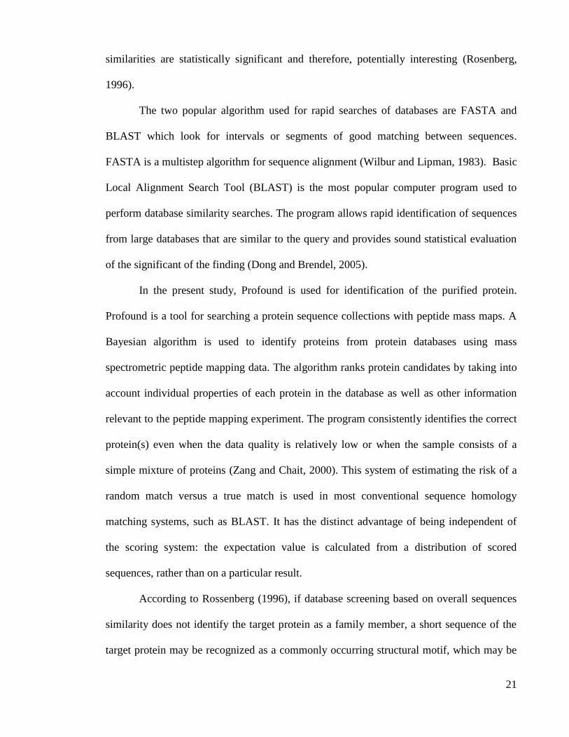
were also being purified and isolated from mushroom (Table 2.1). Their dose compensation
and the mode of action is a subject for research. Undoubtedly, the antimicrobial potential of
extracts of several medicinal mushroom type and other Basidiomycetes not yet exploited,
must warrant further examination (Rai et al., 2005).
Table 2.1 : Compounds showing antimicrobial activities from mushrooms.
Mushrooms Bioactive compounds Bioactivity
Cheimonophylum candissimum Cheimonophyllon A-E Antibacterial
Weak antifungal
Clitocybe cyathiformis Cyathiformine A Antibacterial and
antifungal
Clitocybe diatreta Diatretol Antibacterial
Coprinus atramentarius Illudin C2, Illudin C3 Antimicrobial
Crepidotus fulvotomentosus Strobilurin E Antifungal
30
Favolaschia pustulosa 9-methoxystrobilurin L Antifungal and
antibacterial
Favolaschia sp. Favolon Antifungal
Flagelloscypha pilatii Pilatin Antibiotic
Ganoderma lucidum Ganoderan Antiviral
Lentinula edodes Lentinan Antiviral
Mniopetalum sp. Mniopetals Antimicrobial
Mycena sp. Strobilurin M,
Tetrachloropyrocatechol
Antifungal, cytostatic
Antifungal, antibacterial
Omphalotus illudens Illudinic acid Antibacterial
Oudemansiella radiate Oudemansin x Antifungal
Poria cocos Lanostene Phospolipase A2
inhibitor (group of anti-
inflammatory agents)
Source : Rai et al. (2005)
2.8 Anti Human Immunodeficiency Virus -1 Reverse Transcriptase activity
2.8.1 Human Immunodeficiency Virus (HIV)
The human immunodeficiency virus (HIV) is a lentiviruses (a member of the
retrovirus family), which are pathogens that causes an illness trajectory, characterized by
long period of clinical latency and asymptomatic infection caused by weak humoral
immune response and persistent viremia (Green, 1997). According to Barre-Sanoussi
(1996), there are two different viral strains of HIV been identified since 1983 : HIV type 1
(HIV-1), which is distributed worldwide, and the HIV type 2 (HIV-2), which is seen
predominantly in West Africa. Both viruses can cause primary immunodeficiency in
human.

31
HIV-1 can infect cells that possess the CD4+ marker / receptor such as CD4+ T-
lymphocytes (T-helper cells), macrophage / monocytes, some bone marrow progenitor cells
and follicular dendritic cells, resulting in direct and indirect effects on human immune
system. Direct interaction of HIV-1 and the affected host CD4+ cell is the direct effects of
HIV-1 on the immune system (Staprans and Feinberg, 1997) while indirect affects and
dysregulation of the immune system is caused by the substances released as part of the
immune response to HIV-1 infection, resulting to the deterioration of CD4+ cell which
leads to the dysfunction and cellular death (Brennan and Porche, 1997)
HIV-1 is transmitted by body fluids that contain HIV-1 or CD4+ HIV-1 infected
lymphocytes. These include serum, seminal fluid, vaginal secretions, amniotic fluids and
breast milk (Staprans and Feinberg, 1997). According to Brennan and Porche (1997), HIV
transmission happens by few mechanisms or behaviors that allow the exposure of these
body secretions to other persons. These include form of unprotected sexual activity,
injection of blood or blood products, and perinatal infection (from mother to the fetus or
infant). In Malaysia, the first reported cases of HIV/AIDS were in 1986, and since then
there are now over 91, 000 reported cases of HIV infections in Malaysia. There are more
than 16,000 people have died from AIDS as of December 2010. The majority of HIV
infections are found in adult aged 30-39 (43% of cases) and those aged 20-29 (33% of
cases) with 90% reported cases are among male
(http://www.rumahjaireh.com/PDFs/AIDSinMalaysia2010.pdf).

32
2.8.2 Anti HIV compounds
Enormous efforts have been dedicated in searching for the cure for HIV/AIDS.
Until now, there is no cure for this pandemic disease. Because HIV-1 is one of the main
targets for inhibiting the reproduction of HIV, many inhibitors to the enzyme have been and
still are extensively discovered in small and big scale screening. However, only a small
number is used in therapy and these compounds are nucleoside and non-nucleoside
inhibitors (De Clercq, 2001). Many natural products such as ribosome inactivating proteins,
alkaloids, flavanoids, lignans, and so on, have been reported to inhibit unique enzymes and
protein crucial to the life cycle of HIV, including the reverse transcription process, virus
entry, the integrase or protease (Cos et al., 2004; De Clercq, 2000). As HIV demonstrates a
high ability to developed resistance against therapeutic agents, new promising compounds
with anti HIV inhibitory activity have to be discovered (Mlinaric et al., 2005).
2.8.3 Anti HIV-1 reverse transcriptase activity
Most of natural inhibitors of HIV-1 reverse transcriptase described are obtained
from plants, but not from fungi (Mlinaric et al., 2005). According to Mlinaric et al. (2005),
the kingdom of fungal which represent a vast and promising source of novel therapeutic
agents are often overlooked. Still, there are many reports of proteins from mushrooms with
inhibitory activity against human immunodeficiency virus type 1 (HIV-1) reverse
transcriptase, an enzyme of paramount importance to the life cycle of the HIV.
In the screening of new compounds with anti HIV-1 RT activities, many strategies
and bioassays have been developed. According to Tan and Pezzuto (1991), the early

33
developed assays for screening of HIV-1RT inhibitory activity were relatively complicated,
time consuming and included work with radioactive material. A simple, non-radioactive
ELISA based assay become commercially available recently. This assay allows rapid,
reliable and safe screenings with a minimum amount of sample (Mlinaric et al., 2005).
In this study, the purified protein was assessed for anti HIV-1 reverse transcriptase
activity using the transcriptase colorimetric enzyme immunoassay. The assay determined
the quantitative retroviral reverse transcriptase activity by incorporation of digoxigenin and
biotin-labeled dUTP into DNA. Lyophilize recombinant HIV-1 RT was used in the
screening. The assay takes advantage of the ability of reverse transcriptase to synthesize
DNA, starting from the template/ primer hybrid poly(A) oligo(dT)15. The digoxigenin and
biotin-labeled nucleotides in an optimized ratio are incorporated into one of the same DNA
molecule, which is freshly synthesized by the reverse transcriptase (RT). The detection and
quantification of synthesized DNA as a parameter for RT activity follows sandwich ELISA
protocol. Biotin labeled DNA binds to the surface of microtiter plate modules that have
been precoated with streptavidin. In the next step, an antibody to digoxigenin, conjugated to
peroxidase, binds to the digoxigenin-labeled DNA. In the final step, the peroxidase
substrate is added. The peroxidase enzyme catalyzes the cleavage of the substrate,
producing a colored reaction product. The absorbance of the samples at 405nm can be
determined using microtiter plate (ELISA) reader and is directly correlated to the level of
RT activity.

34
2.9 Hemolytic activity
In the present study, the ability of the purified protein to induce hemolysis against
erythrocytes was investigated. Hemolysis test is used to see potential toxicity issues early in
the drug discovery process. Besides, the examination of the in vitro cytotoxicity of the
purified protein is important before they can be considered in clinical use (Javadpour et al.,
1996). According to Zafloff et al. (2002), erythrocyte is used in the test because its
represent prokaryote cyte negatively charged cell containing lipid bilayer. This is due to the
fact that erythrocyte plasma membrane is a natural membrane in the body which contains
amnionic surface and a lipid monolayer. The activity was compared to melittin, a protein
from honeybee venom which can cause total hemolysis at a very low concentration
(Dempsey, 1990).