Lipid-like self-assembling peptide nanovesicles for drug delivery
6
Lipid-like Self-Assembling Peptide Nanovesicles for Drug Delivery Dimitrios G. Fatouros, † Dimitrios A. Lamprou, ‡ Andrew J. Urquhart, ‡ Spyros N. Yannopoulos, § Ioannis S. Vizirianakis, † Shuguang Zhang, ∥ and Sotirios Koutsopoulos* ,⊥ † School of Pharmacy, Aristotle University of Thessaloniki, GR-54124 Thessaloniki, Greece ‡ Strathclyde Institute of Pharmacy and Biomedical Sciences (SIPBS), University of Strathclyde, 161 Cathedral Street, Glasgow G4 0RE, United Kingdom § Foundation for Research and Technology, Hellas − Institute of Chemical Engineering Sciences (FORTH/ICE-HT), P.O. Box 1414, GR-26504 Patra, Greece ∥ Center for Bits & Atoms, Massachusetts Institute of Technology, 77 Massachusetts Avenue, Cambridge, Massachusetts 02139, United States ⊥ Center for Biomedical Engineering, Massachusetts Institute of Technology, 77 Massachusetts Avenue, Cambridge, Massachusetts 02139, United States ABSTRACT: Amphiphilic self-assembling peptides are func- tional materials, which, depending on the amino acid sequence, the peptide length, and the physicochemical conditions, form a variety of nanostructures including nanovesicles, nanotubes, and nanovalves. We designed lipid- like peptides with an aspartic acid or lysine hydrophilic head and a hydrophobic tail composed of six alanines (i.e., ac-A 6 K- CONH 2 , KA 6 -CONH 2 , ac-A 6 D-COOH, and DA 6 -COOH). The resulting novel peptides have a length similar to biological lipids and form nanovesicles at physiological conditions. AFM microscopy and light scattering analyses of the positively charged lipid-like ac-A 6 K-CONH 2 , KA 6 -CONH 2 peptide formulations showed individual nanovesicles. The negatively charged ac-A 6 D- COOH and DA 6 -COOH peptides self-assembled into nanovesicles that formed clusters that upon drying were organized into necklace-like formations of nanovesicles. Encapsulation of probe molecules and release studies through the peptide bilayer suggest that peptide nanovesicles may be good candidates for sustained release of pharmaceutically active hydrophilic and hydrophobic compounds. Lipid-like peptide nanovesicles represent a paradigm shifting system that may complement liposomes for the delivery of diagnostic and therapeutic agents. KEYWORDS: lipid-like peptides, designer peptide surfactants, liposome alternatives, tunable peptides, controlled release ■ INTRODUCTION Molecular self-assembly has enabled the fabrication of nanostructures and the development of advanced functional materials. 1 The design and synthesis of biologically inspired molecules with self-assembling properties has significantly advanced the field of biomaterials and includes peptide, oligonucleotide, and polysaccharide systems. 2 Depending on the amino acid sequence, self-assembling peptides have varying properties and have been tested in biomedicine as permissive biological scaffolds for regenerative medicine and drug delivery. 3−8 These short, self-assembling peptides are non- toxic, non-immunogenic, and degrade to natural amino acids which can be physiologically metabolized. Self-assembly of amphiphilic lipid-like peptides leads to the formation of stable nanotubes, nanodoughnuts, nanovalves, nanovesicles, or micelles similar to lipids and chemical surfactants. 9 The type and size of the self-assembled peptide nanostructures depend on the peptide concentration, the peptide specific critical micelle concentration (CMC), the amino acid sequence, geometrical constraints (defined by the size of the amino acid side groups), the ionic strength, and the pH of the medium. 9,10 These factors affect peptide alignment, packing density, and strength of the intermolecular interactions between the monomers, which determine the formation of hierarchical supramolecular structures of different morpholo- gies and properties. 9,11 Since their discovery in 2002, 9 self- assembling lipid-like peptides have been studied by many groups; these studies improved our understanding and highlighted the importance of the system. 12−16 The develop- ment of lipid-like self-assembling peptides with surfactant properties has opened new avenues for applications in biotechnology for the stabilization of membrane proteins more effectively than commercial detergents 11,17,18 and in nanotechnology for the construction of energy conversion devices. 19 Lipid-like self-assembling peptides are amenable to molecular design allowing modifications in the number, type, and order of amino acids on the peptide chain as well as incorporation of Received: February 7, 2014 Accepted: May 12, 2014 Published: May 12, 2014 Research Article www.acsami.org © 2014 American Chemical Society 8184 dx.doi.org/10.1021/am501673x | ACS Appl. Mater. Interfaces 2014, 6, 8184−8189
Transcript of Lipid-like self-assembling peptide nanovesicles for drug delivery

Lipid-like Self-Assembling Peptide Nanovesicles for Drug DeliveryDimitrios G. Fatouros,† Dimitrios A. Lamprou,‡ Andrew J. Urquhart,‡ Spyros N. Yannopoulos,§
Ioannis S. Vizirianakis,† Shuguang Zhang,∥ and Sotirios Koutsopoulos*,⊥
†School of Pharmacy, Aristotle University of Thessaloniki, GR-54124 Thessaloniki, Greece‡Strathclyde Institute of Pharmacy and Biomedical Sciences (SIPBS), University of Strathclyde, 161 Cathedral Street, Glasgow G40RE, United Kingdom§Foundation for Research and Technology, Hellas − Institute of Chemical Engineering Sciences (FORTH/ICE-HT), P.O. Box 1414,GR-26504 Patra, Greece∥Center for Bits & Atoms, Massachusetts Institute of Technology, 77 Massachusetts Avenue, Cambridge, Massachusetts 02139,United States⊥Center for Biomedical Engineering, Massachusetts Institute of Technology, 77 Massachusetts Avenue, Cambridge, Massachusetts02139, United States
ABSTRACT: Amphiphilic self-assembling peptides are func-tional materials, which, depending on the amino acidsequence, the peptide length, and the physicochemicalconditions, form a variety of nanostructures includingnanovesicles, nanotubes, and nanovalves. We designed lipid-like peptides with an aspartic acid or lysine hydrophilic headand a hydrophobic tail composed of six alanines (i.e., ac-A6K-CONH2, KA6-CONH2, ac-A6D-COOH, and DA6-COOH).The resulting novel peptides have a length similar to biologicallipids and form nanovesicles at physiological conditions. AFM microscopy and light scattering analyses of the positively chargedlipid-like ac-A6K-CONH2, KA6-CONH2 peptide formulations showed individual nanovesicles. The negatively charged ac-A6D-COOH and DA6-COOH peptides self-assembled into nanovesicles that formed clusters that upon drying were organized intonecklace-like formations of nanovesicles. Encapsulation of probe molecules and release studies through the peptide bilayersuggest that peptide nanovesicles may be good candidates for sustained release of pharmaceutically active hydrophilic andhydrophobic compounds. Lipid-like peptide nanovesicles represent a paradigm shifting system that may complement liposomesfor the delivery of diagnostic and therapeutic agents.
KEYWORDS: lipid-like peptides, designer peptide surfactants, liposome alternatives, tunable peptides, controlled release
■ INTRODUCTIONMolecular self-assembly has enabled the fabrication ofnanostructures and the development of advanced functionalmaterials.1 The design and synthesis of biologically inspiredmolecules with self-assembling properties has significantlyadvanced the field of biomaterials and includes peptide,oligonucleotide, and polysaccharide systems.2 Depending onthe amino acid sequence, self-assembling peptides have varyingproperties and have been tested in biomedicine as permissivebiological scaffolds for regenerative medicine and drugdelivery.3−8 These short, self-assembling peptides are non-toxic, non-immunogenic, and degrade to natural amino acidswhich can be physiologically metabolized.Self-assembly of amphiphilic lipid-like peptides leads to the
formation of stable nanotubes, nanodoughnuts, nanovalves,nanovesicles, or micelles similar to lipids and chemicalsurfactants.9 The type and size of the self-assembled peptidenanostructures depend on the peptide concentration, thepeptide specific critical micelle concentration (CMC), theamino acid sequence, geometrical constraints (defined by thesize of the amino acid side groups), the ionic strength, and the
pH of the medium.9,10 These factors affect peptide alignment,packing density, and strength of the intermolecular interactionsbetween the monomers, which determine the formation ofhierarchical supramolecular structures of different morpholo-gies and properties.9,11 Since their discovery in 2002,9 self-assembling lipid-like peptides have been studied by manygroups; these studies improved our understanding andhighlighted the importance of the system.12−16 The develop-ment of lipid-like self-assembling peptides with surfactantproperties has opened new avenues for applications inbiotechnology for the stabilization of membrane proteinsmore effectively than commercial detergents11,17,18 and innanotechnology for the construction of energy conversiondevices.19
Lipid-like self-assembling peptides are amenable to moleculardesign allowing modifications in the number, type, and order ofamino acids on the peptide chain as well as incorporation of
Received: February 7, 2014Accepted: May 12, 2014Published: May 12, 2014
Research Article
www.acsami.org
© 2014 American Chemical Society 8184 dx.doi.org/10.1021/am501673x | ACS Appl. Mater. Interfaces 2014, 6, 8184−8189

active peptide sequences to facilitate cell penetration or reactivechemical groups such as fluorescent dyes or biotin. The ease ofproduction and the wide scope of modification allow for thesynthesis of designer sequences with “tailor-made” tunableproperties. In this work, we set out to investigate thephysicochemical of cationic and anionic lipid-like peptides,and determine the release kinetics of model drug compoundsthrough peptide formulations as a first step towards thedevelopment of a peptide-based drug delivery system.
■ MATERIALS AND METHODSLipid-like Self-Assembling Peptides. Ac-A6K-CONH2, KA6-
CONH2, ac-A6D-COOH, and DA6-COOH were received in powder(SynBioSci, Livermore, CA). The purity of the peptides was 90−94%as determined by electrospray ionization-quadrupole-time-of-flight(ESI-Q-TOF) mass spectrometry. Peptides were dispersed in PBS pH7.4, probe sonicated for 10 min to facilitate dispersion, andequilibrated for 15 min to allow for self-association of the monomers.CMC of Lipid-like Peptides. DLS was used to determine the
peptides’ CMC (PDDLS/Batch setup, Precision Detectors, Franklin,MA). Solutions of different peptide concentrations in PBS pH 7.4were filtered through 0.45 μm pore size filters prior to measuring.Scattered light was detected at 90° and the number of photons wasrecorded and processed by Precision Deconvolve. The solventviscosity and the refractive index of the buffer were taken as 0.894cP and 1.33, respectively, at 25 °C.Particle Size Determination. Normalized intensity (I)−time (t)
autocorrelation functions g(2)(q,t) = ⟨I(q,t)I(q,0)⟩/⟨I(q,0)⟩2 of thepeptide vesicle dispersions in PBS at 25 °C were measured (n ≥ 4)over a broad time scale (10−7−104 s) using a full multiple τ digitalcorrelator with 280 channels spaced quasi-logarithmically (ALV−5000/FAST) and the 671 nm line of a diode pumped solid state laser(operated at a power <5 mW). The scattered light was collected by asingle-mode optical fiber, transferred to an avalanche photo-detectorand then to the digital correlator for analysis. In dilute suspensions, thenormalized electric field (E)−time autocorrelation function g(1)(q,t) =⟨E(q,t)E*(q,0)⟩/⟨E(q,0)⟩2 is related to the experimentally recordedg(2)(q,t)2 through the Siegert relation:20 g(2)(q,t) = B[1 + f*|g(1)(g,t)|2]where B describes the long delay time behavior of g(2)(q,t) and f* is aninstrumental factor (in our system f* ∼ 0.95). Hence, g(1)(t) (forsimplicity we drop the q-dependence) was analyzed as a weighted sumof independent exponential contributions g(1)(t) = ∫ L(τ) exp(−t/τ)dτ= ∫ L(ln τ) exp(−t/τ)d ln τ. The distribution of relaxation times L(lnτ) = τL(τ) was obtained by inverse Laplace transformation of g(1)(q,t)using the CONTIN algorithm.21 The apparent hydrodynamic radii ofthe suspended particles were determined using the Stokes−Einsteinrelation Rh = kBT/6πηD, where kB is the Boltzmann constant, η is theviscosity of the solvent, and D is the diffusion coefficient of the particlewhich is equal to D = (1/τ)q2, where τ is the relaxation time ofg(1)(q,t).AFM of Peptide Nanovesicles. 3 μL of the peptide vesicle
dispersions (20 mg/mL of peptides in PBS filtered through 0.45 μmfilters) were deposited on mica (G250-2, rms ∼ 0.4 nm; Agar ScientificLtd, Essex, U.K.), rinsed with 200 μL of water (Millipore) after 1 minof equilibration and dried in nitrogen gas stream for 5 min or in air for1 h. Images were obtained immediately in air using a BrukerMultiMode NanoScope 3D Controller Scanning Probe Microscope(Digital Instruments) operated in tapping mode and soft siliconprobes (FESP; nominal length lnom = 225 μm, width wnom = 28 μm, tipradius Rnom = 8 nm, resonant frequency υnom = 75 kHz, spring constantknom = 2.8 N m−1; Veeco Instruments SAS, France). Images werecollected from two samples of each peptide system at random spotsurface sampling (at least eight areas).Release through Peptide Nanovesicles. Peptides in powder
were mixed to a final concentration of 5 mg/mL with 1 mM CFsolution in PBS and the suspension was probe-sonicated for 10 min.After equilibration for 1 h, 1 mL of the nanovesicle suspension wasplaced in Microcon YM-10 membrane tubes (10 kDa cut-off). Non-
encapsulated CF was removed by centrifugation at 11 000 rpm for 20min. Then the vesicles were re-suspended in PBS pH 7.4 to a finalvolume of 1 mL and were incubated at 25 °C. CF released throughpeptide nanovesicles (n = 4) was collected by centrifugation of themembrane tubes at 11 000 rpm for 20 min, the fluorescence intensityof the filtrate was measured, fresh PBS pH 7.4 was added to a finalvolume of 1 mL, and the suspension was incubated. This process wasrepeated at specific time points, and a graph was created to show thetime course of released CF through peptide nanovesicles. Allmeasurements were carried out in a PerkinElmer LS-50B spectropho-tometer using 1 cm quartz cuvettes. The excitation wavelength was at470 nm, and the emission maximum was observed at 520 nm.
Nile red was used as model compound to study the uptake ofhydrophobic molecules by lipid-like peptide nanovesicles. Peptidepowder (5 mg) was added to 1 mL PBS pH 7.4 containing 3.14 μMNile red (i.e., maximum concentration of Nile red in water),25 thesuspension was probe sonicated for 10 min, placed in Microcon YM-10 membrane tubes (n = 4) and incubated at 25 °C. To determine therelease kinetics we measured fluorescence intensity of the peptidenanovesicle suspension, removed the released Nile red bycentrifugation of the membrane tubes at 11 000 rpm for 20 min andadded fresh PBS pH 7.4 to 1 mL. This process was repeated at specifictime points. Fluorescence emission was recorded from 550 to 700 nmusing 1 cm quartz cuvettes (excitation was at 545 nm).
Effect of Lipid-like Peptides on Cell Viability. Caco-2 cells(passage 40) were cultured at 37°C in growth medium (i.e., DMEM,supplemented with 10% v/v FBS, 1% w/v non-essential amino acidsand 100 μg/mL penicillin and streptomycin) in 5% v/v CO2humidified atmosphere. The medium was changed every 2−3 daysuntil cells reached 80% confluency in culture, and then, cells weretrypsinized and subcultured. The effect of lipid-like peptides on cellviability and proliferation was studied using the 3-(4,5-dimethylthiazol-2-yl)-2,5-diphenyl tetrazolium (MTT) assay (Trevigen Inc., MD).Caco-2 cells grown to a density of 4 × 10³ cells/cm2 in 96-well plateswere incubated for 3 and 24 h in growth medium containing 0.2 or 1.0mg/mL of lipid-like peptides. Then, 10 μL of MTT reagent wereadded, and the plates were incubated for 3 h at 37 °C. Absorbance ofthe formazan product was determined at 600 nm with a TeknikaELISA plate reader after 1 h.
■ RESULTS AND DISCUSSIONA class of self-assembling peptides (i.e., ac-A6K-CONH2, KA6-CONH2, ac-A6D-COOH, and DA6-COOH) was designed tomimic lipids of biological membranes. These peptides have ahydrophobic tail composed of six alanines, a hydrophilic head,which is an amino acid with charged side group and a length of∼2.5 nm (Figure 1). As lipid-based systems, addition of lipid-like peptides to water or an electrolyte solution results information of a turbid suspension due to self-assembly of thepeptide monomers to minimize the interaction betweenhydrophobic domains and polar environment.9 We determinedthe CMC of lipid-like peptides in phosphate buffer saline (PBS,100 mM KH2PO4, 10 mM Na2HPO4, 137 mM NaCl, 2.7 mMKCl at pH 7.4) to be 0.12 mg/mL for A6K-CONH2, 0.09 mg/mL for KA6-CONH2, 0.08 mg/mL for ac-A6D-COOH, and0.06 mg/mL for DA6-COOH.
Atomic Force Microscopy (AFM) Imaging. We usedAFM to study the morphology of lipid-like peptide assemblies.Previously, we reported that self-assembly of lipid-like peptidesresults in uncontrolled formation of various supramolecularstructures including vesicles, micelles, and nanotubes.9,10
Herein, the experimental conditions typically resulted innanovesicle formation (Figure 2). Size distribution analyses ofthe peptide nanovesicles showed that self-assembly of thelysine-containing, positively charged ac-A6K-CONH2 and KA6-CONH2 peptides results in larger particles (126 and 169 nm,respectively) compared to those observed from association of
ACS Applied Materials & Interfaces Research Article
dx.doi.org/10.1021/am501673x | ACS Appl. Mater. Interfaces 2014, 6, 8184−81898185

the negatively charged ac-A6D-COOH and DA6-COOH (28and 44 nm, respectively) (Table 1, Figure 2 insets). This is dueto the smaller side chain of aspartic acid compared to lysine,which allows for better packing of ac-A6D-COOH and DA6-COOH peptides in the nanovesicle bilayer as suggestedpreviously by molecular modeling studies.9
AFM imaging of ac-A6D-COOH and DA6-COOH peptideassemblies revealed dispersed nanovesicles as well as necklace-like ultra-structures (Figure 2 C−D). Such formations areobserved at the micrometer scale in colloidal systems, when adrop of the suspension evaporates on a solid surface and havebeen described by two dimensional crystallization laws,22 or thecoffee-ring effect.23 It may be that such phenomena arereproduced at the nanometer scale due to evaporation of thesolvent and repulsion of like-charged peptide nanovesicles. Thediameter of necklaces composed of ac-A6D-COOH and DA6-COOH nanovesicles is 200 and 159 nm, respectively (Table 1).Image analysis showed that ac-A6D-COOH necklaces have 20± 5 individual (average diameter 28 nm) ac-A6D-COOHnanovesicles per necklace whereas necklaces composed oflarger (average diameter 44 nm) DA6-COOH nanovesicleshave 9 ± 3 nanovesicles per necklace.These observations prompted inquiry about the mechanism
of necklace formation. Changing the sample drying conditions(from quick nitrogen gas drying to slow air drying for 1 h)revealed metastable nanovesicle clusters (Figure 2 E−F)suggesting that prior to necklace formation ac-A6D-COOHand DA6-COOH peptide nanovesicles formed loosely boundclusters with diameter 113 and 135 nm, respectively. It is likelythat during quick sample drying under nitrogen gas thenegatively charged peptide nanovesicle clusters disassemble andspread on the like-charged mica surface leading to nanovesiclenecklace-like formations. Theoretical calculations assumingtight packing of hard spheres, revealed that 20 ac-A6D-COOH nanovesicles with diameter 28 nm and 9 DA6-COOH nanovesicles with diameter 44 nm form clusters withdiameter 97 and 120 nm, respectively.24 These values correlatewell with the AFM determined sizes of the ac-A6D-COOH (i.e.,113 nm) and DA6-COOH (i.e., 135 nm) nanovesicle clusters.
Dynamic Light Scattering (DLS). The hydrodynamicradii, Rh, of the peptide nanovesicle suspensions weredetermined by DLS from which the intensity-time correlationfunctions and the corresponding inverse Laplace transformanalyses of lipid-like peptide nanovesicles in PBS werecalculated (Figure 3). Analysis of the light scattering datayielded monomodal peptide vesicle size distributions except inthe case of KA6-CONH2 suspensions in which peak analysisshowed the presence of particles with average diameter of 164and 906 nm; the latter probably represent aggregates ofindividual nanovesicles.DLS measurements of the ac-A6K-CONH2 and KA6-CONH2
nanovesicle diameter are in agreement with the size of micadeposited nanovesicles as determined by AFM. However, DLSanalysis of ac-A6D-COOH and DA6-COOH peptide nano-vesicle suspensions revealed the presence of particles withdiameter 99 and 137 nm, respectively (Figure 3). These valuesdeviate from the nanovesicle diameter determined by AFM fordispersed ac-A6D-COOH and DA6-COOH nanovesicles but
Figure 1. Molecular modeling, amino acid sequence, and chargedistribution of lipid-like self-assembling peptides. The peptide length issimilar to biological phospholipids. The hydrophobic domain of thepeptides consists of six alanines. Color code: cyan, carbon; red,oxygen; blue, nitrogen; white, hydrogen. Illustrations were generatedby VMD. Highlighted domains represent amino acids with positivecharge (blue), negative charge (red), and hydrophobic side chains(grey). Using pKa values from the literature the net charge of thepeptides at pH 7.4 was calculated to be ac-A6K-CONH2 (+1), KA6-CONH2 (+2), ac-A6D-COOH (−2), DA6-COOH (−1).
Figure 2. AFM of lipid-like peptide nanovesicles on mica. (A−DInsets) size distribution histograms of peptide nanovesicles weregenerated from AFM image analysis. (C and D) ac-A6D-COOH andDA6-COOH peptide nanovesicle necklace-like formation upon quickdrying of the samples under nitrogen gas. (C and D Insets) ImageJimage processing showing the surface topology of the necklace-likeformations. (E and F) ac-A6D-COOH and DA6-COOH peptidenanovesicle clusters formed upon slow air drying of the samples. (Eand F Insets) Graphical representation of 20 and 9 tightly packed ac-A6D-COOH and DA6-COOH nanovesicles was generated by http://www.randomwalk.de/sphere/insphr/spheresinsphr.html.
ACS Applied Materials & Interfaces Research Article
dx.doi.org/10.1021/am501673x | ACS Appl. Mater. Interfaces 2014, 6, 8184−81898186
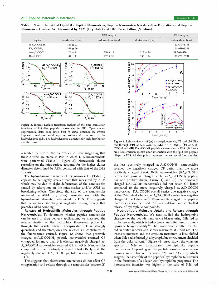
resemble the size of the nanovesicle clusters suggesting thatthese clusters are stable in PBS in which DLS measurementswere performed (Table 1, Figure 3). Nanovesicle clusterspreading on the mica surface accounts for the higher clusterdiameter determined by AFM compared with that of the DLSanalysis.The hydrodynamic diameter of the nanovesicles (Table 1)
appears to be slightly smaller than that measured by AFMwhich may be due to slight deformation of the nanovesiclescaused by adsorption on the mica surface and/or AFM tipbroadening effects. Therefore, the size of the nanovesiclesmeasured by AFM (dry state) correlates well with thehydrodynamic diameter determined by DLS. This suggeststhat nanovesicle shrinking is negligible during drying thatprecedes AFM scanning.Release of Hydrophilic Molecules through Peptide
Nanovesicles. To determine whether peptide nanovesiclescan be used in drug delivery applications, we measured therelease kinetics of the hydrophilic fluorescent probe CFthrough the vesicle bilayer. Encapsulated CF is stronglyquenched, and therefore, only the released CF contributes tothe fluorescence emitted. Figure 4A shows that positivelycharged ac-A6K-CONH2 peptide nanovesicles retained CFentrapped for more than 6 h whereas negatively charged ac-A6D-COOH nanovesicles released CF in ∼3 h. Nanovesiclescomposed of the positively charged KA6-CONH2 and thenegatively charged DA6-COOH peptides released CF within∼1 h.This suggests that electrostatic interactions do not affect CF
encapsulation and release through the nanovesicles because (i)
the less positively charged ac-A6K-CONH2 nanovesiclesretained the negatively charged CF better than the morepositively charged KA6-CONH2 nanovesicles (KA6-CONH2carries two positive charges while ac-A6K-CONH2 peptidehas one positive charge, Figure 1) and (ii) the negativelycharged DA6-COOH nanovesicles did not retain CF bettercompared to the more negatively charged ac-A6D-COOHnanovesicles (DA6-COOH overall carries one negative chargeat the C-terminal whereas ac-A6D-COOH carries two negativecharges at the C-terminal). These results suggest that peptidenanovesicles can be used for encapsulation and controlledrelease of hydrophilic compounds.
Hydrophobic Molecule Uptake and Release throughPeptide Nanovesicles. We next studied the hydrophobiccharacter of the peptide nanovesicle bilayer using Nile red asprobe molecule, which is hydrophobic and often used to assessliposome bilayer stability.25 The fluorescence emitted by Nilered in water is weak and shows maximum at ∼660 nm. Theintensity increases and the emission maximum is blue shiftedwhen Nile red is buried in a hydrophobic environment shieldedfrom the polar solvent.25 Figure 4B, inset, shows the emissionspectra of Nile red incorporated into lipid-like peptidenanovesicles. Depending on the peptide formulation emissionmaxima were observed between 621 and 633 nm, whichsuggests that assembly of the peptides’ hydrophobic tails resultsin the formation of a bilayer with hydrophobic properties. Thefluorescence intensity was higher in the case of Nile red
Table 1. Size of Individual Lipid-Like Peptide Nanovesicles, Peptide Nanovesicle Necklace-Like Formations and PeptideNanovesicle Clusters As Determined by AFM (Dry State) and DLS Curve Fitting (Solution)
AFM analysis DLS analysis
peptide vesicle diam. (nm) necklace diam. (nm) cluster diam. (nm) particle diam. (nm)
ac-A6K-CONH2 126 ± 23 122 (30−173)KA6-CONH2 169 ± 29 164 (65−350)ac-A6D-COOH 28 ± 9 200 ± 11 113 ± 26 99 (40−320)DA6-COOH 44 ± 11 159 ± 26 135 ± 24 137 (70−690)
Figure 3. Inverse Laplace transform analysis of the time correlationfunctions of lipid-like peptide nanovesicles in PBS. Open circles,experimental data; solid lines, best fit curve obtained by inverseLaplace transform; solid squares, volume distributions of thehydrodynamic radii. The hydrodynamic diameters for each distributionare also shown. Figure 4. Release kinetics of (A) carboxyfluorescein, CF and (B) Nile
red through (●) ac-A6K-CONH2, (▲) KA6-CONH2, (⧫) ac-A6D-COOH and (■) DA6-COOH peptide nanovesicles in PBS. (B Inset)Nile Red emission spectra upon interaction with the lipid-like peptidebilayer in PBS. All data points represent the average of four samples.
ACS Applied Materials & Interfaces Research Article
dx.doi.org/10.1021/am501673x | ACS Appl. Mater. Interfaces 2014, 6, 8184−81898187

interacting with ac-A6D-COOH nanovesicles compared to thatemitted by Nile red incorporated in ac-A6K-CONH2 nano-vesicles. This result suggests that the ac-A6D-COOH bilayeraccommodated more Nile red molecules with better shieldingfrom water compared to Nile red inside the ac-A6K-CONH2bilayer. Ac-A6D-COOH and ac-A6K-CONH2 have similarCMCs and therefore, the observed differences in the emittedfluorescence of nanovesicle incorporated Nile red is likely dueto better packing of the ac-A6D-COOH peptide monomers inthe bilayer. This results in the presentation of a morehydrophobic environment for incorporating Nile red in theac-A6D-COOH bilayer compared to the ac-A6K-CONH2bilayer. The fluorescence intensity of Nile red interactingwith KA6-CONH2 and DA6-COOH nanovesicles was signifi-cantly lower suggesting less Nile red incorporation in thesepeptides’ bilayers. Furthermore, we studied the release kineticsthrough peptide nanovesicles, and we found slow Nile redrelease through ac-A6D-COOH and ac-A6K-CONH2 nano-vesicles reaching significant levels after 6 and 4 h, respectively(Figure 4B). KA6-CONH2 and DA6-COOH nanovesiclesappeared to be less stable and released Nile red within 2 and1 h, respectively. Notably, the release kinetics of Nile redthrough the peptide nanovesicles follow the order of Nile redfluorescence emission incorporated inside the hydrophobicenvironment of the peptide bilayer (Figure 4B and inset). As inthe case of CF release, we observed that ac-A6K-CONH2 andac-A6D-COOH nanovesicles retain Nile red for prolongedperiods of time suggesting that these peptide nanovesicles canbe used for sustained delivery of hydrophilic and hydrophobiccompounds.Cell Viability. To determine the effect of lipid-like peptides
on Caco-2 cell viability, we used the MTT assay. Figure 5
shows a constant increase in cell numbers after 3 and 24 hincubation with and without 0.2 or 1.0 mg/mL lipid-likepeptides (P < 0.05). These results suggest that ac-A6K-CONH2,KA6-CONH2, ac-A6D-COOH, and DA6-COOH lipid-likepeptides do not affect Caco-2 cell proliferation comparedwith the control.
■ CONCLUSIONS
Lipid-like peptides were designed to mimic natural lipids havinga hydrophilic head and a hydrophobic domain. Although allpeptides form nanovesicles some of them do not retain modeldrug probe molecules for prolonged times. Peptide design byaltering amino acid sequence and charge distribution provides ameans to control the loading capacity and the drug releasekinetics through the nanovesicles. Of the two leading lipid-likepeptide nanovesicle systems (i.e., ac-A6K-CONH2 and ac-A6D-COOH), the latter may be more suitable for drug delivery.Negatively charged drug carriers, such as the ac-A6D-COOHnanovesicles, are preferable for intravenous administrationbecause they result in electrostatic repulsions with the like-charged surface of blood cells and vessel walls, which allows forprolonged circulation in the bloodstream.Liposomes were proposed as drug delivery carriers in the
1970s. A significant amount of work has been done to increaseliposomal stability in serum, prolong drug release and reduceside effects associated with immunogenicity and toxicity ofliposomes. Lipid-like peptides are nontoxic, nonimmunogenicand may encapsulate or incorporate and slowly release bothhydrophilic and hydrophobic drug molecules and may present aplatform to append to existing liposomal drug release systems.Peptide self-assembly is similar to that of lipids and fatty
acids. However, peptides differ from these systems because thepeptide bilayer is stabilized by a combination of hydrophobicinteractions of the hydrophobic amino acids’ side groups andhydrogen bonding of the peptides’ polar backbones. Therefore,the bilayer’s internal chemistry differs between liposomes andpeptide nanovesicles. However, lipid-like peptides readily mixwith lipids to form hybrid peptide/lipid liposome systems.26
The incorporation of lipid-like peptides in liposomes conferredfunctionality and modulated the liposome bilayer curvature andstability of the formulation. Furthermore, lipid-like peptides canbe easily modified and tailored to incorporate other moleculessuch as sugars and functional motifs, including cell signalingand cell penetrating peptides to allow the synthesis of celltargeting drug delivery systems.Depending on their sequence peptide nanovesicles can retain
and slowly release both hydrophilic and hydrophobiccompounds. We believe that these simple, inexpensive, andnontoxic peptides will open new paths in the field of vesicle-mediated drug delivery systems.
■ AUTHOR INFORMATION
Corresponding Author*Tel.: +1-617-752-2042. Fax: +1-617-258-5239. Email: [email protected].
NotesThe authors declare no competing financial interest.
■ ACKNOWLEDGMENTS
The Center for Environmental Health Sciences (CEHS; GrantNo. NIEHS ES002109) at MIT is acknowledged for massspectrometric analysis and technical support. We also thank Dr.M. Spanakis for expert technical assistance with cell assay.
■ ABBREVIATIONS:
ac; acetyl group; AFM; atomic force microscopy; CMC; criticalmicelle concentration; DLS; dynamic light scattering; CF; 5,6-carboxyfluorescein
Figure 5. Effect of 0.2 and 1.0 mg/mL lipid-like peptides on Caco-2cell viability after (A) 3 h and (B) 24 h in culture. In each box chart,the bottom (×) shows the minimum value and marks the 0thpercentile. The bottom of the box marks the 25th percentile and thetop of the box marks the 75th percentile. The square symbol (□) inthe box marks the mean. The top (×) shows the maximum value and100th percentile. n = 4 sample points. The differences in the numberof viable cells at each condition compared to the control are significant(P < 0.05).
ACS Applied Materials & Interfaces Research Article
dx.doi.org/10.1021/am501673x | ACS Appl. Mater. Interfaces 2014, 6, 8184−81898188

■ REFERENCES(1) Whitesides, G. M.; Mathias, J. P.; Seto, C. T. Molecular Self-Assembly and NanochemistryA Chemical Strategy for the Synthesisof Nanostructures. Science 1991, 254, 1312−1319.(2) Koutsopoulos, S. Molecular Fabrications of Smart Nano-biomaterials and Applications in Personalized Medicine. Adv. DrugDelivery Rev. 2012, 64, 1459−1476.(3) Kisiday, J.; Jin, M.; Kurz, B.; Hung, H.; Semino, C.; Zhang, S.;Grodzinsky, A.J. Self-Assembling Peptide Hydrogel Fosters Chon-drocyte Extracellular Matrix Production and Cell Division: Implica-tions for Cartilage Tissue Repair. Proc. Natl. Acad. Sci. U.S.A. 2002, 99,9996−10001.(4) Koutsopoulos, S.; Zhang, S. Long-Term Three-DimensionalNeural Tissue Cultures in Functionalized Self-Assembling PeptideHydrogels, Matrigel, and Collagen I. Acta Biomater. 2013, 9, 5162−5169.(5) Davis, M. E.; Motion, J. P. M.; Narmoneva, D.A.; Takahashi, T.;Hakuno, D.; Kamm, R. D.; Zhang, S.; Lee, R.T. Injectable Self-Assembling Peptide Nanofibers Create Intramyocardial Microenviron-ments for Endothelial Cells. Circulation 2005, 111, 442−450.(6) Koutsopoulos, S.; Unsworth, L.D.; Nagai, Y.; Zhang, S.Controlled Release of Functional Proteins through Designer Self-Assembling Peptide Nanofiber Hydrogel Scaffold. Proc. Natl. Acad. Sci.U.S.A. 2009, 106, 4623−4628.(7) Nagai, Y.; Unsworth, L.D.; Koutsopoulos, S.; Zhang, S. SlowRelease of Molecules in Self-Assembling Peptide Nanofiber Scaffold. J.Controlled Release 2006, 115, 18−25.(8) Koutsopoulos, S.; Zhang, S. Two-Layered Injectable Self-Assembling Peptide Scaffold Hydrogels for Long-Term SustainedRelease of Human Antibodies. J. Controlled Release 2012, 160, 451−458.(9) Vauthey, S.; Santoso, S.; Gong, H.; Watson, N.; Zhang, S.Molecular Self-Assembly of Surfactant-like Peptides to Form Nano-tubes and Nanovesicles. Proc. Natl. Acad. Sci. U.S.A. 2002, 99, 5355−5360.(10) Santoso, S.; Hwang, W.; Hartman, H.; Zhang, S. Self-Assemblyof Surfactant-like Peptides with Variable Glycine Tails to FormNanotubes and Nanovesicles. Nano Lett. 2002, 2, 687−691.(11) Koutsopoulos, S.; Kaiser, L.; Eriksson, H. M.; Zhang, S.Designer Peptide Surfactants Stabilize Diverse Functional MembraneProteins. Chem. Soc. Rev. 2012, 41, 1721−1728.(12) Meng, Q. B.; Kou, Y. Y.; Ma, X.; Liang, Y. J.; Guo, L.; Ni, C. H.;Liu, K. L. Tunable Self-Assembled Peptide Amphiphile Nanostruc-tures. Langmuir 2012, 28, 5017−5022.(13) Wang, Q. R.; Yu, J.; Zhang, X.; Liu, D. J.; Zheng, J. H.; Pan, Y.;Lin, Y. J. Controlled Biosilification Using Self-Assembled ShortPeptides A(6)K and V6K. RSC Adv. 2013, 3, 2784−2793.(14) Castelletto, V.; Nutt, D. R.; Hamley, I. W.; Bucak, S.; Cenker,C.; Olsson, U. Structure of Single-Wall Peptide Nanotubes: In SituFlow Aligning X-ray Diffraction. Chem. Commun. 2010, 46, 6270−6272.(15) Qiu, F.; Chen, Y. Z.; Zhao, X. J. Comparative Studies on theSelf-Assembling Behaviors of Cationic and Catanionic Surfactant-likePeptides. J. Colloid Interface Sci. 2009, 336, 477−484.(16) Xu, H.; Wang, J.; Han, S. Y.; Wang, J. Q.; Yu, D. Y.; Zhang, H.Y.; Xia, D. H.; Zhao, X. B.; Waigh, T. A.; Lu, J. R. Hydrophobic-Region-Induced Transitions in Self-Assembled Peptide Nanostruc-tures. Langmuir 2009, 25, 4115−4123.(17) Matsumoto, K.; Vaughn, M.; Bruce, B. D.; Koutsopoulos, S.;Zhang, S. Designer Peptide Surfactants Stabilize Functional Photo-system-I Membrane Complex in Aqueous Solution for ExtendedTime. J. Phys. Chem. B 2009, 113, 75−83.(18) Wang, X. Q.; Corin, K.; Baaske, P.; Wienken, C. J.; Jerabek-Willemsen, M.; Duhr, S.; Braun, D.; Zhang, S. G. Peptide Surfactantsfor Cell-Free Production of Functional G Protein-Coupled Receptors.Proc. Natl. Acad. Sci. U.S.A. 2011, 108, 9049−9054.(19) Das, R.; Kiley, P. J.; Segal, M.; Norville, J.; Yu, A. A.; Wang, L.;Trammell, S. A.; Reddick, L. E.; Kumar, R.; Stellacci, F.; Lebedev, N.;Schnur, J.; Bruce, B. D.; Zhang, S.; Baldo, M. Integration of
Photosynthetic Protein Molecular Complexes in Solid-State ElectronicDevices. Nano Lett. 2004, 4, 1079−1083.(20) Berne, B. J.; Pecora, R. Dynamic Light Scattering. Wiley: NewYork, 1976.(21) Provencher, S. W. A Constrained Regularization Method forInverting Data Represented by Linear Algebraic or Integral Equations.Comput. Phys. Commun. 1982, 27, 213−227.(22) Denkov, N. D.; Velev, O. D.; Kralchevsky, P. A.; Ivanov, I. B.;Yoshimura, H.; Nagayama, K. 2-Dimensional Crystallization. Nature1993, 361, 26−26.(23) Deegan, R. D.; Bakajin, O.; Dupont, T. F.; Huber, G.; Nagel, S.R.; Witten, T. A. Capillary Flow as the Cause of Ring Stains fromDried Liquid Drops. Nature 1997, 389, 827−829.(24) Birgin, E. G.; Sobral, F. N. C. Minimizing the ObjectDimensions in Circle and Sphere Packing Problems. Comput. Oper.Res. 2008, 35, 2357−2375.(25) Greenspan, P.; Fowler, S. D. Spectrofluorometric Studies of theLipid Probe, Nile Red. J. Lipid Res. 1985, 26, 781−789.(26) Yaghmur, A.; Laggner, P.; Zhang, S.; Rappolt, M. TuningCurvature and Stability of Monoolein Bilayers by Designer Lipid-likePeptide Surfactants. PLoS One 2007, 5, e1.
ACS Applied Materials & Interfaces Research Article
dx.doi.org/10.1021/am501673x | ACS Appl. Mater. Interfaces 2014, 6, 8184−81898189