Integrating Toxicological Assessments in Material Selection ...
Upload
independentCategory
view
3download
0
ORIGINAL ARTICLE
Labaditin, a cyclic peptide with rich biotechnological potential:preliminary toxicological studies and structural changesin water and lipid membrane environment
S. C. Barbosa • E. M. Cilli • Luis G. Dias •
Rodrigo G. Stabeli • P. Ciancaglini
Received: 12 February 2010 / Accepted: 1 June 2010 / Published online: 19 June 2010
� Springer-Verlag 2010
Abstract Cyclic peptides isolated from the plants of the
Euphorbiaceae family have been largely studied due to
their rigid conformation, which is considered significant for
biologic activity. The peptide Labaditin (L0) and its open
chain analogs (L1) were synthesized by the solid-phase
peptide synthesis technique (Fmoc/tBu), and purified to
elucidate its interaction with membrane models. A shift in
kmax emission and Stern–Volmer constants values indicate
that both tryptophans migrate to a more apolar environ-
ment, with L1 decreasing less than L0. A circular dichroism
(CD) study revealed that L0 was kept unstructured in
aqueous media as much as in the presence of dipalmitoil-
phosphatidylcholine liposomes. The thermodynamic stud-
ies by differential calorimetry (DSC) show a DH increase
(50 and 18 kcal/mol, for L0 and L1, respectively) with
peptide concentrations, which is indicative of lipids asso-
ciating with peptides, resulting in the inability of the lipids
to participate in the main transition. Therefore, all CD,
DSC, and fluorescence data suggest a greater L0 membrane
insertion. A probable mechanism for Labaditin interaction
is based initially on the hydrophobic interaction of the
peptide with the lipid membrane, conformational change,
peptide adsorption on the lipid surface, and internalization
process. Peptide’s antibacterial effect was also evaluated
and revealed that only L0 showed reduction in viability in
Gram-positive bacteria while no effects to the Gram-
negative.
Keywords Labaditin � Peptide synthesis � Liposome �Fluorescence � Antibacterial activity �Membrane interaction
Introduction
Labaditin (VWTVWGTIAG) is a cyclic peptide (CP)
found in superior plants of the jatropha genus of the Eu-
phorbiaceae family, popularly known as Jarak gurita
(Indonesia) and Mana (Philippines). Phytochemically, this
genus is known for having numerous classes of secondary
metabolites such as phytosterols, flavonoids, alkaloids,
terpenes, lignins, and, more recently, the above-mentioned
cyclic peptides (Tan and Zhou 2006; Zhang et al. 2009).
These molecules isolated from the vegetal biomass present
antimicrobial, antitumor, cytotoxic effects, and anti-HIV
activities (Craik et al. 1999; Daly et al. 1999; Menezes and
Jared 2002; Ake et al. 2004; Reddy et al. 2004).
The cyclic peptides belong to the Caryophyllaceae
group (Tan and Zhou 2006; Zhang et al. 2009). One of the
structural characteristics of these CP is the presence of
7–10 groups with a high proportion of hydrophobic amino
acids (Auvin et al. 1997). Biobollein (9 residues) and
Labaditin (10 residues), isolated from Jatropha Multifida,
Electronic supplementary material The online version of thisarticle (doi:10.1007/s00726-010-0648-6) contains supplementarymaterial, which is available to authorized users.
S. C. Barbosa � L. G. Dias � P. Ciancaglini (&)
Departamento de Quımica, FFCLRP-USP,
Ribeirao Preto, SP, Brazil
e-mail: [email protected]
E. M. Cilli
Departamento de Bioquımica e Biotecnologia,
IQ-UNESP-Univ. Estadual Paulista, Araraquara, SP, Brazil
R. G. Stabeli
Centro de Estudos de Biomoleculas Aplicadas a Medicina
(CEBio), Nucleo de Saude (NUSAU), Universidade Federal de
Rondonia (UNIR), Fundacao Oswaldo Cruz Noroeste
(FIOCRUZ), 76812-245 Porto Velho, RO, Brazil
123
Amino Acids (2011) 40:135–144
DOI 10.1007/s00726-010-0648-6

were the first CPs of this group to be described in literature
(Kosasi et al. 1989; Tan and Zhou 2006).
Another important characteristic of the Labaditin
peptide is the amino acid composition since it has two
tryptophan residues in its structure. According to Ridder
et al. (2005), tryptophan has a fundamental role in the
molecule’s interaction with the biological membranes.
According to these authors, the substitution of this residue
for another one, also hydrophobic, significantly decreases
the interaction of the peptide with the membrane.
Therefore, due to their great interaction ability with the
membrane (Xiao and Pei 2007), these peptides have
attracted a lot of biological and biotechnological interest,
although their therapeutical application can be associated
with some problems such as low water solubility leading to
low absorption, bio-availability, and high sample aggre-
gation. In order to overcome the low solubility of some
drugs, some organic solvents are used in these compounds’
formulation, such as ethanol or yet compounds derived
from polyethoxylated ricinus oil (Torchilin 2004).
The nanotechnological insights for the use of Labaditin
as a pharmaceutical model is its encapsulating into lipo-
somes structures, which will guarantee preservation of the
solubility mediated for biological systems and drug deliv-
ery when associated with some biomarkers, such as
tumoral antibodies for example. But in order to have this
development, it is necessary to know this CP’s action
mechanism, toxicity, preferential target, etc.
The other important biotechnological characteristic is
the cyclic chain of these peptides which guarantees more
resistance to the proteolytic degradation than its linear
analogous, due to the lack of N or C-terminal groups and
reduced conformational freedom (Xiao and Pei 2007).
Besides that, the lower entropy associated with this greater
rigidity makes these molecules have a higher affinity to and
specificity with receptors (Rezai et al. 2006a, b; Xiao and
Pei 2007).
Based on the biological activities exhibited, Labaditin
can be considered as an interesting prototype in the
development of new drugs. Allied to the peptides syn-
thesis techniques in solid phase, the study of small cyclic
peptides has shown to be a powerful approach, mainly
due to the easiness of obtention and to the diverse
structural modifications possible in the original peptide
chain, allowing the development of more bioactive mol-
ecules. One of the feasible strategies in order to know
more details on the action mechanisms and the peptide
compounds interactions is the study with natural and
artificial membrane systems (Nakamura et al. 1998;
Anderluh et al. 1999). The knowledge of the structure–
activity relationship of Labaditin and its analogous is an
important factor to unveil the most common form the
molecule assumes while exercising its activity (Auvin-
Guette et al. 1999), because its action mode has not been
elucidated yet. This is possible due to the high sensibility
of the indol fluorophore to the environment surrounding it
(Chattopadhyay and London 1987; Chen and Barkley
1998; Powl et al. 2003).
Differential scanning calorimetry (DSC) has also been
applied to probe the thermodynamic profiles of the lipid
assemblies and the consequences of peptide interactions in
terms of ordering and cooperative properties of the bilayers
(Lohner et al. 1999). These studies will be done and
evaluated in different conditions, including interactions
with natural (erythrocyte) and artificial (liposomes) mem-
brane systems in order to understand the possible action
mechanism of these peptides.
Besides the interaction and structural modification
systems, the antibacterial activity in Gram-positive and
-negative microorganisms was evaluated, objectifying new
possibilities of therapeutical applications through a nano-
biotechnological approach.
Materials and methods
Peptide synthesis, cyclization, and purification
The L1 peptide (VWTVWGTIAG) was manually prepared
by solid-phase peptide synthesis (SPPS) as previously
described by Merrifield (1963) according to the standard
Na-Fmoc protecting-group strategy (Atherton and Shepard
1989) using the experimental steps described in Castro
et al. (2009). After cleavage, L0 was obtained by L1’s
cyclization (Baraguey et al. 1998). The peptides L0 and L1
were purified by semi-preparative HPLC using a reverse
phase C18 column and identified by electrospray mass
spectrometry. All the details of this section are available in
the Supplementary Material.
Liposome preparation
Large unilamellar vesicles (LUV) constituted by 1 mg/mL
of dipalmitoilphosphatidylcholine (DPPC) were prepared
by the extrusion method, using the experimental procedure
described by Simao et al. (2010).
Dynamic light scattering measurements
Determination of size distributions (liposomes or aggre-
gates) were carried out by dynamic light scattering (DLS),
using a N5 Submicron Particle Size Analyzer (Beckman
Coulter, Inc., Fullerton, CA, USA). The average value
diameters were obtained from the unimodal distribution.
136 S. C. Barbosa et al.
123

Circular dichroism (CD) spectroscopy
The CD spectra were recorded at 25�C with a Jasco 810
spectropolarimeter, purged with nitrogen gas, and with a
0.1 cm quartz cuvette. Initially, the L0 and L1 peptides
concentration effect was evaluated by varying the peptide
concentration from 5 to 270 lM, in aqueous media, pH 7.0.
Each Peptide solution (L0 and L1), in different concentra-
tions and separately, were incubated for 2 h with DPPC
constituted liposomes up to 14 lipids for each peptide (mol
ratio). The spectra were recorded using a 1-nm spectral
bandwidth, 10 nm/min scan speed, 2 s response time with
six accumulations, and a wavelength range of 250–190 nm
was chosen to minimize the noise and error during the
measurement. Typically, the CD spectra showed in the
figures correspond to the average after buffer (or LUV)
spectra subtraction and baseline correction.
Tryptophan fluorescence spectroscopy assays
Fluorescence measurements were made with a Spectronic
SLM 8100 spectrofluorometer equipped with a single-
grating emission monochromator and a double-grating
excitation monochromator. Excitation was carried out at
280 nm to irradiate tryptophan groups, and emission
spectra were measured from 300 to 500 nm, at 25�C and
pH 7.0. Quartz cells with optical paths of 0.5 cm were
used.
Fluorescence intensity and maximum wavelengths
(kmax) were obtained for each peptide (L0 and L1) varying
the concentration from 2 to 73 lM in aqueous media and
also in the presence of DPPC liposomes (100 lL). Thecorresponding base line obtained in the same conditions
but without the peptide was always previously subtracted
from the peptide spectrum.
Structural alterations in peptides due to their binding or
interaction with liposomes were monitored by quenching
of tryptophan fluorescence with acrylamide in the presence
and absence of liposomes according to the Stern–Volmer
equation:
F0=F ¼ 1þ Ksv � Q½ �;where F0 and F are the fluorescence intensities in the
absence and presence of quencher, respectively, [Q] is the
concentration of quencher, and Ksv is the Stern–Volmer
quenching constant. Fluorescence quenching measure-
ments of tryptophan with acrylamide quencher were made
by serial addition of small aliquots (5 lL) of concentratedacrylamide stock solution (6 M) in a cuvette containing a
fixed concentration of 50 lM of the peptide solution, alone
or in the presence of liposome, in a final molar ratio about
14 lipid/peptide (L/P), up to a final concentration of 1 M
acrylamide.
Differential scanning calorimetry assay
Transition phase temperatures (Tc) of the DPPC-LUV
membranes prepared in the absence or presence of peptide
(L0 and L1 in a 0–50-lM range concentration) were studied
by DSC. All LUV suspensions and reference buffer were
degassed under vacuum (140 mbar) for 30 min prior to
use. The samples were scanned from 20 to 65�C at an
average heating rate of 1�C/min, and the recorded ther-
mograms were analyzed using Nano-DSC II software
(Calorimetry Sciences Corporation, CSC 6100, UT, USA).
Samples containing the peptide alone (100 lM), dis-
solved in the same buffer, exhibited no thermal events over
the temperature range of 0–100�C. This indicates that the
endothermic events observed in this study arise solely from
phase transitions of the DPPC vesicles. A minimum of at
least three heating and cooling scans were performed for
each analysis, and all thermograms was reproducible. In
order to ensure homogeneity in the analysis of the effect of
the selected peptides on the lipid phase transitions, we have
chosen the simplest baseline correction to introduce the
least amount of variability when comparing thermograms
from different sets of experiments.
Peptide antibacterial effect
The antibacterial test was done with both L1 and L0 pep-
tides using the following bacteria lineages: Gram-positive
(Streptococcus mutans) and Gram-negative (Aggregatib-
acter actinomycetemcomitans) obtained and cultivated
according to Paulino et al. (2005) and Goulart et al. (2009),
respectively.
Before the incubation with different concentrations of
peptide (0.5 and 100 lM), the cells (A600nm = 0.5) were
serially diluted in Tryptic Soy Broad (TSB) medium to
obtain 103 Colony Forming Units (CFU)/mL and distrib-
uted (1 mL) in assay tubes (125 9 15 mm). One set of
tubes was submitted to a control cell experiment without
any peptide addition.
Cells were grown by dropping and spreading 50 lL of
previously diluted cell suspension directly onto Tryptic
Soy Agar (TSA), and incubating it in a candle jar for 72 h
at 37�C. After this period, the CFU/mL was calculated
(Goulart et al. 2009). All experiments were done in
triplicate.
Peptide hemolytic activity
Erythrocytes were isolated from human freshly collected
blood. Blood was centrifuged (4,7309g by 15 min at 4�C)in sodium citrate 3.8% (1:4 v/v), and the sediment was
washed over three times in this same solution. It was then
resupended in saline solution PBS (10 mM Na2HPO4,
Labaditin, a cyclic peptide with rich biotechnological potential 137
123

2 mM KH2PO4, 137 mM NaCl, and 2.7 mM KCl, pH 7.4).
Erythrocytes (2 9 107, determined by Neubauer chamber)
were incubated in PBS buffer and with different peptide
concentrations (previously determined) for 30 min at 37�C.The solution was then centrifuged (4,7309g for 10 min,
4�C) to remove the lysed cells. Hemoglobin release,
determined through supernatant spectrophotometer absor-
bance at 414 nm, was correlated with the lysis of eryth-
rocytes. Cells’ maximum lysis (100%) was attained by the
incubation of an erythrocyte suspension with 0.1% (v/v) of
Triton X-100. All experiments were done in triplicate.
Results and discussion
Study of peptide interaction with lipid interface
Peptides were found to have limited solubility in water
(maximum about 270 lM), and this has also raised the
possibility that the peptide is in an aggregated state in
solution at the low concentrations employed for the CD
measurements (Yang et al. 1994). The L0 and L1 peptide
solutions were also analyzed with the light scattering
technique. It was possible to evidence the presence of
structures as high as 100 nm, with high polydispersion
index, for both peptides, indicating the formation of
aggregates in these conditions. In addition, it should be
noted that up to 50 lM no scattering was observed for both
peptides, but the formation of complex by peptide–peptide
interaction was not discarded (data not shown).
The concentration dependence of the far-UV CD spec-
trum of the peptides L0 and L1, pH 7.0, were investigated,
and essentially, the concentration range was from 30 to
270 lM (Fig. 1). The spectrum obtained presented increase
of ellipticity intensity [h] according to the increase in the
concentration of L0 peptide in 205 nm. No band is attributed
to the presence of interaction between tryptophan’s side
chains, in part, that can be attributed to the cyclic confor-
mation which significantly restricts changes between chains
(Fig. 1a). For L1, a minimum at approximately 197 nm was
observed, with increase of ellipticity intensity according to
the increase in the concentration of peptide, and a maximum
at 224 nm, with decrease of ellipticity intensity according to
the increase in the concentration of peptide (Fig. 1b). By
increasing the peptide concentration, the band which refers
to the side chain interactions of tryptophan becomes less
intense, evidencing the decrease of intermolecular associa-
tions verified by the positive band around 220–320 nm
(Grishina and Woody 1994; Woody 1996; Andersson et al.
2001). This CD spectrum obtained for L1 suggests that
structuring of the peptide involves at least some degree of
intermolecular association (Yang et al. 1994). A possible
explanation for this behavior can be given analyzing the
peptide’s isoelectric point (pI). The theoretical pI of L1 is
6.0, close to the pH studied, which justifies the observed
behavior, because with zero charges and in high concen-
trations, these molecules are more susceptible to intermo-
lecular aggregations due to less repulsion. The same applies
to L0 since it does not present charged groups in its structure,
as well as C- and N-terminal groups, which are compro-
mised in cyclization.
The structural changes due to peptide–membrane inter-
actions are essential to understand the action and regulation
mechanism of the biological activity of molecules (Kelly
et al. 2005). The effect of lipids on the backbone confor-
mation of L0 and L1 peptides was studied by far-UV CD
spectroscopy. The CD spectrum of the peptide L0 in water,
pH 7.0, has a minimum at approximately 207 nm and
maximum at 190 nm, characteristic of an unordered
structure (Yang et al. 1994). Addition of DPPC liposome to
the solution of L0 resulted in change in the peptide CD
spectrum (Fig. 2a). The effect was concentration depen-
dent, and the spectrum of L0 in the presence of DPPC, in
different peptide/lipids molar ratios (P/L), has a minimum
Fig. 1 Circular dichroism spectrum of L0 (a) and L1 (b) peptides inconcentrations from 5 to 270 lM, in aqueous media, pH 7.0, as
described in ‘‘Materials and methods’’
138 S. C. Barbosa et al.
123

at approximately 190 nm and a maximum at 210 nm, the
characteristic features of a b-structure (Yang et al. 1994).
An increase in magnitude of the ellipticity according to P/L
ratio increase also showed, possibly due to the saturation of
the peptides on the lipids layer.
The CD spectrum of the peptide L1 in water, pH 7.0, has
a minimum at approximately 197 nm and maximum at
224 nm, characteristic of a random coil structure with
interactions among molecules of tryptophan (Yang et al.
1994; Andersson et al. 2001). Addition of DPPC liposome
to the solution of L1, in different peptide/lipids molar ratios
(P/L), resulted in change in the peptide CD spectrum to
a minimum at approximately 195 nm and the disappear-
ance of the maximum at 224 nm (Fig. 2b). This is probably
due to the interaction of the peptide with the lipid layer,
which leads to the separation of the tryptophans (Sforca
et al. 2005; Woody 1996; Kelly and Price 2000; Andersson
et al. 2001).
A similar result was obtained with the Ab (1–40) pep-
tide, where a fast change in CD spectrum was observed, in
the presence of the ganglioside lipid; it changed from
random coil structure to b-structure (Choo-Smith and
Surewicz 1997). Another study with a 21-residue peptide,
derived from a human prion protein, showed a conforma-
tional change from a-helix to b-structure after interaction
with SDS micelles (Kelly and Price 2000).
The tryptophan amino acid was used as a probe
to analyze the local environment and the influence of
the N-terminus modifications (Ladokhin et al. 2000). The
presence of a tryptophan residue allows the study of the
conformational changes that can occur in these molecules.
The greater or smaller proximity of the side chains and
terminus ionizable groups of the peptide with this residue
can affect the fluorescence intensity (Van der Wel et al.
2007) by the quenching process. This amino acid also can
be used as a probe to analyze the local environment; it
Fig. 2 Circular dichroism spectrum of the L0 (a) and L1 (b) in
different concentrations (from 5 to 100 lM) and separately, in
aqueous media, pH 7.0, incubated for 2 h with DPPC liposomes up to
14 lipids for each peptide (mol ratio), and the CD spectrum was
recorded as described in ‘‘Materials and methods’’
Fig. 3 Peptides fluorescence emission spectra of different concen-
trations (2–73 lM): a L0 and b L1 in the presence of DPPC liposome,
according to ‘‘Materials and methods’’. The arrow indicates an
increase of peptide concentration in solution
Labaditin, a cyclic peptide with rich biotechnological potential 139
123

allows the evaluation of the migration from one environ-
ment to the other. These studies are possible due the var-
iation of fluorescence emission spectroscopy and
fluorescence quenching (Park et al. 1995; Ladokhin et al.
2000).
DPPC–liposomes interaction was studied with different
peptide concentrations. As can be seen in Fig. 3a, the
increase of L0 and L1 concentration results in the decrease
of fluorescence intensity. This reduction in fluorescence
intensity can be attributed to the conformational changes
suffered by the peptide. The modification in structure can
promote a greater approximation of the side chains of some
amino acids with the indol group of tryptophan, acting as
suppressors and affecting this parameter (Chen and Barkley
1998; Ladokhin et al. 2000). It is important to note that
only one maximum was found in the spectra, indicating
that both tryptophans have the same behavior in this
membrane mimetic, probably due to their proximity and/or
strong interaction between them.
It should be noted that the fluorescence spectra have also
revealed a blue shift of kmax emission for the L0 peptide as
much as for the L1 (Fig. 4). Initially, both peptides pre-
sented a discrete reduction in kmax (L0 reduced 5 nm and
L1 only 3 nm) for the mol ratio P/L of around 0.005. This
value remains constant with the increase of the P/L ratio to
0.009 and 0.011, for L0 and L1, respectively. After this
baseline, a new reduction in kmax was observed, reaching
a new level at 339 and 344 nm for L0 and L1, respectively.
This behavior of kmax, apparently in two stages, followed
by the reduction in fluorescence intensity with the increase
of P/L ratio, can be correlated with distinct phenomena that
occur during the interaction of the peptide with the lipid
present in liposome. Therefore, in small P/L relationships,
as the first step, the peptide probably undergoes an
aggregation stage in contact with the lipid membrane,
which results in its adsorption in the interface (when they
are present in the interface at a ratio of approximately one
peptide for every 14 lipids). This interpretation is associ-
ated with the low value of fluorescence reduction and kmax
that is attributed to the tryptophan migration to a more
apolar environment (Christiaens et al. 2002; Lakowicz
2006). In the second step, due to the increase in the peptide
adsorption in the lipid interface, possibly, a redistribution
of the peptides in the more internal layer is occurring.
Hence, the peptide that is probably more immersed in the
liposome is L0 because it has the lowest kmax emission
(L0 339 nm and L1 344 nm).
It should be highlighted that the susceptibility of the
bilayer depends on the peptide concentration on the
membrane surface, which is, when a limit ratio between the
peptide and lipid concentration is reached; it leads to the
immersion on the lipid bilayer, depending on its compo-
sition (Huang 2000). Besides that, the phospholipid com-
position of the membrane, and its size and charge can
influence in the orientation, insertion and depth of the
peptide in relation to the membrane surface (Deber and Li
1995).
The fluorescence quenching can be numerically mea-
sured from the obtention of the Stern–Volmer (Ksv) con-
stant for each peptide, in a lipid microenvironment as in an
aqueous environment. Ksv is a parameter obtained from the
quenching tangent curve, given by the fluorescence
Fig. 4 Peptides kmax emission variation of L0 and L1 studied in
aqueous media alone, or in the presence of DPPC liposomes,
according to ‘‘Materials and methods’’
Fig. 5 Fluorescence quenching (F0/F) of the Tryptophans present in
L0 (squares) and L1 (circles) in the absence (open symbols) and in the
presence (full symbols) of DPPC liposomes, pH 7.0, with acrylamide
quencher. These experiments were made by serial addition of small
aliquots (5 lL) of concentrated acrylamide solution stock (6 M) in a
cuvette containing a fixed concentration (50 lM) of peptide solution
alone or in the presence of 14 lipid/peptide ratios, up to a final
concentration of 1 M acrylamide, according to ‘‘Materials and
methods’’
140 S. C. Barbosa et al.
123
intensity initial value in relation to the punctual values
obtained (F0/F) in the presence of increasing concentra-
tions of acrylamide (Deva and Behere 1999; Eftink and
Ghrion 1976; Christiaens et al. 2002; Kelkar and Chatto-
padhyay 2006; Lakowicz 2006; Sahu and Behera 2008).
As can be seen in Fig. 5, both peptides show tryptophan
fluorescence quenching by the acrylamide—fluorescence
intensity decreases. This reduction of fluorescence can be
due to a higher exposition of tryptophan in the apolar media.
The linear curve obtained in this study probably indicates
that both tryptophans have the same quenching by acryl-
amide and the same insertion in the mimetic membrane.
In another experiment, the fluorescence quenching was
also done with the L0 and L1 peptides, separately, in
aqueous solution, yielding information about the trypto-
phan exposition level in the absence of liposome (Fig. 5).
All the Ksv constants determined in the presence and
absence of liposomes are summarized in Table 1. We can
observe that peptides have shown a greater quenching in
aqueous medium (higher values of Ksv) than in the pres-
ence of DPPC liposomes (lower values of Ksv) due to
a greater exposition of tryptophan when in solution. Thus,
we can conclude that the L0 peptide interacts more with the
lipids present in liposomes than L1 peptide because it has
a lower Ksv.
It should be noted that each of the two peptides studied
(L0 and L1) have two tryptophan residues in their structure;
however, through this quenching experiment it was not
possible to differentiate them because we have obtained
only one line in the regression value of F0/F. This can be
explained by the fact that both tryptophans have similar
behavior or yet, due to a limitation of the technique that
does not allow us to differentiate them.
In the DSC study, the disturbances undergone by the
lipids present in DPPC liposomes in the presence of L0 and
L1 peptides were observed. Surface adsorption, membrane
insertion, and peptide-specific bonding are frequently fol-
lowed by system energy changes that can be conveniently
measured by the DSC technique. For this study, a mem-
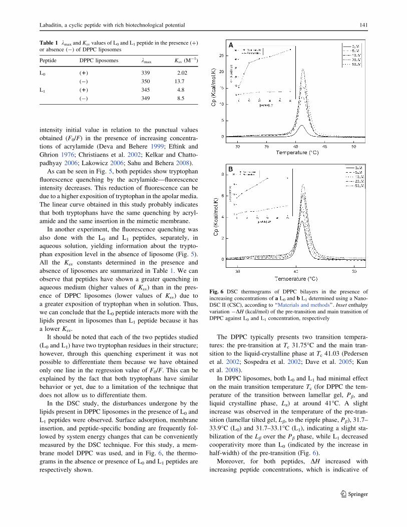
brane model DPPC was used, and in Fig. 6, the thermo-
grams in the absence or presence of L0 and L1 peptides are
respectively shown.
The DPPC typically presents two transition tempera-
tures: the pre-transition at Tc 31.75�C and the main tran-
sition to the liquid-crystalline phase at Tc 41.03 (Pedersen
et al. 2002; Sospedra et al. 2002; Dave et al. 2005; Kun
et al. 2008).
In DPPC liposomes, both L0 and L1 had minimal effect
on the main transition temperature Tc (for DPPC the tem-
perature of the transition between lamellar gel, Pb, and
liquid crystalline phase, La) at around 41�C. A slight
increase was observed in the temperature of the pre-tran-
sition (lamellar tilted gel, Lb, to the ripple phase, Pb), 31.7–
33.9�C (L0) and 31.7–33.1�C (L1), indicating a slight sta-
bilization of the Lb over the Pb phase, while L1 decreased
cooperativity more than L0 (indicated by the increase in
half-width) of the pre-transition (Fig. 6).
Moreover, for both peptides, DH increased with
increasing peptide concentrations, which is indicative of
Table 1 kmax and Ksv values of L0 and L1 peptide in the presence (?)
or absence (-) of DPPC liposomes
Peptide DPPC liposomes kmax Ksv (M-1)
L0 (1) 339 2.02
(-) 350 13.7
L1 (1) 345 4.8
(-) 349 8.5
Fig. 6 DSC thermograms of DPPC bilayers in the presence of
increasing concentrations of a L0 and b L1 determined using a Nano-
DSC II (CSC), according to ‘‘Materials and methods’’. Inset enthalpyvariation -DH (kcal/mol) of the pre-transition and main transition of
DPPC against L0 and L1 concentration, respectively
Labaditin, a cyclic peptide with rich biotechnological potential 141
123

lipids associating with peptides or greater lipid packing and/
or lipid sequestering, resulting in the inability of the lipids to
participate in the main transition (Inset of Fig. 6). This
variation was bigger for L0 peptide, increased to 50.8 kcal/
mol, while L1 increased to 18.2 kcal/mol (Inset of Fig. 6).
In a study done with DPPC and alamethicin, an anti-
microbial peptide, through DSC, a great increase of the
peak after the peptide–lipid interaction was observed and
conferred to the peptide–lipid interaction (Dave et al.
2005). In another study, done with cyclic antimicrobial
peptide rhesus theta defensin (RTD-1) and open chain
analog (oRTD-1), the peptides were not able to notably
alter the phase transition properties of DPPC. They
decreased the cooperativity of the pre-transition, resulting
in a very flat wide peak (Abuja et al. 2004), similar to what
L0 and L1 peptides had shown.
Antibacterial and hemolytic effects of peptides
After the advent of high-throughput experiments and post-
genomic technologies, more than 80% of drug substances
were natural products or inspired by a natural compound.
This fact illustrates the importance of natural products for
human health (Calderon et al. 2009). The modern nanobio-
technology considers the design and engineer modifications
of bioactive peptides for new antibiotic formulations
(Thennarasu and Nagaraj 1996). Thus, the cytotoxic
potential, in vitro, of L0 and L1 peptides over the bacterial
lineages Streptococcus mutans (Gram-positive) and Aggre-
gatibacter actinomycetemcomitans (Gram-negative) was
evaluated, according to ‘‘Materials and methods’’. The
results show that only the L0 peptide had viability reduction
in a concentration-dependent manner; however, for S. mu-
tans there was a 25% reduction in the presence of 5 lM of
the peptide and 56% with 100 lM (Fig. 7). These results
indicate that L0 kills Gram-positive bacteria membrane, and
probably acts on the inner membrane.
Similar results were obtained with the linear SPFK
peptide, 13 residues, in the presence of the Escherichia coli
bacteria (Sitaram et al. 1992). Peptides derived from toxiri
pardaxin (PX), 18 amino acids, exhibited antimicrobial
activity against Escherichia coli and not against Gram-
positive microorganisms (Thennarasu and Nagaraj 1996).
Studies directed toward structure–function correlation with
short bioactive peptides such as L0 and L1, which are
composed of only ten residues and thus are easily obtain-
able by chemical synthesis, would be appropriate for pos-
sible therapeutic uses (Sitaram et al. 1992).
For the Gram-negative bacteria, no cytotoxicity was
observed, neither for L0, nor for L1, in the concentration
range up to 100 lM.
L0 and L1 peptides have not shown hemolytic activity up
to concentrations of 100 lM. It should be clear that for a
peptide to be toxic it does not have to forcibly be hemo-
lytic. In many cases, toxicity of the peptide is associated
with the membrane rearrangement-generated processes,
specific interaction, enzyme/protein membrane inactiva-
tion, phase state changes forming membrane microdo-
mains, etc., even if it does not break its integrity. This
reason can explain the fact that the peptide is toxic only to
Gram-positive and not to Gram-negative bacteria.
Similar results have been described for peptides from
pardaxin toxin and SPF fragment of the seminalplasmin
peptide (SPFK). On the latter study, besides having used
the same methodology, the SPF peptide is highly hydro-
phobic and has tryptophan in its composition, such as la-
baditin (Thennarasu and Nagaraj 1996; Shin et al. 2001).
Conclusion
The Fmco/tBu method was adequate for the L1 synthesis
and cyclization for the obtention of Labaditin (L0). The
HPLC purification was efficient (over 95%), and Mass
spectrometry has confirmed the planned peptides. Both L0
and L1 interact with liposome. That was proved through the
kmax shift through fluorescence, the conformational change
observed by CD and DSC, with a significant increase of
enthalpy. Independent of the technique used, the L0 peptide
always showed greater interaction with DPPC liposomal
systems than its linear analogous L1.
By using this technique it is also possible to observe the
conformational restriction influence of the cyclic peptide
(L0) when compared to the linear analogous (L1). Hence,
cyclic peptides are considered good models for the study of
Fig. 7 Peptide toxicity of L1 (white bars) and L0 (gray bars) in
Gram-positive bacteria culture (Streptococcus mutans) for 30 min.
Bacteria survival percentages were obtained according to ‘‘Materials
and methods’’
142 S. C. Barbosa et al.
123

the structure–interaction relationship, as already described
by other authors (Rezai et al. 2006a, b; Kwon and Kodadek
2007). This is probably due to the fact that cyclization can
increase permeability in the membrane eliminating termi-
nal charges and internally favoring conformations by
hydrogen bonds. Based on that, according to what was
described by Seelig (2004), the adsorption and insertion
into the membrane can be followed by conformational
transitions, favored by, in part, a hydrogen bond system.
There are two very well-known conformational transitions
induced by membrane, a-helix to b-structure (Kelly and
Price 2000) done by amphypatic peptides (Seelig 2004)
and of random coil structure to b-structure (Choo-Smith
and Surewicz 1997) done by hydrophobic peptides.
A probable interaction mechanism of the L0 peptide can be
based, initially, on the hydrophobic peptide–membrane
interaction. Next, the adsorption of the peptide on the lipid
surface occurs. The exact location of this adsorption layer
is difficult to define, depends on the forces involved. On
this layer, peptides are directly in contact with the lipids,
all accessible. On the third step, the bond process occurs
with consequent conformational change of the peptide.
This mechanism is based on the kmax emission variations
observed (Fig. 4), where initially, a discrete kmax emission
reduction occurs followed by a constant plateau, according
to the increase of the P/L ratios. In this condition, the
membrane adsorption should be happening. After this
plateau, a new reduction in the kmax emission values is
observed, reaching a new plateau at 339 nm, which can be
related to the insertion moment. A similar example that
was well documented is the example of the Cyclosporine A
hydrophobic peptide. When a cyclic decapeptide interacts
with the membrane, it alters its conformation from random
coil to b-structure (Seelig 2004). Labaditin shows similar
characteristics, including immunosuppressant activity
(Kosasi et al. 1989).
Acknowledgments The authors are grateful to Conselho Nacional
de Desenvolvimento Cientıfico e Tecnologico (CNPq), Fundacao ao
Amparo a Pesquisa do Estado de Sao Paulo (FAPESP), Coordenacao
de Aperfeicoamento de Nıvel Superior—Projeto NanoBiotec
(CAPES) for financial support and Priscila Cerviglieri for linguistic
advice. PC, EMC and RGS are senior researchers of the CNPq, and
SCB was the recipient of Master fellowship from FAPESP.
References
Abuja PM, Zenza A, Trabib M, Craikb DJ, Lohner K (2004) The
cyclic antimicrobial peptide RTD-1 induces stabilized lipid-
peptide domains more efficiently than its open-chain analogue.
FEBS Lett 566:301–306
Ake RC, Rejon GE, Pat MF, Rodriguez LMP, Sanchez SRP (2004)
Bioactive terpenoids from roots and leaves of Jatropha gaumeri.Soc Quim Mex 48:11–14
Anderluh G, Barlic A, Podlesek Z, Macek P, Pungercar J, Gubensek
F, Zecchini ML, Dalla SM, Menestrina G (1999) Cysteine-
scanning mutagenesis of an eukaryotic pore-forming toxin from
sea anemone-topology in lipid membranes. Eur J Biochem
263:128–136
Andersson D, Carlsson U, Freskgard P (2001) Contribution of
tryptophan residues to the CD spectrum of the extracellular
domain of human tissue factor. Eur J Biochem 268:1118–1128
Atherton E, Shepard RC (1989) Solid phase peptide synthesis:
a practical approach. IRL Press, Oxford
Auvin C, Baraguey C, Blond A, Lezenven F, Pousset JL, Bodo B
(1997) Curcacycline B, a cyclic nonapeptide from Jatrophacurcas enhancing rotamase activity of cyclophHin. Tetrahedron
Lett 38:2845–2848
Auvin-Guette C, Baraguey C, Blond A, Xavier HS, Pousset JL, Bodo
B (1999) Pohlianins A, B and C, cyclic peptides from the latex of
jatropha pohliana ssp. molissima. Tetrahedron Lett 55:11495–
11510
Baraguey C, Guette CA, Blond A, Cavelier F, Lezenven F, Pousset
JL, Bodo B (1998) Isolation, structure and synthesis of
chevalierins A, B and C, cyclic peptides from the latex of
jatropha chevalieri. J Chem Soc 1:3033–3039
Calderon LA, Silva-Jardim I, Zuliani JP, Silva AA, Ciancaglini P,
Silva LHP, Stabeli RG (2009) Amazonian biodiversity: a view of
drug development for leishmaniasis and malaria. J Braz Chem
Soc 20:1011–1023
Castro MS, Ferreira TCG, Cilli EM, Crusca E, Mendes-Giannini MJS,
Sebben A, Ricart CA, Sousa MV, Fontes W (2009) Hylin a1, the
first cytolytic peptide isolated from the arboreal South American
frog Hypsiboas albopunctatus (‘‘spotted treefrog’’). Peptides
30:291–296
Chattopadhyay A, London E (1987) Parallax method for direct
measurement of membrane penetration depth utilizing fluores-
cence quenching by spin-labeled phospholipids. Biochemistry
26:39–45
Chen Y, Barkley MD (1998) Toward understanding tryptophan
fluorescence in proteins. Am Chem Soc 37:9976–9982
Choo-Smith LP, Surewicz WST (1997) The interaction between
Alzheimer amyloid b(1–40) peptide and ganglioside GM1-
containing membranes. FEBS Lett 402:95–98
Christiaens B, Symoens S, Vanderheyden S, Engelborghs Y, Joliot
A, Prochiantz A, Vandekerckhove J, Rosseneu M, Vanloo B
(2002) Tryptophan fluorescence study of the interaction of
penetratin peptides with model membranes. Eur J Biochem
269:2918–2926
Craik DJ, Daly NL, Bond T, Waine C (1999) Plant cyclotides:
a unique family of cyclic and knotted proteins that defines the
cyclic cystine knot structural motif. J Mol Biol 294:1327–1336
Daly NL, Koltay A, Gustafson KR, Boyd MR, Casas-Finet JR, Craik
DJ (1999) Solution structure by NMR of circulin A. J Mol Biol
285:333–345
Dave PC, Billington E, Pan Y, Straus SK (2005) Interaction of
alamethicin with ether-linked phospholipid bilayers: oriented
circular dichroism, 31P solid-state NMR, and differential scan-
ning calorimetry studies. Biophys J 89:2434–2442
Deber CM, Li S (1995) Peptides in membranes: helicity and
hydrophobicity. Biopolymers 37:295–318
Deva MSZW, Behere DV (1999) Fluorescence and circular dichroism
spectroscopic studies on bovine lactoperoxidase. Biometals
12:219–225
Eftink MR, Ghrion CA (1976) Exposure of tryptophanyl residues in
proteins. Quantitative determination by fluorescence quenching
studies. Biochemistry 15:672–680
Goulart RC, Bolean M, Paulino TP (2009) Photodynamic therapy in
planktonic and biofilm cultures of aggregatibacter actinomyce-
temcomitans. Photomed Laser Surg. doi:10.1089/pho.2009.2591
Labaditin, a cyclic peptide with rich biotechnological potential 143
123
Grishina IB, Woody RW (1994) Contributions of tryptophan side
chains to the circular dichroism of globular proteins: exciton
couplets and coupled oscillators. Faraday Discuss 99:245–262
Huang HW (2000) Action of antimicrobial peptides: two-state model.
Biochemistry 39:8347–8352
Kelkar DA, Chattopadhyay A (2006) Monitoring ion channel
conformations in membranes utilizing a novel dual fluorescence
quenching approach. Biochem Biophys Res Commun 353:483–
488
Kelly SM, Price NC (2000) The use of circular dichroism in the
investigation of protein structure and function. Curr Protein Pept
Sci 1:349–384
Kelly SM, Jess TJ, Price NC (2005) How to study proteins by circular
dichroism. Biochim Biophys Acta 1751:119–139
Kosasi S, Vandersluis WG, Boelens R, Thart L, Labadie RPL (1989)
Labaditin A novel cyclic decapeptide from the latex of jatropha-
multifida (Euphorbiaceae). FEBS Lett 256:91–96
Kun H, Minnes R, Mastai Y (2008) Effects antifreeze peptides on the
thermotropic properties of a model membrane. J Bioenerg
Biomembr 40:389–396
Kwon YU, Kodadek T (2007) Quantitative comparison of the relative
cell permeability of cyclic and linear peptides. Chem Biol
14:671–677
Ladokhin AS, Jayasinghe S, White SH (2000) How to measure and
analyze tryptophan fluorescence in membranes properly and why
bother? Anal Biochem 285:235–245
Lakowicz JR (2006) Principles of fluorescence spectroscopy, 3rd edn.
Springer, New York, p 395
Lohner K, Staudegger E, Prenner EJ, Lewis RN, Kriechbaum M,
Degovics G, McElhaney RN (1999) Effect of staphylococcal
delta-lysin on the thermotropic phase behavior and vesicle
morphology of dimyristoylphosphatidylcholine lipid bilayer
model membranes. Differential scanning calorimetric, 31P
nuclear magnetic resonance and Fourier transform infrared
spectroscopic, and X-ray diffraction studies. Biochemistry
38:16514–16528
Menezes H, Jared C (2002) Immunity in plants and animals: common
ends through different means using similar tools. Comp Biochem
Physiol 132:1–7
Merrifield RB (1963) Solid phase peptide synthesis: synthesis of
a tetrapeptide. J Am Chem Soc 85:2149–2154
Nakamura M, Sekino-Suzuki N, Mitsui K, Ohno-Iwashita Y (1998)
Contribution of tryptophan residues to the structural changes in
perfringolysin O during interaction with liposomal membranes.
J Biochem 123:1145–1155
Park NG, Yamato Y, Lee S, Sugihara G (1995) Interaction of
mastoparan-B from venom of a hornet in Taiwan with
phospholipid bilayers and its antimicrobial activity. Biopolymers
36:793–801
Paulino TP, Ribeiro KF, Thedei G Jr, Tedesco AC, Ciancaglini P
(2005) Use of hand held photopolymerizer to photoinactivate
Streptococcus mutans. Arch Oral Biol 50:353–359
Pedersen TB, Frokjaer S, Mouritsen OG, Jørgensen K (2002) A
calorimetric study of phosphocholine membranes mixed with
desmopressin and its diacylated prodrug derivative (DPP). Int
J Pharm 233:199–206
Powl AM, East MJ, Lee AG (2003) Lipid-Protein interactions studied
by introduction of tryptophan residue: the mechanosensitive
channel MscL. Biochemistry 42:14306–14317
Reddy KVR, Yedery RD, Aranha C (2004) Antimicrobial peptides:
premises and promises. Int J Antimicrob Agents 24:536–547
Rezai T, Bock JE, Zhou MV, Kalyanaraman C, Lokey RS, Jacobson
MP (2006a) Conformational flexibility, internal hydrogen bond-
ing, and passive membrane permeability: successful in silico
prediction of the relative permeabilities of cyclic peptides. J Am
Chem Soc 128:14073–14080
Rezai T, Yu B, Millhauser GL, Jacobson MP, Lockey S (2006b)
Testing the conformational hypothesis of passive membrane
permeability using synthetic cyclic peptide diastereomers. Am
Chem Soc 128:2510–2511
Ridder A, Shupjen P, Unterreitmeier S, Langosch D (2005) Trypto-
phan supports interaction of transmembrane helices. J Mol Biol
354:894–902
Sahu S, Behera PK (2008) Fluorescence quenching of 2-naphthol by
methyl acrylamide in micellar medium. Indian J Chem
47A:1516–1519
Seelig J (2004) Thermodynamics of lipid-peptide interactions.
Biochim Biophys Acta 1666:40–50
Sforca ML, Machado A, Figueredo RCR, Oyama S Jr, Silva FD,
Miranda A, Dafre S, Teresa M, Miranda M, Spisni A, Pertinhez
TA (2005) The micelle-bound structure of antimicrobial peptide
derived from the a-chain of bovine hemoglobin isolated from the
tick Boophilus microplus. Biochemistry 44:6440–6451
Shin SY, Lee SH, Yang ST, Park EJ, Lee DG, Lee MK, Eom SH,
Song WK, Kim Y, Hahm KS, Kim JI (2001) Antibacterial,
antitumor and hemolytic activities of a-helical antibiotic peptide,P18 and its analogs. J Pept Res 58:504–514
Simao AMS, Ciancaglini P, Yadav MC, Narisawa S, Bolean M,
Pizauro JM, Hoylaerts MF, Millan JL (2010) Proteoliposomes
harboring alkaline phosphatase and nucleotide pyrophosphatase
as matrix vesicles’ biomimetics. J Biol Chem 285:7598–7609
Sitaram N, Chandy M, Pillai VNR, Nagaraj R (1992) Change of
glutamic acid to lysine in a 13-residue antibacterial and
hemolytic peptide results in enhanced antibacterial activity
without increase in hemolytic activity. Antimicrob Agents
Chemother 36:2468–2472
Sospedra P, Mestres C, Haro I, Munoz M, Busquets MA (2002) Effect
of amino acid sequence change on peptide-membrane interac-
tion. Am Chem Soc 18:1231–1237
Tan NH, Zhou J (2006) Plant cyclopeptides. Chem Rev 106:840–895
Thennarasu S, Nagaraj R (1996) Specific antimicrobial and hemolytic
activities of 18 residue peptides derived from the amino terminal
region of the toxin pardaxin. Protein Eng 9:1219–1224
Torchilin VP (2004) Polymeric immunomicelles: carriers of choice
for targeted delivery of water-insoluble pharmaceuticals. Poly-
meric Immunomicelles 4:63–68
Van Der Wel PCA, Reed ND, Greathouse DV, Koeppe RE (2007)
Orientation and motion of tryptophan interfacial anchors in
membrane spanning peptides. Biochemistry 46:7514–7524
Woody RW (1996) Theory of circular dichroism of protein. In:Fasman GD (ed) Circular dichroism and the conformational
analysis of biomolecules, chap 2. Plenum Press, New York, pp
25–68
Xiao Q, Pei D (2007) High-Throughput synthesis and screening of
cyclic peptide antibiotics. J Med Chem 50:3132–3137
Yang JJ, Pitkeathly M, Radford SE (1994) Far-UV circular dichroism
reveals a conformational switch in a peptide fragment from the
b-Sheet of hen lysozyme. Biochemistry 33:7345–7353
Zhang XP, Zhang ML, Su AH, Huo CH, Gu YC, Shi QW (2009)
Chemical constituents of the plants from genus Jatropha. Chem
Biodivers 6:2166–2183
144 S. C. Barbosa et al.
123
Copyright © 2022 FDOKUMEN