lessons from Iron Mountain, California
15
Ž . Chemical Geology 169 2000 383–397 www.elsevier.comrlocaterchemgeo Geochemical and biological aspects of sulfide mineral dissolution: lessons from Iron Mountain, California Katrina J. Edwards a,b, ) , Philip L. Bond a , Greg K. Druschel a , Molly M. McGuire c , Robert J. Hamers c , Jillian F. Banfield a a Department of Marine Chemistry and Geochemistry, McLean Lab, Mail Stop No. 8, Woods Hole Oceanographic Institution, Falmouth, MA 02543, USA b UniÕersity of Wisconsin-Madison, Department of Geology and Geophysics, 1215 W. Dayton St., Madison, WI 53706, USA c UniÕersity of Wisconsin-Madison, Department of Chemistry, 1101 UniÕersity AÕenue, Madison, WI 53706, USA Received 21 July 1999; accepted 3 January 2000 Abstract Ž . The oxidative dissolution of sulfide minerals leading to acid mine drainage AMD involves a complex interplay between microorganisms, solutions, and mineral surfaces. Consequently, models that link molecular level reactions and the microbial communities that mediate them to field scale processes are few. Here we provide a mini-review of laboratory and field-based studies concerning the chemical, microbial, and kinetic aspects of sulfide mineral dissolution and generation of AMD at the Richmond ore body at Iron Mountain, California. q 2000 Elsevier Science B.V. All rights reserved. Keywords: Acid mine drainage; Pyrite; Sulfide dissolution; Microorganisms 1. Site description Iron Mountain is considered as one of the most Ž . unique acid mine drainage AMD sites because of the extremely acidic, metal-rich waters encountered there. Iron Mountain is a massive sulfide ore body within rhyolitic host rock, located in the West Shasta Ž . Mining District of Northern California Fig. 1 . The ore body was mined between the 1860s and the Ž 1960s for Ag, Au, Cu, Fe, Zn, and pyrite for . sulfuric acid . Prior to the late 1980s, when Super- fund monies were used to construct a waste treat- ) Corresponding author. Tel.: q 1-508-289-3620. Ž . E-mail address: [email protected] K.J. Edwards . ment facility that is still in use today, the drainage from Iron Mountain flowed without treatment into the Sacramento river, with deleterious environmental consequences such as massive fish kills. Currently, waste is diverted from disused subsurface mines to the treatment facility for neutralization of acidity and precipitation of metals. Ž A number of mines Richmond, Hornet, Lawson, . and others generate acidic waters at Iron Mountain. However, the effluent from the Richmond mine tun- Ž . Ž y1 . nels Fig. 1 is the most metal-rich up to 200 g l Ž . and acidic down to pH sy3.5 reported anywhere Ž . in the world Alpers et al., 1994; Nordstrom, 2000 . Hence, a great deal of research and environmental monitoring has focused on the Richmond ore body, tunnel system, and effluent. A compilation of the 0009-2541r00r$ - see front matter q 2000 Elsevier Science B.V. All rights reserved. Ž . PII: S0009-2541 00 00216-3
-
Upload
khangminh22 -
Category
Documents
-
view
0 -
download
0
Transcript of lessons from Iron Mountain, California

Ž .Chemical Geology 169 2000 383–397www.elsevier.comrlocaterchemgeo
Geochemical and biological aspects of sulfide mineraldissolution: lessons from Iron Mountain, California
Katrina J. Edwards a,b,), Philip L. Bond a, Greg K. Druschel a, Molly M. McGuire c,Robert J. Hamers c, Jillian F. Banfield a
a Department of Marine Chemistry and Geochemistry, McLean Lab, Mail Stop No. 8, Woods Hole Oceanographic Institution, Falmouth,MA 02543, USA
b UniÕersity of Wisconsin-Madison, Department of Geology and Geophysics, 1215 W. Dayton St., Madison, WI 53706, USAc UniÕersity of Wisconsin-Madison, Department of Chemistry, 1101 UniÕersity AÕenue, Madison, WI 53706, USA
Received 21 July 1999; accepted 3 January 2000
Abstract
Ž .The oxidative dissolution of sulfide minerals leading to acid mine drainage AMD involves a complex interplay betweenmicroorganisms, solutions, and mineral surfaces. Consequently, models that link molecular level reactions and the microbialcommunities that mediate them to field scale processes are few. Here we provide a mini-review of laboratory and field-basedstudies concerning the chemical, microbial, and kinetic aspects of sulfide mineral dissolution and generation of AMD at theRichmond ore body at Iron Mountain, California. q 2000 Elsevier Science B.V. All rights reserved.
Keywords: Acid mine drainage; Pyrite; Sulfide dissolution; Microorganisms
1. Site description
Iron Mountain is considered as one of the mostŽ .unique acid mine drainage AMD sites because of
the extremely acidic, metal-rich waters encounteredthere. Iron Mountain is a massive sulfide ore bodywithin rhyolitic host rock, located in the West Shasta
Ž .Mining District of Northern California Fig. 1 . Theore body was mined between the 1860s and the
Ž1960s for Ag, Au, Cu, Fe, Zn, and pyrite for.sulfuric acid . Prior to the late 1980s, when Super-
fund monies were used to construct a waste treat-
) Corresponding author. Tel.: q1-508-289-3620.Ž .E-mail address: [email protected] K.J. Edwards .
ment facility that is still in use today, the drainagefrom Iron Mountain flowed without treatment intothe Sacramento river, with deleterious environmentalconsequences such as massive fish kills. Currently,waste is diverted from disused subsurface mines tothe treatment facility for neutralization of acidity andprecipitation of metals.
ŽA number of mines Richmond, Hornet, Lawson,.and others generate acidic waters at Iron Mountain.
However, the effluent from the Richmond mine tun-Ž . Ž y1 .nels Fig. 1 is the most metal-rich up to 200 g l
Ž .and acidic down to pHsy3.5 reported anywhereŽ .in the world Alpers et al., 1994; Nordstrom, 2000 .
Hence, a great deal of research and environmentalmonitoring has focused on the Richmond ore body,tunnel system, and effluent. A compilation of the
0009-2541r00r$ - see front matter q 2000 Elsevier Science B.V. All rights reserved.Ž .PII: S0009-2541 00 00216-3
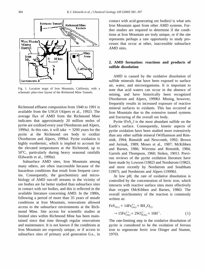
( )K.J. Edwards et al.rChemical Geology 169 2000 383–397384
Fig. 1. Location maps of Iron Mountain, California, with aschematic plan-view layout of the Richmond Mine Tunnels.
Richmond effluent composition from 1940 to 1991 isŽ .available from the USGS Alpers et al., 1992 . The
average flux of AMD from the Richmond Mineindicates that approximately 20 million moles of
Žpyrite are oxidized every year Nordstrom and Alpers,.1999a . At this rate, it will take ;3200 years for the
pyrite at the Richmond ore body to oxidizeŽ .Nordstrom and Alpers, 1999a . Pyrite oxidation ishighly exothermic, which is implied to account forthe elevated temperatures at the Richmond, up to508C, particularly during heavy seasonal rainfallsŽ .Edwards et al., 1999a .
Subsurface AMD sites, Iron Mountain amongmany others, are often inaccessible because of thehazardous conditions that result from frequent cave-ins. Consequently, the geochemistry and micro-biology of AMD run-off streams in the vicinity ofore bodies are far better studied than subsurface sitesin contact with ore bodies, and this is reflected in theavailable literature concerning AMD. In the 1980s,following a period of more than 35 years of unsafeconditions at Iron Mountain, renovations allowedaccess to the subsurface environments at the Rich-mond Mine. Site access for scientific studies atlimited sites within Richmond Mine has been main-tained since that time through regular renovationsand maintenance. It is not known if the conditions atIron Mountain are expressly unique, or if access to
Žsubsurface sites of primary acid generation i.e., in
.contact with acid-generating ore bodies is what setsIron Mountain apart from other AMD systems. Fur-ther studies are required to determine if the condi-tions at Iron Mountain are truly unique, or if the siterepresents perhaps a rare opportunity to study pro-cesses that occur at other, inaccessible subsurfaceAMD sites.
2. AMD formation: reactions and products ofsulfide dissolution
AMD is caused by the oxidative dissolution ofsulfide minerals that have been exposed to surfaceair, water, and microorganisms. It is important tonote that acid waters can occur in the absence ofmining, and have historically been recognizedŽ .Nordstrom and Alpers, 1999b . Mining, however,frequently results in increased exposure of reactivemineral surfaces to oxidants. This has occurred atIron Mountain due to the extensive tunnel systemsand fracturing of the overall ore body.
Ž .Pyrite FeS is the most abundant sulfide on the2
Earth’s surface. Consequently, kinetic aspects ofpyrite oxidation have been studied more extensively
Žthan any other sulfide mineral Williamson and Rim-stidt, 1994; Rimstidt and Newcomb, 1992; Brownand Jurinak, 1989; Moses et al., 1987; McKibbenand Barnes, 1986; Wiersma and Rimstidt, 1984;
.Garrels and Thompson, 1960; Stokes, 1901 . Previ-ous reviews of the pyrite oxidation literature have
Ž . Ž .been made by Lowson 1982 and Nordstrom 1982 ,and more recently by Nordstrom and SouthhamŽ . Ž .1997 , and Nordstrom and Alpers 1999b .
At low pH, the rate of oxidative dissolution iscontrolled by the concentration of ferric iron, whichinteracts with reactive surface sites more effectively
Ž .than oxygen McKibben and Barnes, 1986 . Theoverall stoichiometry of the reaction is commonlywritten as:
FeS q14Fe3q q8H O2Žs. Žaq . 2 Ž l.
™15Fe2q q2SO2y q16Hq. 1Ž .Žaq . 4Žaq .
The rate-limiting step in the oxidative dissolution ofpyrite is considered to be the oxidation of ferrous
Žiron to regenerate ferric iron Singer and Stumm,.1970 .

( )K.J. Edwards et al.rChemical Geology 169 2000 383–397 385
Ž .Reaction 1 describes the overall stoichiometryof oxidative dissolution reactions, but it does notdescribe the individual steps that must occur in theoxidation of sulfide to sulfate because of the largenumber of electrons that is transferred. Intermediatespecies such as elemental sulfur, sulfoxy compounds,and sulfites may play an extremely important role in
Žthe overall reaction kinetics Nordstrom and South-.ham, 1997 .
The most widely accepted model of sulfide min-eral dissolution was proposed by Singer and StummŽ .1968 . This model describes the sequential oxida-tion of surface sulfur atoms to form the thiosulfate
Ž 2q.anion, which is then liberated along with Fe intosolution. The thiosulfate anion is subsequently oxi-
Ž .dized to sulfate. In this case the overall reaction 1Ž .can be separated into the surface reaction 2 and
Ž .solution phase reaction 3 :
FeS q6Fe3qq3H O™S O2yq7Fe2qq6Hq,2 2 2 3
2Ž .
S O2yq8Fe3qq5H O™2SO2yq8Fe2qq10Hq.2 3 2 4
3Ž .
An important aspect of this model is that it predictsthe formation of only water-soluble products. How-ever, numerous reports have shown that elemental
Žsulfur also forms at surfaces McGuire et al., 1999,.in review; Sasaki et al., 1995; see below . A com-
plete model of pyrite dissolution must incorporatethe formation of all observed surface products.
2.1. Intermediate dissolution products on sulfidemineral surfaces
Chemical changes taking place on the mineralsurface are important in at least two respects. Forma-tion of secondary minerals at the surface has thepotential for forming inert layers that might inhibitdiffusion of oxidants to the surface, thereby slowingdissolution. Additionally, intermediate sulfur prod-ucts that develop on surfaces can be used as anenergy source for some microorganisms. The specia-tion of intermediates surface products and the kinet-ics of their production are crucial for understandinghow they, and the microbial communities they sup-port, impact overall sulfide dissolution rates.
Raman and X-ray photoelectron spectroscopyŽ .XPS have recently become quite commonly uti-lized techniques for the determination of chemicalspeciation at the surfaces of pyrite and other metalsulfides. Since elemental sulfur has no electronic
Ž .dipole and therefore no infrared absorption , Ramanis a particularly good probe of elemental sulfur. Tobetter understand the role of microorganisms in alter-ing surface chemistry during dissolution, we haveused Raman spectroscopy to analyze surfaces reactedin the laboratory with enrichment cultures of mi-croorganisms known to be important members of the
Žmicrobial community at the Richmond Mine Ed-.wards et al., 1997, 1998, 1999b . The enrichment
culture contained the iron-oxidizing species, Ferro-plasma acidarmanus and Leptospirillum ferrooxi-dans, as well as a sulfur oxidizer, Thiobacillus cal-dus. Pyrite was reacted with enrichment culturesunder conditions within the ranges observed at Iron
Ž .Mountain pH 1.5 and 378C . Fig. 2 shows a compar-ison of the Raman spectrum of a pyrite single crystal
Ž .exposed to the enrichment culture Fig. 2C and thespectrum of a sample reacted abiotically in acid forthe same length of time. The original starting surfaceŽ .Fig. 2A shows three primary peaks, 342, 377, and435 cmy1, that arise from bulk pyrite. After reactionfor 22 days in acid, the surface shows little change in
Ž . Ž .Fig. 2. Raman spectra of pyrite crystals. A Unreacted; BŽ .abiotically reacted with sulfuric acid, pH 1.5 for 22 days; C
reacted with a mixed enrichment culture of iron- and sulfur-Žoxidizing microorganisms for 22 days modified after McGuire et
.al., 1999, in review .

( )K.J. Edwards et al.rChemical Geology 169 2000 383–397386
overall surface chemical composition, even when thescale is expanded by a factor of 10. However, in thepresence of iron-oxidizing microorganisms, whichcan rapidly regenerate Fe3q, the surfaces developrather extensive sulfur layers. In this case, the Ra-man spectrum contains three very large, intense peaksat 470, 217, and 151 cmy1 that are characteristic ofelemental sulfur, primarily in the form of S rings.8
Calibration of the Raman data indicates that thesulfur layer is at least several hundreds of Angstromsthick if distributed uniformly over the mineral sur-
Ž .face McGuire et al., 1999, in review .The formation of sulfur must arise from a differ-
ent mechanism than the original Stumm model. Forexample, one possibility is direct attack on a sulfur
Ž q.site by a hydronium ion H , via a mechanism suchas:
MSqFe3qqHq™M2qq1r2H S qFe2q, 4Ž .2 n
1r2H S qFe3q™1r8S qFe2qqHq. 5Ž .2 n 8
If the reaction of pyrite to sulfur is dominant, andsulfur accumulations do not limit surface oxidationrates, then the reaction of pyrite could be written as:
FeS q2Fe3q™2S0 q3Fe2q. 6Ž .2ŽS.
In this case, 1 mol of pyrite oxidation consumes only3q Ž .2 mol of Fe , compared to 14 in reaction 1 . This
is important, because the rate of pyrite oxidation israte-limited by ferric iron supply. Also notable from
Ž .reaction 6 is the absence of hydrogen ion release tosolution, compared with 16 hydrogen ions released
Ž .in reaction 1 , emphasizing that the acid is gener-ated from the oxidation of sulfur, for example:
S0 q1.5O qH O™SO2yq2Hq. 7Ž .2 2 4
Ž .Bahatti et al., 1993 . Further studies are needed toquantify the formation of sulfur during pyrite oxida-tion in order to determine if the oxidation of elemen-
Žtal sulfur involves important, rate-limiting in terms.of acid generation reactions.
Fig. 3 shows dissolution rate data, as indicated bythe total iron concentration in solution over time, forpyrite reacted with the enrichment culture and re-acted abiotically in acid. The difference in dissolu-tion rate between these is approximately a factor of 2Ž .McGuire et al., 1999, in review . If the rate of sulfurformation correlates linearly with the dissolution rate,
Fig. 3. Dissolution data for pyrite. Data for total iron release areshown for pyrite reacted abiotically in sulfuric acid, pH 1.5, andreacted in the presence of a mixed enrichment culture of iron- and
Žsulfur-oxidizing microorganisms, also at pH 1.5 modified after.McGuire et al., in review .
a twofold difference in sulfur accumulation betweenthe two samples is the maximum that would beexpected because some sulfur will be consumed bythe sulfur oxidizers in the enrichment culture. How-ever, the Raman measurements on pyrite indicatethat in the abiotic experiment, there is no detectablesulfur, while the microbial sample shows sulfur peaksthat exceed those of the bulk pyrite by at least a
Ž .factor of 10 Fig. 2 . Based on the signal-to-noiselevel of these measurements, the amount of sulfur onthe abiotic sample is at least a factor of 100 less thanthat in the microbial experiment. The observation ofa 100-fold difference in surface sulfur concentrationson samples whose nascent dissolution rates are dif-ferent by a factor of 2 suggests that there may be anactivation energy barrier to the formation of elemen-tal sulfur at the surface.
2.2. Sulfur remoÕal and reaction kinetics
Determining intermediate reactions involved inpyrite oxidation is critical because the overall reac-tion rate will be limited by the slowest kinetic step.If secondary phases such as sulfur form and inhibitdiffusion to the surface, then at long times the over-all dissolution rate will be controlled by the rate ofoxidation of the surface product, rather than the rateof oxidation of the metal sulfide mineral itself. In thecase of pyrite discussed above, quantitative analysisof the sulfur layer indicates that it is hundreds ofAngstroms in thickness if distributed uniformlyacross the surface.
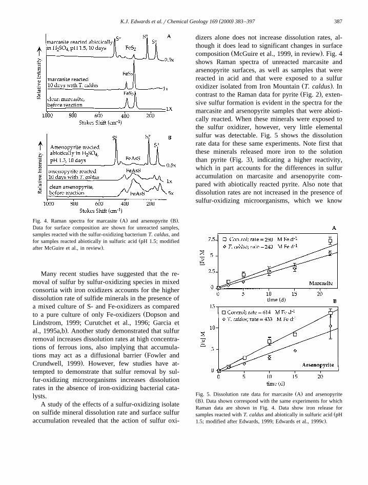
( )K.J. Edwards et al.rChemical Geology 169 2000 383–397 387
Ž . Ž .Fig. 4. Raman spectra for marcasite A and arsenopyrite B .Data for surface composition are shown for unreacted samples,samples reacted with the sulfur-oxidizing bacterium T. caldus, and
Žfor samples reacted abiotically in sulfuric acid pH 1.5; modified.after McGuire et al., in review .
Many recent studies have suggested that the re-moval of sulfur by sulfur-oxidizing species in mixedconsortia with iron oxidizers accounts for the higherdissolution rate of sulfide minerals in the presence ofa mixed culture of S- and Fe-oxidizers as compared
Žto a pure culture of only Fe-oxidizers Dopson andLindstrom, 1999; Curutchet et al., 1996; Garcia et
.al., 1995a,b . Another study demonstrated that sulfurremoval increases dissolution rates at high concentra-tions of ferrous ions, also implying that accumula-
Žtions may act as a diffusional barrier Fowler and.Crundwell, 1999 . However, few studies have at-
tempted to demonstrate that sulfur removal by sul-fur-oxidizing microorganisms increases dissolutionrates in the absence of iron-oxidizing bacterial cata-lysts.
A study of the effects of a sulfur-oxidizing isolateon sulfide mineral dissolution rate and surface sulfuraccumulation revealed that the action of sulfur oxi-
dizers alone does not increase dissolution rates, al-though it does lead to significant changes in surface
Ž .composition McGuire et al., 1999, in review . Fig. 4shows Raman spectra of unreacted marcasite andarsenopyrite surfaces, as well as samples that werereacted in acid and that were exposed to a sulfur
Ž .oxidizer isolated from Iron Mountain T. caldus . InŽ .contrast to the Raman data for pyrite Fig. 2 , exten-
sive sulfur formation is evident in the spectra for themarcasite and arsenopyrite samples that were abioti-cally reacted. When these minerals were exposed tothe sulfur oxidizer, however, very little elementalsulfur was detectable. Fig. 5 shows the dissolutionrate data for these same experiments. Note first thatthese minerals released more iron to the solution
Ž .than pyrite Fig. 3 , indicating a higher reactivity,which in part accounts for the differences in sulfuraccumulation on marcasite and arsenopyrite com-pared with abiotically reacted pyrite. Also note thatdissolution rates are not increased in the presence ofsulfur-oxidizing microorganisms, which we know
Ž .Fig. 5. Dissolution rate data for marcasite A and arsenopyriteŽ .B . Data shown correspond with the same experiments for whichRaman data are shown in Fig. 4. Data show iron release for
Žsamples reacted with T. caldus and abiotically in sulfuric acid pH.1.5; modified after Edwards, 1999; Edwards et al., 1999c .

( )K.J. Edwards et al.rChemical Geology 169 2000 383–397388
Fig. 6. The SEM image of sulfur-rich deposits on arsenopyrite,after abiotic reaction for 22 days at 378C, pHs1.5. Examples of
Ž .sulfur-rich deposits by EDX analysis; data not shown are shownwith arrows. Scale bar is 50 mm.
from the Raman data were effectively removingŽ .sulfur from the surfaces Fig. 4 . If sulfur were
inhibiting dissolution, iron release rates would slowas sulfur accumulates at the surface. The fact that thedissolution rates in the presence of T. caldus werenot higher than those of the abiotic control reactionssuggests that either the sulfur layers are permeable toreactants and products of the surface reaction, or thatthe sulfur is not distributed uniformly across thesurface. Indeed, some scanning electron microscopy
Ž .images Fig. 6 reveal agglomerates of sulfur presenton the surface, surrounded by relatively AcleanBareas. The ecological role of sulfur oxidizers, andwhy rates of sulfide dissolution are increased inmixed populations of iron- and sulfur oxidizers, re-main poorly understood.
3. Biodiversity at Iron Mountain: seasonal andspatial relationships between microbial communi-ties and geochemistry
Considerable prior attention has been given to thebiology of drainage streams that deliver acidic,metal-rich waters from ore bodies or tailings piles tolarger water ways. The microbial populations associ-ated with these run-off environments are generallylow in both biomass and diversity, and molecular
analyses have suggested that most taxa in theseenvironments are readily obtained via culturing tech-
Žniques Rawlings, 1995; Goebel and Stackebrandt,1994b, 1995; Pizarro et al., 1996; Vasquez and
.Espejo, 1997 . Our studies of run-off streams at IronŽMountain Edwards et al., 1999a; Schrenk et al.,
.1998 are consistent with other studies that suggestthat the dominant organisms detected are usually T.
Žferrooxidans and L. ferrooxidans Rawlings et al.,.1999; Walton and Johnson, 1992 . However, our
studies have also found that there is more that wecan learn about the microbial diversity involved insulfide dissolution by studying the microbial com-
Ž .munities at subsurface sites see below .At Iron Mountain, as at other sites, the acidophilic
community includes Eukarya, Bacteria and Archaea.At subsurface sites that receive no sunlight, chemo-synthetic prokaryotes that utilize the reduced ironandror sulfur from pyrite for energy and fix CO for2
cell carbon are the foundation of the mine biota.ŽMixotrophic assimilate organic or inorganic carbon
.as well as oxidize iron andror sulfur and hetero-Ž .trophic utilize organic carbon for energy prokary-
otes are often detected and play an important role inŽ .the ecology Johnson, 1998 . Filamentous fungi and
protozoa are the most common eukaryotes observedŽ .Schleper et al., 1995 . Protozoa are typically foundin biofilms, where they graze on bacterial cells within
Ž .them Fig. 7 .Because of the large number of studies that have
identified T. ferrooxidans and L. ferrooxidans inAMD run-off environments, and the relative ease ofculturing these bacteria, laboratory studies have con-centrated on the role these two species play insulfide dissolution. However, in the environment,diverse communities of acidophilic organisms mayinfluence leaching rates and effect sulfide dissolu-tion. Other species of both bacteria and archaea havebeen obtained from AMD environments, and a greatdeal has been learned by characterization of these.However, in order to assess the natural abundance ofdifferent species of microorganisms and thereby in-fer their relative importance to sulfide dissolution, itis difficult and usually inaccurate to enumerate or-ganisms by culturing, and molecular methods arerequired for quantitative analysis. Molecular methods
Ž .include fluorescent in situ hybridization FISH us-ing oligonucleotide probes targeting ribosomal ri-

( )K.J. Edwards et al.rChemical Geology 169 2000 383–397 389
Fig. 7. DAPI-stained fluorescent image of protists within the subaerial biofilm that formed following a partial cave-in within the RichmondŽ .mine. Scale bar is 25 mm and applies to both images. Protist cells are large 10–20 mm diameter , roughly spherical, and contain many
brightly fluorescent bodies, some of which contain ingested prokaryotes.
Ž .bonucleic acid rRNA , and detection of ribosomalRNA gene sequences by polymerase chain reactionŽ . ŽPCR , cloning and sequence analysis Amann et al.,
.1995; Pace et al., 1986 .The need for in situ molecular enumeration of
biodiversity has been exemplified in many environ-Žments, AMD sites among others e.g., Hugenholtz et
al., 1998; Johnson, 1998, 1991; Amann et al., 1996;Goebel and Stackebrandt, 1994a,b; Barns et al.,
.1994 . As one example, an early study at Iron Moun-tain tentatively identified T. ferrooxidans in drainage
Ž .waters Nordstrom, 2000 . While this is consistentwith more recent in situ molecular studies of run-offstreams at Iron Mountain, it may tell us little aboutthe microbial community at subsurface sites of pri-mary acid generation. Studies using species-specificoligonucleotide probes to characterize populationsfrom the Richmond Mine found that T. ferrooxidansrarely occurs at sites in contact with the ore bodyŽ .Edwards et al., 1999a; Schrenk et al., 1998 . Inaddition to problems with inferring microbial popula-tions at acid-generating sites by studying run-offwaters, the use of culturing techniques often selectsan organism that can grow the fastest under theconditions provided, which may not reflect its natu-ral abundance in the environment. For example, atthe Richmond Mine, even when the abundance of T.ferrooxidans is below detection limit using FISH,the species can be cultured from the Richmond Mine
Žusing A9KB medium adjusted to pH 2.5 unpublished.data . While culturing techniques are vital for
physiological assessment of microorganisms, these
examples reinforce the importance of using quantita-tive, in situ molecular methods at primary sites ofacid generation to identify key players involved insulfide dissolution, in order to target and verifyspecies of relevance.
3.1. Seasonal Õariations in microbial communities
Extensive analysis of samples from the RichmondMine collected throughout 1 year indicated that sub-stantial fluctuations in microbial community struc-ture correlate with significant fluctuations in rainfall,
Žionic strength, and temperature Fig. 8; Edwards et.al., 1999a . Heavy rainfall in the winter is generally
followed by summer droughts. Peaks and lows indischarge from the Richmond Mine correlate closelywith precipitation, suggesting transit times for flow
Žon the scale of days to a few weeks P. Ekoniak,.May 1999, personal communication . During times
Ž .of low rainfall summer droughts , the ionic strengthof effluent is maximal.
Cell populations are found to be highest at theRichmond Mine during the dry summer and fallmonths. FISH indicates densities of 107–109 cellsmly1 total for bacteria, archaea and eukaryotes within
Ž .sediments pore fluids and attached to surfaces fromŽ .July through September Edwards et al., 1999a .
During the dry months, dense communities of mi-Ž 9 y1.croorganisms )10 cells ml , commonly re-
Ž .ferred to as slime streamers see below , are alsocommon. During the winter months, bacteria domi-
Ž .nate the microbial population Fig. 8 . However,

( )K.J. Edwards et al.rChemical Geology 169 2000 383–397390
Fig. 8. Seasonal variations in environmental conditions and micro-Ž . Ž .bial populations at the Richmond Mine over 1 year 1997 . aŽ .Conductivity, rainfall, pH, and temperature conditions. b Rela-
tive abundance of bacteria, archaea, eukarya, and the species, L.ferrooxidans and T. ferrooxidans. Microbial populations were
Ž .quantitated using FISH data from Edwards et al., 1999a .
during the summer and fall months, archaea repre-sent a significant proportion of the microbial popula-tion. Correlations between high ionic strength andhigh archaeal numbers, and loss of archaea anddilution of solutions by rainfall, e.g., suggest proba-
ble geochemical controls on microbial communitystructure. However, analysis of the bacterial andarchaeal impact on AMD generation requiresmetabolic analysis, and thus requires culturing andisolation of relevant species.
From enrichment cultures that were started withinoculum collected from the Richmond mine duringthe summer months when archaea are in high abun-
Ždance, an iron-oxidizing archaea was isolated fer1;.Bond et al., 1999; Edwards, 2000 . Analysis of 16S
rRNA gene for fer1 indicates that the available se-Ž .quence 849 bases is identical to that of a recently
described autotroph and bioleaching isolate, F. aci-dophilum. However, the archaea isolated from IronMountain are metabolically distinct. In comparisonto F. acidophilum, fer1 has more extreme pH toler-
Ž .ance grows well down to pH 0 , higher temperatureŽ .optimum ;458C , and is capable of heterotrophic
growth on yeast extract. Due to these physiologicaldistinctions, the isolate has been tentatively given thespecies name acidarmanus, within the genus Ferro-plasma. In situ analyses of the microbial communityusing species-specific probes for F. acidarmanusindicate that this archaea occurs in quite high abun-dance in the environment, comprising up to 85% ofthe total microbial community in some environmentsŽ .Bond et al., 1999; Edwards, 2000 . This species isinferred to be the archaea originally identified during
Ž .the 1-year study Edwards et al., 1999a . Given itspredominance and widespread distribution, thisiron-oxidizing species must play a significant role inAMD generation at Iron Mountain.
3.2. Population eÕolution oÕer time: re-colonizationeÕents
Occasionally, periods of extremely heavy rainfallresult in heavy washouts of the fine-grained pyritesediments within the Richmond Mine. When thisoccurs, the sediments must be removed in order toprovide access, and to keep the pipes that divert theacid waters from the mine clear of debris. One such
Ž .occurrence April 1998 provided an opportunity tostudy the evolution of the microbial population dur-ing re-colonization.
After the flow had subsided and the sedimentsremoved in April 1998, very few microorganisms

( )K.J. Edwards et al.rChemical Geology 169 2000 383–397 391
were detectable on sediment surfaces or in sedimentŽ 4 y1.pore fluid -10 cells ml , and slime streamers
were washed away entirely. We attribute this towashout and removal of the microbially colonized
Župper sediment region where there is sufficient oxy-.gen diffusion to support growth . Shortly after this
Ž .episode July 1998 , molecular analyses found thatthe majority of 16S rRNA gene sequences detectedfrom sediment samples were from Leptospirillum
ŽGroup II type, as represented by clone BU01 Fig..9 . It has been suggested that Leptospirillum Group
ŽII types are the naturally abundant species Goebel.and Stackebrandt, 1995 and these may be the major
types occurring in the mine. It is interesting to notethat while these species accounted for F20% ofmicrobial populations associated with sediments dur-
Ž .ing 1997 Edwards et al., 1999b , they appear as thedominant initial colonizing species after this distur-
Ž .bance unpublished data .Re-colonization and return to the usual mine biota
Ž .slime streamers, etc. associated with the sedimentsafter this redistribution occurred only after a lengthy
Ž .lag time ;4–6 months . FISH analyses of themicrobial communities by November 1998 showedthat F. acidarmanus dominated the microbial com-munity, particularly in some slime streamers and
Ž .sediments Bond et al., 1999; Edwards, 2000 . Cells
Žhybridizing with a probe for Leptospirillum Groups.Iq II were detected in many samples, but these
made up only low proportions of biofilms and sedi-Ž .ments unpublished data . One explanation is that the
Leptospirillum dominating the 16S rRNA gene se-quences detected in July 1998 decreased in abun-dance some months later and were succeeded by anabundance of F. acidarmanus. Hence, F. acidar-manus was a dominant second generation microbialconstituent during re-colonization. Communities re-structuring and changing geochemical conditionsduring re-colonization are undoubtedly coupled;hence, this sequence may only be applicable underthe geochemical conditions that prevailed during thistime. However, it seems probable that such reorgani-zation occurs regularly following annual winterfloods.
3.3. Taxonomic indications of redox gradients
Periodic slumps of pyritic material from overlyingstoops are common events within subsurface tunnelsat Iron Mountain. In November 1998, a slump devel-oped from a partial cave-in of an above mine tunnel.Gelatinous material dripping from above formed a
Ž .thick up to 10 mm , subaerial biofilm on the slumpsurface. DAPI staining and molecular analysis indi-
Fig. 9. Phylogenetic dendogram representing the evolutionary history of Iron Mountain mine clones with Leptospirillum species. This wasinferred from comparison of DNA sequences, comprising 1163 nucleotides, of the 16S rRNA gene. Statistical confidence intervals of thetree topology are shown as bootstrap values at the branch points. Evolutionary distances are indicated by the sum of horizontal branchlengths; scale bar represents changes per nucleotide. Modified after Bond et al., in review.

( )K.J. Edwards et al.rChemical Geology 169 2000 383–397392
Žcates the presence of novel diversity Bond et al., in.review . The major 16S rRNA gene sequence de-
tected, represented by clone BA29 in Fig. 9, dis-Ž .tantly 91% similar related to Leptospirillum se-
quences, likely represents a new probable iron-oxidizing species or genus.
In addition to Leptospirillum, the subaerial biofilmcontained organisms with sequences of varying simi-larity to cultured organisms of the Acidimicrobiumgroup. Most of these were )99% similar to therecently described iron-oxidizing heterotroph, Fer-
Ž .romicrobium acidophilus Bond et al., in review .Other sequences detected clustered within the delta
Ž .subdivision of the Proteobacteria with low ;80%similarity to these sequences. Detection of thesesequences suggests that anaerobic respiration, sulfateor metal reduction may occur in the slime. It is likelythat redox gradients and cycling of S and Fe com-pounds occur within the subaerial biofilm. To our
Ž .knowledge, extremely acidophilic pH-3 sulfate-reducing species have not previously been described,although acid-tolerant, if not acidophilic sulfate re-ducers have been cultured in solutions down to pH 3ŽB. Johnson, December 1999, personal communica-
.tion .
3.4. DiÕersity at Iron Mountain: spatial considera-tions
Our analyses at Iron Mountain have found thatlimited types of organisms often dominate microbialcommunities in small-scale environments. This find-ing is consistent with that of other investigators whoconcluded that AMD communities are characterized
Žby quite limited diversity Goebel and Stackebrandt,.1995; Johnson, 1998 . However, as can be seen from
the above summary, the overall populations withinthe mine are quite diverse because the communitystructures change with time and space in response tochanges in geochemical and physical conditions. As
Ž .noted by Goebel and Stackebrandt 1994b, 1995 ,many of the previously identified organisms fromthese environments are cultivable and have beenisolated. However, our recent molecular investiga-tions indicate that novel 16S rRNA sequences andpoorly studied species are present in extremely acidicenvironments. Development of new isolation proce-
dures is required to obtain pure cultures for charac-terization of the novel organisms.
4. Kinetics of sulfide dissolution: evaluation ofabiotic vs. microbial contributions to AMD
Microbial dissolution rates measured by numerousworkers over several decades suggest considerablevariability in the degree to which microorganisms
Ž .accelerate oxidative dissolution Olson, 1991 . It isimportant to quantitatively measure microbially me-diated dissolution rates under a variety of conditionsin order to model AMD systems. However, thevariability among microbial dissolution rates hasmade extrapolation to environmental settings diffi-cult.
In order to better constrain microbial dissolutionŽ .rates, Olson 1991 reported a comparison of leach-
ing rates determined in eight different laboratoriesthat all used the same strain of T. ferrooxidans, thesame pyrite source, and the same leaching methodŽ .conditions, etc. . The results of these studies deter-mined that T. ferrooxidans increased dissolution ofpyrite relative to controls by an average of 34 times
Ž .the abiotic rate Olson, 1991 . The important findingfrom this study was that microbial dissolution rateswere fairly repeatable when experimental proceduresand microbial strains were the same. However, evenconsidering the consistency of these data, extrapola-tion to field settings is still problematic because twofundamentally crucial factors are often not consid-ered: cell densities and surface area for reaction.First, cell densities are important because rates of
Žpyrite dissolution are controlled by the rate of mi-.crobial re-oxidation of ferrous iron, which is neces-
sarily linked to numbers of actively metabolizingcells. For the interlaboratory comparison, this wasapparently not important, because enough parameterswere constrained to ensure approximately compara-ble cell numbers in each experiment. However, envi-ronmental population sizes may differ dramaticallyfrom the optimized conditions used for laboratoryexperiments. Second, surface area is important be-cause abiotic dissolution also contributes to the fluxof iron and sulfur from pyrite surfaces, and theserates depend upon the surface area available for

( )K.J. Edwards et al.rChemical Geology 169 2000 383–397 393
reaction. Surface area is routinely determined forabiotic geochemical experiments, but not so oftendetermined for microbial experiments. In sum, bothcell number and surface area data are required fordetermination of rate constants that would allowextrapolation to the environment. Cell-normalizedrate constants have been commonly determined formany microbially mediated processes, such as bacte-
Žrial sulfate reduction e.g., Kirsten and Canfield,.1997 . In the case of sulfide oxidation, however,
cell-normalized rates are not reported, due in part tothe technical difficulties associated with accuratelydetermining cell densities attached to sulfide parti-cles.
In recent work, we determined cell-normalizeddissolution rates for different mixed enrichment cul-tures and iron-oxidizing isolates that were obtained
Žfrom the Richmond Mine Edwards, 1999; Edwards.et al., 1998, 1999b . Experiments were conducted
under varying conditions of oxygenation, pH, andŽavailable surface area, type of sulfide mineral pyrite,
.marcasite, arsenopyrite and cell densities, so totaldissolution rates varied considerably. However, cell-normalized dissolution rates clustered more tightly,from 2=10y8 to 6=10y7
mM Fe celly1 dayy1
Ž y7 y1 y1.medians3=10 mM Fe cell day . Thus,for order-of-magnitude environmental calculations, itmay be possible to approximate the microbial contri-bution to dissolution by a constant. This is importantbecause given information regarding two key envi-ronmental parameters, cell densities of iron-oxidizingmicroorganisms and surface area for reactions, themicrobial contribution to sulfide dissolution can beestimated.
4.1. Application of cell-normalized dissolution ratesto predict microbial contribution to AMD production
Cell-normalized dissolution rates provide the linknecessary to make predictive estimates of the micro-bial contribution to AMD production in the environ-ment. Here we briefly give an example of how thiscan be applied to a system such as the RichmondMine at Iron Mountain. Although this is not intendedto be a strictly quantitative analysis of AMD genera-tion at Iron Mountain, it highlights where futureanalysis may be needed.
Fig. 10 shows a simplified diagram of the dis-charge system for the Richmond Tunnel SystemŽ .Fig. 1 . We will estimate the maximum microbialcontribution to iron release into the Richmond mine
Ž .effluent Fig. 10 , and approximate the amount ofpyritic material that would need to react to producethe iron load coming from the Richmond Mine.These calculations are based on a recent measure-ment of iron concentrations in the Richmond effluentŽ y1 .;30 g l ; May 1998, unpublished data and flow
Ž y1rate ;100 l min ; courtesy of Stauffer Manage-.ment .
In Fig. 10, the mine tunnel environments aredivided into two simplified AzonesB. Zone 1 consistsof water flowing over pyrite sediments that are colo-nized by biofilms. Most of these biofilms exist asstreamers in the water that are anchored within the
Ž .sediments, rather than as a surface coating Fig. 11 .As noted above, the biofilms are comprised predomi-nately of iron-oxidizing prokaryotes, although theparticular species varies with time and space. Al-though the proportion of eukarya in biofilms varies,for these calculations, we will use the estimate froma recent study that determined eukarya to comprise
Ž;15% of the volume of a biofilm Bond et al.,.1999; Edwards, 2000 . We estimate that condensed
biofilms are ;2 cm thick. Determining the prokary-otic cell density within biofilms is difficult becausethey are held within voluminous extracellular poly-meric material that is not easily disaggregated with-out cell disruption. But if we estimate that each cell
3 Žoccupies ;8 mm using cubic geometry and esti-mating 2 mm diameter for each cellqextracellular
. 11material and is closely packed, then ; 10prokaryotic cells can be accommodated in 1 cm3 ofbiofilm. An upper estimate for the aerial extent offlowing water and biofilm material, based on and theneed for flowing water to accommodate streamersand the estimated geometry of dendritic flow throughthe tunnels, is ;30 m2, or ;6=103 cm3. At 1011
cells cmy3, the biofilms contain ;6=1014 totalcells. Hence, if we use the median cell-normalizeddissolution rate of 3=10y13 M Fe celly1 dayy1, themaximum contribution from the biofilms is ;200M Fe dayy1, or -1% of the 105 M of total iron
Ž .output from the tunnel system Fig. 10 . Since this isa relatively minor contribution to the total iron flux,we will consider other sources, namely sediment

( )K.J. Edwards et al.rChemical Geology 169 2000 383–397394
Ž .Fig. 10. Schematic diagram to represent a cross-section through the combined length Richmond Mine tunnels, that are shown in plan formŽ .in Fig. 1. The tunnel floors contain deep )1 m accumulations of fine-grained pyrite sediments, which are represented as a wedge in the
Ž .diagram. Some surface water flows in the tunnels, and is colonized by biofilms Fig. 11 . However, much of fluid flow within the tunnels issubsurface, resulting in loosely consolidated, saturated sediments. The depth of oxygen penetration is unknown; an oxygenated, saturated
Ž .portion of the wedge not scaled is illustrated by the stippled pattern.
piles and the microbial cells associated with them, inthe following calculations.
The remainder of the tunnel system consists ofpyrite sediments at varying degrees of saturation andoxygenation. Much of the sediment is very looselyconsolidated because subsurface water flows, whichgives the tunnel floors a quicksand-like consistencyin many areas. The surface area of the bulk sedi-ments, which include some silicate materials, ishighly variable. A measured value is ;15=10y2
2 y1 Ž .m g Edwards et al., 1998 . This is comparable tothe surface area used in laboratory experiments fordetermination of abiotic dissolution rates, at 2.3=
10y2 my2 gy1, which was entirely sulfide materialŽ .Edwards, 1999; Edwards et al., 1999c . For the
purposes of example calculations, we will use therate determined in this study, 1.2=10y4 M Fecmy1 dayy1 for the abiotic iron release rate.
To determine the microbial contribution to ironrelease, we need information on cell densities, andestimates of pore fluid space within the sediments.For these calculations, we will again be estimatingan upper contribution to iron release by microorgan-isms. We estimate that 1 cm3 of very loosely consol-
Ž .idated sediment contains ;2 g ;150–500 mm ofpyrite, which requires ;500 ml of solution to satu-rate pore space. The cell density of prokaryotes in
Žsediments at the Richmond Mine attached to sur-.faces and in pore fluids , as noted above, is maximal
in the summer months, at ;2=109 cells mly1

( )K.J. Edwards et al.rChemical Geology 169 2000 383–397 395
Fig. 11. Example of biofilms within the Richmond Mine. Stream width is ;1 m. Eukaryotic filaments are anchored within the underlyingsediments and are colonized by iron-oxidizing prokaryotes, which comprise most of the biomass.
Ž . 9 y3 9Edwards et al., 1999a , or 10 cells cm . At 10cells cmy3, using the cell normalized rate constant of3=10y7
mM Fe celly1 dayy1, we obtain an upperestimate of the microbial contribution to dissolutionwithin the sediments of 3=10y4 M Fe cmy1 dayy1.This calculation suggests that the abiotic and micro-bial contributions to iron release differ by less thanthe order of magnitude confidence we have for thecalculation. This assessment contrasts laboratorymeasurements that indicate up to 34-fold increases inlaboratory-determined microbial dissolution rates rel-
Ž .ative to controls Olson, 1991; Paciorek et al., 1981 .Moreover, these calculations suggest that the highrate of AMD generation at Iron Mountain may belargely due to the larger than anticipated inorganiccontribution resulting from the high-surface areaavailable for reaction. The temperature dependenceof rates may also be a factor. Unlike the inorganicrate, the microbial rate seems to have a limited
Ž .temperature dependence Norris, 1990 . Reactionswithin the Richmond Mine occur at ;208C highertemperatures than those used in most laboratory stud-ies, which may also explain the inferred greatercontribution of inorganic dissolution at this site.Additionally, pore fluid constraints within saturated
sediments will greatly reduce microbial populationdensities relative to most laboratory experiments.Population size will be pivotal to what the microbialcontribution to dissolution in the environment canbe.
Summing the abiotic and microbial contributions,we can estimate the release of iron from saturated,sufficiently oxygenated sediments to be ;4=10y4
M Fe dayy1 cmy3. Hence, if all of the iron in theeffluent were coming from the tunnel sediments,;250 m3 of reacting sediment would be required toproduce 105 M Fe dayy1. This volume of sedimentsis easily accommodated within the Richmond MineŽ .Figs. 1 and 10 . However, it is difficult to estimatethe depth to which pyrite dissolution occurs in thesediments because the rate of oxygen transport intosediments is poorly constrained. Determination ofthe mechanisms and rates of oxygen transport anddiffusion has been previously recognized to be offirst-order importance for assessing AMD productionat Iron Mountain. Given the accessibility of pyriteaccumulations in the tunnels to air and water, weemphasize the importance of explicit considerationof oxygen transport into saturated and semi-saturatedsediments in future analyses.

( )K.J. Edwards et al.rChemical Geology 169 2000 383–397396
Acknowledgements
D. Kirk Nordstrom is thanked for stimulatingdiscussion. We also thank Iron Mountain Mine forsite access and Stauffer Management for providingassistance. Support for this work was provided byNSF grant CHE-9521731.
References
Alpers, C.N., Nordstrom, D.K., Burchard, J.M., 1992. Compila-tion and interpretation of water quality and discharge foracidic waters at Iron Mountain, Shasta County, California.U.S. Geol. Surv. Bull., No. 91-4150.
Alpers, C.N., Nordstrom, D.K., Thompson, J.M., 1994. Environ-mental geochemistry of sulfide oxidation. In: Alpers, C.N.,
Ž .Blowes, D.W. Eds. , American Chemical Society SymposiumSeries. American Chemical Society, Washington.
Amann, R.I., Ludwig, W., Schleifer, K., 1995. Phylogenetic iden-tification and in situ detection of individual microbial cellswithout cultivation. Microbiol. Rev. 59, 143–169.
Amann, R., Snaidr, J., Wagner, M., Ludwig, W., Schleifer, K.H.,1996. In situ visualization of high genetic diversity in a naturalmicrobial community. J. Bacteriol. 178, 3496–3500.
Barns, S.M., Fundyga, R.E., Jeffries, M.W., Pace, N.R., 1994.Remarkable archaeal diversity detected in a Yellowstone Na-tional Park hot spring environment. Proc. Natl. Acad. Sci. U.S. A. 91, 1609–1613.
Bhatti, T.B., Bigham, J.M., Carlson, L., Tuovinen, O.H., 1993.Mineral products of Pyrrhotite oxidation by Thiobacillus fer-rooxidans. Appl. Environ. Microbiol. 59, 1984–1990.
Bond, P.L., Edwards, K.J., Gihring, T.M., Banfield, J.F., 1999.Microbial ecology of an acid mine drainage environment.Geological Society of America, 31st Annual Meeting. TheGeological Society of America, Boulder.
Bond, P.L., Smriga, S.P., Banfield, J.F., in review. Phylogeny ofmicroorganisms populating a thick, subaerial, predominantlylithotrophic biofilm at an extreme acid mine drainage site. Inreview.
Brown, A.D., Jurinak, J.J., 1989. Mechanism of pyrite oxidationin aqueous mixtures. J. Environ. Qual. 18, 545–550.
Curutchet, G., Tedesco, P., Donati, E., 1996. Combined degrada-tion of covellite by Thiobacillus thiooxidans and Thiobacillusferrooxidans. Biotechnol. Lett. 18, 1471–1476.
Dopson, M., Lindstrom, E.B., 1999. Potential role of Thiobacilluscaldus in arsenopyrite bioleaching. Appl. Environ. Microbiol.65, 36–40.
Edwards, K.J., 1999. The geomicrobiology of sulfide dissolution.PhD Thesis, University of Wisconsin, Madison, 311 pp.
Edwards, K.J., Schrenk, M.O., Banfield, J.F., Hamers, R.J., 1997.Oxidative dissolution of pyrite by microorganisms at IronMountain, California. The Geological Society of America,29th Annual Meeting. The Geological Society of America,Boulder.
Edwards, K.J., Schrenk, M.O., Hamers, R., Banfield, J.F., 1998.Microbial oxidation of pyrite: experiments using microorgan-isms from an extreme acidic environment. Am. Mineral. 83,1444–11453.
Edwards, K.J., Gihring, T.M., Banfield, J.F., 1999a. Seasonalvariations in microbial populations and environmental condi-tions at an extreme acid mine drainage environment. Appl.Environ. Microbiol. 65, 3627–3632.
Ž .Edwards, K.J. et al., 1999b. Geomicrobiology of pyrite FeS2
dissolution: a case study at Iron Mountain. Calif. Geomicro-biol. J. 16, 155–179.
Edwards, K.J., McGuire, M.M., Hamers, R.J., Banfield, J.F.,1999c. Kinetics and surface microstructural evolution of mi-crobially mediated sulfide dissolution: implications for model-ing acid mine drainage generation. Ninth Annual V.M. Gold-schmidt Conference. Lunar and Planetary Institute, Houston.
Edwards, K.J., Bond, P.L., Gihring, T.M., Banfield, J.F., 2000.An iron-oxidizing extreme acidophile important in acid minedrainage. Sci. 287, 1797–1799.
Fowler, T.A., Crundwell, F.K., 1999. Leaching of zinc sulfide byThiobacillus ferrooxidans: bacterial oxidation of the sulfurproduct layer increases the rate of zinc sulfide dissolution athigh concentrations of ferrous ions. Appl. Environ. Microbiol.65, 5285–5292.
Garcia, O. Jr., Bigham Jerry, M., Olli, H., 1995a. Oxidation ofgalena by Thiobacillus ferrooxidans and Thiobacillus thiooxi-dans. Can. J. Microbiol. 41, 508–514.
Garcia, O. Jr., Bigham Jerry, M., Tuovinen Olli, H., 1995b.Sphalerite oxidation by Thiobacillus ferrooxidans andThiobacillus thiooxidans. Can. J. Microbiol. 41, 578–584.
Garrels, R.M., Thompson, M.E., 1960. Oxidation of pyrite by ironsulfate solutions. Am. J. Sci. 259, 57–67.
Goebel, B.M., Stackebrandt, E., 1994a. The biotechnological im-portance of molecular biodiversity studies for metal bioleach-ing. In: Priest, F.G., Ramos-Cormenzana, A., Tindall, B.J.Ž .Eds. , Bacterial Diversity and Systematics. Plenum.
Goebel, B.M., Stackebrandt, E., 1994b. Cultural and phylogeneticanalysis of mixed microbial populations found in natural andcommercial bioleaching environments. Appl. Environ. Micro-biol. 60, 1614–1621.
Goebel, B.M., Stackebrandt, E., 1995. Molecular analysis of themicrobial biodiversity in a natural acidic environment. In:
Ž .Jerez, C.A., Vargas, T., Toledo, H., Wiertz, J.V. Eds. ,International Biohydrometallurgy Symposium IBS-95. Univer-sity of Chile, Santiago, Chile, pp. 43–52.
Hugenholtz, P., Goebel, B.M., Pace, N.R., 1998. Impact of cul-ture-independent studies on the emerging phylogenetic view ofbacterial diversity. J. Bacteriol. 180, 4765–4775.
Johnson, D.B., 1991. Diversity of microbial life in highly acidic,Ž .mesophilic environments. In: Berthelin, J. Ed. , Diversity of
Environmental Biogeochemistry. Elsevier, Amsterdam.Johnson, D.B., 1998. Biodiversity and ecology of acidophilic
microorganisms. FEMS Microbiol. Ecol. 27, 307–317.Kirsten, S.H., Canfield, D.E., 1997. Sulfur isotope fractionation
during bacterial sulfate reduction in organic-rich sediments.Geochim. Cosmochim. Acta 61, 5351–5361.
( )K.J. Edwards et al.rChemical Geology 169 2000 383–397 397
Lowson, R.T., 1982. Aqueous oxidation of pyrite by molecularoxygen. Chem. Rev. 82, 461–497.
McGuire, M.M., Edwards, K.J., Banfield, J.F., Hamers, R.J.,1999. Formation of elemental sulfur on sulfide mineral sur-faces. Geological Society of America, 31st Annual Meeting.The Geological Society of America, Boulder.
McGuire, M.M., Edwards, K.J., Banfield, J.F., Hamers, R.J., inreview. Surface chemistry of sulfide minerals during micro-bially mediated oxidative dissolution.
McKibben, M.A., Barnes, H.L., 1986. Oxidation of pyrite in lowtemperature acidic solutions; rate laws and surface textures.Geochim. Cosmochim. Acta 50, 1509–1520.
Moses, C.O., Nordstrom, D.K., Herman, J.S., Mills, A.L., 1987.Aqueous pyrite oxidation by dissolved oxygen and by ferriciron. Geochim. Cosmochim. Acta 51, 1561–1572.
Nordstrom, D.K., 2000. Hydrogeochemical and microbiologicalfactors affecting the heavy metal chemistry of an acid minedrainage system. PhD Thesis, Stanford University, Stanford,230 pp.
Nordstrom, D.K., 1982. Aqueous pyrite oxidation and the conse-quent formation of secondary iron minerals. In: Kittrick, J.A.,
Ž .Fanning, D.S., Hossner, L.R., Kral, D.M., Hawkins, S. Eds. ,Acid Sulfate Weathering; Proceedings of a Symposium. SoilScience Society of America, Madison, pp. 37–56.
Nordstrom, D.K., Alpers, C.N., 1999a. Negative pH, efflorescentmineralogy, and consequences for environmental restoration atthe Iron Mountain Superfund site, California. Proc. Natl.Acad. Sci. U. S. A. 96, 3455–3462.
Nordstrom, D.K., Alpers, N., 1999b. Geochemistry of acid mineŽ .waters. In: Plumlee, G., Logsdon, M. Eds. , Reviews in
Economic Geology. Society of Economic Geologists, Little-ton, pp. 133–160.
Nordstrom, D.K., Southham, G., 1997. The geomicrobiology ofacid mine drainage. Geomicrobiology: Interactions BetweenMicrobes and Minerals. In: Banfield, J.F., Nealson, K.H.Ž .Eds. , Reviews in Mineralogy Mineralogical Society ofAmerica, Washington, pp. 361–390.
Norris, P.R., 1990. Acidophilic bacteria and their activity inŽ .mineral sulfide oxidation. In: Erlich, H.L., Brierley, C. Eds. ,
Microbial Mineral Recovery. McGraw-Hill, New York.Olson, G.J., 1991. Rate of pyrite bioleaching by Thiobacillus
ferrooxidans: results of an interlaboratory comparison. Appl.Environ. Microbiol. 57, 642–644.
Pace, R.N., Stahl, D.A., Lane, D.J., Olsen, G.J., 1986. Theanalysis of natural microbial population by ribosomal RNAsequences. Adv. Microbial. Ecol. 9, 1–55.
Paciorek, K.J.L., Kratzer, R.H., Kimble, P.F., Toben, W.A.,Vatasescu, A.L., 1981. Degradation of massive pyrite: physi-cal, chemical and bacterial effects. Geomicrobiol. J. 2, 363–374.
Pizarro, J., Jedlicki, E., Orellana, O., Romero, J., Espejo, R.T.,1996. Bacterial populations in samples of bioleached copperore as revealed by analysis of DNA obtained before and aftercultivation. Appl. Environ. Microbiol. 62, 1323–1328.
Rawlings, D.E., 1995. Restriction enzyme analysis of 16S rRNAgenes for the rapid identification of Thiobacillus ferrooxidansand Leptospirillum ferrooxidans in leaching environments. In:
Ž .Jerez, C.A., Vargas, T., Toledo, H., Wietz, J.V. Eds. , Pro-ceedings of the International Biohydrometallurgy SymposiumIBS-95. University of Chile, Santiago, pp. 9–17.
Rawlings, D.E., Tributsch, H., Hansford, G.S., 1999. Reasonswhy A LeptospirillumB-like species rather Thiobacillus fer-rooxidans are the dominant iron-oxidizing bacteria in manycommercial processes for the biooxidation of pyrite and re-lated ores. Microbiology 145, 5–13.
Rimstidt, D.E., Newcomb, W.D., 1992. Measurement and analysisof rate data: the rate of reaction of ferric iron with pyrite.Geochim. Cosmochim. Acta 57, 1919–1934.
Sasaki, K., Tsunekawa, M., Ohtsuka, T., Konno, H., 1995. Confir-mation of a sulfur-rich layer on pyrite after oxidative dissolu-
Ž .tion by Fe III ions around pH 2. Geochim. Cosmochim. Acta59, 3155–3158.
Schleper, C. et al., 1995. Picrophilus gen. nov., fam. nov.: a novelaerobic, heterotrophic, thermoacidophilic genus and familycomprising archaea capable of growth around pH 0. J. Bacte-riol. 177, 7050–7059.
Schrenk, M.O., Edwards, K.J., Goodman, R.M., Hamers, R.J.,Banfield, J.F., 1998. Distribution of Thiobacillus ferrooxidansand Leptospirillum ferrooxidans: implications for generationof acid mine drainage. Science 279, 1519–1522.
Singer, P.C., Stumm, W., 1968. Kinetics of the oxidation offerrous iron. Proceedings of the 2nd Symposium on Coal MineDrainage Research. Bituminous Coal Research. National CoalAssociation, Pittsburg, pp. 12–34.
Singer, P.C., Stumm, W., 1970. Acidic mine drainage: the rate-de-termining step. Science 167, 1121–1123.
Stokes, H.N., 1901. On pyrite and marcasite. U.S. Geol. Surv.Bull., No. 186.
Vasquez, M., Espejo, R.T., 1997. Chemolithotropic bacteria incopper ores leached at high sulfuric acid concentrations. Appl.Environ. Microbiol. 63, 332–334.
Walton, K.C., Johnson, D.B., 1992. Microbiological and chemicalcharacteristics of an acidic stream draining a disused coppermine. Environ. Pollut. 76, 169–175.
Wiersma, C.L., Rimstidt, J.D., 1984. Rates of reaction of pyriteand marcasite with ferric iron at pH 2. Geochim. Cosmochim.Acta 48, 85–92.
Williamson, M.A., Rimstidt, J.D., 1994. The kinetics and electro-chemical rate-determining step of aqueous pyrite oxidation.Geochim. Cosmochim. Acta 58, 5443–5454.