Laser cleaning: a study on greyish alteration induced on non-patinated marbles
6
J. Cult. Heritage 1 (2000) S55–S60 © 2000 E ´ ditions scientifiques et me ´dicales Elsevier SAS. All rights reserved S1296-2074(00) 00152-7/FLA Laser cleaning: a study on greyish alteration induced on non-patinated marbles Alfredo Aldrovandi, Carlo Lalli, Giancarlo Lanterna*, Mauro Matteini Laboratori di Restauro, Opificio delle Pietre Dure, Viale F. Strozzi, I, 50100 Florence, Italy Abstract – During a conservative operation, a restorer noted a grey shade on a ‘fresh’ (recently fractured) marble surface during laser cleaning. Following this report, the authors began a study on ten Carrara white marble specimens polished on the surface and treated in the same restoration conditions using two types of laser equipment: the first one a Q-switched laser (20-ns pulse), the second one a short free-running laser with a medium pulsewidth of 20 ms. In a few specimens a grey shade appears on the surface. These specimens were investigated by optical microscopy in reflected and raking light and by SEM/EDX. After that, the same samples were embedded in resin and polished to obtain cross sections to be analysed in the same way. Results show that the white marble of the greyish samples contains very small fragments of pyrite (iron sulphide) of dark shiny aspect and that after cleaning the aspect of the fragment appreciably changes. The morphological studies allow two different behaviours of dark inclusion to be observed following the different laser pulses: the first one occurs after short-pulse laser treatment in the form of micro-explosion surrounding the pyrite grains (mechanical effect); the second one (medium pulsewidth) appears like a fusion and spread of particles on the surface. Results are reported showing microphotographs and SEM/EDS documentation. © 2000 E ´ ditions scientifiques et me ´dicales Elsevier SAS Keywords: laser cleaning / greyish alteration / non-patinated marbles 1. Introduction Patinas and encrustations of a greater or lesser thickness usually cover the surface of old marble objects and monuments. The main target of cleaning is the removal of non-esthetical deposits, as well as dangerous contaminants included in the porosity, while ancient inert patinas, when present, are pre- served. The latter, mostly consist of thin layers of yellowish or light brown oxalate, strongly resistant to acid atmospheres of urban sites. Their genesis is not yet well understood, but their very old origin has been thoroughly established. As a consequence, recent marble statues present no evident oxalate patinas but, nevertheless, may be covered by gyp- sum black encrustations, which were formed during the last century. Thus, laser cleaning of a recent marble object (or artefacts integrated with new mar- ble portions) covered only by black crusts but with- out any internal patinas, represents therefore a possibility to be taken into consideration in the field of conservation. Sometimes, lasers are also used on recent marble objects for the removal of inappropri- ate discoloured fixatives and consolidants applied in past restorations. All the above-mentioned cases deal with the possi- bility that, during cleaning, a laser beam comes in direct contact with a relatively ‘fresh’ marble sur- face. The question, at this point, concerns the be- haviour of the lithotypes. In fact, during a recent conservative operation, a restorer noted the appear- ance of a grey shade during laser cleaning on some inserts of marble integrations, a relatively ‘fresh surface’. To highlight the phenomenon, following this report, the authors began a study on Carrara white marble specimens polished on the surface and * Correspondence and reprints: [email protected]
-
Upload
independent -
Category
Documents
-
view
3 -
download
0
Transcript of Laser cleaning: a study on greyish alteration induced on non-patinated marbles

J. Cult. Heritage 1 (2000) S55–S60© 2000 Editions scientifiques et medicales Elsevier SAS. All rights reserved
S1296-2074(00)00152-7/FLA
Laser cleaning: a study on greyish alteration induced onnon-patinated marbles
Alfredo Aldrovandi, Carlo Lalli, Giancarlo Lanterna*, Mauro Matteini
Laboratori di Restauro, Opificio delle Pietre Dure, Viale F. Strozzi, I, 50100 Florence, Italy
Abstract – During a conservative operation, a restorer noted a grey shade on a ‘fresh’ (recently fractured) marble surfaceduring laser cleaning. Following this report, the authors began a study on ten Carrara white marble specimens polished onthe surface and treated in the same restoration conditions using two types of laser equipment: the first one a Q-switchedlaser (20-ns pulse), the second one a short free-running laser with a medium pulsewidth of 20 ms. In a few specimens a greyshade appears on the surface. These specimens were investigated by optical microscopy in reflected and raking light andby SEM/EDX. After that, the same samples were embedded in resin and polished to obtain cross sections to be analysedin the same way. Results show that the white marble of the greyish samples contains very small fragments of pyrite (ironsulphide) of dark shiny aspect and that after cleaning the aspect of the fragment appreciably changes. The morphologicalstudies allow two different behaviours of dark inclusion to be observed following the different laser pulses: the first oneoccurs after short-pulse laser treatment in the form of micro-explosion surrounding the pyrite grains (mechanical effect);the second one (medium pulsewidth) appears like a fusion and spread of particles on the surface. Results are reportedshowing microphotographs and SEM/EDS documentation. © 2000 Editions scientifiques et medicales Elsevier SAS
Keywords: laser cleaning / greyish alteration / non-patinated marbles
1. Introduction
Patinas and encrustations of a greater or lesserthickness usually cover the surface of old marbleobjects and monuments. The main target of cleaningis the removal of non-esthetical deposits, as well asdangerous contaminants included in the porosity,while ancient inert patinas, when present, are pre-served. The latter, mostly consist of thin layers ofyellowish or light brown oxalate, strongly resistantto acid atmospheres of urban sites. Their genesis isnot yet well understood, but their very old originhas been thoroughly established. As a consequence,recent marble statues present no evident oxalatepatinas but, nevertheless, may be covered by gyp-sum black encrustations, which were formed duringthe last century. Thus, laser cleaning of a recentmarble object (or artefacts integrated with new mar-
ble portions) covered only by black crusts but with-out any internal patinas, represents therefore apossibility to be taken into consideration in the fieldof conservation. Sometimes, lasers are also used onrecent marble objects for the removal of inappropri-ate discoloured fixatives and consolidants applied inpast restorations.
All the above-mentioned cases deal with the possi-bility that, during cleaning, a laser beam comes indirect contact with a relatively ‘fresh’ marble sur-face. The question, at this point, concerns the be-haviour of the lithotypes. In fact, during a recentconservative operation, a restorer noted the appear-ance of a grey shade during laser cleaning on someinserts of marble integrations, a relatively ‘freshsurface’. To highlight the phenomenon, followingthis report, the authors began a study on Carrarawhite marble specimens polished on the surface and
* Correspondence and reprints: [email protected]
A. Aldrovandi et al. / J. Cult. Heritage 1 (2000) S55–S60S56
treated in the same restoration conditions. In thispaper some preliminary results are presented.
2. Materials and methods
Ten pieces (about 10×5 cm) of various whiteCarrara marble were treated with two Nd:YAG
(1.064 mm) lasers simulating typical laser cleaningconditions used in stone restoration. The two typesof equipment used for the experiments were:
a) Q-switched laser with a pulse duration of 20 ns(QUANTEL manufacturer, short pulse);
b) short free-running laser with a pulse durationof 20 ms (EL-EN manufacturer, medium pulse);
both with an energy set in the range 100–200 mJcorresponding to a fluence of 1–5 J/cm2.
After the treatment an evident grey shade ap-peared on the surface of a few specimens, visible tothe naked eye, as shown in figure 1. Two newspecimens, 5×5×1 cm plates, were prepared fromthe same kind of marbles as those showing the greyshade; the surface of each one was prepared bypolishing it (Buehler™ Metaserv polisher, Struers™silicon carbide paper pc1200/4000) to obtainequivalent specimens. A central portion was shieldedwith adhesive tape while the lateral areas weretreated with the different lasers.
After the laser treatments, the phenomenon wasevident on both areas of each specimen.
3. Sample analyses
The areas of each specimen were observed anddocumented by optical microscopy (Zeiss Axioplan2) in reflected and grazing light. After that, eachspecimen was cut into three parts corresponding tothe untreated, short-pulse-treated and long-pulse-treated areas; then they were mounted on 13 mm Øaluminium stubs by conductive adhesive (Agar sci-entific™ quick drying silver paint), sputtered withgraphite and investigated by SEM (Leica™Stereosacn 440) for morphological aspect andanalysed by Oxford™ Pentafet ATW EDX detectorfor element analysis. Finally, the same samples, re-moved from stubs, were embedded in polyesterresin, ground and polished to obtain cross sectionsto be reanalysed in the same way (OM and SEM).
4. Discussion of the results
Microscopic observation of the Carrara whitemarble specimens show a constant, weak distribu-tion of very small grains of dark shiny aspect; thegrains are distributed statistically. Grazing light ob-servation of the same samples shows a slight in-creasing in surface roughness.
SEM investigation of morphology confirms thegrain distribution and differentiates (back scatteredelectrons, BSE) a compact and heavy element com-
Figure 1. Two marble fragments with areas turned greyafter treatment with lasers in different conditions: top,short free-running from 50 to 300 mJ, step 50 mJ; bot-tom, Q-switched: left 100 mJ and right 200 mJ.
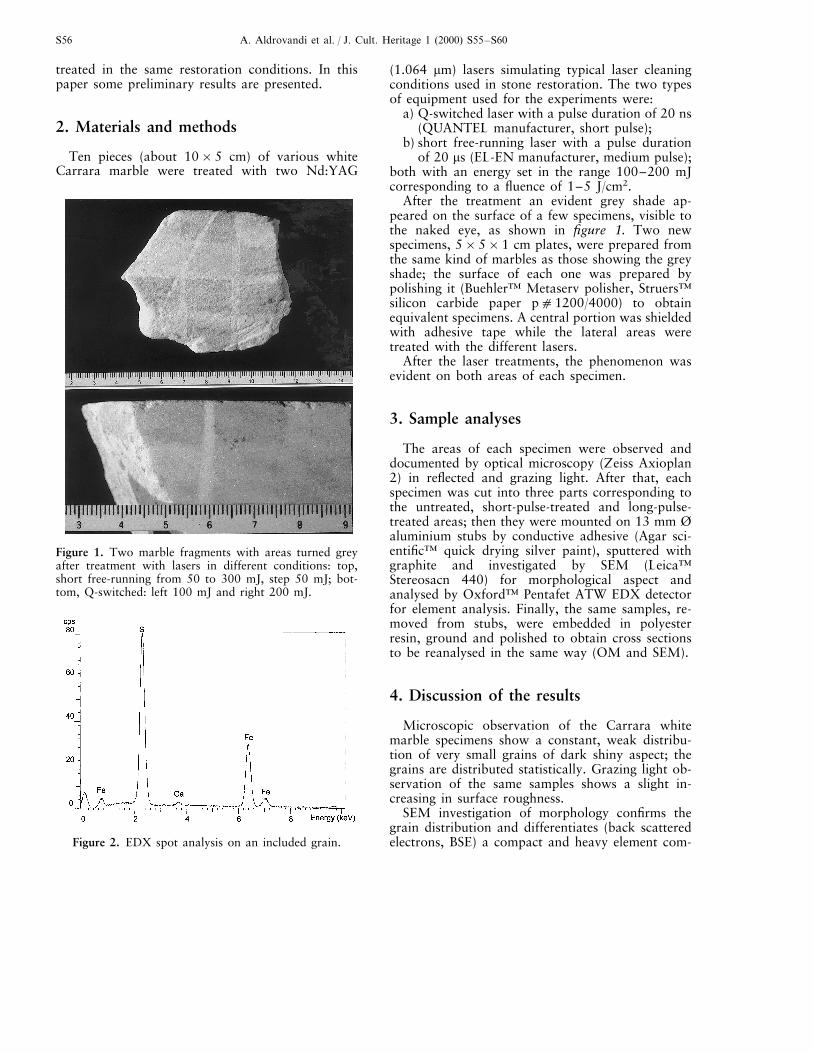
Figure 2. EDX spot analysis on an included grain.

A. Aldrovandi et al. / J. Cult. Heritage 1 (2000) S55–S60 S57
position corresponding to the grains compared tocalcite. EDX analyses on grains revealed the pres-ence of iron and sulphur as principal components(figure 2); therefore, we can confirm these grains tobe pyrite grains (iron sulphide) as previously re-ported [1–3]. After laser cleaning the surface aspectappreciably changes. It is possible to observe twodifferent behaviours: the first one occurs after short-pulse laser treatment and shows a sort of micro-ex-plosion leaving a crater surrounding the pyritegrains and a spread of small black dots all around;the second one, after the medium-pulse laser treat-ment, appears like an extension of the black pyritespot on the surface with a smoother shape. Figure 3reports a comparison among untreated (a), short-pulse-treated (b) and medium-pulse-treated (c) lasersphotographed at two magnifications, ×80 and×160. Both of them clearly contribute to the in-crease in the grey shade of the surfaces.
SEM analyses with a back scattered electron de-tector (BSE) show the presence of very small grains
(dimension B10 mm) with composition differen-tiable from the calcite matrix in the untreated refer-ence specimen. After the treatments, the same grainswith larger dimensions are recognisable (about100 mm), but their morphology differs according tothe pulse duration: the short one seems to leave thesurface more rough, whereas the longer one seemsto melt the black grains and spread them all around.
EDX spot analyses on mentioned zones confirmthe grain composition as the same of pyrite: ironand sulphur peaks are present. EDX element mapsallow a localising of the distribution of sulphur andiron before and after treatments: in both cases alarger area affected by redistribution of these ele-ments is visible. Their general morphology and dis-tribution element maps regarding calcium, sulphurand iron are reported in figure 4.
The SEM BSE images (figure 5, left column) com-pared with secondary electrons (SE) images (figure5, right column) of the same area of surfaces treatedwith different pulse lasers illustrate the morphology
Figure 3. Macrophotographs showing details of the marble specimen surface of untreated (a), treated with short-pulselaser (b) and medium-pulse laser (c). Top, original magnification ×80; bottom, original magnification ×160.

A. Aldrovandi et al. / J. Cult. Heritage 1 (2000) S55–S60S58
Figure 3. Continued
Figure 4. SEM morphology and analysis on mechanical effects: a) secondary electron image; b) back scattered image of thesame area; c) sulphur map; d) calcium map; e) iron map.

A. Aldrovandi et al. / J. Cult. Heritage 1 (2000) S55–S60 S59
Figure 5. Effects of different pulsed lasers on a pyrite grain (SEM micrographs: left BSE, right SE image. a) Mechanicaleffect of a short-pulse laser; b) thermal effect of a medium-pulse laser.
of two distinct behaviours of material: short-pulselaser treatment induces a mechanical effect similarto a micro-explosion of pyrite grains (figure 5,
top), while long pulses probably generate a ther-mal effect like a melting of same particles (figure5, bottom)..

A. Aldrovandi et al. / J. Cult. Heritage 1 (2000) S55–S60S60
5. Conclusions
Both the reported effects increase the local di-mension and distribution of pyrite fragments,larger black spots produce a clearly visible greyshade on the surface.
Unfortunately, the studies on cross sections gaveno significant results because the observed speci-mens did not furnish any information about su-perficial pyrite grains, even with further polishingof the samples: probably the diffusion of this phe-nomenon is so rare, although the optical effect iseasily recognisable, that the chance of dissecting acrater is remote.
According to these preliminary results authorscan conclude that experimentation in this fieldshould be further investigated by the use of com-plementary techniques, such as micro-X-ray dif-fraction, Mossbauer and X-ray photoelectronspectroscopy, etc., in order to better explain thechange induced on the pyrite grains. Also, the useof lasers with different pulse lengths could helpprovide the right interpretation of this phe-nomenon.
In conclusion, grey shading effects only occur ona number of new marbles or on recent cracks onold ones, because these is no form of patina toprotect the surface (and the components) of themarble. Non-patinated white marbles are not so
frequently found in conservation of art objects;nevertheless, it must be observed that possible dis-advantages can occur when laser cleaning is ap-plied in such special cases.
The application of preliminary tests and a care-ful laser cleaning on works of art must take intoaccount these facts which can affect a good con-servative operation.
Acknowledgements. The authors would like tothank the restorer Alberto Casciani for the reportand for the tests with short-pulse laser; RobertoPini and Salvatore Siano of CNR-IEQ for medium-pulse laser tests and equipment; Angelita Mairanifor English revision.
References
[1] Pieri M., Pigmentazione e tonalita cromatiche deimarmi, U. Hoepli, Milan, 1957.[2] Pieri M., I marmi d’Italia, graniti e pietre ornamen-tali, U. Hoepli, Milan, 1964.[3] Manganelli del Fa C., Lazzarini L., Genesi, compo-sizione e alterazione del marmo, in OPD Restauro, nu-mero speciale su Restauro del marmo, Opere e problemi,Opus libri, Florence, 1986.
..