six padas each. Be divides it into four stanzas, the first with six ...
Upload
independentCategory
view
2download
0
Središnja medicinska knjižnica
Bojanić I., Dubravčić K., Batinić D., Golubić Ćepulić B., Mazić S., Hren
D., Nemet D., Labar B. (2011) Large volume leukapheresis: Efficacy
and safety of processing patient's total blood volume six times.
Transfusion and Apheresis Science, 44 (2). pp. 139-47. ISSN 1473-
0502
http://www.elsevier.com/locate/issn/14730502 http://www.sciencedirect.com/science/journal/14730502 http://dx.doi.org/10.1016/j.transci.2011.01.005 http://medlib.mef.hr/981
University of Zagreb Medical School Repository
http://medlib.mef.hr/

Bojanic, 1
Type of contribution: original article Date of preparation: 10th June 2010.
Number of pages: 32
Number of tables: 5
Number of figures: 2
Title: Large volume leukapheresis: Efficacy and safety of processing
patient's total blood volume six times
Names of authors: Ines Bojanica, Klara Dubravcicb, Drago Batinicb, Branka Golubic Cepulica, Sanja Mazica, Darko Hrenc, Damir Nemetd, Boris Labard
a Department of Transfusion Medicine and Cellular Therapy, University Hospital Centre Zagreb, Kispaticeva 12, Croatia b Department of Immunology, University Hospital Centre Zagreb, Kispaticeva 12, Croatia c University of Split, Faculty of Philosophy, Zajceva bb, Split, Croatia d Department of Internal Medicine, University Hospital Centre Zagreb, Kispaticeva 12, Croatia
Ines Bojanic, MD, PhD, Department of Transfusion Medicine and Cellular Therapy,
University Hospital Centre Zagreb, Kispaticeva 12, Croatia; [email protected]
Klara Dubravcic, Msc, Department of Immunology, University Hospital Centre Zagreb,
Kispaticeva 12, Croatia; [email protected]
Drago Batinic, MD, PhD, Department of Immunology, University Hospital Centre
Zagreb, Kispaticeva 12, Croatia, [email protected]
Branka Golubic Cepulic, MD, Department of Transfusion Medicine and Cellular
Therapy, University Hospital Centre Zagreb, Kispaticeva 12, Croatia; bgolubic@kbc-
zagreb.hr
Sanja Mazic, MD, Department of Transfusion Medicine and Cellular Therapy, University
Hospital Centre Zagreb, Kispaticeva 12, Croatia; [email protected]
Darko Hren, PhD, University of Split Faculty of Philosophy, Zajceva bb, Split, Croatia;
Damir Nemet, MD, PhD, Department of Internal Medicine, University Hospital Centre
Zagreb, Kispaticeva 12, Croatia; [email protected]
Boris Labar, MD, PhD, Department of Internal Medicine, University Hospital Centre
Zagreb, Kispaticeva 12, Croatia; [email protected]
Corresponding author: Ines Bojanic, Department of Transfusion Medicine and Cellular Therapy, University Hospital Centre Zagreb, Kispaticeva 12, 10000 Zagreb, Croatia; tel, +385-1-2388708; fax. +385-1-2388699; e-mail: [email protected]

Bojanic, 2
Abstract
Large volume leukapheresis: Efficacy and safety of processing patient's total blood
volume six times
Large-volume leukapheresis (LVL) differs from standard leukapheresis by increased
blood flow and altered anticoagulation regimen. An open issue is to what degree a further
increase in processed blood volume is reasonable in terms of higher yields and safety. In
30 LVL performed in patients with haematologic malignancies, 6 total blood volumes
were processed. LVL resulted in a higher CD34+ cell yield, without change in graft
quality. Although a marked platelet decrease can be expected, LVL is safe and can be
recommended as the standard procedure for patients who mobilize low numbers of
CD34+ cells and when high number of CD34+ cells are required.

Bojanic, 3
1. Introduction
Peripheral blood has become widely accepted source of hematopoietic stem cells
because of easier accessibility and faster engraftment [1]. Since rapid and sustained
engraftment following high-dose therapy depends on the numbers of stem cells reinfused,
efforts are directed towards harvesting sufficient numbers of CD34+ cells. Quality of the
peripheral blood stem cell (PBSC) graft is affected by the effectiveness of mobilization of
stem cells, proper timing of collection, and the efficiency of collection technique [2].
Although sufficient PBSCs may be obtained in a single leukapheresis, the majority of
patients require repeated procedures. An alternative to repeated aphereses could be
processing larger volumes of blood during one procedure. Large-volume leukapheresis
(LVL) involves processing patient's total blood volume (TBV) at least three times during
single procedure, and differs from standard leukapheresis by processing larger blood
volume, increasing blood flow rate, use of additional anticoagulant heparin and longer
duration of procedure [1-5]. Unfortunately, there is no standardized protocol for LVL and
between centers there are differences regarding blood volume processed and duration of
procedure. An open issue is to what degree a further increase in processed blood volume
is reasonable in terms of higher cell yields and safety.
The purpose of this study was to investigate efficiency and safety of processing 6
TBVs. We prospectively analyzed the kinetics of peripheral blood cells and CD34+ cell
yield during LVL, and intraapheresis recruitment of CD34+ cells. Finally, we evaluated
whether the prolonged procedure and processing 6 instead of 4 TBVs altered the quality
of leukapheresis product.

Bojanic, 4
2. Patients and methods
2.1. Patients
Study was performed on group of 30 patients treated at University Hospital
Centre Zagreb (table 1.). All patients were candidates for high-dose chemotherapy
followed by autologous PBSC transplantation. PBSCs were mobilized by combination of
disease specific chemotherapy protocols and 10 µg/kg/day s.c. granulocyte colony-
stimulating factor (G-CSF) filgrastim (Neupogen, Roche, Switzerland). Study was
approved by the local ethics committee, and written informed consent for the PBSC
collection by LVL and additional testing was obtained from all patients.
2.2. Methods
2.2.1. PBSC collection
PBSCs were collected using a Cobe-Spectra cell separator (MNC program,
software version 6.0) (Gambro BCT, Lakewood, CO, USA). Venous access was
established using a dual lumen central venous catheter (Dualise-Cath, Vygon, France),
placed in subclavian (27 patients), jugular (2 patients) or femoral vein (1 patient).
Leukapheresis started when the peripheral blood CD34+ cell count reached
10x106/L. A minimum of 30x109/L platelets was required before leukapheresis. Patients
with preapheresis counts of ≥20x106/L CD34+ cells were considered good mobilizers,
while those with a CD34+ count <20x106/L were considered poor mobilizers. Twelve
patients were poor mobilizers, and 18 were good mobilizers.
Bojanic, 5
The target yield of CD34+ cells was 3.5x10⁶/kg of body weight (BW), except in
patients with multiple myeloma who were scheduled for double transplantation with
target yield of 7x10⁶/kg BW.
During LVL the total volume of processed blood equaled 6 patients' TBVs,
calculated by the instrument, based on weight, height and gender. Inlet flow rate was set
according to instrument calculations. Collection rate was 1.0 ml/min while the collected
fraction was maintained under manual control at a hematocrit of approximately 1%.
A combination of solution citrate dextrose formula A (ACD-A, Baxter, Deerfield,
IL, USA) and heparin (Heparin, Belupo, Croatia) were used as anticoagulants. The
addition of 6 IU of heparin per 1 ml of ACD-A allowed us to enhance the ACD-A to
whole blood ratio to 1:24. Therefore the inlet flow rate was doubled and in the same time
twice the blood volume was processed.
In order to investigate the volume-dependent kinetics of CD34+ cell yield after
each TBV processed, WBC collection set was modified. Collection bag was replaced
with a six collection bag set which were connected to the WBC set collection line by
sterile connection device. Modified set allowed removal of the collected cells every time
one TBV had been processed and therefore product collected during processing each
TBV could be analyzed.
When the optimal interface for MNC collection was established, the first
collection bag was opened; bags were changed after processing 1x, 2x, 3x, 4x, 5x and 6x
the patients' TBV. All aphereses were performed by the same operator and parameters
were held constant throughout the procedure.

Bojanic, 6
Patients were monitored for adverse reactions and recorded reactions were
classified according to National Cancer Institute Common Toxicity Criteria (CTC-NCI)
Classification, Version 2 [4]. In order to prevent symptoms of hypocalcemia all patients
during LVL received 0.5 g calcium chloride/10 kg BW in 100 ml of saline solution.
2.2.2. Laboratory evaluation of peripheral blood and leukapheresis products
Peripheral blood samples taken preapheresis and after processing each TBV, and
samples from each leukapheresis bag were analyzed for WBC, MNC, platelet and CD34+
cell counts. Afterwards, products collected in the first four bags were pooled into one
bag, while products collected in the fifth and sixth bag were pooled into another bag.
Samples were taken from each pooled bag and quality of products was compared with
respect to CD34+ cell count. An intraindividual comparison of the collected CD34+ cells
was done with respect to their differentiation- (CD38, CD90, HLA-DR) and lineage-
associated markers (CD117, CD33, CD41). Numbers of colony-forming units:
granulocyte macrophage (CFU-GM), burst forming units-erythroid (BFU-E) and mixed
units (CFU-MIX) were determined in two pooled bags as well. Finally, products from
both bags were pooled into one final bag and analysed for WBC, MNC, platelet and
CD34+ cell count.
Cell yields were expressed as both the content of cells in a product and the cell
yield per kg of patient's BW. Total yield was defined as the sum of the yields of 6 bags.
Cumulative yield was defined as the sum of the yields collected during processing each
TBV. CD34+ cell collection efficiency [6, 7] and recruitment factor [8-10] for the various
cell populations were calculated as previously described.
Bojanic, 7
Complete blood counts on the peripheral blood samples and leukapheresis
products were obtained on an automated cell counter ADVIA 120 (Bayer, Leverkusen,
Germany). WBC differential counts were performed manually using Wright-Giemsa-
stained specimens. Mononuclear cells (MNC) were defined as the sum of monocytes and
lymphocytes.
CD34+ cells were analysed by flow cytometry using FACSCalibur (BD
Biosciences, Heidelberg, Germany) following standard ISHAGE procedure for cell
staining with anti-CD34-PE (clone 8G12) and anti-CD45-FITC (clone 2D1) monoclonal
antibodies (BD Biosciences) [11]. CD34+ cell subsets were analyzed using monoclonal
antibodies to CD38, HLA-DR, CD90, CD117, CD41 and CD33 (BD Biosciences).
Short-term culture assay for CFU-GM, BFU-E, CFU-MIX) was performed using
MethoCult H4433 medium (StemCell Technologies, Vancouver, Canada) [12]. The
number of colonies was evaluated after 14 days of incubation at 37°C in a humidified
atmosphere with 5% CO2.
Electrolytes calcium, potassium, phosphorus, and magnesium were tested in
peripheral blood samples obtained pre- and postapheresis using Olympus AU 400
analyzer (Olympus Diagnostica, Tokyo, Japan).
2.2.3. Statistical analysis
Data were tested for normality using Kolmogorov Smirnov test. All distributions
were normal, thus means and standard deviations as descriptors and parametric
procedures were used for all analyses.
Repeated measures ANOVA was used to test changes in peripheral blood cells
during LVL. If repeated measures ANOVA resulted in statistically significant F-ratio, we

Bojanic, 8
used paired samples t-tests to test the differences between neighbouring points of
measurement. In these cases we used Bonferonni's correction for multiple comparisons:
to preserve the overall p<0.05 level of statistical significance, p<0.008 was considered
statistically significant when there were 6 comparisons, and p<0.007 when there were 7
comparisons. Furthermore we used within-between subjects ANOVA to test the possible
effects of diagnosis (multiple myeloma vs. lymphoma) or level of CD34+ cell
mobilization (good vs. poor mobilizers) on changes in peripheral blood cells during LVL.
A 2x2 between subjects ANOVA was used to test the effect of diagnosis and level of
CD34+ cell mobilization on recruitment factor. Independent samples t-test was used to
test the differences in CD34+ cell yield between patients with multiple myeloma and
those with lymphoma, as well as between good and poor mobilizers. We used Spearman's
rho coefficient to test the association between CD34+ cell total yield with preapheresis
CD34+ cell count. The influence of processing 6 TBVs as opposed to standard processing
of 4 TBVs on success of collecting necessary amount of CD34+ cells in patients with
lymphoma was tested using McNemar's test. Differences in CD34+ cell subpopulations in
product collected during processing the first 4 TBVs and product collected during
processing the fifth and sixth TBVs were tested using paired samples t-test. Level of
statistical significance was set at 0.05 for all analyses and they were performed using
SPSS 13.0 for Windows (SPSS Inc., Chicago, IL).

Bojanic, 9
3. Results
Thirty aphereses performed in 30 patients were analyzed. A mean volume of
26.97±5.48 l (range 17.83-37.29 l) was processed during 305.8±7.6 min (range 290-324
min) with a mean flow rate of 91.7±18.8 ml/min (60.2-131.9 ml/min). The patients
received a mean volume of 1120.8±230.1 ml ACD-A (range 740-1510 ml) and
6725.2±1380.6 U of heparin (range 4440-9060 U). The mean volume of total apheresis
products was 291.9±13.2 ml (range 255-335ml). After processing 6 TBVs the mean total
CD34+ cell yield was 4.71±3.79 (range 0.91-16.90) x108/kg BW, and was significantly
higher in good mobilizers than in poor mobilizers, (6.54±3.93 x10⁶/kg vs. 1.98±0.79
x106/kg, p<0.001). There were no significant differences in CD34+ yield between the
myeloma and lymphoma patients.
There was a strong correlation between the preapheresis peripheral blood CD34+
cell count and the total CD34+ cells yield (ρ=0.860, p<0.001). In both good and poor
mobilizers, the total CD34+ yield correlated significantly with the preapheresis CD34
count (ρ=0.83 and ρ=0.89, respectively; p<0.0001).
3.1. Kinetics of peripheral blood cells during LVL
The baseline hematological parameters and their changes during apheresis after
processing each of the 6 TBVs are summarized in table 2. and figure 1. LVL significantly
decreased the circulating WBC, MNC, and CD34+ cell counts (p<0.001). Post-hoc
analyses revealed that this effect was due to the significant difference in number of WBC,
MNC and CD34+ cells between the baseline values and values after processing the first
TBV (p<0.001). We found no significant interaction between the change in peripheral
Bojanic, 10
blood CD34+ cell count and diagnosis (p=0.212) or the level of mobilization of CD34+
cells (p=0.62). Platelet count decreased significantly during LVL through each of the
seven points of measurement (p<0,001).
3.2. Kinetics of PBSC collection
PBSCs harvested during processing each TBV volume were collected in a
separate bag, with mean volume of 48.6±5.1 ml. Yields of cells and CD34+ cell
collection efficiency during LVL are summarized in table 3. and figure 2. Statistically
significant change in yield of WBC, MNC, and CD34+ cells during LVL were found
(p<0.001). Post-hoc analyses revealed that this effect was due to the significantly higher
yield of all cells while processing the second TBV compared to the first TBV(p<0.001).
Afterwards the mean yield of WBC, MNC and CD34+ cells remained stable while
processing the remaining 5 TBVs. No significant interaction between the kinetic of
CD34+ cell yield and diagnosis (p=0.380) or level of CD34+ cells mobilization were
found (p=0.886). Although the total amount of harvested CD34+ cells was higher for
good mobilizers, the kinetics of CD34+ cell collection during different stages of LVL
were the same for all patients.
Cummulative yield of CD34+ cells increased continuously during LVL with each
processed TBV (p<0.001), which confirmed that the number of CD34+ cells collected
during apheresis is related to the volume of blood processed.
The average total CD34+ cell collection efficiency during LVL was 70.9±33.2%.
CD34+ cell collection efficiency was lowest during the first TBV processing, but
afterward remained stable. No significant interaction between the change in CD34+ cell

Bojanic, 11
collection efficiency and diagnosis (p=0.309) or the level of CD34+ cell mobilization
were found (p=0.173).
3.3. Comparison of the total CD34+ cell yield collected during processing 4 TBVs vs. 6
TBVs
Processing 6 TBVs versus 4 TBVs resulted in significantly higher CD34+ cell
yields; 4.71±3.79x10⁶/kg vs. 3.28±2.89x10⁶/kg, respectively (p<0.001). If leukapheresis
was stopped after processing 4 TBVs, the target CD34+ cell yield would have been
achieved after only one procedure in 9 (30%) patients. However, when the procedure was
prolonged to processing 6 TBVs, the target dose was successfully achieved in 20 (66.7%)
patients (p=0.001).
The results for the group of 20 myeloma patients were analyzed separately
because their target dose was doubled. With processing 6 TBVs, satisfactory CD34+
counts for tandem transplantation was achieved in 6 (30%) patients, for single
transplantation in 9 (45%) patients, while counts <3.5x10⁶ CD34+/kg were collected in 5
(25%) patients. If leukapheresis was stopped after processing 4 TBVs, only one patient
(5%) would have collected enough CD34+ cells for tandem transplantation and 6 (30%)
for single transplantation
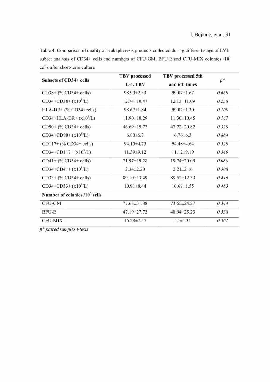
3.4. Quality of products collected during different stages of LVL
The intraindividual comparison of quality of cells collected while processing 4
TBVs and cells collected while processing the fifth and sixth TBVs are presented in table
4. The CD34+ cell subset analysis revealed no significant difference in the composition

Bojanic, 12
of products collected during different stages of LVL. Likewise, the number of CFU-GM,
BFU-E and CFU-MIX collected from processing 4 TBVs was not significantly different
from the number of CFUs collected from processing the fifth and sixth TBVs.
3.5. Recruitment of cells during LVL
Total number of cells in peripheral blood preapheresis and postapheresis, total
number of cells in the leukapheresis products, and recruitment factor for WBC, MNC,
granulocytes, CD34+ cells and platelets are presented in table 5. The recruitment factor
was calculated for each of these cells in order to find out whether cell-specific
recruitment occurred during LVL. The highest recruitment factor (3.2±1.6) was found for
CD34+ cells. Statistically significant difference in CD34+ cell recruitment factor was
found between good (2.8±0.4) and poor mobilizers (4.1±0.5), respectively (p=0.044).
There was no difference in recruitment factors concerning patients' diagnoses (p=0.112).
3.6. Side effects and electrolyte changes during LVL
All LVLs were well tolerated, and none of procedures had to be discontinued.
Apheresis related adverse reactions were restricted to mild symptoms of citrate toxicity.
Only one patient experienced mild perioral paresthesias, classified as grade 1. Although
significant decreases in platelet count were observed after LVL, no bleeding episodes
occurred, and there was no need for transfusion support.
All analysed electrolytes decreased significantly with respect to the basal values:
calcium 5.18±4.10%, (p=0.005), potassium 11.75±7.46% (p<0.001), magnesium
11.98±5.44% (p<0.001), and phosphorus 17.22±16.06% (p=0.024). Although calcium

Bojanic, 13
concentration decreased significantly with respect to basal values, it still remained within
referent values (2.38±0.40 mmol/L vs. 2.24±0.22mmol/L) [13]. Other analysed
electrolytes decreased markedly below referent interval: potassium 3.92±0.35 mmol/L vs.
3.47±0.38 mmol/L, magnesium 0.71±0.08 vs. 0.65±0.09, and phosphorus 0.87±0.28
mmol/L vs. 0.73±0.2 mmol/L [13]. Nevertheless, none of our patients experienced
clinically relevant side effects related to changes in these electrolytes.

Bojanic, 14
4. Discussion
Leukapheresis has been successfully used in PBSC collection for over 20 years,
however several topics still remain unresolved regarding the optimal method of collection
and the volume of processed blood. Processing of larger blood volumes in single LVL
may improve CD34+ cell yield, consequently reducing the number of required
procedures and diminishing the total cost of collections [2, 4]. Another rationale for use
of LVL is the narrow peak of the CD34+ cells in the peripheral blood, present only for a
short period after mobilization, and therefore the optimal time for successful collection
would not likely be missed [14].
Results of previous studies which compared standard vs. LVL processing varied
considerably [2, 4, 10, 14-18]. It is difficult to establish a control group with comparable
patients due to high interindividual variability regarding patients' characteristics, such as
age, gender, TBV, stage of disease, previous treatment, and baseline blood count values.
Even if the same patient is analyzed the following day, peripheral blood CD34+ cell
count could halve or double overnight. The optimal study design would be to compare
products collected during the same procedure at different time, which was done in this
study, and differences caused by interindividual variability were excluded. Studies of
kinetics of PBSC enrichment during LVL showed variable results [3, 8-10, 19-31], which
could be consequence of difference in volumes of processed blood. Therefore, processed
blood volume in our study was standardized and strictly related to the patient's TBV.
Processing 6 TBVs was accomplished by doubling inlet flow rate and additional use of
heparin, along with prolongation of procedure to 5 hours. Duration of LVL could have
been prolonged even more, but was limited to 5 hours because of patients’ comfort and

Bojanic, 15
tolerance, and consistence with working hours of apheresis unit, quality control and cell
processing laboratories. Time-consuming LVL raises an issue of patient’s compliance,
but previous studies showed that cooperation for prolonged apheresis procedure could be
achieved even in pediatric patients [17, 25, 26].
Strong correlation between the preapheresis CD34+ cell count and the total
CD34+ cell yield was found, both in good and poor mobilizers. Accordingly to our
results as well as other reports [5, 22, 27, 29], even in LVL setting, the preapheresis
CD34+ cell count is still the best predictor of the outcome of PBSC collection, although
the volume of blood processed during LVL also affects the total yield.
LVL is feasible only if the level of CD34+ cells in the blood is maintained
throughout the procedure. Although a decline in CD34+ cell level was documented in all
patients, our study showed that even after processing 6 TBVs there was no exhaustion of
the peripheral blood CD34+ cells. Kinetic study showed that decrease in CD34+ count is
related to the number of the TBVs processed. The drop in CD34+ cell count was most
evident at the beginning of leukapheresis, as cells were packed into the apheresis device
set [3, 26]. At the same time, separator draws patient's blood while returns saline which
filled set lines, which can cause hemodilution and contribute to CD34+ cell decrease.
Our results confirm that CD34+ cells were collected at a steady rate throughout
the LVL [9, 19, 26, 30]. The lowest CD34+ cell yield and collection efficiency were
observed at the very beginning of LVL, which can be explained by the time required to
establish interface, and by a dilution effect in the first collection bag, caused by the
automated filling with saline [19, 29, 31]. In the remaining collection period, the CD34+
cell yield and collection efficiency were stable, without any time-dependent changes, as

Bojanic, 16
observed by others [9, 10]. Furthermore, the MNC purity remained constant during LVL,
confirming that enhanced blood volume processing doesn’t affect the product quality in
terms of higher granulocyte contamination during the final stages of apheresis [19]. It is
important to emphasize that in spite of decrease of peripheral blood CD34+ cell count
during LVL, the total cumulative CD34+ cell yield increased steadily after each blood
volume processed. Kinetics of CD34+ cell collection were similar and stable regardless
of diagnosis or level of CD34+ cell mobilization, therefore all patients could have benefit
from LVL [32].
Use of modified sets enabled us to compare how many cells would be collected
after processing standard 4 TBVs vs. 6 TBVs. CD34+ cell yield was higher when
leukapheresis was extended, which could be particularly beneficial in poor mobilizers.
Our results confirmed that likelihood for collecting target number of CD34+ cells was
greater with use of LVL than with standard leukapheresis [4, 28].
Current protocols for myeloma patients require tandem transplantation [33, 34],
and it is common practice to collect sufficient cells for two grafts. That is sometimes
challenging in heavily pretreated patients, particularly those who previously received
lenalidomide [4, 35]. Moreover, some data demonstrated different mobilization of PBSCs
and myeloma cells, with peak levels of myeloma cells occurring few days after CD34+
cells [36-38]. Since LVL allows collection of more CD34+ cells, fewer days are required
to obtain target cell dose, and use of LVL could result in lower tumor contamination of
graft [36-38]. Zubair et al. [33] estimated that using LVL in myeloma patients, the cost
savings could be greater than $7,597 per patient.

Bojanic, 17
Leukapheresis product consists of hematopoietic stem and progenitor cells in
different stages of differentiation [39-41]. Previous studies, [39, 41, 42] as well as ours,
reported that the majority of collected CD34+ cell coexpressed CD38, HLA-DR or CD33
associated with commitment to different hematopoietic lineages. Our study first analysed
CD34+ cell subpopulations in product collected while processing as many as 6 TBVs,
and showed no difference in the composition of products collected during different stage
of LVL. This finding argues against a preferential release of particular CD34+ cell
subsets during the LVL [27]. Our data confirm that LVL results in a higher CD34+ cell
yield without change in graft quality.
Higher CD34+ cell yield harvested by processing a larger volume of blood was
explained with a steady recruitment of PBSCs during leukapheresis [8, 9, 18, 19]. In our
study, threefold more CD34+ cells were collected than were present in blood before
leukapheresis. The recruitment was limited to MNC and CD34+ cells, which were
predominantly removed during the procedure, which was also observed by other authors
[8, 23, 24, 43]. Interestingly, recruitment factor for CD34+ cells in our study was
significantly higher in poor mobilizers than in good mobilizers, which points to the
importance of LVL in patients who mobilize low number of CD34+ cells [17]. The
underlying mechanisms of PBSCs recruitment during LVL are not clear, and few
hypotheses have been proposed. Some authors explained recruitment of PBSCs merely
by the stimulating effect of G-CSF administration [44], while others suggested that it was
mediated by a negative feedback mechanism caused by the decline of the peripheral
blood CD34+ cell count during LVL [8, 10]. Another possible mechanism is modification

Bojanic, 18
of PBSCs homing and transmigration caused by changes in their microenvironment,
induced by use of heparin [15, 45, 46] and citrate-induced hypocalcemia [27, 47].
Results of our study are in favour of LVL but some drawbacks should also be
mentioned. LVL is definitely time-consuming because processing 6 TBVs requires 5
hours. Implementation of LVL has implications on working hours of apheresis
department as well as quality control and cell processing laboratory. Timely completion
of LVL requires high blood flow rates. In some studies it wasn't possible to establish the
required inlet flow rate, and the procedures were shortened because of catheter related
problems [10, 18]. We didn't observe any problem related to high inlet flow rate, but all
our patients had apheresis catheter which we recommend for LVL. LVL may result in an
excess of collected CD34+ cells, therefore it shouldn’t be used in patients who mobilized
high number of CD34+ cells.
A limiting factor for widespread use of LVL may be patients’ tolerance of the
procedure [2, 3]. Larger volumes of infused anticoagulants causes electrolyte imbalance
such as hypocalcemia, metabolic alkalosis, hypokalemia and hypomagnesemia [48]. In
agreement with previous reports [33, 48], we did not observe increase in number of
adverse events during LVL collection. Buchta et al. [49] showed that prophylactic
calcium infusion during LVL reduced the incidence of citrate-related symptoms without
affecting the technical performance or the number of CD34+ cells collected, as was
confirmed by our results. Our study also confirmed marked platelet decrease after
processing each TBV with halved platelet count after LVL [3, 48]. The additional heparin
administration enables the reduction of infused volume of citrate anticoagulant, but might
represent an additional risk factor for bleeding complications in thrombocytopenic
Bojanic, 19
patients with central venous catheters [50]. Heparin-induced thrombocytopenia (HIT) and
associated thrombotic complications could develop in a newly exposed patient, or rapid-
onset complications might occur in a patient with a recent prior history o HIT [51].
Processing up to 6 TBVs according to our results could be performed safely because no
severe adverse reactions occurred, including bleeding complications. Although it lasts
longer, patients will probably tolerate an extra hour of collection easier than another
procedure on consecutive days which would increase the total cost of treatment and
expose them to risks of central venous line complications and additional leukaphereses
[24].
LVL could be strongly recommend for patients who mobilized low number of
CD34+ cells, and patients who need high dose of CD34+ cells, including double
transplantation or in vitro processing of leukapheresis product where a significant loss of
cells is expected. If role of LVL is considered in setting of new mobilizing agents, such
as plerixafor used in case of previously unsuccessful mobilization [52, 53], there is no
doubt that LVL should be performed to collect maximum PBSCs in one procedure. From
the data presented, we conclude that processing of 6 TBVs during LVL is efficient and
safe technique, which significantly reduces the number of aphereses needed to obtain
target number of CD34+ cells. Whether or not further increase of the processed blood
volume could improve the quality of graft must be evaluated in forthcoming studies.

Bojanic, 20
References
[1] Moog R. Management strategies for poor peripheral blood stem cell mobilization.
Transfus Apher Sci 2008; 38:229-236.
[2] Gasová Z, Marinov I, Vodvárková S, Böhmová M, Bhuyian-Ludvíková Z. PBPC
collection techniques: standard versus large volume leukapheresis (LVL) in donors and in
patients. Transfus Apher Sci 2005; 32:167-176.
[3] Rowley S, Yu J, Gooley T, et al. Trafficking of CD34+ cells into the peripheral
circulation during collection of peripheral blood stem cells by apheresis. Bone Marrow
Transplant 2001; 28:649-656.
[4] Abrahamsen J, Stamnesfet S, Liseth K, Hervig T, Bruserud O. Large-volume
leukapheresis yields more viable CD34+ cells and colony-forming units than normal-
volume leukapheresis, especially in patients who mobilize low numbers of CD34+ cells.
Transfusion 2005; 45:248-253.
[5] Monacada V, Bolan C, Yau Y, Leitman S. Analysis of PBPC cell yields during
large-volume leukapheresis of subjects with a poor mobilization response to filgrastim.
Transfusion 2003; 43:495-501.
[6] Rowley S, Prather K, Bui K. Collection of peripheral blood progenitor cells with
an automated leukapheresis system. Transfusion 1999; 39:1200-1206.
[7] Schreiner T, Wiesneth M, Krug E, et al. Collection of allogeneic peripheral blood
progenitor cells by two protocols on an apheresis system. Transfusion 1998; 38:1051-
1055.
Bojanic, 21
[8] Knudsen L, Nikolaisen K, Gaarsdal E, Johnsen H. Kinetic studies during
peripheral blood stem cell collection show CD34+ cell recruitment intra-apheresis. J Clin
Apheresis 2001; 16:114-119.
[9] Cull G, Ivey J, Chase P, Picciuto R, Herrmann R, Cannell P. Collection and
recruitment of CD34+ cells during large-volume leukapheresis. J Hematother 1997;
6:309-314.
[10] Moller A, Dickmeiss E, Geisler C, Christensen L. Recruitment of CD34+ cells
during large-volume leukapheresis. J Hematoth Stem Cell 2001; 10:837-853.
[11] Keeney M, Chin-Yee I, Weir K, Popma J, Nayar R, Sutherland D. Single platform
flow cytometric absolute CD34+ cell counts based on the ISHAGE gudielines.
International Society of Hematotherapy and Graft Engineering. Cytometry 1998; 34:61-
70.
[12] Smalcelj R, Kusec V, Thune S, Petrovecki M. Circulating hematopoietic
progenitors are not altered in patients with post-transplant erythrocytosis. Haematologica
1998; 83:948-949.
[13] Painter P, Cope J, Smith J, Reference information for the clinical laboratory. In:
Burtis C, Ashwood E, Bruns D, editors. Tietz textbook of clinical chemistry and
molecular diagnostics. St. Louis: Elsevier Saunders, 2006: 2251-2318.
[14] Abe T, Makimoto A, Kawano Y, et al. Intra-apheresis recruitment of blood
progenitor cells in children. Transfusion 1998; 38:944-950.
[15] Humpe A, Riggert J, Munzel U, et al. A prospective, randomized, sequential,
crossover trial of large-volume versus normal-volume leukapheresis procedures: effect on
progenitor cells and engraftment. Transfusion 1999; 39:1120-1127.

Bojanic, 22
[16] Smolowicz A, Villman K, Tidefelt U. Large-volume apheresis for the harvest of
peripheral blood progenitor cells for autologous transplantation. Transfusion 1997;
37:188-192.
[17] Torrabadella M, Olivé T, Ortega JJ, Massuet L. Enhanced HPC recruitment in
children using LVL and a new automated apheresis system. Transfusion 2000; 40:404-
410.
[18] Passos-Coelho J, Machado M, Lucio P, Leal-Da-Costa F, Silva M, Parraiera A.
Large-volume leukaphereses may be more efficient than standard-volume leukaphereses
for collection of peripheral blood progenitor cells. J Hematother 1997; 6:465-474.
[19] Cassens U, Ostkamp-Ostermann P, van der Werf N, Garritsen H, Ostermann H,
Sibrowski W. Volume-dependent collection of peripheral blood progenitor cells during
large-volume leukapheresis for patients with solid tumours and haematological
malignancies. Transfus Med 1999; 9:311-320.
[20] Hillyer C, Lackey Dr, Hart K, et al. CD34+ progenitors and colony-forming units-
granulocyte macrophage are recruited during large-volume leukapheresis and
concentrated by counterflow centrifugal elutriation. Transfusion 1993; 33:316-321.
[21] Hillyer C, Tiegerman K, Berkman E. Increase in circulating colony-forming units-
granulocyte-macrophage during large-volume leukapheresis: evaluation of a new cell
separator. Transfusion 1991; 31:327-332.
[22] Passos-Coelho J, Braine H, Davis J, Huelskamp A. Predictive factors for
peripheral- blood -progenitor-cell collections using a single large-volume leukapheresis
after cyclophosphamide and granulocyte-macrophage colony stimulating factor
mobilization. J Clin Oncol 1995; 13:705-714.

Bojanic, 23
[23] Smolowicz A, Villman K, Berlin G, Tidefelt U. Kinetics of peripheral blood stem
cell harvests during a single apheresis. Transfusion 1999; 39:403-409.
[24] Bojko P, Stellberg W, Küdde C, et al. Kinetic study of CD34+ cells during
peripheral blood stem cell collections. J Clin Apher 1999; 14:18-25.
[25] Gorlin J, Vamvakas E, Cooke E, et al. Large-volume leukapheresis in pediatric
patients: processing more blood diminishes the apparent magnitude of intra-apheresis
recruitment. Transfusion 1996; 36:879-885.
[26] Yamaguchi E, Yamato K, Miyata Y. Kinetics of peripheral blood stem cell
collection in large-volume leukapheresis for pediatric patients undergoing chemotherapy
and adult patients before chemotherapy. J Hematother Stem Cell Research 2000; 9:565-
572.
[27] Murea S, Goldschmidt H, Hahn U, Pförsich M, Moos M, Haas R. Successful
collection and transplantation of peripheral blood stem cells in cancer patients using
large-volume leukaphereses. J Clin Apheresis 1996; 11:185-194.
[28] Bojko P, Scharifi M, Stössel K, Seeber S. Comparison of processing four and five
times the patients' blood volume during peripheral blood stem cell collection and analysis
of CD34+38-and CD34+49d+ subsets during apheresis. J Cancer Res Clin Oncol 2002;
128:19-28.
[29] Fabritiis P, Gonzalez M, Meloni G, et al. Monitoring of CD34+ cells during
leukapheresis allows a single, successful collection of hemopoietic progenitors in patients
with low numbers of circulating stem cells. Bone Marrow Transplant 1999; 23:1229-
1236.

Bojanic, 24
[30] Humpe A, Riggert J, Meineke I, et al. A cell-kinetic model of CD34+ cell
mobilization and harvest: development of a predictive algorithm for CD34+ cell yield in
PBPC collections. Transfusion 2000; 40:1363-1370.
[31] Cassens U, Momkvist P, Zuehlsdorf M, et al. Kinetics of standardized large
volume leukapheresis in patients do not show a recruitment of peripheral blood
progenitor cells. Bone Marrow Transplant 2001; 28:13-20.
[32] Majado M, Minguela A, Gonzalez-Garcia C, et al. Large-volume-apheresis
facilitates autologous transplantation of hematopoietic progenitors in poor mobilizer
patients. J Clin Apheresis 2009; 24:12-17.
[33] Zubair A, Rymer R, Young J, et al. Multiple myeloma patients receiving large
volume leukapheresis efficiently yield enough CD34+ cells to allow double transplants. J
Clin Apheresis 2009; 24:6-11.
[34] Barlogie B, Attal M, Crowley J, et al. Long-term follow-up of autotransplantation
trials for multiple myeloma: update of protocols conducted by the Intergroupe
Francophone du Myelome, Southwest Oncology Group, and University of Arkansas for
Medical Sciences. J Clin Oncol 2010; 28:1209-1214.
[35] Kumar A, Dispenzieri A, Lacy M, et al. Impact of lenalidomide therapy on stem
cell mobilization and engraftment post-peripheral blood stem cell transplantation in
patients with newly diagnosed myeloma. Leukemia 2007; 21:2035-2042.
[36] Gazitt Y, Tian E, Barlogie B, et al. Different mobilization of myeloma cells and
normal hematopoietic stem cells in multiple myeloma after treatment with
cyclophosphamide and granulocyte-macrophage colony-stimulating factor. Blood 1996;
87:805-811.

Bojanic, 25
[37] Desikan K, Jagannath S, Siegel D, et al. Collection of more hematopoietic
progenitor cells with large volume leukapheresis in patients with multiple myeloma.
Leukemia Lymphoma 1997; 28:501-508.
[38] Gazitt Y, Reading C, Hoffman R, et al. Purified CD34+Lin-Thy+ stem cells do
not contain clonal myeloma cells. Blood 1995; 86:381-389.
[39] Stewart A, Imrie K, Keating A, Anania S, Nayar R, Sutherland D. Optimizing the
CD34+ and CD34+Thy-1+ stem cell content of peripheral blood collections. Exp
Hematol 1995; 23:1619-1627.
[40] Haas R, Mohle R, Murea S, et al. Characterization of peripheral blood progenitor
cells mobilized by cytotoxic chemotherapy and recombinant human granulocyte colony-
stimulating factor. J Hematother 1994; 3:323-330.
[41] Humpe A, Riggert J, Koch S, Legler T, Munzel U, Kohler M. Prospective,
randomized, sequential, crossover trial of large-volume vs. normal volume leukapheresis
procedures: effects on subpopulations of CD34+ cells. J Clin Apheresis 2001; 16:109-
113.
[42] Haas R, Mohle R, Pforsich M, et al. Blood-derived autografts collected during
granulocyte-colony-stimulating factor-enhanced recovery are enriched with early Thy-
1+hematopoietic progenitor cells. Blood 1995; 85:1936-1943.
[43] Benjamin R, Linsley L, Fountain D, et al. Preapheresis peripheral blood CD34+
mononuclear cell counts as predictors of progenitor cell yield. Transfusion 1997; 37:79-
85.

Bojanic, 26
[44] Cassens U, Barth I, Baumann C, et al. Factors affecting the efficacy of peripheral
blood progenitor cells collections by large-volume leukaphereses with standardized
processing volumes. Transfusion 2004; 44:1593-1602.
[45] Watt S, Williamson J, Genevier H, et al. The heparin binding PECAM-1 adhesion
molecule is expressed by CD34+ hematopoietic precursor cells with early myeloid and B-
lymphoid cell phenotypes. Blood 1993; 82:2649-2663.
[46] Yong K, Watts M, Shaun Thomas N, Sullivan A, Ings S, Linch D. Transmigration
of CD34+ cells across specialized and nonspecialized endothelium requires prior
activation by growth factors and is mediated by PECM-1 (CD31). Blood 1998; 91:1196-
1205.
[47] Mohle R, Haas R, Hunstein W. Expression of adhesion molecules and c-kit on
CD34+ hematopoietic progenitor cells: Comparison of cytokine mobilized blood stem
cells with normal bone marrow and peripheral blood. J Hematother 1993; 2:483-489.
[48] Humpe A, Riggert J, Munzel U, Köhler M. A prospective, randomized, sequential
crossover trial of large-volume versus normal-volume leukapheresis procedures: effects
on serum electrolytes, platelet counts, and other coagulation measures. Transfusion 2000;
40:368-374.
[49] Buchta C, Macher M, Bieglmayer C, Hocker P, Dettke M. Reduction of adverse
reactions during autologous large-volume PBSC apheresis by continuous infusion of
calcium-gluconate. Transfusion 2003; 43:1615-1621.
[50] Malachowski M, Comenzo R, Hillyer C, Tiegerman K, Berkman E. Large-volume
leukapheresis for peripheral blood stem cell collection in patients with hematologic
malignancies. Transfusion 1992; 32:732–735.

Bojanic, 27
[51] Linenberger M, Collection of cellular therapy products by apheresis. In: Areman
E, Loper K, editors. Cellular Therapy: Principles, methods, and regulations. Bethesda:
AABB, 2009: 251-264.
[52] Choi H, Yong C, Yoo B. Plerixafor for stem cell mobilization in patients with
noon-Hodgkin's lymphoma and multiple myeloma. Ann Pharmacother 2010; 44:117-126.
[53] Fruehauf S, Veldwijk M, Seeger T, et al. A combination of granulocyte-colony-
stimulating factor (G-CSF) and plerixafor mobilizes more primitive peripheral blood
progenitor cells than G-CSF alone: results of a European phase II study. Cytotherapy
2009; 11:992-1101.

Bojanic, 28
Tables
Table 1. Patients' characteristics
Gender (male / female) 16 / 14
Age (years)* 50.1±10.9 (19-61)
Body weight (kg)* 78.3±15.4 (57–108)
Total blood volume (ml)* 4618.6±140.5 (3463–6761)
Diagnosis (N)
Multiple myeloma 20
Non-Hodgkin's lymphoma 8
Hodgkin's disease 2
Mobilization (N)
cyclophosphamide (4 g/m2) 20
HDIM 7
ICE 1
mini BEAM 1
DHAP 1
*mean±SD (range); HDIM=high-dose ifosfamide, mitoxantrone; ICE=ifosfamide,
carboplatin, etoposide; mini BEAM=carmustine, etoposide, cytarabine, melphalan;
DHAP=dexamethasone, cytarabine, cisplatin

Bojanic, 29
Table 2. Kinetics of WBC, MNC, CD34+ cells and platelets in the peripheral blood during LVL
(mean ± SD). Levels of statistical significance were calculated comparing the results after each
processed total blood volume (TBV) with the following
Peripheral blood cells
Processed
TBV
WBC x
109/L
WBC
decrease
from
baseline %
MNC
x109/L
MNC
decrease
from
baseline %
CD34+
x106/L
CD34+
cell
decrease
from
baseline
%
Platelet
x 109/L
Platelets
decrease
from
baseline
%
Baseline 14.39
±7.68 100
2.02±
0.81 100
48.35
±44.80 100
99.75
±40.17 100
p* <0.001 <0.001 <0.001 <0.001 <0.001 <0.001 <0.001 <0.001
1x 12.10
±6.91
83.20
± 10.74
1.38
±0.57
69.27
± 12.69
27.86
±23.19
63.46
±20.38
79.06
±33.55
79.26
±9.06
p* NS NS p=0.004 NS NS NS <0.001 <0.001
2x 11.83
±6.69
82.48
± 12.64
1.26
±0.51
64.23
± 14.71
26.71
±25.29
58.17
±18.01
71.75
±32.69
72.24
±12.73
p* NS NS 0.005 0.007 NS NS <0.001 <0.001
3x 11.64
±6.57
80.07
± 10.20
1.15
±0.41
58.58
± 10.37
23.42
±23.00
51.20
±17.02
63.41
±25.46
64.55
±11.23
p* NS NS 0.006 0.003 NS NS <0.001 <0.001
4x 11.41
±6.59
78.01
± 12.39
1.05
±0.38
53.98
± 11.91
21.02
±19.73
47.72
±21.27
58.75
±25.41
59.88
±13.61
p* NS NS NS NS NS NS <0.001 <0.001
5x 11.16
±6.47
76.67
± 14.50
1.01
±0.35
52.54
± 13.01
19.63
±16.13
46.47
±19.08
55.20
±25.14
56.65
±16.21
p* NS NS NS NS NS NS <0.001 <0.001
6x 11.09
±6.53
76.01
± 14.56
12.14
±8.46
50.35
± 16.21
17.61
±14.05
45.95
±18.64
49.44
±21.46
51.65
±15.89
F6,156 19.168 46.828 42.029 138.430 15.368 61.054 62.312 130.902
p** <0.001 <0.001 <0.001 <0.001 <0.001 <0.001 <0.001 <0.001
p* paired samples t-tests; p** repeated measures ANOVA

Bojanic, 30
Table 3. Yields of WBC, MNC and CD34+ cells collected in separate bags and CD34+ cell
collection efficiencies (CE) during each total blood volume processed (mean±SD). Levels of
statistical significance were calculated comparing the results after each processed total blood
volume (TBV) with the following
Yield
Processed
TBV
WBC
x109/L
WBC
x108/kg
MNC
x109/L
MNC
x108/kg
CD34+
x106/L
CD34+
x106/kg
CD34+ cell
CE (%)
1x 149.7
±61.4
0.87
±0.43
80.3
±39.5
0.45
±0.23
12.6
±10.7
0.71
±0.61
40.6
±18.7
p* NS p<0.001 p<0.001 p<0.001 NS p=0.004 p<0.001
2x 162.1
±57.8
1.02
±0.41
88.4
±36.6
0.56
±0.22
14.3
±14.2
0.87
±0.73
68.6
±25.3
p* NS NS NS NS NS NS NS
3x 150.9
±68.3
0.97
±0.36
82.2
±33.5
0.53
±0.19
14.5
±16.2
0.87
±0.84
74.4
±32.6
p* NS NS NS NS NS NS NS
4x 152.6
±48.6
0.96
±0.32
80.5
±29.8
0.50
±0.19
13.4
±14.1
0.82
±0.82
73.8
±29.2
p* NS NS NS NS NS NS NS
5x 156.7
±46.3
0.98
±0.28
81.6
±32.1
0.48
±0.18
13.6
±10.1
0.80
±0.60
74.4
±24.2
p* NS NS NS NS NS NS NS
6 159.1
±46.0
1.01
±0.34
79.6
±27.5
0.49
±0.19
12.9
±9.1
0.82
±0.51
79.5
±28.3
F5,125 1.489 3.187 3.979 4.808 0.242 3.082 9.927
p** 0.198 0.041 0.002 <0.001 0.943 0.012 <0.001
Total
product
149.7
±61.4
5.71
±1.97
80.3
±39.5
3.32
±3.86
126.2
±107.2
4.71
±3.79
70.9
±33.2
p* paired samples t-test; p** repeated measures ANOVA
I. Bojanic, et al. 31
Table 4. Comparison of quality of leukapheresis products collected during different stage of LVL:
subset analysis of CD34+ cells and numbers of CFU-GM, BFU-E and CFU-MIX colonies /105
cells after short-term culture
Subsets of CD34+ cells TBV processed
1.-4. TBV
TBV processed 5th
and 6th times p*
CD38+ (% CD34+ cells) 98.90±2.33 99.07±1.67 0.669
CD34+CD38+ (x108/L) 12.74±10.47 12.13±11.09 0.238
HLA-DR+ (% CD34+cells) 98.67±1.84 99.02±1.30 0.100
CD34+HLA-DR+ (x108/L) 11.90±10.29 11.30±10.45 0.147
CD90+ (% CD34+ cells) 46.69±19.77 47.72±20.82 0.320
CD34+CD90+ (x108/L) 6.80±6.7 6.76±6.3 0.884
CD117+ (% CD34+ cells) 94.15±4.75 94.48±4.64 0.529
CD34+CD117+ (x108/L) 11.39±9.12 11.12±9.19 0.349
CD41+ (% CD34+ cells) 21.97±19.28 19.74±20.09 0.080
CD34+CD41+ (x108/L) 2.34±2.20 2.21±2.16 0.508
CD33+ (% CD34+ cells) 89.10±13.49 89.52±12.33 0.416
CD34+CD33+ (x108/L) 10.91±8.44 10.68±8.55 0.483
Number of colonies /105 cells
CFU-GM 77.63±31.88 73.65±24.27 0.344
BFU-E 47.19±27.72 48.94±25.23 0.558
CFU-MIX 16.28±7.57 15±5.31 0.301
p* paired samples t-tests

I. Bojanic, et al. 32
Table 5. Preapheresis and postapheresis total number of cells in peripheral blood, total number of
collected cells in the leukapheresis products and recruitment factor
Parameters Preapheresis Postapheresis Total number of
harvested cells
Recruitment
factor*
WBC (x108) 676.70±416.73 527.36±368.78 439.69±156.81 1.64±0.54
MNC (x108) 96.32±45.07 46.56±20.49 205.47±103.26 2.72±0.69
Granulocyte
(x108) 580.37±385.53 480.79±355.94 234.21±137.95 1.42±0.51
CD34+ cells
(x106) 226.80±204.88 83.37±68.77 540.12±562.95 3.23±1.61
Platelets
(x109) 469.47±210.93 227.70±87.77 313.72±183.12 1.17±0.20
*Recruitment factor = absolute number of cells in blood postapheresis + absolute number of
cells collected/ absolute number of cells in blood preapheresis

I. Bojanic, et al. 33
Figures
Figure 1. Decrease of WBC, MNC, CD34+ cells and platelets during LVL expressed as a mean
percentage from baseline values
0
10
20
30
40
50
60
70
80
90
100
baseline 1x 2x 3x 4x 5x 6x
Total blood volume processed
Percent of baseline values (%)
platelets
WBC
MNC
CD34+ cells

I. Bojanic, et al. 34
Figure 2. Yields of WBC, MNC and CD34+ cells / kg BW collected after each blood volume
processed during large volume leukapheresis expressed as a mean
0.00
0.20
0.40
0.60
0.80
1.00
1.20
1x 2x 3x 4x 5x 6x
Total blood volume processed
Yield
CD34+ cells x10E6/kg BW
MNC x10E8/kg BW
WBC x10E8/kg BW
Copyright © 2022 FDOKUMEN