L-leucine, beta-hydroxy-beta-methylbutyric acid (HMB) and creatine monohydrate prevent...
9
RESEARCH ARTICLE Open Access L-leucine, beta-hydroxy-beta-methylbutyric acid (HMB) and creatine monohydrate prevent myostatin-induced Akirin-1/Mighty mRNA down-regulation and myotube atrophy Christopher Brooks Mobley 1 , Carlton D Fox 1 , Brian S Ferguson 1 , Rajesh H Amin 3 , Vincent J Dalbo 4 , Shawn Baier 5 , John A Rathmacher 5 , Jacob M Wilson 2 and Michael D Roberts 1* Abstract Background: The purpose of this study was to examine if L-leucine (Leu), β-hydroxy-β-methylbutyrate (HMB), or creatine monohydrate (Crea) prevented potential atrophic effects of myostatin (MSTN) on differentiated C2C12 myotubes. Methods: After four days of differentiation, myotubes were treated with MSTN (10 ng/ml) for two additional days and four treatment groups were studied: 1) 3x per day 10 mM Leu, 2) 3x per day 10 mM HMB, 3) 3x per day 10 mM Crea, 4) DM only. Myotubes treated with DM without MSTN were analyzed as the control condition (DM/CTL). Following treatment, cells were analyzed for total protein, DNA content, RNA content, muscle protein synthesis (MPS, SUnSET method), and fiber diameter. Separate batch treatments were analyzed for mRNA expression patterns of myostatin-related genes (Akirin-1/Mighty, Notch-1, Ski, MyoD) as well as atrogenes (MuRF-1, and MAFbx/Atrogin-1). Results: MSTN decreased fiber diameter approximately 30% compared to DM/CTL myotubes (p < 0.001). Leu, HMB and Crea prevented MSTN-induced atrophy. MSTN did not decrease MPS levels compared to DM/CTL myotubes, but MSTN treatment decreased the mRNA expression of Akirin-1/Mighty by 27% (p < 0.001) and MyoD by 26% (p < 0.01) compared to DM/CTL myotubes. shRNA experiments confirmed that Mighty mRNA knockdown reduced myotube size, linking MSTN treatment to atrophy independent of MPS. Remarkably, MSTN + Leu and MSTN + HMB myotubes had similar Akirin-1/Mighty and MyoD mRNA levels compared to DM/CTL myotubes. Furthermore, MSTN + Crea myotubes exhibited a 36% (p < 0.05) and 86% (p < 0.001) increase in Akirin-1/Mighty mRNA compared to DM/CTL and MSTN-only treated myotubes, respectively. Conclusions: Leu, HMB and Crea may reduce MSTN-induced muscle fiber atrophy by influencing Akirin-1/Mighty mRNA expression patterns. Future studies are needed to examine if Leu, HMB and Crea independently or synergistically affect Akirin-1/Mighty expression, and how Akirin-1/Mighty expression mechanistically relates to skeletal muscle hypertrophy in vivo. Keywords: Myostatin, GDF8, Akirin-1, Atrophy, Skeletal muscle * Correspondence: [email protected] 1 School of Kinesiology, Molecular and Applied Sciences Laboratory, Auburn University, 301 Wire Road, Office 286, Auburn, AL 36849, USA Full list of author information is available at the end of the article © 2014 Mobley et al.; licensee BioMed Central Ltd. This is an Open Access article distributed under the terms of the Creative Commons Attribution License (http://creativecommons.org/licenses/by/4.0), which permits unrestricted use, distribution, and reproduction in any medium, provided the original work is properly credited. The Creative Commons Public Domain Dedication waiver (http://creativecommons.org/publicdomain/zero/1.0/) applies to the data made available in this article, unless otherwise stated. Mobley et al. Journal of the International Society of Sports Nutrition 2014, 11:38 http://www.jissn.com/content/11/1/38
Transcript of L-leucine, beta-hydroxy-beta-methylbutyric acid (HMB) and creatine monohydrate prevent...

Mobley et al. Journal of the International Society of Sports Nutrition 2014, 11:38http://www.jissn.com/content/11/1/38
RESEARCH ARTICLE Open Access
L-leucine, beta-hydroxy-beta-methylbutyric acid(HMB) and creatine monohydrate preventmyostatin-induced Akirin-1/Mighty mRNAdown-regulation and myotube atrophyChristopher Brooks Mobley1, Carlton D Fox1, Brian S Ferguson1, Rajesh H Amin3, Vincent J Dalbo4, Shawn Baier5,John A Rathmacher5, Jacob M Wilson2 and Michael D Roberts1*
Abstract
Background: The purpose of this study was to examine if L-leucine (Leu), β-hydroxy-β-methylbutyrate (HMB), or creatinemonohydrate (Crea) prevented potential atrophic effects of myostatin (MSTN) on differentiated C2C12 myotubes.
Methods: After four days of differentiation, myotubes were treated with MSTN (10 ng/ml) for two additional days andfour treatment groups were studied: 1) 3x per day 10 mM Leu, 2) 3x per day 10 mM HMB, 3) 3x per day 10 mM Crea,4) DM only. Myotubes treated with DM without MSTN were analyzed as the control condition (DM/CTL). Followingtreatment, cells were analyzed for total protein, DNA content, RNA content, muscle protein synthesis (MPS, SUnSETmethod), and fiber diameter. Separate batch treatments were analyzed for mRNA expression patterns of myostatin-relatedgenes (Akirin-1/Mighty, Notch-1, Ski, MyoD) as well as atrogenes (MuRF-1, and MAFbx/Atrogin-1).
Results: MSTN decreased fiber diameter approximately 30% compared to DM/CTL myotubes (p < 0.001). Leu, HMB andCrea prevented MSTN-induced atrophy. MSTN did not decrease MPS levels compared to DM/CTL myotubes, but MSTNtreatment decreased the mRNA expression of Akirin-1/Mighty by 27% (p < 0.001) and MyoD by 26% (p < 0.01) comparedto DM/CTL myotubes. shRNA experiments confirmed that Mighty mRNA knockdown reduced myotube size, linkingMSTN treatment to atrophy independent of MPS. Remarkably, MSTN + Leu and MSTN+ HMB myotubes had similarAkirin-1/Mighty and MyoD mRNA levels compared to DM/CTL myotubes. Furthermore, MSTN + Crea myotubes exhibiteda 36% (p < 0.05) and 86% (p < 0.001) increase in Akirin-1/Mighty mRNA compared to DM/CTL and MSTN-only treatedmyotubes, respectively.
Conclusions: Leu, HMB and Crea may reduce MSTN-induced muscle fiber atrophy by influencing Akirin-1/Mighty mRNAexpression patterns. Future studies are needed to examine if Leu, HMB and Crea independently or synergistically affectAkirin-1/Mighty expression, and how Akirin-1/Mighty expression mechanistically relates to skeletal muscle hypertrophyin vivo.
Keywords: Myostatin, GDF8, Akirin-1, Atrophy, Skeletal muscle
* Correspondence: [email protected] of Kinesiology, Molecular and Applied Sciences Laboratory, AuburnUniversity, 301 Wire Road, Office 286, Auburn, AL 36849, USAFull list of author information is available at the end of the article
© 2014 Mobley et al.; licensee BioMed Central Ltd. This is an Open Access article distributed under the terms of the CreativeCommons Attribution License (http://creativecommons.org/licenses/by/4.0), which permits unrestricted use, distribution, andreproduction in any medium, provided the original work is properly credited. The Creative Commons Public DomainDedication waiver (http://creativecommons.org/publicdomain/zero/1.0/) applies to the data made available in this article,unless otherwise stated.
Mobley et al. Journal of the International Society of Sports Nutrition 2014, 11:38 Page 2 of 9http://www.jissn.com/content/11/1/38
BackgroundMyostatin (MSTN) is a key negative regulator of matureskeletal muscle myofiber growth [1-3]. In this regard,MSTN has been shown to reduce muscle protein synthesisby abrogating mTORC1 signaling [4,5] and increase muscleproteolytic mechanisms [6,7]. Furthermore, several studieshave implicated physical inactivity-induced up-regulation inMSTN as a potential regulator in age-related skeletalmuscle loss [8-13], and there is supporting evidence sug-gesting that serum and skeletal muscle MSTN are elevatedwith aging [14,15].The effects of exercise have also been implicated in
MSTN pathway signaling. Specifically, Louis et al. [16] re-ported that MSTN mRNA expression is depressed withinhours following either endurance or resistance exercise.Dalbo et al. [17] similarly reported that resistance exercisedecreased skeletal muscle myostatin mRNA levels upto 6 hours post-exercise. Likewise, recent evidencesuggests that skeletal muscle MSTN increases threedays following detraining from 90 days of resistanceexercise; an event which preceded subtle but rapid type IIfiber atrophy [18].While exercise reduces skeletal muscle myostatin
expression, nutritional strategies to reduce myostatinsignaling are also warranted. In this regard, select nutri-ents have been shown to increase skeletal muscle anabolicsignaling mechanisms. Leucine is a well-known activatorof mTOR signaling [19-21], and leucine has additionallybeen shown to reduce muscle proteolysis [22]. The leucinemetabolite beta-hydroxy- beta-methylbutyric acid (HMB)has also been well-described with regard to its effects onwhole-body muscle mass accretion [23-25], as well asits ability to independently activate skeletal musclemTOR signaling and reduce proteolytic signaling [26-28].Creatine monohydrate has been less studied with regardsto mTOR pathway modulation, although some evi-dence exists suggesting that creatine monohydrate isable to increase myotube differentiation throughpoorly understood mechanisms [29]. Notwithstanding,ample literature has demonstrated that creatine monohy-drate supplementation is able to increase muscle massand strength [30-35].While a plethora of literature reports the effects of
these nutritional supplements on skeletal muscle ana-bolic and/or anti-catabolic mechanisms, no informationto our knowledge is known regarding how or if thesesupplements can abrogate facets of MSTN signaling.Therefore, the purpose of this study was to determinewhether differentiated/mature myotubes treated withleucine, HMB or creatine monohydrate in the presence ofMSTN affected: a) myotube diameter, b) select anabolicindices (Protein: DNA, RNA: DNA), and c) the mRNAexpression patterns of genes associated with myostatinsignaling.
MethodsCell culture methodsC2C12 myoblasts (graciously donated by RHA), pas-sage no. 4–10, were maintained in growth medium(GM; DMEM, 10% FBS, 1% penicillin/streptomycin,0.1% gentamycin) under standard culture conditionsat 37°C in a 5% CO2 atmosphere. Myoblasts were grownon 145 mm plates (Griener Bio-One GmbH, Maybachstr,Frickenhausen, GER) at a density of 7.5 × 10⁵ in 10 ml ofgrowth medium for protein analyses, or on 12-well plates(Griener Bio-One GmbH) at a density of 5 × 10⁵ formRNA analyses. Differentiation was induced 48 hafter myoblast growth reached 80%–90% confluencyby removing the growth medium and replacing it withdifferentiation medium (DM; DMEM, 2% (vol/vol)horse serum, 1% penicillin/streptomycin). DM was thenreplaced every 24 h for 4 d.
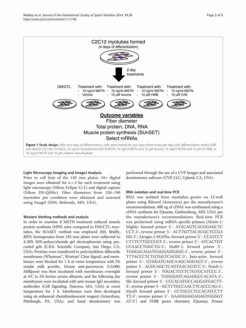
Treatment methods (DM only, MSTN only, MSTN +Leucine, MSTN + HMB, MSTN + Creatine)The study design is illustrated in Figure 1 below. Briefly,after four days of differentiation, cells were treated threetimes per day with one of the following treatments for48 h: 1) DM and vehicle (10 mM Tris-NaCl); denoted as‘DM/CTL’ only, 2) 10 ng/ml rGDF-8 MSTN (R&D Systems,Minneapolis, MN, USA); denoted as ‘MSTN’, 3) 10 ng/mlMSTN and 10 μM (13 ug/ml) leucine (EMD Chemicals,Inc., San Diego, CA, USA); denoted as MSTN+Leu, 4)10 ng/ml MSTN and 10 μM (13 ug/ml) of free acid HMB(Metabolic Technologies, Inc., Ames, IA, USA);denoted as ‘MSTN+HMB’, or 5) 10 ng/ml MSTN and10 μM (12 ug/ml) creatine monohydrate (BodyBuilding.com, Boise, ID, USA); denoted as ‘MSTN+Crea’. TheMSTN treatment dosage was based upon two priorstudies showing that 10–30 ng/ml reduces myotubediameter in differentiated C2C12 myotubes [36,37].The leucine, HMB and creatine monohydrate dosages werebased upon prior C2C12 literature showing biologicalresponses to similar dosages for each respective ingredient[21,38,39]. On the last day of treatment, and 30–45 minprior to cell lysis, cells were pulse-labeled with 1 μM ofpuromycin hydrochloride (Millipore, Temecula, CA, USA)in phosphate-buffered saline for subsequent muscle proteinsynthesis (MPS) assessment.Cells grown on 145 mm plates were lysed using
RIPA buffer (Tris base; pH 8.0, NaCl, NP-40, sodiumdeoxycholate, SDS) containing protease and phosphataseinhibitors (Ameresco, Solon, OH, USA). After cells werelysed, RIPA homogenates were analyzed for total protein,total DNA, and total RNA using a Qubit Fluorometer(Life Technologies, Grand Island, NY). RIPA homogenateswere then spun down at 500xg for 5 min, and supernatantswere stored for Western blotting analyses as describedbelow.
Outcome variablesFiber diameter
Total protein, DNA, RNA Muscle protein synthesis (SUnSET)
Select mRNAs
C2C12 myotubes formed (4 days of differentiation)
2-day treatments
DM/CTL Treatment with: - 10 ng/ml MSTN- Vehicle
Treatment with: - 10 ng/ml MSTN- 10 µM leucine
Treatment with: - 10 ng/ml MSTN- 10 µM HMB
Treatment with: - 10 ng/ml MSTN- 10 µM CrM
Figure 1 Study design. After four days of differentiation, cells were treated for two days (three times per day) with differentiation media (DM)and vehicle (10 mM Tris-NaCl), 10 ng/ml recombinant GDF-8 MSTN, 10 ng/ml MSTN and 10 μM leucine, 10 ng/ml MSTN and 10 μM of HMB, or10 ng/ml MSTN and 10 μM creatine monohydrate.
Mobley et al. Journal of the International Society of Sports Nutrition 2014, 11:38 Page 3 of 9http://www.jissn.com/content/11/1/38
Light Microscopy Imaging and ImageJ AnalysisPrior to cell lysis of the 145 mm plates, 10× digitalimages were obtained for n = 2 for each treatment usinglight microscopy (Nikon Eclipse Ci-L) and digital capture(Nikon DS-QilMc). Fiber diameters from 150–190myotubes per condition were obtained and assessedusing ImageJ (NIH, Bethesda, MD, USA).
Western blotting methods and analysisIn order to examine if MSTN treatment reduced muscleprotein synthesis (MPS) rates compared to DM/CTL myo-tubes, the SUnSET method was employed [40]. Briefly,RIPA homogenates from 145 mm plates were subjected to4-20% SDS-polyacrylamide gel electrophoresis using pre-casted gels (C.B.S. Scientific Company, San Diego, CA,USA). Proteins were transferred to polyvinylidene difluoridemembranes (Whatman™, Westran® Clear Signal), and mem-branes were blocked for 1 h at room temperature with 5%nonfat milk powder. Mouse anti-puromycin (1:5,000;Millipore) was then incubated with membranes overnightat 4°C in 5% bovine serum albumin, and the following daymembranes were incubated with anti-mouse IgG secondaryantibodies (Cell Signaling, Danvers, MA, USA) at roomtemperature for 1 h. Membranes were then developedusing an enhanced chemiluminescent reagent (Amersham,Pittsburgh, PA, USA), and band densitometry was
performed through the use of a UVP Imager and associateddensitometry software (UVP, LLC, Upland, CA, USA).
RNA isolation and real-time PCRRNA was isolated from myotubes grown on 12-wellplates using Ribozol (Ameresco) per the manufacturer’srecommendations. 400 ng of cDNA was synthesized using acDNA synthesis kit (Quanta, Gaithersburg, MD, USA) perthe manufacturer’s recommendations. Real-time PCRwas performed using mRNA specific primers {Akirin-1/Mighty: forward primer 5′- ATACAGTCACGGAGCTCCCT-3′, reverse primer 5′- ACTTGTTACACGCTCCGAGG-3′; Atrogin-1/MAFbx: forward primer 5′- CCATCCTCTTTCTTGCCCGT-3′, reverse primer 5′- ATCACTGTCCAACCTGGCTG-3′; MuRF-1: forward primer 5′-TGGGACAGATGAGGAGGAGG-3′, reverse primer 5′-TTTACCCTCTGTGGTCACGC-3′; beta-actin: forwardprimer 5′- GTGGATCAGCAAGCAGGAGT-3′, reverseprimer 5′- ACGCAGCTCAGTAACAGTCC-3′; Notch-1:forward primer 5′- TGGACTGTTCTGTGCATCCC-3′,reverse primer 5′- TGGGGATCAGAGGCCACATA-3′;Ski: forward primer 5′- CCCACATGCCAGGATGACTT-3′, reverse primer 5′- GCTTTGCCAACTTCACCCAG-3′;MyoD: forward primer 5′- CCTGCCCTCCACATCCTTTT-3′, reverse primer 5′- GAAGGGGGAGAGTGGGGTAT-3′} and SYBR green chemistry (Quanta). Primer

Mobley et al. Journal of the International Society of Sports Nutrition 2014, 11:38 Page 4 of 9http://www.jissn.com/content/11/1/38
efficiency curves for all genes were generated and efficien-cies ranged between 90% and 110%.
shRNA experiments to confirm that Akirin-1/Mightyaffects myotube sizeA separate batch of C2C12 myoblasts was seeded in12-well plates at a density of 5 × 10⁵ cells per plate.Cells were grown to 80-90% confluency and thentransfection growth media containing Lipofectamine3000 (Life Technologies) was added to myoblasts per themanufacturer’s recommendations. Specifically, n = 4–5wells were transfected with green fluorescent protein(GFP) reporter plasmids (GeneCopoeia, Rockville, MD,USA) containing either: a) scrambled shRNA, or b)Akirin-1/Mighty shRNA. Twenty-four hours aftertransfection, cells were differentiated as mentionedabove. Cells were then allowed to differentiate for4 days prior to imaging on an inverted fluorescentmicroscope (Olympus IX71). 10× digital fluoromtericFITC-filtered images were obtained for each transfec-tion condition and myotube areas of GFP-positivemyotubes were quantified using ImageJ. Cells were thenlysed with Ribozol (Ameresco) per the manufacturer’srecommendations and Akirin-1/Mighty mRNA knock-down was confirmed using real-time PCR methods asmentioned above.
MSTN
MSTN + Crea
D
23.4 17.9 23.0 22.8 23.905
1015202530
mic
rons
Fiber diameterA
ba a a a
723 526 587 652 8570
200400600800
10001200
mg
pro/
mg
DN
A
Protein: DNAB
aa
a a,b
b
11.1 8.2 15.1 17.3 24.20
5
10
15
20
25
30
ug R
NA
/ug
DN
A
RNA: DNAC
aa
b,ca,b
c
Figure 2 Efects of MSTN in the absence or presence of leucine, HMB,Data are presented as means ± standard error. Data in A show that MSTN treaMSTN-induced atrophy. Data in B & C are n = 5-6 plates per treatment. Muscl+Leu, MSTN+HMB and MSTN+Crea samples were separately analyzed for MPSperformed where applicable; difference superscript letters = p < 0.05 and NS =white bar is 100 μm.
StatisticsAll data are presented as means ± standard error. For alldata, statistics were performed between treatments usingan ANOVA with LSD post-hoc comparisons whenapplicable. All statistics were performed using IBM SPSSversion 22.0 and significance was determined at p < 0.05.
ResultsL-leucine, HMB and creatine monohydrate preventmyostatin-induced myotube atrophyTwo days of MSTN only treatment significantly reducedmyotube diameter by approximately 30% compared tothe DM/CTL condition (p < 0.001; Figure 2A). However, theMSTN+Leu, MSTN+HMB and MSTN+Crea treatmentsrescued this atrophy effect. MSTN treatment tendedto decrease the protein: DNA compared to the DM/CTLcondition (p = 0.084, Figure 2B; index of muscle hyper-trophy [41]). However, MSTN+Crea myotubes exhibited asignificantly greater protein: DNA ratio compared to theMSTN only treatment (p < 0.01, Figure 2B). MSTN+ Leu,MSTN+HMB, and MSTN+Crea myotubes all exhibiteda greater RNA: DNA ratio compared to the MSTN onlycondition (Figure 2C; index of translational capacity andhypertrophic potential [41]).Interestingly, two days of MSTN treatment did not
affect MPS rates compared to DM/CTL only-treated
1.00 0.910.0
0.5
1.0
1.5
2.0
Pur
o D
ensi
ty
Muscle protein synthesis (SUnSET)E
p = 0.70
or creatine on markers of skeletal muscfle hypertrophy. Legend:tment decreases myotube diameter, while Leu, HMB, and Crea rescuee protein synthesis data in E are n = 4 treatments per plate; of note, MSTN(data not shown). One-way ANOVA with LSD post-hoc comparisonsno significant differences. In subfigure D of 10x light micrographs,

Mobley et al. Journal of the International Society of Sports Nutrition 2014, 11:38 Page 5 of 9http://www.jissn.com/content/11/1/38
myotubes (Figure 2E). Thus, the MSTN-induced reductionin myotube diameter appears to be independent ofMPS rates. Notably, MPS rates were also determinedin a separate batch of DM/CTL (n = 2), MSTN + Leu(n = 6), MSTN+HMB (n = 6), and MSTN+Crea (n = 6)myotubes. Compared to DM/CTL myotubes, MSTN+ Leuand MSTN+HMB caused non-significant increases inMPS rates (38% and 31%, respectively; data not shown),whereas MSTN+Crea did not increase MPS levels.
Myostatin-induced down-regulation of Akirin-1/MightymRNA is rescued by creatine treatmentsGiven that MSTN-treated myotubes atrophied independentof MPS rates, we next examined if select atrogenes(MAFbx/atrogin-1, MuRF-1), MSTN signaling repressors(Notch-1, Ski), and/or MSTN transcriptional targets(Akirin-1/Mighty, MyoD) were affected at the mRNA levelby MSTN treatment with or without leucine, HMB orcreatine monohydrate treatments. Compared to DM/CTLmyotubes, the MSTN-only treatment did not affect themRNA expression patterns of MuRF-1, Ski, Notch-1.However, MAFbx/atrogin-1 mRNA levels were depressedcompared to DM/CTL myotubes (Figure 3A/B).The MSTN-only treatment did depress Akirin-1/Mighty
mRNA and MyoD mRNA levels by 27% (p < 0.001) and26% (p < 0.01) compared to DM/CTLmyotubes (Figure 3C).Moreover, MSTN-treated myotubes co-treated with leucineor HMB reversed MSTN-induced Akirin-1/MightymRNA down-regulation (p < 0.05); specifically, MSTN+Leu exhibited an 18% increase and MSTN+HMB exhibited
Figure 3 Effects of MSTN in the absence or presence of leucine, HMBData are presented as means ± standard error (n = 6–9 plates per treatmenexpression of explicit atrogenes (A), MSTN signaling repressors (B), and MSAkirin-1/Mighty mRNA expression was modestly-to-strongly correlated withMighty mRNA expression was modestly correlated with the early differentiacomparisons performed in sub-figures A/B/C; difference superscript lettersMyoD mRNA compared to all other groups (p < 0.01).
a 27% increase in Akirin-1/Mighty mRNA compared toMSTN-treated myotubes. MSTN+Crea-treated myotubesexhibited a significant up-regulation in Akirin-1/MightymRNA levels by 36% compared to DM/CTL myotubes(p < 0.05) and 86% compared to MSTN-only treated myo-tubes (p < 0.001). The expression of Akirin-1/MightymRNA was modestly (0.50 < r < 0.80) to strongly (r > 0.80)correlated with select measured anabolic indices (Pro:DNA r = 0.98, p = 0.004; fiber diameter r = 0.72, p > 0.05;Figure 3D/E) as well as the early differentiation markerMyoD mRNA (r = 0.61, p > 0.05; Figure 3F).
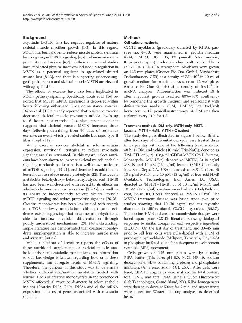
Knockdown of Akirin-1/Mighty mRNA decreases myotubesizeAs mentioned above, Akirin-1/Mighty mRNA expressionpatterns between treatments exhibited modest to strongcorrelations to select hypertrophy indices. Thus, we nextsought to determine if experimentally decreasing Akirin-1/Mighty mRNA using shRNA knockdown affected myotubesize. Indeed, GFP-positive myotubes transfected with theAkirin-1/Mighty shRNA plasmid exhibited a drasticreduction in myotube area (−72%, p < 0.001; Figure 4A).Knockdown in transfected wells was also confirmed atthe mRNA level (Figure 4B). Therefore, we posit thatMSTN-induced atrophy observed in the current studyis likely linked to the MSTN-induced down-regulation inAkirin-1/Mighty mRNA. Furthermore, leucine and HMBappear to prevent this down-regulation and creatinemonohydrate increases myotube Akirin-1/Mighty mRNAlevels in spite of MSTN treatment.
, or creatine on the expression of MSTN-related mRNAs. Legend:t). Effects of MSTN with or without each ingredient on the mRNATN transcriptional targets (C). Sub-figures D and E demonstrate thatmeasured hypertrophy variables. Sub-figure F shows that Akirin-1/tion marker MyoD mRNA. One-way ANOVA with LSD post-hoc= p < 0.05. In sub-figure C, ** indicates that MSTN down-regulated

Figure 4 Experiment demonstrating Akirin-1/Mighty mRNA knockdown affects myotube size. Legend: Data are presented as means ± standarderror (n = 4–5 plates per treatment). Effects of shRNA-mediated Akirin-1/Mighty mRNA knockdown on myotube size versus a scrambled shRNA controlcondition (A), and confirmation that Akirin-1 mRNA was reduced in shRNA-transfected myotubes (B). Photographs in sub-figure C are 10x representativeimages of GFP-positive cells (arrows) that were transfected with either a scrambled (CTL)-shRNA or Akirin-1/Mighty-shRNA.
Mobley et al. Journal of the International Society of Sports Nutrition 2014, 11:38 Page 6 of 9http://www.jissn.com/content/11/1/38
DiscussionWe used an in vitro approach to investigate whetherleucine, HMB, or creatine monohydrate could rescuethe atrophic effects of MSTN in C2C12 myotubes.MSTN has previously been shown to inhibit myoblastproliferation, myotube differentiation and protein synthesisin the C2C12 cell line [42-44]. Specifically, one of the afore-mentioned studies demonstrated that myotubes treatedwith high doses of recombinant MSTN (~1-6 μg/ml whichis 100-600x the dose used in the current study) significantlydepressed protein synthesis in myotubes [42]. However, therelatively low concentration of MSTN treatments used inthe current study reduced myotube size independent ofmuscle protein synthesis. This finding is in agreement withother studies which used MSTN treatment dosages similarto our study (10–30 ng/ml) and reported MSTN toreduce myotube diameter by inhibiting differentiation inC2C12 myotubes [36,37]. Thus, we hypothesize thatMSTN-induced atrophy in the current study was likely dueto diminished myotube differentiation rather than decreasesin muscle protein synthesis and/or increases in muscleproteolysis; a hypothesis which is further supported by theMSTN-induced down-regulation in MyoD mRNA.Interestingly, while myotubes treated with MSTN
only showed a significant decrease in myotube diameter,this effect was reversed in all three treatment groups(MSTN+ Leu, MSTN+HMB, and MSTN+Crea). Thereis ample evidence to suggest that leucine and HMB areable to increase muscle protein synthesis in vitro andin vivo [26,27,45,46]. Thus, it is difficult to reconcile whyMSTN+ Leu and MSTN+HMB treatments in the currentstudy did not statistically increase muscle protein synthesiscompared to DM/CTL myotubes. However, all treatmentsoccurred in myotubes that were not amino acid deprived;this being a condition which may be obligatory for leucine
and HMB to exert more profound muscle proteinsynthesis effects [47]. Furthermore, the MSTN + Creatreated group demonstrated the greatest hypertrophiceffect in spite of the fact that creatine monohydrate likelydoes not affect markers of muscle protein synthesis [29]and/or muscle protein synthesis rates [48] in vivo. Thus,we hypothesized that leucine, HMB and creatine monohy-drate treatments all independently counteracted low-doseMSTN-induced atrophy through potential genetic mecha-nisms related to myotube differentiation.Of the mRNAs measured in the current study select
hypertrophic indices, including the protein: DNA andmyofiber diameter, were strongly and modestly correlatedwith Akirin-1/Mighty gene expression, respectively.Furthermore, our main findings with Akirin-1/Mightygene expression were as follows: 1) leucine and HMB canreverse MSTN-induced down-regulation in Akirin-1/Mighty mRNA; 2) in spite of MSTN treatment, creatinemonohydrate up-regulated Akirin-1/Mighty mRNAwhile exhibiting the most potent anabolic effects; and 3)Akrin-1/Mighty mRNA knockdown via shRNA transfec-tion reduced myofiber size. Hence, our findings support thehypothesis that the transcriptional modulation of Akirin-1/Mighty mRNA by leucine/HMB/creatine monohydratemay be a mechanism whereby these ingredients promotemyotube hypertrophy in vitro in spite of MSTN treatments.Our finding that each of these purported anabolic
ingredients rescues MSTN-induced Akirin-1/MightymRNA down-regulation is indeed difficult to interpretfrom a mechanistic and practical viewpoint regardingthe preservation of myofiber size. Furthermore, this isthe first study to show that each of these ingredientsaffects (directly or indirectly) the mRNA expression ofAkirin-1/Mighty mRNA. Resistance exercise has beenshown to increase Akirin-1/Mighty mRNA expression

Mobley et al. Journal of the International Society of Sports Nutrition 2014, 11:38 Page 7 of 9http://www.jissn.com/content/11/1/38
patterns 6 h and 48 h following an acute exercise bout inrodents [49]; of note these rodents were trained 6 weeksprior to the acute bout and the exercise-induced changesin Akrin-1/Mighty strongly predicted the hypertrophicresponse to the 6-week training bout. The authorsconcluded that, while Akrin-1/Mighty may play a rolein the activation of satellite cells, how Akirin-1/Mighty promotes the hypertrophic response to exercisehas yet to be determined. Notwithstanding, it appears thatAkirin-1/Mighty plays a role in exercise-induced skeletalmuscle hypertrophy. The expression of mRNA expressionof akirin genes in skeletal muscle are sensitive to fastingand re-feeding in Artic charr [50], and we have observedmixed gastrocnemius Akirin-1/Mighty mRNA to increaseapproximately 90% 3 h after rats fed 10 human equivalentgrams of whey protein concentrate (which is ~12-14%leucine) when compared to 18-h fasted rats (p < 0.05;unpublished observations). Hence, our finding thatpurported anabolic ingredients directly or indirectlyaffect myotube Akirin-1/Mighty mRNA expression isnot unfounded given that other hypertrophic stimuli(i.e., resistance exercise and protein feeding) have alsobeen shown to increase the expression of this gene inskeletal muscle. In this regard, future research is neededto elucidate if: 1) decrements in Akirin-1/Mighty mRNAexpression accompany and/or causal to muscle wastingconditions such as sarcopenia, cachexia, and disuseatrophy; and 2) if each of the anabolic ingredients studiedherein mitigate these conditions through Akirin-1/Mightygene expression changes.While the exact mechanisms are unknown as to how
Akirin-1/Mighty regulates muscle mass, it is a proventranscriptional target of myostatin [49,51,52]. Recentevidence also suggests that the Akirin-1/Mighty genemodulates satellite cell proliferation and differentiationfollowing muscle injury, and there is interplay betweenAkirin-1/Mighty down-regulation and the inhibition ofdifferentiation-promoting genes such as MyoD [53]. Thereis a paucity of literature examining how leucine and/orHMB affect Akirin-1/Mighty mRNA expression patterns,though recent in vivo evidence suggests that chronicallyconsuming a leucine-rich pre-exercise beverage increasesskeletal muscle MyoD and MRF4 mRNA [54]. Furthermore,adding various concentrations (10–100 μg/ml) of HMB toserum-starved myoblasts has been shown to inducemyoblast proliferation and MyoD expression, suggestive ofenhanced myoblast differentiation [55]. However, Akirin-1/Mighty mRNA was not assessed in the aforementionedstudies making it difficult to reconcile whether theseeffects were mitigated through Akrin-1/Mighty mRNAgene expression.Of particular interest was the ability of creatine
monohydrate to promote the up-regulation of Akirin-1/Mighty mRNA in the presence of myostatin. Creatine
monohydrate supplementation has been shown to reducethe catabolic response of hind limb immobilization in rats[56]. Additionally, Johnston et al. [57] have demonstratedthat short-term creatine monohydrate supplementation(7 days) attenuates losses of muscle mass and strength dur-ing upper-arm immobilization in young men. Furthermore,prolonged creatine monohydrate supplementation has beenreported to increase satellite cell proliferation and differen-tiation in resistance-trained subjects versus a placebo group[58]. While the anabolic/anti-catabolic mechanisms ofcreatine monohydrate remain poorly understood, creatinemonohydrate supplementation has been shown to increasecellular fluid retention and modulate the expression ofmyogenic transcription factors related to skeletal musclehypertrophy [59,60]. With regards to the former mechan-ism, Häussinger [61] reported that an increase in cellularfluid/swelling acts as an anabolic proliferative signal,whereas cell shrinkage is catabolic and anti-proliferative.However, creatine monohydrate has been shown to pro-mote myotube hypertrophy in vitro by enhancing myotubedifferentiation compared to DM/CTL-treated myotubes[38]; an effect the authors suggested may be independent ofthe intracellular osmolarity effects of creatine monohydrate.Therefore, the ability of creatine monohydrate to up-regulate Akirin-1/Mighty mRNA may be a primary mech-anism involved in the ability of creatine monohydrate tostimulate skeletal muscle hypertrophy independent ofmuscle protein synthesis and/or its effects on osmolarity. Inthis regard, future research should continue to examinethe ability of creatine monohydrate supplementation tomodulate Akirin-1/Mighty mRNA expression in vivo.
ConclusionsWe demonstrate that leucine, HMB, and creatine mono-hydrate reverse myostatin-induced atrophy in myotubes;this potentially results from the independent action ofeach ingredient modulating Akirin-1/Mighty mRNAexpression. Furthermore, our findings suggest that, inspite of MSTN treatments, creatine monohydrate treatmentup-regulates Akirin-1/Mighty mRNA which leads to ahypertrophic effect clearly independent of muscle proteinsynthesis. Future in vivo studies should continue toexamine how leucine, HMB, and/or creatine monohydrateindependently or synergistically affect Akirin-1/Mightygene expression. More importantly, while Akirin-1/Mightygene expression is needed for the maintenance ofmyofiber size as reported herein, further research isneeded in order to examine how Akirin-1/Mighty geneexpression mechanistically relates to skeletal musclehypertrophy in vivo.
AbbreviationsHMB: Beta-hydroxy-beta-methylbutyric acid; Leu: L-leucine; Crea: Creatinemonohydrate; MSTN: Myostatin; DM: Differentiation media; CTL: Control;SUnSET: Surface sensing of translation.

Mobley et al. Journal of the International Society of Sports Nutrition 2014, 11:38 Page 8 of 9http://www.jissn.com/content/11/1/38
Competing interestsBesides SB, and JAR, none of the authors have non-finacial and/or financialcompeting interests. SB, and JAR are employed by Metabolic Technologies,Inc., but both authors intellectually contributed to study design and datawrite-up. Therefore, all co-authors agreed that their intellectual input into thisproject warranted co-authorship.
Authors’ contributionsCDF, BSF, RHA, VJD, SB, JAR, JMW, MDR. This person has made substantialcontributions to conception and design, or acquisition of data, or analysisand interpretation of data. CBM, MDR. This person primarily was involved indrafting the manuscript or revising it critically for important intellectualcontent. CBM, CDF, BSF, RHA, VJD, SB, JAR, JMW, MDR. This person gave finalapproval of the version to be published. CBM, CDF, BSF, RHA, VJD, SB, JAR,JMW, MDR This person agrees to be accountable for all aspects of the workin ensuring that questions related to the accuracy or integrity of any part ofthe work are appropriately investigated and resolved.
AcknowledgementsInternal funding from Auburn University’s School of Kinesiology was used tofund this study. The authors thank Dr. David Pascoe and Dr. Andreas Kavazisfor intellectual input, as well as Dr. Heidi Kluess for allowing us to use herlaboratory’s fluorescent microscope.
Author details1School of Kinesiology, Molecular and Applied Sciences Laboratory, AuburnUniversity, 301 Wire Road, Office 286, Auburn, AL 36849, USA. 2Departmentof Health Sciences and Human Performance, University of Tampa, Tampa, FL,USA. 3Harrison School of Pharmacy, Auburn University, Auburn, AL, USA.4School of Medical and Applied Sciences, Central Queensland University,Rockhampton, QLD, Australia. 5Metabolic Technologies, Inc, Ames, IA, USA.
Received: 15 May 2014 Accepted: 30 June 2014Published: 13 August 2014
References1. Jackman RW, Kandarian SC: The molecular basis of skeletal muscle
atrophy. Am J Physiol Cell Physiol 2004, 287(4):C834–C843.2. Chen YW, Gregory CM, Scarborough MT, Shi R, Walter GA, Vandenborne K:
Transcriptional pathways associated with skeletal muscle disuse atrophyin humans. Physiol Genomics 2007, 31(3):510–520.
3. Elkina Y, von Haehling S, Anker SD, Springer J: The role of myostatin in musclewasting: an overview. J Cachexia Sarcopenia Muscle 2011, 2(3):143–151.
4. Hulmi JJ, Tannerstedt J, Selanne H, Kainulainen H, Kovanen V, Mero AA:Resistance exercise with whey protein ingestion affects mTOR signalingpathway and myostatin in men. J Appl Physiol (1985) 2009,106(5):1720–1729.
5. Goodman CA, McNally RM, Hoffmann FM, Hornberger TA: Smad3 inducesatrogin-1, inhibits mTOR and protein synthesis, and promotes muscleatrophy in vivo. Mol Endocrinol 2013, 27(11):1946–1957.
6. McFarlane C, Plummer E, Thomas M, Hennebry A, Ashby M, Ling N, Smith H,Sharma M, Kambadur R: Myostatin induces cachexia by activating theubiquitin proteolytic system through an NF-kappaB-independent,FoxO1-dependent mechanism. J Cell Physiol 2006, 209(2):501–514.
7. Joulia-Ekaza D, Cabello G: Myostatin regulation of muscle development:molecular basis, natural mutations, physiopathological aspects. Exp CellRes 2006, 312(13):2401–2414.
8. Wu M, Fannin J, Rice KM, Wang B, Blough ER: Effect of aging on cellularmechanotransduction. Ageing Res Rev 2011, 10(1):1–15.
9. Dalbo VJ, Roberts MD, Sunderland KL, Poole CN, Stout JR, Beck TW, Bemben M,Kerksick CM: Acute loading and aging effects on myostatin pathwaybiomarkers in human skeletal muscle after three sequential bouts ofresistance exercise. J Gerontol A Biol Sci Med Sci 2011, 66(8):855–865.
10. Sakuma K, Yamaguchi A: Molecular mechanisms in aging and currentstrategies to counteract sarcopenia. Curr Aging Sci 2010, 3(2):90–101.
11. Leger B, Derave W, De Bock K, Hespel P, Russell AP: Human sarcopeniareveals an increase in SOCS-3 and myostatin and a reduced efficiency ofAkt phosphorylation. Rejuvenation Res 2008, 11(1):163–175B.
12. Siriett V, Salerno MS, Berry C, Nicholas G, Bower R, Kambadur R, Sharma M:Antagonism of myostatin enhances muscle regeneration duringsarcopenia. Mol Ther 2007, 15(8):1463–1470.
13. Tobin JF, Celeste AJ: Myostatin, a negative regulator of muscle mass:implications for muscle degenerative diseases. Curr Opin Pharmacol 2005,5(3):328–332.
14. Kawada S, Tachi C, Ishii N: Content and localization of myostatin in mouseskeletal muscles during aging, mechanical unloading and reloading.J Muscle Res Cell Motil 2001, 22(8):627–633.
15. Yarasheski KE, Bhasin S, Sinha-Hikim I, Pak-Loduca J, Gonzalez-Cadavid NF:Serum myostatin-immunoreactive protein is increased in 60–92 year oldwomen and men with muscle wasting. J Nutr Health Aging 2002,6(5):343–348.
16. Louis E, Raue U, Yang Y, Jemiolo B, Trappe S: Time course of proteolytic,cytokine, and myostatin gene expression after acute exercise in humanskeletal muscle. J Appl Physiol (1985) 2007, 103(5):1744–1751.
17. Dalbo VJ, Roberts MD, Hassell S, Kerksick CM: Effects of pre-exercise feedingon serum hormone concentrations and biomarkers of myostatin andubiquitin proteasome pathway activity. Eur J Nutr 2013, 52(2):477–487.
18. Jespersen JG, Nedergaard A, Andersen LL, Schjerling P, Andersen JL: Myostatinexpression during human muscle hypertrophy and subsequent atrophy:increased myostatin with detraining. Scand J Med Sci Sports 2011,21(2):215–223.
19. Anthony JC, Anthony TG, Kimball SR, Jefferson LS: Signaling pathwaysinvolved in translational control of protein synthesis in skeletal muscleby leucine. J Nutr 2001, 131(3):856S–860S.
20. Stipanuk MH: Leucine and protein synthesis: mTOR and beyond. Nutr Rev2007, 65(3):122–129.
21. Haegens A, Schols AM, van Essen AL, van Loon LJ, Langen RC: Leucineinduces myofibrillar protein accretion in cultured skeletal musclethrough mTOR dependent and -independent control of myosin heavychain mRNA levels. Mol Nutr Food Res 2012, 56(5):741–752.
22. Combaret L, Dardevet D, Rieu I, Pouch MN, Bechet D, Taillandier D, Grizard J,Attaix D: A leucine-supplemented diet restores the defective postprandialinhibition of proteasome-dependent proteolysis in aged rat skeletalmuscle. J Physiol 2005, 569(Pt 2):489–499.
23. Nissen S, Sharp R, Ray M, Rathmacher JA, Rice D, Fuller JC Jr, Connelly AS,Abumrad N: Effect of leucine metabolite beta-hydroxy-beta-methylbutyrateon muscle metabolism during resistance-exercise training. J Appl Physiol(1985) 1996, 81(5):2095–2104.
24. Slater GJ, Jenkins D: Beta-hydroxy-beta-methylbutyrate (HMB)supplementation and the promotion of muscle growth and strength.Sports Med 2000, 30(2):105–116.
25. Jowko E, Ostaszewski P, Jank M, Sacharuk J, Zieniewicz A, Wilczak J, Nissen S:Creatine and beta-hydroxy-beta-methylbutyrate (HMB) additively increaselean body mass and muscle strength during a weight-training program.Nutrition 2001, 17(7–8):558–566.
26. Eley HL, Russell ST, Baxter JH, Mukerji P, Tisdale MJ: Signaling pathwaysinitiated by beta-hydroxy-beta-methylbutyrate to attenuate the depressionof protein synthesis in skeletal muscle in response to cachectic stimuli.Am J Physiol Endocrinol Metab 2007, 293(4):E923–E931.
27. Wilson GJ, Wilson JM, Manninen AH: Effects of beta-hydroxy-beta-methylbutyrate (HMB) on exercise performance and body compositionacross varying levels of age, sex, and training experience: A review.Nutr Metab (Lond) 2008, 5:1.
28. Wilson JM, Lowery RP, Joy JM, Walters JA, Baier SM, Fuller JC Jr, Stout JR,Norton LE, Sikorski EM, Wilson SM, Duncan NM, Zanchi NE, Rathmacher J:beta-Hydroxy-beta-methylbutyrate free acid reduces markers ofexercise-induced muscle damage and improves recovery inresistance-trained men. Br J Nutr 2013, 110(3):538–544.
29. Deldicque L, Atherton P, Patel R, Theisen D, Nielens H, Rennie MJ, Francaux M:Effects of resistance exercise with and without creatine supplementationon gene expression and cell signaling in human skeletal muscle. J ApplPhysiol (1985) 2008, 104(2):371–378.
30. Earnest CP, Snell PG, Rodriguez R, Almada AL, Mitchell TL: The effect ofcreatine monohydrate ingestion on anaerobic power indices, muscularstrength and body composition. Acta Physiol Scand 1995, 153(2):207–209.
31. Hultman E, Soderlund K, Timmons JA, Cederblad G, Greenhaff PL: Musclecreatine loading in men. J Appl Physiol (1985) 1996, 81(1):232–237.
32. Clarkson PM, Rawson ES: Nutritional supplements to increase musclemass. Crit Rev Food Sci Nutr 1999, 39(4):317–328.
33. Francaux M, Poortmans JR: Effects of training and creatine supplement onmuscle strength and body mass. Eur J Appl Physiol Occup Physiol 1999,80(2):165–168.
Mobley et al. Journal of the International Society of Sports Nutrition 2014, 11:38 Page 9 of 9http://www.jissn.com/content/11/1/38
34. Becque MD, Lochmann JD, Melrose DR: Effects of oral creatinesupplementation on muscular strength and body composition. Med SciSports Exerc 2000, 32(3):654–658.
35. Kreider RB: Effects of creatine supplementation on performance andtraining adaptations. Mol Cell Biochem 2003, 244(1–2):89–94.
36. Lach-Trifilieff E, Minetti GC, Sheppard K, Ibebunjo C, Feige JN, Hartmann S,Brachat S, Rivet H, Koelbing C, Morvan F, Hatakeyama S, Glass DJ: An antibodyblocking activin type II receptors induces strong skeletal muscle hypertrophyand protects from atrophy. Mol Cell Biol 2014, 34(4):606–618.
37. Trendelenburg AU, Meyer A, Rohner D, Boyle J, Hatakeyama S, Glass DJ:Myostatin reduces Akt/TORC1/p70S6K signaling, inhibiting myoblastdifferentiation and myotube size. Am J Physiol Cell Physiol 2009,296(6):C1258–C1270.
38. Deldicque L, Theisen D, Bertrand L, Hespel P, Hue L, Francaux M: Creatineenhances differentiation of myogenic C2C12 cells by activating both p38and Akt/PKB pathways. Am J Physiol Cell Physiol 2007, 293(4):C1263–C1271.
39. Smith HJ, Wyke SM, Tisdale MJ: Mechanism of the attenuation ofproteolysis-inducing factor stimulated protein degradation in muscle bybeta-hydroxy-beta-methylbutyrate. Cancer Res 2004, 64(23):8731–8735.
40. Goodman CA, Hornberger TA: Measuring protein synthesis with SUnSET:a valid alternative to traditional techniques? Exerc Sport Sci Rev 2013,41(2):107–115.
41. Nader GA, Hornberger TA, Esser KA: Translational control: implications forskeletal muscle hypertrophy. Clin Orthop Relat Res 2002, (403 Suppl):S178–S187.
42. Taylor WE, Bhasin S, Artaza J, Byhower F, Azam M, Willard DH Jr, Kull FC Jr,Gonzalez-Cadavid N:Myostatin inhibits cell proliferation and protein synthesisin C2C12 muscle cells. Am J Physiol Endocrinol Metab 2001, 280(2):E221–E228.
43. Rios R, Carneiro I, Arce VM, Devesa J: Myostatin is an inhibitor ofmyogenic differentiation. Am J Physiol Cell Physiol 2002, 282(5):C993–C999.
44. McCroskery S, Thomas M, Maxwell L, Sharma M, Kambadur R: Myostatinnegatively regulates satellite cell activation and self-renewal. J Cell Biol2003, 162(6):1135–1147.
45. Dardevet D, Sornet C, Bayle G, Prugnaud J, Pouyet C, Grizard J: Postprandialstimulation of muscle protein synthesis in old rats can be restored by aleucine-supplemented meal. J Nutr 2002, 132(1):95–100.
46. Pimentel GD, Rosa JC, Lira FS, Zanchi NE, Ropelle ER, Oyama LM, Nascimento CMO d, de Mello MT, Tufik S, Santos RV: beta-Hydroxy-beta-methylbutyrate(HMbeta) supplementation stimulates skeletal muscle hypertrophy in rats viathe mTOR pathway. Nutr Metab (Lond) 2011, 8(1):11.
47. Talvas J, Obled A, Fafournoux P, Mordier S: Regulation of protein synthesisby leucine starvation involves distinct mechanisms in mouse C2C12myoblasts and myotubes. J Nutr 2006, 136(6):1466–1471.
48. Parise G, Mihic S, MacLennan D, Yarasheski KE, Tarnopolsky MA: Effects ofacute creatine monohydrate supplementation on leucine kinetics andmixed-muscle protein synthesis. J Appl Physiol (1985) 2001, 91(3):1041–1047.
49. MacKenzie MG, Hamilton DL, Pepin M, Patton A, Baar K: Inhibition ofmyostatin signaling through Notch activation following acute resistanceexercise. PLoS One 2013, 8(7):e68743.
50. Macqueen DJ, Kristjansson BK, Johnston IA: Salmonid genomes have aremarkably expanded akirin family, coexpressed with genes fromconserved pathways governing skeletal muscle growth and catabolism.Physiol Genomics 2010, 42(1):134–148.
51. Marshall A, Salerno MS, Thomas M, Davies T, Berry C, Dyer K, Bracegirdle J,Watson T, Dziadek M, Kambadur R, Bower R, Sharma M: Mighty is a novelpromyogenic factor in skeletal myogenesis. Exp Cell Res 2008, 314(5):1013–1029.
52. Salerno MS, Dyer K, Bracegirdle J, Platt L, Thomas M, Siriett V, Kambadur R,Sharma M: Akirin1 (Mighty), a novel promyogenic factor regulates muscleregeneration and cell chemotaxis. Exp Cell Res 2009, 315(12):2012–2021.
53. Dong Y, Pan JS, Zhang L: Myostatin suppression of Akirin1 mediatesglucocorticoid-induced satellite cell dysfunction. PLoS One 2013,8(3):e58554.
54. Shelmadine B, Cooke M, Buford T, Hudson G, Redd L, Leutholtz B,Willoughby DS: Effects of 28 days of resistance exercise and consuming acommercially available pre-workout supplement, NO-Shotgun(R), onbody composition, muscle strength and mass, markers of satellite cellactivation, and clinical safety markers in males. J Int Soc Sports Nutr2009, 6:16.
55. Kornasio R, Riederer I, Butler-Browne G, Mouly V, Uni Z, Halevy O:Beta-hydroxy-beta-methylbutyrate (HMB) stimulates myogenic cellproliferation, differentiation and survival via the MAPK/ERK and PI3K/Aktpathways. Biochim Biophys Acta 2009, 1793(5):755–763.
56. Aoki MS, Lima WP, Miyabara EH, Gouveia CH, Moriscot AS: Deleteriuoseffects of immobilization upon rat skeletal muscle: role of creatinesupplementation. Clin Nutr 2004, 23(5):1176–1183.
57. Johnston AP, Burke DG, MacNeil LG, Candow DG: Effect of creatinesupplementation during cast-induced immobilization on the preservationof muscle mass, strength, and endurance. J Strength Cond Res 2009,23(1):116–120.
58. Olsen S, Aagaard P, Kadi F, Tufekovic G, Verney J, Olesen JL, Suetta C, Kjaer M:Creatine supplementation augments the increase in satellite cell andmyonuclei number in human skeletal muscle induced by strength training.J Physiol 2006, 573(Pt 2):525–534.
59. Willoughby DS, Rosene JM: Effects of oral creatine and resistance trainingon myogenic regulatory factor expression. Med Sci Sports Exerc 2003,35(6):923–929.
60. Hespel P, Op’t Eijnde B, Van Leemputte M, Urso B, Greenhaff PL, Labarque V,Dymarkowski S, Van Hecke P, Richter EA: Oral creatine supplementationfacilitates the rehabilitation of disuse atrophy and alters the expressionof muscle myogenic factors in humans. J Physiol 2001, 536(Pt 2):625–633.
61. Haussinger D, Roth E, Lang F, Gerok W: Cellular hydration state: animportant determinant of protein catabolism in health and disease.Lancet 1993, 341(8856):1330–1332.
doi:10.1186/1550-2783-11-38Cite this article as: Mobley et al.: L-leucine, beta-hydroxy-beta-methylbutyricacid (HMB) and creatine monohydrate prevent myostatin-induced Akirin-1/Mighty mRNA down-regulation and myotube atrophy. Journal of theInternational Society of Sports Nutrition 2014 11:38.
Submit your next manuscript to BioMed Centraland take full advantage of:
• Convenient online submission
• Thorough peer review
• No space constraints or color figure charges
• Immediate publication on acceptance
• Inclusion in PubMed, CAS, Scopus and Google Scholar
• Research which is freely available for redistribution
Submit your manuscript at www.biomedcentral.com/submit