Journal pone 0091255
13
An Alternative Chemical Redox Method for the Production of Bispecific Antibodies: Implication in Rapid Detection of Food Borne Pathogens Mohammad Owais 1 , Shadab Kazmi 1 , Saba Tufail 1 , Swaleha Zubair 2 * 1 Interdisciplinary Biotechnology Unit, Aligarh Muslim University, Aligarh, India, 2 Women’s College, Aligarh Muslim University, Aligarh, India Abstract Bi-functional antibodies with the ability to bind two unrelated epitopes have remarkable potential in diagnostic and bio- sensing applications. In the present study, bispecific antibodies that recognize human red blood cell (RBC) and the food borne pathogen Listeria monocytogenes (L. monocytogenes) were engineered. The procedure involves initial reduction of a mixture of anti-RBC and anti-Listeria antibodies followed by gradual re-oxidation of the reduced disulphides. This facilitates association of the separated antibody chains and formation of hybrid immunoglobulins with affinity for the L. monocytogenes and human RBC. The bispecific antibodies caused the agglutination of the RBCs only in the presence of L. monocytogenes cells. The agglutination process necessitated the specific presence of L. monocytogenes and the red colored clumps formed were readily visible with naked eyes. The RBC agglutination assay described here provides a remarkably simple approach for the rapid and highly specific screening of various pathogens in their biological niches. Citation: Owais M, Kazmi S, Tufail S, Zubair S (2014) An Alternative Chemical Redox Method for the Production of Bispecific Antibodies: Implication in Rapid Detection of Food Borne Pathogens. PLoS ONE 9(3): e91255. doi:10.1371/journal.pone.0091255 Editor: Dipshikha Chakravortty, Indian Institute of Science, India Received September 19, 2013; Accepted February 11, 2014; Published March 17, 2014 Copyright: ß 2014 Owais et al. This is an open-access article distributed under the terms of the Creative Commons Attribution License, which permits unrestricted use, distribution, and reproduction in any medium, provided the original author and source are credited. Funding: The financial assistance was provided by Indian Council of Agriculture Research (www.icar.nic.in) against project sanction (NF/PHT-3028/2012–13). The funders had no role in study design, data collection and analysis, decision to publish, or preparation of the manuscript. Competing Interests: The authors have declared that no competing interests exist. * E-mail: [email protected] Introduction Bispecific antibodies (BsAbs) are crafted using either intact antibodies or their fragments such as single domain antibodies, Fabs (fragments antigen binding) and scFvs (single-chain fragments variable) [1]. The unique ability of BsAbs to bind two distinct epitopes simultaneously is the hallmark of their therapeutic potential. Additionally, BsAbs obtained by fusing two or more antibodies which bind different epitopes on the same antigen have been traditionally used for increasing the avidity of antigen antibody interaction [2,3]. BsAbs prepared by fusing an antibody specific for an effector cell to a target cell-specific second antibody have also been used for activating innate and adaptive immune responses of the host [3,4]. In spite of the robustness of the fabricated antibody based strategies, potential of BsAbs has not been fully explored either in bio-sensing applications or detection and screening of organisms affecting health of higher animals. Various existing methods employed for preparation of BsAbs suffer with inherent limitations; (A) Chemical cross-linking of two antibody molecules or their fragments [5] sometimes results in inactivation, unfolding or aggregation of the synthesized BsAbs [6]. (B) Fusion of two or three different cell lines to form a quadroma or trioma [7], a strategy necessitating lengthy cell culturing but usually gives poor yield of the BsAbs [8]. (C) Recombinant DNA based approach involving cloning and expression of single chain variable fragment (scFv) fusions or diabodies, scFv-Fc fusions and single variable domain IgGs as well as dual-variable domain IgG [9–12]. The procedure entails good technical expertise and sophisticated instrumentation. For detection and screening of various pathogens, techniques based on cell culture, PCR and immuno-assays are widely employed [13]. While these strategies are highly sensitive as well as consistent, they unfortunately are time consuming and expensive. The large number of outbreaks of infections globally, and illnesses they manifest, advocate the need for simple and rapid procedures for identification of the causative pathogen. Annually, on an average about 1–2 million people are estimated to be infected by bacteria, of which 70% are food borne [14,15]. Addressing a problem of such magnitude, especially in the under developed and poor countries is possible only if simple, rapid and inexpensive diagnostic tools for detection of various pathogens become available. We describe in this article a strategy employing BsAbs for the specific and rapid detection of L. monocytogenes in food and other biological samples. BsAbs recognizing the cell surface antigens of human erythrocytes and L. monocytogenes were generated using a modified reduction/oxidation procedure [5]. The hybrid antibod- ies induced the agglutination of human erythrocytes specifically in the presence of L. monocytogenes cells and the resulting red cell clumps were large enough to be visible to the naked eye. BsAbs prepared both from monoclonal as well as polyclonal antibodies were equally effective in inducing the agglutination. BsAbs were successfully employed for the highly precise, specific and ready detection of L. monocytogenes. This inexpensive but robust sensing technique neither requires sophisticated instrumentation nor any special skill and can be used in any modest clinical laboratory. L. monocytogenes was chosen for the study, since listeriosis is most widespread amongst various food-borne pathogens and PLOS ONE | www.plosone.org 1 March 2014 | Volume 9 | Issue 3 | e91255
Transcript of Journal pone 0091255
An Alternative Chemical Redox Method for theProduction of Bispecific Antibodies: Implication in RapidDetection of Food Borne PathogensMohammad Owais1, Shadab Kazmi1, Saba Tufail1, Swaleha Zubair2*
1 Interdisciplinary Biotechnology Unit, Aligarh Muslim University, Aligarh, India, 2 Women’s College, Aligarh Muslim University, Aligarh, India
Abstract
Bi-functional antibodies with the ability to bind two unrelated epitopes have remarkable potential in diagnostic and bio-sensing applications. In the present study, bispecific antibodies that recognize human red blood cell (RBC) and the foodborne pathogen Listeria monocytogenes (L. monocytogenes) were engineered. The procedure involves initial reduction of amixture of anti-RBC and anti-Listeria antibodies followed by gradual re-oxidation of the reduced disulphides. This facilitatesassociation of the separated antibody chains and formation of hybrid immunoglobulins with affinity for the L.monocytogenes and human RBC. The bispecific antibodies caused the agglutination of the RBCs only in the presence of L.monocytogenes cells. The agglutination process necessitated the specific presence of L. monocytogenes and the red coloredclumps formed were readily visible with naked eyes. The RBC agglutination assay described here provides a remarkablysimple approach for the rapid and highly specific screening of various pathogens in their biological niches.
Citation: Owais M, Kazmi S, Tufail S, Zubair S (2014) An Alternative Chemical Redox Method for the Production of Bispecific Antibodies: Implication in RapidDetection of Food Borne Pathogens. PLoS ONE 9(3): e91255. doi:10.1371/journal.pone.0091255
Editor: Dipshikha Chakravortty, Indian Institute of Science, India
Received September 19, 2013; Accepted February 11, 2014; Published March 17, 2014
Copyright: � 2014 Owais et al. This is an open-access article distributed under the terms of the Creative Commons Attribution License, which permitsunrestricted use, distribution, and reproduction in any medium, provided the original author and source are credited.
Funding: The financial assistance was provided by Indian Council of Agriculture Research (www.icar.nic.in) against project sanction (NF/PHT-3028/2012–13). Thefunders had no role in study design, data collection and analysis, decision to publish, or preparation of the manuscript.
Competing Interests: The authors have declared that no competing interests exist.
* E-mail: [email protected]
Introduction
Bispecific antibodies (BsAbs) are crafted using either intact
antibodies or their fragments such as single domain antibodies,
Fabs (fragments antigen binding) and scFvs (single-chain fragments
variable) [1]. The unique ability of BsAbs to bind two distinct
epitopes simultaneously is the hallmark of their therapeutic
potential. Additionally, BsAbs obtained by fusing two or more
antibodies which bind different epitopes on the same antigen have
been traditionally used for increasing the avidity of antigen
antibody interaction [2,3]. BsAbs prepared by fusing an antibody
specific for an effector cell to a target cell-specific second antibody
have also been used for activating innate and adaptive immune
responses of the host [3,4]. In spite of the robustness of the
fabricated antibody based strategies, potential of BsAbs has not
been fully explored either in bio-sensing applications or detection
and screening of organisms affecting health of higher animals.
Various existing methods employed for preparation of BsAbs
suffer with inherent limitations; (A) Chemical cross-linking of two
antibody molecules or their fragments [5] sometimes results in
inactivation, unfolding or aggregation of the synthesized BsAbs
[6]. (B) Fusion of two or three different cell lines to form a
quadroma or trioma [7], a strategy necessitating lengthy cell
culturing but usually gives poor yield of the BsAbs [8]. (C)
Recombinant DNA based approach involving cloning and
expression of single chain variable fragment (scFv) fusions or
diabodies, scFv-Fc fusions and single variable domain IgGs as well
as dual-variable domain IgG [9–12]. The procedure entails good
technical expertise and sophisticated instrumentation.
For detection and screening of various pathogens, techniques
based on cell culture, PCR and immuno-assays are widely
employed [13]. While these strategies are highly sensitive as well
as consistent, they unfortunately are time consuming and
expensive. The large number of outbreaks of infections globally,
and illnesses they manifest, advocate the need for simple and rapid
procedures for identification of the causative pathogen. Annually,
on an average about 1–2 million people are estimated to be
infected by bacteria, of which 70% are food borne [14,15].
Addressing a problem of such magnitude, especially in the under
developed and poor countries is possible only if simple, rapid and
inexpensive diagnostic tools for detection of various pathogens
become available.
We describe in this article a strategy employing BsAbs for the
specific and rapid detection of L. monocytogenes in food and other
biological samples. BsAbs recognizing the cell surface antigens of
human erythrocytes and L. monocytogenes were generated using a
modified reduction/oxidation procedure [5]. The hybrid antibod-
ies induced the agglutination of human erythrocytes specifically in
the presence of L. monocytogenes cells and the resulting red cell
clumps were large enough to be visible to the naked eye. BsAbs
prepared both from monoclonal as well as polyclonal antibodies
were equally effective in inducing the agglutination.
BsAbs were successfully employed for the highly precise, specific
and ready detection of L. monocytogenes. This inexpensive but robust
sensing technique neither requires sophisticated instrumentation
nor any special skill and can be used in any modest clinical
laboratory. L. monocytogenes was chosen for the study, since listeriosis
is most widespread amongst various food-borne pathogens and
PLOS ONE | www.plosone.org 1 March 2014 | Volume 9 | Issue 3 | e91255
leads to very high fatality rate (25%–30%) [16]. The L.
monocytogenes contamination has prompted imposition of zero
tolerance limits by U.S. regulatory agencies [17,7]. Unfortunately,
the detection methods currently in vogue require sophisticated
instrumentation and are slow needing a time lapse (at least 24 hrs)
to deliver concrete information [7].
Materials and Methods
Chemicals and reagentsAll the chemicals and reagents used were of the highest purity
available. Phenylmethanesulfonylfluoride, b–mercaptoethanesulfo-
nic acid sodium salt, b–mercaptoethanol, Ethanolamine, Sephar-
ose-4B, Freund’s complete and incomplete adjuvant, Bicinchoninic
acid (BCA) protein estimation kit, Tween-20 and FITC conjugated
mouse anti-rabbit antibody were purchased from Sigma-Aldrich
Chemicals (St Louis, MO) and used as received. Horseradish
peroxidase-conjugated anti-rabbit IgG were purchased from
Bangalore Genei (India) Pvt. Ltd. (Bangalore, India). Mouse
monoclonal anti-Listeria LZH1 IgG1 and mouse monoclonal
IgG2a specific for the human erythrocyte membrane protein
(Protein4.2 (2G-12)) were procured from Santa Cruz Biotechnol-
ogy, Inc., California and Abnova Corporation respectively.
Ethics statementThe study was approved by the Institutional Animal Ethics
Committee of the Interdisciplinary Biotechnology Unit, Aligarh
Muslim University, Aligarh, India. All animal experiments were
performed according to the National Regulatory Guidelines issued
by the Committee for the Purpose of Control and Supervision of
Experiments on Animals (CPCSEA). Our approval ID was 332/
CPCSEA, Ministry of Environment and Forests, Paryavaran
Bhavan, Government of India.
AnimalsInbred female rabbits, 8–10 weeks old, were obtained from the
Department Animal House Facility, Interdisciplinary Biotechnol-
ogy Unit, AMU, India. The animals were kept on standard pellet
diet and water ad libitum. All the rabbits were housed under
standard conditions at the Department Animal House Facility.
Blood CollectionThe study was conducted on discarded blood procured from
Blood Bank, Department of Pathology (R. No.-BBL-04/SC/P/
1996), Jawaharlal Nehru Medical College, Aligarh Muslim
University, Aligarh, India.
Preparation of erythrocyte ghostThe erythrocyte ghosts were prepared using published proce-
dure as standardized in our laboratory [18]. Briefly, whole blood
(human) was mixed with anticoagulant solution (EDTA, 2.7%)
and centrifuged at 1500 g for 10 minutes. Buffy coat was discarded
and the RBC pellet was washed with 20 mM phosphate buffered
saline, PBS (pH 7.4). The RBC pellet was lysed with chilled
hypotonic buffer (5 mM Tris-HCl, pH 7.01) in 1:40 ratio. This
suspension was stirred at 4uC for 1 hour. The hemolysed
suspension was centrifuged at 10,000 g to get the lysed erythro-
cytes. The pellet was washed with chilled hypotonic buffer until a
white pellet was obtained. To initiate vesiculation (resealing), each
ml of packed unsealed ghost was diluted to 40 ml with ice cold
20 mM PB in the presence of 1 mM MgSO4 solution and stirred
for 2 hours at 4uC. The suspension was pelleted at 10,000 g and
washed twice with the same buffer [18,19]. Membrane proteins
associated with erythrocyte ghost were analysed in 10% sodium
dodecyl sulphate-polyacrylamide gel electrophoresis (SDS-PAGE).
Preparation of antigen from Listeria monocytogenesL. monocytogenes (ATCC 15313) was procured from ATCC
(American Type Culture Collection, Manassas, VA). InlB protein
of L. monocytogenes was purified using the protocol described
elsewhere [20,21]. Briefly, L. monocytogenes was cultured on Brain
Heart Infusion broth medium at 37uC overnight. The exponen-
tially growing bacterial cells were harvested at 5000 g for 10
minutes and washed with 150 mM Normal Saline twice. The
pelleted bacteria were immediately re-suspended in approx. 0.5%
of the original culture volume using 1M Tris-HCl buffer (pH 8).
Re-suspended bacteria were incubated for 60 min on ice. The
bacterial suspension was pelleted at 12000 g for 20 minutes and
the supernatant was collected [20,21]. The concentration of the
purified protein was determined by the BCA method of protein
estimation [22].
Generation of affinity columnsCross-linked erythrocyte stroma was prepared to isolate anti-
RBC and in-house prepared bispecific antibodies. We followed
published procedure for the preparation of cross-linked stroma
[23,24]. Briefly, blood (50 ml) was centrifuged at 1500 g.
Supernatant along with buffy coat was removed and cells were
washed three times with normal saline. A 10% suspension of
erythrocytes was prepared in 5 mM hypotonic phosphate buffer
(50 ml) and mixed with digitonin (250 ml, 5 mg/ml) with stirring
at 4uC. It was centrifuged at 12,000 g for 10 minutes at 0uC. The
pellet was thoroughly washed with normal saline till the
supernatant was free from protein as determined by absorbance
at 280 nm. About 4 g of stroma was obtained from 50 ml of
blood. The stroma, thus prepared, was washed two times with
10 mM Tris buffer (pH 7.4) and 10 mg/ml (wet weight) was
suspended in the same buffer. It was mixed with an equal volume
of 6.25% solution of glutaraldehyde. The mixture was stirred for
4.5 hours at 37uC followed by stirring at 4uC for 12 hours. All the
operations were carried out in dark. After stipulated time period,
the stroma was centrifuged at 12,000 g for 10 mins at 4uC to
obtain pellet. This pellet was washed with normal saline till the
supernatant was free of any protein.
Cyanogen bromide activated Sepharose 4B was used to prepare
the affinity column of a Listeria monocytogenes cell surface protein,
InlB [25]. Briefly, 10 g Sepharose 4B was washed with distilled
water followed by the addition of 2 M Na2CO3 solution. Further,
1 g CNBr in acetonitrile was added with constant stirring. After
5 mins, the activated gel was washed with the coupling buffer
(0.1 M NaHCO3, 0.5 M NaCl, pH 8.5). Gel was suspended in the
coupling buffer containing InlB protein. Unbound protein was
quantitated in washed out elutes. Remaining active groups were
blocked using excess of ethanolamine for 2 hours at room
temperature.
Production of anti-RBC antibodyPrimary immunization was executed by immunizing female
rabbit with 150 mg (wet weight) of RBC ghost in combination with
complete Freund’s adjuvant. The first booster was given with
100 mg of RBC ghost in incomplete Freund’s adjuvant emulsion
after 21 days of first immunization. The second booster was
administered after 28 days of primary immunization. On the fifth
day of second booster, animals were bled and sera were collected.
RBC surface antigen specific antibodies were purified from sera
using affinity column of cross-linked erythrocyte stroma to obtain
antibodies specific for RBCs. Briefly, sera were incubated with the
Bispecific Antibody: Food Borne Pathogen Detection
PLOS ONE | www.plosone.org 2 March 2014 | Volume 9 | Issue 3 | e91255

cross-linked stroma to allow specific interaction and then RBC
stroma was washed extensively with PBS (pH 7.4) to remove
unbound non-specific antibodies. Bound antibodies were eluted
using 0.1 M Glycine-HCl, pH 2.7 and the pH of elute was
neutralized by the addition of a 1/10 dilution of 1.0 M Tris-HCl,
pH 9.0. The eluted fraction from the RBC stroma was dialyzed
against three changes of PBS (pH 7.4).
Immunization protocol for the development of Listeriacell surface protein specific antibody
Female rabbits were immunized with Listeria InlB cell surface
protein (100 mg) emulsified with complete Freund’s adjuvant. The
booster dose (50 mg antigen) was administered on day 21 post
primary immunization. On the fifth day of the booster, blood was
collected to obtain sera. Antibodies were purified from sera using
the in-house prepared Listeria affinity column to obtain specifically
anti-Listeria antibodies. Antibodies bound to the Listeria affinity
column were eluted using 0.1 M Glycine-HCl, pH 2.7 and the pH
of elute was neutralized by the addition of a 1/10 dilution of
1.0 M Tris-HCl, pH 9.0. The eluted fraction from the Listeria
affinity column was dialyzed against three changes of PBS
(pH 7.4).
Construction of BsAb employing Redox methodologyThe redox method was employed for the preparation of
bispecific antibodies. A schematic representation of the redox
method used for BsAb production is shown in Figure 1.
Increasing concentrations (0–60 mM) of reducing agents, viz. b–
mercaptoethanesulfonic acid sodium salt and b–mercaptoethanol
were analyzed for efficient reduction of the disulphide bonds of
both monoclonals as well as polyclonals antibodies (data not shown
for b–mercaptoethanol). The monoclonal antibodies were reduced
by adding an equal volume of two fold concentrated b-
mercaptoethanesulphonic acid sodium salt solution (60 mM) in
distilled water to a mixture of 1 mg of mouse monoclonal anti-L.
monocytogenes LZH1 IgG1 and mouse monoclonal IgG2a raised
against the human erythrocyte membrane protein (Protein4.2 (2G-
12)) in PBS and incubated at 37uC for 25 minutes. Thereafter,
monovalent arms were exposed to oxidizing conditions by dialysis
against three buffer exchanges of phosphate buffered saline (PBS),
pH 7.4 for 24 hours at 4uC [9]. The same set of protocol was
followed for the construction of the in-house prepared bispecific
antibody, employing polyclonal antibodies specific for human
erythrocyte or L. monocytogenes cell surface antigens. Bispecific
antibodies from both sources (monoclonal and polyclonal) were
purified from parent antibody mixtures over sequential affinity
columns (as discussed in detail later) to ensure that only pure and
surface antigen specific BsAbs were isolated.
Hybrid BsAb purificationBsAbs from both the sources (monoclonal as well as polyclonal)
were purified from parent antibodies using cross-linked erythro-
cyte stroma. The bound antibody was eluted using 0.1 M Glycine-
HCl, pH 2.7 and the pH of elute was neutralized by the addition
of a 1/10 dilution of 1.0 M Tris-HCl, pH 9.0. The eluted fraction
from the RBC stroma was dialyzed against three changes of PBS
and subsequently purified over affinity purified L. monocytogenes
membrane proteins. The bound BsAbs were eluted using 0.1 M
Glycine-HCl, pH 9.0 and subsequently extensively dialysed
against PBS [9].
Binding of hybrid BsAbs to L. monocytogenes cell surfaceproteins and human red blood cell surface antigens
The specificity of in-house prepared mBsAbs as well as pBsAbs
was ascertained by studying its interaction with specific antigens.
To check the binding efficacy of BsAb to Listeria and RBC surface
antigens, ELISA was performed. Firstly, the plates were coated
Figure 1. Schematic representation of production and purifi-cation of Bispecific antibody (BsAb). Anti-RBC antibody and anti-listeria antibody are mixed and reduced using reducing agent, resultingin mvAb fragments. When dialysed into oxidising conditions antibodiesare reformed resulting in a mixture of BsAb of different parent lineage.The resulting elute, after affinity column 1 and 2, contains pure BsAbspecific for both RBC and Listeria surface antigens.doi:10.1371/journal.pone.0091255.g001
Bispecific Antibody: Food Borne Pathogen Detection
PLOS ONE | www.plosone.org 3 March 2014 | Volume 9 | Issue 3 | e91255
with RBC antigens. The antigen-coated plates were washed with
PBS and blocked with 200 ml per well of 1% bovine serum
albumin (BSA) in PBS at 37uC for 4 hour. Bispecific antibody
(1:2000 dilution) was dispensed (100 ml per well) and the plate was
then incubated at 37uC for another 2 hours, followed by washing
three times with PBS containing 0.05% Tween-20. The plates
were further incubated with Listeria antigen InlB. After usual
washing steps, the plates were incubated with anti-InlB antibodies
raised in rabbit. After excessive washing of the plates, 100 ml of
(1:5000 dilution of stock) HRP conjugated goat anti-rabbit IgG
antibodies (3u antibody) were added to the wells. The plates were
further incubated at 37uC for 1 hour. The plates were washed
again before the addition of 100 ml of substrate solution (6 mg O-
phenylenediamine dihydrochloride (OPD) salt dissolved in 12 ml
of substrate buffer with 5 ml of 30% H2O2) and were finally
incubated at 37uC for 40 minutes. The reaction was terminated by
the addition of 50 ml of 7% H2SO4. The absorbance was read at
490 nm with a microtitre plate reader (Bio-Rad, Hercules, CA
94547 USA). Similarly, the specificity of bispecific antibodies was
also tested by coating Listeria antigen, InlB on the plate and
detecting the signal using anti-RBC antibodies raised in rabbit.
Briefly, BsAbs pre-incubated with erythrocyte antigens were used
as primary antibody. After routine washing steps, the wells were
exposed to anti erythrocyte antibodies. Next, plate was incubated
with HRP conjugated goat anti rabbit and color was developed
using routine protocol.
Western Blot AssayWestern blot analysis was used to study the interaction of
mBsAbs as well as pBsAbs with RBC ghost proteins and Listeria cell
surface proteins. Briefly, RBC ghost proteins (50 mg) were resolved
by electrophoresis on 10% sodium dodecyl sulfate polyacrylamide
gel and then electroblotted onto PVDF membrane. The blot was
blocked overnight with 3% nonfat dry milk and probed with
BsAbs at 1:1000 dilution. After stipulated incubation, the strips
were washed thrice in PBST and further incubated for 1 hr at
37uC with horseradish peroxidase-conjugated mouse anti-rabbit
antibody (1:5000). The strips were washed with PBST three times
and finally the immunoblots were developed on X-ray film by
enhanced chemiluminiscence (ECL) using ECL kit, BioRad.
Western blot of Listeria membrane proteins (30 mg) was also
analyzed, following the same method described as above.
Dot blot assayListeria and RBC antigens were dispensed onto PVDF strips
carefully and allowed to dry at room temperature. The strips were
rinsed briefly in phosphate-buffered saline (PBS; pH 7.4) containing
Tween-20 (PBST) and were incubated overnight at 4uC in 5% non-
fat dry milk in PBST to block the residual binding sites on the paper.
The strips were rinsed three times in PBST. The antigen coated
strips were incubated with BsAb of 1:1000 dilution. Immunoreac-
tive dots were detected by horseradish peroxidase-conjugated
mouse anti-rabbit IgG using chromogen 3,39-diaminobenzidine
tetrahydrochloride.
Interaction of RBCs and Listeria monocytogenes cells withBsAb
Interaction of BsAb (of both monoclonal and polyclonal origin)
with RBCs as well as L. monocytogenes surface proteins was further
assessed using fluorescence microscopy. RBCs (20 ml from 60%
hematocrit stock) were mixed with 103 Listeria cells in the presence
of BsAbs. After agglutination, the cells were incubated with FITC
conjugated mouse anti-rabbit antibody for 30 minutes and washed
with PBS and finally transferred onto a glass slide to be analyzed
under fluorescence microscope [26].
Fluorescence activated cell sorter based binding analysisFACS analysis was performed using the procedure described
elsewhere standardized as per requirement of the experiment [26].
Briefly, L. monocytogenes cells (16106) were incubated with in-house
prepared BsAb (1 mg/ml) for 30 minutes at 4uC and washed with
dilution buffer (0.01 M phosphate buffer saline, pH 7.4 containing
1% bovine serum albumin, and 0.1% sodium azide) three times
and finally re-suspended in 500 ml of 2% paraformaldehyde.
FITC-tagged anti-rabbit secondary IgG was used as detection
antibody. The percentage of positive cells were measured at
various time intervals using fluorescence activated cell sorter
(Easycyte Mini; Guava Technologies, Hayward, CA), and the data
were analyzed accordingly.
Agglutination assayIn the RBC and Listeria agglutination test, 103 Listeria cells were
mixed with 20 ml of human erythrocyte (60% hematocrit stock)
and dispensed to the wells of a 96-well microtitre plate. Two fold
serial dilution of BsAbs (poly/monoclonal origin) was made before
mixing RBC and Listeria cells, in 100 ml of PBS. After 10 minutes
of incubation at room temperature, the mixture was scored
visually to assess degree of agglutination. To confirm RBC and
Listeria agglutination, the agglutinated mass was observed under a
light microscope.
Sensitivity analysis of agglutination assay with pureListeria cultures
Overnight-grown bacterial cultures were centrifuged to harvest
cell pellet and washed with PBS three times. Bacterial pellet was
serially diluted in PBS to get desired number of bacterial cells. A
known number of cells with density 102, 103, 105 and 107 were
dispensed in various wells of microtiter plates. Subsequently, fixed
number of RBCs (20 ml of 60% hematocrit) were added to each
well and the mixture was finally reacted with mBsAbs as well as
pBsAbs. The minimum number of Listeria cells that could induce
agglutination determined the level of sensitivity.
Detection of Listeria monocytogenes in contaminatedfood samples by agglutination assay
Beef, mutton and chicken were purchased from local grocery
stores in Aligarh, U.P., India. Absence of L. monocytogenes in
procured meat samples was confirmed by following the procedure
as described elsewhere [27]. Briefly, 25 g of each meat sample was
mashed and kept in sterile container. The sample was enriched
using 250 ml of half-strength Fraser Broth (K FB: Difco Lab,
Sparks, MD, USA) followed by homogenisation of the meat
samples for 2 min using the Stomacher 400 (Seward, Norfolk,
UK). The sample homogenates were then incubated at 37uC for
18 h. Several aliquots were collected from each sample and tested
by agglutination assay as described above.
For artificially contaminated samples, bacterial cell suspension
with approx. density (16103 CFU) was used to inoculate 25 g of
meat samples (approx. 40 CFU/g). After incubation for 15 min at
room temperature to allow bacterial adaptation, enrichment step
was followed as described elsewhere [27]. Ten millilitres of
enriched samples was withdrawn from each sample, centrifuged
(16, 000 g for 10 min), and the pellets were resuspended in 10 ml
of PBS. A specific volume was used to test the presence of L.
moncytogenes by agglutination assay.
Bispecific Antibody: Food Borne Pathogen Detection
PLOS ONE | www.plosone.org 4 March 2014 | Volume 9 | Issue 3 | e91255
Statistical analysisStatistical significance (P value) was ascertained by performing t
tests on antibody titre data by using Sigma plot statistics software
package (v 10 and 11; SigmaPlot, San Jose). P values,0.05 were
considered significant.
Results
As a proof of the concept, anti-Listeria mouse monoclonal LZH1
antibody specific for Listeria cell surface protein and mouse
monoclonal antibody directed against human erythrocyte mem-
brane protein (Protein 4.2 [2G-12]), were used in the construction
of a mBsAb. The LZH1 has IgG1 isotype and recognizes cell
surface protein of L. monocytogenes. The monoclonal antibody
directed against transmembrane protein 4.2 of erythrocyte belongs
to IgG2a isotype. LZH1 recognizes a 23 kDa protein of Listeria
monocytogenes and 2G-12 is reactive to Protein 4.2 of RBCs which
has mol. wt. of 72 kDa.
The specificity of hybrid BsAb consisting of one arm recogniz-
ing Listeria surface antigen and other recognizing protein 4.2 was
established by its potential to induce formation of agglutinated cell
mass that contained both erythrocyte and Listeria cells. Similarly,
possibility of construction of similar hybrid antibody using
polyclonal antibodies was also examined. Rabbit polyclonal
antibodies raised against intact human erythrocytes in rabbits
and those against InlB surface protein of L. monocytogenes were used
for the formation of pBsAb. The mBsAb and pBsAb recognized
both L. monocytogenes cells as well as human erythrocytes
simultaneously.
Specificity of antibody generated against RBC ghosts andListeria monocytogenes surface antigen, InlB in rabbit sera
Sera were isolated from the blood of immunized rabbit and
assayed for anti-RBC as well as anti-Listeria cell surface antibodies
by ELISA, fluorescence microscopy and dot blot as depicted by
Figure 2. The results enumerate generation of antibodies specific
for RBCs and L. monocytogenes. Fig. 2A demonstrates potential
immunogenicity of the RBC antigens and InlB Listeria antigen.
With increase in antigen concentration, absorbance at 492 nm
was found to increase consistently and was significantly higher
than controls (P,0.01). Fluorescence restricted to the surface of
Listeria monocytogenes and RBCs further enumerates generation of
antibodies specific for Listeria and RBC cell surface (Fig. 2B). The
antigen-antibody interaction was validated by dot blot assay
showing positive result for specificity of InlB and RBC antigens
with respective antibodies developed upon their immunization
(Fig. 2C). The controls exhibited negative results.
Construction of bispecific antibodies and evaluation oftheir specificity by ELISA, dot blot and Western blotanalyses
Attempts were made to use increasing concentrations (0–60 mM)
of reducing agents, viz. b–mercaptoethanesulfonic acid sodium salt
and b–mercaptoethanol to reduce the disulphide bonds of
antibodies (data not shown for b–mercaptoethanol). It was found
that b–mercaptoethanesulfonic acid sodium salt (60 mM) as well as
b–mercaptoethanol (50 mM) were equally efficient in the formation
of the monovalent antibody fragment (data not shown for b–
mercaptoethanol). A pilot study suggested that the reduction and re-
oxidation of the mixture of monovalent antibody from two different
sources (anti-human erythrocyte and anti-Listeria cell surface
antigens) led to the formation of bispecific antibody. As shown in
Fig. 3A, 60 mM of b–mercaptoethanesulfonic acid sodium salt
caused the optimum reduction of disulphide linkages of both anti-
RBC and anti-Listeria antibodies of monoclonal origin. Similar to
monoclonal antibodies, 60 mM of b–mercaptoethanesulfonic acid
sodium salt redendered efficient reduction of antibodies of
polyclonal nature to mvAbs (Fig. S1A). The controlled re-oxidation
of treated antibodies led to the generation of BsAbs (Fig. 3B andS1B). The affinity purified BsAbs were evaluated for their potential
to bind RBC cell surface as well as Listeria monocytogenes cell surface
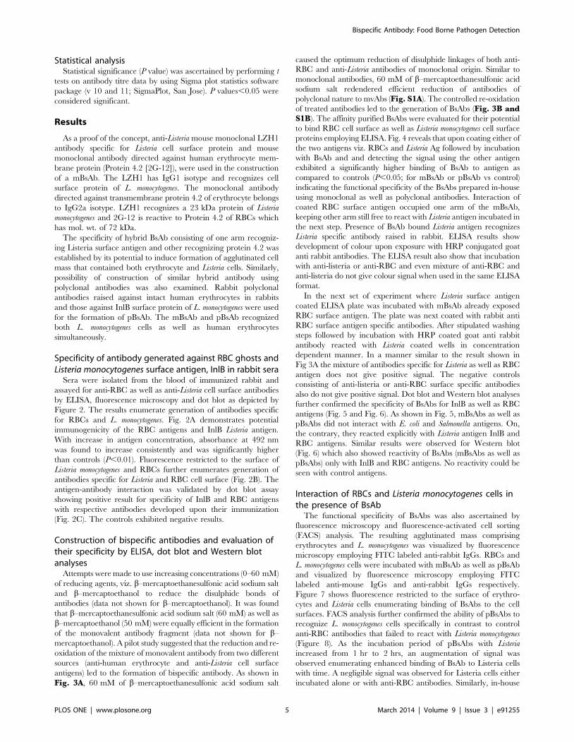
proteins employing ELISA. Fig. 4 reveals that upon coating either of
the two antigens viz. RBCs and Listeria Ag followed by incubation
with BsAb and and detecting the signal using the other antigen
exhibited a significantly higher binding of BsAb to antigen as
compared to controls (P,0.05; for mBsAb or pBsAb vs control)
indicating the functional specificity of the BsAbs prepared in-house
using monoclonal as well as polyclonal antibodies. Interaction of
coated RBC surface antigen occupied one arm of the mBsAb,
keeping other arm still free to react with Listeria antigen incubated in
the next step. Presence of BsAb bound Listeria antigen recognizes
Listeria specific antibody raised in rabbit. ELISA results show
development of colour upon exposure with HRP conjugated goat
anti rabbit antibodies. The ELISA result also show that incubation
with anti-listeria or anti-RBC and even mixture of anti-RBC and
anti-listeria do not give colour signal when used in the same ELISA
format.
In the next set of experiment where Listeria surface antigen
coated ELISA plate was incubated with mBsAb already exposed
RBC surface antigen. The plate was next coated with rabbit anti
RBC surface antigen specific antibodies. After stipulated washing
steps followed by incubation with HRP coated goat anti rabbit
antibody reacted with Listeria coated wells in concentration
dependent manner. In a manner similar to the result shown in
Fig 3A the mixture of antibodies specific for Listeria as well as RBC
antigen does not give positive signal. The negative controls
consisting of anti-listeria or anti-RBC surface specific antibodies
also do not give positive signal. Dot blot and Western blot analyses
further confirmed the specificity of BsAbs for InlB as well as RBC
antigens (Fig. 5 and Fig. 6). As shown in Fig. 5, mBsAbs as well as
pBsAbs did not interact with E. coli and Salmonella antigens. On,
the contrary, they reacted explicitly with Listeria antigen InlB and
RBC antigens. Similar results were observed for Western blot
(Fig. 6) which also showed reactivity of BsAbs (mBsAbs as well as
pBsAbs) only with InlB and RBC antigens. No reactivity could be
seen with control antigens.
Interaction of RBCs and Listeria monocytogenes cells inthe presence of BsAb
The functional specificity of BsAbs was also ascertained by
fluorescence microscopy and fluorescence-activated cell sorting
(FACS) analysis. The resulting agglutinated mass comprising
erythrocytes and L. monocytogenes was visualized by fluorescence
microscopy employing FITC labeled anti-rabbit IgGs. RBCs and
L. monocytogenes cells were incubated with mBsAb as well as pBsAb
and visualized by fluorescence microscopy employing FITC
labeled anti-mouse IgGs and anti-rabbit IgGs respectively.
Figure 7 shows fluorescence restricted to the surface of erythro-
cytes and Listeria cells enumerating binding of BsAbs to the cell
surfaces. FACS analysis further confirmed the ability of pBsAbs to
recognize L. monocytogenes cells specifically in contrast to control
anti-RBC antibodies that failed to react with Listeria monocytogenes
(Figure 8). As the incubation period of pBsAbs with Listeria
increased from 1 hr to 2 hrs, an augmentation of signal was
observed enumerating enhanced binding of BsAb to Listeria cells
with time. A negligible signal was observed for Listeria cells either
incubated alone or with anti-RBC antibodies. Similarly, in-house
Bispecific Antibody: Food Borne Pathogen Detection
PLOS ONE | www.plosone.org 5 March 2014 | Volume 9 | Issue 3 | e91255

Figure 2. Specificity characterization of antibody generated against RBC ghosts and Listeria monocytogenes in rabbit sera. A) ELISAanalysis: Antibody titer against Listeria surface antigens and RBC ghost proteins. Data are represented as mean 6 standard error of three independentexperiments (P values; Anti-RBC Ab vs control ,0.01, Anti-List Ab vs control ,0.01). B) Fluorescence microscopy: Anti-Listeria antibody interactionwith cell surface proteins of Listeria monocytogenes (a and b), anti-RBC antibody binding to human RBCs (c and d). C) Dot blot analysis: a and bexhibit recognition of Listeria surface antigens by monospecific anti-Listeria antibody and interaction of RBC ghost proteins by anti-RBC antibodyrespectively. The controls where InlB antigen of Listeria (c) and RBC antigens (d) were spotted onto PVDF membrane respectively. InlB antigens wereallowed to react with anti-RBC while RBC antigens were allowed to interact with anti-InlB monospeific antibodies respectively. At least threeindependent experiments were performed for each sample and data are representative of three independent experiments with similar observations.doi:10.1371/journal.pone.0091255.g002
Figure 3. Optimisation of Redox method. Non-Reducing SDS-PAGE analysis of mvAb and BsAb of monoclonal origin. A range of b-mercaptoethanesulphonic acid concentrations were analysed for effective reduction monoclonal anti-erythrocyte antibodies. b-mercaptoethane-sulphonic acid concentration of 60 mM (reducing condition) efficiently cleaved the inter- disulphide bridges between heavy chains and lead toformation of mvAb ($75 kD) of monoclonal anti-RBC antibodies (A). Similarly, 60 mM of b-mercaptoethanesulphonic also rendered effectivereduction of monoclonal anti-Listeria antibodies (data not shown). Dialysis against PBS (oxidizing condition) resulted in reformation of mvAb againstRBC and Listeria into BsAb (B). The lanes marked mAb, red., oxid. and M in B enumerate anti-Listeria monoclonal antibody, reduction of anti-ListeriamAb using 60 mM of b-mercaptoethanesulphonic acid, oxidation of anti-RBC mvAb and anti-Listeria mvAb and protein marker respectively. H2L2 =whole antibody, HL = mvAb, H = heavy chain, red. = reducing condition, oxid. = oxidizing condition. At least two independent experiments werecarried out for each sample and data are representative of two independent experiments with similar observations.doi:10.1371/journal.pone.0091255.g003
Bispecific Antibody: Food Borne Pathogen Detection
PLOS ONE | www.plosone.org 6 March 2014 | Volume 9 | Issue 3 | e91255
prepared BsAbs did not recognize unrelated organisms such as E.
coli and Salmonella typhimurium (data not shown).
Detection of Listeria monocytogenes cells byagglutination assay and its sensitivity determination
To establish further that both the anti-RBC and the anti-Listeria
antigen binding sites were located on the two arms of the same
bispecific antibody, we performed agglutination tests by incubating
two fold serial dilution of mBsAbs as well as pBsAbs with the
mixture of RBCs and Listeria cells. Listeria monocytogenes cells
containing many copies of Listeria antigens (cf. InlB) can act as a
bridge between two or more BsAbs and accentuate agglutination.
As shown in figure 9, both mBsAbs and pBsAbs caused the
agglutination in the presence of Listeria cells. The BsAbs mediated
agglutination was also studied in 96 well plate formats (Figure 10).
In concordance with previous result, mBsAbs as well as pBsAbs
were found to be equally efficient in inducing agglutination in the
presence of Listeria cells. Figure 11 shows fluorescence micrograph
of agglutinated mass, where the BsAbs of monoclonal as well as
polyclonal origins facilitating the agglutination of two cell
populations (L. monocytogenes as well as erythrocytes) are highlighted
by FITC-conjugated antibody.
Keeping into consideration the fact that to evaluate potential of
BsAbs for analysis of food samples, it was necessary to check their
sensitivity in agglutination assay. Various number of Listeria cells
were incubated with a fixed density of RBCs in the presence of
BsAbs (both mBsAbs and pBsAbs) to determine the sensitivity of
agglutination assay. Listeria cells when incubated with either
mBsAbs or pBsAbs at a density of at least 103 cells were found to
induce agglutination of the RBCs (Figure 12). The specificity of in-
house prepared BsAb was established employing non-related
pathogens such as E. coli and S. typhimurium. The presence of two
unrelated pathogens even at very high concentration (107 cells) did
not induce agglutination of the erythrocyte (Figure 12).
Figure 4. Specificity determination of in-house prepared BsAb by ELISA. Either Listeria Ag or RBC antigen was coated followed byrecognition with BsAb so that its one arm was fully captured with the antigen. Further RBC Ag or Listeria Ag (which was not used for coating) wasadded for stipulated time period. The plate was thoroughly washed and allowed to interact with Rabbit anti RBC antibody or Rabbit anti Listeriaantibody. Finally, the signal was detected using HRP-tagged goat anti-rabbit Ab. In case of controls instead of BsAb, anti-RBC Ab or anti-Lis Ab ormixture of the two was added. A) RBC antigen was coated and anti-Lis mAb was used for detection. mBsAb was employed. B) Listeria Ag coated andanti-RBC mAb detected the signal. mBsAb was used. C) RBC antigen was coated and anti-Lis pAb was used for detection. pBsAb was exploited. D)Listeria Ag coated and anti-RBC pAb detected the signal. pBsAb was used. Only BsAbs could give significant signals. On the other hand, the controlsgave very feeble signals validating the formation of BsAb. At least three independent experiments were performed and data are represented as meanof at three independent experiments 6 SD value. (P values; BsAb vs anti-RBC control ,0.05, BsAb vs anti-Lis control ,0.05, BsAb vs anti-RBC and anti-Lis mixture ,0.05).doi:10.1371/journal.pone.0091255.g004
Bispecific Antibody: Food Borne Pathogen Detection
PLOS ONE | www.plosone.org 7 March 2014 | Volume 9 | Issue 3 | e91255

Validation of bispecific antibody based agglutinationassay detection method in food samples
As the ultimate purpose of the development of bispecific
antibodies was to use it for the detection of L. monocytogenes in food,
it is obligatory to validate its performance in food samples. The
commercially available food samples were confirmed to be free of
detectable pathogens. Therefore, testing the agglutination assay in
food was carried out by artificially inoculating with L. monocytogenes
meat (beef, mutton and chicken) samples. The bispecific antibodies
were found to be capable of agglutinating the blood when
incubated with artificially contaminated food samples (Figure 13).
Discussion
Infections and infectious diseases continue to plague human
population globally inspite of the improved hygiene, sanitation and
availability of a spectrum of antibiotics. Annually, a quarter of
population in US and ,20% population in UK are estimated to
be affected by food-borne diseases [28]. There have been multiple
outbreaks caused by Escherichia coli O157:H7 and Salmonella
respectively in US in years 2006 and 2007. The Escherichia coli
O157:H7 outbreaks have been associated with green leafy
vegetables like spinach and lettuce and a total of 199 people were
infected in 26 states by consuming contaminated spinach, out of
which 3 succumbed to the infection [29]. A Salmonella outbreak
spread through tomatoes and peanut butter caused severe illness in
288 individuals [28].
Data from developing/poor countries are fragmentary but that
available show that at any given time millions suffer from bacterial
infections or are under constant threat. Illiteracy, poverty and lack
of access to hygiene, healthy and uninfected food can aggravate
the problem. Besides causing food poisoning, food-borne patho-
gens can evoke other disease manifestations like stomach ulcers
(eg. Helicobacter pylori), tuberculosis (Mycobacterium tuberculosis),
meningitis (species of Streptococci, Neisseria), cholera (Vibrio cholerae),
sexually transmitted diseases, and nosocomial infections [30]. The
rapid detection of pathogens and other microbial contaminants in
food is therefore highly critical for ensuring the safety of
consumers.
The widespread occurrence of food-borne infections combined
with apparent lack of simple and rapid bio-sensing technique
persuaded us to develop an assay that can detect the presence of
pathogen specifically with naked eye. We were successful in
developing a simple strategy to rapidly but accurately identifying
the presence of Listeria without the need of any sophisticated
instrumentation. The agglutination of erythrocytes, which is
induced by a bifunctional antibody recognizing surface antigen
of Listeria and erythrocytes only in the presence of Listeria cells
could be visualized by the naked eye in 10-30 minutes. To the best
of our knowledge, this is the first report employing erythrocyte
surface specific bi-specific antibodies to screen the presence of
pathogens in sample foods and beverages.
The following can be inferred from the data of the present
study:
i. Redox method reported in the present study causes reduction
of parent antibodies leading to formation of monovalent
population. The oxidation mediated reassociation of light &
heavy chain leads to formation of BsAb that has dual
specificity for both erythrocyte as well as Listeria surface
protein simultaneously.
ii. The in-house developed BsAb antibody can recognize both
isolated (free) as well as cell surface bound antigen (Listeria
surface antigen InlB) of Listeria monocytogenes with one arm
while other arm can interact with erythrocyte surface antigen.
i. The co-incubation of erythrocyte as well as Listeria (whole
bacteria) in the presence of BsAb ensues in agglutination of
RBC as well as Listeria monocytogenes.
ii. The interaction of BsAb is specific as no agglutination takes
place in the presence of unrelated food borne bacteria such as
E. coli or Salmonella typhimurium.
iii. Erythrocyte agglutination is rapid and sensitive and can be
seen with both microscope as well as naked eyes. The
agglutination can take place in the presence of 102–103
Listeria cells.
Once formation of BsAb from monoclonal antibodies (mouse
monoclonal anti-Listeria LZH1 IgG1 and mouse monoclonal
IgG2a raised against the human erythrocyte membrane protein
(Protein4.2 [2G-12]) was established by Western blot analysis
(Figure 5), we were encouraged to construct BsAb of polyclonal
origin. Remarkably, it was also possible to prepare the BsAbs with
antibodies raised against intact red blood cells and InlB, an
indispensible protein of Listeria. Specific agglutination could be
induced by the BsAb constructed from either monoclonal or
polyclonal antibodies. The use of antibodies recognizing InlB, an
important surface antigen of virulent Listeria makes this approach
highly cost effective and addresses the risk of food sample
contamination with such important bacteria. We prefer to use
Figure 5. Dot blot analysis. A) a and b show recognition of Listeriasurface antigen and RBC ghost proteins by mBsAb respectively. c and dexhibit controls where PVDF membrane was coated with E. coli andSalmonella Ags respectively and allowed to react with mBsAb (specificto Listeria monocytogenes and RBCs). A negative result was obtained incontrols. B) a and b exhibit binding of pBsAb to Listeria surface proteinand RBC membrane proteins respectively. PVDF membrane coated withE. coli and Salmonella Ags shows negative result when allowed to reactwith pBsAb (c and d respectively). At least three independentexperiments were performed for each sample and data are represen-tative of three independent experiments with similar observations.doi:10.1371/journal.pone.0091255.g005
Bispecific Antibody: Food Borne Pathogen Detection
PLOS ONE | www.plosone.org 8 March 2014 | Volume 9 | Issue 3 | e91255
pBsAb over mBsAb as the latter does not jeopardizes loss of
recognition of single surface molecule in the mutated forms.
Reports that Listeria monocytogenes isolate can undergo a point
mutation that abolishes the recognition by a monoclonal antibody
is available [30].
The specificity of the BsAbs constructed from monoclonal as
well as polyclonal antibodies was ascertained by conventional
assays including ELISA, dot blot and Western blot analysis but
agglutination assay was the center stage experimental strategy.
This obviously overcame some of the limitations of ELISA,
Western blot or dot blot assays. Antibodies were raised against
RBC ghost and L. monocytogenes membrane protein (Inl B) as
revealed by ELISA, dot blot assay and fluorescence microscopy
(Figure 2). The ELISA result shown in Figure 4 confirms the
formation of bispecific antibody as depicted by comparatively
increased absorbance in response to BsAb binding with respect to
controls. The generated bispecific antibody could effectively bind
RBC ghosts and the surface antigens of L. monocytogenes was
confirmed by dot blot and Western blot analyses as revealed in
Figure 5 and Figure 6 respectively and cell surface restricted
fluorescence as depicted by fluorescence microscopic image
(Figure 7). The potential of the constructed bispecific antibody,
to recognize populations of L. monocytogenes was further confirmed
by fluorescence activated cell sorting analysis as well (Figure 8).
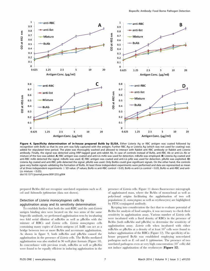
Figure 6. Western blot analysis. A) a and b show the reactivity of mBsAb to RBC ghost proteins and Listeria surface protein respectively while cand d exhibit incapability of mBsAb to react with E. coli and Salmonella Ags respectively. Similarly,pBsAb reacted well with RBC ghost proteins andListeria InlB protein (Ba and Bb respectively). On the other hand negative result is observed for E. coli and Salmonella proteins when pBsAb is used forrecognition (Bc and Bd respectively). At least three independent experiments were performed for each sample and data are representative of threeindependent experiments with similar observations.doi:10.1371/journal.pone.0091255.g006
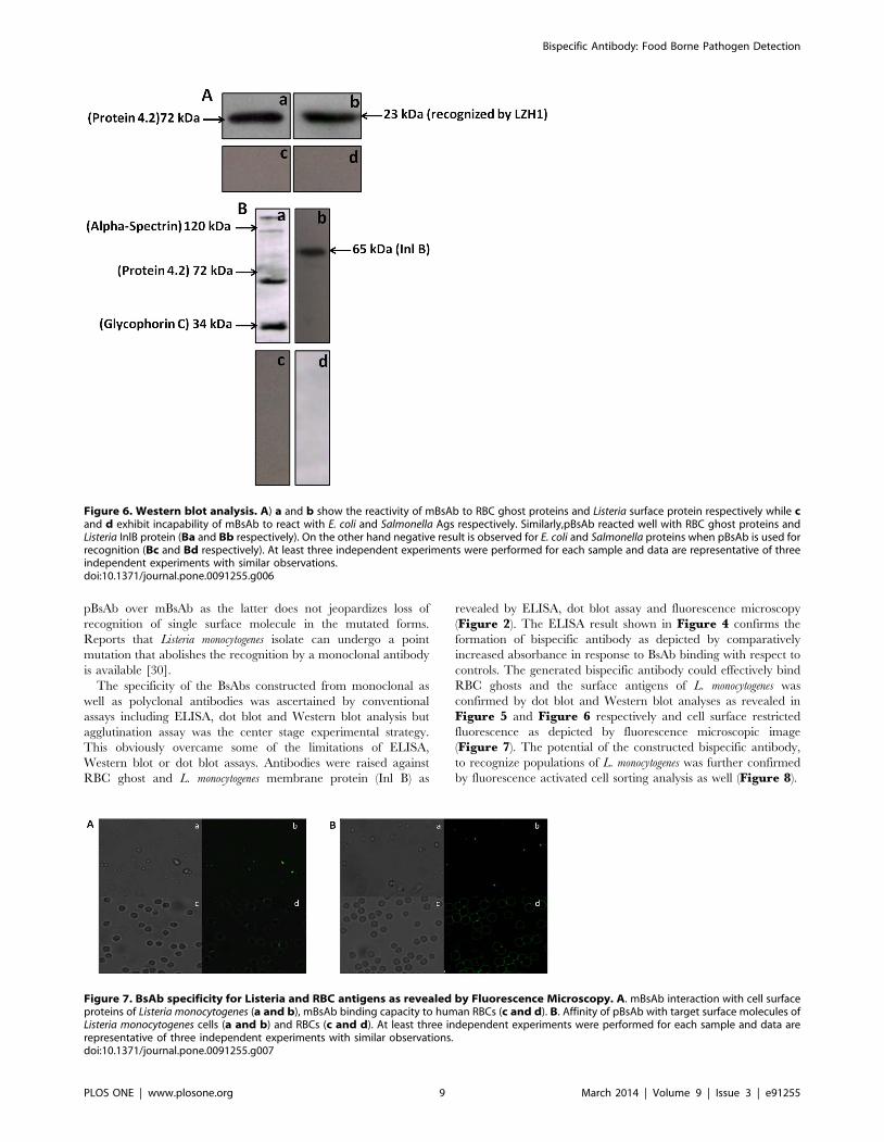
Figure 7. BsAb specificity for Listeria and RBC antigens as revealed by Fluorescence Microscopy. A. mBsAb interaction with cell surfaceproteins of Listeria monocytogenes (a and b), mBsAb binding capacity to human RBCs (c and d). B. Affinity of pBsAb with target surface molecules ofListeria monocytogenes cells (a and b) and RBCs (c and d). At least three independent experiments were performed for each sample and data arerepresentative of three independent experiments with similar observations.doi:10.1371/journal.pone.0091255.g007
Bispecific Antibody: Food Borne Pathogen Detection
PLOS ONE | www.plosone.org 9 March 2014 | Volume 9 | Issue 3 | e91255

Interestingly, the agglutination was evident under a light
microscope (Figure 9) and also to the naked eyes (Figure 10).
The higher dilution of antibody does not reveal the presence of an
agglutinated mass because of the decrease in the concentration of
bispecific antibodies. That bispecific antibodies adhered to the
agglutinated mass could be clearly seen in the fluorescence
microscopic image (Figure 11). The agglutination assay was
found to be sensitive to agglutinate cells with density more than or
equal to 103 cells (Figure 12). The BsAbs of either monoclonal or
polyclonal origin failed to agglutinate RBCs in the presence of
unrelated pathogens viz. E.coli and S. tyhimurium even at very high
densisities (Figure 12) revealing the specificity of the in-house
generated BsAbs. Moreover, as depicted in figure 13, the
agglutination assay was also able to detect L. monocytogenes from
artificially inoculated meat samples (beef, chicken and mutton)
although the commercially procured food samples were found to
be free from bacterial contamination (data not shown). A
schematic representation of the agglutination process employing
bispecific antibodies capable of recognizing RBCs and Listeria
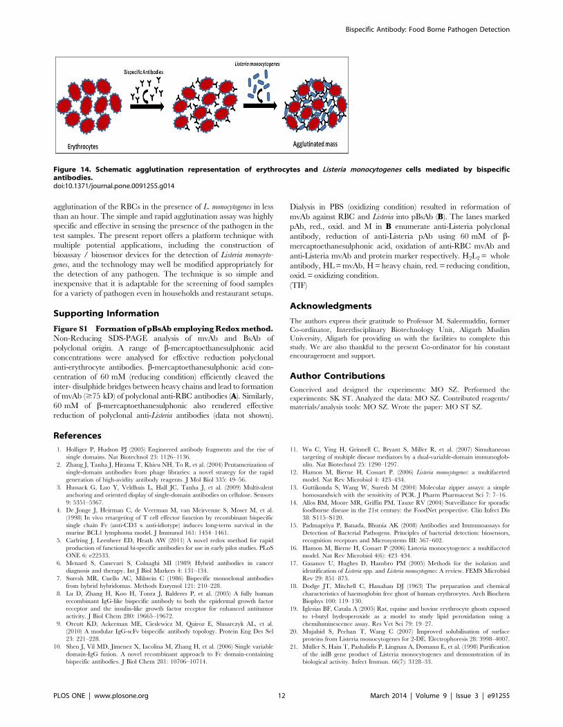
monocytogenes is depicted in Figure 14.
Finally, in the present report, we describe the construction of
BsAb based biosensor for the detection of L. monocytogenes employing
either monoclonal antibodies specific to human erythrocyte
membrane protein antigen (Protein 4.2 (2G-12)) or Listeria
monocytogenes surface antigen, or polyclonal antibodies recognizing
surface structures present on two cell populations. The use of
polyclonal antibodies recognizing more than one surface antigen,
makes production of BsAbs simpler and convenient. The remark-
able specificity of the technique employed is evident from the
absence of cross-reactivity of the in-house prepared BsAbs with
other pathogens such as E. coli and S. typhimurium. The strategy is
remarkably cost effective as it does not necessitate any sophisticated
instrumentation or expensive chemicals. The rapid onset of
Figure 8. Interaction of Listeria monocytogenes cells with BsAb employing fluorescence-activated cell sorting analysis. The cells withlog fluorescence intensities were gated BsAb and Listeria cells. (a) Listeria monocytogenes cells only, (b) Anti-RBC Ab incubated with L. monocytogenescells (c) Bispecific antibody of polyclonal origin (pBsAb) incubated with Listeria cells for 1 hr. At least three independent experiments were performedfor each sample and data are representative of three independent experiments with similar observations.doi:10.1371/journal.pone.0091255.g008
Figure 9. Bispecific antibody mediated haemagglutination.mBsAb (A) as well as pBsAb (B) rendered agglutination of RBCs andListeria cells on a glass slide. A given volume (50 ml of 200 ng/ml) ofBsAb specific against both RBCs and Listeria cell surface Ag was mixedwith Listeria cells-negative (Aa, Ba) or Listeria cells-positive (Ab, Bb).After 1 hr of incubation, the RBC agglutination results were observedunder a microscope (right panel; magnification, 20X). At least threeindependent experiments were performed for each sample and dataare representative of three independent experiments with similarobservations.doi:10.1371/journal.pone.0091255.g009
Figure 10. Demonstration of hemagglutination employingmBsAb (A) and pBsAb (B) against human red blood cells andListeria cells. The wells contain a constant number of RBC (20 ml of60% hematocrit) and Listeria cells (103 cells) plus serial two-folddilutions of BsAb. The spread pattern in the experimental seriesindicates positive hemagglutination through well 5 and 4 respectivelyfor mBsAb and pBsAb incubated wells. The wells marked as a, b and cwere incubated with anti-Lis Ab, anti-RBC Ab and a mixture of thesetwo respectively. At least three independent experiments wereperformed for each sample and data are representative of threeindependent experiments with similar observations.doi:10.1371/journal.pone.0091255.g010
Bispecific Antibody: Food Borne Pathogen Detection
PLOS ONE | www.plosone.org 10 March 2014 | Volume 9 | Issue 3 | e91255

agglutination reaction visible to the naked eyes makes it highly
suitable for maintaining the quality control over food supply in
various eatery outlets.
Conclusion
We describe in this report the successful application of a
bispecific antibody for the rapid and specific detection of Listeria in
food and other biological samples. The BsAbs induced visible
Figure 11. Fluorescence microscopy demonstrating RBC andListeria cells agglutination in the presence of mBsAb (A) andpBsAb (B) generated for simultaneous interaction with bothRBC as well Listeria cell surface antigen. Phase contrast (Aa and Ba)and fluorescence images (Ab and Bb) at x20. Same as Ba and Bb at x40(Bc and Bd). At least three independent experiments were performedfor each sample and data are representative of three independentexperiments with similar observations.doi:10.1371/journal.pone.0091255.g011
Figure 12. Sensitivity of agglutination assay. Various densities of Listeria cells (from wells 1-10) were incubated with fixed number of RBC cellsin serial two-fold dilutions of BsAbs. Listeria cells with a density of 103 or above induced agglutination of RBCs in the presence of mBsAbs (A) as wellas pBsAbs (B). Wells 11 and 12 harbor E. coli and S. typhimurium cells. No agglutination is observed when these unrelated pathogens are incubatedwith BsAbs (mBsAbs or pBsAbs) even at densities as high as 107 cells. At least three independent experiments were performed for each sample anddata are representative of three independent experiments with similar observations.doi:10.1371/journal.pone.0091255.g012
Figure 13. Detection of Listeria monocytogenes using agglutina-tion assay in inoculated meat (beef, chicken and mutton)samples. mBsAbs as well as pBsAbs rendered agglutination of RBCswhen incubated with Listeria inoculated meat. The wells marked as ‘B’are controls lacking Listeria cells. At least three independent experi-ments were performed for each sample and data are representative ofthree independent experiments with similar observations.doi:10.1371/journal.pone.0091255.g013
Bispecific Antibody: Food Borne Pathogen Detection
PLOS ONE | www.plosone.org 11 March 2014 | Volume 9 | Issue 3 | e91255
agglutination of the RBCs in the presence of L. monocytogenes in less
than an hour. The simple and rapid agglutination assay was highly
specific and effective in sensing the presence of the pathogen in the
test samples. The present report offers a platform technique with
multiple potential applications, including the construction of
bioassay / biosensor devices for the detection of Listeria monocyto-
genes, and the technology may well be modified appropriately for
the detection of any pathogen. The technique is so simple and
inexpensive that it is adaptable for the screening of food samples
for a variety of pathogen even in households and restaurant setups.
Supporting Information
Figure S1 Formation of pBsAb employing Redox method.Non-Reducing SDS-PAGE analysis of mvAb and BsAb of
polyclonal origin. A range of b-mercaptoethanesulphonic acid
concentrations were analysed for effective reduction polyclonal
anti-erythrocyte antibodies. b-mercaptoethanesulphonic acid con-
centration of 60 mM (reducing condition) efficiently cleaved the
inter- disulphide bridges between heavy chains and lead to formation
of mvAb ($75 kD) of polyclonal anti-RBC antibodies (A). Similarly,
60 mM of b-mercaptoethanesulphonic also rendered effective
reduction of polyclonal anti-Listeria antibodies (data not shown).
Dialysis in PBS (oxidizing condition) resulted in reformation of
mvAb against RBC and Listeria into pBsAb (B). The lanes marked
pAb, red., oxid. and M in B enumerate anti-Listeria polyclonal
antibody, reduction of anti-Listeria pAb using 60 mM of b-
mercaptoethanesulphonic acid, oxidation of anti-RBC mvAb and
anti-Listeria mvAb and protein marker respectively. H2L2 = whole
antibody, HL = mvAb, H = heavy chain, red. = reducing condition,
oxid. = oxidizing condition.
(TIF)
Acknowledgments
The authors express their gratitude to Professor M. Saleemuddin, former
Co-ordinator, Interdisciplinary Biotechnology Unit, Aligarh Muslim
University, Aligarh for providing us with the facilities to complete this
study. We are also thankful to the present Co-ordinator for his constant
encouragement and support.
Author Contributions
Conceived and designed the experiments: MO SZ. Performed the
experiments: SK ST. Analyzed the data: MO SZ. Contributed reagents/
materials/analysis tools: MO SZ. Wrote the paper: MO ST SZ.
References
1. Holliger P, Hudson PJ (2005) Engineered antibody fragments and the rise of
single domains. Nat Biotechnol 23: 1126–1136.
2. Zhang J, Tanha J, Hirama T, Khieu NH, To R, et al. (2004) Pentamerization of
single-domain antibodies from phage libraries: a novel strategy for the rapid
generation of high-avidity antibody reagents. J Mol Biol 335: 49–56.
3. Hussack G, Luo Y, Veldhuis L, Hall JC, Tanha J, et al. (2009) Multivalent
anchoring and oriented display of single-domain antibodies on cellulose. Sensors
9: 5351–5367.
4. De Jonge J, Heirman C, de Veerman M, van Meirvenne S, Moser M, et al.
(1998) In vivo retargeting of T cell effector function by recombinant bispecific
single chain Fv (anti-CD3 x anti-idiotype) induces long-term survival in the
murine BCL1 lymphoma model. J Immunol 161: 1454–1461.
5. Carlring J, Leenheer ED, Heath AW (2011) A novel redox method for rapid
production of functional bi-specific antibodies for use in early pilot studies. PLoS
ONE 6: e22533.
6. Menard S, Canevari S, Colnaghi MI (1989) Hybrid antibodies in cancer
diagnosis and therapy. Int J Biol Markers 4: 131–134.
7. Suresh MR, Cuello AC, Milstein C (1986) Bispecific monoclonal antibodies
from hybrid hybridomas. Methods Enzymol 121: 210–228.
8. Lu D, Zhang H, Koo H, Tonra J, Balderes P, et al. (2005) A fully human
recombinant IgG-like bispecific antibody to both the epidermal growth factor
receptor and the insulin-like growth factor receptor for enhanced antitumor
activity. J Biol Chem 280: 19665–19672.
9. Orcutt KD, Ackerman ME, Cieslewicz M, Quiroz E, Slusarczyk AL, et al.
(2010) A modular IgG-scFv bispecific antibody topology. Protein Eng Des Sel
23: 221–228.
10. Shen J, Vil MD, Jimenez X, Iacolina M, Zhang H, et al. (2006) Single variable
domain-IgG fusion. A novel recombinant approach to Fc domain-containing
bispecific antibodies. J Biol Chem 281: 10706–10714.
11. Wu C, Ying H, Grinnell C, Bryant S, Miller R, et al. (2007) Simultaneous
targeting of multiple disease mediators by a dual-variable-domain immunoglob-
ulin. Nat Biotechnol 25: 1290–1297.
12. Hamon M, Bierne H, Cossart P. (2006) Listeria monocytogenes: a multifaceted
model. Nat Rev Microbiol 4: 423–434.
13. Guttikonda S, Wang W, Suresh M (2004) Molecular zipper assays: a simple
homosandwich with the sensitivity of PCR. J Pharm Pharmaceut Sci 7: 7–16.
14. Allos BM, Moore MR, Griffin PM, Tauxe RV (2004) Surveillance for sporadic
foodborne disease in the 21st century: the FoodNet perspective. Clin Infect Dis
38: S115–S120.
15. Padmapriya P, Banada, Bhunia AK (2008) Antibodies and Immunoassays for
Detection of Bacterial Pathogens. Principles of bacterial detection: biosensors,
recognition receptors and Microsystems III: 567–602.
16. Hamon M, Bierne H, Cossart P (2006) Listeria monocytogenes: a multifaceted
model. Nat Rev Microbiol 4(6): 423–434.
17. Gasanov U, Hughes D, Hansbro PM (2005) Methods for the isolation and
identification of Listeria spp. and Listeria monocytogenes: A review. FEMS Microbiol
Rev 29: 851–875.
18. Dodge JT, Mitchell C, Hanahan DJ (1963) The preparation and chemical
characteristics of haemoglobin free ghost of human erythrocytes. Arch Biochem
Biophys 100: 119–130.
19. Iglesias BF, Catala A (2005) Rat, equine and bovine erythrocyte ghosts exposed
to t-butyl hydroperoxide as a model to study lipid peroxidation using a
chemiluminescence assay. Res Vet Sci 79: 19–27.
20. Mujahid S, Pechan T, Wang C (2007) Improved solubilisation of surface
proteins from Listeria monocytogenes for 2-DE. Electrophoresis 28: 3998–4007.
21. Muller S, Hain T, Pashalidis P, Lingnau A, Domann E, et al. (1998) Purification
of the inlB gene product of Listeria monocytogenes and demonstration of its
biological activity. Infect Immun. 66(7): 3128–33.
Figure 14. Schematic agglutination representation of erythrocytes and Listeria monocytogenes cells mediated by bispecificantibodies.doi:10.1371/journal.pone.0091255.g014
Bispecific Antibody: Food Borne Pathogen Detection
PLOS ONE | www.plosone.org 12 March 2014 | Volume 9 | Issue 3 | e91255

22. Likhite N and Warawdekar UM (2011) A Unique Method for Isolation and
Solubilization of Proteins after Extraction of RNA from Tumor Tissue UsingTrizol. J Biomol Tech 22: 37–44.
23. Bratcher RL, Chong CA, Dray S (1974) The isolation & characterization of an
antisheep red blood cell antibody having limited heterogeneity. J Immunol112:1337–1346
24. Singhal A, Bali A and Gupta CM (1986) Antibody-mediated targeting ofliposomes to erythrocytes in whole blood. Biochem Biophys Acta 880:72–77.
25. Porath J, Olin B, Granstrand B (1983) Immobilized metal affinity chromatog-
raphy of serum proteins on gel immobilized group III A metal ions. ArchBiochem Biophys 225, 543–547.
26. Chauhan A, Zubair S, Tufail S, Sherwani A, Sajid M, et al. (2011) Fungus-mediated biological synthesis of gold nanoparticles: potential in detection of liver
cancer. Int J Nanomedicine 6: 2305–2319.
27. Ohk SH, Koo OK, Sen T, Yamamoto CM, Bhunia AK (2010) Antibody-
aptamer functionalized fibre-optic biosensor for specific detection of Listeriamonocytogenes from food. J Appl Microbiol 109:808–17.
28. Kendall PA, Hillers VV, Medeiros LC (2006) Food safety guidance for older
adults. Clin Infect Dis 42: 1298–1304.29. CDC. Update on multi-state outbreak of E. coli O157:H7 infections from fresh
spinach. (2006) In: Department of Health and Human Services (ed) E. coliO157:H7 Outbreak in Spinach. Center for Disease control and Prevention (CDC),
Atlanta, Georgia. Available: http://www.cdc.gov/foodborne/ecolispinach/
100606.htm.30. Bhunia AK (2006). Detection of significant bacterial pathogens and toxins of
interest in homeland security. In: Amass SF, Bhunia AK, Chaturvedi AR, DolkDR, Peeta S, and Atallah MJ, (eds) The Science of Homeland Security. Purdue
University Press, West Lafayette, Indiana. pp. 109–149.
Bispecific Antibody: Food Borne Pathogen Detection
PLOS ONE | www.plosone.org 13 March 2014 | Volume 9 | Issue 3 | e91255