Journal of the ASEAN Federation of Endocrine Societies
112
REVIEW ARTICLE COVID-19 and Thyroid Diseases: How the Pandemic Situation Affects Thyroid Disease Patients ORIGINAL ARTICLES Clinical Characteristics, Residual Beta-Cell Function and Pancreatic Auto-Antibodies in Thai people with Long-Standing Type 1 Diabetes Mellitus Prevalence of Vitamin B12 Deficiency and its Associated Factors among Patients with Type 2 Diabetes Mellitus on Metformin from a District in Malaysia The Association between Maternal Serum Vitamin D Levels and Gestational Diabetes Mellitus among Filipino Patients: A Cross-Sectional Study Prevalence and Risk Factors for Hypovitaminosis D among Healthy Adolescents in Kota Bharu, Kelantan Efficacy of Repetitive Transcranial Magnetic Stimulation (rTMS) in Inducing Weight Loss among Obese Filipino Patients: A Randomized Controlled Trial Relationship between Plasma Adiponectin Level and Corrected QT Interval in Smoker and Non-smoker Adult Male Subjects CASE REPORTS Triple Synchronous Tumors Presenting as Right Nasolabial Basal Cell Carcinoma, Papillary Thyroid Carcinoma and Prolactinoma: A Rare Case Report Who were those MEN hiding behind the Ulcers?: A Case Report Primary Partial Empty Sella presenting with Prepubertal Hypogonadotropic Hypogonadism: A Case Report Bilateral Genu Valgum in an Adolescent with Primary Hyperparathyroidism: A Case Report and Review of Literature CASE SERIES Agoitrous Graves’ Hyperthyroidism with Markedly Elevated Thyroid Stimulating Immunoglobulin Titre displaying Rapid Response to Carbimazole with Discordant Thyroid Function Legions of Presentations of Myxedema Coma: A Case Series from a Tertiary Hospital in India Dome-Shaped Pituitary Enlargement in Primary Hypothyroidism: Avoiding Neurosurgical Interventions Journal of the ASEAN Federation of Endocrine Societies J A E Vol. 35 No. 2 November 2020 | ISSN 0857-1074 (Print)| eISSN 2308-118x (Online) www.asean-endocrinejournal.org Now Indexed in Indexed in PubMed Central
-
Upload
khangminh22 -
Category
Documents
-
view
1 -
download
0
Transcript of Journal of the ASEAN Federation of Endocrine Societies

REVIEW ARTICLECOVID-19 and Thyroid Diseases: How the Pandemic Situation Affects Thyroid Disease Patients
ORIGINAL ARTICLESClinical Characteristics, Residual Beta-Cell Function and Pancreatic Auto-Antibodies in Thai people with Long-Standing Type 1 Diabetes Mellitus
Prevalence of Vitamin B12 Deficiency and its Associated Factors among Patients with Type 2 Diabetes Mellitus on Metformin from a District in Malaysia
The Association between Maternal Serum Vitamin D Levels and Gestational Diabetes Mellitus among Filipino Patients: A Cross-Sectional Study
Prevalence and Risk Factors for Hypovitaminosis D among Healthy Adolescents in Kota Bharu, Kelantan
Efficacy of Repetitive Transcranial Magnetic Stimulation (rTMS) in Inducing Weight Loss among Obese Filipino Patients: A Randomized Controlled Trial
Relationship between Plasma Adiponectin Level and Corrected QT Interval in Smoker and Non-smoker Adult Male Subjects
CASE REPORTSTriple Synchronous Tumors Presenting as Right Nasolabial Basal Cell Carcinoma, Papillary Thyroid Carcinoma and Prolactinoma: A Rare Case Report
Who were those MEN hiding behind the Ulcers?: A Case Report
Primary Partial Empty Sella presenting with Prepubertal Hypogonadotropic Hypogonadism: A Case Report
Bilateral Genu Valgum in an Adolescent with Primary Hyperparathyroidism: A Case Report and Review of Literature
CASE SERIESAgoitrous Graves’ Hyperthyroidism with Markedly Elevated Thyroid Stimulating Immunoglobulin Titre displaying Rapid Response to Carbimazole with Discordant Thyroid Function
Legions of Presentations of Myxedema Coma: A Case Series from a Tertiary Hospital in India
Dome-Shaped Pituitary Enlargement in Primary Hypothyroidism: Avoiding Neurosurgical Interventions
Journal of theASEAN Federation ofEndocrine SocietiesJournal of theASEAN Federation ofEndocrine SocietiesVol. 35 No. 2 November 2020 | ISSN 0857-1074 (Print)| eISSN 2308-118x (Online)
www.asean-endocrinejournal.orgwww://asean-endocrinejournal.org
Vol. 32 No. 2 November 2017 | ISSN 0857-1074 | eISSN 2308-118x
The Journal of the ASEAN Federation of Endocrine Societies (JAFES) is an open-access, peer-reviewed, English language, medical and health science journal that is published two times a year by the ASEAN Federation of Endocrine Societies (AFES). Its editorial policies are aligned with the policies of the International Committtee of Medical Journal Editors (www.icmje.org).
JAFES welcomes manuscripts on all aspects of endocrinology and metabolism in the form of original articles, review articles, case reports, feature articles (clinical practice guidelines, clinical case seminars, clinical practice guidelines, book reviews, et cetera), editorials, letters to the Editor, brief communications and special announcements. Authors may include members and non-members of the AFES.
Authors are required to accomplish, sign and submit scanned copies of the JAFES Author Form consisting of: (1) Authorship Certification, that the manuscript has been read and approved by all authors, and that the requirements for authorship have been met by each author; (2) the Author Declaration, that the article represents original material that is not being considered for publication or has not been published or accepted for publication elsewhere; (3) the Statement of Copyright Transfer [accepted manuscripts become the permanent property of the JAFES and are licensed with an Attribution-Share Alike-Non-Commercial Creative Commons License. Articles may be shared and adapted for non-commercial purposes as long as they are properly cited]; and the ICMJE form for Disclosure of Potential Conflicts of Interest. For original articles, authors are required to submit a scanned copy of the Ethics Review Approval of their research. Consent forms, as appropriate, have been secured for the publication of information about patients.
Articles and any other material published in the JAFES represent the work of the author(s) and should not be construed to reflect the opinions of the Editors or the Publisher. JAFES does not charge any article processing or submission fees to authors. It likewise does not ask for subscription fees to gain access to scholarly content.
EDITORIAL CONTACT INFORMATION: Journal of the ASEAN Federation of Endocrine Societies | Unit 2005, 20th floor, Medical Plaza Ortigas, San Miguel Avenue, Ortigas Center, Pasig City, Philippines 1605 | Editorial Coordinator: Amado O. Tandoc III, MD, FPSP | Telefax: (+632) 637-3162 | E-mail: [email protected]
Copyright © 2017 by the Journal of the ASEAN Federation of Endocrine Societies
Now Indexed inIndexed inPubMedCentral

* This Advertisement is a complimentary service of the JAFES for member societies/organizations.
SAVEtheDATE
AOCE
AFESICE2022
20TH INTERNATIONAL CONGRESS OF ENDOCRINOLOGY18TH ASIA OCEANIA CONGRESS OF ENDOCRINOLOGY
21ST ASEAN FEDERATION OF ENDOCRINE SOCIETIES CONGRESS
25 - 28 August 2022
www. i c e 2 0 2 2 s i n g a p o r e . c om
Held in Supported by Managed by
Organised by

The Journal of the ASEAN Federation of Endocrine Societies (JAFES) is an open-access, peer-reviewed, English language, medical and health science journal that is published two times a year by the ASEAN Federation of Endocrine Societies (AFES). Its editorial policies are aligned with the policies of the International Committee of Medical Journal Editors (www.icmje.org), and resolves ethical issues using recommendations and guidelines of the Committee on Publication Ethics (COPE). It is a member of the World Association of Medical Editors (WAME) and CrossRef, and indexed in PubMed Central, Scopus, Web of Science (WoS), ASEAN Citation Index (ACI), Directory of Open Access Journals (DOAJ), Western Pacific Index Medicus (WPRIM) and APAMED Central.
JAFES welcomes manuscripts on all aspects of endocrinology and metabolism in the form of original articles, review articles, case reports, feature articles (clinical practice guidelines, clinical case seminars, clinical practice guidelines, book reviews, et cetera), editorials, letters to the Editor, brief communications and special announcements. Authors may include members and non-members of the AFES.
Authors are required to accomplish, sign and submit scanned copies of the JAFES Author Form consisting of: (1) Authorship Certification, that the authors contributed substantially to the work; that the manuscript has been read and approved by all authors, and that the requirements for authorship have been met by each author; (2) the Author Declaration, that the article represents original material that is not being considered for publication or has not been published or accepted for publication elsewhere; that the article does not infringe or violate any copyrights or intellectual property rights, and that no references have been made to predatory/suspected journals; (3) the Author Contribution Disclosure, which lists the specific contributions of authors; and (4) the Author Publishing Agreement which retains author copyright, grants publishing and distribution rights to JAFES, and allows JAFES to apply and enforce an Attribution Non-Commercial Creative Commons user license. Authors are also required to submit the signed ICMJE form for Disclosure of Potential Conflicts of Interest. For original articles, authors are required to submit a scanned copy of the Ethics Review Approval of their research as well as registration in trial registries as appropriate. For manuscripts reporting data from studies involving animals, authors are required to submit a scanned copy of the Institutional Animal Care and Use Committee approval. Consent forms, as appropriate, have been secured for the publication of information about patients.
Articles and any other material published in the JAFES represent the work of the author(s) and should not be construed to reflect the opinions of the Editors or the Publisher. JAFES does not charge any article processing or submission fees to authors. It likewise does not ask for subscription fees to gain access to scholarly content.
EDITORIAL CONTACT INFORMATION: Journal of the ASEAN Federation of Endocrine Societies | Unit 2005, 20th floor, Medical Plaza Ortigas, San Miguel Avenue, Ortigas Center, Pasig City, Philippines 1605 | Editorial Coordinator: Amado O. Tandoc III, MD, FPSP | Telefax: (+632) 8637-3162 | E-mail: [email protected]; [email protected].
Journal of theASEAN Federation ofEndocrine SocietiesJournal of theASEAN Federation ofEndocrine SocietiesVol. 35 No. 2 November 2020 | ISSN 0857-1074 (Print)| eISSN 2308-118x (Online)
www.asean-endocrinejournal.orgwww://asean-endocrinejournal.org
Vol. 32 No. 2 November 2017 | ISSN 0857-1074 | eISSN 2308-118x
The Journal of the ASEAN Federation of Endocrine Societies (JAFES) is an open-access, peer-reviewed, English language, medical and health science journal that is published two times a year by the ASEAN Federation of Endocrine Societies (AFES). Its editorial policies are aligned with the policies of the International Committtee of Medical Journal Editors (www.icmje.org).
JAFES welcomes manuscripts on all aspects of endocrinology and metabolism in the form of original articles, review articles, case reports, feature articles (clinical practice guidelines, clinical case seminars, clinical practice guidelines, book reviews, et cetera), editorials, letters to the Editor, brief communications and special announcements. Authors may include members and non-members of the AFES.
Authors are required to accomplish, sign and submit scanned copies of the JAFES Author Form consisting of: (1) Authorship Certification, that the manuscript has been read and approved by all authors, and that the requirements for authorship have been met by each author; (2) the Author Declaration, that the article represents original material that is not being considered for publication or has not been published or accepted for publication elsewhere; (3) the Statement of Copyright Transfer [accepted manuscripts become the permanent property of the JAFES and are licensed with an Attribution-Share Alike-Non-Commercial Creative Commons License. Articles may be shared and adapted for non-commercial purposes as long as they are properly cited]; and the ICMJE form for Disclosure of Potential Conflicts of Interest. For original articles, authors are required to submit a scanned copy of the Ethics Review Approval of their research. Consent forms, as appropriate, have been secured for the publication of information about patients.
Articles and any other material published in the JAFES represent the work of the author(s) and should not be construed to reflect the opinions of the Editors or the Publisher. JAFES does not charge any article processing or submission fees to authors. It likewise does not ask for subscription fees to gain access to scholarly content.
EDITORIAL CONTACT INFORMATION: Journal of the ASEAN Federation of Endocrine Societies | Unit 2005, 20th floor, Medical Plaza Ortigas, San Miguel Avenue, Ortigas Center, Pasig City, Philippines 1605 | Editorial Coordinator: Amado O. Tandoc III, MD, FPSP | Telefax: (+632) 637-3162 | E-mail: [email protected]
Copyright © 2017 by the Journal of the ASEAN Federation of Endocrine Societies
&Indexed
WEB OF SCIENCE ™
PubMed Central

ELIZABETH PAZ-PACHECOEditor-in-Chief
CECILIA A. JIMENOVice Editor-in-Chief
GABRIEL V. JASUL JR.MADE RATNA SARASWATI
WAN NAZAIMOON WAN MOHAMUDKYU KYU MAUNG
LIM SU-CHICHAICHARN DEEROCHANAWONG
NGUYEN THY KHUEAssociate Editors
MARY ANN R. ABACANLORNA R. ABAD
MARISSA M. ALEJANDRIAPIA D. BAGAMASBAD
YUPIN BENJASURATWONGCHNG CHIAW LING
NOR AZMI KAMARUDDINTINT SWE LATT
KHOO CHIN MENGNORLAILA MUSTAFA
NURAIN MOHD NOORNATHANIEL S. ORILLAZA JR.PAUL MATTHEW D. PASCO
AGUNG PRANOTOCATHERINE LYNN T. SILAO
ROGELIO V. TANGCONGUYEN VAN TUAN
MYO WINEditorial Board
MARIA LUISA PATRICIA B. GATBONTONChief Manuscript Editor
AIMEE A. ANDAG-SILVAMA. CECILLE S. AÑONUEVO-CRUZ
ELAINE C. CUNANANManuscript Editors
ROBERTO C. MIRASOLBusiness Manager
CATHERINE JESSICA MERCADO-LAZARORadiology Editor
JERICO B. GUTIERREZCreative Editor
MARITA V.T. REYESJOSE MA. C. AVILA
BENITO M. PACHECOEditorial Board Advisers
AMADO O. TANDOC IIIEditorial Coordinator
MELISSA O. TANDOCSecretary/Website Administrator
JOHANN FABRIAN Q. BOLINAOKIM L. COCHON
ETHEL M. ESTANISLAOAL JOSEPH R. MOLINA
JESUS N. SAROL JR.Statisticians
151
155
158
163
169
176
181
190
200
210
215
220
224
233
238
244248251254255
EDITORIALBetter Normal, A Silver Lining in 2020: JAFES is Accepted for Indexing in PubMed CentralElizabeth Paz-Pacheco
REVIEW ARTICLECOVID-19 and Thyroid Diseases: How the Pandemic Situation Affects Thyroid Disease PatientsLaurentius Aswin Pramono
ORIGINAL ARTICLESClinical Characteristics, Residual Beta-Cell Function and Pancreatic Auto-Antibodies in Thai people with Long-Standing Type 1 Diabetes MellitusYotsapon Thewjitcharoen, Sirinate Krittiyawong, Somboon Vongterapak, Soontaree Nakasatien, Suphab Aroonparkmongkol, Ishant Khurana, Assam El-Osta, Thep Himathongkam
Prevalence of Vitamin B12 Deficiency and its Associated Factors among Patients with Type 2 Diabetes Mellitus on Metformin from a District in MalaysiaGayathri Devi Krishnan, Miza Hiryanti Zakaria, Norhayati Yahaya
The Association between Maternal Serum Vitamin D Levels and Gestational Diabetes Mellitus among Filipino Patients: A Cross-Sectional StudyCarmen Carina Cabrera, Oliver Allan Dampil, Albert Macaire Ong-Lopez
Prevalence and Risk Factors for Hypovitaminosis D among Healthy Adolescents in Kota Bharu, KelantanSuhaimi Hussain and Maged Elnajeh
Efficacy of Repetitive Transcranial Magnetic Stimulation (rTMS) in Inducing Weight Loss among Obese Filipino Patients: A Randomized Controlled TrialMargaret Encarnacion, Oliver Allan Dampil, Ludwig Damian, Maria Leila Doquenia, Divina Cristy Redondo-Samin, Mary Karen Woolbright
Relationship between Plasma Adiponectin Level and Corrected QT Interval in Smoker and Non-smoker Adult Male SubjectsYin Thu Theint, Ei Ei Khin, Ohnmar Myint Thein, Mya Thanda Sein
CASE REPORTSTriple Synchronous Tumors Presenting as Right Nasolabial Basal Cell Carcinoma, Papillary Thyroid Carcinoma and Prolactinoma: A Rare Case ReportMateo Te III, Donnah Bless Lumanlan-Mosqueda, Kenny Jun Demegillo
Who were those MEN hiding behind the Ulcers?: A Case ReportShazatul Reza Binti Mohd Redzuan and Yong Sy Liang
Primary Partial Empty Sella presenting with Prepubertal Hypogonadotropic Hypogonadism: A Case ReportMaria Angela Matabang and Buena Sapang
Bilateral Genu Valgum in an Adolescent with Primary Hyperparathyroidism: A Case Report and Review of LiteratureSiow Ping Lee, Shu Teng Chai, Leh Teng Loh, Norhaliza Mohd Ali
CASE SERIESAgoitrous Graves’ Hyperthyroidism with Markedly Elevated Thyroid Stimulating Immunoglobulin Titre displaying Rapid Response to Carbimazole with Discordant Thyroid FunctionYin Chian Kon, Brenda Su Ping Lim, Yingshan Lee, Swee Eng Aw, Yoko Kin Yoke Wong
Legions of Presentations of Myxedema Coma: A Case Series from a Tertiary Hospital in IndiaNirmalya Roy, Anirban Majumder, Debmalya Sanyal, Soumyabrata Roy Chaudhuri, Suman Sarkar, Ankan Pathak
Dome-Shaped Pituitary Enlargement in Primary Hypothyroidism: Avoiding Neurosurgical InterventionsSatyam Chakraborty, Mona Tiwari, Rajan Palui, Kajari Bhattacharya, Kalyan Kumar Gangopadhyay
Instructions to AuthorsAuthorship Form ICMJE Form for Disclosure of Potential Conflicts of InterestPatient Consent FormPeer Reviewers
Emerging Sources Citation Index
COVID-19 continues to redefine the way we live and the way we work. We have no recourse but to embrace a different way of life: wearing masks and face shields in public, establishing physical and social distancing, and practicing hand hygiene at all times. As medical practitioners, educators, and researchers, we adapt our profession and practice in the context of this public health threat.
We have realized that there are novel and innovative ways to take care of our patients, confer and meet with our colleagues, teach our students, and mentor our Residents and Fellows. When it concerns our researches for purposes of understanding a disease and improving a policy, data gathering among patients in clinics and hospitals remains limited; yet different research designs and strategies still enable meaningful studies.
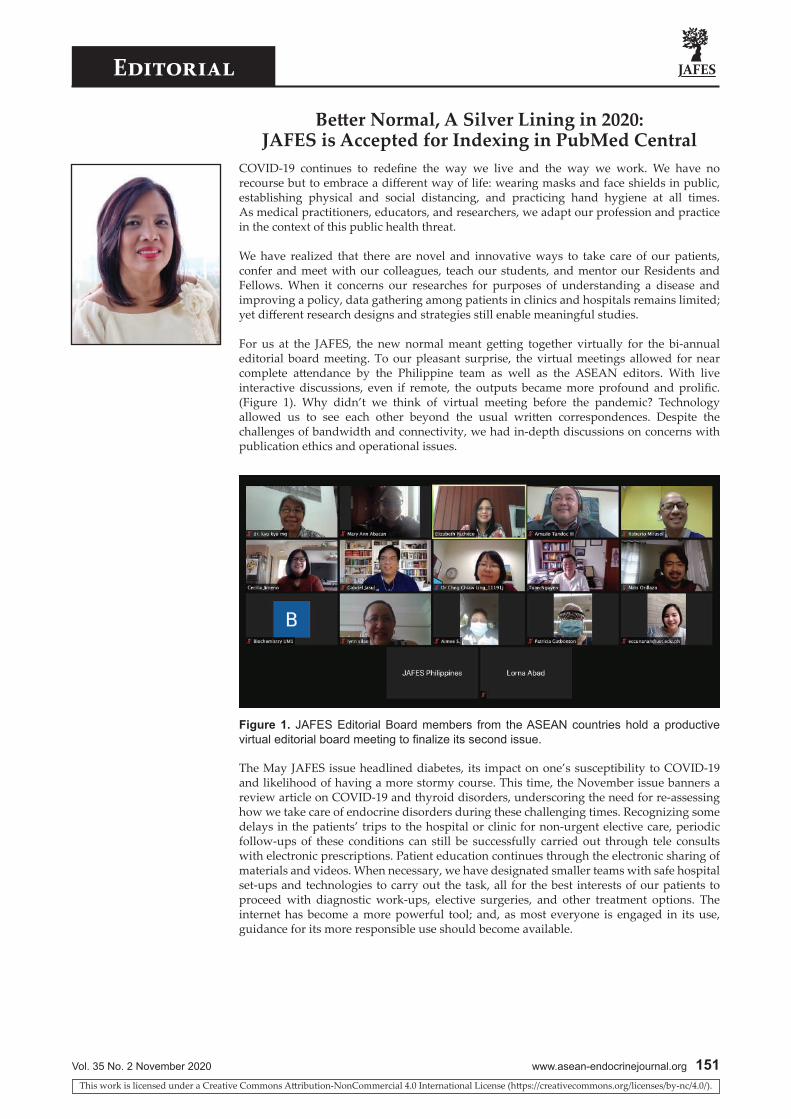
For us at the JAFES, the new normal meant getting together virtually for the bi-annual editorial board meeting. To our pleasant surprise, the virtual meetings allowed for near complete attendance by the Philippine team as well as the ASEAN editors. With live interactive discussions, even if remote, the outputs became more profound and prolific. (Figure 1). Why didn’t we think of virtual meeting before the pandemic? Technology allowed us to see each other beyond the usual written correspondences. Despite the challenges of bandwidth and connectivity, we had in-depth discussions on concerns with publication ethics and operational issues.
The May JAFES issue headlined diabetes, its impact on one’s susceptibility to COVID-19 and likelihood of having a more stormy course. This time, the November issue banners a review article on COVID-19 and thyroid disorders, underscoring the need for re-assessing how we take care of endocrine disorders during these challenging times. Recognizing some delays in the patients’ trips to the hospital or clinic for non-urgent elective care, periodic follow-ups of these conditions can still be successfully carried out through tele consults with electronic prescriptions. Patient education continues through the electronic sharing of materials and videos. When necessary, we have designated smaller teams with safe hospital set-ups and technologies to carry out the task, all for the best interests of our patients to proceed with diagnostic work-ups, elective surgeries, and other treatment options. The internet has become a more powerful tool; and, as most everyone is engaged in its use, guidance for its more responsible use should become available.
Better Normal, A Silver Lining in 2020:JAFES is Accepted for Indexing in PubMed Central
Vol. 35 No. 2 November 2020 151www.asean-endocrinejournal.org
JAFESEditorial
Figure 1. JAFES Editorial Board members from the ASEAN countries hold a productive virtual editorial board meeting to finalize its second issue.
This work is licensed under a Creative Commons Attribution-NonCommercial 4.0 International License (https://creativecommons.org/licenses/by-nc/4.0/).

________________________________________
https://doi.org/10.15605/jafes.035.02.15
Elizabeth Paz-PachecoEditor-in-Chief
Amid the uncertainties and challenges brought on by the COVID-19 pandemic, we celebrate another major milestone in the continuing journey of the JAFES. We formally announce here our acceptance to PubMed Central, (Figure 2), after being included in Scopus and Clarivate Analytics Emerging Sources Citation Index in the last 2 years. Launched in 2000, PubMed Central is a free archive of full-text biomedical and life sciences journal articles, serving as a digital counterpart to the print journal collection of the US National Library of Medicine. As a participating journal, JAFES shall be depositing full text articles starting from 2017 and these shall be available 100% open access and searchable also in MedLine.
The pandemic shook us out of our comfort zones, obliged us a new look at doing things, and led us to improve our ways and the effects on the people that we serve. We all look toward a better normal.
We wish everyone a safe and healthy end of 2020 and hope for a better New Year 2021!
Vol. 35 No. 2 November 2020
152
www.asean-endocrinejournal.org
Editorial
US National Library of MedicineNational Institutes of Health
Figure 2. JAFES passed the scientific quality and technical review by NML for PMC.

We are pleased to announce that the Journal of the ASEAN Federation of Endocrine Societies (JAFES) has been accepted for indexing in PubMed Central (PMC) after undergoing in-depth review of editorial content and policies, including a rigorous technical assessment.
This achievement is a significant addition to the JAFES’ list of credentials. JAFES is currently indexed in Scopus®, Emerging Sources Citation Index™ (ESCI) under Clarivate™ Analytics, ASEAN Citation Index (ACI), the Directory of Open Access Journals (DOAJ), and the Western Pacific Region Index Medicus (WPRIM).
All articles published in JAFES from 2017 to present shall now be indexed and made available 100% Open Access through PubMed Central, and searchable through PubMed. This is part of our commitment to authors to ensure wide reach of their scientific findings, and to our readers to assure you of the quality of our content.
Follow us in:
PubMed Central is afree archive of full-textbiomedical andlife sciences journalarticles, serving as a digital counterpart tothe print journal collection of theUS National Libraryof Medicine.
Journal of theASEAN Federation ofEndocrine Societies

* This Advertisement is a complimentary service of the JAFES for member societies/organizations.

COVID-19 and Thyroid Diseases:How the Pandemic Situation Affects Thyroid Disease Patients
Laurentius Aswin Pramono
Department of Public Health and Nutrition, School of Medicine and Health Sciences, Atma Jaya Catholic University of Indonesia, Jakarta, IndonesiaDepartment of Internal Medicine, Saint Carolus Hospital, Jakarta, Indonesia
Abstract
Patients with thyroid diseases need special attention during this COVID-19 pandemic. There is a paucity of publications that review the effect of coronavirus infection on thyroid disease patients, such as those with hyperthyroidism, hypothyroidism, thyroid nodules and cancer. This article aims to collect reviews and statements about how the COVID-19 pandemic has affected the management of thyroid disease patients.
Key words: COVID-19, thyroid disease, hyperthyroidism, hypothyroidism, thyroid cancer
________________________________________
ISSN 0857-1074 (Print) | eISSN 2308-118x (Online)Printed in the PhilippinesCopyright © 2020 by Pramono.Received: May 25, 2020. Accepted: July 13, 2020.Published online first: July 30, 2020.https://doi.org/10.15605/jafes.035.02.01
Corresponding author: Laurentius Aswin Pramono, MD, MSc (Epidemiology)Academic Staff, Department of Public Health and NutritionSchool of Medicine and Health SciencesAtma Jaya Catholic University of IndonesiaJalan Pluit Raya 2, North Jakarta, IndonesiaE-mail: [email protected], [email protected]: https://orcid.org/0000-0001-7271-7594
INTRODUCTION
The World Health Organization (WHO) announced the global pandemic situation caused by severe acute respiratory syndrome coronavirus 2 (SARS-CoV-2) as coronavirus disease 2019 (COVID-19) in March 2020.1 The disease has spread widely all over the world, including Southeast Asian countries.1 The pandemic is a great burden to all communities and populations. Older citizens and people with chronic and degenerative diseases are prone to severe illness if infected by the virus.2 Furthermore, morbidity and mortality caused by COVID-19 are higher in patients with chronic, metabolic and degenerative diseases such as diabetes, hypertension, cardiovascular disease, cancer, stroke and autoimmune diseases.3
As COVID-19 is a new illness, its effects on patients with thyroid disease are not yet known.4 Because of the prevalence of thyroid disease—1 to 2% for spontaneous hypothyroidism, 0.5 to 2% for hyperthyroidism in women (10 times more common than in men), and 1% and 5% for clinically detectable thyroid nodules in men and women, respectively—it may be surmised that patients with the disease may be affected directly and indirectly by the pandemic situation.5 This review aims to collect publications which discuss consequences of COVID-19 in thyroid disease patients.
PAThObIOlOgy
There are a limited number of publications on the effect of viral infections on thyroid pathology. A study on severe acute respiratory syndrome (SARS) patients during the outbreak in 2003 found that triiodothyronine (T3) and thyroxine (T4) levels were lower in SARS patients compared to controls, during the acute and convalescent phases.6
Autopsy findings in patients with SARS showed destruction of follicular and parafollicular thyroid cells which led to low T3 and T4.7 Viral infections can affect thyroid hormone synthesis causing low T4 and T3, a common phenomenon known as non-thyroidal illness syndrome.
Chronic viral infections can later lead to autoimmune disease, including thyroid diseases, such as Graves’ disease and Hashimoto’s thyroiditis. Viral infections account for major environmental factors in subacute and chronic auto-immune thyroiditis.8 Coxsackie virus, echovirus, Epstein-Barr virus, herpes simplex virus, mumps and parvovirus are thought to cause autoimmune thyroiditis. Presently, there are no published studies on the effect of COVID-19 and the risk for later autoimmune thyroid disease.
STATEmENTS FROm ThyROID SOCIETIES
Various thyroid interest groups, including the American Thyroid Association (ATA), the European Thyroid Association (ETA), the British Thyroid Foundation (BTF) and the American Association for Clinical Endocrinologists (AACE), have released statements in their respective official websites for the guidance of thyroid patients during the COVID-19 pandemic.4,9-11 The statements provide information for physicians and patients on how to deal with specific thyroid concerns during the pandemic.
Thyroid disorders may be generally divided to three major conditions, namely, hyperthyroidism, hypothyroidism, and thyroid nodules and cancer. Hyperthyroidism encompasses conditions caused by overactive thyroid function, resulting in elevated levels of thyroid hormones (free T4 and free T3) and low levels of thyroid stimulating hormone (TSH). This includes Graves’ disease, toxic multinodular goiter (Plummer’s disease) and toxic nodular
Vol. 35 No. 2 November 2020 155www.asean-endocrinejournal.org
Journal of theASEAN Federation ofEndocrine SocietiesReview Article
This work is licensed under a Creative Commons Attribution-NonCommercial 4.0 International License (https://creativecommons.org/licenses/by-nc/4.0/).

be prescribed for 3 or 4 months to limit clinic visits. Calcium and vitamin D must also be continued with the same dose.
ThyROID NODUlE AND CANCER
The urgency to perform fine needle aspiration (FNA) biopsy or thyroid cyst aspiration is determined by the patient’s risk factors, the characteristics of the nodule and clinical judgment.9 Generally, it is safe to perform FNA biopsy or cytology if standard personal protective equipment (PPE) are worn.13 If the nodule is highly suspicious for malignancy, has indicators of thyrotoxicosis, or with concomitant compression signs and symptoms, prompt referral to a head and neck, thyroid or oncologic surgeon to prepare for thyroidectomy is recommended. If the nodule is deemed moderately to highly suspicious and would be aided by preoperative cytology, FNA biopsy may be performed with a moderate level of PPE. If the nodule has very low to low suspicion of malignancy, biopsy may be postponed and the nodule may be observed by repeating thyroid ultrasonography at 3 to 6 months.
Thyroid cancer patients generally do not have a higher risk for COVID-19 infection. Majority of thyroid cancers are well-differentiated, specifically papillary and follicular thyroid cancer.4,9,10 Both types are slow growing, rarely aggressive, and infrequently metastasize to distant organs. Some well-differentiated cancers with aggressive character and higher stages from vascular invasion, lymph node metastases and distant spread pose a higher risk for viral infection including COVID-19. Morbidity and mortality can also increase, especially in those with lung metastases.11
Poorly-differentiated thyroid cancers, specifically medullary and anaplastic types, have a higher risk for morbidity and mortality from COVID-19 infection. These patients, and also those with aggressive variants of well-differentiated thyroid cancer, sometimes receive tyrosine kinase inhibitors (sorafenib, vandetanib or lenvatinib) after total thyroidectomy or radioactive iodine therapy. These agents suppress the immune system and may confer a higher risk of severe pneumonia in outbreak situations.4,9-11 Patients who have previously received external beam radiotherapy to the neck also have a higher risk for severe illness from COVID-19.4 All high risk thyroid cancer patients must then be advised to self-isolate and stay at home during the pandemic.
The decision to proceed with thyroid surgery during the pandemic is based on consideration of the surgeon, the patient’s condition, the hospital (i.e., availability of operating room), the availability of personal protective equipment (PPE) and adequate screening prior to surgery. Hospitals in Jakarta, such as the Saint Carolus Hospital, perform examinations including chest X-ray, chest computerized tomography scan, complete blood count, C-reactive protein, lactate dehydrogenase, rapid antibody test for IgM/IgG SARS-CoV2, real time-polymerase chain reaction for SARS-CoV2 of nose and throat specimens, and adequate preoperative consultation and screening with an internal medicine specialist or pulmonologist and an anesthesiologist.14 With the application of a complete general preoperative assessment including specific screening to prevent the spread of COVID-19, thyroid surgical procedures can be performed safely.
goiter (toxic adenoma). Hypothyroidism is a condition of low free T4 and free T3 levels, and high TSH, as seen in csongenital hypothyroidism, endemic goiter (iodine deficiency disorder), and chronic autoimmune thyroid disease (Hashimoto’s thyroiditis). Thyroid nodules and cancer are in the same category of neoplasia. The treatment of thyroid cancer includes surgery (thyroidectomy), radioactive iodine therapy (radioablation), and TSH suppression therapy with levothyroxine.
hyPERThyROIDISm
The ATA, BTF and AACE have stated that autoimmune thyroid disease which resulted to hyperthyroidism (Graves’ disease, multinodular toxic goiter or toxic adenoma) does not increase the risk for COVID-19 infection, morbidity or mortality.9-11 Patients maintained on anti-thyroid drugs or levothyroxine also do not have a higher risk for COVID-19 infection. It is important to continue thyroid medications since being untreated or suboptimally controlled may increase the risk of viral infection or complications.4,11
Treatment with anti-thyroid drugs (ATD) like propylthiouracil, carbimazole, methimazole may cause agranulocytosis, a very rare adverse reaction with symptoms resembling COVID-19. These include fever, sore throat and muscle pain.9,11 Physician assessment must be done promptly to ascertain the cause of the symptoms.
ATDs must be continued with the same dose and titration, as was done prior to the pandemic. The medications may be prescribed for two to three months to minimize the need for frequent clinic visits.9-11 Maintaining euthyroidism as long as possible can prevent more severe conditions such as thyroid storm, an unexpected severe complication of viral infections in uncontrolled hyperthyroid patients.12
In Graves’ orbitopathy or other forms of thyroid-related eye disease, the use of high dose (500 mg) intravenous glucocorticoid (methylprednisolone) weekly for six weeks may suppress the immune system and increase the risk for infection, including COVID-19; or worsen blood glucose control which can lead to prolonged length of stay in the hospital.11 The decision to give this therapy has to be made after having fully discussed the risks and benefits with the patient. The patient should be well advised to observe and maintain physical hygiene and distancing while on high dose corticosteroid therapy.
hyPOThyROIDISm
The various etiologies of hypothyroidism such as Hashimoto’s thyroiditis, post-thyroidectomy for thyroid nodule or thyroid cancer, post-radioactive iodine therapy or congenital hypothyroidism, are all treated with levothyroxine to maintain normal thyroid hormone levels. Hashimoto’s thyroiditis is a chronic autoimmune condition that may present with other autoimmune conditions. The BTF reassures patients that the condition does not increase the risk of COVID-19 infection, morbidity or mortality.4
Levothyroxine must be continued with the same dose as before the pandemic.4,9-11 Once the patient has attained euthyroidism (normal free T4 and TSH levels) with a known total dose in a day or a week, the medication may
Vol. 35 No. 2 November 2020
156
www.asean-endocrinejournal.org
Laurentius Aswin Pramono How the COVID-19 Pandemic Situation Affects Thyroid Disease Patients

Other issue is about radioactive iodine therapy during the COVID-19 pandemic. The ATA clearly states that radioactive iodine (RAI) therapy for patients with possible residual thyroid tissue after total thyroidectomy can be delayed for six months and is still effective.9 Moreover, RAI therapy does not increase the risk, morbidity and mortality of COVID-19.4,9,11 It should be noted that RAI therapy is highly dependent on the availability of adequate healthcare facilities. If the indication for RAI therapy is Graves’ hyperthyroidism or toxic multinodular goiter, anti-thyroid medications may be preferred, as these provide an easier and simpler means of achieving euthyroidism.
CONClUSION
During this time of the COVID-19 pandemic, thyroid disease patients must receive optimal therapy for each of their conditions, such as hyperthyroidism, thyroid eye disease, hypothyroidism, thyroid nodules and thyroid cancer. Statements from the American Thyroid Association, the European Thyroid Association, the British Thyroid Foundation and the American Association for Clinical Endocrinologists (AACE) can guide clinicians, physicians, endocrinologists and thyroid surgeons in their therapeutic decisions.
AcknowledgmentsThe author expresses his gratitude to the Department of Public Health and Nutrition, School of Medicine and Health Sciences, Atma Jaya Catholic University of Indonesia; the Department of Internal Medicine, Saint Carolus Hospital; and the Saint Carolus COVID-19 Outbreak Team for supporting the faculty in their teaching and academic activities, in clinical work, and in their endeavors in writing studies and reviews during the time of the COVID-19 pandemic.
Statement of AuthorshipThe author certifies fulfillment of ICMJE authorship criteria.
Author DisclosureThe author declared no conflicts of interest.
Funding SourceNone.
References 1. Ge H, Wang X, Yuan X, et al. The epidemiology and clinical
information about COVID-19. Eur J Clin Microbiol Infect Dis. 2020;39(6):1011-9. PMID: 32291542. PMCID: PMC7154215. https://doi.org/10.1007/s10096-020-03874-z.
2. Liu K, Chen Y, Lin R, Han K. Clinical features of COVID-19 in elderly patients: A comparison with young and middle-aged patients. J Infect. 2020;80(6):e14-8. PMID: 32171866. PMCID: PMC7102640. https://doi.org/10.1016/j.jinf.2020.03.005.
3. Yang J, Zheng Y, Gou X, et al. Prevalence of comorbidities and its effects in patients infected with SARS-CoV-2: A systematic review and meta-analysis. Int J Infect Dis. 2020;94:91-5. PMID: 32173574. PMCID: PMC7194638. https://doi.org/10.1016/j.ijid.2020.03.017.
4. British Thyroid Foundation. Thyroid disease and coronavirus (COVID-19). 4 May 2020. https://www.btf-thyroid.org/news/thyroid-disease-and-coronavirus-covid-19.
5. Vanderpump MPJ. Epidemiology of thyroid disorders. In: Luster M, Duntas L, Wartofsky L, eds. The Thyroid and Its Diseases: A Comprehensive Guide for the Clinician. Cham, Switzerland: Springer, 2019.
6. Pal R, Banerjee M. COVID-19 and the endocrine system: Exploring the unexplored. J Endocrinol Invest. 2020;43(7):1027-31. PMID: 32361826. PMCID: PMC7195612. https://doi.org/10.1007/s40618-020-01276-8.
7. Wei L, Sun S, Xu C, et al. Pathology of the thyroid in severe acute respiratory syndrome. Hum Pathol. 2007;38(1):95-102. PMID: 16996569. PMCID: PMC7112059. https://doi.org/10.1016/j.humpath.2006.06.011.
8. Desailloud R, Hober D. Viruses and thyroiditis: An update. Virol J. 2009;6(5). https://doi.org/10.1186/1743-422X-6-5.
9. American Thyroid Association. Novel coronavirus (COVID-19) and the thyroid: Resources. https://www.thyroid.org/covid-19/.
10. European Thyroid Association. COVID-19: Information and recommendations for patients with thyroid diseases. ETA Public Health Board Statement. https://www.eurothyroid.com/news/covid-19-thyroid-diseases.html.
11. American Association of Clinical Endocrinologists. AACE position statement: Coronavirus (COVID-19) and people with thyroid disease. https://www.aace.com/recent-news-and-updates/aace-position-statement-coronavirus-covid-19-and-people-thyroid-disease.
12. Baharoon SA. H1N1 infection-induced thyroid storm. Ann Thorac Med. 2010;5(2):110-2. PMID: 20582177. PMCID: PMC2883193. https://doi.org/10.4103/1817-1737.62475.
13. Vigliar E, Iaccarino A, Bruzzese D, et al. Cytology in the time of coronavirus disease (COVID-19): An Italian perspective. J Clin Pathol. 2020; jclinpath-2020-206614. PMID: 32312717. PMCID: PMC7211103. https://doi.org/10.1136/jclinpath-2020-206614.
14. Hardi F, Shinto R, Putra AC, Fahmi M, Wiyono WH, Pramono LA, et al. Coronavirus disease (Covid-19) service in Saint Carolus Hospital. Updated 29 April 2020. Saint Carolus Hospital, Jakarta, Indonesia.
Vol. 35 No. 2 November 2020
157
www.asean-endocrinejournal.org
Laurentius Aswin PramonoHow the COVID-19 Pandemic Situation Affects Thyroid Disease Patients
Table 1. Conditions that warrant urgent (<4 weeks) thyroid surgery1. Thyroid cancer which is life-threatening (large size), with local invasion to the trachea or recurrent laryngeal nerve, with aggressive features (rapid growing,
adherent to nearby organs, with distant metastases)2. Graves’ disease or toxic adenoma with severe or life-threatening symptoms, which cannot be controlled by anti-thyroid medications3. Goiter or enlargement of the thyroid gland with respiratory or gastrointestinal tract compressive symptoms4. Open core-biopsy (and removal, total or near-total thyroidectomy) for nodules highly suspicious for thyroid cancer, such as medullary thyroid cancer
(high calcitonin and with highly suspicious ultrasonographic characteristics), anaplastic thyroid cancer or thyroid lymphoma if other diagnostic modalities are equivocal or inconclusive
5. Pregnant patient with thyrotoxic and compressive symptoms presenting a life-threatening condition for the mother and fetus and cannot be controlled with anti-thyroid medications
Adapted from the American Thyroid Association. Novel coronavirus (COVID-19) and the thyroid. https://www.thyroid.org.9
Authors are required to accomplish, sign and submit scanned copies of the JAFES Author Form consisting of: (1) Authorship Certification, that authors contributed substantially to the work, that the manuscript has been read and approved by all authors, and that the requirements for authorship have been met by each author; (2) the Author Declaration, that the article represents original material that is not being considered for publication or has not been published or accepted for publication elsewhere, that the article does not infringe or violate any copyrights or intellectual property rights, and that no references have been made to predatory/suspected predatory journals; (3) the Author Contribution Disclosure, which lists the specific contributions of authors; and (4) the Author Publishing Agreement which retains author copyright, grants publishing and distribution rights to JAFES, and allows JAFES to apply and enforce an Attribution-Non-Commercial Creative Commons user license. Authors are also required to accomplish, sign, and submit the signed ICMJE form for Disclosure of Potential Conflicts of Interest. For original articles, authors are required to submit a scanned copy of the Ethics Review Approval of their research as well as registration in trial registries as appropriate. For manuscripts reporting data from studies involving animals, authors are required to submit a scanned copy of the Institutional Animal Care and Use Committee approval. For Case Reports or Series, and Images in Endocrinology, consent forms, are required for the publication of information about patients; otherwise, appropriate ethical clearance has been obtained from the institutional review board. Articles and any other material published in the JAFES represent the work of the author(s) and should not be construed to reflect the opinions of the Editors or the Publisher.

Clinical Characteristics, Residual beta-Cell Functionand Pancreatic Auto-Antibodies in Thai peoplewith long-Standing Type 1 Diabetes mellitus
Yotsapon Thewjitcharoen,1 Sirinate Krittiyawong,1 Somboon Vongterapak,1 Soontaree Nakasatien,1
Suphab Aroonparkmongkol,2 Ishant Khurana,3 Assam El-Osta,3 Thep Himathongkam1
1Diabetes and Thyroid Center, Theptarin Hospital, Bangkok, Thailand2Division of Pediatric Endocrinology, Department of Pediatrics, Faculty of Medicine, Chulalongkorn University, Bangkok, Thailand
3Epigenetics in Human Health and Disease Laboratory, Department of Diabetes, Central Clinical School,Faculty of Medicine Nursing and Health Sciences, Monash University, Melbourne, Australia
Abstract
Objectives. To describe the characteristics of long-standing T1DM in Thai patients and assess residual beta-cell function with status of pancreatic autoantibodies.
Methodology. This is a cross-sectional study of Thai subjects with T1DM and disease duration ≥ 25 years seen at the Theptarin Hospital. Random plasma C-peptide and pancreatic auto-antibodies (Anti-GAD, Anti-IA2, and Anti-ZnT8) were measured. Patients who developed complications were compared with those who remained free of complications.
Results. A total of 20 patients (males 65%, mean age 49.4±12.0 years, BMI 22.5±3.1 kg/m2, A1C 7.9±1.6%) with diabetes duration of 31.9±5.1 years were studied. Half of the participants remained free from any diabetic complications while the proportions reporting retinopathy, nephropathy, and neuropathy were 40%, 30%, and 15%, respectively. HDL cholesterol was significantly higher and triglyceride concentration significantly lower in patients who were free from diabetic nephropathy but not in those who were free from other complications. The prevalence rates of anti-GAD, anti-IA2, and anti-ZnT8 were 65%, 20%, and 10%, respectively. None of the patients who tested negative for both anti-GAD and anti-IA2 was positive for anti-ZnT8. Residual beta-cell function based on detectable random plasma C-peptide (≥ 0.1 ng/mL) and MMTT was found in only 3 patients (15%). There was no relationship between residual beta-cell function and protective effects of diabetic complications.
Conclusion. Endogenous insulin secretion persists in some patients with long-standing T1DM and half of long-standing T1DM in Thai patients showed no diabetic complications. HDL cholesterol was significantly higher and triglyceride concentration significantly lower in patients who were free from diabetic nephropathy.
Key words: type 1 diabetes mellitus, long-standing, residual beta-cell function, pancreatic autoantibodies, Thai people
INTRODUCTION
Emerging evidence in Caucasian populations suggests that endogenous insulin secretion persists in long-standing type 1 diabetes mellitus (T1DM). This is protective against severe hypoglycemia and is implicated in the reduced incidence of microvascular complications.1-3 Following onset of diabetes, patients with T1DM exhibit diverse amounts of residual c-peptide, indicating varying levels of endogenous insulin production and beta cell function. Persistence of residual c-peptide is associated with improved glycemic control and reduced risk of complications and its preservation has been used as a
clinical endpoint in clinical trials. However, the clinical significance of long-duration T1DM in Asian populations remained poorly understood. A recent study of 95 Chinese people with T1DM duration of ≥30 years revealed that almost 70% of the participants remained free from diabetic complications.4 Interestingly, residual beta-cell function assessed by plasma C-peptide ≥0.075 nmol/L was observed in 15% of study participants but pancreatic auto-antibodies had been detected in less than 20% of patients. Furthermore, favorable lipid profiles were observed in these participants and closely corresponded with the Golden Years Cohort from United Kingdom and the Joslin 50-Year Medalist cohort from United States.
________________________________________
ISSN 0857-1074 (Print) | eISSN 2308-118x (Online)Printed in the PhilippinesCopyright © 2020 by Thewjitcharoen et al.Received: May 25, 2020. Accepted: July 28, 2020.Published online first: August 3, 2020.https://doi.org/10.15605/jafes.035.02.02
Corresponding author: Yotsapon Thewjitcharoen, MDDiabetes and Thyroid Center, Theptarin Hospital3858 Rama IV Rd., Long Toey, Bangkok 10110, ThailandTel. No.: +66-02-3487000 Fax No.: +66-02-2498774E-mail: [email protected]: https://orcid.org/0000-0002-2317-4041
Vol. 35 No. 2 November 2020158 www.asean-endocrinejournal.org
Journal of theASEAN Federation ofEndocrine Societies Original Article
This work is licensed under a Creative Commons Attribution-NonCommercial 4.0 International License (https://creativecommons.org/licenses/by-nc/4.0/).

3-6 months apart. Neuropathy was detected based on annual monofilament test and/or vibration perception threshold testing. Macrovascular complications including coronary artery disease, stroke, and peripheral vascular disease were noted.
Plasma C-peptide was measured by chemiluminescent immunometric assay (IMMULITE®, Siemens) with an inter-assay coefficient of variation 3.3% at plasma C-peptide 0.2 nmol/L. Mixed meal tolerance test (MMTT) was measured if random plasma C-peptide was ≥ 0.03 nmol/L. MMTT was done by ingestion of 6 mL/kg of Ensure® up to 360 mL (1 calorie/mL; 65% carbohydrates, 21% protein and 14% fat) after overnight fasting (at least 8 h) and withholding of insulin injection or oral agents (at least 12 h). Plasma C-peptide and plasma glucose were obtained at 0 and 90 min after the ingestion. Pancreatic auto-antibodies (Anti-GAD, Anti-IA2, and Anti-ZnT8) were assessed by ELISA method (RSR ®, UK). All the cut-off values for positivity of pancreatic auto-antibodies were based on the manufacturer label. Cut-off point for anti-GAD positivity is 5 U/mL with a specificity of 98% and sensitivity of 92%. Cut-off point for anti-IA2 positivity is 7.5 U/mL with a specificity of 100% and sensitivity of 68%. Cut-off value for ZnT8A positivity is 15 U/ml with a specificity of 97% and sensitivity of 76%. Participants who developed complications were compared with those that remained free of diabetic complications. All participants provided informed consent and the Ethics Committee of Theptarin Hospital approved the study (EC 09/2018).
Statistical analysesContinuous variables were presented as mean (SD) and categorical variables were presented as proportions. Comparisons between T1DM without any complication and T1DM with complication were done using unpaired Student’s t-test for continuous data and Chi-square test for categorical data. P-value ≤0.05 was considered statistically significant. All statistical analyses were conducted using the Statistical Package for the Social Sciences (version 17.0; SPSS, Chicago, IL, USA).
ObjECTIVES
To better understand the clinical features of long-standing T1DM in Thai people, we evaluated the clinical characteristics of long-standing T1DM (duration of diabetes ≥25 years) in Thai patients and assessed residual beta-cell function together with the status of pancreatic autoantibodies.
mEThODOlOgy
A cross-sectional study of Thai participants with T1DM registered at Theptarin Hospital, a tertiary diabetes center in Bangkok was performed from January 2019 to June 2019. T1DM was defined based on the clinical presentations of abrupt onset of symptoms including polyuria, polydipsia or unexplained weight loss, diabetic ketoacidosis (DKA) and insulin requirement from the time of diagnosis for control of hyperglycemia. Plasma C-peptide was measured in all T1DM cases and potential cases of misdiagnosis of T1DM were excluded if fasting C-peptide is ≥0.2 nmol/L after several years of onset of DM.5 If pancreatic autoantibodies were negative or unknown, then insulin must have been started at or shortly after diagnosis and used continually thereafter. Other types of diabetes including latent autoimmune diabetes in adults (LADA) and Maturity Onset Diabetes of the Young (MODY) were excluded. None of the T1DM patients in our cohort underwent islet cell transplantation or pancreatic transplantation. No HLA haplotype was done in our routine care of patients with T1DM. Long-standing T1DM was defined as disease duration ≥25 years. Demographic data, mean glycated hemoglobin (HbA1c) in the previous 12 months, lipid profiles, serum creatinine, history of acute diabetic complications including severe hypoglycemia in the previous 12 months, chronic diabetic complications, and other co-morbidities during the study period were noted. Retinopathy was detected with the regular dilated eye examinations by ophthalmologists annually. Nephropathy was defined as persistent microalbuminuria greater than 30 mg of albumin per g of creatinine from spot urine on at least 2 occasions,
Vol. 35 No. 2 November 2020
159
www.asean-endocrinejournal.org
Yotsapon Thewjitcharoen, et alClinical and Pathological Characteristics in Long-Standing Type 1 DM in Thai People
Table 1. Clinical characteristics and laboratory data of Thai people with long-standing type 1 diabetes mellitusAll patients
(n=20)Free of any complication
(n=10)With Dm complications
(n=10) p-value
Age (yrs) 49.4±12.0 47.3±11.9 51.4±12.3 0.459Male/Female 13/7 8/2 5/5 0.160Age at diagnosis (yrs) 17.5±9.4 16.4±9.2 18.5±10.0 0.632Pre-pubertal onset (%) 35% 50% 25% 0.160Initial presentation with DKA (%) 70% 60% 80% 0.235Duration of DM (yrs) 31.9±5.1 30.9±5.1 32.9±5.1 0.392Current Smoking (%) 10% 10% 10% 0.763BMI (kg/m2) 22.5±3.1 22.0±2.5 23.0±3.7 0.501Daily insulin usage (unit/kg) 40.7±14.9 38.6±8.9 42.8±19.5 0.547HbA1c (mmol/mol) 63±2 57±1 68±2 0.176HbA1c (%) 7.9±1.6 7.4±1.1 8.4±1.9 0.176SBP (mmHg) 120±14 118±12 121±16 0.577DBP (mmHg) 69±9 70±10 67±8 0.459Total Cholesterol (mmol/l) 4.6±0.8 4.9±1.0 4.2±0.4 0.088Triglyceride (mmol/l) 0.8±0.4 0.8±0.4 0.9±0.4 0.370HDL (mmol/l) 1.9±0.5 2.1±0.5 1.7±0.5 0.097LDL (mmol/l) 2.7±0.8 2.9±0.9 2.5±0.5 0.237Detectable random plasma C-peptide (%) 15% 10% 20% 0.435
RESUlTS
From a total of 89 T1DM cases in our hospital, 20 long-standing T1DM participants were identified and studied. Baseline clinical data (males 65%, mean age 49.4±12.0 years, BMI 22.5±3.1 kg/m2, HbA1c 63±2 mmol/mol, 7.9±1.6%) with duration of diabetes 31.9±5.1 years were shown in Table 1. DKA was the initial presentation in 14 patients from the cohort of 20 T1DM patients (70%) with long-standing duration of diabetes. Severe hypoglycemia in the previous 12 months was found in 35% of all patients. Half of the participants remained free from any diabetic complications while the proportions reporting retinopathy, nephropathy, and neuropathy were 40%, 30%, and 15%, respectively. Even though HDL cholesterol tended to be higher in participants who were free from any diabetic complications, it did not reach statistical significance (2.1 mmol/L vs. 1.7 mmol/L, p-value = 0.097). However, HDL cholesterol was significantly higher (2.1 mmol/L vs. 1.5 mmol/L, p-value = 0.011) and triglyceride concentrations were significantly lower (1.7 mmol/L vs. 2.5 mmol/L, p-value = 0.036) in participants who were free from diabetic nephropathy but not in those who were free from other complications.
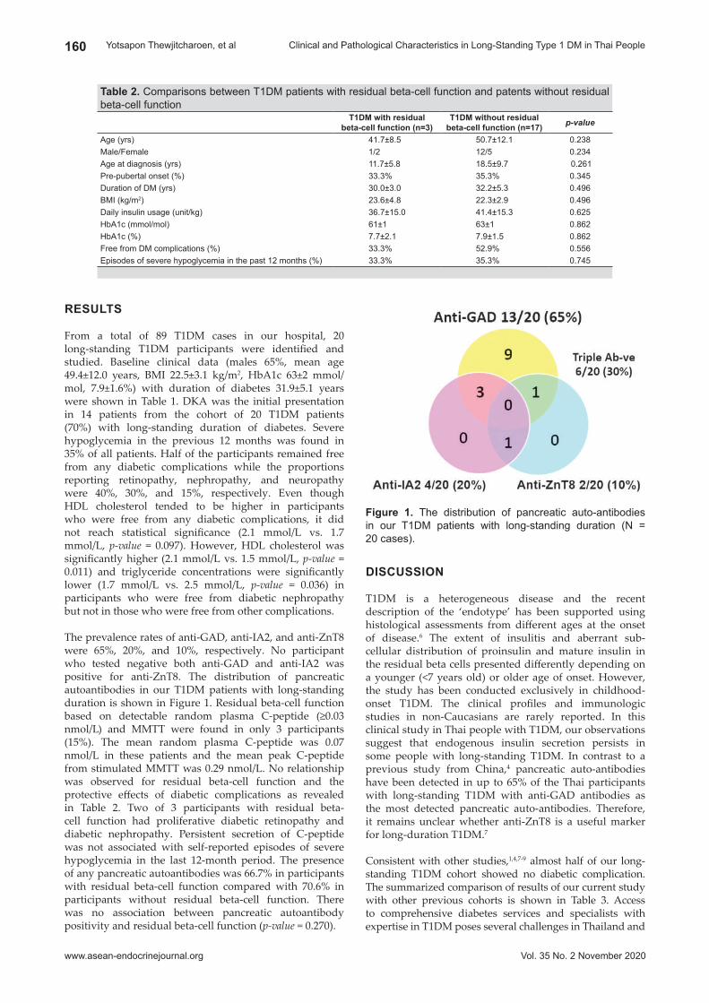
The prevalence rates of anti-GAD, anti-IA2, and anti-ZnT8 were 65%, 20%, and 10%, respectively. No participant who tested negative both anti-GAD and anti-IA2 was positive for anti-ZnT8. The distribution of pancreatic autoantibodies in our T1DM patients with long-standing duration is shown in Figure 1. Residual beta-cell function based on detectable random plasma C-peptide (≥0.03 nmol/L) and MMTT were found in only 3 participants (15%). The mean random plasma C-peptide was 0.07 nmol/L in these patients and the mean peak C-peptide from stimulated MMTT was 0.29 nmol/L. No relationship was observed for residual beta-cell function and the protective effects of diabetic complications as revealed in Table 2. Two of 3 participants with residual beta-cell function had proliferative diabetic retinopathy and diabetic nephropathy. Persistent secretion of C-peptide was not associated with self-reported episodes of severe hypoglycemia in the last 12-month period. The presence of any pancreatic autoantibodies was 66.7% in participants with residual beta-cell function compared with 70.6% in participants without residual beta-cell function. There was no association between pancreatic autoantibody positivity and residual beta-cell function (p-value = 0.270).
DISCUSSION
T1DM is a heterogeneous disease and the recent description of the ‘endotype’ has been supported using histological assessments from different ages at the onset of disease.6 The extent of insulitis and aberrant sub-cellular distribution of proinsulin and mature insulin in the residual beta cells presented differently depending on a younger (<7 years old) or older age of onset. However, the study has been conducted exclusively in childhood-onset T1DM. The clinical profiles and immunologic studies in non-Caucasians are rarely reported. In this clinical study in Thai people with T1DM, our observations suggest that endogenous insulin secretion persists in some people with long-standing T1DM. In contrast to a previous study from China,4 pancreatic auto-antibodies have been detected in up to 65% of the Thai participants with long-standing T1DM with anti-GAD antibodies as the most detected pancreatic auto-antibodies. Therefore, it remains unclear whether anti-ZnT8 is a useful marker for long-duration T1DM.7
Consistent with other studies,1,4,7-9 almost half of our long-standing T1DM cohort showed no diabetic complication. The summarized comparison of results of our current study with other previous cohorts is shown in Table 3. Access to comprehensive diabetes services and specialists with expertise in T1DM poses several challenges in Thailand and
Vol. 35 No. 2 November 2020
160
www.asean-endocrinejournal.org
Yotsapon Thewjitcharoen, et al Clinical and Pathological Characteristics in Long-Standing Type 1 DM in Thai People
Table 2. Comparisons between T1DM patients with residual beta-cell function and patents without residual beta-cell function
T1Dm with residual beta-cell function (n=3)
T1Dm without residual beta-cell function (n=17) p-value
Age (yrs) 41.7±8.5 50.7±12.1 0.238Male/Female 1/2 12/5 0.234Age at diagnosis (yrs) 11.7±5.8 18.5±9.7 0.261Pre-pubertal onset (%) 33.3% 35.3% 0.345Duration of DM (yrs) 30.0±3.0 32.2±5.3 0.496BMI (kg/m2) 23.6±4.8 22.3±2.9 0.496Daily insulin usage (unit/kg) 36.7±15.0 41.4±15.3 0.625HbA1c (mmol/mol) 61±1 63±1 0.862HbA1c (%) 7.7±2.1 7.9±1.5 0.862Free from DM complications (%) 33.3% 52.9% 0.556Episodes of severe hypoglycemia in the past 12 months (%) 33.3% 35.3% 0.745
Figure 1. The distribution of pancreatic auto-antibodies in our T1DM patients with long-standing duration (N = 20 cases).

determinants associated with long-term survival in this unique population.
AcknowledgmentsThe authors wish to thank Dr. Tinapa Himathongkam for the excellent language editing and the staff at the Diabetes and Thyroid Center, Theptarin Hospital for taking care of all the patients.
Statement of AuthorshipAll authors certified fulfillment of ICMJE authorship criteria.
Author DisclosureThe authors declared no conflict of interest.
Funding SourceThis work was supported by the grant for promoting research in Theptarin Hospital (Grant No. 1/2561).
References 1. Keenan HA, Sun JK, Levine J, et al. Residual insulin production and
pancreatic beta-cell turnover after 50 years of diabetes: Joslin Medalist Study. Diabetes. 2010; 59(11):2846-53. PMID: 20699420. PMCID: PMC2963543. https://doi.org/10.2337/db10-0676.
2. Wang L, Lovejoy NF, Faustman DL. Persistence of prolonged C-peptide production in type 1 diabetes as measured with an ultrasensitive C-peptide assay. Diabetes Care. 2012;35(3):465-70. PMID: 22355018. PMCID: PMC3322715. https://doi.org/10.2337/dc11-1236.
3. Davis AK, DuBose SN, Haller MJ, et al. Prevalence of detectable C-Peptide according to age at diagnosis and duration of type 1 diabetes. Diabetes Care. 2015; 38(3):476-81. PMID: 25519448. https://doi.org/10.2337/dc14-1952.
4. Liu W, Han X, Wang Y, et al. Characteristics and ongoing autoimmunity of patients with long-standing type 1 diabetes living in China. Diabetes Care. 2018;41(6):e97-8. PMID: 29615395. https://doi.org/10.2337/ dc18-0046.
5. Jones AG, Hattersley AT. The clinical utility of C-peptide measurement in the care of patients with diabetes. Diabet Med. 2013;30(7):803–17. PMID: 23413806. PMCID: PMC3748788. https://doi.org/10.1111/dme.12159.
6. Hanna SJ, Powell WE, Long AE, et al. Slow progressors to type 1 diabetes lose islet autoantibodies over time, have few islet antigen specific CD8+ T cells and exhibit a distinct CD95hi B cell phenotype. Diabetologia. 2020;63(6):1174-85. PMID: 32157332. PMCID: PMC7228996. https://doi.org/10.1007/s00125-020-05114-7.
7. Trisorus C, Aroonparkmongkol S, Kongmanas HB, Sahakitrungruang T. Prevalence of islet autoantibodies in Thai juvenile-onset type 1 diabetes. Pediatr Int. 2018;60(11):1002-7. https://doi.org/10.1111/ped.13687.
8. Bain SC, Gill DV, Dyer PH, et al. Characteristics of type 1 diabetes of over 50 years duration (the golden years cohort). Diabet Med. 2003;20(10):808-11. PMID:14510860. https://doi.org/10.1046/j.1464-5491.2003.01029.x.
other countries in Southeast Asia; however, T1DM patients could have long life expectancy similar to the general population if they adhere to self-diabetes management and have good support system. A recent mechanistic study among long-standing T1DM in the United States showed that elevated medium-sized HDL particles and elevated levels of HDL-associated paraoxonase 1 (PON1) which is an atheroprotective enzyme might contribute to vascular protection in this group of people.10
The limitations of the study should be acknowledged. First, this was a cross-sectional study from a tertiary diabetes care center in Bangkok. Our institute has an advantage as a comprehensive diabetes center in Thailand for over three decades. Therefore, to be generalizable, our findings should be confirmed in more heterogeneous healthcare services across Southeast Asia. Second, the mean HbA1c values were obtained in the past 12 months. The long-term mean glycemic control since the onset of disease might be different from the present results. Third, some residual or undocumented factors affecting diabetic complications such as the frequency and severity of DKA, distinct protective genetic factors, nutrition status or intake of various supplements could not be completely ruled out. Fourth, the modest sample size of our study would affect the statistical power. Multicenter studies are required to verify our present results and create the prospective registry for T1DM in Southeast Asia. Finally, measured conventional plasma C-peptide levels in this study might misclassify some participants who had very low level of preserved insulin secretion if ultra-sensitive plasma C-peptide measurements were used.
CONClUSION
In conclusion, our observations highlight the emergence of long-standing type 1 diabetes mellitus in an Asian population that is considered to be under-represented. Further multi-center studies in Asian populations with ultra-sensitive plasma C-peptide measurements and detailed mechanistic study together with the assessment of genetic and epigenetic indices should be considered in these individuals with long-standing T1DM. These studies will provide a better understanding of the contributing
Vol. 35 No. 2 November 2020
161
www.asean-endocrinejournal.org
Yotsapon Thewjitcharoen, et alClinical and Pathological Characteristics in Long-Standing Type 1 DM in Thai People
Table 3. Comparisons between our present study in Thai people with long-standing T1DM with other published seriesCountry Dm duration (yrs) Complications Persistent C-peptideJoslin 50-year Medalist Study(United States, N=411)1
56.2±5.8 PDR 55%MAU 13%DN 61%CVD 48%
Minimal C-peptide(0.1-0.6 ng/mL) = 64.4%Sustained C-peptide(≥ 0.6 ng/mL) = 2.6%
Diabetes UK The Golden Years cohort(United Kingdom, N=400)8
55.8±5.4 PRP 43%DKD 36%
N/A
Chinese Study(China, N=95)4
37.3±6.8 DR 68%DKD 34%DN 61%CVD 14%
C-peptide ≥ 0.2 ng/mL = 14.7%
Japanese Study(Japan, N=29)9
55.4±3.9 PDR 59%DKD 46%CVD 25%
C-peptide > 0.4 ng/mL = 6.9%
Theptarin cohort (Thailand, N=20)
31.9±5.1 DR 40%DKD 30%DN 10%CVD 0%
C-peptide ≥ 0.1 ng/mL = 15.0%
Abbreviations: CVD – Cardiovascular Disease; DKD – Diabetic Kidney Disease; DN – Diabetic Neuropathy; DR – Diabetic Retinopathy; MAU – Microalbuminuria; PDR- Proliferative Diabetic Retinopathy; PRP – Panretinal Photocoagulation
9. Otani T, Kasahara T, Miura J, Uchigata Y, Babazono T. Clinical background of Japanese patients with type 1 diabetes mellitus who have received insulin therapy for 50 years or longer. Diabetol Int. 2019;10(4):288-94. PMID: 31592405. PMCID: PMC6763551. https://doi.org/10.1007/s13340-019-00393-x.
10. Vaisar T, Kanter JE, Wimberger J, et al. High concentration of medium-sized HDL particles and enrichment in HDL paraoxonase 1 associated with protection from vascular complications in people with long-standing type 1 diabetes. Diabetes Care. 2020;43(1):178-86. PMID: 31597668. PMCID: PMC6925582. https://doi.org/10.2337/dc19-0772.
Vol. 35 No. 2 November 2020
162
www.asean-endocrinejournal.org
Yotsapon Thewjitcharoen, et al Clinical and Pathological Characteristics in Long-Standing Type 1 DM in Thai People
JAFESExperience the new JAFES.
Visit us at www.ASEAN-endocrinejournal.org.
Authors are required to accomplish, sign and submit scanned copies of the JAFES Author Form consisting of: (1) Authorship Certification, that authors contributed substantially to the work, that the manuscript has been read and approved by all authors, and that the requirements for authorship have been met by each author; (2) the Author Declaration, that the article represents original material that is not being considered for publication or has not been published or accepted for publication elsewhere, that the article does not infringe or violate any copyrights or intellectual property rights, and that no references have been made to predatory/suspected predatory journals; (3) the Author Contribution Disclosure, which lists the specific contributions of authors; and (4) the Author Publishing Agreement which retains author copyright, grants publishing and distribution rights to JAFES, and allows JAFES to apply and enforce an Attribution-Non-Commercial Creative Commons user license. Authors are also required to accomplish, sign, and submit the signed ICMJE form for Disclosure of Potential Conflicts of Interest. For original articles, authors are required to submit a scanned copy of the Ethics Review Approval of their research as well as registration in trial registries as appropriate. For manuscripts reporting data from studies involving animals, authors are required to submit a scanned copy of the Institutional Animal Care and Use Committee approval. For Case Reports or Series, and Images in Endocrinology, consent forms, are required for the publication of information about patients; otherwise, appropriate ethical clearance has been obtained from the institutional review board. Articles and any other material published in the JAFES represent the work of the author(s) and should not be construed to reflect the opinions of the Editors or the Publisher.

________________________________________
ISSN 0857-1074 (Print) | eISSN 2308-118x (Online)Printed in the PhilippinesCopyright © 2020 by Krishnan et al.Received: April 4, 2020. Accepted: August 7, 2020.Published online first: August 25, 2020.https://doi.org/10.15605/jafes.035.02.03
Corresponding author: Gayathri Devi Krishnan, MRCP (UK)Clinical Specialist and Fellow in EndocrinologyMinistry of Health, MalaysiaHospital Raja Perempuan Zainab II, 15586 Kota Bharu, Kelantan, MalaysiaTel. No.: 09-745 2000Fax No.: 09-748 6951E-mail: [email protected]: https://orcid.org/0000-0002-2384-3401
Prevalence of Vitamin B12 Deficiency and its Associated Factorsamong Patients with Type 2 Diabetes mellitus on metformin
from a District in malaysiaGayathri Devi Krishnan,1 Miza Hiryanti Zakaria,2 Norhayati Yahaya1
1Hospital Raja Perempuan Zainab II, Kota Bharu, Kelantan, Malaysia2Hospital Tengku Ampuan Afzan, Kuantan, Pahang, Malaysia
Abstract
Introduction. Vitamin B12 deficiency is more common among metformin-treated subjects although the prevalence is variable. Many factors have been associated with this. The aim of this study is to determine the prevalence of vitamin B12 deficiency and its associated factors among patients with type 2 diabetes mellitus (DM) who are on metformin.
Methodology. A total of 205 patients who fit eligibility criteria were included in the study. A questionnaire was completed, and blood was drawn to study vitamin B12 levels. Vitamin B12 deficiency was defined as serum B12 level of ≤300 pg/mL (221 pmol/L).
Results. The prevalence of vitamin B12 deficiency among metformin-treated patients with type 2 DM patients was 28.3% (n=58). The median vitamin B12 level was 419 (±257) pg/mL. The non-Malay population was at a higher risk for metformin-associated vitamin B12 deficiency [adjusted odds ratio (OR) 3.86, 95% CI: 1.836 to 8.104, p<0.001]. Duration of metformin use of more than five years showed increased risk for metformin-associated vitamin B12 deficiency (adjusted OR 2.06, 95% CI: 1.003 to 4.227, p=0.049).
Conclusion. Our study suggests that the prevalence of vitamin B12 deficiency among patients with type 2 diabetes mellitus on metformin in our population is substantial. This is more frequent among the non-Malay population and those who have been on metformin for more than five years.
Key words: Vitamin B12, metformin, deficiency, type 2 diabetes mellitus, type 2 DM
INTRODUCTION
Type 2 diabetes mellitus is a major non-communicable disease in Malaysia for which metformin is one of the most commonly prescribed first line medications. Multiple cross-sectional studies have reported a wide range in prevalence of biochemical vitamin B12 deficiency with metformin exposure, ranging from 5.8% to as high as 30%.1-5 Vitamin B12 deficiency associated with metformin use is thought to occur due to vitamin B12 malabsorption at the terminal ileum.5-7
Vitamin B12 deficiency is clinically important as it is a reversible cause of bone marrow failure and nerve damage.8 Neurological damage as a result of metformin-induced vitamin B12 deficiency can present as peripheral neuropathy and may be mistaken for diabetic neuropathy.8
Because vitamin B12 deficiency and its associated complications are treatable and potentially reversible,
early detection and treatment are clinically important in patients with diabetes who are on metformin.9
The first large scale study among Asians designed to investigate the prevalence and risk factors associated with vitamin B12 deficiency was conducted among Koreans in 2014. It reported vitamin B12 deficiency in 9.5% of the patients who were on metformin.9 Interestingly, another study among the South African population demonstrated that subjects of black South African descent on metformin had a lower prevalence of B12 deficiency, suggesting that different ethnic origins may influence the prevalence of metformin-associated vitamin B12 deficiency.10 This study is the first of its kind that investigated the association between ethnicity and vitamin B12 deficiency among metformin-treated type 2 DM patients.
Duration of use and dose of metformin have also been shown to influence vitamin B12 levels. A meta-analysis of six randomized controlled trials showed a significant
* This study was presented as a poster at the ASEAN Federation of Endocrine Societies (AFES) Congress in 2019 at Manila, Philippines.
Vol. 35 No. 2 November 2020 163www.asean-endocrinejournal.org
Journal of theASEAN Federation ofEndocrine SocietiesOriginal Article
This work is licensed under a Creative Commons Attribution-NonCommercial 4.0 International License (https://creativecommons.org/licenses/by-nc/4.0/).

0.05, the required sample size was 178.16 Sample size was augmented by 15% to take into account missing data. The final sample size was determined to be 205.
Statistical AnalysisDescriptive analyses of all the demographic and outcome variables were performed. Results of the continuous variables are described with mean and standard deviation or median and interquartile range and results of categorical variables are described with frequency and percentage. Test of normality was used to determine the distribution of the outcome variables. Independent sample t-test was used for normally distributed variables, and Mann-Whitney U-test or Fisher Exact test for variables with a skewed distribution. Pearson Chi-Square test was used to determine association between categorical predictors variables and outcome variables. The vari ables with p-value <0.2 in the univariate analy sis were included in the multivariate analysis. Multiple logistic regression analysis was performed to assess the independent predictive effect of the variables on the risk for vitamin B12 deficiency. All statistical analyses were performed using Statistical Package for Social Science (SPSS) Version 22.0. A p-value of less than 0.05 was considered significant.
RESUlTS
Two hundred fifty-two patients with type 2 DM were screened from two study centers. Forty-six patients were subsequently excluded. A total of 205 patients from two study centers were finally included in the study (Figure 1). Majority (51.7%, n=106) were recruited from a tertiary hospital while 48.3% (n=99) were from a health clinic.
Table 1 shows the baseline demographic data of our study population. A total of 79 (38.5%) males and 126 (61.5%) females were enrolled. Majority of the patients were of Malay race (78%) while the remaining were non-Malay (15.6% Chinese and 6.3% Indian). The median age of the
reduction in vitamin B12 levels induced by metformin and suggested that this may be dose dependent.11 In another large study published in the same year, Korean patients on higher doses (metformin >1 g daily) and with longer treatment duration (>4 years) were more likely to be deficient in vitamin B12.9
Some studies have found lower serum levels of vitamin B12 in smokers, but the exact mechanism for this is still poorly understood.12 It is thought that smokers generally have poor dietary intake. The second National Health and Nutrition Survey (NHANES II) found that smokers have a lower intake of most vitamins and were less likely to have consumed fruit, vegetables, vitamins and mineral supplements. Proton pump inhibitors (PPI) and histamine 2 receptor antagonists (H2RA) may lead to malabsorption of vitamin B12 due to inhibition of gastric acid secretion and reduced production of the intrinsic factor.13 Excessive alcohol intake is also linked to vitamin B12 deficiency. This has been attributed to intestinal malabsorption due to altered binding of intrinsic factor and alcohol-induced ileal damage.10,14
The primary objective of this study is to determine the prevalence of vitamin B12 deficiency among patients with type 2 DM who are on metformin in Malaysia. Our secondary objective is to determine the associated factors contributing to vitamin B12 deficiency in this cohort.
mEThODOlOgy
Study Population This was a cross-sectional prevalence study. A total of 252 patients with type 2 DM were screened from two study centers in the district of Kuantan, Pahang in Malaysia. Patients who turned up for their scheduled clinic appointment at the type 2 diabetes clinic in the two centers were seen screened and recruited during their routine clinic visit between September 2018 and February 2019. Patients aged 18 years old and above with a diagnosis of type 2 DM who were on metformin for at least 6 preceding months were screened. Participants were recruited based on eligibility and willingness to participate. Forty-six patients were excluded based on the exclusion criteria, while one declined to join. Patients who had pernicious anaemia; prior bariatric surgery, gastrectomy, colectomy or inflammatory bowel disease; ongoing critical illnesses; malignancy; liver cirrhosis or renal impairment (creatinine ≥265 µmol/L) were excluded. Subjects who were vegetarians, recipients of vitamin B12 injections or supplements within the past 3 months, pregnant or lactating were excluded as well. Once informed consent was obtained, all participants were interviewed based on a standardized questionnaire (Appendix 1). Blood extraction for serum vitamin B12 levels was done. Vitamin B12 deficiency was defined as serum B12 level ≤300 pg/mL (221 pmol/L). This encompasses vitamin B12 levels defined as low and borderline low.3,9,15 Serum vitamin B12 level was measured by chemiluminescent microparticle Intrinsic Factor assay using the 7K61 ARCHITECT B12 Reagent Kit.
Sample size was calculated based on the 9.5% prevalence of B12 deficiency among type 2 diabetes patients on metformin.9 Using the sample size calculator for estimations with type I error probability and precision of
Vol. 35 No. 2 November 2020
164
www.asean-endocrinejournal.org
Gayathri Devi Krishnan, et al Vitamin B12 Deficiency in Type 2 DM Patients on Metformin
Figure 1. Study design summarizing sample recruitment.
Screened for eligibility and met inclusion criteria(n=252)
Enrolled in the study(n=205)
Vitamin B12 ≤300 pg/mL(n=58)
Vitamin B12 >300 pg/mL(n=147)
Met exclusion criteria (n=47)• Liver cirrhosis (n=3)• Malignancy (n=1)• Vegetarian (n=1)• On Vitamin B12 supplements (n=41)• Declined to participate (n=1)
to 9.70, p<0.01). Duration of metformin use of more than five years was associated with more than a two-fold risk for vitamin B12 deficiency (OR 2.27, 95% CI: 1.21 to 4.27, p=0.01). The other studied factors did not reveal a significant association with vitamin B12 deficiency in our study population (Table 2).
In the multivariate analysis, after adjusting for age, smoking status, duration of diabetes and HbA1c, the non-Malay population remained at a significantly higher risk for metformin-associated vitamin B12 deficiency (adjusted OR 3.86, 95% CI: 1.836 to 8.104, p<0.001) (Table 3). Metformin use for a duration of more than five years showed an increased risk for metformin-associated vitamin B12 deficiency (adjusted OR 2.06, 95% CI: 1.003 to 4.227, p=0.049).
DISCUSSION
Vitamin B12 deficiency has been long known to adversely affect health, causing anaemia and neuropathy among other complications. Metformin, a widely used anti-diabetes drug, has been reported as a risk factor for vitamin B12 deficiency. To the best of our knowledge, this is the first study in Southeast Asia designed to investigate the prevalence vitamin B12 deficiency among metformin-treated patients with type 2 diabetes mellitus.
patients was 56 years. The median duration of diabetes was 72 months with only 15.1% of patients achieving HbA1c ≤7%. HbA1c value was not available for 10 participants. The median body mass index (BMI) was 29 kg/m2. We were unable to obtain the BMI for one participant whose height was not measured as he was unable to stand. Most patients (51.2%) were treated with metformin for more than 5 years. Majority of the included patients (83.9%) were on a metformin dose of more than 1 gram daily. Concomitant medications were largely sulfonylureas (42.9%), insulin (55.1%) and statins (81%). A small proportion of patients were on H2 receptor antagonists (1.5%) and proton pump inhibitors (1%). There were a few smokers (8.3%) and alcoholic beverage consumers (0.9%).
Vitamin B12 deficiency was defined as serum B12 level ≤300 pg/mL (221 pmol/L). The prevalence of vitamin B12 deficiency among metformin-treated type 2 DM patients was 28.3% (n=58). The median vitamin B12 level was 419 (±257) pg/mL. Among the population deficient in vitamin B12, 56.9% were of Malay race while 43.1% were non-Malays. In the normal vitamin B12 category, 86.4% were of Malay race.
Univariate analysis showed that participants of non-Malay race had a significantly higher risk for metformin-associated vitamin B12 deficiency (OR 4.81, 95% CI: 2.39
Vol. 35 No. 2 November 2020
165
www.asean-endocrinejournal.org
Gayathri Devi Krishnan, et alVitamin B12 Deficiency in Type 2 DM Patients on Metformin
Table 1. Baseline demographics according to Vitamin B12 levelaCharacteristic Total (n=205) Deficient in Vitamin B12 (n=58) Normal Vitamin b12 (n=147) p-value
Age, yr (±SD) 56 (15.0) 59 (13.7) 55 (14.0) 0.039b
Gender (%)MenWomen
79 (38.5)126 (61.5)
24 (41.3)34 (58.6)
55 (37.4)92 (62.6)
0.599c
Race (%)MalayNon-Malay
160 (78.1)45 (21.9)
33 (56.9)25 (43.1)
127 (86.4)20 (13.6)
<0.001c
Duration of diabetes, month (±SD) 72 (84.0) 90 (123.0) 60 (96.0) 0.045b
Current smoker (%) 17 (8.3) 8 (13.8) 9 (6.1) 0.092d
Alcohol intake (%) 2 (0.9) 0 (0) 2 (1.4) 1.000d
BMIe (kg/m2) 28.9 (6.3) 28.8 (6.2) 28.0 (6.3) 0.626b
Duration of metformin use (%)≤5 years>5 years
100 (48.8)105 (51.2)
20 (34.5)38 (65.5)
80 (54.4)67 (45.6)
0.010c
Daily dose of metformin (%)≤1000 mg>1000 mg
33 (16.1)172 (83.9)
7 (12.1)51 (87.9)
26 (17.7)121 (82.3)
0.324c
HbA1cf ≤7%>7%
31 (15.1)174 (84.9)
13 (22.4)45 (77.6)
18 (12.2)129 (87.8)
0.067c
Concomitant medication (%)SulfonylureaDPP-4g inhibitorAlpha-glucosidase inhibitorSGLT2h inhibitorGLP-1i receptor agonistInsulinStatinH2RA j
PPIk
88 (42.9)16 (7.8)2 (1.0)3 (1.5)1 (0.5)
113 (55.1)166 (81.0)
3 (1.5)2 (1)
25 (43.1)4 (6.9)0 (0)
1 (1.7)1 (1.7)
26 (44.8)47 (81.0)1 (1.7)1 (1.7)
63 (42.9)12 (8.16)2 (1.36)2 (1.36)
0 (0)87 (59.2)119 (80.9)
2 (1.4)1 (0.7)
0.974c
1.000d
1.000d
1.000d
0.283d
0.063c
0.989c
1.000d
0.487d
a Values were expressed as mean (SD) for normally distributed continuous variables, median (interquartile range) for not normally distributed continuous variables and n (%) for categorical variables.
b Mann Whitney testc Chi square testd Fisher exact teste BMI, body mass indexf HbA1c, glycosylated hemoglobing DPP-4, dipeptidyl peptidase-4h SGLT2, sodium glucose cotransporter-2i GLP-1, glucagon-like peptide-1j H2RA, H2 receptor antagonistk PPI, proton pump inhibitor

was a significant protective factor for vitamin B12 deficiency among metformin-treated patients.21 This was the first study to report ethnic differences in vitamin B12 levels among metformin-exposed type 2 DM patients. Higher levels of the vitamin binding proteins transcobalamin II and haptocorrin in black individuals have been described in South African settings, explaining their relatively elevated vitamin B12 levels.21 The difference in prevalence of vitamin B12 deficiency among different ethnic groups in Asia has not been studied. The currently utilized cut-off points and definitions of vitamin B12 deficiency do not consider the possible effects of ethnicity.21 Further research is needed to determine why Malay ethnicity seemed protective against metformin-associated vitamin B12 deficiency.
Duration of metformin use of more than five years conferred a greater than two-fold increased risk for vitamin B12 deficiency (p=0.049) in our population. Several studies have shown a significant positive association between duration of metformin use and vitamin B12 deficiency.3,9,17,22 In a large-scale study among Koreans (n=799), daily metformin dosage and treatment duration were the most consistent risk factors for vitamin B12 deficiency.9 Secondary analysis from the Diabetes Prevention Program Outcomes Study (DPPOS) showed that 13 years after randomization, there was a 13% increased risk for vitamin B12 deficiency per year of total metformin use.17 The results of our study echo these findings of increased risk for vitamin B12 deficiency with longer duration of metformin use.
Age, sex, body mass index, smoking, duration of diabetes and HbA1c levels did not show a statistically significant association with vitamin B12 deficiency in our population. There was no significant association between vitamin B12 deficiency and the use of other anti-diabetes medications (Table 2).
Previous studies have linked vitamin B12 deficiency with the use of PPI and H2RA among metformin-treated patients. These observations were supported by the concept that gastric acidity is vital for vitamin B12 absorption, and that PPI and H2RA result in reduction in acid discharge by gastric parietal cells.20,23,24 However, this finding was controversial.1,11 Our study did not find a significant association between use of PPI or H2RA and vitamin B12 deficiency. This could be attributed to the very small number of patients in our study who were on PPI or H2RA (n=5).
Vitamin B12 deficiency is clinically important as it can cause anemia, bone marrow failure, peripheral neuropathy and cognitive impairment.8,9,25 Neuropathy secondary to metformin-associated vitamin B12 deficiency may be mistaken for peripheral neuropathy secondary to diabetes-associated microvascular complications, as
The prevalence of vitamin B12 deficiency in our study population is 28.3%, which falls at the upper end of global prevalence. The worldwide prevalence of vitamin B12 deficiency among metformin users ranges between 4.3 to 30%.1,9,17,18 Vitamin B12 deficiency associated with metformin use is thought to occur due to vitamin B12 malabsorption. It is postulated that metformin interferes with the calcium-dependent membrane action responsible for vitamin B12-intrinsic factor absorption in the terminal ileum.6,7,19,20 The substantial prevalence of vitamin B12 deficiency in our population should prompt consideration for routine screening of this deficiency among metformin-treated type 2 DM patients.
Our study demonstrated that race and duration of metformin use were the most consistent associated factors with vitamin B12 deficiency among metformin users. This association remained evident after adjusting for potential confounding factors by multivariate analysis.
The most significant association was race. Non-Malay race was associated with an approximately four-fold increased risk for metformin-associated vitamin B12 deficiency even after adjusting for potential confounders (p<0.001). A study conducted in Africa found that Black South African descent
Vol. 35 No. 2 November 2020
166
www.asean-endocrinejournal.org
Gayathri Devi Krishnan, et al Vitamin B12 Deficiency in Type 2 DM Patients on Metformin
Table 2. Univariate logistic regression analysisORa 95% CI p-value
GenderMaleFemale
1.181
0.63 - 2.20 0.60
RaceNon-MalayMalay
4.811
2.39 - 9.70 <0.01
Daily metformin dose (g/day)>1000 mg≤1000 mg
1.571
0.64 - 3.84 0.33
Metformin treatment duration>5 years≤5 years
2.271
1.21 - 4.27 0.01
HbA1c b
≤7%>7%
2.071
0.94 - 4.56 0.07
Age, yr 1.03 1.00 - 1.06 0.06BMIc, kg/m2 1.02 0.98 - 1.07 0.33Diabetes duration, month 1.00 1.00 - 1.01 0.03Smoking 2.45 0.90 - 6.70 0.08Sulfonylurea 1.01 0.55 - 1.87 0.97DPP-4d inhibitor 0.83 0.26 - 2.70 0.76SGLT-2e inhibitor 1.27 0.11 - 14.30 0.85PPIf 2.56 0.16 - 41.65 0.51H2RA g 1.27 0.11 - 14.30 0.85a OR, odds ratiob HbA1c, glycosylated hemoglobinc BMI, body mass indexd DPP-4, dipeptidyl peptidase-4e SGLT2, sodium glucose cotransporter-2f PPI, proton pump inhibitorg H2RA, H2 receptor antagonist
Table 3. Multiple logistic regression analysisAdjusted ORa 95% CI P Cov and Snell R square Nagelkerke R square
Non-Malay race 3.86 1.836 - 8.104 <0.001 0.13 0.19Duration of metformin use >5 years 2.06 1.003 - 4.227 0.049Non-smoker 0.36 0.120 - 1.059 0.063HbA1cb ≤ 7% 2.32 0.934 - 5.751 0.070Constant 3.24 0.056a OR, odds ratiob HbA1c, glycosylated hemoglobin

Statement of AuthorshipAll authors certified fulfillment of ICMJE authorship criteria.
Author DisclosureThe authors declared no conflicts of interest.
Funding SourceThe Malaysian Endocrine and Metabolic Society (MEMS) funded this research.
References 1. Nervo M, Lubini A, Raimundo FV, et al. Vitamin B12 in metformin-
treated diabetic patients: A cross-sectional study in Brazil. Rev Assoc Med Bras (1992). 2011;57(1):46-9. PMID: 21390459.
2. Tomkin GH, Hadden DR, Weaver JA, Montgomery DAD. Vitamin B12 status of patients on long-term metformin therapy. Br Med J. 1971;2(5763):685-7. PMID: 5556053. PMCID: PMC1796258. https://doi.org/10.1136/bmj.2.5763.685.
3. Reinstatler L, Qi YP, Williamson RS, Garn JV, Oakley GPJ. Association of biochemical B12 deficiency with metformin therapy and vitamin B12 supplements: The National Health and Nutrition Examination Survey, 1999-2006. Diabetes Care. 2012;35(2):327-33. PMID: 22179958. PMCID: PMC3263877. https://doi.org/10.2337/dc11-1582.
4. Romero JMC, Lozano JMR. Vitamin B(12) in type 2 diabetic patients treated with metformin. Endocrinol Nutr. 2012;59(8):487-90. PMID: 22981397. https://doi.org/10.1016/j.endonu.2012.06.005.
5. Pongchaidecha M, Srikusalanukul V, Chattananon A, Tanjariyaporn S. Effect of metformin on plasma homocysteine, vitamin B12 and folic acid: A cross-sectional study in patients with type 2 diabetes mellitus. J Med Assoc Thai. 2004;87(7):780-7. PMID: 15521233.
6. Berchtold P, Bolli P, Arbenz U, Keiser G. Disturbance of intestinal absorption following metformin therapy (observations on the mode of action of biguanides). Diabetologia. 1969;5(6):405-12. PMID: 5372893. https://doi.org/10.1007/BF00427979.
7. Caspary WF, Zavada I, Reimold W, Deuticke U, Emrich D, Willms B. Alteration of bile acid metabolism and vitamin-B12-absorption in diabetics on biguanides. Diabetologia. 1977;13(3):187-93. PMID: 873086. https://doi.org/10.1007/BF01219698.
8. Bell DSH. Metformin-induced vitamin B12 deficiency presenting as a pe ripheral neuropathy. South Med J. 2010;103(3):265-7. PMID: 20134380. https://doi.org/10.1097/SMJ.0b013e3181ce0e4d.
9. Ko SH, Ko SH, Ahn YB, et al. Association of vitamin B12 deficiency and metformin use in patients with type 2 diabetes. J Korean Med Sci. 2014;29(7):965-72. PMID: 25045229. PMCID: PMC4101785. https://doi.org/10.3346/jkms.2014.29.7.965.
10. Shaw LM, Strømme JH, London JL, Theodorsen L. International Federation of Clinical Chemistry (IFCC), Scientific Committee, Analytical Section. IFCC methods for the measurement of catalytic concentration of enzymes. Part 4. IFCC method for gamma-glutamyltransferase [(gamma-glutamyl)-peptide: amino acid gamma-glutamyltransferase, EC 2.3.2.2]. J Clin Chem Clin Biochem. 1983;21(10);633-46. PMID: 6139407.
11. Liu Q, Li S, Quan H, Li J. Vitamin B12 status in metformin treated patients: Systematic review. PLoS One. 2014; 9(6):e100379. PMID: 24959880. PMCID: PMC4069007. https://doi.org/10.1371/journal.pone.0100379.
12. Khan A, Shafiq I, Shah MH. Prevalence of vitamin B12 deficiency in patients with type 2 diabetes mellitus on metformin: A study from Khyber Pakhtunkhwa. Cureus. 2017;9(8):e1577. PMID: 29057189. PMCID: PMC5647126. https://doi.org/10.7759/cureus.1577.
13. Toh BH, van Driel IR, Gleeson PA. Pernicious anemia. N Engl J Med. 1997;337(20):1441-8. PMID: 9358143. https://doi.org/10.1056/NEJM199711133372007.
14. Lindenbaum J. Folate and vitamin B12 deficiencies in alcoholism. Semin. Hematol. 1980;17(2):119-29. PMID: 6990497.
15. Long AN, Atwell CL, Yoo W, Solomon SS. Vitamin B(12) deficiency associated with concomitant metformin and proton pump inhibitor use. Diabetes Care. 2012;35(12):e84. PMID: 23173145. PMCID: PMC3507616. https://doi.org/10.2337/dc12-0980.
16. Daniel WW. Biostatistics: A foundation for analysis in the health sciences. 7th ed. New York: John Wiley & Sons, 1999.
17. Aroda VR, Edelstein SL, Goldberg RB, et al. Long-term metformin use and vitamin B12 deficiency in the Diabetes Prevention Program Outcomes Study. J Clin Endocrinol Metab. 2016;101(4):1754-61. PMID: 26900641. PMCID: PMC4880159. https://doi.org/10.1210/jc.2015-3754.
18. Pflipsen MC, Oh RC, Saguil A, et al. The prevalence of vitamin B(12) deficiency in patients with type 2 diabetes: A cross-sectional study. J Am Board Fam Med. 2009;22(5):528-34. PMID: 19734399. https://doi/org/10.3122/jabfm.2009.05.090044.
19. Berchtold P, Dahlqvist A, Gustafson A, Asp NG. Effects of a biguanide (metformin) on vitamin B12 and folic acid absorption and intestinal
both diseases can result in reduced vibration sense and diminished proprioception.8,26 There is no definitive clinical or electrophysiological test that can differentiate diabetic peripheral neuropathy from vitamin B12-associated neuropathy.8 This may lead to inappropriate use of tricyclic antidepressants and anticonvulsants to manage symptoms.8,27,28
Recognition of metformin-associated vitamin B12 deficiency is imperative as it is potentially treatable and reversible. Multivitamin use seemed to protect type 2 DM patients from B12 deficiency.18 Randomized trials among adults taking supplemental vitamin B12 doses as low as 6 to 9 mcg daily show higher serum B12 levels compared with placebo.29,30
We regard the prevalence of vitamin B12 deficiency of 28.3% in our study population as a substantial percentage, as it affects over a quarter of the type 2 DM patients who are on metformin. This provides a valuable manual for clinicians to consider testing for vitamin B12 levels especially among type 2 DM patients who are of non-Malay race and who have been on metformin for more than five years. Although the clinical implication of our findings has not been demonstrated in this study, the potential complications of vitamin B12 deficiency has been well documented in literature.
Our study had several limitations. It was conducted in two centers from a single district, which may not be representative of the entire Malaysian population. We were unable to measure serum homocysteine and serum methylmalonic acid, early markers of vitamin B12 deficiency, as this was cost-prohibitive. The study was not sufficiently powered for some of the factors that were evaluated as an association with B12 deficiency in our population. We did not include a detailed dietary history which could be a potential contributing factor to low vitamin B12 levels.
CONClUSION
Given the mounting evidence associating metformin exposure with low vitamin B12 levels, assessment of serum vitamin B12 levels among metformin-treated patients should be incorporated into routine clinical practice. According to the 2019 National Health and Morbidity Survey (NHMS), the prevalence of type 2 DM in Malaysia is 18.3% among adults above the age of 18.31
Metformin is widely recommended as a first line agent in the treatment of type 2 DM. Our study supports evidence that use of metformin is indeed associated with vitamin B12 deficiency and the prevalence of this association in our study population is significant. Testing for vitamin B12 deficiency among metformin-treated type 2 DM patients should be strongly considered especially among patients who are of non-Malay race and those who have been on metformin for more than five years.
AcknowledgmentsThe authors thank the Malaysian Endocrine and Metabolic Society (MEMS) for funding the research. They also extend their gratitude to Dr. Wan Hasmawati Binti Wan Ismail, family medicine physician, and the staff of Klinik Kesihatan Paya Besar, for accommodating their research.
Vol. 35 No. 2 November 2020
167
www.asean-endocrinejournal.org
Gayathri Devi Krishnan, et alVitamin B12 Deficiency in Type 2 DM Patients on Metformin

enzyme activities. Scand J Gastroenterol. 1971;6(8):751-4. PMID: 5139118. https://doi.org/10.3109/00365527109179948.
20. Bauman WA, Shaw S, Jayatilleke E, Spungen AM, Herbert V. Increased intake of calcium reverses vitamin B12 malabsorption induced by metformin. Diabetes Care. 2000;23(9):1227-31. PMID: 10977010. https://doi.org/10.2337/diacare.23.9.1227.
21. Fernandes-Costa F, Metz J. A comparison of serum transcobalamin levels in white and black subjects. Am J Clin Nutr. 1982;35(1):83-6. PMID: 7064880. https://doi.org/10.1093/ajcn/35.1.83.
22. Ahmed MA, Muntingh G, Rheeder P. Vitamin B12 deficiency in metformin-treated type 2 diabetes patients, prevalence and association with peripheral neuropathy. BMC Pharmacol Toxicol. 2016;17:44. PMID: 27716423. PMCID: PMC5054613. https://doi.org/10.1186/s40360-016-0088-3.
23. Valuck RJ, Ruscin JM. A case-control study on adverse effects: H2 blocker or proton pump inhibitor use and risk of vitamin B12 deficiency in older adults. J Clin Epidemiol. 2004;57(4):422-8. PMID: 15135846. https://doi.org/10.1016/j.jclinepi.2003.08.015.
24. Ruscin JM, Page RL 2nd, Valuck RJ. Vitamin B(12) deficiency associated with histamine(2)-receptor antagonists and a proton-pump inhibitor. Ann Pharmacother. 2002;36(5):812-6. PMID: 11978157. https://doi.org/10.1345/aph.10325.
25. Hin H, Clarke R, Sherliker P, et al. Clinical relevance of low serum vitamin B12 concentrations in older people: The Banbury B12 study. Age Ageing. 2006;35(4):416-22. PMID: 16709605. https://doi.org/10.1093/ageing/afl033.
26. Lindenbaum J, Healton EB, Savage DG, et al. Neuropsychiatric disorders caused by cobalamin deficiency in the absence of anemia or macrocytosis. N Engl J Med. 1988; 318(26):1720-8. PMID: 3374544. https://doi.org/10.1056/NEJM198806303182604.
27. Naha K, Dasari S, Vivek G, Prabhu M. Vitamin B12 deficiency: An unusu al cause for recurrent generalised seizures with pancytopaenia. BMJ Case Reports. 2012;10.1136/bcr-2012-006632. PMID: 22948998. PMCID: PMC4543758. https://doi.org/10.1136/bcr-2012-006632.
28. Durand C, Mary S, Brazo P, Dollfus S. Psychiatric manifestations of vita min B12 deficiency: A case report. Encephale. 2003;29(6):560-5. PMID: 15029091.
29. McKay DL, Perrone G, Rasmussen H, Dallal G, Blumberg JB. Multivitamin/mineral supplementation improves plasma B-vitamin status and homocysteine concentration in healthy older adults consuming a folate-fortified diet. J Nutr. 2000;130(12):3090-6. PMID: 11110875. https://doi.org/10.1093/jn/130.12.3090.
30. Wolters M, Hermann S, Hahn A. Effect of multivitamin supplementation on the homocysteine and methylmalonic acid blood concentrations in women over the age of 60 years. Eur J Nutr. 2005;44(3):183-92. PMID: 15309436. https://doi.org/10.1007/s00394-004-0510-2.
31. Institute for Public Health 2020. National Health and Morbidity Survey (NHMS) 2019.: Non-communicable diseases, healthcare demand, and health literacy—Key Findings. http://iku.moh.gov.my/images/IKU/Document/REPORT/NHMS2019/Infographic_Booklet_NHMS_2019-English.pdf.
Vol. 35 No. 2 November 2020
168
www.asean-endocrinejournal.org
Gayathri Devi Krishnan, et al Vitamin B12 Deficiency in Type 2 DM Patients on Metformin
Authors are required to accomplish, sign and submit scanned copies of the JAFES Author Form consisting of: (1) Authorship Certification, that authors contributed substantially to the work, that the manuscript has been read and approved by all authors, and that the requirements for authorship have been met by each author; (2) the Author Declaration, that the article represents original material that is not being considered for publication or has not been published or accepted for publication elsewhere, that the article does not infringe or violate any copyrights or intellectual property rights, and that no references have been made to predatory/suspected predatory journals; (3) the Author Contribution Disclosure, which lists the specific contributions of authors; and (4) the Author Publishing Agreement which retains author copyright, grants publishing and distribution rights to JAFES, and allows JAFES to apply and enforce an Attribution-Non-Commercial Creative Commons user license. Authors are also required to accomplish, sign, and submit the signed ICMJE form for Disclosure of Potential Conflicts of Interest. For original articles, authors are required to submit a scanned copy of the Ethics Review Approval of their research as well as registration in trial registries as appropriate. For manuscripts reporting data from studies involving animals, authors are required to submit a scanned copy of the Institutional Animal Care and Use Committee approval. For Case Reports or Series, and Images in Endocrinology, consent forms, are required for the publication of information about patients; otherwise, appropriate ethical clearance has been obtained from the institutional review board. Articles and any other material published in the JAFES represent the work of the author(s) and should not be construed to reflect the opinions of the Editors or the Publisher.
Appendix 1. Association of Vitamin B12 deficiency and Metformin in Type 2 Diabetes Mellitus
Data Collection SheetDate Centre
Name of Investigator
Subject InformationSubject Initials Subject ID (IC number)Gender M / F Race Malay / Chinese / Indian / Others
Others (please specify):____________Age (years)Height (cm) Weight (kg) BMI (kg/m2)Current Smoker Yes / No Alcohol consumption (units/week or units/month-
see appendix)Duration of diabetes mellitus (since diagnosis)Current metformin dose (daily dose)Total duration of metformin use (in years/months)Medications: (tick relevant box)Sulphonylurea Proton pump inhibitor (PPI)DPP-IV antagonist H2 antagonist GLP-1 agonist StatinSGLT-2 inhibitor ACEi/ARBAlpha glucosidase inhibitor AspirinThiazolidinediones InsulinOthers (please specify)
lab Investigations
Test Value DateVitamin B12 level (pg/mL)Fasting Blood Sugar (mmol/L)HbA1c (%) *(latest available value)

The Association between maternal Serum Vitamin D levelsand gestational Diabetes mellitus among Filipino Patients:
A Cross-Sectional StudyCarmen Carina Cabrera,1 Oliver Allan Dampil,1 Albert Macaire Ong-Lopez2
1Section of Endocrinology, Diabetes and Metabolism, Department of Internal Medicine, St. Luke’s Medical Center, Quezon City, Philippines2Department of Medicine, St. Luke’s Medical Center, Quezon City, Philippines
Abstract
Objectives. To determine the association between low maternal serum vitamin D and gestational diabetes mellitus (GDM) among Filipino women in St. Luke’s Medical Center, Quezon City.
Methodology. A cross-sectional study involving pregnant women at outpatient clinics in a tertiary hospital in the Philippines. Simultaneous testing for fasting blood sugar, 75g oral glucose tolerance test and serum vitamin D was done. Participants were classified as GDM versus non-GDM, and normal versus low serum vitamin D. Univariate and multivariate statistics were done to determine relationship between vitamin D and GDM.
Results. Of 211 included women, 198 (93.8%) had low vitamin D levels, and 56 (26.5%) had GDM. Vitamin D was significantly higher in the GDM group (21.0±8.1 vs 18.8±5.3 ng/mL, p=0.0189). The proportion of women with low vitamin D levels was significantly higher among those without GDM (96.1% vs 87.5%, OR=0.28, p=0.029]. After adjusting for age, parity, history of GDM and pre-pregnancy BMI, no significant association was observed (adjusted OR=0.66, p=0.522). No correlation was seen between vitamin D and FBS (r=0.28, p=0.095), 1-hour post-75 g OGTT (r=0.26, p=0.643), and 2-hour post-75 g OGTT (r=0.28, p=0.113).
Conclusion. There was an association found between maternal serum vitamin D level and GDM in the univariate analysis, but none was evident after adjusting for possible confounders. The unanticipated high prevalence of low vitamin D levels among pregnant Filipinos needs to be verified in future studies.
Key words: gestational diabetes mellitus, gestational diabetes, vitamin D deficiency
INTRODUCTION
gestational Diabetes mellitus (gDm)GDM is defined by the American Diabetes Association (ADA) as diabetes diagnosed in the second or third trimester of pregnancy that is not clearly either type 1 or type 2 diabetes.1 Its occurrence, like type 2 diabetes mellitus, is increasing reaching a global prevalence of 15% to 20%,2 while locally in the Philippines, it was reported to be at 14%.3 It carries the risk of adverse maternal, fetal and neonatal outcomes including increased birth weight above the 90th percentile, as well as a higher incidence of neonatal hypoglycemia and primary cesarean section demonstrated in the large-scale multinational cohort study called The Hyperglycemia and Adverse Pregnancy Outcome (HAPO) study.4 Other reported risks increased by GDM are the development of preeclampsia and dystocia, and the predisposition of both the mother and offspring to develop obesity, type 2 diabetes mellitus and the metabolic syndrome. Recognized risk factors in the development of GDM include advanced maternal age, obesity, ethnicity, family history of diabetes, and a previous
history of abnormal glucose metabolism and polycystic ovarian syndrome. Parity per se was not found to have any direct link to GDM appearance.5 Recently, vitamin D was identified as a potential contributor to its occurrence.
Vitamin D and its Extra-skeletal EffectsThere is gaining interest in the role of vitamin D in diabetes mellitus. Studies found that its function extends beyond calcium and bone metabolism. It was demonstrated in animal models to improve pancreatic exocrine function and insulin sensitivity.6 Calcitriol or 1,25(OH)2D, the form of vitamin D produced in the kidneys, was shown in animal models to have effects on the synthesis, secretion, and actions of insulin.7,8 It enhances insulin-dependent glucose transport by inducing insulin receptor expression.
Pregnancy and Vitamin DPhysiologic changes in vitamin D metabolism during pregnancy are still incompletely understood. Studies showed an increase in vitamin D binding proteins by 7% to 152% 9,10,11,12 as well as serum 1,25(OH)2D by 104% to 134% with minimal effect on serum 25(OH)D.13,14
________________________________________
ISSN 0857-1074 (Print) | eISSN 2308-118x (Online)Printed in the PhilippinesCopyright © 2020 by Cabrera et al.Received: May 20, 2020. Accepted: August 16, 2020.Published online first: September 1, 2020.https://doi.org/10.15605/jafes.035.02.04
Corresponding author: Carmen Carina G. Cabrera, MDFellow-in-training, Section of Endocrinology, Diabetes and MetabolismSt. Luke’s Medical Center, Quezon City279 E Rodriguez Sr. Ave. Quezon City, 1112, PhilippinesTel. No.: +638-723-0101 E-mail: [email protected]: https://orcid.org/0000-0001-6190-9152
Vol. 35 No. 2 November 2020 169www.asean-endocrinejournal.org
Journal of theASEAN Federation ofEndocrine SocietiesOriginal Article
This work is licensed under a Creative Commons Attribution-NonCommercial 4.0 International License (https://creativecommons.org/licenses/by-nc/4.0/).
Serum 1,25(OH)2D levels are expected to return to pre-pregnancy levels by weeks 8 to 10 postpartum. Both forms of vitamin D cross the placenta to the growing fetus, the latter theorized to be the predominant metabolite.15
There is still a lack of consensus on the definition of normal vitamin D levels among pregnant women. Based on the systematic review by the Institute of Medicine (IOM), serum 25(OH)D levels below 20 ng/mL (50 nmol/L) are considered insufficient.16 The Endocrine Society Clinical Practice Guidelines define vitamin D insufficiency as 25(OH)D levels of >20 ng/mL (50 nmol/L) and <30 ng/mL (75 nmol/L), and vitamin D deficiency as levels ≤20 ng/mL.17 The National Institute for Health and Clinical Excellence guidelines, on the other hand, define vitamin D insufficiency as 25(OH)D levels less than 10 ng/mL (25 nmol/L).18 However, these cutoff values are based on optimal levels in maintaining skeletal health in the general population. There remains the need to establish a normal range among pregnant women.
gDm and Vitamin DIn gestational diabetes mellitus, vitamin D acts as a potential immunosuppressant that downregulates the expression of pro-inflammatory marker such as TNF-α and IL-2.19 Many observational studies found an association between low maternal levels of serum vitamin D and GDM. A case-control study done in Istanbul by Parildar et al., (2013) among 44 pregnant women with GDM and 78 non-GDM pregnant women showed a lower mean serum vitamin D level among GDM patients (14.3±8.2 ng/ml) versus that of controls (23.2±8.3 ng/ml, p=0.001). Vitamin D deficiency (defined as vitamin D of ≤20 ng/mL) prevalence was 56.8% among GDM patients and 35.8% among non-GDM patients.20 Another nested case-control study by Wen et al.,(2017) which included 4718 pregnant women from China, 1280 of whom were diagnosed with GDM, found that maternal serum 25(OH)D were significantly lower in women with GDM [42.4 (34.5, 54.0) nmol/L] compared to controls [44.4 (36.0, 58.8) nmol/L, p<0.001]. Seventy percent of women with GDM had vitamin D <50 nmol/L compared to 60.5% in the control group.21
There were other studies, however, that found no significant link between the two conditions. Makgoba et al., (2011) conducted a case-control study in Europe involving 248 women in the first trimester of pregnancy, 90 of whom developed GDM. They found no correlation between mean vitamin D levels among those with GDM (18.9±10.7 ng/ml) versus those without GDM (19.0±10.7, p=0.874), even after adjustment for possible confounders (p=0.784).22 A case-control study by Pleskacova et al., (2015) among 47 pregnant women with GDM and 29 healthy controls measured mid-gestational and early postpartum vitamin D levels. They found a high prevalence of vitamin D deficiency in both groups (95.7% in women with GDM, 93.1% in controls), but mean levels were not significantly different [28.5 (21.0, 34.0) nmol/L in women with GDM, 31.7 (24.0, 40.0) in controls; p>0.05].23
Meta-analyses and systematic reviews aimed to absolve these conflicting data. One done by Aghajafari (2013) on the role of vitamin D in pregnancy included 31 studies published between 1980 and 2012, 10 studies of which had gestational diabetes as the outcome, with a total of 687 cases and 3425 controls. They reported that low vitamin
D levels were associated with GDM [pooled odds ratio (OR)=1.49, (1.18-1.89)] with no evidence of heterogeneity (I2=0%).24 Participants in this meta-analysis, however, included Americans, Asians, Europeans, Canadians and Middle Easterners. There was no representative study for Southeast Asians.
A recently published article by Hu et al., (2018) pooled data from 29 observational studies which included 28982 participants, with more than half being Asian of Chinese and Korean descent, 4634 of whom were diagnosed with GDM. It was demonstrated that low levels of vitamin D significantly increased the risk for GDM by 39% (pooled OR=1.39, [1.20, 1.60]) albeit with moderate heterogeneity (I2=50.2%, p=0.001). Vitamin D levels were significantly lower among patients with GDM compared with controls with a pooled effect of -4.79 (-6.43, -3.15) nmol/L and significant heterogeneity (I2=65.0%, p<0.001).2
Several limitations of these meta-analyses may hinder the applicability of results in the Filipino population. These include the observational nature of included studies, as well as the diversity of study populations in terms of ethnicity with inadequate representation of Southeast Asians. Other confounders that were not considered were adiposity and laboratory techniques in the measurement of serum 25(OH)D. In this light, local data is needed to assess its applicability in the Filipino community.
The current guidelines of the American College of Obstetrics and Gynecology (ACOG)25 and World Health Organization26 do not recommend routine screening for vitamin D deficiency among pregnant women, except those who are considered high risk – only then would screening and treatment be initiated. Current knowledge points toward a possible link between GDM and low maternal vitamin D levels, but the challenge lies in its translation to clinical recommendation whether achieving optimal levels of vitamin D can actually prevent GDM and its associated sequelae. Establishing an association between the two conditions among Filipinos can pave the way for future local studies on causality and benefit of vitamin D screening and correction in pregnant patients to prevent GDM, in the hope of ultimately improving maternal and fetal outcomes in the country.
This study aimed to determine the association of low levels of maternal serum vitamin D levels and GDM among Filipino patients in St. Luke’s Medical Center, Quezon City.
mEThODOlOgy
This was a single-center study. Target population included both social service and private outpatients in St. Luke’s Medical Center, Quezon City, a private tertiary hospital in the Philippines, from April 2019 to January 2020. Table 1 enumerates the inclusion and exclusion criteria of participants.
Description of Study Procedure This was a cross-sectional study involving pregnant women seen at both private and social service outpatient clinics at St. Luke’s Medical Center, Quezon City from April 2019 to January 2020.
Vol. 35 No. 2 November 2020
170
www.asean-endocrinejournal.org
Carmen Carina Cabrera, et al Maternal Serum Vitamin D Levels and GDM among Filipino Patients

Consent was obtained from obstetricians and endocrinologists to screen their patients for inclusion in this study. All pregnant women who were in their second or third trimester scheduled to undergo FBS and 75 g OGTT as standard of care, who met eligibility criteria were included in the study. Informed consent was obtained by the study investigator or designated representative during their clinic visit. The study investigator or designated representative gathered demographic information which included age, pre-pregnancy BMI, personal history of abnormal glucose metabolism (prediabetes, impaired glucose tolerance, impaired fasting glucose), previous GDM, history of poor obstetric outcome (including but not limited to macrosomia, fetal demise, spontaneous abortion), and family history of diabetes.
During their scheduled blood draw for FBS and OGTT, blood samples were likewise taken through venipuncture by the medical technologist to measure serum vitamin D levels. These tests were done at the St. Luke’s Medical Center laboratory. Patients were then classified as to having GDM and no GDM, as well as to having low and normal vitamin D levels (Figure 1).
Description of Test Procedures The measurement of total vitamin D was done at St. Luke’s Medical Center, Quezon City serology laboratory using an in vitro diagnostic electrochemiluminescent process according to the manufacturer’s instructions. Reported deviations are as follows: for concentrations up to 15ng/ml, deviation is ≤1.5 ng/ml; for concentrations >15 ng/ml, deviation is ≤10%.
DiagnosisThe diagnosis of GDM was based on the ADA criteria,1 and was made when any of the following plasma glucose values were met or exceeded with a 75 g OGTT during the second or third trimester of pregnancy: fasting blood sugar
of 92 mg/dl; 1-hour post-OGTT of 180 mg/dl; or 2-hour post-OGTT of 153 mg/dl.
Since there is still a lack of consensus on definitions of vitamin D deficiency and insufficiency among pregnant women, the Endocrine Society Clinical Practice Guideline17 definition was applied. Patients were classified as having low levels of vitamin D when serum level of total vitamin D was ≤30 ng/ml. Those with low vitamin D levels were further classified as vitamin D insufficient when levels were 21-30 ng/ml, and vitamin D deficient at levels ≤20 ng/ml.
Outcome measuresThe primary outcome of this study was the prevalence of low maternal levels of vitamin D among patients with GDM. The secondary outcomes were the prevalence of vitamin D insufficiency and vitamin D deficiency among patients with GDM, the mean vitamin D level among patients with GDM, and the correlation between FBS and 75g OGTT levels with maternal serum vitamin D levels.
Sample Size EstimationBased on a level of significance of 5% and a power of 90%, a minimum of 190 patients were required for this study. This was derived from preliminary data from an article by Parildar et al., (2013)20 which reported a prevalence of 56.8% of vitamin D deficiency among pregnant women with GDM and 35.8% in pregnant women without GDM. Controlling for 2 more variables in the analysis (age, parity), with an additional 10% for each control variable, final sample size required was 228. The computed sample size assumes that the proportion of patients to be assigned to the two groups is equal.
Data Processing and AnalysisData was processed and encoded using Microsoft Excel. Statistical analysis was done using STATA version 14. Determination of the relationship between maternal serum vitamin D levels and GDM was analyzed using univariate and multivariate statistics. Chi-square test and logistic regression were done in the univariate analysis of qualitative and quantitative independent variables, respectively. Multiple logistic regression was utilized in the multivariate analysis. Crude and adjusted OR and the 95% confidence interval were also calculated. Pearson’s r was calculated to determine the correlation of vitamin D and parameters of OGTT. Level of significance was set at α = 0.05.
Ethical ConsiderationsThe clinical protocol and all relevant documents were reviewed and approved by the SLMC Institutional Ethics
Vol. 35 No. 2 November 2020
171
www.asean-endocrinejournal.org
Carmen Carina Cabrera, et alMaternal Serum Vitamin D Levels and GDM among Filipino Patients
Figure 1. Schematic diagram of study design from time of inclusion of participants.
Table 1. Eligibility criteriaInclusion criteria• Pregnant Filipino women above 18 years oldExclusion criteria• Pregnant women who fulfill diagnostic criteria for diabetes mellitus
before 24 weeks age of gestation• Pregnant women with:
o History of type 1 or type 2 diabetes mellitus prior to pregnancyo Multifetal pregnancyo Use of artificial reproductive technologyo Fetal abnormalitieso Chronic liver or renal failureo Parathyroid or bone metabolism abnormalitieso Malignancy
GDM No GDM
Normal vitamin D Normal vitamin DLow vitamin D Low vitamin D
Total vitamin D Total vitamin D
Pregnant women included in the study

Review Committee. As respect to patient confidentiality, anonymity of patient records was ensured by assigning a unique code to each patient. The principal investigators were responsible for the integrity of the data that was recorded. The protection of confidentiality of the data was guaranteed by the manner of dissemination of study results. Written and signed informed consent were obtained and data collection forms were compiled and stored in an envelope. Data was tabulated in Microsoft Excel format and saved in a CD. These will be kept and filed in the Diabetes, Thyroid and Endocrine Center under the Section of Endocrinology for 5 years before they are shredded.
RESUlTS
Two hundred eighty-nine pregnant women were screened and gave their consent to be included in this study. However, only 211 complied with the required test procedures and were subsequently included in the analysis. Fifty-six of these women (26.5%) were diagnosed with GDM by ADA criteria, and 155 (73.5%) without GDM. Women with GDM had a significantly higher average age compared to those without GDM (p<0.001). Both groups likewise differed significantly in terms of parity. Majority of those with GDM had 1-2 previous deliveries, while most of those without GDM were nulliparous or had 1-2 previous deliveries (p=0.008). The proportion of women with a previous history of GDM was significantly higher among those with GDM (p=0.004) There was no significant difference between the two groups in terms of pre-pregnancy BMI, history of poor obstetric outcome, abnormal glucose metabolism, PCOS or family history of diabetes. The summary of baseline characteristics is seen in Table 2.
Low vitamin D levels were seen in 198 of the participants, accounting for 93.8% of the entire group. Vitamin D level was significantly higher in the GDM group (21.0±8.1 vs 18.7±5.3 ng/mL, p=0.0189). The proportion of patients with low vitamin D levels was significantly higher among those without GDM [GDM 49 (87.5%) vs no GDM 149 (96.1%)]. Calculating for the odds ratio (OR), having low vitamin D levels was significantly associated with a lower likelihood for GDM (OR=0.28, p=0.029). However, after adjusting for age, parity, history of GDM, and pre-pregnancy BMI no significant association was observed (OR=0.66, p=0.522). The same trend was demonstrated on subgroup analysis of those with vitamin D insufficiency (OR=0.32, p=0.069; adjusted OR=0.55, p=0.433) and vitamin D deficiency (OR=0.27, p=0.025; adjusted OR=0.65, p=0.511). These are summarized in Table 3. No correlation was found between Vitamin D and FBS (Figure 2, r=0.28, p=0.095), 1-hour post 75 g OGTT (Figure 3, r=0.26, p=0.643), and 2-hour post 75 g OGTT (Figure 4, r=0.28, p=0.113).
DISCUSSION
Vitamin D acts as a potential immunosuppressant that downregulates the expression of pro-inflammatory markers which are associated with the development of GDM.19 It is also known to influence insulin secretion thereby affecting circulating glucose levels.27 Hence, low concentrations of vitamin D is a potential risk factor for developing GDM. GDM, on the other hand, is associated with several adverse maternal and fetal outcomes including increased birthweight, neonatal hypoglycemia, increased incidence for primary cesarean section, preeclampsia and dystocia, and the predisposition of both the mother and offspring to develop obesity, type 2 diabetes mellitus and the metabolic syndrome.
Vol. 35 No. 2 November 2020
172
www.asean-endocrinejournal.org
Carmen Carina Cabrera, et al Maternal Serum Vitamin D Levels and GDM among Filipino Patients
Table 2. Baseline characteristicsgDm
(n=56, 26.5%)No gDm
(n=155, 73.5%) p value
Age (yrs) 33.2±5.6 28.7±5.2 0Pre-pregnancy BMI (kg/m2) 23.0±3.6 22.1±3.6 0.12Parity n (%)
Nulliparous 13 (23.2) 71 (45.8)1-2 previous deliveries 40 (71.4) 74 (47.7) 0.008≥3 previous deliveries 3 (5.4) 10 (6.5)
Previous history of GDM n (%) 14 (25.0) 15 (9.7) 0.004History of poor obstetric outcome n (%) 4 (7.1) 19 (12.3) 0.292History of abnormal glucose metabolism n (%) 1 (1.8) 1 (0.6) 0.461History of polycystic ovarian syndrome n (%) 8 (14.3) 12 (7.7) 0.183Family history of diabetes n (%) 21 (37.5) 73 (47.1) 0.272Fasting blood glucose (mg/dL) 93.6±21.3 76.8±7.4 075g oral glucose tolerance (mg/dL)
1st hour (mg/dL) 192.1±34.7 133.6±26.2 02nd hour (mg/dL) 162.2±29.0 110.9±21.0 0
Vitamin D level (ng/mL) 21.0±8.1 18.7±5.3 0.0189
Table 3. Vitamin D levels among patients with GDMgDm
(n=56, 26.5%)No gDm
(n=155, 73.5%) p value Unadjusted OR (95% CI) p value Adjusted OR
*(95% CI) p value*
Normal Vitamin D (n=13) 7 (12.5) 6 (3.9)0.021
Reference – Reference –Low Vitamin D (n=198) 49 (87.5) 149 (96.1) 0.28 (0.09-0.88) 0.029 0.66 (0.18-2.36) 0.522Vitamin D insufficiency (n=59) 16 (69.6) 43 (87.8) 0.061 0.32 (0.09-1.09) 0.069 0.55 (0.12-2.48) 0.433Vitamin D deficiency (n=139) 33 (82.5) 106 (94.6) 0.018 0.27 (0.08-0.85) 0.025 0.65 (0.18-2.38) 0.511*adjusted for pre-pregnancy BMI, age, parity and history of GDM

In this study, pregnant women diagnosed with GDM had a significantly higher mean age and history of previous GDM, both of which are established risk factors for GDM. Other risk factors for GDM such as BMI, a history of abnormal glucose metabolism, PCOS, family history for diabetes, were not found to have any direct link to GDM appearance in the participants. Increased parity was also seen in the GDM group, although parity per se has not been shown to be a risk factor for its development.5
There is still no established cutoff to define normal vitamin D levels among pregnant women. Several ranges have been used to define vitamin D sufficiency,16,17,18 but these were set on the basis of optimal levels to maintain skeletal health in the general population. There remains the prerequisite to determine the normal range for pregnant women. Employing the cutoffs recommended by the Endocrine Society Clinical practice guidelines to define vitamin D insufficiency (>20 to <30 ng/mL, or >50 to 75 nmol/L) and deficiency (≤20 ng/mL, or ≤50 nmol/L),17 there was a high overall prevalence of 93.8% for low vitamin D levels among pregnant women included in this study. This is similar to a study on 74 pregnant Czech women which found
an overall prevalence of 94.7%.23 Other studies, however, reported lower rates. An incidence rate of vitamin D deficiency (<50 nmol/L) among 98 pregnant Chinese women was reported at 20.4%.19 A larger cohort of 4718 Chinese women were found to have a higher prevalence rate of 63.1%21 with a median 25(OH)D concentration of 43.7 nmol/L. Another study in Nepal involving 79 pregnant women revealed an even higher rate of 81%.28
Local data on the prevalence of vitamin D among pregnant Filipino women are still lacking. For the general population, however, the overall prevalence of combined vitamin D deficiency and insufficiency was 48.7%. In the same report, low vitamin D levels were highest in people residing in NCR (54.1%), with a higher prevalence in females (62.5%), in the age group of 20-39 years old (55.5%)29– all factors of which were similar to the profile of the women included in this study which could explain the relatively increased rates observed. Avoidance of sun exposure, whether intentional (i.e., to maintain fair skin for aesthetic reasons, to prevent sunburns) or unintentional (i.e., occupation setting mostly indoors) likely contributes to the low vitamin D levels in this population. It is unclear if pregnancy per se contributes to this decrease.
Important to note as well, although not quantified, is that women who were included in this study were already on vitamin D supplementation as part of standard of prenatal care. Yet, there remained a high prevalence of low vitamin D levels. A study by Lau et al., found that among 147 pregnant Australian women on daily vitamin D supplementation of 400 IU or 500 IU, 41% remained vitamin D deficient.30 This might imply that the amount of supplementation given as standard of care is not enough to augment already low vitamin D levels.
Vitamin D levels were significantly higher among patients with GDM (GDM 21.0±8.1 ng/mL vs no GDM 18.7±5.3 ng/mL, p=0.0189). However, the absolute difference between both groups may be small clinically. Pregnant women with low vitamin D levels were found to have lower odds of having GDM (OR=0.28, p=0.029). These findings contradict the initial hypothesis of this study. To our knowledge, there have been no studies reporting an association between high vitamin D levels and the occurrence of GDM. We attribute this finding to random chance or a type I error, as the initial calculated sample size of 228 was not reached. Furthermore, after adjusting for, age, parity, history of GDM, and pre-pregnancy BMI, no significant association was observed (adjusted OR=0.66, p=0.522). The same finding was true when stratified according to vitamin D insufficient (adjusted OR=0.55, p=0.433) and deficient individuals (adjusted OR=0.65, p=0.511). Similar findings of non-association were reported by previous studies. A study on 76 pregnant Czech women by Pleskacova et al., found a prevalence of vitamin D deficiency of 95.7% among those with GDM and 93.1% among controls (p=NS)23. Makgoba et al., studied 248 women and found a rate of 57% among those with GDM versus 62.2% among those without GDM (p=0.502)22. However, the results of this current study are in contrast with many other studies including meta-analyses by Aghajafari and Lu. In the former, 10 studies were included in the analysis and it was found that GDM was associated with insufficient vitamin D levels compared with controls (pooled OR=1.49
Vol. 35 No. 2 November 2020
173
www.asean-endocrinejournal.org
Carmen Carina Cabrera, et alMaternal Serum Vitamin D Levels and GDM among Filipino Patients
Figure 2. Scatter plot of FBS against total vitamin D.
Figure 3. Scatter plot of 1-hour post 75 g oral glucose tolerance against total vitamin D.
Figure 4. Scatter plot of 2-hour post 75 g oral glucose tolerance against total vitamin D.

using random effects model, I2 = 0%).24 In the latter which analyzed 20 observational studies that contained a total of 16,515 pregnant women, maternal vitamin D insufficiency was found to be associated with greater risk for GDM (RR=1.45, p<0.0001).31
The lack of association between GDM and low maternal serum vitamin D levels in this study may be due to the high overall prevalence of low vitamin D levels. Hence, vitamin D levels were plotted against FBS, 1-hour and 2-hour blood glucose post 75 g OGTT. However, still no significant correlations were found. The study by Makgoba et al., found no association between low vitamin D levels and GDM, but found a negative correlation between vitamin D and fasting glucose (p=0.009) and blood glucose 2-hours after a glucose load (p=0.002) at 28 weeks of gestation.22 Similarly, Burris et al., found an inverse association between vitamin D levels of women in their second trimester and blood glucose levels after a 50 g oral glucose load. However, only 5% of these women developed GDM.32 Only the study by Soheilykhah among Iranian women found no correlation between vitamin D levels and FBS despite reporting a higher prevalence of vitamin D deficiency among women with impaired glucose tolerance and GDM.33
This investigation is limited by a few factors. Many studies have demonstrated a likely link between low maternal serum vitamin D levels and GDM. However, if present, this association is probably very small and may have been diluted by the unanticipated high prevalence of low serum vitamin D levels, which was the reason it was not detected in this study. Given the high rates of low vitamin D levels found in this investigation, a study with a power of 90% and level of significance of 5% will require at least 261 participants to detect if an association indeed exists.
Furthermore, the cutoffs to define vitamin D sufficiency, insufficiency and deficiency were based on levels to maintain skeletal health in the general population. A different level of vitamin D may be required to achieve optimal glycemic control and prevention of GDM among pregnant women. Thus, future studies should aim to determine this threshold.
CONClUSION
There was an association found between maternal serum vitamin D level and GDM in the univariate analysis but none was evident after adjusting for possible confounders. Given the high prevalence of low vitamin D levels among pregnant Filipino women, the absence of an association between vitamin D and GDM in this study cannot be firmly established. This unanticipated high prevalence of low vitamin D levels needs to be verified in future studies.
AcknowledgmentsThe authors would like to thank the consultants of the Section of Endocrinology, Diabetes and Metabolism of St. Luke’s Medical Center, Quezon City for their guidance in the preparation of this paper, as well as the doctors from the Department of Obstetrics and Gynecology of the same institution for graciously sharing their patients with us.
Statement of AuthorshipThe author certified fulfillment of ICMJE authorship criteria.
Author DisclosureThe author declared no conflicts of interest.
Funding SourceThis work was supported by the 2019 Philippine Society of Endocrinology, Diabetes and Metabolism (PSEDM) Philippine Research Initiative in Diabetes and Endocrinology (PRIDE) research grant, as well as the St. Luke’s Medical Center Research and Biotechnology Division.
References 1. American Diabetes Association. Classification and diagnosis of
diabetes:Standards of medical care in diabetes-2018. Diabetes Care. 2018;41(Suppl.1):S13-S27. PMID: 29222373. https://doi.org/10.2337/dc18-S002.
2. Hu L, Zhang Y, Wang X, et al. Maternal vitamin D status and risk of gestational diabetes: A meta-analysis. Cell Physiol Biochem. 2018;45(1):291-300. PMID: 29402818. https://doi.org/10.1159/000486810.
3. Litonjua AD, Boedisantoso R, Serirat S, et al. AFES Study Group on diabetes in pregnancy: Preliminary data on prevalence. Philipp J Int Med. 1996;34(2):67-8.
4. HAPO Study Cooperative Research Group, Metzger BE, Lowe LP, et al. Hyperglycemia and adverse pregnancy outcomes. N Engl J Med. 2008;358(19):1991-2002. PMID: 18463375. https://doi.org/10.1056/NEJMoa0707943
5. Seghieri G, De Bellis A, Anichini R, Alviggi L, Fanconi F, Breschi MC. Does parity increase insulin resistance during pregnancy? Diab Med. 2005;22(11):1574-80. PMID: 16241924. https://doi.org/10.1111/j.1464-5491.2005.01693.x.
6. Burris HH, Camargo Jr CA. Vitamin D and gestational diabetes mellitus. Curr Diab Rep. 2013;14(1):451. PMID: 24277676. PMCID: PMC3895371. https://doi.org/10.1007/s11892-013-0451-3.
7. Bourlon PM, Billaudel B, Faure-Dussert A. Influence of vitamin D3 deficiency and 1,25 dihydroxyvitamin D3 on de novo insulin biosynthesis in the islets of the rat endocrine pancreas. J Endocrinol. 1999;160(1):87–95. PMID: 9854180. https://doi.org/10.1677/joe.0.1600087.
8. Cade C, Norman AW. Vitamin D3 improves impaired glucose tolerance and insulin secretion in the vitamin D-deficient rat in vivo. Endocrinology. 1986;119(1):84–90. PMID: 3013599. https://doi.org/10.1210/endo-119-1-84.
9. Barragry JM, Corless D, Auton J, et al. Plasma vitamin D-binding globulin in vitamin D deficiency, pregnancy and chronic liver disease. Clin Chim Acta 1978;87(3):359–65. PMID: 79455. https://doi.org/10.1016/0009-8981(78)90179-1.
10. Bikle DD, Gee E, Halloran B, Haddad JG. Free 1,25-dihydroxyvitamin D levels in serum from normal subjects, pregnant subjects, and subjects with liver disease. J Clin Invest. 1984;74(6):1966-71. PMID: 6549014. PMCID: PMC425383. https://doi.org/10.1172/JCI111617.
11. Bouillon R, Van Assche FA, Van Baelen H, Heyns W, De Moor P. Influence of the vitamin D-binding protein on the serum concentration of 1,25-dihydroxyvitamin D3. Significance of the free 1,25-dihydroxyvitamin D3 concentration. J Clin Invest. 1981;67(3):589–96. PMID: 6894152. PMCID: PMC370606. https://doi.org/10.1172/JCI110072.
12. Haddad JG Jr, Walgate J. Radioimmunoassay of the binding protein for vitamin D and its metabolites in human serum: Concentrations in normal subjects and patients with disorders of mineral homeo-stasis. J Clin Invest. 1976;58(5):1217–22. PMID: 1086857. PMCID: PMC333290. https://doi.org/10.1172/JCI108575.
13. Papapetrou PD. The interrelationship of serum 1,25-dihydroxy-vitamin D, 25-hydroxyvitamin D and 24,25-dihydroxyvitamin D in pregnancy at term: A meta-analysis. Hormones (Athens). 2010;9(2): 136-44. PMID: 20687397. https://doi.org/10.14310/horm.2002.1263.
14. Sanchez PA, Idrisa A, Bobzom DN, et al. Calcium and vitamin D status of pregnant teenagers in Maiduguri, Nigeria. J Natl Med Assoc. 1997;89(12):805–11. PMID: 9433060. PMCID: PMC2608295.
15. Brannon PM, Picciano MF. Vitamin D in pregnancy and lactation in humans. Annu Rev Nutr. 2011;31: 89–115. PMID: 21756132. https://doi.org/10.1146/annurev.nutr.012809.104807.
16. Institute of Medicine (US) Committee to Review Dietary Reference Intakes for Vitamin D and Calcium; Ross AC, Taylor CL, Yaktine AL, et al., editors. Dietary Reference Intakes for Calcium and Vitamin D. Washington (DC): National Academies Press (US); 2011. https://www.ncbi.nlm.nih.gov/books/NBK56050/.
17. Holick MF, Binkley NC, Bischoff-Ferrari HA, et al. Evaluation, treatment and prevention of vitamin D deficiency: An Endocrine Society clinical practice guideline. J Clin Endocrinol Metab. 2011;96(7): 1911–30. PMID: 21646368. https://doi.org/10.1210/jc.2011-0385.
18. National Institute for Health and Care Excellence. Vitamin D: Supplement use in specific population groups. 2014. https://www.nice.org.uk/guidance/ph56/resources/vitamin-d-supplement-use-in-specific-population-groups-pdf-1996421765317. Accessed July 31, 2018.
Vol. 35 No. 2 November 2020
174
www.asean-endocrinejournal.org
Carmen Carina Cabrera, et al Maternal Serum Vitamin D Levels and GDM among Filipino Patients

19. Liu Y, Jin Q, Bao Y, Li S, Wang J, Qiu L. Investigation of the vitamin D nutritional status in women with gestational diabetes mellitus in Beijing. Lipids Health Dis. 2017;16(1):22. PMID: 28129773. PMCID: PMC5273824. https://doi.org/10.1186/s12944-017-0412-y.
20. Parildar H, Unal AD, Desteli GA, Cigerli O, Demirag NG. Frequency of Vitamin D deficiency in pregnant diabetics at Baskent University Hospital, Istanbul. Pak J Med Sci. 2013;29(1):15-20. PMID: 24353500. PMCID: PMC3809191. https://doi.org/10.12669/pjms.291.2896.
21. Wen J, Hong Q, Zhu L, Xu P, Fu Z, Cui X. Association of maternal serum 25-hydroxyvitamin D concentrations in second and third trimester with risk of gestational diabetes and other pregnancy outcomes. Int J Obes. 2017;41(4):489-96.
22. Makgoba M, Nelson SM, Savvidou M, Messow CM, Nicolaides K, Sattar N. First-trimester circulating 25-hydroxyvitamin D levels and development of gestational diabetes mellitus. Diabetes Care. 2011;34(5):1091-3. PMID: 21454797. PMCID: PMC3114479. https://doi.org/10.2337/dc10-2264.
23. Pleskačová A, Bartáková V, Pácal L, et al. Vitamin D status in women with gestational diabetes mellitus during pregnancy and post- partum. BioMed Res Int. 2015;2015:1-7.
24. Aghajafari F, Nagulesapillai T, Ronksley PE, Tough SC, O’Beirne M, Rabi DM. Association between maternal serum 25-hydroxyvitamin D level and pregnancy and neonatal outcomes: Systematic review and meta-analysis of observational studies. BMJ. 2013;346:f1169. PMID: 23533188. https://doi.org/10.1136/bmj.f1169.
25. ACOG Committee on Obstetric Practice. ACOG Committee Opinion No. 495. Vitamin D: screening and supplementation during pregnancy. Obstet Gynecol. 2011;118(1):197–8. PMID: 21691184. https://doi.org/10.1097/AOG.0b013e318227f06b.
26. World Health Organization. WHO recommendations on antenatal care for a positive pregnancy experience. 2016. http://www.who.int/reproductivehealth/publications/maternal_perinatal_health/anc-positive-pregnancy-experience/en/. Accessed July 31, 2018.
27. Chiu KC, Chu A, Go VL, Saad MF. Hypovitaminosis D is associates with insulin resistance and beta cell dysfunction. Am J Clin Nutr. 2004;79:820-5.
28. Shrestha D, Budhathoki S, Pokhrel S. et al. Prevalence of vitamin D deficiency in pregnant women and their babies in Bhaktapur, Nepal. BMC Nutr. 2019;5:31. https://doi.org/10.1186/s40795-019-0294-7.
29. Angeles-Agdeppa I, Perlas LA, Capanzana MV. Vitamin D status of Filipino adults: Evidence from the 8th National Nutrition Survey 2013. Mal J Nutr. 2018;24(3):395-406. https://nutriweb.org.my/mjn/publication/24-3/i.pdf.
30. Lau SL, Gunton JE, Athayde NP, Byth K, Wah Cheung N. Serum 25-hydroxyvitamin D and glycated haemoglobin levels in women with gestational diabetes mellitus. Med J Aust. 2011;194(7):334–7. PMID: 21470081.
31. Lu M, Xu Y, Lv L, Zhang M. Association between vitamin D status and the risk of gestational diabetes mellitus: A meta-analysis. Arch Gynecol Obstet. 2016;293(5):959-66. PMID: 26825733. https://doi.org/10.1007/s00404-016-4010-4.
32. Burris HH, Rifas-Shiman SL, Kleinman K et al. Vitamin D deficiency in pregnancy and gestational diabetes mellitus. Am J Obstetr Gynecol. 2012;207(3):182.e1–8. PMID: 22717271. PMCID: PMC3432741. https://doi.org/10.1016/j.ajog.2012.05.022.
33. Soheilykhah S, Mojibian M, Rashidi M, Rahimi-Saghand S, Jafari F. Maternal vitamin D status in gestational diabetes mellitus. Nutr Clin Pract. 2010;25(5):524–7. PMID: 20962313. https://doi.org/10.1177/0884533610379851.
Vol. 35 No. 2 November 2020
175
www.asean-endocrinejournal.org
Carmen Carina Cabrera, et alMaternal Serum Vitamin D Levels and GDM among Filipino Patients
JAFESSend your paper to the publication pathway.
Instructions to Authors atwww.ASEAN-endocrinejournal.org.
Authors are required to accomplish, sign and submit scanned copies of the JAFES Author Form consisting of: (1) Authorship Certification, that authors contributed substantially to the work, that the manuscript has been read and approved by all authors, and that the requirements for authorship have been met by each author; (2) the Author Declaration, that the article represents original material that is not being considered for publication or has not been published or accepted for publication elsewhere, that the article does not infringe or violate any copyrights or intellectual property rights, and that no references have been made to predatory/suspected predatory journals; (3) the Author Contribution Disclosure, which lists the specific contributions of authors; and (4) the Author Publishing Agreement which retains author copyright, grants publishing and distribution rights to JAFES, and allows JAFES to apply and enforce an Attribution-Non-Commercial Creative Commons user license. Authors are also required to accomplish, sign, and submit the signed ICMJE form for Disclosure of Potential Conflicts of Interest. For original articles, authors are required to submit a scanned copy of the Ethics Review Approval of their research as well as registration in trial registries as appropriate. For manuscripts reporting data from studies involving animals, authors are required to submit a scanned copy of the Institutional Animal Care and Use Committee approval. For Case Reports or Series, and Images in Endocrinology, consent forms, are required for the publication of information about patients; otherwise, appropriate ethical clearance has been obtained from the institutional review board. Articles and any other material published in the JAFES represent the work of the author(s) and should not be construed to reflect the opinions of the Editors or the Publisher.

Prevalence and Risk Factors for hypovitaminosis Damong healthy Adolescents in Kota bharu, Kelantan
Suhaimi Hussain and Maged Elnajeh
Hospital University Science Malaysia
Abstract
Objective. We aim to study the prevalence and risk factors of hypovitaminosis D among healthy adolescents in Kota Bharu, Kelantan based on the most recent Paediatric Consensus guideline.
Methodology. Ten public schools were selected from Kota Bharu, Kelantan. We analysed their demography (age, gender, ethnicity, income), measured their anthropometry (height, weight, BMI) and finally analysed their vitamin D and intact-Parathyroid hormone levels.
Results. The prevalence of hypovitaminosis D was 16.9% among healthy teenagers with mean age of 15.9±1.39 years. Multivariate analysis showed female gender (adjusted OR, 95% CI): 23.7 (5.64, 100.3) and Chinese 0.24 (0.07, 0.84) were the significant predictors for hypovitaminosis D.
Conclusion. The prevalence of healthy adolescents with hypovitaminosis D in Kota Bharu, Kelantan was 16.9% using the most recent cut off value of 30 nmol/L from the global consensus 2016. Female and Malay were the significant risk factors associated with hypovitaminosis D. Higher cut off value would result in overestimation of prevalence rate of hypovitaminosis D.
Key words: hypovitaminosis D, adolescents, nutritional rickets
INTRODUCTION
Hypovitaminosis D or vitamin D deficiency is a cause of nutritional rickets. Rickets is due to defective bone mineralization in growing children. Vitamin D plays a major role in calcium regulation and as calcium is the main mineral in the bones, deficiency of vitamin D would lead to failure to absorb calcium and therefore it results in poor bone mineralization or rickets.1-4 Vitamin D deficiency during teenage years compromise bone mass and put teenagers at risk of adverse consequences of skeletal health; and there is also increase in likelihood of other medical problems in the future such as diabetes, hypertension, metabolic syndrome and certain cancers such as colorectal cancer.5-7 The prevalence of vitamin D deficiency worldwide varies widely depending on the definition used in the trials. It can be as low as 0.4% in China and as high as 86% in Middle East if it is based on the definition of vitamin D level <30 nmol/L. With higher cut off used, the reported prevalence ranged from 5-99%.8-10 Global consensus 2016 recommendations on prevention and management of nutritional rickets has defined vitamin D deficiency using <30 nmol/L as the critical cut off below which nutritional rickets is more likely to occur.11 This definition is consistent with that of The Institute of Medicine (IOM).12 Malaysian Health and Adolescents Longitudinal Research Team Study (MyHeARTs) reported the prevalence
of vitamin D deficiency was 33% among adolescents with mean age of 15 years old from Selangor, Perak and the capital of Malaysia, Kuala Lumpur. However, the definition was based on higher cut off or adult value of 50 nmol/L.13
Eighty percent of the source of vitamin D is from skin synthesis following exposure to sun light. UVB from the sunlight would convert 7-dehydrocholesterol in the skin to cholecalciferol which will later be hydroxylated to inactive vitamin D in the liver then active vitamin D in the kidney. Any factors that may interfere with skin synthesis related to sociodemographic factors would lead to vitamin D deficiency.14-16 Any diseases that would affect liver, kidney and gut would also impair synthesis of vitamin D since liver and kidney are involved in the steps for hydroxylation of vitamin D while gut is the site at which active vitamin D and parathyroid hormone act to increase calcium and oral vitamin D absorption from the food. Known risk factors associated with vitamin D deficiency are poor sunlight exposure, sunscreen usage, darker skin, poor diet, older age, clothing, obesity, female gender and geographical location away from equator.17-33 The purpose of this trial was to investigate the prevalence of vitamin D deficiency using the most recent definition from the 2016 Global consensus in Kelantan which is one of the poorest states in Malaysia, and to study factors associated with vitamin D deficiency. Healthy adolescents were selected
________________________________________
ISSN 0857-1074 (Print) | eISSN 2308-118x (Online)Printed in the PhilippinesCopyright © 2020 by Hussain et al.Received: June 18, 2020. Accepted: August 23, 2020.Published online first: September 17, 2020.https://doi.org/10.15605/jafes.035.02.05
Corresponding author: Suhaimi Hussain, MDConsultant Paediatric EndocrinologistPaediatric Department, Hospital University Science MalaysiaJln Raja Perempuan Zainab 2, 16150, Kota Bharu, Kelantan, MalaysiaTel. No.: +6097676536Fax No.: +6097673370ORCiD: https://orcid.org/0000-0002-7164-3076
Vol. 35 No. 2 November 2020176 www.asean-endocrinejournal.org
Journal of theASEAN Federation ofEndocrine Societies Original Article
This work is licensed under a Creative Commons Attribution-NonCommercial 4.0 International License (https://creativecommons.org/licenses/by-nc/4.0/).

as teenage years is marked by the highest bone mineral accrual and if they do not have the optimal storage of vitamin D, they are more likely to face all detrimental effects of hypovitaminosis D as an adult.34
mEThODOlOgy
Sampling designThere were 45 public secondary schools in Kota Bharu based on the list provided by the Kelantan State Education. It was decided that ten schools were sufficient in order to recruit the required sample size. Participants were selected via nonprobability sampling. Specifically, students were recruited via purposive sampling. With the assistance and supervision of the teachers in charge, an invitation to participate were given to students in class who met the inclusion criteria. The students who expressed their interest were invited for a briefing by the primary research team.
Research toolSubjects who gave consent, were asked to fill in the questionnaire to explore about sociodemographic factors. Height and weight were measured using standardized instruments and methods. Blood for 25-(OH)Vit D, intact-PTH were taken in EDTA tubes, centrifuged and stored at -80 degree centigrade. Vitamin D was analysed using Elecsys Vitamin D Cobas that utilised Electro Chemiluminescent Immunoassay for the quantitative measurement of total vitamin D. The Elecsys Vitamin D demonstrated good overall performance with a precision testing that showed within-run coefficient of variations (CVs) of <7%, within-laboratory CVs of 9.5%, between-laboratory precision CVs of <10.1% and a functional sensitivity below 9.8 nmol/L (at CV 12.9%).
Inclusion and exclusionHealthy adolescents with age from 13-18 years old, without major medical problems such as kidney, liver and gut diseases were included. The information with regard to the health status and intake of vitamin D supplements were answered by parents in the questionnaire. Those who were on vitamin D supplements and had incomplete data entry were excluded.
Statistical analysis Data analysis was performed using SPSS (IBM) version 22. Numerical data (age, weight, height, vitamin D, I-PTH) were expressed as mean and standard deviation while categorical data (gender, race, BMI) were presented as number and percentage. Simple and multiple logistic regression were applied to study the factors that affect vitamin D deficiency.
The prevalence of vitamin D deficiency was determined using a single proportion formula at confidence level/z =95% or 1.96, margin of error at 5%/ standard value of 0.05 and an estimated prevalence from Perez-Lopez et al., Jan 2010= 28%. By using the formula, a sample of 310 subjects was required to obtain a 95% confidence interval of 5% around a prevalence estimate of 20% and in order to allow for an expected 20% drop out rate (62), a total of 372 students were needed. Sample size for calculating factors associated with hypovitaminosis D was calculated with Pocock’s formula for comparing two proportions. Both numerical and categorical data were selected for the
analysis of factors associated with hypovitaminosis D in the binary logistic regression with hypovitaminosis D as the dependent variable or outcome. From the results of simple logistic regression, selected variables / independent variables that have p value <0.25 were included in the multiple logistic regression. As for the multiple logistic regression analysis, we used forward and backward LR initially and it was decided to use the output of backward LR for the presentation of the final results.
Ethical ApprovalThe study was approved by the University Ethical Board with its reference USM/PPP/Ethics Com. /2012(60)
RESUlTS
A total of 361 healthy adolescents with the mean age (SD);15.9±1.39 years, age range (13-18 years) were recruited from 10 public schools in Kota Bharu, Kelantan. The predominant race was Malay, 307 (85%) while 54 (15%) were Chinese students. Female subjects outnumbered male with 227 (62.9%) vs 134 (37.1%). The mean vitamin D level was 19.62 ng/mL, 95% CI (18.77, 20.47) with interval estimates 19.62±0.8466. The average vitamin D level lies between 18.7 and 20.46 ng/mL. The prevalence of hypovitaminosis D / vitamin D deficiency based on the definition of <30 nmol/L or 12 ng/mL was 16.3%. The mean weight and height (SD) were 53.0±16.8 kg, 156.2±19.2 cm. The mean (SD) of vitamin D level and intact-PTH were 19.6±8.2 ng/mL and 33.4±18.0 pmol/L respectively (Table 1).
Significant factors associated with vitamin D deficiency after univariate analysis were older age, crude OR (95% CI); 0.87 (0.72, 1.06), female gender; 22.1 (5.30, 92.3), Malay; 3.79 (1.14, 12.59), family income <RM 2000; 4.32 (1.12, 16.59) and obesity; 2.74 (1.16, 6.49) (Table 2).
However, multivariate analysis revealed only Chinese race; adjusted OR (95% CI); 0.24 (0.07, 0.84) and female gender; 23.7 (5.64, 100.3) were significant prognostic factors for hypovitaminosis D (Table 3).
DISCUSSION
The prevalence of healthy adolescents with vitamin D deficiency in Kota Bharu, Kelantan was 16.3% if it is based on the paediatric cut off <30 nmol/L. In MyHeARTs
Vol. 35 No. 2 November 2020
177
www.asean-endocrinejournal.org
Suhaimi Hussain, et alHypovitaminosis D among Healthy Adolescents in Kota Bharu, Kelantan
Table 1. Demographics of participantsVariable Results
Age (years)# 15.9±1.39Gender*
MaleFemale
134 (37.1)227 (62.9)
Weight (kg)# 53.0±16.8Height (cm)# 156.2±19.2Vitamin D (ng/mL)# 19.6±8.2i-PTH (pmol/L)# 33.4±18.0Race*
MalayChinese
307 (85)54 (15)
BMI*ObeseNormalUnderweight
27 (7.5)292 (80.9)40 (11.1)
* n (%) # mean±SD

benefit beyond the recommended levels.12 Based on inverse relationship between PTH and vitamin D, the level of vitamin D at which PTH is plateauing with increasing level of vitamin D is defined as the cut off for vitamin D deficiency.37 The effect of high PTH with vitamin D deficiency would result in an increase in bone resorption or skeletal effects but which level of vitamin D that is associated with other non-skeletal effects is still unknown and controversial and this might contribute to different cut-off values of vitamin D used in many other trials.6
Based on univariate analysis, significant factors associated with increased odds to have hypovitaminosis D among healthy teenagers were older age, female gender, Malay race, poor socioeconomic status and obesity; OR (95% CI): 0.87 (0.72,1.06), 22.1 (5.30, 92.3), 3.79 (1.14, 12.59), 4.32 (1.12,16.59), 2.74 (1.16, 6.49). From multivariate analysis, there were only two significant predictors for hypovitaminosis D which were female gender and race. We found that female gender had 22.1 times increased odds than male gender to have hypovitaminosis D while Chinese had 74% reduced odds to have hypovitaminosis D compared to Malay. The risk factors associated with hypovitaminosis D are similar to data found in most of the other vitamin D studies.17-31 From MyHeARTs study, the factors that were identified were female gender, obesity, wearing long sleeves, Malay and Indian ethnicity. Female; 5.5 (3.4-7.5), Malay; 3.2 (1.3-8.0), Indians; 4.3 (1.6-12.0) and wearing long sleeves; 2.4 (1.1, 5.4) were the factors associated with increased odds to have hypovitaminosis D after multivariate analysis.13 A cross sectional study of 402 healthy school children aged 7-12 years old in Kuala Lumpur found that 35.3% of the children had serum 25(OH)D <37.5 nmol/L. The subjects were younger and it was found that the high prevalence was higher in obese boys with chi square=5.958; p=0.016. As expected when a higher value for cut off was chosen, the prevalence would be higher.38 Compared to other South East Asia countries from SEANUTS survey conducted in 2010/2011 in Indonesia, Malaysia, Thailand and Vietnam, the prevalence of vitamin D deficiency among children with age range 0.5-12 years old were 4.1% in Malaysia, 2% in Thailand, and 11.1% in Vietnam. The subjects were younger than our cohort and the cut off value to define vitamin D deficiency was similar to our definition. Our prevalence is higher most probably since our subjects are older. Older age/ adolescents have higher metabolic demands for vitamin D, owing to the rapid growth of the skeleton during puberty and are therefore at higher risk of vitamin D deficiency.39 Among risk factors associated with vitamin D deficiency from SEANUTS were older age, girls, wearing head scarf or long trousers and darker skins in certain ethnicity especially Indian. In Malaysia (SEANUTS), significant differences were noted between races in which Indians had lower value of vitamin D; 45.6±2.9) compared to Malays; 53.7±1.2, and Chinese; 56.2±1.7 nmol/L. Older age; 1.4 (1.2, 1.5), female gender; 1.8 (1.0, 3.1) had increased odds to have hypovitaminosis D. In Thailand (SEANUTS), older age; 1.1 (1.0,1,2) female gender; 2.2 (1.3,3.7) were associated with increased odds of hypovitaminosis D. In Vietnam (SEANUTS), significant factors that were associated with increased odds for hypovitaminosis D were older age; 1.1 (0.9, 14) and female gender 1.0 (06,1.7) and the same were also seen in Indonesia (SEANUTS), with values for older age at 1.1 (1.0, 1.2) and for female gender at 2.7 (1.3, 5.5).40
(Malaysian Health and Adolescents Longitudinal Research Team Study) the prevalence of vitamin D deficiency was 33% among healthy adolescents from 15 schools in Selangor, Perak and Kuala Lumpur which are located in states with higher revenue than Kelantan but the definition of vitamin D deficiency used was 50 nmol/L that was higher than our definition of 30 nmol/L.13As a higher cut off value was used to define hypovitaminosis D, therefore their prevalence rate was higher. Their prevalence might be similar to us if only a lower cut off was used to define hypovitaminosis D since higher value would result in over diagnosing vitamin D deficiency and furthermore the cut off of 50 nmol/L is mostly used in adult trials. This current definition was based on strong evidence supported by the increased incidence of nutritional rickets with 25(OH)D concentration <30 nmol/L based on the latest global consensus recommendations on prevention and management of nutritional rickets.11 It is also consistent with the latest recommendation by The Institute of Medicine (IOM).12 There are many studies with different cut off level to diagnose Vitamin D deficiency, insufficiency and sufficiency but with higher cut off values used would likely to overestimate the burden of vitamin D deficiency across all age groups and this might also lead to unnecessary treatment with vitamin D.35,36 IOM (2010) committee has based its recommendation as deficiency <30 nmol/L, insufficiency 30-50 nmol/L and sufficiency 50-75 nmol/L on the indicators of bone health as review of many literature did not suggest any additional
Vol. 35 No. 2 November 2020
178
www.asean-endocrinejournal.org
Suhaimi Hussain, et al Hypovitaminosis D among Healthy Adolescents in Kota Bharu, Kelantan
Table 3. Multivariate analysis for factors associated with vitamin D deficiency
Variable Crude ORa
(95% CI)Adjusted ORb
(95% CI)
Wald statistic
(df)P valueb
RaceMalayChinese
1.000.26 (0.07, 0.87) 0.24 (0.07, 0.84) 4.99 (1) 0.025
GenderMaleFemale
1.0022.1 (5.30, 92.3) 23.7 (5.64, 100.3) 18.6 (1) 0.0001
a Simple logistic regressionb Multiple logistic regression, the model reasonably fits well. Model
assumptions are met. There are no interaction & multicollinearity problems.
Variance Inflation Factor <10Correlation is weak that indicates no multicollinearity Hosmer-Lemeshow goodness-of-fit p-value=0.966, not significant and therefore the model fitsOverall correct classification=83.8%Area under the curve=0.762 (95% CI=0.706, 0.818)
Table 2. Univariate analysis for factors affecting vitamin D level
Variable Crude OR (95% CI) P value*Age 0.87 (0.72, 1.06) 0.191Gender
MaleFemale
1.0022.1 (5.30, 92.3)
0.0001
Race Chinese Malay
1.003.79 (1.14, 12.59)
0.029
Socio-economy<RM 2000>RM 2000
1.004.32 (1.12, 16.59)
0.033
BMINormalObesityUnderweight
1.002.74 (1.16,6.49)0.61 (020, 1.79)
0.0220.37
i-PTH 1.00 (0.99, 1.02) 0.249* Simple logistic regression
Solar radiation (UVB band of 290-315 nm) stimulates synthesis of pre-vitamin D in the skin from 7-dehydrocholesterol to cholecalciferol, an inactive metabolite of vitamin D.32 The sun exposure is affected by many factors such as latitude, altitude, season, time of the day, cloud cover, air quality and personal factors which are life style, clothing, time spent outdoor and use of sunscreen. The dose-response of circulating 25(OH)D to cutaneous UVB exposure is dependent on skin pigmentation, age, body composition, genetic factors and baseline 25(OH)D.17
Kota Bharu is the capital of Kelantan and it is located at 6.133 N 102.23 E. Abundant sunlight is received throughout the year as Malaysia is located close to the equator. Most of the Malays have darker skin compared to Chinese and this contributed to higher proportion of Malay teenagers with vitamin D deficiency since sunlight is the main source of endogenous vitamin D synthesis. The mean vitamin D (Malay vs Chinese) was 19.0 vs 23.0 ng/mL; (p=0.001). Males have higher level of vitamin D compared to females; 25.4 vs 16.2 ng/mL. Males were significantly taller and heavier compared to females; (162.1 vs 152.8 cm: p=0.006), (56.1 vs 51.1 kg: p<0.001). Majority of the subjects had normal BMI; 292 (80.9%) and the proportion of obesity was only 7.5%. Individuals with obesity are often vitamin D deficient as vitamin D is trapped in adipose tissue potentially because of insufficient lipolytic stimulation and tissue dysfunction/adaptation resulting from adipose expansion.41 As males were significantly heavier and taller compared to females with normal BMI, this implies higher lean mass than fat mass and therefore higher level of vitamin D compared to female. Other possible associations for a difference in the level of vitamin D between genders are the extent of clothing/head cover and time spent outdoor which were not analysed in this study.
In general, higher rate of obesity and increasing BMI in females are some of the known reasons for lower vitamin D levels in females.42,43 Obesity is associated with hypovitaminosis D for some other reasons too. Most of them have less outdoor activity and therefore less sun exposure There is alteration in the vitamin D feedback mechanism with higher production of 1, 25(OH) 2D, that exerts negative feedback control on the hepatic synthesis of serum 25(OH)D. There is also a change in the metabolic clearance due to enhanced uptake by adipose tissue and decreased bioavailability of vitamin D from cutaneous and dietary sources because of its deposition in body fat compartment.42,43
Our study has a few limitations such as limited detailed surveys on diet and other personal/lifestyle factors. The inclusion of dietary survey will be useful since most often the food intake is deficient in vitamin D content. Exploring other lifestyle/personal factors such as time spent in outdoors, clothing, use of sunscreen are some of the important clues that may explain the underlying reasons for vitamin D deficiency. There was some selection bias in terms of number of females compared to males; (227 vs 134) most likely related to convenient sampling and races; Malay vs Chinese; (307 vs 54) since Kelantan has predominantly Malay ethnicity. Initially a total of 367 were recruited but 7 had to be excluded due to missing blood results asso- ciated with insufficient blood volume (3) and inadequate number of other races; Indian (2) and Siamese (1).
CONClUSION
The prevalence of hypovitaminosis D among healthy adolescents in Kota Bharu, Kelantan was 16.9% based on the most recent cut off value of 30 nmol/L. Female gender and Malay race were the significant risk factors associated with hypovitaminosis D. Higher cut off limit used to diagnose vitamin D deficiency would result in overestimation of the prevalence rate.
Statement of AuthorshipBoth authors certified fulfillment of ICMJE authorship criteria.
Author DisclosureBoth authors declared no conflict of interest.
Funding SourceThe University Science Malaysia provided short term grant funding for the study with reference number 304/PPSP/613-10055.
References 1. Munns CF, Simm PJ, Rodda CP, et al. Incidence of vitamin D
deficiency rickets among Australian children: An Australian Paediatric Surveillance Unit study. Med J Aust. 2012; 196(7):466-8. PMID: 22509879. https://doi.org/10.5694/mja11.10662.
2. Dawodu A, Agarwal M, Sankarankutty M, Hardy D, Kochiyil J, Badrinath P. Higher prevalence of vitamin D deficiency in mothers of rachitic than nonrachitic children. J Paediatr. 2005;147(1):109-11. PMID: 16027707. https://doi.org/10.1016/j.jpeds.2005.03.001.
3. Specker BL, Ho ML, Oestreich A, et al. Prospective study of vitamin D supplementation and rickets in China. J Pediatr. 1992;120(5):733-9. PMID: 1578308. https://doi.org/10.1016/s0022-3476(05)80236-7.
4. Ward LM, Gaboury I, Ladhani M, Zlotkin S. Vitamin D-deficiency rickets among children in Canada. Can Med Assoc J. 2007;177(2): 161-6. PMID: 17600035.PMCID: PMC1913133. https://doi.org/10.1503/cmaj.061377.
5. Pike JW, Gooze LL, Haussler MR. Biochemical evidence for 1,25-dihydroxyvitamin D receptor macromolecules in parathyroid, pancreatic, pituitary, and placental tissues. Life Sci. 1980; 26(5):407–14. https://doi.org/10.1016/0024-3205(80)90158-7.
6. Mithal A, Wahl DA, Bonjour JP, et al. Global vitamin D status and determinants of hypovitaminosis D status. Osteoporosis Int. 2009;20(11):1807-20. PMID: 19543765. https://doi.org/10.1007/s00198-009-0954-6.
7. Colston K, Hirt M, Feldman D. Organ distribution of the cytoplasmic 1,25-dihydroxycholecalciferol receptor in various mouse tissues. Endocrinology. 1980;107(6):1916–22. PMID: 6253281. https://doi.org/ 10.1210/endo-107-6-1916.
8. Marwaha RK, Tandon N, Reddy DR, et al. Vitamin D and bone mineral density status of healthy schoolchildren in northern India. Am J Clin Nutr 2005;82(2):477-82. PMID: 16087996. https://doi.org/10.1093/ajcn.82.2.477.
9. Angurana SK, Angurana RS, Mahajan G, Kumar N, Mahajan V. Prevalence of vitamin D deficiency in apparently healthy children in north India. J Pediatr Endocrinol Metab 2014; 27(11-12):1151-6. PMID: 25006749. https://doi.org/10.1515/jpem-2013-0387.
10. Ritu G, Gupta A. Vitamin D deficiency in India: Prevalence, causalities and interventions. Nutrients. 2014;6(2):729-75. PMID: 24566435. PMCID: PMC3942730. https://doi.org/10.3390/nu6020729.
11. Munns C, Shaw N, Kiely M, et al. Global consensus recommendations on prevention and management of nutritional rickets. J Clinical Endocrinol Metab. 2016;101(2):394-415. PMID: 26745253. PMCID: PMC4880117. https://doi.org/10.1210/jc.2015-2175.
12. Ross AC, Manson JE, Abrams SA, et al. The 2011 report on dietary reference intakes for calcium and vitamin D from the Institute of Medicine: What clinicians need to know. J Clin Endocrinol Metab. 2011;96(1):53-8. PMID: 21118827. PMCID: PMC3046611. https://doi.org/10.1210/jc.2010-2704.
13. Al-Sadat N, Majid HA, Sim PY, et al. Vitamin D deficiency in Malaysian adolescents aged thirteen years: Findings from the Malaysian Health and Adolescents Longitudinal Research Team study (MyHeARTs). BMJ Open. 2016;18(6):e010689. PMID: 27540095. PMCID: PMC5013370. https://doi.org/10.1136/bmjopen-2015-010689.
14. Holick MF, Schnoes HK, DeLuca HF. Identification of 1,25-dihydroxycholecalciferol, a form of vitamin D3 metabolically active in the intestine. Proc Natl Acad Sci U S A. 1971;68(4):803-4. PMID: 4323790. PMCID: PMC389047. https://doi.org/10.1073/pnas.68.4.803.
15. Holick MF, Schnoes HK, DeLuca HF, Suda T, Cousins RJ. Isolation and identification of 1,25-dihydroxycholecalciferol. A metabolite of
Vol. 35 No. 2 November 2020
179
www.asean-endocrinejournal.org
Suhaimi Hussain, et alHypovitaminosis D among Healthy Adolescents in Kota Bharu, Kelantan

vitamin D active in intestine. Biochemistry. 1971;10(14):2799–804. PMID: 4326883. https://doi.org/10.1021/bi00790a023.
16. Jones G, Strugnell SA, DeLuca HF. Current understanding of the molecular actions of vitamin D. Physiol Rev. 1998;78(4):1193–231. PMID: 9790574. https://doi.org/10.1152/physrev.1998.78.4.1193.
17. Webb AR, Kline L, Holick MF. Influence of season and latitude on the cutaneous synthesis of vitamin D3: Exposure to winter sunlight in Boston and Edmonton will not promote vitamin D3 synthesis in human skin. J Clin Endocrinol Metab. 1988;67(2):373-8. PMID: 2839537. https://doi.org/10.1210/jcem-67-2-373.
18. Agarwal KS, Mughal MZ, Upadhyay P, Berry JL, Mawer EB, Puliyel JM. The impact of atmospheric pollution on vitamin D status of infants and toddlers in Delhi, India. Arch Dis Child. 2002; 87(2):111-3. PMID: 12138058. PMCID: PMC1719192. https://doi.org/10.1136/adc.87.2.111.
19. Harris SS, Dawson-Hughes B. Seasonal changes in plasma 25-hydroxyvitamn D concentrations of young American black and white women. Am J Clin Nutr. 1998;67(6):1232-6. PMID: 9625098. https://doi.org/10.1093/ajcn/67.6.1232.
20. Petersen B, Wulf HC, Triguero-Mas M, et al. Sun and ski holidays improve vitamin D status, but are associated with high levels of DNA damage. J Invest Dermatol. 2014; 134(11):2806-13. PMID: 24844860. https://doi.org/10.1038/jid.2014.223.
21. Richter K, Breitner S, Webb A, et al. Influence of external, intrinsic and individual behaviour variables on serum 25(OH)D in a German survey. J Photochem Photobiol B. 2014; 140C:120-9. PMID: 25116947. https://doi.org/10.1016/j.jphotobiol.2014.07.018.
22. Terushkin V, Bender A, Psaty EL, Engelsen O, Wang SQ, Halpern AC. Estimated equivalency of Vitamin D production from natural sun exposure versus oral vitamin D supplementation across Seasons at two US latitudes. J Am Acad Dermatol. 2010; 62(6):929.e1-9. PMID: 20363523. https://doi.org/10.1016/j.jaad.2009.07.028.
23. Holick MF. McCollum Award Lecture, 1994: Vitamin D-new horizons for the 21th century. Am J Clin Nutr. 1994;60(4):619-30. PMID: 8092101. https://doi.org/10.1093/ajcn/60.4.619.
24. Bodekær M, Petersen B, Thieden E, et al. UVR exposure and vitamin D in a rural population. A study of outdoor working farmers, their spouses and children. Photochem Photobiol Sci. 2014; 13(11):1598-1606. https://doi.org/10.1039/C4PP00188E.
25. Kift R, Berry JL, Vail A, Durkin MT, Rhodes LE, Webb AR. Lifestyle factors including less cutaneous sun exposure contribute to starkly lower vitamin D levels in U.K. South Asians compared with the white population. Br J Dermatol. 2013;169(6):1272-8. PMID: 23855783. https://doi.org/10.1111/bjd.12518.
26. Thieden E, Philipsen PA, Heydenreich J, Wulf CH. UV radiation exposure related to age, sex, occupation, and sun behavior based on time-stamped personal dosimeter readings. Arch Derm. 2004;140(2): 197-203. PMID: 14967793. https://doi.org/10.1001/archderm.140.2.197.
27. Diffy BL. Is casual exposure to summer sunlight effective at maintaining adequate vitamin D status? Photodermatol Photoimmunol Photomed. 2010;26(4):172-6. PMID: 20626818. https://doi.org/10.1111/j.1600-0781.2010.00518.x.
28. Farrar MD, Kift R, Felton SJ, et al. Recommended summer sunlight exposure amounts fail to produce sufficient vitamin D status in UK adults of South Asian origin. Am J Clin Nutr. 2011; 94(5):1219-24. PMID: 21918215. https://doi.org/10.3945/ajcn.111.019976.
29. Farrar MD, Webb AR, Kift R, et al. Efficacy of a dose range of simulated sunlight exposures in raising vitamin D status in South Asian adults: Implications for targeted guidance on sun exposure. Am J Clin Nutr. 2013;97(6):1210-6. PMID: 23615828. https://doi.org/10.3945/ajcn.112.052639.
30. MacLaughlin J, Holick MF. Aging decreases the capacity of human skin to produce vitamin D3. J Clin Invest. 1985;76(4):1536-8. PMID: 2997282. PMCID: PMC424123. https://doi.org/10.1172/JCI112134.
31. Webb AR, Kift R, Durkin MT, et al. The role of sunlight exposure in determining the vitamin D status of the U.K. white adult population. Br J Dermatol. 2010;163(5):1050-5. PMID: 20716215. https://doi.org/10.1111/j.1365-2133.2010.09975.x.
32. Holick MF. Environmental factors that influence the cutaneous production of vitamin D. Am J Clin Nutr. 1995;61(3 Suppl):638S-645S. PMID: 7879731. https://doi.org/10.1093/ajcn/61.3.638S.
33. Wagner CL, Greer FR, American Academy of Pediatrics Section on Breastfeeding, American Academy of Pediatrics Committee on Nutrition. Prevention of rickets and vitamin D deficiency in infants, children, and adolescents. Pediatrics. 2008;122(5):1142–52. PMID: 18977996. https://doi.org/10.1542/peds.2008-1862.
34. Theintz G, Buchs B, Rizzoli R, et al. Longitudinal monitoring of bone mass accumulation in healthy adolescents: Evidence for a marked reduction after 16 years of age at the levels of lumbar spine and femoral neck in female subjects. J Clin Endocrinol Metab. 1992;75(4): 1060-5. PMID: 1400871. https://doi.org/10.1210/jcem.75.4.1400871.
35. Ladhani S, Srinivasan L, Buchanan C, Allgrove J. Presentation of vitamin D deficiency. Arch Dis Child. 2004;89(8):781-4. PMID: 15269083. PMCID: PMC1720051. https://doi.org/10.1136/adc.2003.031385.
36. Lulseged S. Severe rickets in a children’s hospital in Addis Ababa. Ethiop Med J. 1990; 28(4):175–81. PMID: 2249677.
37. Thacher TD, Clark BL. Vitamin D insufficiency. Mayo Clin Proc. 2011; 86(1):50-60. PMID: 21193656. PMCID: PMC3012634. https://doi.org/10.4065/mcp.2010.0567" https://doi.org/10.4065/mcp.2010.0567.
38. Khor GL, Chee WSS, Shariff ZM, et al. High prevalence of vitamin D insufficiency and its association with BMI-for-age among primary school children in Kuala Lumpur, Malaysia. BMC Public Health. 2011;11:95. PMID: 21310082. PMCID: PMC3045951. https://doi.org/10.1186/1471-2458-11-95.
39. Perez-López FR, Perez-Roncero G, López-Baena MT. Vitamin D and adolescent health. Adolesc Health Med Ther.2010; 1-8. PMID: 28028383. PMCID: PMC5175574. https://doi.org/10.2147/AHMT.S7472.
40. Poh BK, Rojroongwasinkul N, Nguyen BKL, et al. 25-hydroxy-vitamin D demography and the risk of vitamin D insufficiency in the South East Asian Nutrition Surveys (SEANUTS). Asia PAC J Clin Nutr. 2016; 25(3):538-48. PMID: 27440689. https://doi.org/10.6133/apjcn.092015.02.
41. Hengist A, Perkin O, Gonzalez JT, et al. Mobilizing vitamin D from adipose tissue: The potential impact of exercise. Nutr Bull. 2019;(44).25-35. https://doi.org/10.1111/nbu.12369.
42. Pereira-Santos M, Costa PRF, Assis AMO, Santos CAST, Santos DB. Obesity and vitamin D deficiency: A systematic review and meta-analysis. Obes Rev. 2015;16(4):341–9. PMID: 25688659. https://doi.org10.1111/obr.12239.
43. Pourshahidi LK. Vitamin D and obesity: current perspectives and future directions. Proc Nutr Soc. 2015; 74(2):115–24. https://doi.org/10.1017/S0029665114001578.
Vol. 35 No. 2 November 2020
180
www.asean-endocrinejournal.org
Suhaimi Hussain, et al Hypovitaminosis D among Healthy Adolescents in Kota Bharu, Kelantan
Authors are required to accomplish, sign and submit scanned copies of the JAFES Author Form consisting of: (1) Authorship Certification, that authors contributed substantially to the work, that the manuscript has been read and approved by all authors, and that the requirements for authorship have been met by each author; (2) the Author Declaration, that the article represents original material that is not being considered for publication or has not been published or accepted for publication elsewhere, that the article does not infringe or violate any copyrights or intellectual property rights, and that no references have been made to predatory/suspected predatory journals; (3) the Author Contribution Disclosure, which lists the specific contributions of authors; and (4) the Author Publishing Agreement which retains author copyright, grants publishing and distribution rights to JAFES, and allows JAFES to apply and enforce an Attribution-Non-Commercial Creative Commons user license. Authors are also required to accomplish, sign, and submit the signed ICMJE form for Disclosure of Potential Conflicts of Interest. For original articles, authors are required to submit a scanned copy of the Ethics Review Approval of their research as well as registration in trial registries as appropriate. For manuscripts reporting data from studies involving animals, authors are required to submit a scanned copy of the Institutional Animal Care and Use Committee approval. For Case Reports or Series, and Images in Endocrinology, consent forms, are required for the publication of information about patients; otherwise, appropriate ethical clearance has been obtained from the institutional review board. Articles and any other material published in the JAFES represent the work of the author(s) and should not be construed to reflect the opinions of the Editors or the Publisher.

Efficacy of Repetitive Transcranial Magnetic Stimulation (rTMS)in Inducing Weight loss among Obese Filipino Patients:
A Randomized Controlled TrialMargaret Encarnacion,1 Oliver Allan Dampil,1 Ludwig Damian,2 Maria Leila Doquenia,2
Divina Cristy Redondo-Samin,3 Mary Karen Woolbright3
1Department of Internal Medicine, Section of Endocrinology, Diabetes and Metabolism, St. Luke’s Medical Center, Quezon City, Philippines2Department of Neurology, St. Luke’s Medical Center, Quezon City, Philippines
3Department of Clinical Nutrition, St. Luke’s Medical Center, Quezon City, Philippines
Abstract
Objective. To determine the efficacy of rTMS in decreasing body mass index (BMI) versus sham stimulation among obese Filipino patients.
Methodology. This was a single-center, randomized, sham-controlled, single-blind, parallel group trial. Participants were 15-65 years old with BMI ≥30 kg/m2 and weight stable for 6 weeks. Participants were randomized to receive real rTMS or sham stimulation. Each underwent 4 sessions of stimulation over 2 weeks. Anthropometrics, total caloric intake (TCI), and VAS score for appetite were taken at baseline, 2, 4, 6, and 12 weeks.
Results. A total of 31 patients were randomized with 15 to the treatment and 14 to sham stimulation completing treatment, with 2 lost to follow-up. A significant decrease in BMI was noted after 4 weeks from the start of rTMS in the treatment group, (0.6±0.6, p-value=0.001), with weight change of -1.3±1.3 kg (p-value=0.009), but was no longer observed at 6 weeks onwards. No severe adverse effects were noted.
Conclusion. rTMS to the DLPFC effectively decreased BMI (0.6±0.6) and weight (-1.3±1.3 kg) from baseline to 4 weeks. At 6-12 weeks after rTMS however, there was no longer a significant difference, indicating that 4 sessions of rTMS may not be enough to produce a prolonged effect on weight loss.
Key words: obese, repetitive transcranial magnetic stimulation, weight loss
bACKgROUND
Obesity is characterized by excessive fat accumulation, causing adverse effects on health and well-being.1,2 According to the Asia-Pacific guidelines, obesity is defined as a body mass index (BMI) of more than 25 kg/m2. It is considered as a fast-growing epidemic which occurs in about 500 million adults, with its prevalence increasing in adolescents and children.3 Obesity is linked to several disease entities that are leading causes of morbidity and mortality worldwide. These include type 2 diabetes mellitus, cardiovascular disease, cancer, and metabolic syndrome.4
Previously, non-pharmacologic treatment such as lifestyle modification was the first line of treatment in obesity. However, recent studies have shown that the success rate of lifestyle modification alone is low.5
Several studies have explored the link between food cravings and incidence of obesity. In patients who were obese, there is a lack of stimulation of the dorsolateral prefrontal cortex (DLPFC) in response to food, leading
to increased food cravings.6 Furthermore, stimulation of this area also reduced the neural activity in more remote areas like the orbitofrontal and anterior cingulate cortex,7 and this further reduces food cravings. With less cravings, it is speculated that there will be decreased food consumption and overall weight loss.
rTMS is a non-invasive neuromodulation procedure that involves delivering magnetic waves at a high frequency. Research from animal studies have shown that activity in the prelimbic region (the homologous prefrontal cortex in rodents) is decreased by chronic cocaine use, and stimulation of the prelimbic cortex decreases compulsive cocaine seeking which is similar to addictive behavior in humans. Therefore, when the dorsolateral prefrontal cortex is stimulated by rTMS, it may decrease cortical activity and improve cognitive control. These studies were then applied to human behavior.8
rTMS delivered to the left DLPFC has been associated with reduction in cravings and subjective urge to smoke, both of which are associated with addictive behavior.9 It is also currently accepted as a treatment option for several
________________________________________
ISSN 0857-1074 (Print) | eISSN 2308-118x (Online)Printed in the PhilippinesCopyright © 2020 by Encarnacion et al.Received: June 7, 2020. Accepted: September 10, 2020.Published online first: October 6, 2020.https://doi.org/10.15605/jafes.035.02.06
Corresponding author: Margaret C. Encarnacion, MDSection of Endocrinology, Diabetes and MetabolismDepartment of Medicine, St. Luke’s Medical Center, Quezon City279 E. Rodriguez Sr. Avenue, Quezon City, Philippines 1112Telefax: +632-7230101 local 5210E-mail: [email protected]: https://orcid.org/ 0000-0003-4029-7032
Vol. 35 No. 2 November 2020 181www.asean-endocrinejournal.org
Journal of theASEAN Federation ofEndocrine SocietiesOriginal Article
This work is licensed under a Creative Commons Attribution-NonCommercial 4.0 International License (https://creativecommons.org/licenses/by-nc/4.0/).

stimulation over a period of 12 weeks: change in body mass index, change in appetite and food cravings and change in actual total caloric intake (TCI) 2) To describe the safety of rTMS, including serious adverse effects like seizures.
mEThODOlOgy
Trial DesignThis was a single-center, randomized, sham-controlled, single-blind, parallel group trial. Only the participants, and not the study staff, were blinded to the treatment given.
ParticipantsParticipants included social service and private Filipino outpatients SLMC QC who were 15-65 years old with BMI ≥30 kg/m2 who remained weight stable (±5%) for 6 weeks. Exclusion criteria were: history of prior rTMS, history of head injury or epilepsy/ seizure disorder, pacemaker, body metallic implants and other contraindications to MRI or rTMS, use of weight loss drugs within the past year or very low calorie diet, pregnancy or breastfeeding, eating disorder or substance dependence, current psychiatric illness or use of psychotropic medications, unstable cardiovascular disease (recent MI or stroke within 1 year, heart failure, acute limb ischemia, severe peripheral arterial occlusive disease), neurologic deficits based on initial physical exam, presence of other underlying causes of obesity (hypothyroidism, Cushing’s syndrome, hypogonadism, insulinoma) noted on history or physical examination, and current use of cochlear implants.
Randomization/AllocationParticipants were randomized to receive either real rTMS plus standard of care non-pharmacologic therapy or sham stimulation plus standard of care non-pharmacologic therapy through sealed randomization envelopes, in a 1:1 ratio.
The primary investigator and the staff who performed rTMS were aware of the treatment allocation whereas participants were blinded.
InterventionBaseline assessment included anthropometrics, laboratory results, Visual Analog Scale (VAS) score for appetite and average total daily caloric intake.
Anthropometric measurements included weight, height and body mass index. The height of each participant was measured up to the nearest 0.1 centimeter. The weight was measured using the same standing weight scale in the SLMC QC Weight Management Center, up to the nearest 0.1 kilogram. Body mass index was determined by dividing the weight of the participant in kilograms from the square of the height in meters. Waist circumference was measured to the nearest centimeter at the end of the normal expiration in a horizontal plane immediately superior to the left iliac crest (using the National Health and Nutrition Examination Survey protocols). Blood pressure was measured using an aneroid sphygmomanometer, and heart rate was obtained through palpation of radial pulse over 1 minute.
Each participant also underwent the following laboratory tests prior to initiation of the study: Thyroid stimulating
neuropsychiatric disease conditions like depression, bipolar disorder, Alzheimer’s disease and Parkinson’s disease.2
TMS is generally well tolerated and has been used for several years. Reported mild adverse effects of rTMS occur in about 5% of 1270 sessions among 113 patients who underwent rTMS according to a study by Maizey et al., in 2012.10 Among these patients, 37% of reported mild adverse effects were related to anxieties and expectations regarding TMS.10 These mild adverse effects included mild headache, stinging skin sensation, and nausea.
According to the Safety Guidelines on TMS published by Rossi et al., in 2009, the risk of rTMS to induce seizures is very low, at less than 1% of the population. In a review of accidental seizure events during TMS, 3 or 4 instances of seizures that occurred and have temporal relationship with receipt of TMS, 6 of 8 instances occurred in patients taking epileptogenic medications or have seizures already occurring as part of their disease, and 3 of 8 cases may represent non-epileptic events such as anxiety or syncope.11
Local pain and headache are also described and may occur in 28% and 39% respectively. The percentage of those who discontinued treatment due to pain is <2%, and was often relieved by oral pain relievers like NSAIDS.11
On review of existing literature, most studies explored the effect of rTMS on reducing food cravings after only 1 session of treatment. The assessment of impact on food cravings and appetite came immediately after the rTMS. These showed that rTMS did reduce food cravings, however this did not result in immediate reduction in food intake. One of the reasons cited was that the evaluation of treatment came after only one session, and thus its longer-term benefits were not explored.9
A study conducted by Se-Hong Kim in 20184 is a randomized, single-blind, sham-controlled trial conducted in Korea which enrolled 60 participants, divided equally and received either rTMS to the left DLPFC or sham stimulation over 2 weeks. Results showed that at the 4th week, participants who received rTMS showed a significant weight loss from baseline after 4 sessions (-1.35±2.31 kg vs 0.45±1.28 kg), reduction in BMI, fat mass and visceral adipose tissue compared to sham stimulation. These participants also had lesser appetite and consumed less kilocalories per day. This study is more beneficial to current clinical setting as it has significant influence in the management of obesity.
One of the limitations cited in the study was that the effect of rTMS was only studied up to 2 weeks after the last session. The long-term or permanent effect of rTMS even after the intervention has been discontinued has not yet been fully explored. Likewise, a similar study has not yet been conducted in the Filipino population.
ObjECTIVES
The general objective of the study was to compare the efficacy of rTMS in decreasing BMI versus sham stimulation among obese patients at St. Luke’s Medical Center, Quezon City (SLMC QC). Specific objectives were: 1) To describe and compare the following parameters among obese Filipino patients who received rTMS versus sham
Vol. 35 No. 2 November 2020
182
www.asean-endocrinejournal.org
Margaret Encarnacion, et al Efficacy of rTMS in Inducing Weight Loss among Obese Filipino Patients

hormone, fasting blood sugar, glycohemoglobin, total cholesterol, triglycerides, high density lipoprotein, low density lipoprotein, 12-lead ECG, serum creatinine and complete blood count.
Appetite was measured using 10 cm visual analog scales measuring “urge to eat”, “hunger,” and “prospective food consumption.” These were obtained at the start of the study, immediately after rTMS (week 2), 2 weeks after rTMS (week 4), 4 weeks after rTMS (week 6), and 10 weeks after rTMS (week 12). These scales are 10cm line scales with each end labeled with opposite attributes e.g., for hunger – “not hungry at all” and on the other end “extremely hungry,” with different attributes of increasing intensity of hunger placed at 1 cm intervals. The participants were asked to encircle the category which best described his/her hunger state.
TCI was measured through a 3-day food diary, taken at baseline, at 2 weeks, at 4 weeks, at 6 weeks, and at 12 weeks of intervention. Food and beverage intake were recorded over 3 nonconsecutive days, including one weekend day. Average daily TCI was monitored and calculated by a nutritionist.
Study participants underwent either rTMS or sham stimulation according to the group to which they were randomized. There was a total of 4 rTMS sessions done at St. Luke’s Medical Center Global City Institute of Neurosciences, provided over 2 non-consecutive days a week for 2 weeks. The TAMAS CR Technology device with either real or sham butterfly magnetic coil was used to administer rTMS. After mapping the abductor pollicis brevis site in the left motor cortex, the motor threshold for each participant was obtained as the minimum stimulus needed to induce contraction of the right thumb.4
For the treatment group, the site for stimulation of the left DLPFC was 5 cm anterior to and in the same parasagittal plane as the site of maximal abductor pollicis brevis stimulation. Twenty trains of 5 seconds with 55-second intertrain intervals were given at a frequency of 10 Hz and intensity of 110% of the participant’s motor threshold, providing a total of 1000 pulses over 20 minutes.4
In the sham group, the sham-coil was placed over the interhemispheric fissure at the vertex, and stimulation is at low intensity (10% of resting motor threshold), enough to produce similar skin sensations as real rTMS.4 Blinding was achieved in this way: both arms received a form of stimulation but the area and intensity are different. The staff and investigators were aware of their allocation but did not disclose such to the participants.
As part of standard of care, each participant in both treatment and sham groups was enrolled in a 6-week standard weight management clinic, which included nutrition counseling, 18 sessions of consultation (3-4 times a week), and guided exercise/use of gym. Prior to entry in the weight management program, each participant underwent cardiac clearance. The same physician also evaluated the patient at the end of the program. The rTMS sessions were done during the first 2 weeks of the 6-week weight management intervention. Figure 1 represents a schematic diagram of the study design.
OutcomesPrimary outcome measure was a change in BMI from baseline to 4, 6, and 12 weeks. Secondary outcome measures were: weight change from baseline, change in average total daily caloric intake and change in VAS score for appetite. These were obtained at baseline, after the last session of transcranial stimulation, at week 4, week 6 and at week 12.
Sample SizeThe sample size was calculated based on the comparison of change in BMI, the primary outcome of choice, before and after treatment for the TMS group versus sham group. Assuming that the change in BMI for the TMS group is -0.43±0.79 SD, and for the sham, 0.18±0.49, (Se-Hong Kim, et al., 2018), with an alpha error of 5% and power of 80%, and a 1-tailed alternative hypothesis, sample size deduced was 15 per group, for a total of 30 patients for 2 groups.
Statistical methodsDescriptive statistics were used to summarize the clinical characteristics of the patients. Frequency and proportion were used for nominal variables, median and range for ordinal variables, and mean and SD for interval/ratio variables. Per-protocol analysis was done. In this analysis, only those who completed the treatment allocation and follow-up were included in the study. The results were expressed as the mean ± standard deviation (SD).
Between-group differences on outcome variables measured at baseline and at weeks 2, 4, 6, and 12 respectively were analyzed using ANCOVA with treatment group as factor and baseline values as covariates. Effect sizes were calculated for statistical differences between-group. Mixed linear model was used for within group differences for those that were measured at baseline, at week 2, at week 4, week 6, and at week 12.
Vol. 35 No. 2 November 2020
183
www.asean-endocrinejournal.org
Margaret Encarnacion, et alEfficacy of rTMS in Inducing Weight Loss among Obese Filipino Patients
Figure 1. Schematic diagram of study design.
Patients who fit the inclusion criteria and have none of the exclusion criteria
Randomization to real rTMS group or placebo stimulation group
Real rTMS for 2 weeks + weight management
program for 6 weeks
Placebo stimulation for 2 weeks + weight
management program for 6 weeks
Posttest 1 (after the intervention)
Posttest 3 (at 4 weeks after intervention)
Posttest 2 (at 2 weeks after intervention)
Posttest 4 (at 10 weeks after intervention)
WEEK 2
WEEK 0
WEEK 4
WEEK 6
WEEK 12
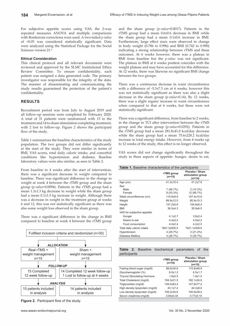
For subjective appetite scores using VAS, the 2-way repeated measures ANOVA and multiple comparisons with Bonferroni corrections were used. A two-tailed p-value of <0.05 was considered statistically significant. Data were analyzed using the Statistical Package for the Social Sciences version 21.4
Ethical ConsiderationThis clinical protocol and all relevant documents were reviewed and approved by the SLMC Institutional Ethics Review Committee. To ensure confidentiality, each patient was assigned a data generated code. The primary investigator was responsible for the integrity of the data. The manner of disseminating and communicating the study results guaranteed the protection of the patient’s confidentiality.
RESUlTS
Recruitment period was from July to August 2019 and all follow-up sessions were completed by February 2020. A total of 31 patients were randomized with 15 to the treatment and 14 to sham stimulation completing treatment, with 2 lost to follow-up. Figure 2 shows the participant flow of the study.
Table 1 summarizes the baseline characteristics of the study population. The two groups did not differ significantly at the start of the study. They were similar in terms of BMI, VAS scores, total daily caloric intake, and comorbid conditions like hypertension and diabetes. Baseline laboratory values were also similar, as seen in Table 2.
From baseline to 4 weeks after the start of intervention, there was a significant decrease in weight compared to baseline. There was significant difference in the change in weight at week 4 between the rTMS group and the sham group (p-value=0.0094). Patients in the rTMS group had a mean 1.3±1.3 kg decrease in weight while the sham group had a mean 0.1±1.5 kg increase in weight. Although there was a decrease in weight in the treatment group at weeks 6 and 12, this was not statistically significant as there was also some weight loss observed in the sham group.
There was a significant difference in the change in BMI compared to baseline at week 4 between the rTMS group
and the sham group (p-value=0.0017). Patients in the rTMS group had a mean 0.6±0.6 decrease in BMI while the sham group had a mean 0.1±0.6 increase in BMI. Furthermore, large effect sizes were observed in change in body weight (0.786 to 0.996) and BMI (0.742 to 0.990) indicating a strong relationship between rTMS and these outcomes. At 6 weeks however, there was a plateau in BMI from baseline but the p-value was not significant. The plateau in BMI at 6 weeks posttest coincides with the weight plateau and may have accounted for this difference. At 12 weeks, there was likewise no significant BMI change between the two groups.
There was a continuous decrease in waist circumference with a difference of -5.3±7.3 cm at 6 weeks, however this was not statistically significant as there was also a slight decrease in the sham group (p-value=0.14). By 12 weeks, there was a slight regain/ increase in waist circumference when compared to that at 6 weeks, but these were not statistically significant.
There was a significant difference, from baseline to 2 weeks, in the change in TCI after intervention between the rTMS group and the sham group (p-value=0.0292). Patients in the rTMS group had a mean 281.8±41.0 kcal/day decrease while the sham group had a mean 75.6±228.2 kcal/day increase in total energy intake. However, from 4 weeks up to 12 weeks of the study, this effect is no longer observed.
VAS scores did not change significantly throughout the study in three aspects of appetite- hunger, desire to eat,
Vol. 35 No. 2 November 2020
184
www.asean-endocrinejournal.org
Margaret Encarnacion, et al Efficacy of rTMS in Inducing Weight Loss among Obese Filipino Patients
Figure 2. Participant flow of the study.
Fulfilled inclusion criteria and randomized (n=30)
Real rTMS +weight management
n=15
Sham +weight management
n=15
15 Completed12 week follow-up
14 Completed 12 week follow-up1 Lost to follow-up at 4 weeks
15 patients included in analysis
14 patients includedin analysis
FOLLOW-UP
ALLOCATION
ANALYSIS
Table 1. Baseline characteristics of the participantsrTmS group
(n=15)
Placebo / Sham stimulation group
(n=14)Age (yrs) 41.3±10.4 41.2±7.6Sex
MaleFemale
7 (46.7%)8 (53.3%)
2 (14.3%)12 (85.7%)
Waist circumference (cm) 110.6±9.9 107.4±10.8Weight 89.9±12.0 85.9±15.3Height 157.2±5.6 154.6±6.0BMI 36.0±4.3 35.9±6.5VAS for subjective appetite
Hunger 4.1±2.7 3.6±2.4Desire to eat 4.4±2.2 4.0±2.2Food consumption 4.4±2.4 4.2±2.5
Total daily caloric intake 1851.3±605.5 1621.1±428.6Hypertension 4 (26.7%) 3 (21.4%)Diabetes Mellitus 4 (26.7%) 5 (35.7%)
Table 2. Baseline biochemical parameters of the participants
rTmS group(n=16)
Placebo / Sham stimulation group
(n=15)Fasting blood sugar (mg/dl) 98.6±34.6 115.8±46.9Glycohemoglobin (%) 6.0±1.2 6.5±1.7Thyroid Stimulating Hormone 1.9±0.5 1.9±1.0Total Cholesterol (mg/dl) 184.3±31.5 182.1±34.6Triglycerides (mg/dl) 149.4±63.4 147.8±77.2High density lipoprotein (mg/dl) 43.1±7.2 44.3±8.6Low density lipoprotein (mg/dl) 109.2±34.6 105.9±38.5Serum creatinine (mg/dl) 0.84±0.34 0.77±0.15

and prospective food consumption. There was a significant difference in hunger scores after intervention between the rTMS group and the sham group (p-value=0.023) at 2 weeks. Patients in the rTMS group had a mean 0.5±2.9 increase while the sham group had a mean 0.9±2.2 increase in hunger scores. At 4 up to 12 weeks however, there was no longer an observed difference between the two groups. There was no significant difference in desire to eat and prospective food consumption between the two groups from baseline up to 12 weeks of the study.
Within-group differences were analyzed through mixed linear regression model. These showed that within the rTMS group, there was a significant decrease in weight, waist circumference and VAS score for prospective food consumption when baseline values are compared to 12 weeks. However, when compared to the sham group (between-group difference) based on ANCOVA results reported above, the changes were not significant.
Safety rTMS was well-tolerated by the participants. 2 patients in the treatment group reported transient mild headache (graded 3/10 post 1st session of rTMS for 1 patient, and graded 4/10 post 3rd and 4th session of rTMS for 1 patient). These occurred immediately after rTMS and were resolved within 24 hours. No other adverse event was reported and no participant dropped out of the study because of headache. No adverse effect was reported in the sham stimulation group.
DISCUSSION
Efficacy of rTMS in Decreasing BMIThe weight loss in this study is comparable to that of the study by Kim et al., in 20184 which also showed a significant decrease in weight and BMI at 4 weeks after the rTMS. The weight loss in this study was -1.3±1.3 kg in the treatment group versus -1.35±2.31 kg in the study by Kim et al. Since Week 2 anthropometric values were not measured in their study, the researchers are unable to compare the 2-week data to that of other studies.
Vol. 35 No. 2 November 2020
185
www.asean-endocrinejournal.org
Margaret Encarnacion, et alEfficacy of rTMS in Inducing Weight Loss among Obese Filipino Patients
Table 3. Within-group differences in study outcomes assessed using mixed linear regressionrTmS group
(n=15)Placebo/Sham Stimulation group
(n=14)Within Group Differences Within Group Differences
Coefficient Standard Error P-value Coefficient Standard Error P-valueWeight
After 2weeksAfter 4 weeksAfter 6 weeksAfter 12 weeks
-0.66-1.33-1.36-2.87
1.331.331.331.33
0.6230.3150.3050.030
0.090.11-0.15-0.31
0.480.480.480.48
0.8470.8240.7560.515
BMIAfter 2weeksAfter 4 weeksAfter 6 weeksAfter 12 weeks
-1.16-0.60-0.56-1.21
0.650.650.650.65
0.0760.3590.3870.064
0.090.08-0.03-0.21
0.230.230.230.23
0.6820.7460.8910.377
Waist CircumferenceAfter 2weeksAfter 4 weeksAfter 6 weeksAfter 12 weeks
-1.8-3.7-5.3-4.1
1.451.451.451.45
0.2160.0110.0000.005
-2.3-2.5-3.2-3.0
1.141.141.141.14
0.0420.0270.0050.008
TCIAfter 2weeksAfter 4 weeksAfter 6 weeksAfter 12 weeks
-281.1-282.4-236.8-153.8
124.3124.3124.3124.3
0.0240.0230.0570.216
75.610
-81.1-114.6
87.387.387.387.3
0.3870.9090.3530.189
VAS HungerAfter 2weeksAfter 4 weeksAfter 6 weeksAfter 12 weeks
0.5-0.6-0.6-1.1
0.670.670.670.67
0.4280.3610.3400.088
-0.9-0.4-0.40.0
0.610.610.610.61
0.1290.4980.4761.000
VAS Desire to EatAfter 2weeksAfter 4 weeksAfter 6 weeksAfter 12 weeks
-0.3-1.1-1.4-1.0
0.500.500.500.50
0.5230.0320.0040.039
-1.1-0.9-0.9-0.4
0.550.550.550.55
0.0460.0900.1180.482
VAS Food consumptionAfter 2weeksAfter 4 weeksAfter 6 weeksAfter 12 weeks
-0.4-1.2-1.1-1.4
0.560.560.560.56
0.5110.0390.0450.012
-1.0-1.1-1.0-0.7
0.620.620.620.62
0.0890.0690.0930.325
Table 4. Correlation of change in VAS scores from baseline to 12 weeks with change in total caloric intake from baseline to 12 weeks
Coefficient P-valueVAS Hunger 0.13 0.3441VAS Desire to Eat -0.01 0.9398VAS Food Consumption -0.03 0.8356
Table 5. Correlation of change in weight, BMI, Waist Circumference from baseline to 12 weeks with change in total caloric intake from baseline to 12 weeks
Coefficient P-valueWeight -0.17 0.3704BMI -0.17 0.3740Waist Circumference -0.27 0.1501

These results are also supported by 2 other studies conducted in 2019 (both published after our study protocol has been completed and subject recruitment was already ongoing). In the study by Kim et al., in 2019, 8 sessions of rTMS over 4 weeks were done and resulted in more weight loss of 2.75±2.3 kg.12 Alvarado-Reynoso and Tututi’s study employed a longer treatment period, with 5 rTMS sessions every week for 2 weeks, then once a week on weeks 3, 4, 6, 8, 12, 20 and 28 coupled with a low -carbohydrate diet. They also found a continuous decline in weight up to the last session at 28 weeks.13
The proposed explanation is that targeting the DLPFC helps decrease deranged eating behaviors and excessive food cravings. Decreased food cravings, in turn, decreases food intake and aids in weight loss.4,12 The researchers did a correlation analysis and found that change in caloric intake was not significantly associated with change in BMI, waist circumference and weight. Although it is accepted in studies that less food intake leads to weight loss, several factors may have affected the results of this study. One may be that the sample size was not adequate for the correlation analysis done. Food intake through 3-day food diary may also be inaccurately collected by the subjects, thus a significant decrease in intake in relation to weight loss was also not observed.
There was no longer a significant change observed at weeks 6 and 12, however, indicating that although rTMS may be effective in decreasing body weight, the effects may not be permanent. The studies done by Kim and Alvarado-Reynoso employed assessment of weight immediately after the last session, and therefore were not able to explore the long-term effects of rTMS even if this intervention is no longer present. This current study may imply that regular sessions of rTMS may need to be given in order to maintain weight loss.
As to waist circumference, although there was a continuous decrease with a difference of -5.3+7.3 cm at 6 weeks, this was not statistically significant when compared to that of the sham group. It is possible that a steady decline in waist circumference may be observed if the study is further extended, or that there was inter-observer variability in measuring the waist circumference at each follow-up.
It is important to note that one of the subjects in the rTMS group displayed a significantly higher decrease in weight and BMI compared to the other subjects in both groups, and this may be a possible outlier in the study. Effect of rTMS on TCIIn the first 2 weeks, the TCI was significantly lower in the treatment group. However, for the succeeding 2 weeks
Vol. 35 No. 2 November 2020
186
www.asean-endocrinejournal.org
Margaret Encarnacion, et al Efficacy of rTMS in Inducing Weight Loss among Obese Filipino Patients
Figure 3. Comparison of weight from baseline between rTMS vs sham group.
Figure 4. Comparison of BMI from baseline between rTMS vs sham group.
Figure 5. Comparison of waist circumference from baseline between rTMS vs sham group.
Figure 6. Change in total daily caloric intake from baseline between rTMS vs sham group.
88.988.3
87.688.3
86.185.9 86 86 86 85.6
8081828384858687888990
Baseline Week 2 Week 4 Week 6 Week 12
rTMS group (n=15) sham group (n=14)
Wei
ght (
kg)
100
102
104
106
108
110
112
Baseline Week 2 Week 4 Week 6 Week 12
rTMS group (n=15) sham group (n=14)
Wai
st C
ircum
fere
nce
(cm
)
1000
1100
1200
1300
1400
1500
1600
1700
1800
1900
Baseline Week 2 Week 4 Week 6 Week 12
rTMS group (n=15) sham group (n=14)
Tota
l Cal
oric
Inta
ke (k
cal/d
ay)
34.2
34.4
34.6
34.8
35
35.2
35.4
35.6
35.8
36
36.2
Baseline Week 2 Week 4 Week 6 Week 12
rTMS group (n=15) sham group (n=14)
Bod
y M
ass
Inde
x (B
MI)

onwards, there was no significant difference anymore between the two groups. This initial decrease coincided with the duration of rTMS procedure indicating that TCI may be decreased by rTMS but the effect is not permanent.
This may be reflective of the short-term impact of rTMS in decreasing appetite and food cravings. Thus, when the rTMS was no longer continued, there was a corresponding increase in intake in the treatment group. The permanency or reversibility of effects of rTMS on weight loss and food cravings in obese patients has not yet been studied, yet it is an area that needs to be explored. In a study by Mally et al., long-term effect of rTMS was studied in patients with Parkinson’s disease. rTMS was given 2 times a day for 7 days (1 Hz, 100 stimuli per day) and was repeated at least twice a year for 3 years. There was significant decrease in progression of Parkinson’s disease with the repeated stimulation over 3 years. They proposed that prolonged stimulation produced prolonged inhibition in intracortical connections which delayed progression of the disease.14
Therefore, increasing the total number of rTMS to the DLPFC may potentially induce longer lasting effects on weight loss and caloric intake as well.
The result of this study is in contrast to the study by Kim et al., where there was a continued decrease in total caloric intake at Week 4.4 However, since there was no Week 2 assessment in their study, a comparison of intake between Week 2 and Week 4, and therefore a possibility that there was lesser caloric intake at Week 2 than at Week 4, was not determined.
Change in VAS scores for appetite and change in total caloric intake showed no significant association when correlation analysis was done. A meta-analysis by Lowe et al., in 2017 supported this study’s result, where they found that food cravings decreased after multiple stimulation however actual food consumption after both single and multiple sessions for rTMS was found to be inconsistent among different studies.2 A study by Uher et al., showed that a decrease in subjective food craving did not necessarily translate to less food consumption between the two groups.6 Differences in methodology i.e., specific brand of stimulation device, frequency and intensity of stimulation and higher BMI cutoffs in this study may account for the incongruent results.2,6,12 It is also possible that the sample size in this study was not powered to observe a correlation between food cravings and food intake, as this study used change in BMI in computing for the sample size.
Another is the possible discrepancy in the method of collecting data for total caloric intake through the use of food diaries. Since it is subjective and based on recall, factors such as inaccurate recording of intake, writing down only of days with the least amount of oral intake, or inability to recall all food taken may have played a role in the inability to show significant results at week 4 onwards.
Effect of rTMS on appetiteResults showed that there was no significant difference in appetite between the treatment and sham groups from baseline to 12 weeks. This is comparable to the study by Kim et al., where there was no significant difference in the sham and treatment groups in terms of hunger and desire to eat; however, this study failed to show a significant
reduction in prospective food consumption as mentioned in the former study.4 In their 2019 study, however there was no significant difference in prospective food consumption, but there was a significant reduction in hunger and desire to eat.12
There can be several reasons why there are contradicting results in the VAS scores for appetite. One reason may
Vol. 35 No. 2 November 2020
187
www.asean-endocrinejournal.org
Margaret Encarnacion, et alEfficacy of rTMS in Inducing Weight Loss among Obese Filipino Patients
Figure 7. Change in hunger scores from baseline between rTMS vs sham group.
Figure 9. Change in prospective food consumption from baseline between rTMS vs sham group.
Figure 8. Change in desire to eat from baseline between rTMS vs sham group.
0
0.5
1
1.5
2
2.5
3
3.5
4
4.5
5
Baseline Week 2 Week 4 Week 6 Week 12
rTMS group (n=15) sham group (n=14)
VAS
Scor
e
0
0.5
1
1.5
2
2.5
3
3.5
4
4.5
5
Baseline Week 2 Week 4 Week 6 Week 12
rTMS group (n=15) sham group (n=14)
VAS
Scor
e
0
0.5
1
1.5
2
2.5
3
3.5
4
4.5
Baseline Week 2 Week 4 Week 6 Week 12
rTMS group (n=15) sham group (n=14)
VAS
Scor
e

be the differences in fasting time prior to taking the VAS scores. In the 2018 study by Kim et al., it was taken after 4 hours of fasting while in their 2019 study, it was after only 2 hours.4,12 In this study, the fasting hours prior to testing were consistent for each participant, but was not uniform across all subjects, and these ranged from 2-5 hours of fasting (i.e., participant 1 fasted for 2 hours prior to each assessment, participant 2 fasted for 4 hours prior to assessment). Another reason may be that caloric intake during the study was not standardized. Only the specific timing of last meal was consistent for each participant, however the actual intake/ meal prior to each assessment of appetite was not the same for all participants and therefore these may have affected their subjective scores.
Another reason is the possible placebo effect on the control group. Most of the patients in the sham group believed themselves to be in the treatment group and therefore these may have given lower ratings on perceived appetite.
Lastly, the differences in technique of rTMS application may also account for the different results in weight and appetite/ food cravings. In our study, we used 20 trains of 5 seconds at a frequency of 10 Hz and intensity of 110% of the participant’s motor threshold. In the study by Kim et al., in 2019, 40 trains of 5 seconds with frequency of 10 Hz and intensity of 110% motor threshold was used.12 Alvarado-Reynoso in 2019 used 10 trains of 100 pulses given at 10 Hz and 90% of motor threshold, with more sessions employed compared to the two studies.13 While all these produced weight loss, the degree of weight loss and reduction in food cravings were different among the 3 studies.
Neuroendocrine effects of rTMS on food cravings have been studied. Ferrulli et al., in 2018, reported that orexigenic pathways have been altered as a result of TMS, producing an increase in norepinephrine and B-endorphins, while salivary cortisol is decreased. This suggests a potential role of TMS in inducing dopaminergic activation and modulation of the food-reward system.15 With the advent of these biochemical tests that provide objective and measurable assessment of appetite, evaluation of food cravings no longer need to be purely subjective, thus increasing the accuracy of results. It still needs to be established, however, if alteration of the neuroendocrine pathways translates into actual reduction in food intake.
limitations There were only 4 sessions of rTMS done due to cost and inability to determine safety of the procedure. As this is the first study done among Filipinos, the researchers opted to use the same rTMS protocol that was used among obese patients in Korea, which employed 4 sessions of rTMS. Furthermore, fully accurate measurement of appetite and intake may not have been achieved due to the subjective nature of the VAS scores for appetite, as well as accounting for some inaccurately recorded food diaries. Another possible limitation may be that this did not have adequate sample size and power to declare statistical significance of the reported differences in changes in other outcomes (changes in desire to eat, hunger and total caloric intake between two groups) as the sample size in this study was computed based on BMI change from baseline.
CONClUSION
rTMS to the DLPFC effectively decreases BMI and weight from baseline to 4 weeks in the treatment group compared to the sham group, with a decrease in weight by -1.3±1.3 kg and decrease in BMI by 0.6±0.6. At 6-12 weeks after rTMS however, there was no longer a significant difference, indicating that 4 sessions of rTMS are not enough to produce a permanent effect on weight loss. Although there was an initial significant decrease in total caloric intake in the first 2 weeks by about 200 kilocalories a day, it failed to show a consistent decline in total caloric intake after 2 weeks from the last session of rTMS. Furthermore, subjective scoring showed no difference as to hunger, desire to eat and prospective food consumption in the treatment group versus the sham group.
RecommendationsThe researchers recommend a more controlled food intake and fasting time prior to testing for subjective appetite in order to have a better estimate of appetite/ hunger that is not related to quantity of food consumed prior to the testing. Body fat analysis may also help to determine if visceral adipose tissue decreases with rTMS, as this is an important risk factor for cardiovascular disease in obese patients. To eliminate bias and placebo effect, the researchers recommend exploring the usefulness of measuring neuroendocrine hormones like leptin, B-endorphins, cortisol and norepinephrine as objective markers of appetite in order to supplement the much more subjective food diaries and VAS scoring to measure hunger and appetite of the participants.
Acknowledgments The authors sincerely thank the active consultants and trainees of SLMC QC for helping in enrolling participants in this study. They would also like to thank the SLMC QC Research and Biotechnology group, statistician, and the participants who volunteered to take part in the research.
Statement of AuthorshipAll authors certified fulfillment of ICMJE authorship criteria.
Author DisclosureThe authors declared no conflicts of interest.
Funding SourceFunding for rTMS, weight management program, and the laboratory tests done came from the SLMC-QC Research and Biotechnology Division and the PSEDM-Abbott Nutrition Year 2019 research grant.
References 1. WHO Expert Consultation. Appropriate body-mass index for
Asian populations and its implications for policy and intervention strategies [Erratum in Lancet. 2004; 13;363(9412):902]. Lancet. 2004;363(9403):157-163. PMID: 14726171. https://doi.org/10.1016/S0140-6736(03)15268-3.
2. Lowe CJ, Vincent C, Hall PA. Effects of noninvasive brain stimulation on food cravings and consumption: A meta-analytic review. Psychosom Med. 2017;79(1):2-13. PMID: 27428861. https://doi.org/10.1097/PSY.0000000000000368.
3. Garvey WT, Mechanick JI, Brett EM, et al. American Association of Clinical Endocrinologists and American College of Endocrinology Clinical Practice Guidelines for Comprehensive Medical Care of Patients with Obesity. Endocr Pract. 2016;22 Suppl 3:1-203. PMID: 27219496. https://doi.org/10.4158/EP161365.GL.
4. Kim SH, Chung JH, Kim TH, et al. The effects of repetitive transcranial magnetic stimulation on eating behaviors and body weight in obesity: A randomized controlled study. Brain Stimul. 2018;11(3): 528-35. PMID: 29326022. https://doi.org/10.1016/j.brs.2017.11.020.
Vol. 35 No. 2 November 2020
188
www.asean-endocrinejournal.org
Margaret Encarnacion, et al Efficacy of rTMS in Inducing Weight Loss among Obese Filipino Patients

5. Mann T, Tomiyama AJ, Westling E, Lew AM, Samuels B, Chatman J. Medicare's search for effective obesity treatments: Diets are not the answer. Am Psychol. 2007;62(3):220-33. PMID: 17469900. https://doi.org/10.1037/0003-066X.62.3.220.
6. Uher R, Yoganathan D, Mogg A, et al. Effect of left prefrontal repetitive transcranial magnetic stimulation on food craving. Biol Psychiatry. 2005;58(10):840-2. PMID: 16084855. https://doi.org/10.1016/j.biopsych.2005.05.043.
7. Mishra BR, Sarkar S, Praharaj SK, Mehta VS, Diwedi S, Nizamie SH. Repetitive transcranial magnetic stimulation in psychiatry. Ann Indian Acad Neurol. 2011;14(4):245-251. PMID: 22346010. PMCID: PMC3271460. https://doi.org/10.4103/0972-2327.91935.
8. Cooper S, Robison AJ, Mazei-Robison M. Reward circuitry in addiction. Neurotherapeutics. PMID: 28324454. PMCID: PMC5509624. https://doi.org/10.1007/s13311-017-0525-z.
9. Nahas Z, Teneback CC, Kozel A, et al. Brain effects of TMS delivered over prefrontal cortex in depressed adults: Role of stimulation frequency and coil-cortex distance. J Neuropsychiatry Clin Neurosci. 2001;13(4):459-70. PMID: 11748315. https://doi.org/10.1176/jnp.13.4.459.
10. Maizey L, Allen CPG, Dervinis M, et al. Comparative incidence rates of mild adverse effects to transcranial magnetic stimulation. Clin Neurophysiol. 2013;124(3):536-44. PMID: 22986284. https://doi.org/10.1016/j.clinph.2012.07.024.
11. Rossi S, Hallett M, Rossini PM, Pascual-Leone A, Safety of TMS Consensus Group. Safety, ethical considerations, and application guidelines for the use of transcranial magnetic stimulation in clinical practice and research. Clin Neurophysiol. 2009;120(12): 2008-39. PMID: 19833552. https://doi:10.1016/j.clinph.2009.08.016.
12. Kim SH, Chung JH, Kim TH, et al. The effects of repetitive transcranial magnetic stimulation on body weight and food consumption in obese adults: A randomized controlled study. Brain Stimul. 2019;12(6): 1556-64. PMID: 31378600. https://doi.org/10.1016/j.brs.2019.07.020
13. Alvarado-Reynoso, B, Ambriz-Tututi, M. The effects of repetitive transcranial magnetic stimulation in combination with a low-carbohydrate diet in overweight or obese patients. A randomized controlled trial. Obesity Medicine 2019;14. https://doi.org/10.1016/ j.obmed.2019.100095.
14. Málly J, Farkas R, Tóthfalusi L, Stone TW. Long-term follow-up study with repetitive transcranial magnetic stimulation (rTMS) in Parkinson's disease. Brain Res Bull. 2004;64(3):259-63. PMID: 15464863. https:// doi.org/10.1016/j.brainresbull.2004.07.004.
15. Ferrulli A, Adamo M, Massarini S, Luzi L. Deep transcranial magnetic stimulation acutely modulates neuro-endocrine pathways underlying obesity. Endocrine Abstracts. 2018; 56. OC6.2. https:// doi.org/10.1530/endoabs.56.OC6.2.
Vol. 35 No. 2 November 2020
189
www.asean-endocrinejournal.org
Margaret Encarnacion, et alEfficacy of rTMS in Inducing Weight Loss among Obese Filipino Patients
JAFESA new venue for publishing your original articles.
Visit www.ASEAN-endocrinejournal.org forInstructions to Authors.
Authors are required to accomplish, sign and submit scanned copies of the JAFES Author Form consisting of: (1) Authorship Certification, that authors contributed substantially to the work, that the manuscript has been read and approved by all authors, and that the requirements for authorship have been met by each author; (2) the Author Declaration, that the article represents original material that is not being considered for publication or has not been published or accepted for publication elsewhere, that the article does not infringe or violate any copyrights or intellectual property rights, and that no references have been made to predatory/suspected predatory journals; (3) the Author Contribution Disclosure, which lists the specific contributions of authors; and (4) the Author Publishing Agreement which retains author copyright, grants publishing and distribution rights to JAFES, and allows JAFES to apply and enforce an Attribution-Non-Commercial Creative Commons user license. Authors are also required to accomplish, sign, and submit the signed ICMJE form for Disclosure of Potential Conflicts of Interest. For original articles, authors are required to submit a scanned copy of the Ethics Review Approval of their research as well as registration in trial registries as appropriate. For manuscripts reporting data from studies involving animals, authors are required to submit a scanned copy of the Institutional Animal Care and Use Committee approval. For Case Reports or Series, and Images in Endocrinology, consent forms, are required for the publication of information about patients; otherwise, appropriate ethical clearance has been obtained from the institutional review board. Articles and any other material published in the JAFES represent the work of the author(s) and should not be construed to reflect the opinions of the Editors or the Publisher.

Relationship between Plasma Adiponectin level andCorrected QT Interval in Smoker and Non-smoker Adult male Subjects
Yin Thu Theint, Ei Ei Khin, Ohnmar Myint Thein, Mya Thanda Sein
Department of Physiology, University of Medicine 2, Yangon, Republic of the Union of Myanmar
Abstract
Objective. This study determined the relationship between plasma adiponectin level and corrected QT interval (QTc) in smokers and non-smokers.
Methodology.This cross-sectional analytical study was undertaken in 30 smokers and 30 non-smokers. Plasma adiponectin level was determined by enzyme-linked immunosorbent assay (ELISA). The QT interval was measured by routine 12-lead ECG with Lead II rhythm and QTc was calculated.
Results. Mean plasma adiponectin level was significantly lower in smokers (27.89±15 µg/ml) than that of non-smokers (52.13±21.57µg/ml) (p<0.001). Mean QTc interval was significantly longer in smokers than that of non-smokers (415.37±29.9 versus 395.63±26.13 ms, p<0.01). Higher risk of low adiponectin level (odds ratio [OR],8.1; 95% confidence interval [CI],1.61-40.77) and QTc interval prolongation (OR,6; 95%CI,1.17-30.73) were observed in smokers. There was weak significant negative correlation between plasma adiponectin level and QTc interval in the study population (n=60, r=-0.407, p=0.001). Moreover, low plasma adiponectin level was significantly associated with prolonged QTc interval in the study population (n=60, Fisher's exact p value<0.05). Risk of QTc interval prolongation was 4.3 times higher in subjects with low plasma adiponectin level (OR,4.27; 95% CI,1.05-17.46).
Conclusion. Smokers have greater risk for low plasma adiponectin level and prolonged QTc interval. There is a relationship between plasma adiponectin level and QTc interval.
Key words: smoker, adiponectin, QTc
INTRODUCTION
Adiponectin is a 247 amino-acid protein secreted from adipocytes1 and exerts beneficial effects on the cardio-vascular system by directly acting on vascular smooth muscle cells, endothelial cells and cardiac myocytes.2,3,4
It can mediate anti-atherosclerotic, anti-fibrotic, anti-apoptotic and anti-inflammatory effects.3,4,5 Thus, low plasma adiponectin level is associated with increased prevalence of cardiovascular diseases (CVD). Animal study reported that adiponectin plays a role in expression of transient outward potassium channel (Ito) and duration of action potential in rat ventricular muscles.6 A loss of Ito channel protein expression and function was associated with action potential prolongation7 which was reflected by QT prolongation.8 The QT interval represents the time from onset of ventricular depolarization to completion of repolarization. When QT interval is prolonged, repolarization is irregular with increased incidence of ventricular arrhythmias and sudden death.8
QTc means corrected QT interval with heart rate because normal QT inter val decreases as heart rate increases. QTc interval was calculated using Bazett’s formula as follows: QTc=QT/√RR.9QTc interval of more than 440ms in men and 460ms in women is considered prolonged.10 So, it can be assumed that adiponectin might be associated with QT interval. Only a few studies are available focusing on the role of adiponectin in QTc interval in humans. In a study done in Japan, it was shown that QTc interval had inverse correlation with adiponectin in healthy male subjects.11 Further studies are needed to explore the association between adiponectin and QTc interval.
According to previous studies,12-15 the circulating adiponectin level was significantly decreased in smokers compared to non-smokers. Present study selected the smokers as participants to determine the association between plasma adiponectin level and QTc interval. Thus, present study investigated the relationship between plasma adiponectin level and corrected QT interval in male adult smokers and non-smokers.
________________________________________
ISSN 0857-1074 (Print) | eISSN 2308-118x (Online)Printed in the PhilippinesCopyright © 2020 by Theint et al.Received: June 7, 2020. Accepted: October 2, 2020.Published online first: October 19, 2020.https://doi.org/10.15605/jafes.035.02.09
Corresponding author: Prof. Mya Thanda Sein, MBBS, MMedSc (Physiology), PhD, Dip Med Edu, DCMEDepartment of Physiology, University of Medicine 2Yangon, Republic of the Union of Myanmar 11031Tel. No.: 95-95109450Fax No.: 95-1-9690265E-mail: [email protected]: https://orcid.org/0000-0002-5135-3418
* This research work was presented in the 20th AFES Congress 2019, Philippines and was awarded best poster presentation.
Vol. 35 No. 2 November 2020190 www.asean-endocrinejournal.org
Journal of theASEAN Federation ofEndocrine Societies Original Article
This work is licensed under a Creative Commons Attribution-NonCommercial 4.0 International License (https://creativecommons.org/licenses/by-nc/4.0/).

According to the adiponectin level derived from Fan et al.17
X1 = mean plasma adiponectin level of smoker = 2.49 µg/mlX2 = mean plasma adiponectin level of non-smoker
= 3.23 µg/mlσ1 = standard deviation of smoker = 0.35σ2= standard deviation of non-smoker = 0.37sample size n1= 9
According to the QTc value derived from Sharma et al.18
X1 = mean QTc interval of smoker = 413.9 msX2 = mean QTc interval of non-smoker = 377.9 msσ1 = standard deviation of smoker = 34.17σ2= standard deviation of non-smoker = 20.88sample size n2= 23
A total of 60 male adult subjects (30 smokers and 30 non-smokers) were recruited in the present study. The study was conducted in the morning between 7 to 9 am in the fasting state. At the beginning, fasting blood sugar level was determined by pricking the fingertip using a glucose meter. Subjects having fasting blood sugar (FBS) levels >110 mg/dl were excluded from the study. Routine 12 lead ECG was performed using ECG machine (CM 100, Shenzhen Comen Medical Instruments Co., Ltd, China) after the subjects were allowed to lie down comfortably on the bed with attachment of limb electrodes for 15 minutes. Paper speed was 25 mm/s and manual calibration was carefully adjusted at 10 mm/mV before recording. Lead II rhythm strip was also taken for 10 seconds. QT interval was measured from the start of Q-wave to the end of T-wave in a normal beat. Heart rate was calculated from average R-R intervals of the beat within 10 seconds. QT intervals and R-R intervals were measured using vernier caliper. Then, QTc interval was calculated by using Bazett’s formula (QTc=QT/√RR).9 Then, 3 milliliters of fasting venous blood was drawn from the antecubital vein under aseptic condition using a disposable syringe and needle for each subject. Blood samples were collected into a tube containing EDTA disodium anticoagulant and carried to the common research laboratory of the University of Medicine 2. On arrival, plasma separation was done by centrifuging at 2000 rpm at 4°C for 10 minutes and stored at -20°C until sample analysis. Plasma adiponectin level was determined by Adiponectin ELISA Kit (ab99968, Abcam, UK).
Data entry and analysis were done by SPSS software (version 22, SPSS Inc., Chicago, IL, USA). Data were described by mean ± SD. Independent "t" test was used to compare the plasma adiponectin level and QTc interval between smokers and non-smokers. Correlation studies were computed by Pearson’s correlation. Chi-square test was used to determine whether there are significant associations between smoking, plasma adiponectin level and QTc interval. Values of p<0.05 were accepted as statistically significant. This study was approved by Ethics Review Committee, University of Medicine 2, Yangon.
RESUlTS
The general characteristics of the study population are shown in Table 1. Mean age, BMI, SBP, DBP and FBS of two
mEThODOlOgy This cross-sectional analytical study was undertaken in apparently healthy male subjects 18-40 years old, residing in Hlaingtharyar Township,Yangon, Myanmar from April 2018 to December 2018. The present study used the non-probability convenience sampling method. No (8) Quarter was selected from 22 Quarters in Hlaingtharyar Township since that quarter has high population density. We asked for approval from the local administrator for recruitment of apparently healthy male subjects between 18-40 years old. Voluntary written informed consent was obtained after thorough explanation of research purpose and procedures. All participants underwent history taking and physical examination. Subjects with no acute illness and no known history of hypertension, diabetes, ischemic heart disease, cerebrovascular accident, arrhythmia, peripheral arterial disease, renal disease and bronchial asthma were regarded as apparently healthy subjects. Individuals with the following characteristics were excluded from the study: those who consumed heavy alcohol (more than 3 units of alcohol per day) that decreased adiponectin levels as a result of increased tumor necrosis factor-α; individuals who chewed betel quid with tobacco containing nicotine that affects both adiponectin and QT interval; individuals who are currently taking drugs that altered adiponectin concentration like omega 3 fish oil, niacin and statin; individuals who are currently using antimicrobial agents such as fluoroquinolones, erythromycin, antidepressant agents such as amitriptyline that prolonged QT interval. Each participant was interviewed by using a questionnaire to collect history of cigarette smoking including average number of cigarettes smoked per day and duration of cigarette smoking. Those who currently smoke a minimum of 10 cigarettes per day for at least 5 years were selected as smokers and those who have never smoked any form of tobacco in their life and with no history of smoking in their family members were defined as non-smokers. Subjects with normal body mass index (BMI) (18.5-24.9 kg/m2) were selected. Resting arterial blood pressure was measured in lying position using a mercury sphygmomanometer and a stethoscope by an indirect method. The average of three measurements taken over a one-minute interval was used. Subjects having systolic blood pressure (SBP) >120 mmHg and diastolic blood pressure (DBP) >80 mmHg were excluded according to American Health Association guideline 2017.
Sample size was calculated by using Rosner's formula.16
n = number of subjects for each groupδ = X2 – X1σ1
2 = variance of X1 σ2
2 = variance of X2 Z1-α
= the Z value from normal distribution associated with a probability of 1-αZ1-β = the Z value from normal distribution associated with a probability of 1-β
If α = 0.05, Z1-α= 1.96If β = 0.01, Z1-β = 2.326
Vol. 35 No. 2 November 2020
191
www.asean-endocrinejournal.org
Yin Thu Theint, et alPlasma Adiponectin Level and QTc in Adult Male Subjects

as low adiponectin level in the present study and it was observed in 11 out of 30 (36.7%) smokers and 2 out of 30 (6.7%) non-smokers. Therefore, risk of lower plasma adiponectin level was 8.1 times higher in smokers than non-smokers (odds ratio (OR), 8.1; 95% confidence interval (CI), 1.61-40.77). Also, smokers had higher proportion of low adiponectin level compared to non-smokers (z value=2.84, p=0.005).
In the present study, 9 out of 30 (30%) smokers and 2 out of 30 (6.7%) non-smokers had prolonged QTc interval (>440 ms). Therefore, risk of prolonged QTc interval was 6 times higher in smokers than non-smokers (OR, 6.0; 95% CI=1.17-30.73). Smokers also had a higher proportion of prolonged QTc interval compared to non-smokers (z value=2.33, p=0.02).
It was also noted that 5 out of 13 (38.5%) subjects with low plasma adiponectin level had prolonged QTc interval and 6 out of 47 (12.8%) subjects with normal plasma adiponectin level had prolonged QTc interval (>440 ms). Therefore, risk of prolonged QTc interval was 4.3 times higher in subjects with low plasma adiponectin level than subjects with normal plasma adiponectin level (OR, 4.27; 95%CI, 1.05-17.46). Table 2 showed that there is a significant association between low plasma adiponectin level and prolonged QTc interval in the whole population (n=60, Fisher's exact p value <0.05).
Table 2. The association between low plasma adiponectin level and prolonged QTc interval in total population (n=60)
VariableQTc interval n (%)
*p valueProlonged NormalSubjects with <0.05Low adiponectinNormal adiponectin
5(38.5%)6(12.8%)
8 (61.5%)41 (87.2%)
*Fisher's Exact test
groups showed no significant differences indicating that both groups were comparable to each other. Heart rate of the two groups showed significant difference.
Table 1. Baseline characteristics of the subjects
ParametersNon-smokers
(mean±SD)(n=30)
Smokers(mean±SD)
(n=30)p value
Age (years) 25.43±3.52 26.47±4.1 0.29BMI (kg/m2) 20.95±2.1 21.67±1.66 0.15SBP (mmHg) 112±7.14 113.3±7.58 0.49DBP (mmHg) 70.3±7.18 72±7.14 0.37FBS (mg/dl) 103±10.8 107.2±3.98 0.06HR 72.13±9.45 81.53±11.89 0.001
Figure 1 shows the comparison of plasma adiponectin level between smokers and non-smokers. There was a significantly lower mean plasma adiponectin level in smokers compared with non-smokers (27.89±15 versus 52.13±21.57 µg/ml) (p<0.001). Mean QTc intervals were 415.37±29.9 and 395.63±26.13 ms for smoker and non-smoker groups respectively. Mean QTc interval of smokers was significantly higher than that of non-smokers (p<0.01) (Figure 2).
Correlation between plasma adiponectin level and QTc interval in the study population is illustrated in Figure 3. There was a weak negative correlation between plasma adiponectin level and QTc interval in the whole study population (r=-0.407, p=0.001, n=60) (Figure 3A). This was statistically significant. When the study population was subdivided into smokers and non-smokers, significant weak negative correlation was only observed in smokers (n=30) (r=- 0.434, p=0.017) (Figure 3B) but not in non-smokers (n=30) (r= - 0.175, p=0.35) (Figure 3C). According to Fumeron et al.,19 plasma adiponectin level of healthy individuals presented in a wide range from 20 to 45 µg/ml. Thus, adiponectin value under 20 µg/ml was considered
Vol. 35 No. 2 November 2020
192
www.asean-endocrinejournal.org
Yin Thu Theint, et al Plasma Adiponectin Level and QTc in Adult Male Subjects
*Indicates significant difference (p<0.001)Comparison was done by independent "t" test
Figure 1. Plasma adiponectin level in smokers and non-smokers.
*Indicates significant difference (p<0.01)Comparison was done by independent "t" test
Figure 2. QTc interval in smokers and non-smokers.

by determining carotid intima media thickness,21 lipid profiles22 and ECG23 and have shown that CVD risk is increased in smokers. Constituents of cigarette smoke are mainly nicotine, carbon monoxide, and free radical mediated oxidant gases which not only potentially contribute to cardiovascular diseases24 but also inhibit the adiponectin gene expression.12 Thus, the present study recruited smokers to investigate the role of adiponectin on QTc interval.
In the present study, among the smokers, the mean number of cigarettes smoked per day was 12.5±4.8 and mean cigarette smoking duration was 7.8±2.9 years. The present study showed that significant decrease of mean plasma adiponectin level was observed in smokers compared with non-smokers. The findings agree with the reports of previous studies.12-15 In addition, risk of lower plasma adiponectin level was 8.1 times greater in smokers than non-smokers (OR,8.1; 95% CI, 1.61-40.77). Thus, the present study can conclude that smokers have greater risk for low plasma adiponectin level.
Several explanations have been proposed for the mechanisms by which smoking reduces adiponectin concentration. In previous in vitro and in vivo studies, nicotine itself induces lipolysis through local nicotinic cholinergic (nAchR) and catecholaminergic receptors in adipose tissue25 and inhibits the expression of the adiponectin gene in cultured mouse 3T3-L1 adipocytes.12
Moreover, nicotine also has direct actions on the differentiation of adipocytes by increasing peroxisome proliferator-activated receptor-γ (PPAR-γ), which is essential for inducing differentiation from preadipocytes to mature adipocytes. Supraphysiological activation of PPARγ caused adipogenesis disturbances which may cause enhanced lipolysis and dysfunction of adipokine secretion.26 Smoking provokes oxidative stress and inflammatory cytokines that reduce adiponectin concentration. Oxidative stress disrupts activation of a key molecule, phosphatidylinositol 3-kinase (PI3K) for the secretion of adiponectin in adipocytes.27,28 Inflammatory
DISCUSSION
An animal study has shown that adiponectin affects channel proteins (Ito) in rat ventricular myocyte and hypoadiponectinaemia may be involved in prolongation of QT interval.6 Komatsu et al.,11 reported that there was inverse relationship between adiponectin and QTc interval in healthy men. Contrary to this study, Wu et al.,20 recently reported that there was positive association between adiponectin level and QTc in patients with stable angina. As there is limited information regarding the association between adiponectin and QT interval, further studies are needed to clarify the role of adiponectin on QT interval.
Cigarette smoking is a major cause of cardiovascular diseases. Many studies investigated CVD risk in smokers
Vol. 35 No. 2 November 2020
193
www.asean-endocrinejournal.org
Yin Thu Theint, et alPlasma Adiponectin Level and QTc in Adult Male Subjects
r, Pearson correlation coefficient; n, total number of subjects
Figure 3A. Correlation between plasma adiponectin level and QTc interval in study population.
r, Pearson correlation coefficient; n, total number of subjects
Figure 3C. Correlation between plasma adiponectin level and QTc interval in non-smokers (n=30).
r, Pearson correlation coefficient; n, total number of subjects
Figure 3b. Correlation between plasma adiponectin level and QTc interval in smokers (n=30).

Corrected QT interval was significantly prolonged in smokers compared to non-smokers.Thus, 8.1 times greater risk of low plasma adiponectin and 6 times greater risk of QTc interval prolongation were observed in smokers compared with non-smokers.
In addition, risk of prolonged QTc interval was 4.3 times higher in subjects with low plasma adiponectin level than subjects with normal plasma adiponectin level. A significant weak negative correlation as well as a significant association between plasma adiponectin level and QTc interval was observed in the whole study population. Thus, it can be concluded that relationship exists between plasma adiponectin level and QTc interval.
limitation of the studyAs a cross sectional analytical study, it cannot clearly establish the causative effect of adiponectin level on QTc interval. This study is not powered as an interventional study which provides specific conclusions to clarify the link between adiponectin and QTc interval. Moreover, due to relatively small sample size, the confidence intervals of the odds ratios are very wide e.g., smoking and QTc: OR 6 (1.17–30.73). The possibility of second hand smoke was not accounted for in both groups, especially for the non-smokers in the present study.
AcknowledgmentsThe authors thank all the participants of the Hlaingtharyar Township who willingly gave consent to this study. The authors also wish to acknowledge the External Grant Committee, Department of Medical Research, Lower Myanmar, Republic of Myanmar for the financial support and the Ethics Review Committee, University of Medicine 2, Yangon, Republic of Myanmar for providing ethical clearance.
Statement of AuthorshipAll authors certified fulfillment of ICMJE authorship criteria.
Author DisclosureThe authors declared no conflicts of interest.
Funding SourceThe study was financially supported by the External Grant Committee of the Department of Medical Research, Lower Myanmar, Republic of Myanmar.
References 1. Scherer PE, Williams S, Fogliano M, Baldini G, Lodish HF. A
novel serum protein similar to C1q, produced exclusively in adipocytes. J Biol Chem.1995;270(45):26746-9.PMID: 7592907. https://doi.org/10.1074/jbc.270.45.26746.
2. Arita Y, Kihara S, Ouchi N, et al. Adipocyte-derived plasma protein adiponectin acts as a platelet-derived growth factor-BB-binding protein and regulates growth factor-induced common postreceptor signal in vascular smooth muscle cell. Circulation. 2002;105(24):2893-8. PMID: 12070119. https://doi.org/10.1161/01.cir.0000018622.84402.ff.
3. Chen H, Montagnani M, Funahashi T, Shimomura I, Quon MJ. Adiponectin stimulates production of nitric oxide in vascular endothelial cells. J Biol Chem. 2003;278(45):45021-6. PMID: 12944390. https://doi.org/10.1074/jbc.M307878200.
4. Shibata R, Sato K, Pimentel DR, et al. Adiponectin protects against myocardial ischemia-reperfusion injury through AMPK- and COX-2-dependent mechanisms. Nat Med. 2005;11(10):1096-103. PMID: 16155579. PMCID: PMC2828682. https://doi.org/10.1038/nm1295.
5. Kumada M, Kihara S, Ouchi N, et al. Adiponectin specifically increased tissue inhibitor of metalloproteinase-1 through interleukin-10 expression in human macrophages. Circulation. 2004;109(17):2046-9. PMID: 15096450. https://doi.org/10.1161/01.CIR.0000127953.98131.ED.
6. Wang L. Protective role of adiponectin in volume overload-induced heart failure. A dissertation for Ph.D. Auburn University, Alabama. December 13, 2014. https://etd.auburn.edu/bitstream/handle/10415/4407/Lili%20Wang%20Dissertation.pdf?sequence=2.
cytokines such as TNF and IL-6 had been found to have negative interaction with adiponectin secretion in in-vivo and in-vitro studies.29,30 Another reason for low adiponectin concentration in smokers might be due to impaired vessel wall. Adiponectin accumulates in the injured vascular walls increasing consumption of circulating adiponectin.31,32
Mean QTc interval of smokers was 415.37±29.9 ms and significantly longer than non-smokers (395.63±26.13 ms) (p<0.01) in the present study. The finding of the present study agreed with previous studies.18, 33,34 Contrary to the present study, Devi et al.,23 showed that there was no significant difference in QT interval between smokers and non-smokers. In the present study, 9 out of 30 (30%) smokers and 2 out of 30 (6.7%) non-smokers had prolonged QTc interval (>440 ms). Therefore, risk of prolonged QTc interval was 6 times greater in smokers than non-smokers (OR,6; 95% CI,1.17-30.73). Based on the findings in the present study, we can conclude that smokers have greater risk for QTc interval prolongation. Possible mechanism of prolonged QTc interval might be due to constituents of cigarette smoke such as nicotine, carbon monoxide and oxidant gas. These constituents induce fibrosis at different cardiac sites which in turn lead to altered cardiac conduction and repolarization abnormalities.35 Additionally, nicotine interacts directly with channel protein in ventricular myocytes and blocks cardiac K+ currents (including delayed rectifier current and inward rectifier current) with preferential inhibition of Ito. Thus, it decreased repolarizing current and prolonged action potential, which is reflected as prolongation of the QT interval.36,37 Moreover, smoking induced inflammatory marker, TNF-α decreases Itowhich prolongs action potential duration in rat ventricular myocytes.38 In the present study, 5 out of 13 (38.5%) subjects with low plasma adiponectin level had prolonged QTc interval and 6 out of 47 (12.8%) subjects with normal adiponectin level had prolonged QTc interval (>440 ms). Therefore, risk of prolonged QTc interval was 4.3 times greater in subjects with low plasma adiponectin level than subjects with normal adiponectin level (OR,4.27; 95% CI, 1.05-17.46). It indicated that occurrence of prolonged QTc interval was increased with low plasma adiponectin level. Moreover, significant weak negative correlation was found between plasma adiponectin level and QTc interval in this study population (r=-0.407, p=0.001, n=60). This finding was similar to previous study done by Komatsu et al.,11 reporting that QTc had negative correlation with adiponectin in Japanese healthy men (β=-0.272,p=0.0048). The study also noted that significant weak negative correlation was observed in smokers only (smokers: r=- 0.434, p=0.017, n=30; non-smokers:r=-0.175, p=0.35, n=30).
Furthermore, Wang et al.,6 also reported that adiponectin supplementation restored the duration of action potential and the QT interval on the ECG back to normal by increasing Ito channel protein level in ventricular myocytes. Their findings supported our study since 1µg/ml decrease in plasma adiponectin level was associated with a 0.544 ms prolongation in QTc interval on observed value of the present study.
CONClUSION
We found that mean plasma adiponectin concentration of smokers was significantly lower than that of non-smokers.
Vol. 35 No. 2 November 2020
194
www.asean-endocrinejournal.org
Yin Thu Theint, et al Plasma Adiponectin Level and QTc in Adult Male Subjects

24. Benowitz NL. Cigarette smoking and cardiovascular disease: Pathophysiology and implications for treatment. Prog Cardiovasc Dis. 2003;46(1):91-111. PMID: 12920702. https://doi.org/10.1016/s0033-0620(03)00087-2.
25. Andersson K, Arner P. Systemic nicotine stimulates human adipose tissue lipolysis through local cholinergic and catecholaminergic receptors. Int J Obes Relat Metab Disord. 2001;25(8):1225-32. PMID: 11477508. https://doi.org/10.1038/sj.ijo.0801654.
26. Wang Z, Wang D, Wang Y. Cigarette smoking and adipose tissue: The emerging role in progression of atherosclerosis. Mediators Inflamm. 2017;2017:3102737.PMID: 29445255. PMCID: PMC5763059. https://doi.org/10.1155/2017/3102737.
27. Tirosh A, Potashnik R, Bashan N, Rudich A. Oxidative stress disrupts insulin-induced cellular redistribution of insulin receptor substrate-1 and phosphatidylinositol 3-kinase in 3T3-L1 adipocytes. A putative cellular mechanism for impaired protein kinase B activation and GLUT4 translocation. J Biol Chem. 1999;274(15):10595-602. PMID: 10187855. https://doi.org/10.1074/jbc.274.15.10595.
28. Bogan JS, Lodish HF. Two compartments for insulin-stimulated exocytosis in 3T3-L1 adipocytes defined by endogenous ACRP30 and GLUT4. J Cell Biol. 1999;146(3):609-20. PMID: 10444069. PMCID: PMC2150549. https://doi.org/10.1083/jcb.146.3.609.
29. Hajri T, Tao H, Wattacheril J, Marks-Shulman P, Abumrad NN. Regulation of adiponectin production by insulin: interactions with tumor necrosis factor-α and interleukin-6. Am J Physiol Endocrinol Metab. 2011;300(2):E350-60. PMID: 21062957. PMCID: PMC3280698. https://doi.org/10.1152/ajpendo.00307.2010.
30. Fasshauer M, Kralisch S, Klier M, et al. Adiponectin gene expression and secretion is inhibited by interleukin-6 in 3T3-L1 adipocytes. Biochem Biophys Res Commun. 2003;301(4):1045-50. PMID: 12589818. https://doi.org/10.1016/s0006-291x(03)00090-1.
31. Okamoto Y, Arita Y, Nishida M, et al. An adipocyte-derived plasma protein, adiponectin, adheres to injured vascular walls. Horm Metab Res. 2000;32(2):47-50. PMID: 10741683. https://doi.org/10.1055/s-2007-978586.
32. Miyazaki T, Shimada K, Mokuno H, Daida H. Adipocyte derived plasma protein, adiponectin, is associated with smoking status in patients with coronary artery disease. Heart. 2003;89(6):663. PMID: 12748229. PMCID: PMC1767701. https://doi.org/10.1136/heart.89.6.663.
33. Thomakos P, Liatis S, Kalopita S, et al. Cigarette smoking is associated with prolongation of the QTc interval duration in patients with type 2 diabetes mellitus. Int J Endocrinol. 2013;2013:329189. PMID: 23710173. PMCID: PMC3655597. https://doi.org/10.1155/2013/329189.
34. Özdemir L, Sökmen E. Effect of habitual cigarette smoking on the index of cardiac electrophysiological balance in apparently healthy individuals. J Electrocardiol. 2020;59:41-44. PMID: 31958651. https://doi.org/10.1016/j.jelectrocard.2020.01.003.
35. D'Alessandro A, Boeckelmann I, Hammwhöner M, Goette A. Nicotine, cigarette smoking and cardiac arrhythmia: An overview. Eur J Prev Cardiol. 2012;19(3):297-305. PMID: 22779085. https://doi.org/10.1177/1741826711411738.
36. Wang H, Shi H, Wang Z. Nicotine depresses the functions of multiple cardiac potassium channels. Life Sci. 1999;65(12):PL143-9. PMID: 10503950. https://doi.org/10.1016/s0024-3205(99)00370-7.
37. Wang H, Shi H, Zhang L, et al. Nicotine is a potent blocker of the cardiac A-type K(+) channels. Effects on cloned Kv4.3 channels and native transient outward current. Circulation. 2000;102(10):1165-71. PMID: 10973847. https://doi.org/10.1161/01.cir.102.10.1165.
38. Fernández-Velasco M, Ruiz-Hurtado G, Hurtado O, Moro MA, Delgado C. TNF-alpha downregulates transient outward potassium current in rat ventricular myocytes through iNOS overexpression and oxidant species generation. Am J Physiol Heart Circ Physiol. 2007;293(1):H238-45.PMID: 17337591. https://doi.org/10.1152/ajpheart.01122.2006.
7. Oudit GY, Kassiri Z, Sah R, Ramirez RJ, Zobel C, Backx PH. The molecular physiology of the cardiac transient outward potassium current (I(to)) in normal and diseased myocardium. J Mol Cell Cardiol. 2001;33(5):851-72. PMID: 11343410. https://doi.org/10.1006/jmcc.2001.1376.
8. Yap YG, Camm AJ. Drug induced QT prolongation and torsades de pointes. Heart. 2003;89(11):1363-72. PMID: 14594906. PMCID: PMC1767957. https://doi.org/10.1136/heart.89.11.1363.
9. Bazett HC. An analysis of the time relationships of electrocardiograms. Ann Nonivasive Electrocardiol. 2006;2(2). https://doi.org/10.1111/j.1542-474X.1997.tb00325.x.
10. Burns E. QT interval. http://litfl.com/qt-interval-ecg-library. Accessed February 5, 2018.
11. Komatsu M, Ohfusa H, Sato Y, et al. Strong inverse correlation between serum adiponectin level and heart rate-corrected QT interval in an apparently healthy population: A suggestion for a direct antiatherogenic effect of adiponectin. Diabetes Care.2004;27(5):1237-8.PMID: 15111559. https://doi.org/10.2337/diacare.27.5.1237.
12. Iwashima Y, Katsuya T, Ishikawa K, et al. Association of hypoadiponectinemia with smoking habit in men. Hypertension. 2005;45(6):1094-100.PMID: 15897361. https://doi.org/10.1161/01.HYP.0000169444.05588.4c.
13. Takefuji S, Yatsuya H, Tamakoshi K, et al. Smoking status and adiponectin in healthy Japanese men and women. Prev Med. 2007;45(6):471-5.PMID: 17689602. https://doi.org/10.1016/j.ypmed. 2007.07.001.
14. Sull JW, Kim HJ, Yun JE, Park EJ, Kim G, Jee SH. Serum adiponectin is associated with smoking status in healthy Korean men. Endocr J. 2009;56(1):73-8. PMID: 18840926. https://doi.org/10.1507/endocrj. k08e-231.
15. Tsai JS, Guo FR, Chen SC, et al. Smokers show reduced circulating adiponectin levels and adiponectin mRNA expression in peripheral blood mononuclear cells. Atherosclerosis. 2011;218(1):168-73.PMID: 21605866. https://doi.org/10.1016/j.atherosclerosis.2011.04.025.
16. Rosner B. Hypothesis testing: Two-sample inference. In: Fundamentals of Biostatistics, 7th ed. Cengage Learning. 2010.http://galaxy.ustc.edu.cn:30803/zhangwen/Biostatistics/Fundamentals+of+Biostatistics+8th+edition.pdf.
17. Fan LH, He Y, Xu W, et al. Adiponectin may be a biomarker of early atherosclerosis of smokers and decreased by nicotine through KATP channel in adipocytes. Nutrition. 2015;31(7-8):955-8. PMID: 26059367. https://doi.org/10.1016/j.nut.2015.01.010.
18. Sharma NK, Jaiswal KK, Meena SR, et al. ECG changes in young healthy smokers: A simple and cost-effective method to assess cardiovascular risk according to pack-years of smoking. J Assoc Physicians India. 2017;65(6):26-30.PMID: 28782310.
19. Fumeron F, Aubert R, Siddiq A, et al. Adiponectin gene polymorphisms and adiponectin levels are independently associated with the development of hyperglycemia during a 3-year period: The epidemiologic data on the insulin resistance syndrome prospective study. Diabetes. 2004;53(4):1150-7.PMID: 15047634. https://doi.org/ 10.2337/diabetes.53.4.1150.
20. Wu CC, Chang CS, Hsu CC, et al. Elevated plasma adiponectin levels are associated with abnormal corrected QT interval in patients with stable angina. Int Heart J. 2020;61(1):29-38. PMID: 31956139. https://doi.org/10.1536/ihj.19-270.
21. Fan AZ, Paul-Labrador M, Merz CN, Iribarren C, Dwyer JH. Smoking status and common carotid artery intima-media thickness among middle-aged men and women based on ultrasound measurement: A cohort study. BMC Cardiovasc Disord. 2006;6:42.PMID: 17067397. PMCID: PMC1634872. https://doi.org/10.1186/1471-2261-6-42
22. Yasue H, Hirai N, Mizuno Y, et al. Low-grade inflammation, thrombogenicity and atherogenic lipid profile in cigarette smokers. Circ J. 2006;70(1):8-13. PMID: 16377917. https://doi.org/10.1253/circj.70.8.
23. Devi MRR, Arvind T, Kumar PS. ECG changes in smokers and non smokers-a comparative study. J Clin Diagn Res. 2013;7(5):824-6. PMID: 23814720. PMCID: PMC3681047. https://doi.org/10.7860/JCDR/2013/5180.2950.
Vol. 35 No. 2 November 2020
195
www.asean-endocrinejournal.org
Yin Thu Theint, et alPlasma Adiponectin Level and QTc in Adult Male Subjects
Authors are required to accomplish, sign and submit scanned copies of the JAFES Author Form consisting of: (1) Authorship Certification, that authors contributed substantially to the work, that the manuscript has been read and approved by all authors, and that the requirements for authorship have been met by each author; (2) the Author Declaration, that the article represents original material that is not being considered for publication or has not been published or accepted for publication elsewhere, that the article does not infringe or violate any copyrights or intellectual property rights, and that no references have been made to predatory/suspected predatory journals; (3) the Author Contribution Disclosure, which lists the specific contributions of authors; and (4) the Author Publishing Agreement which retains author copyright, grants publishing and distribution rights to JAFES, and allows JAFES to apply and enforce an Attribution-Non-Commercial Creative Commons user license. Authors are also required to accomplish, sign, and submit the signed ICMJE form for Disclosure of Potential Conflicts of Interest. For original articles, authors are required to submit a scanned copy of the Ethics Review Approval of their research as well as registration in trial registries as appropriate. For manuscripts reporting data from studies involving animals, authors are required to submit a scanned copy of the Institutional Animal Care and Use Committee approval. For Case Reports or Series, and Images in Endocrinology, consent forms, are required for the publication of information about patients; otherwise, appropriate ethical clearance has been obtained from the institutional review board. Articles and any other material published in the JAFES represent the work of the author(s) and should not be construed to reflect the opinions of the Editors or the Publisher.

-endocrinejournal.org
We invite you to visit our website at:
Theis an OPEN
ACCESS, peer reviewed, English language, medical and health science journal that is
JAFES welcomes manuscripts on all aspects of endocrinology and metabolism in the
announcements. Authors may include members and non-members of the AFES.
JAFES DOES NOT CHARGE ANY ARTICLE PROCESSING OR SUBMISSION FEES.
Follow us on Social Media
JAFES Now Included in the Emerging Sources Citation Index
Core Collection
JAFES is also indexed in
Launched in 2017 to cover high-quality, peer-reviewed publica�ons of regional importance and in emerging scien�fic fields, ESCI effec�vely places JAFES in the Web of Science Core Collec�on.
JAFES is also indexed in Scopus, Directory of
Open Access Journals (DOAJ), WHO Western
Pacific Region Index Medicus (WPRIM),
APAMED Central, and the ASEAN Cita�on
Index (ACI).
JAFES' inclusion in the Web of Science Core Collec�on is expected to increase discoverability leading to enhanced and measurable cita�ons of authors' scien�fic outputs.
We are pleased to announce that JAFES has successfully achieved another milestone in 2019, with our acceptance to the Emerging Sources Cita�on Index (ESCI) under Clarivate Analy�cs™ (formerly Thomson Reuters).
Ar�cles published in the JAFES from May 2017, Volume 34 No. 1 henceforth, shall be indexed and available 100% Open Access, accessible through Clarivate Analy�cs™ Web of Science.
Indexing is a testament to the quality of JAFES. It is a valida�on of the interna�onal best prac�ces that the journal has incorporated in its opera�ons over the last nine years.
JAFES utilizes Open Journal Systems,
a powerful open source editorial management
software developed and hosted by the Public
Knowledge Project to streamline end-to-end
journal publication online.
@JAFESPhilippines
@JAFESphil
@JAFESphil

The is a digital code
electronic formats.
As a member of the World
Editors (WAME), JAFES is dedicated to high ethical
WAME’s global network of editors communicate and discuss editorial and publishing issues from which member editors can learn and be guided.
The Ethics (COPE) is the global leader
support its members. JAFES makes use of COPE resources to deliberate on and decide on ethical issues it encounters.
As part of our commitment to our authors and readers, JAFES is continually adopting International Best Practices for Ethical and Scholarly Publication.
The is the
is a not-for-the establishment of a globally-accessible public commons of
use of a to facilitate sharing
Contributor Roles Taxonomy (CRediT) by CASRAI standardizes
specific roles of authors and
scholarly output.
Crossref is a Digital
that enables persistent cross
linking for academic journals. DOIs ensure
published in JAFES are searchable and cross-
linked in the web.
As part of our commitment to our authors and readers, JAFES is continually adopting International Best Practices for Ethical and Scholarly Publication.
JAFES makes use of Cabell’s Black List as part of itseditorial screening processes to ensure that ar�cles
published do not cite findings from predatory journals.
ORCiD provides a persistent digitaliden�fier that dis�nguishes researchers in an
increasingly online world of journal publishing.The unique number supports connec�ons
between researchers and their works.
The EQUATOR (Enhancing the QUAlity and Transparency Of health Research) Network is an interna�onal ini�a�ve that promotes transparent and accurate repor�ng of health research studies to enhance completeness and reliability of medical research literature. JAFES uses the EQUATOR Network checklists as a value-adding component to submi�ed manuscripts.
Triple Synchronous Tumors Presenting asRight Nasolabial basal Cell Carcinoma, Papillary Thyroid Carcinoma
and Prolactinoma: A Rare Case ReportMateo Te III, Donnah Bless Lumanlan-Mosqueda, Kenny Jun Demegillo
Department of Internal Medicine, Southern Philippines Medical Center, Davao City, Philippines
Abstract
Multiple primary tumors are rare, with a published meta-analysis that shows the frequency of second primary tumor at 3-5%, and a third tumor at 0.5%. A 57-year-old female sought consultation due to a persistently bleeding right nasolabial mass. On further history and examination, she also presented with a right anterior neck mass, repeated abortions, secondary amenorrhea, and loss of libido years prior. Serum prolactin was significantly elevated and an incidental finding of a pituitary mass on head and neck CT scan was appreciated. Metastasis and syndromic familial disorder were ruled out. Bromocriptine was given and she underwent total thyroidectomy and wide excision of the right nasolabial mass which turned out to be papillary thyroid carcinoma (PTC) and basal cell carcinoma (BCC) respectively on histopathologic report. On follow up, repeat serum prolactin decreased to normal levels. After extensive literature review, this is the first documented case of triple synchronous tumors with a combination of BCC of the right nasolabial area, PTC and prolactinoma in local, national and international studies. With comprehensive work up and literature search, the diagnosis was established and ultimately the patient benefited from a multidisciplinary management.
Key words: multiple primary, synchronous tumors
INTRODUCTION
A reported meta-analysis of multiple primary tumors show the frequency of second primary tumor as 3-5%, a third tumor as 0.5% and a fourth tumor as 0.3%.1,2 This is a rare case of triple synchronous tumors consisting of basal cell carcinoma of the right nasolabial area, papillary thyroid carcinoma and prolactinoma in a 57-year-old female who presented with infertility, amenorrhea, and loss of sexual desire in the absence of galactorrhea. The incidence of the individual tumor presented is relatively common however this case report highlights that the combination of three common tumors in a single patient is a rare occurrence.
Approaching different tumors involving multiple endocrine organs is challenging. Hence this case report features the diagnostic approach to classifying the nature of multiple tumors whether primary, metastatic or syndromic.
The need to utilize an objective and standardized classification and diagnosis of multiple primary tumors is important. Thus, the definition given by the International Association of Cancer Registries (IACR) was utilized. A 6-month rule interval to diagnosing synchronous from metachronous tumors arising from different sites regardless of the time of onset is observed.3 The patient had three primary tumors of different germline
origins and locations. The manifestation of each tumor has different timeline of appearance however all three tumors were already manifested by the patient and were diagnosed within 6 months during the work up hence considered synchronous.
CASE
A 57-year-old, female, sought consultation due to a persistently bleeding right nasolabial mass.
Five years prior to admission (PTA), a small dark mole was noted by the patient on her right nasolabial area which progressively grew in size to 3x4 cm over five years. The mass ruptured, ulcerated and bled out. Persistent bleeding prompted the consultation.
Past medical history revealed 15 years PTA, a 1x1 cm smooth, non-tender mass was appreciated over the anterior right side of the neck that moved with deglutition. Increase in size, prompted consultation at a local hospital. Fine needle aspiration biopsy (FNAB) was done however she was lost to follow up.
Heredo-familial diseases in the family only revealed a maternal aunt with thyroid cancer of unknown histopathology (Appendix A). She is a street vegetable peddler. She is a non-smoker and non-alcoholic beverage drinker.
________________________________________
ISSN 0857-1074 (Print) | eISSN 2308-118x (Online)Printed in the PhilippinesCopyright © 2020 by Te III et al.Received: July 26, 2020. Accepted: September 30, 2020.Published online first: October 20, 2020.https://doi.org/10.15605/jafes.035.02.08
Corresponding author: Mateo C. Te III, MDSenior Resident, Department of Internal MedicineSouthern Philippines Medical CenterJP Laurel Avenue, Bajada, Davao City, 8000, Davao del SurTel. No.: 082 227-2731E-mail: [email protected]: https://orcid.org/0000-0002-0411-8597
Vol. 35 No. 2 November 2020200 www.asean-endocrinejournal.org
Journal of theASEAN Federation ofEndocrine Societies Case Report
This work is licensed under a Creative Commons Attribution-NonCommercial 4.0 International License (https://creativecommons.org/licenses/by-nc/4.0/).

After her miscarriages (Figure 1), she had amenorrhea at the age of 26 which was associated with loss of sexual desire. There were no headache, dizziness, visual abnormalities, and galactorrhea.
She was examined awake with normal vital signs, and with a body mass index of 29.3 kg/m2 (obese 1 for Asians). As depicted on Figure 2, pertinent physical findings revealed a pedunculated mass with rolled up edges and central ulceration on the right nasolabial area. A nodular, non-tender mass was palpated over the right anterior neck that moved with deglutition. There were no cervical lymphadenopathies and neck vein distension. Breast and female genitalia examination were unremarkable (Tanner V).
Her perinatal history was unremarkable. Her developmental milestones were at par with her peers. She developed secondary sexual characteristics and growth spurt almost at the same rate as her female peers.
Her menarche was at the age of 12 years old, with unremarkable menstrual pattern. The patient’s obstetric profile is G4P0.
Vol. 35 No. 2 November 2020
201
www.asean-endocrinejournal.org
Mateo Te III, et alTriple Synchronous Tumors
Table 1. Obstetric Profileg1 1981 Spontaneous abortion at 8 weeksg2 1982 Spontaneous abortion at 8 weeks g3 1984 Spontaneous abortion at 8 weeksg4 1996 Spontaneous abortion at 16 weeks and underwent
dilatation and curettage with minimal blood loss
Figure 1. History timeline.
1982– 4 consecutive spontaneous abortions– Secondary amenorrhea at the age of 26– Loss of libido– (–) Galactorrhea and visual symptoms
2003-2013– Progressive growth of thyroid mass– FNAB done - unrecalled result– Lost to follow up
2018– Ulcerating and persistently
bleeding mass over the right nasolabial area
2003– 1x1 cm marble-sized thyroid mass– Moves with swallowing– Non-tender, immobile– irregular contour– No consult done
2013– Progressively growing
dark mass with rolled up borders over right nasolabial area
– Non-ulcerating
Figure 2. (A) 3x4 cm pedunculated mass with rolled up borders and central ulceration (right nasolabial area); (b) 6x5 cm nodular mass (right anterior neck).
A b
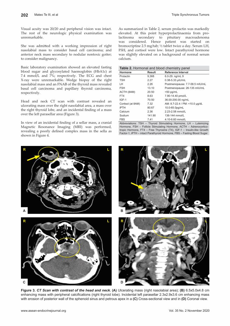
As summarized in Table 2, serum prolactin was markedly elevated. At this point hyperprolactinaemia from pro-lactinoma secondary to pituitary macroadenoma was considered. Hence patient was started on bromocriptine 2.5 mg/tab; ½ tablet twice a day. Serum LH, FSH, and cortisol were low. Intact parathyroid hormone was slightly elevated on a background of normal serum calcium.
Visual acuity was 20/20 and peripheral vision was intact. The rest of the neurologic physical examination was unremarkable.
She was admitted with a working impression of right nasolabial mass to consider basal cell carcinoma; and anterior neck mass secondary to nodular nontoxic goiter; to consider malignancy.
Basic laboratory examination showed an elevated fasting blood sugar and glycosylated haemoglobin (HbA1c) at 7.4 mmol/L and 7%; respectively. The ECG and chest X-ray were unremarkable. Wedge biopsy of the right nasolabial mass and an FNAB of the thyroid mass revealed basal cell carcinoma and papillary thyroid carcinoma, respectively.
Head and neck CT scan with contrast revealed an ulcerating mass over the right nasolabial area, a mass over the right thyroid lobe, and an incidental finding of a mass over the left parasellar area (Figure 3).
In view of an incidental finding of a sellar mass, a cranial Magnetic Resonance Imaging (MRI) was performed, revealing a poorly defined complex mass in the sella as shown in Figure 4.
Vol. 35 No. 2 November 2020
202
www.asean-endocrinejournal.org
Mateo Te III, et al Triple Synchronous Tumors
Figure 3. CT Scan with contrast of the head and neck. (A) Ulcerating mass (right nasolabial area); (b) 6.5x5.0x4.8 cm enhancing mass with peripheral calcifications (right thyroid lobe). Incidental left parasellar 2.3x2.9x3.6 cm enhancing mass with erosion of posterior wall of the sphenoid sinus and petrous apex in a (C) Cross-sectional view and in (D) Coronal view.
b
R l
Table 2. Hormonal and blood chemistry panelhormone Result Reference IntervalProlactin 9,368 6.0-29. ng/mL 9TSH 2.27 0.38-5.33 µIU/mLLH 2.26 Postmenopause: 7.7-58.5 mIU/mLFSH 13.10 Postmenopause: 26-135 mIU/mLACTH (8AM) 20.50 <50 pg/mLFT4 8.63 7.90-14.40 pmol/LIGF-1 70.50 36.00-200.00 ng/mLCortisol (at 8AM) 7.32 AM: 8.7-22.4 / PM: <10.0 µg/dLiPTH 80.67 10.0-65.0pg/mLCalcium 2.36 2.23-2.58 mmol/LSodium 141.90 136-144 mmol/LFBS 7.41 4.10-6.60 mmol/LAbbreviations: TSH – Thyroid Stimulating Hormone, LH – Luteinizing Hormone, FSH – Follicle Stimulating Hormone, ACTH – Adrenocortico-tropic Hormone, FT4 – Free Thyroxine (T4), IGF-1 – Insulin-like Growth Factor-1, iPTH – intact Parathyroid Hormone, FBS – Fasting Blood Sugar.
C
R l
D
R l
A
R l

For Philippines, in 2015, the predicted number of new cases of cancer was about 109,280 and death from cancer was about 66,151 cases.5
The three tumors presented by the patient have relatively common prevalence. Basal Cell Carcinoma (BCC) is the most common skin malignancy with prevalence estimated to be 2.0%, 1.4%, and 0.7%, for Australia, Europe, and the US, respectively.6,7 In Philippines, more than 60% of all skin cancers are of BCC.8 Papillary thyroid carcinoma (PTC) is the most common thyroid malignancy constituting 50% to 90% of well-differentiated thyroid carcinoma worldwide.9 Thyroid cancer was estimated to be the 8th most common malignancy among Filipinos with an incidence of 2%.5 A local study conducted in the Philippine General Hospital – Otorhinolaryngology Department reported that 82.9% of thyroid malignancies admitted were PTC.10 Prolactinoma is the most common pituitary adenoma that accounts for up to 45% of pituitary tumors.11-13 Each tumor presented has a common prevalence but when all three are combined in a single patient, it becomes a rare occurrence.
The Disease and its International, National and local Epidemiology Albeit extensive literature reviews, there are no existing official registry that accounted triple primary tumors
Ophthalmologic evaluation was normal except for a left quadrantanopsia on perimetry studies (Appendix B).
The patient underwent wide excision of the right nasolabial mass with frozen section biopsy for margins and total thyroidectomy. She was then started with synthetic thyroid hormone replacement. Final histopathologic report of the right nasolabial mass and thyroid mass revealed basal cell carcinoma and papillary thyroid carcinoma respectively as shown in Figures 5 and 6. The patient’s final diagnosis was triple synchronous tumors with a combination of right nasolabial basal cell carcinoma, papillary thyroid carcinoma and prolactinoma.
While on bromocriptine, repeat serum prolactin after 6 weeks revealed an exponential decrease from a baseline of 9,368 ng/mL to a normal level at 16.17 ng/mL. Dose of bromocriptine was decreased and the patient was advised to follow up for the surveillance tests. A postoperative radioactive iodine adjuvant therapy was the next plan for the patient.
DISCUSSION
Overall, it is estimated that there were 14.1 million new cases and 8.2 million deaths attributed to cancer worldwide.4
Vol. 35 No. 2 November 2020
203
www.asean-endocrinejournal.org
Mateo Te III, et alTriple Synchronous Tumors
Figure 4. Cranial Magnetic Resonance Imaging (MRI). A 2.2x3.1x3.1 cm poorly defined complex mass predominantly solid in the sella with extension to left parasellar region. (A) T1 Weighted Image (T1WI) – Isointense (b) T1 contrast image – Isointense (C) T2 Weighted Image (T2WI) – Mixed Signals (D) Fluid Attenuation Inversion Recovery (FLAIR) post gadolinium study - homogenous contrast enhancement. Noted encasement of the cavernous portion of the left carotid artery and bony destruction of the left petrous apex bone.
A
C
b
D
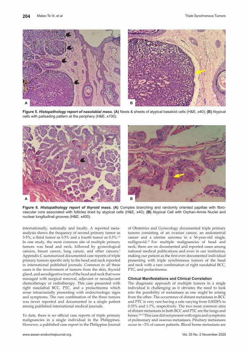
of Obstetrics and Gynecology documented triple primary tumors consisting of an ovarian cancer, an endometrial cancer and a uterine sarcoma in a 56-year-old single, nulligravid.14 For multiple malignancies of head and neck, there are no documented and reported cases among national medical publications and even in our institution, making our patient as the first ever documented individual presenting with triple synchronous tumors of the head and neck with a rare combination of right nasolabial BCC, PTC, and prolactinoma.
Clinical manifestations and Clinical Correlation The diagnostic approach of multiple tumors in a single individual is challenging as it obviates the need to look into the possibility of metastases as one might be arising from the other. The occurrence of distant metastasis in BCC and PTC is very rare having a rate varying from 0.0028% to 0.55% and 1-7%, respectively. The two most common sites of distant metastasis in both BCC and PTC are the lungs and bones.14-15 This case did not present with signs and symptoms of pulmonary and osseous metastases. Pituitary metastases occur in ~3% of cancer patients. Blood borne metastasis are
internationally, nationally and locally. A reported meta-analysis shows the frequency of second primary tumor as 3-5%, a third tumor as 0.5% and a fourth tumor as 0.3%.1,2
In one study, the most common site of multiple primary tumors was head and neck, followed by gynecological cancers, breast cancer, lung cancer, and other cancers.2 Appendix C summarized documented case reports of triple primary tumors specific only to the head and neck reported in international published journals. Common in all these cases is the involvement of tumors from the skin, thyroid gland, and aerodigestive tract of the head and neck that were managed with surgical removal, adjuvant or neoadjuvant chemotherapy or radiotherapy. This case presented with right nasolabial BCC, PTC, and a prolactinoma which arose intracranially presenting with endocrinologic signs and symptoms. The rare combination of the three tumors was never reported and documented in a single patient among published international medical journals.
To date, there is no official case reports of triple primary malignancies in a single individual in the Philippines. However, a published case report in the Philippine Journal
Vol. 35 No. 2 November 2020
204
www.asean-endocrinejournal.org
Mateo Te III, et al Triple Synchronous Tumors
Figure 5. Histopathology report of nasolabial mass. (A) Nests & sheets of atypical basaloid cells (H&E, x40); (b) Atypical cells with palisading pattern at the periphery (H&E, x100).
bA
Figure 6. Histopathology report of thyroid mass. (A) Complex branching and randomly oriented papillae with fibro-vascular core associated with follicles lined by atypical cells (H&E, x40); (b) Atypical Cell with Orphan-Annie Nuclei and nuclear longitudinal grooves (H&E, x400).
bA
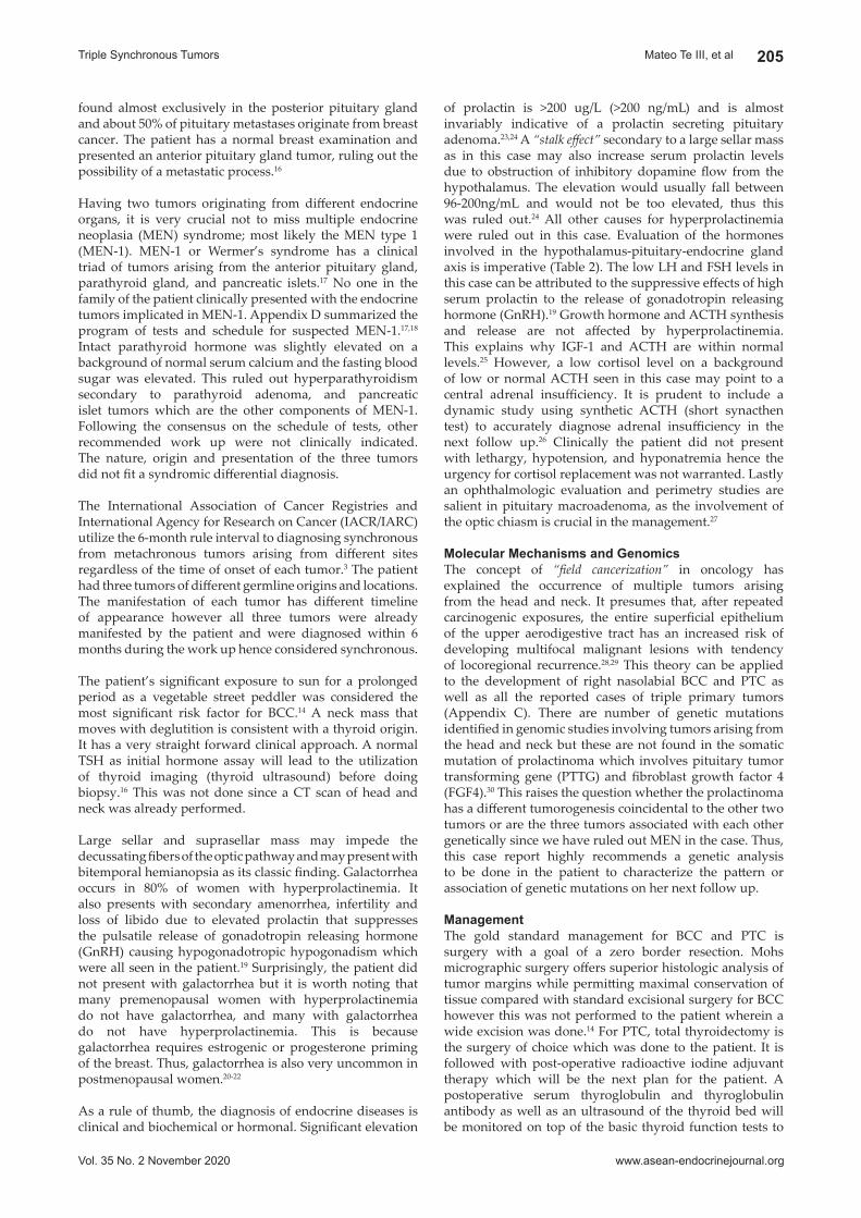
of prolactin is >200 ug/L (>200 ng/mL) and is almost invariably indicative of a prolactin secreting pituitary adenoma.23,24 A “stalk effect” secondary to a large sellar mass as in this case may also increase serum prolactin levels due to obstruction of inhibitory dopamine flow from the hypothalamus. The elevation would usually fall between 96-200ng/mL and would not be too elevated, thus this was ruled out.24 All other causes for hyperprolactinemia were ruled out in this case. Evaluation of the hormones involved in the hypothalamus-pituitary-endocrine gland axis is imperative (Table 2). The low LH and FSH levels in this case can be attributed to the suppressive effects of high serum prolactin to the release of gonadotropin releasing hormone (GnRH).19 Growth hormone and ACTH synthesis and release are not affected by hyperprolactinemia. This explains why IGF-1 and ACTH are within normal levels.25 However, a low cortisol level on a background of low or normal ACTH seen in this case may point to a central adrenal insufficiency. It is prudent to include a dynamic study using synthetic ACTH (short synacthen test) to accurately diagnose adrenal insufficiency in the next follow up.26 Clinically the patient did not present with lethargy, hypotension, and hyponatremia hence the urgency for cortisol replacement was not warranted. Lastly an ophthalmologic evaluation and perimetry studies are salient in pituitary macroadenoma, as the involvement of the optic chiasm is crucial in the management.27
molecular mechanisms and genomics The concept of “field cancerization” in oncology has explained the occurrence of multiple tumors arising from the head and neck. It presumes that, after repeated carcinogenic exposures, the entire superficial epithelium of the upper aerodigestive tract has an increased risk of developing multifocal malignant lesions with tendency of locoregional recurrence.28,29 This theory can be applied to the development of right nasolabial BCC and PTC as well as all the reported cases of triple primary tumors (Appendix C). There are number of genetic mutations identified in genomic studies involving tumors arising from the head and neck but these are not found in the somatic mutation of prolactinoma which involves pituitary tumor transforming gene (PTTG) and fibroblast growth factor 4 (FGF4).30 This raises the question whether the prolactinoma has a different tumorogenesis coincidental to the other two tumors or are the three tumors associated with each other genetically since we have ruled out MEN in the case. Thus, this case report highly recommends a genetic analysis to be done in the patient to characterize the pattern or association of genetic mutations on her next follow up.
managementThe gold standard management for BCC and PTC is surgery with a goal of a zero border resection. Mohs micrographic surgery offers superior histologic analysis of tumor margins while permitting maximal conservation of tissue compared with standard excisional surgery for BCC however this was not performed to the patient wherein a wide excision was done.14 For PTC, total thyroidectomy is the surgery of choice which was done to the patient. It is followed with post-operative radioactive iodine adjuvant therapy which will be the next plan for the patient. A postoperative serum thyroglobulin and thyroglobulin antibody as well as an ultrasound of the thyroid bed will be monitored on top of the basic thyroid function tests to
found almost exclusively in the posterior pituitary gland and about 50% of pituitary metastases originate from breast cancer. The patient has a normal breast examination and presented an anterior pituitary gland tumor, ruling out the possibility of a metastatic process.16
Having two tumors originating from different endocrine organs, it is very crucial not to miss multiple endocrine neoplasia (MEN) syndrome; most likely the MEN type 1 (MEN-1). MEN-1 or Wermer’s syndrome has a clinical triad of tumors arising from the anterior pituitary gland, parathyroid gland, and pancreatic islets.17 No one in the family of the patient clinically presented with the endocrine tumors implicated in MEN-1. Appendix D summarized the program of tests and schedule for suspected MEN-1.17,18 Intact parathyroid hormone was slightly elevated on a background of normal serum calcium and the fasting blood sugar was elevated. This ruled out hyperparathyroidism secondary to parathyroid adenoma, and pancreatic islet tumors which are the other components of MEN-1. Following the consensus on the schedule of tests, other recommended work up were not clinically indicated. The nature, origin and presentation of the three tumors did not fit a syndromic differential diagnosis.
The International Association of Cancer Registries and International Agency for Research on Cancer (IACR/IARC) utilize the 6-month rule interval to diagnosing synchronous from metachronous tumors arising from different sites regardless of the time of onset of each tumor.3 The patient had three tumors of different germline origins and locations. The manifestation of each tumor has different timeline of appearance however all three tumors were already manifested by the patient and were diagnosed within 6 months during the work up hence considered synchronous.
The patient’s significant exposure to sun for a prolonged period as a vegetable street peddler was considered the most significant risk factor for BCC.14 A neck mass that moves with deglutition is consistent with a thyroid origin. It has a very straight forward clinical approach. A normal TSH as initial hormone assay will lead to the utilization of thyroid imaging (thyroid ultrasound) before doing biopsy.16 This was not done since a CT scan of head and neck was already performed.
Large sellar and suprasellar mass may impede the decussating fibers of the optic pathway and may present with bitemporal hemianopsia as its classic finding. Galactorrhea occurs in 80% of women with hyperprolactinemia. It also presents with secondary amenorrhea, infertility and loss of libido due to elevated prolactin that suppresses the pulsatile release of gonadotropin releasing hormone (GnRH) causing hypogonadotropic hypogonadism which were all seen in the patient.19 Surprisingly, the patient did not present with galactorrhea but it is worth noting that many premenopausal women with hyperprolactinemia do not have galactorrhea, and many with galactorrhea do not have hyperprolactinemia. This is because galactorrhea requires estrogenic or progesterone priming of the breast. Thus, galactorrhea is also very uncommon in postmenopausal women.20-22
As a rule of thumb, the diagnosis of endocrine diseases is clinical and biochemical or hormonal. Significant elevation
Vol. 35 No. 2 November 2020
205
www.asean-endocrinejournal.org
Mateo Te III, et alTriple Synchronous Tumors

Funding SourceNone.
References 1. Bittorf B, Kessler H, Merkel S, et al. Multiple primary malignancies:
An epidemiological and pedigree analysis of 57 patients with at least three tumors. Eur J Surg Oncol. 2001;27(3):302-13. PMID: 11373110. https://doi.org/10.1053/ejso.2001.1112.
2. National Cancer Institute. SEER Program Code Manual, 3rd ed. Bethesda (MD): National Cancer Institute, 1998. https://seer.cancer.gov/archive/manuals/codeman.pdf.
3. North American Association of Central Cancer Registries. Howe HL, ed. A review of the definition for multiple primary cancers in the United States: Workshop proceedings from December 4-6, 2002 in Princeton, New Jersey. https://citeseerx.ist.psu.edu/viewdoc/download?doi=10.1.1.466.3479&rep=rep1&type=pdf.
4. Ferlay J, Soerjomataram I, Dikshit R, et al. Cancer incidence and mortality worldwide: Sources, methods and major patterns in GLOBOCAN 2012. Int J Cancer. 2015;136(5):E359-86. PMID: 25220842. https://doi.org/10.1002/ijc.29210.
5. Laudico AV, Mirasol-Lumague MR, Medina V, Mapua CA, Valenzuela FG, Pukkala E. 2015 Philippine cancer facts and estimates. Philippine Cancer Society: Manila, 2015. http://www.philcancer.org.ph/wp-content/uploads/2017/07/2015-PCS-Ca-Facts-Estimates_CAN090516.pdf.
6. Apalla Z, Lallas A, Sotiriou E, Lazaridou E, Ioannides D. Epidemiological trends in skin cancer. Dermatol Pract Concept. 2017;7(2):1-6. PMID: 28515985. PMCID: PMC5424654. https://doi.org/10.5826/dpc.0702a01.
7. Leiter U, Eigentler T, Garbe C. Epidemiology of skin cancer. Adv Exp Med Biol. 2014;810:120-140. PMID: 25207363. https://doi.org/10.1007/978-1-4939-0437-2_7.
8. Saquian FYN. Basal cell carcinoma of the lip and mentum. Philipp J Otolaryngol Head Neck Surg. 2006;21(1,2):54-6.
9. Schlumberger MJ, Filetti S, Hay ID. Nontoxic diffuse and nodular goiter and thyroid neoplasia. In: Williams Textbook of Endocrinology, 12th ed, Ch. 14; 2011. https://doi.org/10.1016/B978-1-4377-0324-5.00014-6.
10. Gloria JDLS, Pontejos Jr AQY, Grullo PER. Risk factors for recurrent papillary thyroid carcinoma. Philipp J Otolaryngol Head Neck Surg. 2017;32(2):25-9. https://journal.pso-hns.org/wp-content/uploads/2017/ 11/orig-article-Risk-FactorS-2.pdf.
11. Arafah BM, Nasrallah MP. Pituitary tumors: Pathophysiology, clinical manifestations and management. Endocr Relat Cancer. 2001;8(4):287-305. PMID: 11733226. https://doi.org/10.1677/erc.0.0080287.
12. Mindermann T, Wilson CB. Age-related and gender-related occurrence of pituitary adenomas. Clin Endocrinol (Oxf). 1994;41(3):359-64. PMID: 7893282. https://doi.org/10.1111/j.1365-2265.1994.tb02557.x.
13. Ciccarelli A, Daly AF, Beckers A. The epidemiology of prolactinomas. Pituitary. 2005;8(1):3-6. PMID: 16411062. https://doi.org/10.1007/s11102-005-5079-0.
14. Carucci JA, Leffell DJ, Pettersen JS. Basal cell carcinoma. In: Fitzpatrick’s Dermatology in General Medicine. Vol. 1, Ch. 115; 2012.
15. Portela RA, Choby GW, Manni A, Campbell D, Crist H, Goldenberg D. Unusual sites of metastasis of papillary thyroid cancer: Case series and review of the literature. Ear Nose Throat J. 2015;94(8):E43-7. PMID: 26322457.
16. Melmed S, Jameson L .Pituitary tumor syndromes. In: Harrison’s Principles of Internal Medicine. 20th ed, Vol. 2, Ch. 373.
17. Cardino MJT, Lantion-Ang FL. Insulinoma and prolactinoma in a young female: A variant of multiple endocrine endocrine neoplasia 1 (MEN-1). Philipp J Intern Med. 2009;47:137-42.https://www.pcp.org.ph/documents/PJIM/Volume%2047%20(2009)/Number%203%20(May-June)/7-Insulinoma%20and%20Prolactinoma.pdf.
18. Brandi ML, Gagel RF, Bilezikian JP, et al. Guidelines for diagnosis and therapy of MEN type 1 and type 2. J Clin Endocrinol Metab. 2006;86(12):5658-71. PMID: 11739416. https://doi.org/10.1210/jcem. 86.12.8070.
19. Majumdar A, Mangal NS. Hyperprolactinemia. J Human Reprod Sci. 2013;6(3):168-75. PMID: 24347930. PMCID: PMC3853872. https://doi.org/10.4103/0974-1208.121400.
20. Kleinberg DL, Noel GL, Frantz AG. Galactorrhea: A study of 235 cases including 48 with pituitary tumors. N Engl J Med. 1977;296(11):589-600. PMID: 840242. https://doi.org/10.1056/NEJM197703172961103.
21. Tolis G, Somma M, Campenhout JV, Friesen H. Prolactin secretion in sixty-five patients with galactorrhea. Am J Obstet Gynecol. 1974;118(1):91-101. PMID: 4202979. https://doi.org/10.1016/s0002-9378 (16)33651-1.
22. Boyd 3rd AE, Reichlin S, Turksoy RN . Galactorrhea-amenorrhea syndrome: Diagnosis and therapy. Ann Intern Med. 1977;87(2);165-75. PMID: 407824. https://doi.org/10.7326/0003-4819-87-2-165.
23. Kawaguchi T, Ogawa Y, Tominaga T. Diagnostic pitfalls of hyper-prolactinemia: The importance of sequential pituitary imaging.
detect recurrence and evaluate surgical adequacy on the next follow up.31
The cornerstone management for prolactinoma is medical. All macroprolactinomas with a size of >1 cm and symptomatic microprolactinomas with a size of <1 cm warrant medical treatment. Having a pituitary tumor size of 2.2 x 3.1 x 3.1 cm on cranial MRI, patient was managed medically with dopamine agonist in the form of bromocriptine.17 Giving of bromocriptine which is a dopamine agonist simulates the inhibitory action of dopamine to the secretion of prolactin thus decreasing its levels and eventually promoting tumor shrinkage.32 Dose of bromocriptine needs to be adjusted depending on medical response and will be given for approximately 1-2 years. Serum prolactin, cranial MRI and perimetry studies are part of the monitoring parameters for the management of prolactinoma and must be monitored during follow up.27 Prognosis of right nasolabial BCC, PTC and prolactinoma in one patient managed medically and surgically is not reported. Furthermore, there is no significant difference in the outcome of the separate tumours, if managed concurrently or separately since it is known that BCC, PTC and prolactinoma when managed individually like in our patient have a relatively good prognosis and have low incidence of recurrence.9,14
CONClUSION
The incidence of the individual tumor presented by the patient is relatively common however the existence of three common tumors in a single patient is a rare occurrence. The rarity of the case prompted a challenge in diagnosing multiple tumors that involved multiple endocrine organs and hence this case report gives emphasis on two salient points.
First, accurate diagnosis is imperative for an accurate management. In approaching multiple tumors especially involving different endocrine organs, classifying the nature of the tumors whether primary, metastatic or syndromic is very crucial as it greatly impacts the management. The patient was spared from unnecessary surgical removal and complications from pituitary surgery as the cornerstone treatment for prolactinoma is medical. Accurate knowledge on disease prevalence, pathophysiology and symptomatology and correlation to guided history taking and astute physical examination is very important in rare medical conditions. Having this knowledge, we are guided and justified on what specific tests to run in order to work up the patient and rule out close differential diagnoses. Second, the multidisciplinary care approach ensures a holistic and comprehensive management of complicated cases and this case has greatly benefited from this. It has proven that even in the medical realm, more heads are better than one and even two.
Ethical Consideration Patient consent was obtained before submission of the manuscript.
Statement of AuthorshipAll authors certified fulfillment of ICMJE authorship criteria.
Author DisclosureThe authors declared no conflicts of interest.
Vol. 35 No. 2 November 2020
206
www.asean-endocrinejournal.org
Mateo Te III, et al Triple Synchronous Tumors

32. Lyons DJ, Hellysaz A, Broberger C. Prolactin regulates tuberoinfundibular dopamine neuron discharge pattern: Novel feedback control mechanisms in the lactotrophic axis. J Neurosci. 2012; 32(23):8074-83. PMID: 22674282. PMCID: PMC6620951. https://doi.org/10.1523/JNEUROSCI.0129-12.2012.
33. Shah M, Agarwal R, Gupta S, et al. Triple primary malignancies in head-and-neck region: A report of four cases. South Asian J Cancer. 2017;6(4):194-5. PMID: 29404306. PMCID: PMC5763638. https://doi.org10.4103/sajc.sajc_225_17.
34. Singh NJ, Tripathy N, Roy P, Manikantan K, Arun P. Simultaneous triple primary head and neck malignancies: A rare case report. Head and Neck Pathol. 2016;10(2):233-6. PMID: 26477035. PMCID: PMC4838955. https://doi.org/10.1007/s12105-015-0664-7.
35. Yalavarthi S, Pamu PK, Thoondla M, Gupta S. Metachronous malignancies in head and neck region: Report of two cases. Indian J Pathol Microbiol. 2014;57(2):314-6. PMID: 24943776. https://doi.org/10.4103/0377-4929.134728.
36. Umeshappa H, Chandrashekhar M, Shenoy AM, Dinesh Kumar GR. Triple simultaneous primary tumors of the head and neck: A rare case report. Int J Case Rep Images 2014;5(7):488-91. https://doi.org/10.5348/ijcri-201485-CR-10396.
37. Nishikawa K, Kawada J, Fujitani K, et al. A case of metachronous triple cancer with multimodal approach including surgical resection. Gan To Kagaku Ryoho. 2014;41(12):2033-5. PMID: 25731413.
38. Clarke DR, Smallman LA, Rhys-Evans PH. Triple primary malignant neoplasms in the head and neck. J R Soc Med. 1986;79(11):676-7. PMID: 3795214. PMCID: PMC1290538.
BMC Res Notes. 2014;7:555. PMID: 25142896. PMCID: PMC4148930. https://doi.org/10.1186/1756-0500-7-555.
24. Shucart WA. Implications of very high serum prolactin levels associated with pituitary tumors. J Neurosurg. 1980;52(2):226-8. PMID: 7351562. https://doi.org/10.3171/jns.1980.52.2.0226.
25. Lugo G, Pena L, Cordido F. Clinical manifestations and diagnosis of acromegaly. Int J Endocrinol. 2012;2012:540398. PMID: 22518126. PMCID: PMC3296170. https://doi.org/10.1155/2012/540398.
26. Bornstein SR, Allolio B, Arlt W, et al. Diagnosis and treatment of primary adrenal insufficiency: An Endocrine Society Clinical Practice Guideline. 2016;101(2):364-89. PMID: 26760044. PMCID: PMC4880116. https://doi.org/10.1210/jc.2015-1710.
27. Schlechte J.. Approach to the patient: Long term management of prolactinomas. J Clin Endocrinol Metab. 2017;92(8):2861-5. https://doi.org/10.1210/jc.2007-0836.
28. Wei ZH, Gong W, Zhou M, Chen QM. The concept of field cancerization and its clinical application. Zhonghua Kou Qiang Yi Xue Za Zhi. 2016;51(9):562-5. PMID: 27596348. https://doi.org/10.3760/cma.j.issn.1002-0098.2016.09.011.
29. Mohan M, Jagannathan N. Oral field cancerization: An update on current concepts. Oncol Rev. 2014; 8(1): 244. PMID: 25992232. PMCID: PMC4419611. https://doi.org/10.4081/oncol.2014.244.
30. Yatavelli RKR, Bhusal K. Prolactinoma. In: StatPearls [Internet]. Treasure Island (FL): StatPearls Publishing; 2020 Jan. 2020 Aug 10. PMID: 29083585. NBK459347.
31. Indrasena BSH. Use of thyroglobulin as a tumour marker. World J Biol Chem. 2007;8(1): 81-5. PMID: 28289520. PMCID: PMC5329716. https://doi.org/10.4331/wjbc.v8.i1.81.
Vol. 35 No. 2 November 2020
207
www.asean-endocrinejournal.org
Mateo Te III, et alTriple Synchronous Tumors
JAFESUnique, interesting, enlightening.
Your case report and the JAFES.
Authors are required to accomplish, sign and submit scanned copies of the JAFES Author Form consisting of: (1) Authorship Certification, that authors contributed substantially to the work, that the manuscript has been read and approved by all authors, and that the requirements for authorship have been met by each author; (2) the Author Declaration, that the article represents original material that is not being considered for publication or has not been published or accepted for publication elsewhere, that the article does not infringe or violate any copyrights or intellectual property rights, and that no references have been made to predatory/suspected predatory journals; (3) the Author Contribution Disclosure, which lists the specific contributions of authors; and (4) the Author Publishing Agreement which retains author copyright, grants publishing and distribution rights to JAFES, and allows JAFES to apply and enforce an Attribution-Non-Commercial Creative Commons user license. Authors are also required to accomplish, sign, and submit the signed ICMJE form for Disclosure of Potential Conflicts of Interest. For original articles, authors are required to submit a scanned copy of the Ethics Review Approval of their research as well as registration in trial registries as appropriate. For manuscripts reporting data from studies involving animals, authors are required to submit a scanned copy of the Institutional Animal Care and Use Committee approval. For Case Reports or Series, and Images in Endocrinology, consent forms, are required for the publication of information about patients; otherwise, appropriate ethical clearance has been obtained from the institutional review board. Articles and any other material published in the JAFES represent the work of the author(s) and should not be construed to reflect the opinions of the Editors or the Publisher.

Appendix b. Octopus Perimetry Study Report
Vol. 35 No. 2 November 2020
208
www.asean-endocrinejournal.org
Mateo Te III, et al Triple Synchronous Tumors
APPENDICES
Appendix A. Family Pedigree
This perimetry study reports normal visual field of the right eye and a finding of a left Quadrantanopsia. However this perimetry study was taken with poor validity having a positive catch of 67% and a negative catch of 21%. A repeat study is recommended.

Vol. 35 No. 2 November 2020
209
www.asean-endocrinejournal.org
Mateo Te III, et alTriple Synchronous Tumors
Appendix C. Case reports of triple primary tumors specific only to the head & neck33-38
Author Features Age management
Shah, M. et alIndia
(2017)33
Case #11. Carcinoma left buccal Mucosa 2. Carcinoma left upper alveolus and hard palate3. Carcinoma right side base of tongue
— 1. Wide local excision + left supra-omohyoid neck dissection + radiation
2. Subtotal maxillectomy with free flap reconstruction + re-irradiation with concurrent chemotherapy
3. Near total glossectomy + right modified radical neck dissection + re-irradiation
Case #21. Carcinoma left tonsil2. Carcinoma left lateral tongue 3. Carcinoma cervical esophagus
— 1. Radiation with concurrent chemotherapy 2. Wide local excision + ipsilateral radical neck dissection3. Neoadjuvant chemotherapy followed by re-irradiation and
concurrent chemotherapyCase #31. Carcinoma vallecular2. Carcinoma-in-situ middle esophagus 3. Carcinoma post cricoid
— 1. Radiation 2. Re-irradiation with concurrent chemotherapy 3. Neoadjuvant chemotherapy followed by re-irradiation
Case #41. Carcinoma right buccal mucosa 2. Carcinoma right lateral tongue 3. Carcinoma glottis
— 1. Wide local excision + supraomohyoid neck dissection + adjuvant radiation
2. Hemiglossectomy + re-irradiation with concurrent chemotherapy 3. Palliative Chemotherapy
Singh, N. et alIndia
(2015)34
1. Squamous cell carcinoma of larynx 2. Papillary thyroid carcinoma 3. Non-Hodgkin’s Lymphoma
71 yo 1. Total laryngectomy with 2. Total thyroidectomy and bilateral selective neck dissection +
adjuvant radiation followed by radioactive iodine ablation 3. Chemotherapy (Cyclophoshamide and Dexamethasone)
Yalavarthi, S. et alIndia
(2014)35
1. Hodgkin’s lymphoma 2. Mucoepidermoid carcinoma of the salivary gland 3. Follicular variant of papillary thyroid carcinoma
21 yo 1. Chemotherapy 2. Not mentioned 3. Left hemithyroidectomy
Umeshappa, H. et alIndia
(2014)36
1. Adenoid cystic carcinoma of left parapharyngeal mass
2. Follicular thyroid carcinoma 3. Basal cell carcinoma of the right upper lip
63 yo 1. Transparotid-trancervical excision + supraomohyoid neck dissection + adjuvant chemotherapy
2. Left thyroid lobectomy with frozen section with concurrent completion thyroidectomy with central neck compartment dissection + radioactive iodine
3. Wide local excision
Nishikawa, K et alJapan
(2014)37
1. Hypopaharyngeal cancer2. Esophageal cancer3. Tongue cancer
37 yo 1. Hypopaharyngealectomy and cervical lymphadenectomy + adjuvant Radiotherapy
2. Esophagectomy3. Partial and subtotal resection of tongue
Clarke DR. et alUK
(1986)38
1. Papillary thyroid carcinoma 2. Squamous cell carcinoma of the left vocal cord 3. Squamous cell carcinoma and lymphoma of the
left posterior mandible
43 yo 1. Subtotal thyroidectomy2. Laryngectomy and left radical neck dissection 3. Chemotherapy
Appendix D. Program of tests, and schedule for suspected mEN-1
Tumor Age biochemical Test (Annual) ImagingParathyroid Adenoma 8 Calcium, PTH None Gastrinoma 20 Gastrin None Insulinoma 5 FBS, Insulin MRIAnterior Pituitary Tumor 5 Prolactin, IGF-1 MRIForegut Carcinoid 20 None CT-Scan*Adapted from the Consensus Guidelines for MEN-1 and MEN-219,20

Who were those mEN hiding behind the Ulcers?: A Case ReportShazatul Reza Binti Mohd Redzuan and Yong Sy Liang
Endocrine Unit, Medical Department, Hospital Tengku Ampuan Rahimah, Selangor, Malaysia
Abstract
Multiple endocrine neoplasia type 1 (MEN1) is a rare autosomal dominant disease caused by a mutation in the MEN1 gene. We present a 65-year-old man with MEN1 who has primary hyperparathyroidism, microprolactinoma, meningioma and gastrinoma. He had undergone parathyroidectomy followed by tumour excision of meningioma. The duodenal gastrinoma lesion was inoperable as it was close to the superior mesenteric artery with high surgery risk. Medical therapy with octreotide LAR had been initiated and showed good biochemical response as well as disease progression control. Chemoembolization was proposed if the duodenum lesion reduces in size on maintenance treatment with octreotide LAR. This case highlights the challenges in managing this rare condition and octreotide LAR has shown to be effective in controlling the disease progression in MEN1 with inoperable gastrinoma.
Key words: MEN1, gastrinoma, octreotide LAR, meningioma
INTRODUCTION
MEN 1 is an autosomal dominant disorder that is due to mutations in the tumor suppressor gene MEN1 which encodes a 610 amino acid protein, menin. MEN1 is characterized by the occurrence of parathyroid, pancreatic islet and anterior pituitary tumours. Patients with MEN1 have decreased life expectancy and the outcomes of current treatment are not as successful because of multiple tumors, which are larger, more aggressive and resistant to treatment and concurrence of metastases. The prognosis for MEN1 patients might be improved by presymptomatic tumor detection and treatment specific for MEN1 tumors.
CASE
We describe a 65-year-old male who was referred to the endocrine service initially for poorly controlled diabetes. Review of his history revealed a Zollinger-Ellison syndrome that had initially presented with persistent diarrhea, abdominal pain and vomiting when the patient was in his late forties. Gastroduodenoscopy revealed multiple duodenal ulcers and esophagitis. Elevated fasting serum gastrin without proton pump inhibitor (PPI) at 405.8 ng/L (Reference value [RV]: 44-104 ng/L), confirmed the diagnosis. CT scan of the abdomen showed a well defined intensely enhancing nodule at the third part of duodenum (2 cm from the gastroduodenal junction) suggestive of gastrinoma. However, he defaulted follow up for many years prior to his presentation to us.
During our evaluation, he was worked up for MEN associated tumors in view of the history of Zollinger-Ellison syndrome. Further work up revealed high calcium level of 2.88 mmol/L, phosphate level of 0.68 mmol/L (RV: 0.81-1.45
mmol/L) and intact parathyroid hormone (iPTH) of 22.13 pmol/L and a diagnosis of primary hyperparathyroidism was confirmed. Otherwise, he had no clinical symptoms of hypercalcaemia such as bodily ache, constipation, osteoporotic fracture or renal stone. Others relevant blood tests are shown in Table 1. The patient underwent left superior and inferior parathyroidectomy. Histopathology analysis of the parathyroid glands revealed left inferior and superior parathyroid adenoma (Figure 1). His calcium level remained normal after the removal of the glands.
Table 1. Initial blood investigationsInitial blood test Reference value
Corrected Ca 2.88 mmo/L 2.20-2.65 mmol/LPhosphate 0.68 mmol/L 0.81-1.45 mmol/LiPTH 22.13 pmol/L 1.3-9.3 pmol/LProlactinFT4TSHTestosterone
7235 mU/L15.03 pmol/L1.2 mU/L 13.9 nmol/L
<500 mU/L11-22 pmol/L0.3-5.6 mU/L8.0- 31.3 nmol/L
GH 0.82 μg/L <3 μg/LIGF-1 272 μg/L 30-210 μg/L24 Hour catecholamine
Norepinephrine EpinephrineDopamine Gastrin
22.7 μg/day10.0 μg/day16.1 μg/day405.8 ng/L
12.1-85.51.7-22.4 <498.144-104 ng/L
Pituitary MRI was done to screen for pituitary lesion show-ing right parietal lobe extraaxial tumour or meningioma with suprasellar lesion (Figures 2 and 3). Prolactin level was elevated at 7235 mU/L (RV: 86-324). Otherwise, he had no headache, vision problem or hypogonadism symptoms. He has since been on cabergoline 0.5 mg twice weekly. Surgery of right parietal tumour was offered at that time but the patient refused.
________________________________________
ISSN 0857-1074 (Print) | eISSN 2308-118x (Online)Printed in the PhilippinesCopyright © 2020 by Redzuan et al.Received: March 31, 2020. Accepted: October 2, 2020.Published online first: October 30, 2020.https://doi.org/10.15605/jafes.035.02.10
Corresponding author: Shazatul Reza Binti Mohd Redzuan, MDEndocrine Fellow, Hospital Tengku Ampuan Rahimah Jalan Langat, 41200 Klang, Selangor, MalaysiaE-mail: [email protected]: https://orcid.org/0000-0002-1658-1538
Vol. 35 No. 2 November 2020210 www.asean-endocrinejournal.org
Journal of theASEAN Federation ofEndocrine Societies Case Report
This work is licensed under a Creative Commons Attribution-NonCommercial 4.0 International License (https://creativecommons.org/licenses/by-nc/4.0/).

having family history of MEN1 or MEN1 associated tumors. Genetic test was not performed for him due to unavailability of the test at that time. His 3 children were advised to go for health and genetic screening. One of his children tested positive for menin gene; the other 2 children’s status are still unknown.
During follow-up, the patient was taking cabergoline 0.5 mg twice/week, alphacalcidol 0.25 mcg bd, esomeprazole 40 mg daily and basal bolus insulin. He was monitored clinically and CT scan abdomen was arranged in to monitor the size of the duodenal lesion.
Reassessment of the duodenal lesion with CT scan abdomen showed increasing size of the duodenal lesion at 13.0x8.1x5.1 cm (Figure 4). Repeated gastroduodenoscopy demonstrated prominent gastric fold and duodenitis. Endoscopic ultrasound was scheduled but he was not keen. He had symptoms of hypergastrinemia with peak gastrin levels 2013 pmol/L and symptoms improved with proton pump inhibitor (PPI).
He was suspected to have MEN1 due to presence of three primary MEN1 tumours which are gastrinoma, primary hyperparathyroidism and microprolactinoma. He denied
Figure 3. MRI of the pituitary showing small well-defined hypo to isointense nodules within the suprasellar region measuring 0.2x0.4x0.4 cm. (A) coronal view and (b) sagittal view.
bA
Figure 1. HPE of the parathyroid showing tumor cell with clear cytoplasm, arranged in sheets and cords traversed by delicate blood vessels (H&E, 100x).
Figure 2. MRI of the brain showing a well-defined extra axial soft tissue mass in the right parietal lobe with cystic areas noted within the tumor.
Vol. 35 No. 2 November 2020
211
www.asean-endocrinejournal.org
Shazatul Reza Binti Mohd Redzuan, et alWho were those MEN hiding behind the Ulcers?: A Case Report

right craniotomy and tumor excision for the right parasagittal mass. The histopathology examination of the tumor revealed right parietal lobe fibrous meningioma WHO grade 1. The surgery was complicated by massive upper gastrointestinal bleeding with hypovolemic shock.
Ga-68 Dotatate PET-CT was performed to look for metastatic lesions and showed increased uptake at the duodenum, stomach and abdominal nodes (Figure 5). There are multilobulated masses (5x12.5x9.4 cm) at the 3rd part of the duodenum (SUVmax 88.3), two foci at stomach wall at cardia (SUVmax 132), paracaval node at L1/L2 vertebral level (SUVmax 86.6) and aortocaval node at L1L2 vertebral level (SUVmax 69.7) measuring 0.9x1.2 cm and 1.5x1.2 cm respectively. The two foci in the stomach wall at cardia could be ulcer rather than metastasis to the stomach after discussion with nuclear medicine team.
The patient was started on intramuscular octreotide LAR 30 mg monthly after a multidisciplinary discussion involving his surgeon, nuclear medicine physician, interventional radiologist, oncologist and endocrine service to shrink the tumor and subsequently plan for chemo-embolization. However, patient was only keen for octreotide LAR until now. He reported improvement in symptoms especially gastric pain after treatment with proton pump inhibitor and octreotide LAR with better quality of life. His pituitary lesion, prolactin and calcium level remain stable.
Unfortunately, four years later, the patient developed insidious onset left sided body weakness and MRI brain showed right fronto-parietal extra axial enhancing mass with edema, mass effect with midline shift. He underwent
Figure 4. CT scan of the abdomen showing large well-defined multinodular and multilobulated, heterogenously enhancing mass lesion with irregular cystic component, arising from the wall of the 3rd part of the duodenum to proximal jejunum [13.0x8.1x5.1 cm (AP)].
Figure 5. Ga-68 DOTATATE PET-CT showing uptake at 3rd part of duodenum (SUVmax 88.3), stomach (SUVmax 132) and abdominal nodes (SUVmax 69.7).
C
bA
Vol. 35 No. 2 November 2020
212
www.asean-endocrinejournal.org
Shazatul Reza Binti Mohd Redzuan, et al Who were those MEN hiding behind the Ulcers?: A Case Report

as well as limited genetic tests available during that time. He denied having family history of MEN 1 or MEN associated tumor. One of his three children tested positive for menin gene.
Primary hyperparathyroidism is the most common feature of MEN 1 and occurs in approximately 95% of MEN1 patients.5 Pancreatic neuroendocrine tumors (NET), consist of gastrinomas, insulinomas, glucagonomas, vasoactive intestinal polypeptidomas (VIPomas) and nonfunctioning pancreatic NETs occur in approximately 40-70% of MEN1 patients.6 Anterior pituitary tumors consisting of prolactinomas, somatotrophinomas, corticotrophinomas and nonfunctioning adenomas occur in approximately 30-40% of patients.7
In the absence of treatment, these tumors are associated with an earlier mortality. Untreated patients with MEN1 have decreased life expectancy with 50% probability of death by the age of 50 and the cause of death in 50-70% is usually due to a malignant tumor process or sequelae of the disease.8 In particular, malignant pancreatic NET and thymic carcinoid tumor were associated with a marked increase in the risk of death.
Subtotal parathyroidectomy has resulted in persistent or recurrent hypercalcaemia within 10-12 years after surgery in 40-60% of patients. For this case, patient had undergone parathyroidectomy and his calcium level was stable after 6 years of surgery. However, close monitoring of his calcium level together with regular assessment for symptom onset and complications are important, noting that patients with MEN1 have an increased risk of persistent or recurrent hyperparathyroidism due to tendency towards multiglandular disease.9
Gastrinoma is the most frequent functioning pancreato-duoedenal NET that causes gastric acid hypersecretion with the manifestation of the Zollinger-Ellison syndrome (ZES). The hypergastrinemia has a trophic effect on gastric mucosa and gastric enterochromaffin cell. It is diagnosed in at least 50% of MEN1 patients at the age of 50 and with higher prevalence in men.8 ZES is associated with recurrent peptic ulcerations. These ulcers frequently appear small, less than 5 mms in diameter, with multiple nodular lesions arising deep in the mucosa. Gastrinomas usually grow slowly but frequently metastasize to peripancreatic lymph nodes and rarely to the liver.
The ideal treatment for a non-metastatic gastrinoma of the pancreas is surgical excision because the disease related survival in-patient with tumors that are more than 2 cm has improved after surgery.10 In most patients with MEN1, gastrinomas are multiple and occur within the duodenum and surgical cure may be difficult. Most centers do not offer Whipple resection for the majority of MEN1 patients because fewer operations are associated with excellent survival.11 Therefore, the surgical procedure needs
His serum gastrin and chromogranin A had reduced markedly after octreotide LAR (30 mg monthly) treatment (Table 2). PPI was discontinued prior obtaining the serum gastrin level. He has been maintained on this dose (Octerotide LAR 30 mg monthly) to date. Repeated CT scan abdomen after 5 months of octreotide LAR treatment revealed no significant change in the gastric, mesenteric and pancreatic lesion (Figure 6).
Currently he is being monitored closely for disease progression and symptoms of gastrinoma with yearly CT scan abdomen, Ga-68 Dotatate PET CT scan and biochemical investigations specifically fasting gastrin as well as chromogranin A.
DISCUSSION
MEN1 is a rare autosomal dominant disease caused by a mutation in the MEN1 gen (chromosome 11) encoding the tumor suppressor protein menin.1 MEN 1 is suspected when two or more of the most common neuroendocrine tumors are found such as parathyroid tumor, pancreatic islet cell tumor and pituitary tumor. Approximately 80-90% of patients diagnosed with MEN1 show a MEN1 gene mutation.2 The diagnosis of MEN1 can be made clinically on the basis of family history or genetic testing for a MEN1 gene mutation.3 Our patient had gastrinoma/Zollinger-Ellison disease in his late forties, and then a few years later had primary hyperparathyroidism and micro-prolactinoma. This patient also had meningioma which is a rare tumor in MEN1. Meningiomas have been reported in about 8% of MEN1 patients and typically present later in life.4 This case is a rare condition of meningioma in MEN1.
Genetic evaluation to confirm MEN1 gene mutation was not performed in this patient due to financial constraints
Table 2. Serial Investigation after octreotide LAR treatmentbaseline before treatment After octreotide lAR
Date 2000 June 2017 September 2017 August 2018 February 2020Gastrin (RV: 6-55 pmol/L ) 405.8 2013 NA 630 680Chromogranin A (RV: 27.0-94.0 ng/ml ) NA 14256 6564 >770 2500NA: not available
Figure 6. Repeated CT scan of the abdomen after octreotide LAR. Image showed similar size of duodenal lesion.
Vol. 35 No. 2 November 2020
213
www.asean-endocrinejournal.org
Shazatul Reza Binti Mohd Redzuan, et alWho were those MEN hiding behind the Ulcers?: A Case Report
Statement of AuthorshipBoth authors certified fulfillment of ICMJE authorship criteria.
Author DisclosureBoth authors declared no conflicts of interest.
Funding SourceNone.
References 1. Christopoulos C, Papavassiliou E. Gastric neuroendocrine tumors:
Biology and management. Ann Gastroenterol. 2005;18(2):127–40.2. Falchetti A. Genetic screening for multiple endocrine neoplasia
syndrome type 1 (MEN-1): When and how. F1000 Med Rep. 2010;2:14. PMID: 20948872. PMCID: PMC2948394. https://doi.org/10.3410/M2-14.
3. Thakker RV, Newey PJ, Walls GV, et al. Clinical practice guidelines for multiple endocrine neoplasia type 1 (MEN1). J Clin Endocrinol Metab. 2012;97(9):2990-3011. PMID: 22723327. https://doi.org/10.1210/jc.2012-1230.
4. Asgharian B, Chen YJ, Patronas NJ, et al. Meningiomas may be a component tumor of multiple endocrine neoplasia type1. Clin Cancer Res. 2004;10(3);869-80. PMID: 14871962. https://doi.org/10.1158/1078-0432.ccr-0938-3.
5. Brandi ML, Gagel RF, Angeli A, et al. Guidelines for diagnosis and therapy of MEN type 1 and type 2. J Clin Endocrinol Metab. 2001;86(12):5658-71. PMID: 11739416. https://doi.org/10.1210/jcem. 86.12.8070.
6. Thomas-Marques et al. Prospective endoscopic ultrasonographic evaluation of the frequency of nonfunctioning pancreaticoduodenal endocrine tumor in patients with MEN1. Am J Gatroenterol. 2006;101(2):266-73. PMID: 16454829. https://doi.org/10.1111/j.1572-0241.2006.00367.x.
7. Trouillas J, Labat-Moleur F, Sturm N, et al. Pitutary tumor and hyperplasia in MEN 1: A case control study in a series of 77 patient versus 2509 non-MEN1 patient. Am J Surg Pathol. 2008;32(4):534-43. PMID: 18300794. https://doi.org/10.1097/PAS.0b013e31815ade45.
8. Ito T, Igarashi H, Uehara H, Berna MJ, Jensen RT. Causes of death and prognostic factors in multiple endocrine neoplasia type 1: A prospective study: comparison of 106 MEN1/Zollinger-Ellison syndrome patients with 1613 literature MEN1 patients with or without pancreatic endocrine tumors. Medicine (Baltimore). 2013;92(3):135-81. PMID: 23645327. PMCID: PMC3727638. https://doi.org/10.1097/MD.0b013e3182954af1.
9. Doherty GM, Lairmore TC, DeBenedetti MK. Multiple endocrine neoplasia type 1 parathyroid adenoma development over time. World J Surg. 2004;28(11):1139-42. PMID: 15490065. https://doi.org/10.1007/s00268-004-7560-8.
10. Norton JA, Jensen RT. Role of surgery in Zollinger-Ellison syndrome. J Am Coll Surg. 2007;205(4 Suppl):S34-7. PMID: 17916516. https://doi.org/10.1016/j.jamcollsurg.2007.06.320.
11. Norton JA. Surgical treatment and prognosis of gastrinoma. Best Pract Res Clin Gastroenterol. 2005;19(5):799-805. PMID: 16253901. https://doi.org/10.1016/j.bpg.2005.05.003.
12. Berardi R, Morgese F, Torniai M, et al. Medical treatment for gastro-entero-pancreatic neuroendocrine tumors. World J Gastrointest Oncol. 2016;8(4):389-401. PMID: 27096034. PMCID: PMC4824717. https://doi.org/10.4251/wjgo.v8.i4.389.
13. Tomassetti P, Migliori M, Caletti GC, Fusaroli P, Corinaldesi R, Gullo L. Treatment of type II gastric carcinoid tumors with somatostatin analogues. N Engl J Med. 2000;343(8):551-4. PMID: 10954763. https://doi.org/10.1056/NEJM200008243430805.
to be individualized according to preoperative findings and the patient’s preference.
For this case, he was not a suitable candidate for Whipple surgery as the duodenal tumor is close to the superior mesenteric artery and is a high risk surgery. In our patient, multidisciplinary discussion involving a hepatobiliary surgeon, oncology and interventional radiologist had decided to start the patient on octreotide LAR for longer duration and then proceed with chemoembolization of the duodenal tumor.
Somatostatin analogue (SSA) has been demonstrated to control the growth of gastro-entero-pancreatic NETs but no data are available regarding their effects on the growth of MEN1 associated gastrinoma.12 Only case reports or small series support the use of SSA in advanced gastrinoma, therefore it is difficult to quantify their ability to control tumor growth and disease progression. Tomassetti et al., had shown reduction in number and size of carcinoid tumors after 6 months of therapy and gross resolution of disease in 1 year. All the patients in the study had small tumors <1 cm in size, suggesting that LAR SSA maybe beneficial in controlling smaller disease burden.13
After 5 months of the somatostatin analogue treatment, our patient had reduction in the chromogranin A and gastrin level but not the size of duodenal tumor. Otherwise, clinically, the patient was feeling much better with less gastric discomfort and a better quality of life after somatostatin analogue treatment. In our patient, with a huge duodenal gastrinoma, long acting SSA appeared contributory in controlling disease progression.
CONClUSION
We report a rare case of MEN1 with huge duodenal gastrinoma, primary hyperparathyroidism and micro-prolactinoma. The growth of gastrinoma tumor in MEN1 remained unchanged with octreotide LAR and symptoms were controlled with both the octreotide LAR and PPI. This case highlights that octreotide LAR has a potential role to control disease progression as well as improve quality of life and possibly increase survival rates in a patient with MEN1 with an inoperable, huge duodenal gastrinoma.
Ethical ConsiderationsPatient consent was obtained before submission of the manuscript.
Vol. 35 No. 2 November 2020
214
www.asean-endocrinejournal.org
Shazatul Reza Binti Mohd Redzuan, et al Who were those MEN hiding behind the Ulcers?: A Case Report
Authors are required to accomplish, sign and submit scanned copies of the JAFES Author Form consisting of: (1) Authorship Certification, that authors contributed substantially to the work, that the manuscript has been read and approved by all authors, and that the requirements for authorship have been met by each author; (2) the Author Declaration, that the article represents original material that is not being considered for publication or has not been published or accepted for publication elsewhere, that the article does not infringe or violate any copyrights or intellectual property rights, and that no references have been made to predatory/suspected predatory journals; (3) the Author Contribution Disclosure, which lists the specific contributions of authors; and (4) the Author Publishing Agreement which retains author copyright, grants publishing and distribution rights to JAFES, and allows JAFES to apply and enforce an Attribution-Non-Commercial Creative Commons user license. Authors are also required to accomplish, sign, and submit the signed ICMJE form for Disclosure of Potential Conflicts of Interest. For original articles, authors are required to submit a scanned copy of the Ethics Review Approval of their research as well as registration in trial registries as appropriate. For manuscripts reporting data from studies involving animals, authors are required to submit a scanned copy of the Institutional Animal Care and Use Committee approval. For Case Reports or Series, and Images in Endocrinology, consent forms, are required for the publication of information about patients; otherwise, appropriate ethical clearance has been obtained from the institutional review board. Articles and any other material published in the JAFES represent the work of the author(s) and should not be construed to reflect the opinions of the Editors or the Publisher.

Primary Partial Empty Sella presenting with Prepubertal hypogonadotropic hypogonadism: A Case Report
Maria Angela Matabang1 and Buena Sapang2
1Department of Internal Medicine, Ospital ng Makati, Makati City, Philippines2Department of Endocrinology, Cardinal Santos Medical Center, San Juan City, Philippines
Abstract
Primary partial empty sella occurs when less than 50% of an enlarged or deformed sella turcica is filled with cerebro-spinal fluid in the setting of unidentified etiologic pathological conditions. Prepubertal hypogonadotropic hypogonadism presenting as its main manifestation is rare since its peak incidence commonly occurs late at 30 to 40 years of age and has a sexual predilection for female. We described a case of 20-year-old male who presented with micropenis and absent secondary sex characteristics. Work up showed cranial MRI finding of partial empty sella, low testosterone, LH, FSH, Estradiol and Beta HCG levels. Sex hormone replacement may not improve fertility for this case but may help produce and maintain virilization and prevent future complications of hypogonadotropic hypogonadism.
Key words: PES, hypogonadotropic, hypogonadism, micropenis
INTRODUCTION
Empty sella is a radiologic finding pertaining to an enlarged or deformed sella turcica which can be partially (<50%) or completely (>50%) filled with cerebrospinal fluid.1 Its diagnosis is confirmed with magnetic resonance (MR) study of sellar and suprasellar regions or computed tomography (CT) for those with absolute contraindication/s to MR.2
Based on etiology, empty sella can be classified as primary or secondary. Compared to secondary empty sella which is caused by pituitary pathological conditions like previous surgical, pharmacological or radiotherapy treatment, the pathogenesis behind primary empty sella (PES) is unclear.2 Some of the identified mechanisms include incomplete formation of sellar diaphragm and the influence of suprasellar or pituitary promoting factors.1 Its incidence is not that high, ranging from 5.5%-12% as accidental finding in autopsy to 8%-35% in clinical practice. Majority of the prevalence of PES is among women – those with history of at least one completed pregnancy in their physiological history. Moreover, its occurrence in children is also less frequent compared to adults, and it is more or less associated with hypothalamic–pituitary dysfunction, genetic disorders or perinatal complication.2
CASE
RC, a 20-year-old male, sought consult in our institution for small penile size. He was born full-term via normal
vaginal delivery to a 35-year-old G3P3 mother, with no perinatal complications. However, there was intake of an abortifacient (4 tablets of misoprostol) on the 5th week of pregnancy. At birth, there was noted “monggo seed” sized penis and “paper-thin” scrotum. His mother was advised by the local health center physician to seek consult with a surgeon however due to financial difficulties, there were no tests nor consult with a specialist done. His childhood and pre-puberty years were unremarkable. There were no symptoms of palpitations, heat or cold intolerance, polyuria, polydipsia, polyphagia. There were no episodes of frequent urinary tract infection, dysuria, or abdominal pain. There were no instances of elevated blood pressure, headache, change in vision. There were no significant health problems during his childhood except for appendicitis for which he underwent an emergency appendectomy in July 28, 2008 at Ospital ng Makati.
However, at around 14 to 16 years of age, there was persistence of high pitched voice, scant pubic and axillary hair and fat deposition on waist and bilateral breasts. There was minimal growth in penile size at approximately 2-3 centimeters (cm) and a bilateral palpable scrotal sac.
On physical examination, he was ambulatory with stable vital signs. He is overweight with BMI of 29.09 kg/m2 and weight of 74.5 kilograms. His waist to hip ratio is 0.89 with his waist circumference spanning 86 cm and hip circumference at 96.5 cm. He presented with eunuchoid proportion (Figure 1) with a height of 162 cm and arm span of 171 cms. His mother stands 151 cm while his father at 169
________________________________________
ISSN 0857-1074 (Print) | eISSN 2308-118x (Online)Printed in the PhilippinesCopyright © 2020 by Matabang et al.Received: July 31, 2020. Accepted: October 19, 2020.Published online first: November 4, 2020.https://doi.org/10.15605/jafes.035.02.11
Corresponding author: Maria Angela M. Matabang, MDMedical Resident, Ospital ng MakatiSampaguita corner Gumamela Streets, PemboMakati City, Philippines 1218Tel. No.: +632-88826316 to 36E-mail: [email protected]: https://orcid.org/0000-0002-5496-1216
* The abstract was accepted for poster presentation in the SICEM 2019 at the Grand Walkerhill, Seoul Korea in April 18-21, 2019.
Vol. 35 No. 2 November 2020 215www.asean-endocrinejournal.org
Journal of theASEAN Federation ofEndocrine SocietiesCase Report
This work is licensed under a Creative Commons Attribution-NonCommercial 4.0 International License (https://creativecommons.org/licenses/by-nc/4.0/).

Micropenis is defined by Schonfeld and Beebe as Stretched Penile Length (SPL) 2.5 SD less than the mean for age without the presence of any other penile anomalies and presence of internal and external genital organs compatible with a 46 XY karyotype.3 For an average adult patient, mean stretched penile length is 13.3 cm with 9.3 cm as the calculated 2.5 standard deviation less than the mean for age. The patient has a stretched penile length of 3 cm falling more than 2.5 SD below the mean for adult.
To satisfy the criteria for micropenis, pelvic and inguino-scrotal ultrasound was done to confirm the presence of internal genital organs, which revealed small left testicle measuring 0.9x0.5x0.7 cm with chronic parenchymal
cm with computed mid parental height for boys of 166.5 cm. Thyroid gland was not palpable. He has gynecomastia without galactorrhea (Figure 2). Genital and pubic hair development was graded as Tanner Stage 1 (Figure 3). He has a flaccid and stretched penile lengths of 2.5 and 3 cm respectively with width of 4 cm. He has palpable small, firm left testis while non palpable on the right. Neurologic examination was normal except for bilateral anosmia. Evaluation was done by Otorhinolaryngology service which showed recurrent rhinosinusitis. CT scan of paranasal sinuses showed pansinusitis with opacified and widened ostiomeatal units. A trial of steroid therapy was recommended which provided slight relief of anosmia.
Vol. 35 No. 2 November 2020
216
www.asean-endocrinejournal.org
Maria Angela Matabang, et al Primary Partial Empty Sella presenting as Hypogonadism
Figure 1. Eunuchoid proportion (A) height of 162 centimeters; (b) arm span of 171 centimeters.
Figure 2. Bilateral gynecomastia, mode-rately enlarged without skin excess in an overweight 20-year-old male.
Figure 3. Tanner stage 1 (A) flaccid penile length; (b) stretched penile length; (C) penile width and scrotum.
bA
b CA

hypogonadism, where the growth plates are not yet fused and there is lack of sex steroids, growth plates of the extremities continue to grow past the usual age of cessation. As a result, there is decreased upper to lower ratio and an increased arm span for height leading to eunuchoid proportion like in the patient.
Aside from this, determination of skeletal development by x-ray of left hand and wrist is also a useful way of establishing the stage of physiological development which in some cases may not be parallel with chronological age. The patient has a delayed bone age (compatible to 16 and 5/12 to 14 and 5/12 year old male) by the Greulich-Pyle method (Figure 4). Estradiol is a product of aromatization of testosterone and it mediates additional effects of testosterone on bone resorption, epiphyseal closure, sexual desire, and fat deposition. From the physical examination and the patient’s bone x-ray, this low estradiol might have contributed to the unfused ossification centers and eunuchoid proportion. Related to this is that androstenedione is converted to testosterone by 17-beta hydroxysteroid dehydrogenase before it gets aromatized to estradiol. However, androstenedione itself can be aromatized to estrogen which might have contributed to gynecomastia and fat deposition.
The patient’s cranial MRI showed a shallow sella with apparent flattening of pituitary gland at its floor (Figure 5). The results of baseline endocrine tests are summarized in
changes and a non visualized right testicle. The prostate gland measures 2.2x1.6x1.7 cm (~3 grams). A whole abdominal CT scan was done with noted left inguinal hernia and ovoid soft tissue density in right inguinal region possibly representing the right testis. Other findings include hepatic steatosis and nephrocalcinosis on the left. Lastly, to satisfy the criteria, chromosome analysis was done revealing a karyotype with no numerical and structural aberrations and an XY sex chromosome complement in all 50 cells examined. Hence the patient has a male karyotype of 46,XY.
However, the patient did not just present with isolated micropenis. Alongside, he has gynecomastia, persistently high pitched voice and underdeveloped adult sexual characteristics. Most authorities accept the definition of delayed puberty as the absence of secondary sexual development at an age 2 SD above the mean age of onset of puberty. This is the age at which 95% of normal children have already entered puberty. Based on etiology, pubertal delay can be classified into constitutional growth delay or hypogonadism. The latter can be further classified into hypogonadotropic or hypergonadotropic hypogonadism.3
Though stature is the most obvious change in growth, the ratio of the upper and lower segment also changes significantly. Sex steroids are necessary for increase in growth hormone secretion and they directly stimulate epiphyseal plate’s growth and fusion. In prepubertal
Vol. 35 No. 2 November 2020
217
www.asean-endocrinejournal.org
Maria Angela Matabang, et alPrimary Partial Empty Sella presenting as Hypogonadism
Figure 4. Cranial MRI (A) T2 sagittal view; (b) T2 axial view; (C) T2 coronal view. The arrows point to fluid-filled sella.
Figure 5. X-ray of left hand comparing (A) patient’s bone age, (b) normal 16-year-old male and (C) normal 18-year-old male.
b
b
C
C
A
A

is 5:1 with peak incidence occurring at 30 to 40 years, occasionally earlier in women. It occurs less frequently in children and is associated with hypothalamic–pituitary dysfunction, genetic disorders or perinatal complications.
The etiology of PES is unclear but some of the etiopathogenic hypotheses identified include: 1. incomplete formation of sellar diaphragm 2. upper sellar factors (persistent or intermittent intracranial idiopathic hypertension, CSF pulsatility, obesity, systemic hypertension) or 3. pituitary factors (conditions associated with variation of pituitary volume like pregnancy, lactation, menopause, hypophysitis, compensatory pituitary hypertrophy to primary hormonal deficit).2
Patients with PES have varied symptoms, and endocrine dysfunction is one the least common presenting manifestations. In a study done by De Marinis et al., only 19% (n=40, N=213) had documented endocrine abnormalities in which only 22% (n=9, N=40) are male.4 This marginal number of male patients with PES presenting with endocrine problem was also seen in a study done by Maira et al., where in only 12% was documented.5
The prevalent endocrine problem varies in different studies. In a study done by Radha Rani et al., hypocortisolemia was most common (62.5%, n=10, N=16). In the same study, hypogonadism was observed in minority (18.75%, n=3) which presented as amenorrhea and erectile dysfunction.6 On the other hand, in a study done by Ghatnatti et al., hyperprolactinemia was the most common dysfunction (20.8% n=5, N=12) and isolated hypogonadotropic hypogonadism was only observed in 2 patients.7
One study on PES showed hypogonadotropic hypo-gonadism as the most common endocrine problem (19%, n=5; N= 21) which presents as oligomenorrhoea in females and decreased sexual function in male.8 Micropenis and lack of secondary sex characteristics are rare presentations since PES is seldom seen during prepubertal years. Its peak incidence is notable at postpubertal years hence the usual clinical manifestations of decrease in sexual function or erectile dysfunction are observed. One event was documented in 1973 which is almost similar with our case. A 24-year-old, 46XY patient was admitted for evaluation of infertility and lack of development of secondary sex
Table 1 showing hypogonadotropic hypogonadism (low testosterone, FSH and LH). Other biochemical results are summarized in Table 2 which showed normal fasting blood sugar, serum sodium, potassium, AST and ALT. While, lipid profile showed normal levels of total cholesterol and triglyceride but elevated LDL.
With these findings, the patient was diagnosed with Primary Empty Sella (PES) (Partial) which is probably congenital based on history of abortifacient use on first trimester of pregnancy, findings of prepubertal hypogonadotropic hypogonadism and MRI showing a partially empty sella.
He was referred to Endocrinology and Urology services. Sex hormone replacement was recommended. He was offered testosterone therapy however this was not yet started due to lack of funds. Steroid was not initiated since serum cortisol is just borderline low and patient is also asymptomatic. Orchiectomy as prophylaxis for develop-ment of malignancy is not warranted but orchidopexy may be offered to monitor tumor development.
Counseling was offered regarding possible psychosocial impact of the physical changes brought about by the endocrine problem. Since he has a partner it was also suggested in order to help them regarding possible plans to have children. However, they refused the offer since they’ve already decided to adopt a child in the future. The patient verbalized that currently, the psychical changes has no derogatory impact on his personal and social life. For continuation of care and surveillance of the possible impact of the hormonal deficiencies on other organ systems, the patient was endorsed to the succeeding endocrinology rotator and outpatient resident. He was last seen on December 2019 and repeat blood chemistries were requested; however, he was lost to follow up thereafter.
DISCUSSION
Data on epidemiology of PES varies based on means of diagnosis. It is usually an incidental finding in 5.5%-12% of autopsy cases. However, using neuroimaging, its overall incidence has been estimated at 12%, while approximately 9-35% if based on clinical findings as reported in various case series. Its female-to-male ratio
Vol. 35 No. 2 November 2020
218
www.asean-endocrinejournal.org
Maria Angela Matabang, et al Primary Partial Empty Sella presenting as Hypogonadism
Table 1. Serologic and immunologic test resultsTest Result Normal ValuesEstradiol (ECLIA) <5.00 pg/mL Male: 25.8-60.7 pg/mLBeta HCG with Dilution <0.100 mIU/mL 0.0-2.0 mIU/mLFSH (ECLIA) 0.43 mIU/mL Male: 1.5-12.40 mIU/mLLH (ECLIA) 0.22 mIU/mL Male: 1.7-8.60 mIU/mLTestosterone (ECLIA) 0.16 ng/mL Male: 2.8-8.00 ng/mLIGF-1 59.35 ng/mL 115-350 mg/mLSerum Cortisol 160.5 nmol/L (8:31 AM) 6-10 AM: 172-49 nmol/LACTH 27.41 pg/mL (8:31 AM) 7-10 AM: 7.2-63.30 pg/mLProlactin 125.30 mIU/L 86-324 mIU/LFT3 2.95 pg/mL 1.71-3.71 pg/mLFT4 0.77 ng/mL 0.70-1.48 ng/mLThyroid Stimulating Hormone 1.0363 mIU/L 0.35-4.94 uIU/mL
Table 2. Biochemical test resultsTest Result Normal ValuesAST/SGOT 34 5-35 U/LALT/SGPT 34 0-55 U/LBUN 3.5 3.2-7.4 mmol/LCreatinine 46 64-104 umol/LSodium 138 136-145 mmol/LPotassium 4.1 3.5-5.1 mmol/LChloride 102 101-110 mmol/LPhosphorous 1.11 0.74-1.52 mmol/LMagnesium 0.91 0.70-0.91 mmol/LHDL 1.20 up to 1.04 mmol/LVLDL 0.77 ng/mLTriglyceride 0.54 up to 1.70 mmol/LLDL 2.90 up to 2.59 mmol/LCholesterol 3.61 up to 5.18 mmol/LFBS 4.78 3.89-5.49 mmol/L

Author DisclosureBoth authors declared no conflicts of interest.
Funding SourceNone.
References 1. Agarwal JK, Sahay RK, Bhadada SK, Reddy VS, Agarwal NK. Empty
sella syndrome. J Indian Acad Clin Med. 2001;2(3):198-202.2. Chiloiro S, Giampietro A, Bianchi A, et al. Diagnosis of endocrine
disease: Primary empty sella: A comprehensive review. Eur J Endocrinol. 2017;177(6):R275-85. PMID: 28780516. https://doi.org/10.1530/EJE-17-0505.
3. Hitapoğlu N, Kurtoğlu S. Micropenis: Etiology, diagnosis and treatment approaches. J Clin Res Pediatr Endocrinol. 2013;5(4):217-23. PMID: 2437909. PMCID: PMC3890219. https://doi.org/10. 4274/Jcrpe.1135
4. De Marinis L, Bonadonna S, Bianchi A, Maira G, Giustina A. Primary empty sella. J Clin Endocrinol Metab. 2005;90(9):5471–7. PMID: 15972577. https://doi.org/10.1210/jc.2005-0288.
5. Maira G, Anile C, Mangiola A. Primary empty sella syndrome in a series of 142 patients. J Neurosurg. 2005;103(5):831–6. PMID: 16304986. https://doi.org/10.3171/jns.2005.103.5.0831.
6. Rani PR, Maheshwari R, Reddy TSK, Prasad NR, Reddy A. Study of prevalence of endocrine abnormalities in primary empty sella. Indian J Endocrinol Metabol. 2013;17(Suppl 1):S125-6. PMID: 24251130. PMCID: PMC3830276. https://doi.org/10.4103/2230-8210.119527.
7. Ghatnatti V, Sarma D, Saikia U. Empty sella syndrome - beyond being an incidental finding. Indian J Endocrinol Metab. 2012;16(Suppl 2):S321-3. PMID: 23565413. PMCID: PMC3603061. https://doi.org/10.4103/2230-8210.104075.
8. Stelmachowska-Banaś M, Czajka-Oraniec I, Zgliczyński, W. Clinical and hormonal assessment of patients with empty sella on MRI. Postępy Nauk Medycznych. 2014;23(12):814-8. http://www.pnmedycznych.pl/wp-content/uploads/2015/01/pnm_2014_814-818.pdf
9. Thomas Jr HM, Lufkin EG, Ellis 3rd GJ, Hartman CR, Hofeldt FD, Herman RH. Hypogonadotropism and "empty sella": Improvement in 2 cases with clomiphene citrate. Fertil Steril. 1973;24(4):252-9. PMID: 4694501. https://doi.org/10.1016/s0015-0282(16)39609-1.
10. Petak SM, Nankin HR, Spark RF, et al. American Association of Clinical Endocrinologists Medical Guidelines for clinical practice for the evaluation and treatment of hypogonadism in adult male patients--2002 update. Endocr Pract. 2002;8(6):440-56. PMID: 15260010.
characteristics. He is obese with eunuchoid proportion given the height and arm span. He has gynecomastia, bilateral undescended testes, normal prostate and decreased male body hair, however data on micropenis was not mentioned.9
The treatment for these patients entails hormonal replacement. For this case, testosterone therapy may not improve fertility at this point but it may help produce and maintain virilization and prevent future complications of hypogonadotropic hypogonadism like osteoporosis and cardiovascular problems. Lastly, semen analysis and other hormone dynamic tests are recommended to evaluate fertility potential and other possible hormonal problems respectively.10
CONClUSION
Primary empty sella itself is a rare disease entity. Presenting as its manifestation, hypogonadotropic hypogonadism is also uncommon, mostly seen late at 30 to 40 years of age and majority in females. Prompt recognition of prepubertal hypogonadotropic hypogonadism at an early age can maximize both surgical and medical management in these patients. However, since the patient in this case sought consult late, it is also important to reiterate at this point alternative options if there are future plans for reproduction as well as methods to prevent future complications of these hormonal imbalances.
Ethical ConsiderationsPatient consent was obtained before submission of the manuscript.
Statement of AuthorshipBoth authors certified fulfillment of ICMJE authorship criteria.
Vol. 35 No. 2 November 2020
219
www.asean-endocrinejournal.org
Maria Angela Matabang, et alPrimary Partial Empty Sella presenting as Hypogonadism
JAFESHad an intriguing discussion in Grand Rounds?
Share your Clinical Case Seminars [email protected].
Authors are required to accomplish, sign and submit scanned copies of the JAFES Author Form consisting of: (1) Authorship Certification, that authors contributed substantially to the work, that the manuscript has been read and approved by all authors, and that the requirements for authorship have been met by each author; (2) the Author Declaration, that the article represents original material that is not being considered for publication or has not been published or accepted for publication elsewhere, that the article does not infringe or violate any copyrights or intellectual property rights, and that no references have been made to predatory/suspected predatory journals; (3) the Author Contribution Disclosure, which lists the specific contributions of authors; and (4) the Author Publishing Agreement which retains author copyright, grants publishing and distribution rights to JAFES, and allows JAFES to apply and enforce an Attribution-Non-Commercial Creative Commons user license. Authors are also required to accomplish, sign, and submit the signed ICMJE form for Disclosure of Potential Conflicts of Interest. For original articles, authors are required to submit a scanned copy of the Ethics Review Approval of their research as well as registration in trial registries as appropriate. For manuscripts reporting data from studies involving animals, authors are required to submit a scanned copy of the Institutional Animal Care and Use Committee approval. For Case Reports or Series, and Images in Endocrinology, consent forms, are required for the publication of information about patients; otherwise, appropriate ethical clearance has been obtained from the institutional review board. Articles and any other material published in the JAFES represent the work of the author(s) and should not be construed to reflect the opinions of the Editors or the Publisher.

bilateral genu Valgum in an Adolescentwith Primary hyperparathyroidism:
A Case Report and Review of literatureSiow Ping Lee,1 Shu Teng Chai,1 Leh Teng Loh,2 Norhaliza Mohd Ali1
1Department of Medicine, Hospital Sultanah Aminah, Johor, Malaysia2Gleneagles Medini, Johor, Malaysia
Abstract
Primary hyperparathyroidism in children and adolescents is rare and often symptomatic at presentation. A 15-year-old boy presented with bilateral genu valgum for two years. Biochemical results were consistent with primary hyperparathyroidism. Calcium levels normalized two months after removal of a left inferior parathyroid adenoma.
Key words: primary hyperparathyroidism, adolescent, genu valgum, parathyroid neoplasms
INTRODUCTION
Primary hyperparathyroidism (PHPT) is a disorder of bone and mineral metabolism caused by autonomous secretion of parathyroid hormone (PTH). It is mainly seen in adults between 50 and 60 years of age, with an annual incidence of 30 per 100,000 and a lifetime prevalence of one per 1,000. The female to male ratio is approximately 3:1.1 On the other hand, PHPT in children and adolescents is rare, with a prevalence of two to five cases per 100,000 and no apparent sex predilection.1,2 In contrast to adults who are often asymptomatic and commonly recognized during routine biochemical screening, children and adolescents with PHPT are mostly clinically symptomatic with end-organ damage. These include skeletal abnormalities and/or nephrolithiasis at presentation.2
Genu valgum is an unusual manifestation of PHPT in children and adolescents. To date, there is a limited number of published reports describing such presentation. We report a young patient who had bilateral genu valgum as a result of a parathyroid adenoma.
CASE
A 15-year-old boy presented in October 2018 with bilateral knock-knee for two years. He had an uneventful antenatal history and normal developmental milestones. He did not have any knee pain, swelling or stiffness. He had no history of injury, fracture or infection to his knees or legs. None of his family members had disorders related to multiple endocrine neoplasia type 1 (MEN 1) or type 2A (MEN 2A), hyperparathyroidism-jaw tumor (HPT-JT) syndrome or familial isolated hyperparathyroidism (FIHPT).
Physical examination revealed short stature (below 5th percentile) and bilateral genu valgum (Figure 1). There were no bony deformities including those typical of rickets. He had no polydactyly, joint laxity or lumbar kyphosis to suggest skeletal dysplasia. The knee joints were not swollen, tender or warm. Laboratory evaluation showed hypophosphatemia and elevated levels of calcium, alkaline phosphatase and intact parathyroid hormone (iPTH), consistent with the diagnosis of primary hyperparathyroidism (Table 1). Neck ultrasonography revealed a well-defined hypoechoic lesion measuring 1.2x1.5x2.7 cm at the inferior pole of left thyroid lobe. Kidney and liver function tests were normal. Unfortunately, serum vitamin D, urinary calcium, kidney ultrasonography and bone mineral density scan were not performed in this case.
He subsequently underwent left inferior parathyroid-ectomy in December 2018. Histopathologic examination of the resected parathyroid gland confirmed a parathyroid adenoma. His immediate postoperative serum calcium was 1.9 mmol/L, for which he received oral calcium and calcitriol for a month. His calcium levels normalized two months after surgery (Table 1). Meanwhile, corrective osteotomy is currently being contemplated by the pediatric orthopedic surgery team.
DISCUSSION
Our adolescent patient presented with bilateral genu valgum as a result of excessive parathyroid hormone secretion from a parathyroid adenoma. His age at presentation corresponds to those with a similar deformity described in literature (Table 2). Majority of reported cases presented during the adolescent period—between 11 to 17
________________________________________
ISSN 0857-1074 (Print) | eISSN 2308-118x (Online)Printed in the PhilippinesCopyright © 2020 by Lee et al.Received: July 20, 2020. Accepted: September 21, 2020.Published online first: November 29, 2020.https://doi.org/10.15605/jafes.035.02.07
Corresponding author: Siow Ping Lee, MDPhysician and Endocrine FellowEndocrine Unit, Department of MedicineHospital Sultanah Aminah, Jalan Persiaran Abu Bakar Sultan80100 Johor Bahru, Johor, MalaysiaTel. No.: +6072257000Fax No.: +6072242694E-mail: [email protected]: https://orcid.org/0000-0003-0104-6978
Vol. 35 No. 2 November 2020220 www.asean-endocrinejournal.org
Journal of theASEAN Federation ofEndocrine Societies Case Report
This work is licensed under a Creative Commons Attribution-NonCommercial 4.0 International License (https://creativecommons.org/licenses/by-nc/4.0/).

HPT-JT and FIHPT. Our patient most likely has sporadic PHPT due to the absence of a family history of the aforementioned disorders. Moreover, additional screening revealed a normal prolactin level and a normal pituitary gland on magnetic resonance imaging. Solitary parathyroid adenoma appears to be the etiology in all the reported cases, including our patient (Table 2).
Our patient presented with isolated bilateral genu valgum with no other symptoms attributable to hypercalcemia. This piece of information has to be taken with a pinch of salt because it is well-known that children and adolescents with PHPT may have vague and non-specific symptoms involving the gastrointestinal, renal, musculoskeletal and neurological systems.1,2 These non-specific complaints
years old— except for one who came for medical attention at the age of 21.3 Out of these 23 patients with PHPT who manifested either unilateral or bilateral genu valgum, 13 were females. The underlying reason for the occurrence of such deformity in this particular age group remains unclear. It has been postulated that the direct effect of elevated parathyroid hormone on the epiphyseal plate and bone remodeling during the pubertal growth spurt could be the main contributing factor.4
Primary hyperparathyroidism in children and adolescents is caused by either parathyroid adenoma (solitary or multiple) or hyperplasia, which may be sporadic or familial. Parathyroid carcinoma in this age group is rarely reported.2 Familial causes encompass MEN 1 or MEN 2A,
Vol. 35 No. 2 November 2020
221
www.asean-endocrinejournal.org
Siow Ping Lee, et alBilateral Genu Valgum in an Adolescent with Primary Hyperparathyroidism
Table 1. Preoperative and postoperative biochemical profile
Parameters Preoperation(October 2018)
Two months post-operation(February 2019)
Six months post-operation
(june 2019)
Reference range
Corrrected calcium, mmol/L 2.97 2.14 2.13 2.10-2.55Phosphate, mmol/L 1.03 1.64 1.3 1.45-2.10Alkaline phosphatase, U/L 1354 560 377 116-468Intact PTHa, pmol/L 154 – – 1.5-7.6Creatinine, µmol/L 33 55 – 50-77aPTH, parathyroid hormone
Table 2. Primary hyperparathyroidism presenting as genu valgum: A summary of case reports and case series from published literature
Publication year Age, yr gender End-organ damage Etiology Outcome after parathyroidectomyMcClure RD et al6 1945 14 Female Skeletal abnormalities Left inferior parathyroid adenoma Biochemical normalization and
spontaneous correction of genu valgumBalch HE et al3 1953 21 Female Skeletal abnormalities Left inferior parathyroid adenoma Hungry bone syndrome in immediate
post-operative period, followed by biochemical normalization and recalcification of demineralized bones
Lloyd HM et al7 1965 14 Male Skeletal abnormalities Left inferior parathyroid adenoma Biochemical normalization and skeletal improvement
Rapaport D et al4 1986 15 Female Skeletal abnormalities, nephrolithiasis
Right inferior parathyroid adenoma Clinical and biochemical resolution
15 Male Skeletal abnormalities, nephrolithiasis
Right inferior parathyroid adenoma Clinical and biochemical resolution
Kauffmann C et al8 1993 13 Female Skeletal abnormalities Left inferior parathyroid adenoma Biochemical normalization and resolution of bone demineralization
Menon PS et al9 1994 14 Female Skeletal abnormalities, nephrolithiasis
Left superior parathyroid adenoma Normalization of calcium and phosphate
Harman CR et al10 1999 14 Female Skeletal abnormalities – –Walczyk A et al11 2011 15 Male Skeletal abnormalities Right inferior parathyroid adenoma Biochemical resolution and improvement
of BMDa
Dutta D et al21 2013 12 Female Skeletal abnormalities Right inferior parathyroid adenoma Clinical and biochemical resolutionRatnasingam J et al12 2013 15 Female Skeletal abnormalities Right parathyroid adenoma Biochemical resolutionRamkumar S et al13 2014 16 Male Skeletal abnormalities Left inferior parathyroid adenoma Biochemical resolution
13 Male Skeletal abnormalities Right inferior parathyroid adenoma Biochemical resolutionSharma S et al14 2016 15 Female Skeletal abnormalities Left inferior parathyroid adenoma Biochemical resolutionZil-E-Ali A et al15 2016 14 Female Skeletal abnormalities Right inferior parathyroid adenoma Biochemical resolutionArambewela MH et al16 2017 12 Female Skeletal abnormalities Right inferior parathyroid adenoma Resolution of primary
hyperparathyroidismKamath SP et al17 2018 11 Female Skeletal abnormalities Left superior parathyroid adenoma Normalization of iPTHb
12 Male Skeletal abnormalities, nephrolithiasis
Left superior parathyroid adenoma Normalization of iPTHb
Khan KA et al22 2019 17 Male Skeletal abnormalities Left inferior parathyroid adenoma Biochemical resolutionParuk IM et al18 2019 17 Male Skeletal abnormalities Left inferior parathyroid adenoma Clinical and biochemical resolution
13 Male Skeletal abnormalities Right superior parathyroid adenoma Clinical and biochemical resolutionRao KS et al19 2019 12 Female Skeletal abnormalities,
nephrolithiasisRight inferior parathyroid adenoma Hungry bone syndrome in immediate
post-operative period, long term outcome not reported
Yanrismet Y et al20 2019 13 Male Skeletal abnormalities Right inferior parathyroid adenoma Hungry bone syndrome in immediate post-operative period, long term outcome not reported
aBMD, bone mineral densitybiPTH, intact parathyroid hormone
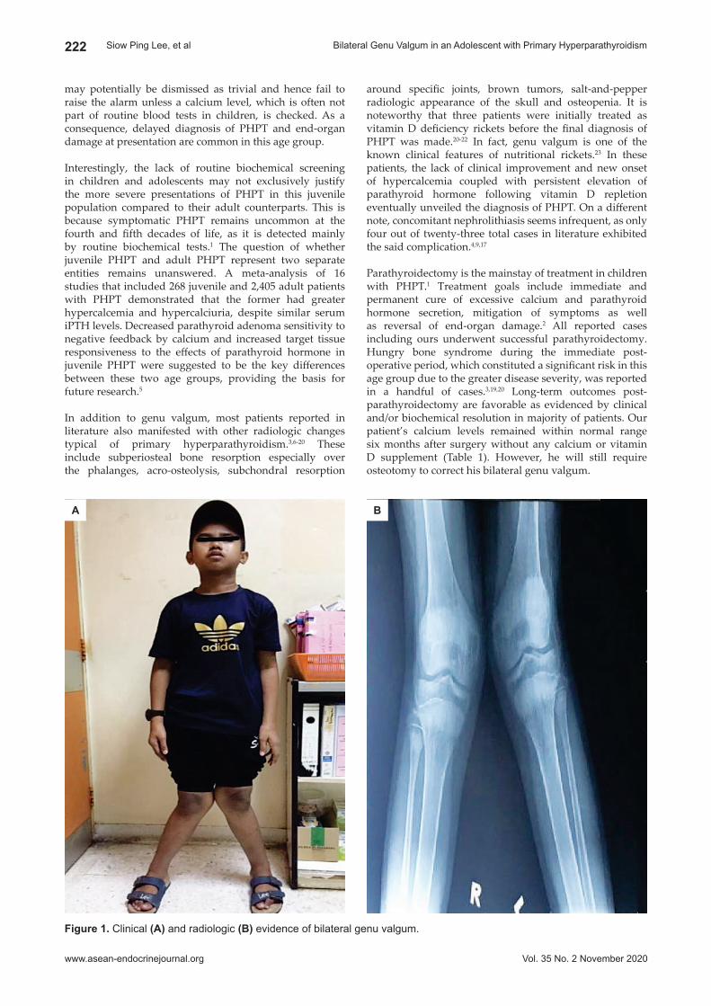
around specific joints, brown tumors, salt-and-pepper radiologic appearance of the skull and osteopenia. It is noteworthy that three patients were initially treated as vitamin D deficiency rickets before the final diagnosis of PHPT was made.20-22 In fact, genu valgum is one of the known clinical features of nutritional rickets.23 In these patients, the lack of clinical improvement and new onset of hypercalcemia coupled with persistent elevation of parathyroid hormone following vitamin D repletion eventually unveiled the diagnosis of PHPT. On a different note, concomitant nephrolithiasis seems infrequent, as only four out of twenty-three total cases in literature exhibited the said complication.4,9,17
Parathyroidectomy is the mainstay of treatment in children with PHPT.1 Treatment goals include immediate and permanent cure of excessive calcium and parathyroid hormone secretion, mitigation of symptoms as well as reversal of end-organ damage.2 All reported cases including ours underwent successful parathyroidectomy. Hungry bone syndrome during the immediate post-operative period, which constituted a significant risk in this age group due to the greater disease severity, was reported in a handful of cases.3,19,20 Long-term outcomes post-parathyroidectomy are favorable as evidenced by clinical and/or biochemical resolution in majority of patients. Our patient’s calcium levels remained within normal range six months after surgery without any calcium or vitamin D supplement (Table 1). However, he will still require osteotomy to correct his bilateral genu valgum.
may potentially be dismissed as trivial and hence fail to raise the alarm unless a calcium level, which is often not part of routine blood tests in children, is checked. As a consequence, delayed diagnosis of PHPT and end-organ damage at presentation are common in this age group.
Interestingly, the lack of routine biochemical screening in children and adolescents may not exclusively justify the more severe presentations of PHPT in this juvenile population compared to their adult counterparts. This is because symptomatic PHPT remains uncommon at the fourth and fifth decades of life, as it is detected mainly by routine biochemical tests.1 The question of whether juvenile PHPT and adult PHPT represent two separate entities remains unanswered. A meta-analysis of 16 studies that included 268 juvenile and 2,405 adult patients with PHPT demonstrated that the former had greater hypercalcemia and hypercalciuria, despite similar serum iPTH levels. Decreased parathyroid adenoma sensitivity to negative feedback by calcium and increased target tissue responsiveness to the effects of parathyroid hormone in juvenile PHPT were suggested to be the key differences between these two age groups, providing the basis for future research.5
In addition to genu valgum, most patients reported in literature also manifested with other radiologic changes typical of primary hyperparathyroidism.3,6-20 These include subperiosteal bone resorption especially over the phalanges, acro-osteolysis, subchondral resorption
Vol. 35 No. 2 November 2020
222
www.asean-endocrinejournal.org
Siow Ping Lee, et al Bilateral Genu Valgum in an Adolescent with Primary Hyperparathyroidism
Figure 1. Clinical (A) and radiologic (b) evidence of bilateral genu valgum.
A b

9. Menon PS, Madhavi N, Mukhopadhyaya S, Padhy AK, Bal CS, Sharma LK. Primary hyperparathyroidism in a 14 year old girl presenting with bone deformities. J Paediatr Child Health. 1994;30(5):441-3. PMID: 7833084. https://doi.org/10.1111/j.1440-1754.1994.tb00698.x.
10. Harman CR, van Heerden JA, Farley DR, Grant CS, Thompson GB, Curlee K. Sporadic primary hyperparathyroidism in young patients: A separate entity? Arch Surg. 1999;134(6): 651-5. PMID: 10367876. https://doi.org/10.1001/archsurg.134.6.651.
11. Walczyk A, Szalecki M, Kowalska A. Primary hyperparathyroidism: A rare endocrinopathy in children. Two case reports. Endokrynol Pol. 2011;62(4):346-50. PMID: 21879476.
12. Ratnasingam J, Tan ATB, Vethakkan SR, et al. Primary hyperparathyroidism: A rare cause of genu valgus in adolescence. J Clin Endocrinol Metab. 2013;98(3):869-70. PMID: 23337722. https://doi.org/10.1210/jc.2012-3839.
13. Ramkumar S, Kandasamy D, Vijay MK, Tripathi M, Jyotsna VP. Genu valgum and primary hyperparathyroidism in children. Int J Case Rep Images. 2014;5(6):401-7. https://doi:10.5348/ijcri-201455-CS-10041.
14. Sharma S, Kumar S. Bilateral genu valgum: An unusual presentation of juvenile primary hyperparathyroidism. Oxf Med Case Reports. 2016;2016(7):141-3. PMID: 27471596. PMCID: PMC4962889. https://doi.org/10.1093/omcr/omw023.
15. Zil-E-Ali A, Latif A, Rashid A, Malik A, Khan HA. Presentation of parathyroid adenoma with genu valgum and thoracic deformities. J Pak Med Assoc. 2016;66(1):101-3. PMID: 26712192.
16. Arambewela MH, Liyanarachchi KD, Somasundaram NP, Pallewatte AS, Punchihewa GL. Case report: Rare skeletal manifestations in a child with primary hyperparathyroidism. BMC Endocr Disord. 2017;17(1):45. PMID: 28732535. PMCID: PMC5521059. https://doi.org/10.1186/s12902-017-0197-z.
17. Kamath SP, Shenoy J, Kini KP, Shetty KB, Pai KA. Primary hyperparathyroidism presenting as bilateral genu valgum. Int J Health Allied Sci. 2018;7(2):114-6. https://doi.org/10.4103/ijhas.IJHAS_69_1.
18. Paruk IM, Pirie FJ, Motala AA. Rickets mimicker: A report of two cases of primary hyperparathyroidism in adolescence. J Endocrinol Metab Diabetes S Afr. 2019; 24(1):23-7. https://doi.org/10.1080/16089677. 2018.1546365.
19. Rao KS, Agarwal P, Reddy J. Parathyroid adenoma presenting as genu valgum in a child: A rare case report. Int J Surg Case Rep. 2019;59:27-30. PMID: 31102836. PMCID: PMC6525286. https://doi.org/10.1016/ j.ijscr.2019.03.063.
20. Yanrismet Y, Tridjaja B. Genu valgum as a rare clinical manifestation in child with primary hyperparathyroidism: A case report. Arch Dis Child. 2019;104(Suppl 3):A276. https://doi.org/10.1136/archdischild-2019-epa.646.
21. Dutta D, Kumar M, Das RN, et al. Primary hyperparathyroidism masquerading as rickets: Diagnostic challenge and treatment outcomes. J Clin Res Pediatr Endocrinol. 2013;5(4):266-9. PMID: 24379038. PMCID: PMC3890227. https://doi.org/10.4274/Jcrpe.1060.
22. Khan KA, Qureshi SU. Primary hyperparathyroidism masquerading as rickets. J Coll Physicians Surg Pak. 2019;29(9):891-4. PMID: 31455490. https://doi.org/10.29271/jcpsp.2019.09.891.
23. Misra M, Pacaud D, Petryk A, Collett-Solberg PF, Kappy M, Drug and Therapeutics Committee of the Lawson Wilkins Pediatric Endocrine Society. Vitamin D deficiency in children and its management: Review of current knowledge and recommendations. Pediatrics. 2008;122(2):398-417. PMID: 18676559. https://doi.org/10.1542/peds. 2007-1894.
In conclusion, primary hyperparathyroidism in children and adolescents is rare and often diagnosed late, with genu valgum being an unusual manifestation of this disorder. Nevertheless, a high index of suspicion is warranted, as prompt parathyroidectomy may lead to cure and reversal of debilitating complications.
AcknowledgmentThe authors would like to thank the Director General of Health Malaysia for the permission to publish this case report.
Ethical ConsiderationPatient consent was obtained before submission of the manuscript.
Statement of AuthorshipAll authors certified fulfillment of ICMJE authorship criteria.
Author DisclosureAll authors declared no conflicts of interest.
Funding SourceNone.
References 1. Roizen J, Levine MA. Primary hyperparathyroidism in children and
adolescents. J Chin Med Assoc. 2012;75(9):425-34. PMID: 22989537. PMCID: PMC3710287. https://doi.org/10.1016/j.jcma.2012.06.012.
2. Alagaratnam S, Kurzawinski TR. Aetiology, diagnosis and surgical treatment of primary hyperparathyroidism in children: New trends. Horm Res Paediatr. 2015. PMID: 25966652. https://doi.org/ 10.1159/000381622.
3. Balch HE, Spiegel EH, Upton AL, Kinsell LW. Hyperparathyroidism: Report of 2 cases with some relatively unusual manifestations. J Clin Endocrinol Metab. 1953;13(6):733-8. PMID: 13061594. https://doi.org/ 10.1210/jcem-13-6-733.
4. Rapaport D, Ziv Y, Rubin M, Huminer D, Dintsman M. Primary hyperparathyroidism in children. J Pediatr Surg. 1986;21(5):395-7. PMID: 3712190. https://doi.org/10.1016/s0022-3468(86)80505-x.
5. Roizen J, Levine MA. A meta-analysis comparing the biochemistry of primary hyperparathyroidism in youths to the biochemistry of primary hyperparathyroidism in adults. J Clin Endocrinol Metab. 2014;99(12):4555-64. PMID: 25181388. PMCID: PMC4255125. https://doi.org/10.1210/jc.2014-2268.
6. McClure RD, Lam CR. End-results in the treatment of hyperparathyroidism. Ann Surg. 1945;121(4):454-66. PMID: 17858584. PMCID: PMC1618130. https://doi.org/10.1097/00000658-194504000-00006.
7. Lloyd HM, Aitken RE, Ferrier TM. Primary hyperparathyroidism resembling rickets of late onset. Brit Med J. 1965;2(5466):853-6. PMID: 5827800. PMCID: PMC1846345. https://doi.org/10.1136/bmj.2.5466.853.
8. Kauffmann C, Leroy B, Sinnassamy P, Carlioz H, Gruner M, Bensman A. [A rare cause of bone pain in children: Primary hyper-parathyroidism caused by adenoma.] Arch Fr Pediatr. 1993;50(9):771-4 [Article in French]. PMID: 8060206.
Vol. 35 No. 2 November 2020
223
www.asean-endocrinejournal.org
Siow Ping Lee, et alBilateral Genu Valgum in an Adolescent with Primary Hyperparathyroidism
Authors are required to accomplish, sign and submit scanned copies of the JAFES Author Form consisting of: (1) Authorship Certification, that authors contributed substantially to the work, that the manuscript has been read and approved by all authors, and that the requirements for authorship have been met by each author; (2) the Author Declaration, that the article represents original material that is not being considered for publication or has not been published or accepted for publication elsewhere, that the article does not infringe or violate any copyrights or intellectual property rights, and that no references have been made to predatory/suspected predatory journals; (3) the Author Contribution Disclosure, which lists the specific contributions of authors; and (4) the Author Publishing Agreement which retains author copyright, grants publishing and distribution rights to JAFES, and allows JAFES to apply and enforce an Attribution-Non-Commercial Creative Commons user license. Authors are also required to accomplish, sign, and submit the signed ICMJE form for Disclosure of Potential Conflicts of Interest. For original articles, authors are required to submit a scanned copy of the Ethics Review Approval of their research as well as registration in trial registries as appropriate. For manuscripts reporting data from studies involving animals, authors are required to submit a scanned copy of the Institutional Animal Care and Use Committee approval. For Case Reports or Series, and Images in Endocrinology, consent forms, are required for the publication of information about patients; otherwise, appropriate ethical clearance has been obtained from the institutional review board. Articles and any other material published in the JAFES represent the work of the author(s) and should not be construed to reflect the opinions of the Editors or the Publisher.

Agoitrous graves’ hyperthyroidism with markedly Elevated Thyroid Stimulating Immunoglobulin Titre displaying Rapid Response
to Carbimazole with Discordant Thyroid FunctionYin Chian Kon,1 Brenda Su Ping Lim,1 Yingshan Lee,1 Swee Eng Aw,2 Yoko Kin Yoke Wong3
1Department of Endocrinology, Tan Tock Seng Hospital, Singapore2Department of Nuclear Medicine and Molecular Imaging, Singapore General Hospital, Singapore
3Department of Epidemiology, Singapore Clinical Research Institute, Singapore
Abstract
We characterize the clinical and laboratory characteristics of 5 patients with Graves’ thyrotoxicosis whose serum free thyroxine (fT4) concentration decreased unexpectedly to low levels on conventional doses of carbimazole (CMZ) therapy. The initial fT4 mean was 40.0 pM, range 25-69 pM. Thyroid volume by ultrasound measured as mean 11 ml, range 9.0-15.6 ml. Initial TSI levels measured 1487% to >4444%. Serum fT4 fell to low-normal or hypothyroid levels within 3.6 to 9.3 weeks of initiating CMZ 5 to 15 mg daily, and subsequently modulated by fine dosage adjustments. In one patient, serum fT4 fluctuated in a “yo-yo” pattern. There also emerged a pattern of low normal/low serum fT4 levels associated with discordant low/mid normal serum TSH levels respectively, at normal serum fT3 levels. The long-term daily-averaged CMZ maintenance dose ranged from 0.7 mg to 3.2 mg. Patients with newly diagnosed Graves' hyperthyroidism who have small thyroid glands and markedly elevated TSI titres appear to be “ATD dose sensitive.” Their TFT on ATD therapy may display a “central hypothyroid” pattern. We suggest finer CMZ dose titration at closer follow-up intervals to achieve biochemical euthyroidism.
Key words: Graves' disease, Thyroid Stimulating Immunoglobulin, responsiveness, carbimazole
INTRODUCTION
In patients with newly diagnosed Graves’ hyperthyroidism started on anti-thyroid drugs (ATDs), various factors affect the rate of decrease of serum free thyroxine (fT4) concentrations. Current guidelines recommend the initial starting dose of ATD to be based on a number of factors, such as initial serum fT4 concentration, thyroid gland size and triiodothyronine (T3) levels.1 In a retrospective multivariate analysis by Choi et al., of 99 patients with new onset Graves’ disease, high level of fT4, high thyroid stimulating hormone receptor antibody (TRAb) titre and absence of goiter were associated with rapid responsiveness to methimazole treatment.2 In their entire cohort, most patients showed normalization of free thyroxine within about 2 months, regardless of the initial free thyroxine level.
Dalan et al., had previously reported on a male patient with newly diagnosed agoitrous Graves’ hyperthyroidism who unexpectedly developed severe hypothyroxinemia within 3 weeks of starting anti-thyroid treatment, and rapidly rebounded back to hyperthyroidism within one week of ATD discontinuation.3 He demonstrated a highly
elevated TRAb titre. Intriguingly, he also demonstrated a discordant profile of suppressed serum TSH level despite concomitantly low serum fT4 and low to normal serum fT3 concentrations during the course of ATD treatment.3 A low serum fT4 may prompt the clinician to stop ATD therapy, which leads to rebound thyrotoxicosis. We report the biochemical and clinical features of a further 5 patients with rapid response to ATD. Early recognition of this phenotype can help to avoid swings between hypothyroidism and hyperthyroidism upon commencing and after adjusting ATD therapy.
CASES
PatientsWe describe 5 patients referred for management of Graves’ hyperthyroidism from July 2008 to March 2017 at the Department of Endocrinology, Tan Tock Seng Hospital, Singapore, who developed hypothyroxinemia after starting oral carbimazole (CMZ). This case series has been approved for publication by our institutional review board (DSRB Ref 2014/00896). Serum TRAb, thyroid stimulating immunoglobulin (TSI), TSH, fT4 and fT3 concentrations,
________________________________________
ISSN 0857-1074 (Print) | eISSN 2308-118x (Online)Printed in the PhilippinesCopyright © 2020 by Kon et al.Received: September 24, 2020. Accepted: November 12, 2020.Published online first: November 19, 2020.https://doi.org/10.15605/jafes.035.02.13
Corresponding author: Yin Chian Kon, BSc (London), MBBS (London), MRCP (UK), FAMS (Endocrinology)Senior Consultant, Department of Endocrinology, Tan Tock Seng Hospital11 Jalan Tan Tock Seng, Singapore, 308433Tel. No.: 65-81263173Fax No.: 65-63573087E-mail: [email protected]: https://orcid.org/0000-0002-9693-2587
* This paper had previously been presented at 37th Annual Meeting of the Endocrine Society of Taiwan, Taipei, Taiwan, on 20 March 2016 and in the 12th Asia and Oceania Thyroid Association (AOTA) Congress, Busan, Korea, on 17 March 2017.
Vol. 35 No. 2 November 2020224 www.asean-endocrinejournal.org
Journal of theASEAN Federation ofEndocrine Societies Case Series
This work is licensed under a Creative Commons Attribution-NonCommercial 4.0 International License (https://creativecommons.org/licenses/by-nc/4.0/).
spectrometry (Elan Dynamic Reaction Cell II; Perkin Elmer) in standard mode using tellurium (Te) as an internal standard. Urine (60 µL) was first diluted 1:25 in diluent (50 mcg/L Te and 10 mcg/L Rh in 1 % tetramethyl-ammonium hydroxide) and then quantified using an aqueous acidic calibration. The analytical measuring range was 10-40,000 mcg/L. Urine creatinine was measured using Roche assay, based on the enzymatic conversion of creatinine by creatininase, creatinase, and sarcosine oxidase to glycine, formaldehyde and hydrogen peroxide. Catalyzed by peroxidase, the liberated hydrogen peroxide reacts with 4-aminophenazone and HTIBa to form a quinone imine chromogen. The colour intensity of the quinone imine chromogen formed is directly proportional to the creatinine concentration in the reaction mixture, utilizing inductively coupled plasma mass spectrometry (ICP-MS) for iodine quantification in urine. This was normalized to urine creatinine, performed by enzymatic colorimetric assay (Roche).
Descriptive analysis The baseline clinical characteristics and initial thyroid function response from all 5 patients are tabulated together with the reference range of each test. When a serum result was expressed as greater or less than a numerical limit, that value of limit was utilized. For each patient, thyroid function (fT4, fT3, TSH), TRAb, TSI, anti-TPO Ab titre, and CMZ dose were summarised graphically across the follow-up calendar time.
RESUlTS
Patients presented with typical symptoms such as weight loss, increased appetite, palpitations, heat intolerance, diarrhoea and tremors. Patients 1 and 4 presented with complications of atrial fibrillation and thyrocardiac failure. Baseline demographics, thyroid function, thyroid volume and echogenicity, starting CMZ dose and initial thyroid function response are shown in Table 1. In all 5 patients, CMZ was started at doses equivalent to, or below, the methimazole (MMI)-equivalent starting dose (CMZ 10 mg equivalent to MMI 6 mg) suggested for the degree of fT4 elevation by the 2016 American Thyroid Association Guidelines.1 The initial free thyroxine mean was 41.5 pM, range 25-69 pM. The mean thyroid volume measured by ultrasound was 11.2 ml, range 9.0-15.6 ml. All patients remained clinically agoitrous throughout the course of follow-up. The range of initial TSI was 1487 % to >4444 %. Serum fT4 plunged to low-normal or hypothyroid levels within 3.6 to 9.3 weeks of initiating oral CMZ 5 to 15 mg daily and were subsequently modulated by fine adjustments in CMZ dosage.
Our 5 patients’ follow-up ranged from 14.7 months to 121.7 months (mean of 66.27 months, SD of 39.3 months). Their long-term biochemical course in response to CMZ treatment are shown in Figures 1-5. In all instances, the date of the dispensation of CMZ was verified to be the same as the date of prescription, suggesting patient compliance to therapy. In patients #1 and #2, whilst on ATD treatment, serum TSH was seen to remain inappropriately suppressed despite fT4 decreasing to and remaining at low normal concentrations, whilst fT3 levels remained normal (Figures 1-2). In patient #2, TSH was seen to be restored to within the lower half of reference range (its suppression was overcome) at
random spot urine iodine-creatinine ratio and thyroid size by ultrasound imaging, performed by our hospital radiology service, were measured. The volume of the thyroid gland was calculated as the sum of right and left thyroid lobes using ellipsoid formula 0.479 x length x depth x width. Low echogenicity in the thyroid gland was classified into 4 categories as previously described: Grade 0, diffuse high-amplitude echoes throughout the whole thyroid lobe; Grade 1, low-amplitude non-uniform echoes in the whole or several regions of the thyroid; Grade 2, several sonolucent regions in the thyroid; and Grade 3, no apparent echoes or very low amplitude echoes throughout the whole thyroid.4
laboratory analysis
Free thyroid hormones and TSHSerum free T4 and free T3 were measured by Access® Thyroid kit in a 2-step competitive immunoassay on a Beckman CoulterTM automated platform. Serum TSH was measured by Access® HYPERsensitive (3rd generation) chemiluminescent sandwich immunoassay on a Beckman Coulter automated platform.
TRAb AssayTRAb was measured using a third generation M22-biotin based ELISA commercial kit (Euroimmun TRAb Fast ELISA IgG) on the Triturus Analyser. Measurements were performed according to the manufacturer's instructions. TBII is detected by inhibiting the binding of M22 to immobilized recombinant human TSHR. The calibration is standardized against WHO 1995 Standard 90/672 (NIBSC 90/672).
Thyroid Stimulating Immunoglobulin (TSI)The thyroid stimulating activity of TSH receptor antibodies is measured by their ability to stimulate susceptible cells to make thyroid adenylate cyclase (cAMP). The immunoglobulin (Ig) fraction of patients’ sera is precipitated with a 20 % solution of polyethylene glycol (PEG). 120 µl of the reconstituted Ig fraction is then incubated with cloned JP-26 cells in a 96-well microtiter culture plate for two hours at 37 oC in 5 % CO2/95 % air atmosphere, using hypotonic buffer. The supernatants are then collected and assayed for cAMP contents in a radioimmunoassay. Results are expressed as percent increase in patient cAMP production as compared to normal human serum (NHS) cAMP production. The calculation formula for TSI results are expressed as %: (Patient’s cAMP / NHS’s cAMP) x 100 % (Reference range of TSI=50 % to 179 %; >179 % indicate stimulating). Each sample is performed in duplicate and the results averaged. The estimated inter-assay coefficient of variation (CV) of the TSI assay is 10 % to 20 %.
Thyroid Peroxidase AntibodyTPO Ab was measured using the commercial kit (ORGENTEC Diagnostika GmbH ELISA IgG) on the Triturus Analyser. Measurements were performed according to the manufacturer's instructions. The calibration is standardized against the international reference preparation WHO MRC66/387 for anti-TPO antibodies as 1000 IU/ml.
Urine Iodine-Creatinine Ratio (UICR)Specimens were sent to Mayo Laboratories. Urine iodine was analysed using inductively coupled plasma mass
Vol. 35 No. 2 November 2020
225
www.asean-endocrinejournal.org
Yin Chian Kon, et alAgoitrous Graves’ Hyperthyroidism responsiveness to Carbimazole

Figure 1. Patient 1 clinical course (A) thyroid function; (b) serum TSH receptor antibody (TRAb) and thyroid stimulating immunoglobulin (TSI) levels; (C) carbimazole (CMZ) therapy-average daily dose. From 22.3.12-25.5.12 (9.4 weeks), serum TSH remained inappropriately suppressed or inappropriately normal in the presence of low serum fT4.
A
b
C
Vol. 35 No. 2 November 2020
226
www.asean-endocrinejournal.org
Yin Chian Kon, et al Agoitrous Graves’ Hyperthyroidism responsiveness to Carbimazole
Table 1. Baseline Clinical Characteristics and Initial Thyroid Function Response After Starting Oral Carbimazole (CMZ)
Patient Ethnicity,Age/gender
USS Thyroid volume
hypoecho-genicity gradea
Initial fT4
Initial TSh
Initial TRabT
InitialTSI
Initial CmZ dose
Time from Starting CmZ to
Next TFT
Next fT4
Next fT3 Next TSh
Random Urine
Iodine/Cr Ratio
(yrs) (cm3) (pM) (mIU/L) (IU/L) (%) (mg/d) (weeks) (pM) (pM) (mIU/L) (mcg/g)1 Chinese / 56 / F 14.3 1 69 0.03 5.4 1487 15 9.3 6 3.7 0.02 1672 Chinese / 81 / F 9.1 1 28 <0.01 >40.0 >4444 5 9.0 8 3.7 0.01 1923 Chinese / 55 / F 7.2 1 49 0.02 >30.0 >4444 15 4.0 5 3.8 0.02 -4 Chinese / 72 / M 15.6 1 25 0.06 34.3 >4000 10 3.6 6 4.6 0.06 1455 Chinese / 82 / M 9.0 0 29 0.05 31.2 3160 10 5.3 6 3.8 0.18 108
Expected range < 15 8-21 0.34-5.64 <0.4 80-179 8-21 3.5-6.0 0.34-5.64 26-705a Grade 0, diffuse high-amplitude echoes throughout the whole thyroid lobe; Grade 1, low-amplitude non-uniform echoes in the whole or several regions of the thyroid; Grade 2, several sonolucent regions in the thyroid; and Grade 3, no apparent echoes or very low amplitude echoes throughout the whole thyroid.

Figure 2. Patient 2 clinical course (A) thyroid function; (b) serum TSH receptor antibody (TRAb) and thyroid stimulating immunoglobulin (TSI) levels; (C) carbimazole (CMZ) therapy-average daily dose. Between 24.10.08 and 8.10.09, fT4 showed a “yo-yo” pattern with small dose adjustments of CMZ. Between 25.11.10 and 30.06.15, TFT showed “central hypothyroid” pattern.
A
b
C
All our patients preferred and were maintained on prolonged low dose CMZ, in averaged daily doses ranging from 0.7 mg to 3.2 mg. Remarkably, in the long-term, patient #2 required the lowest dose of only CMZ 2.5 mg
concomitant fT4 values that were below normal (Figure 2). These patterns resembled “central hypothyroid” patterns. As all patients were non-pregnant, dose of CMZ was adjusted to keep serum fT3 mid-normal as the first priority.
Vol. 35 No. 2 November 2020
227
www.asean-endocrinejournal.org
Yin Chian Kon, et alAgoitrous Graves’ Hyperthyroidism responsiveness to Carbimazole

Figure 3. Patient 3 clinical course (A) thyroid function; (b) serum TSH receptor antibody (TRAb) and thyroid stimulating immunoglobulin (TSI) levels; (C) carbimazole (CMZ) therapy-average daily dose. Patient received block and replace, followed by CMZ only therapy. Although TRAb levels demonstrated a downtrend, paired TSI activity remained discordantly elevated.
A
b
C
DISCUSSION
We present 5 patients with newly diagnosed Graves' hyper-thyroidism whose serum fT4 plunged to unexpectedly
twice per week (averaged to 0.7 mg daily) to maintain euthyroidism, despite having persistently elevated TSI levels at 24 times above upper limit of normal (Figure 2).
Vol. 35 No. 2 November 2020
228
www.asean-endocrinejournal.org
Yin Chian Kon, et al Agoitrous Graves’ Hyperthyroidism responsiveness to Carbimazole

Figure 5. Patient 5 thyroid function course in response to carbimazole (CMZ) therapy. On CMZ 10 mg od for 3.6 weeks, patient became hypothyroxinemic; CMZ was not stopped but decreased to 2.5 mg daily, to avoid rebound thyrotoxicosis.
Figure 4. Patient 4 clinical course (A) thyroid function, serum TSH receptor antibody (TRAb) and thyroid stimulating immunoglobulin (TSI) levels; (b) carbimazole (CMZ) therapy-average daily dose. On 7 weeks of CMZ 10 mg od, patient became primary hypothyroid. CMZ was stopped for 1 week, then resumed on 2.5 mg daily to avoid rebound thyrotoxicosis.
A
b
elevated TRAb and TSI levels around the time of diagnosis. In a retrospective multivariate analysis by Choi et al., of 99 patients with new onset Graves’ disease, high level of fT4, high titre of TRAb and absence of goiter were
low levels after commencing CMZ at doses equivalent to, or below, that suggested for the degree of fT4 elevation by the 2016 American Thyroid Association Guidelines.1 All 5 patients demonstrated absence of goiter with remarkably
Vol. 35 No. 2 November 2020
229
www.asean-endocrinejournal.org
Yin Chian Kon, et alAgoitrous Graves’ Hyperthyroidism responsiveness to Carbimazole

serum T4 and low-normal serum T3 levels. Remarkably, 2 of these patients had non-elevated serum TSH levels.
ATD may reversibly or irreversibly inhibit thyroid peroxidase (TPO) catalyzed iodination, depending on the relative intra-thyroidal concentrations of iodine and ATD.11-13 At low intrathyroidal iodine level, iodination inhibition by ATD is irreversible, but an increase in iodine concentration competitively overcomes the inhibition. It is considered that rapid responders with small thyroid volume and thus small intrathyroidal iodine pool require a markedly elevated TSI level and a fast iodine turnover rate to maintain elevated thyroid hormone levels. The high iodine turnover in turn, contributes to the small iodine pool.
When relatively high doses of ATD are started, the intra-thyroidal ATD to iodide concentration ratio is increased, almost complete blockage of iodide organification ensues, leading to a rapid decrease in serum fT4 relative to serum fT3. Intriguingly, Taurog observed from his animal experiments, that "the change from reversible to irreversible inhibition of iodination with both propylthiouracil and methimazole occurred with seemingly slight elevations in the concentrations of these drugs."12 Perhaps this relates clinically to the marked swing in fT4 levels with slight changes in ATD dose, seen initially for example, in patient 2 (Figure 2).
Azizi reported that Graves’ hyperthyroidism responds more rapidly to MMI in an iodine-deficient area (Teheran) compared with in an iodine-sufficient area (Boston), where urinary iodine/creatinine ratio above 50 g/g represented iodine-sufficiency.14 In a subsequent study he reported that less than the usual recommended doses of MMI or PTU caused a rapid decline of thyroid hormone indices in patients residing in Teheran.15 In opposite contrast, patients with type 1 amiodarone-induced thyrotoxicosis may require higher than usual doses of ATD, because of the drug-derived iodine load.
All four of our patients in whom we performed random urine iodine/creatine ratio had values above 100 µg/g (Mayo Lab lower reference value 70 µg/g), suggesting that the rapid-responder phenotype may also occur in the presence of adequate iodine intake, when small thyroid and markedly elevated TSI titre are concomitantly present.
Besides driving high thyroidal iodine turnover, it is speculated that a high TRAb level may downregulate pituitary TSH secretion by an ultra-short negative feedback loop by acting on the pituitary TSH receptor (TSHR).16,17 The pituitary TSHR, expressed in the human anterior pituitary on folliculo-stellate cells, lies outside the blood-brain barrier and is therefore accessible to these autoantibodies.18 As the pituitary TSHR is also recognized by TSI, this interaction plausibly explains the prolonged serum TSH suppression seen in patients with Graves’ subclinical hyperthyroidism.19
In the course of follow-up, our patients demonstrated a discordant pattern of prolonged suppressed or low-normal TSH with corresponding low-normal or low fT4 levels. The concomitant fT3 levels were normal, hence the inappropriately low TSH is not due to T3-toxicosis.
associated with rapid responsiveness to methimazole treatment.2 Their study, together with our case series, suggest that Graves’ patients presenting with normal-sized thyroid glands and markedly elevated TSI levels predict a “rapid responder” phenotype to conventional doses of ATD. Moreover, we observed the perplexing profile of prolonged suppressed, or inappropriately normal, TSH despite concomitantly low-normal or low serum fT4 during the course of ATD treatment, when serum fT3 was low to normal.
Dalan et al., reported on an agoitrous Graves’ patient with markedly elevated TRAb who rapidly became hypothyroxinemic 2 weeks after being treated with a conventional dose of CMZ (Appendix A, reproduced with permission).3 Presence of thyroid stimulating blocking antibody (TSBAb) was excluded. After CMZ therapy was discontinued for a week, a thyroid uptake scan demonstrated 63% uptake at 4 hours, and 42% uptake at 24 hours, with 24-hr minus 4-hr delta RAIU of -21%, or remarkably high iodine turnover.
Similarly, Gemma et al., reported that the difference between 3-hour and 24-hour RAIU predicted rapidity of response to MMI therapy in Graves’ patients.5 They found that 24-hr minus 3-hr delta RAIU was significantly lower in rapid responders (TFT was normal or low within 1 month) than gradual responders. Delta RAIU was negatively correlated with the reduction in serum fT4 level at two weeks after MMI initiation and positively correlated with the biological half-life of intrathyroidal iodine.
TSH and TSI drive thyroidal iodine turnover. In 1944, Astwood and Bissell demonstrated that when normal rats were administered blocking doses of propylthiouracil (PTU) that rendered them hypothyroid, severe thyroidal iodine depletion occurred within days despite adequate iodine intake.6 Upon discontinuing ATD, the thyroid gland’s ability to re-accumulate iodine after it had been depleted was suppressed by thyroxine treatment or hypophysectomy, presumably by preventing rise of TSH. When hypophesectomy was performed before PTU was given, or if thyroxine was concomitantly administered with PTU, thyroidal iodine loss was prevented or amelio-rated in a graded manner.6 It is now well established that elevated serum TSH or TSI increases thyroidal iodide uptake, increases T3 formation relative to that of T4, and leads to increased iodine turnover via its return to the circulation as thyroid hormone, with relatively more T3 than T4 secreted compared to normal.7-9 Hence a high serum TSI level may be regarded as a marker for increased iodine turnover.
The intra-thyroidal iodine pool seen in untreated and treated Graves’ thyroids are lower than those of normal thyroids.10,11 In 1975, Larsen compared the thyroidal iodine content of 13 patients with Graves’ disease who had undergone thyroidectomy to that of 11 normal human thyroid glands.10 He found that the mean thyroidal iodine content of 2 patients who had received only propranolol (450 µg/g wet weight of thyroid tissue) was lower than normal (630±60 µg/g), but those who had received specific ATD treatment prior to surgery had the lowest levels. In particular, 3 of these patients were found to have very low thyroidal iodine (100±26 µg/g) and markedly low
Vol. 35 No. 2 November 2020
230
www.asean-endocrinejournal.org
Yin Chian Kon, et al Agoitrous Graves’ Hyperthyroidism responsiveness to Carbimazole

References 1. Ross DS, Burch HB, Cooper DS, et al. 2016 American Thyroid Associa-
tion Guidelines for diagnosis and management of hyperthyroidism and other causes of thyrotoxicosis. Thyroid. 2016;26(10):1343-1421. PMID: 27521067. https://doi.org/10.1089/thy.2016.0229.
2. Choi HS, Yoo WS. Free thyroxine, anti-thyroid stimulating hormone receptor antibody titres, and absence of goiter were associated with responsiveness to methimazole in patients with new onset Graves’ disease. Endocrinol Metab. 2017; 32(2):281-7. PMID: 28685517. PMCID: PMC5503874. https://doi.org/10.3803/EnM.2017.32.2.281.
3. Dalan R, Leow MKS, Ng CE. High iodine (substrate) turnover Graves' disease: The intriguing 'rapid-responder' variant of thyro- toxicosis. Ann Clin Biochem 2008;45(Pt6):612-5.PMID:18941129. https://doi.org/10.1258/acb.2008.008098.
4. Takeshima K, Inaba H, Furukawa Y, et al. Elevated serum immunoglobulin G4 levels in patients with Graves‘ disease and their clinical implications. Thyroid 2014;24(4):736-43. PMID: 24256421. https://doi.org/10.1089/thy.2013.0448
5. Gemma R, Nakamura H, Mori T, Andoh S, Suzuki Y, Yoshimi T. The change in 123I-uptake between 3- and 24-hours is useful in predicting early response to methimazole in patients with Graves’ disease. Endocr J. 1996;43(1):61-6. PMID: 8732453. https://doi.org/10.1507/endocrj.43.61.
6. Astwood EB, Bissell A. Effect of thiouracil on the iodine content of the thyroid gland. Endocrinology. 1944;34:282-96. https://doi.org/10.1210/endo-34-4-282.
7. Dunn JT, Dunn AD. Update on intrathyroidal iodine metabolism. Thyroid. 2001;11(5):407-14.PMID:11396699. https://doi.org/10.1089/ 105072501300176363.
8. Lauberg P, Vestergaard H, Nielsen S, et al. Sources of circulating 3,5,3’- triiodothyronine in hyperthyroidism estimated after blocking of type 1 and type 2 iodothyronine deiodinases. J Clin Endocrinol Metab. 2007;92(6):2149-56. PMID: 17389703. https://doi.org/10.1210/jc.2007-0178.
9. Abuid J, Larsen PR. Triidothyronine and thyroxine in hyperthyroidism. Comparison of the acute changes during therapy with antithyroid agents. J Clin Invest. 1974;54(1):201-8. PMID: 4134836. PMCID: PMC301541. https://doi.org/10.1172/JCI107744.
10. Larsen PR. Thyroidal triiodothyronine and thyroxine in Graves’ disease: Correlation with presurgical treatment, thyroid status and iodine content. J Clin Endocrinol Metab. 1975;41(06):1098-104. PMID: 54364. https://doi.org/10.1210/jcem-41-6-1098.
11. Engler H, Taurog A, Luthy C, Dorris ML. Reversible and irreversible inhibition of thyroid peroxidase-catalyzed iodination by thioureylene drugs. Endocrinology. 1983;112(1):86-95. PMID: 6847836. https://doi.org/10.1210/endo-112-1-86.
12. Taurog A. The mechanism of action of the thioureylene antithyroid drugs. Endocrinology.1976;98(4):1031-46.PMID:1278093. https://doi.org/10.1210/endo-98-4-1031.
13. Taurog A, Dorris ML, Guziec FS Jr. Metabolism of 35S- and 14C-labelled 1-methyl-2-mercaptoimidazole in vitro and in vivo. Endocrinology. 1989;124(1):30-9. PMID: 2535809. https://doi.org/10.1210/endo-124-1-30.
14. Azizi F. Environmental iodine intake affects the response to methimazole in patients with diffuse toxic goiter. J Clin Endocrinol Metab. 1985;61(2):374-7. PMID: 3839244. https://doi.org/10.1210/ jcem-61-2-374.
15. Azizi F. Medical treatment of toxic goiter in an area of iodine deficiency. MJIRI 1988;2(2):119-22. http://mjiri.iums.ac.ir/article-1-1264-en.pdf.
16. Prummel MF, Brokken LJS, Wiersinga WM. Ultra-short-loop feedback of thyrotropin secretion. Thyroid. 2004;14(10): 825-9. PMID: 15588378. https://doi.org/10.1089/thy.2004.14.825.
17. Brokken LJ, Scheenhart JW, Wiersinga WM, Prummel MF. Suppression of serum TSH by Graves’ Ig: Evidence for a functional pituitary TSH receptor. J Clin Endocrinol Metab. 2001;86(10): 4814-7. PMID: 11600546. https://doi.org/10.1210/jcem.86.10.7922.
18. Prummel MF, Brokken LJ, Meduri G, Misrahi M, Bakker O, Wiersinga WM. Expression of the thyroid-stimulating hormone receptor in the folliculo-stellate cells of the human anterior pituitary. J Clin Endocrinol Metab. 2000;85(11):4347-53. PMID:11095478. https://doi.org/10.1210/jcem.85.11.6991.
19. Brokken LJS, Wiersinga WM, Prummel MF. Thyrotropin receptor autoantibodies are associated with continued thyrotropin suppression in treated euthyroid Graves’ disease patients. J Clin Endocrinol Metab. 2003;88:4135-8. https://doi.org/10.1210/jc.2003-030430.
20. Vagenakis AG, Braverman LE, Azizi F, Portnay GI, Ingbar SH. Recovery of pituitary thyrotrophic function after withdrawal of prolonged thyroid-suppression therapy. N Engl J Med. 1975;293(14):681-4. PMID: 808728. https://doi.org/10.1056/NEJM197510022931402.
21. Lauberg P. Remission of Graves’ disease during anti-thyroid drug therapy. Time to reconsider the mechanism? Eur J Endocrinol. 2006;155(6):783-6. PMID: 17132745. https://doi.org/10.1530/eje.1.02295.
Thus, persistent highly elevated serum TRAb levels suppressing serum TSH by a short negative feedback loop on pituitary thyrotrophs, offers an alternative explanation to a “delayed recovery” of thyrotrophs, especially if serum TSH remains inappropriately low after 4-6 weeks of relief of endogenous or exogenous hyperthyroidism.20 This short-loop suppression is conceivably overridden when serum fT4 decreases further consequent upon further increase in ATD dose, leading to a rise in serum TSH, i.e., classical biochemical primary hypothyroidism (Appendix B).
Our cases demonstrate long-term maintenance of euthyroidism on low dose (patient #3) or ultra-low dose (patient #2) CMZ despite the presence of persistently and concurrently highly elevated TSI levels. We speculate that euthyroidism could be maintained on such low doses of CMZ because of low intrathyroidal iodine content, a mechanism also alluded to by Lauberg.21 From personal communication, Dalan’s case was subsequently maintained euthyroid on CMZ 2.5 mg on alternate days, and could not be weaned off CMZ because of thyrotoxic relapse.
Our small case series together with Dalan’s case report suggest that Graves’ patients with rapid-responder phenotype require atypically lower initial ATD dosing. If started on conventional doses (10-20 mg CMZ or equivalent), they may become hypothyroxinemic in 4-6 weeks. For such patients, we suggest the initial ATD dose should be lower than usually prescribed, and the follow-up interval shorter than usually arranged. When serum fT4 are at hypothyroid levels, the period of ATD discontinuation should not be too prolonged so as to avoid rebound thyrotoxicosis.
Study limitationsOur case series is limited by the small number of patients, although 4 of the patients had extended follow-up. Future prospective case-controlled studies examining clinical characteristics, therapy and monitoring of rapid-responder Graves’ thyrotoxicosis should be undertaken, to accumulate experience and evidence.
CONClUSION
Patients with newly diagnosed Graves' hyperthyroidism who have small thyroid glands and markedly elevated TSI titres appear to be “ATD dose sensitive.” Free T4 may swing markedly with small dose adjustments of ATD, or a “central hypothyroid” pattern of TFT may be seen. We suggest finer ATD dose titration at closer follow-up intervals to achieve biochemical euthyroidism, guided by monitoring TFT, fT3 and TRAb or TSI.
Ethical ConsiderationsEthics approval was obtained by the authors.
Statement of AuthorshipAll authors certified fulfillment of ICMJE authorship criteria.
Author DisclosureAll authors declared no conflicts of interest.
Funding SourceNone.
Vol. 35 No. 2 November 2020
231
www.asean-endocrinejournal.org
Yin Chian Kon, et alAgoitrous Graves’ Hyperthyroidism responsiveness to Carbimazole

APPENDICES
Vol. 35 No. 2 November 2020
232
www.asean-endocrinejournal.org
Yin Chian Kon, et al Agoitrous Graves’ Hyperthyroidism responsiveness to Carbimazole
Authors are required to accomplish, sign and submit scanned copies of the JAFES Author Form consisting of: (1) Authorship Certification, that authors contributed substantially to the work, that the manuscript has been read and approved by all authors, and that the requirements for authorship have been met by each author; (2) the Author Declaration, that the article represents original material that is not being considered for publication or has not been published or accepted for publication elsewhere, that the article does not infringe or violate any copyrights or intellectual property rights, and that no references have been made to predatory/suspected predatory journals; (3) the Author Contribution Disclosure, which lists the specific contributions of authors; and (4) the Author Publishing Agreement which retains author copyright, grants publishing and distribution rights to JAFES, and allows JAFES to apply and enforce an Attribution-Non-Commercial Creative Commons user license. Authors are also required to accomplish, sign, and submit the signed ICMJE form for Disclosure of Potential Conflicts of Interest. For original articles, authors are required to submit a scanned copy of the Ethics Review Approval of their research as well as registration in trial registries as appropriate. For manuscripts reporting data from studies involving animals, authors are required to submit a scanned copy of the Institutional Animal Care and Use Committee approval. For Case Reports or Series, and Images in Endocrinology, consent forms, are required for the publication of information about patients; otherwise, appropriate ethical clearance has been obtained from the institutional review board. Articles and any other material published in the JAFES represent the work of the author(s) and should not be construed to reflect the opinions of the Editors or the Publisher.
Appendix A. High-iodine-turnover Graves’ patient displaying (1) rapid response to low dose CMZ; (2) rapid rebound thyrotoxicosis after stopping CMZ; (3) “central hypothyroid” TFT pattern.
Appendix b. Proposed Schematic of Small Thyroid-High TSI Rapid-Responder Graves’ Pathophysiology.

legions of Presentations of myxedema Coma:A Case Series from a Tertiary hospital in India
Nirmalya Roy,1 Anirban Majumder,2 Debmalya Sanyal,2 Soumyabrata Roy Chaudhuri,2 Suman Sarkar,1 Ankan Pathak1
1Department of Internal Medicine, KPC Medical College and Hospital, West Bengal University of Health Sciences, Kolkata, India2Department of Endocrinology, KPC Medical College and Hospital, West Bengal University of Health Sciences, Kolkata, India
Abstract
Myxedema coma is associated with decreased mental status and hyponatremia among patients with diagnosed or undiagnosed hypothyroidism. The diagnosis is challenging in the absence of universally accepted diagnostic criteria, but should be considered as a differential even in cases with competing established diagnoses. All patients should receive intensive care level treatment. Even with optimal treatment, mortality is very high.
Key words: myxedema coma, presentations, outcomes, case reports
INTRODUCTION
Myxedema coma is a rare and potentially fatal condition. Even with the best possible treatment, mortality remains very high.1 Untreated hypothyroidism due to any cause including autoimmune disease, iodine deficiency, congenital abnormalities, drugs affecting thyroid function or secondary hypothyroidism can result in myxedema coma. The crisis is usually triggered by stressful events, the most common of which is infections. The incidence of myxedema coma in India is not known, but several case reports and case series have been published in recent years.2-6 In developing countries recognition of this entity is made difficult by its slow onset, lack of awareness among both patients and physicians, and absence of diagnostic facilities in remote areas. Due to its rarity, physicians often fail to identify or keep it as a remote possibility while treating critically ill patients. This case series documents the myriad presentations of myxedema coma encountered in tertiary practice and encourages physicians to keep it in mind as a possibility while treating patients with altered sensorium.
CASES
Case 1 A 75-year-old female with a history of recurrent hospitalization for atrial fibrillation, heart failure, and sepsis was brought to the emergency room with circulatory collapse. She had been on amiodarone therapy for a long period of time, and was never diagnosed as having hypothyroidism. Investigation revealed hyponatremia and subnormal thyroid function. She was treated with conventional care including thyroid hormone and mechanical ventilation support but succumbed after a few days.
Case 2 A 70-year-old male with a long-standing history of hypothyroidism following radio-iodine treatment for Graves' disease was brought to the emergency room with progressively increasing sleepiness and altered sensorium. He did not reveal the history of radio-iodine therapy. Investigations showed community acquired pneumonia which was treated accordingly. Hyponatremia and subnormal thyroid function were detected later once the history of radio-iodine therapy was obtained. He recovered with thyroid hormone replacement and conventional care.
Case 3A 75-year-old female with known hypothyroidism, type 2 diabetes, and hypertension presented in the emergency room with breathing difficulty and altered sensorium. She had hyponatremia and subnormal thyroid function. Investigations revealed the presence of heart failure with reduced ejection fraction. She was treated with standard care and with mechanical ventilatory support but succumbed after a few days.
Case 4 A 72-year-old female with known hypothyroidism presented in the emergency room with a history of bilateral lower limb swelling, facial puffiness and progressive unresponsiveness for four days. History from her attendants suggested that she had a very irregular intake of her thyroid medication. She was presumptively diagnosed as a case of myxedema coma and was treated with standard care before laboratory reports were made available. She eventually succumbed on the next day.
________________________________________
ISSN 0857-1074 (Print) | eISSN 2308-118x (Online)Printed in the PhilippinesCopyright © 2020 by Roy et al.Received: June 22, 2020. Accepted: October 15, 2020.Published online first: November 29, 2020.https://doi.org/10.15605/jafes.035.02.12
Corresponding author: Prof. Anirban Majumder, MBBSKPC Medical College and Hospital, West Bengal University of Health Sciences1F Raja S.C. Mullick Road, Jadavpur, Kolkata, India 700032Tel. No.: 033-6621 1700Fax No.: 033-6621 1768E-mail: [email protected]: https://orcid.org//0000-001-6937-8675
Vol. 35 No. 2 November 2020 233www.asean-endocrinejournal.org
Journal of theASEAN Federation ofEndocrine SocietiesCase Series
This work is licensed under a Creative Commons Attribution-NonCommercial 4.0 International License (https://creativecommons.org/licenses/by-nc/4.0/).

permanent pacemaker, was admitted with massive fluid overload. After the fourth session of sustained low-efficiency dialysis (SLED), he became confused, agitated, and stuporous. Investigations revealed hyponatremia, subnormal thyroid function, and bilateral pleural effusion. He was treated with standard myxedema crisis care and with bi-level positive airway pressure support in the intensive care unit (ICU) and recovered in five days.
Case 10A 66-year-old female with no current levothyroxine treatment was admitted to the ICU for myocardial infarction along with coma and bradycardia. Her core body temperature was documented at 88°F. She had severe hyponatremia and urosepsis. She required mechanical ventilation but did not survive.
Case 11 A 71-year-old female not known to be hypothyroid was admitted to the ICU for myocardial infarction along with stupor and bradycardia. The core temperature was documented to be 90°F. Aside from severe hyponatremia, she was also found to have urosepsis. She required mechanical ventilation but did not survive.
The case records of 11 patients with the diagnosis of myxedema coma between 1st January 2015 and 31st December 2019 admitted to our tertiary care hospital were reviewed (Table 1). Patients with poor clinical and/or biochemical documentation were excluded from our study population. There are review articles and scoring systems to aid diagnosis, but these have limited sensitivity.7 Myxedema coma is best recognized by the clinician when there is a high index of suspicion. The diagnosis in the case series was done clinically by the treating physicians. Data regarding age, sex, date of hospitalization, precipitating events, clinical presentation (central nervous system symptoms, heart rate, blood pressure, temperature), biochemical findings at presentation [free thyroxine (FT4), thyroid-stimulating hormone (TSH), random serum cortisol, serum sodium], management strategy (use of mechanical ventilation or noninvasive ventilation) and outcomes were retrieved from the documentation in the archival department of the hospital (Table 2).
Case 5A 68-year-old male, chronic alcoholic but not known to be hypothyroid, presented in the emergency room with a history of swelling of lower limbs and altered sensorium for two days. He was presumptively diagnosed as a case of hepatic encephalopathy and was treated accordingly. Investigations revealed hyponatremia and subnormal thyroid function. Myxoedema coma management was started three days after his hospitalization but he succumbed after a few days.
Case 6An 83-year-old female, known hypertensive and hypothyroid, with a fractured femur operated one week earlier, became progressively drowsy over the past three days. Herpes zoster -related blisters on the left side of the neck was noted. Investigations revealed hyponatremia with subnormal thyroid function. She developed multi-organ failure and did not survive despite standard care and mechanical ventilatory support.
Case 7A 50-year-old male, known hypothyroid and chronic alcoholic, was brought to the emergency room with inappropriate behavior. He was observed to be aggressive and impulsive. He was presumptively diagnosed and treated as a case of alcohol intoxication but did not improve. Investigations on the next day revealed hyponatremia, subnormal thyroid function, pericardial effusion and community acquired pneumonia. He recovered with conventional care.
Case 8An 80-year-old female with known hypothyroidism, diabetes, and hypertension presented in the emergency room with abnormal mentation. She had a pale puffy face with very slow mentation. Investigations revealed hyponatremia, anemia, subnormal thyroid function with mildly elevated TSH and a huge pericardial effusion. She recovered with standard care for myxedema coma.
Case 9 A 92-year-old male, known to have hypertension, hypothyroidism, chronic kidney disease and with a
Vol. 35 No. 2 November 2020
234
www.asean-endocrinejournal.org
Nirmalya Roy, et al Legions of Presentations of Myxedema Coma
Table 1. Presentations of myxedema coma casesIdentifier Age, yr Sex Season background Precipitating event CNSa symptoms hRb bPc Temperature (°F)Case 1 75 F December Not known hypothyroid Amiodarone Coma 118 90/60 98.4Case 2 70 M October Known hypothyroid Pneumonia Yes 58 100/60 98.7Case 3 70 F November Known hypothyroid, T2DMd and HTNe Heart failure Yes 58 140/80 97.2Case 4 72 F November Known hypothyroid Stopped LT4f Yes 60 100/60 96.7Case 5 68 M December Not known hypothyroid and known
alcoholicHepatic encephalopathy
Coma 95 110/60 95.9
Case 6 83 F September Known hypothyroid Herpes zoster Coma 56 100/60 96Case 7 50 M December Known hypothyroid and alcoholic Pneumonia Yes 50 110/70 97.4Case 8 80 F August Known hypothyroid, T2DMd and HTNe Anaemia Yes 56 150/90 97Case 9 92 M November Known hypothyroid, T2DMd and HTNe SLEDg Yes 78 160/80 97Case 10 66 F November Known hypothyroid and stopped LT4f MIh Coma 58 90/60 88Case 11 71 F October Not known hypothyroid Urosepsis Yes 52 80/58 90
a CNS, central nervous systemb HR, heart ratec BP, blood pressured T2DM, type 2 diabetes mellituse HTN, hypertensionf LT4, levothyroxineg SLED, sustained low-efficiency dialysish MI, myocardial infarction
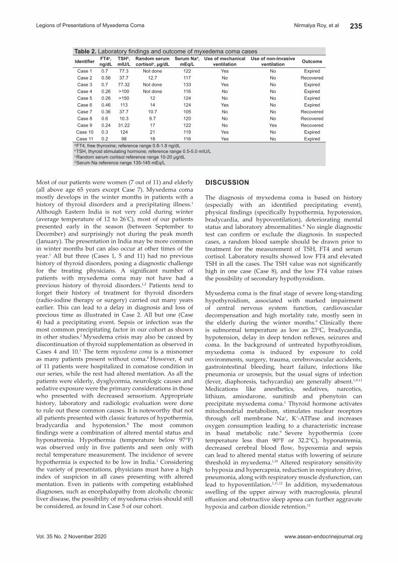
DISCUSSION
The diagnosis of myxedema coma is based on history (especially with an identified precipitating event), physical findings (specifically hypothermia, hypotension, bradycardia, and hypoventilation), deteriorating mental status and laboratory abnormalities.8 No single diagnostic test can confirm or exclude the diagnosis. In suspected cases, a random blood sample should be drawn prior to treatment for the measurement of TSH, FT4 and serum cortisol. Laboratory results showed low FT4 and elevated TSH in all the cases. The TSH value was not significantly high in one case (Case 8), and the low FT4 value raises the possibility of secondary hypothyroidism.
Myxedema coma is the final stage of severe long-standing hypothyroidism, associated with marked impairment of central nervous system function, cardiovascular decompensation and high mortality rate, mostly seen in the elderly during the winter months.9 Clinically there is subnormal temperature as low as 23°C, bradycardia, hypotension, delay in deep tendon reflexes, seizures and coma. In the background of untreated hypothyroidism, myxedema coma is induced by exposure to cold environments, surgery, trauma, cerebrovascular accidents, gastrointestinal bleeding, heart failure, infections like pneumonia or urosepsis, but the usual signs of infection (fever, diaphoresis, tachycardia) are generally absent.1,9-11
Medications like anesthetics, sedatives, narcotics, lithium, amiodarone, sunitinib and phenytoin can precipitate myxedema coma.1 Thyroid hormone activates mitochondrial metabolism, stimulates nuclear receptors through cell membrane Na+, K+-ATPase and increases oxygen consumption leading to a characteristic increase in basal metabolic rate.9 Severe hypothermia (core temperature less than 90°F or 32.2°C), hyponatremia, decreased cerebral blood flow, hypoxemia and sepsis can lead to altered mental status with lowering of seizure threshold in myxedema.1,10 Altered respiratory sensitivity to hypoxia and hypercapnia, reduction in respiratory drive, pneumonia, along with respiratory muscle dysfunction, can lead to hypoventilation.1,11,12 In addition, myxedematous swelling of the upper airway with macroglossia, pleural effusion and obstructive sleep apnea can further aggravate hypoxia and carbon dioxide retention.11
Most of our patients were women (7 out of 11) and elderly (all above age 65 years except Case 7). Myxedema coma mostly develops in the winter months in patients with a history of thyroid disorders and a precipitating illness.1 Although Eastern India is not very cold during winter (average temperature of 12 to 26°C), most of our patients presented early in the season (between September to December) and surprisingly not during the peak month (January). The presentation in India may be more common in winter months but can also occur at other times of the year.1 All but three (Cases 1, 5 and 11) had no previous history of thyroid disorders, posing a diagnostic challenge for the treating physicians. A significant number of patients with myxedema coma may not have had a previous history of thyroid disorders.1,2 Patients tend to forget their history of treatment for thyroid disorders (radio-iodine therapy or surgery) carried out many years earlier. This can lead to a delay in diagnosis and loss of precious time as illustrated in Case 2. All but one (Case 4) had a precipitating event. Sepsis or infection was the most common precipitating factor in our cohort as shown in other studies.2 Myxedema crisis may also be caused by discontinuation of thyroid supplementation as observed in Cases 4 and 10.1 The term myxedema coma is a misnomer as many patients present without coma.8 However, 4 out of 11 patients were hospitalized in comatose condition in our series, while the rest had altered mentation. As all the patients were elderly, dysglycemia, neurologic causes and sedative exposure were the primary considerations in those who presented with decreased sensorium. Appropriate history, laboratory and radiologic evaluation were done to rule out these common causes. It is noteworthy that not all patients presented with classic features of hypothermia, bradycardia and hypotension.8 The most common findings were a combination of altered mental status and hyponatremia. Hypothermia (temperature below 97°F) was observed only in five patients and seen only with rectal temperature measurement. The incidence of severe hypothermia is expected to be low in India.1 Considering the variety of presentations, physicians must have a high index of suspicion in all cases presenting with altered mentation. Even in patients with competing established diagnoses, such as encephalopathy from alcoholic chronic liver disease, the possibility of myxedema crisis should still be considered, as found in Case 5 of our cohort.
Vol. 35 No. 2 November 2020
235
www.asean-endocrinejournal.org
Nirmalya Roy, et alLegions of Presentations of Myxedema Coma
Table 2. Laboratory findings and outcome of myxedema coma casesIdentifier FT4a,
ng/dlTShb,mIU/l
Random serum cortisolc, µg/dl
Serum Nad,mEq/l
Use of mechanical ventilation
Use of non-invasive ventilation Outcome
Case 1 0.7 77.3 Not done 122 Yes No ExpiredCase 2 0.56 37.7 12.7 117 No No RecoveredCase 3 0.7 77.32 Not done 133 Yes No ExpiredCase 4 0.26 >100 Not done 116 No No ExpiredCase 5 0.26 >150 12 124 No No ExpiredCase 6 0.46 113 14 124 Yes No ExpiredCase 7 0.36 37.7 10.7 105 No No RecoveredCase 8 0.6 10.3 9.7 120 No No RecoveredCase 9 0.24 31.22 17 122 No Yes RecoveredCase 10 0.3 124 21 119 Yes No ExpiredCase 11 0.2 98 18 116 Yes No Expired
a FT4, free thyroxine; reference range 0.8-1.8 ng/dLb TSH, thyroid stimulating hormone; reference range 0.5-5.0 mIU/Lc Random serum cortisol reference range 10-20 µg/dLd Serum Na reference range 135-145 mEq/L

Dose, preparation and route of administration of levothyroxine (LT4) have always been a matter of debate. In some institutions, intravenous thyroxine (T4) or a combination of triiodothyronine (T3) and T4 are used. While oral T3 is not available in India, oral T4 is easily available. However, administration of T4 through Ryles tube is equally effective as intravenous T4, with the advantage of easier interpretation of serum T4.1,5 Despite following a standard protocol for myxedema management (empiric antibiotic, dextrose-saline infusion, thyroxine sodium 300 to 500 µg through Ryles tube, intravenous hydrocortisone 100 mg every 8 hours, warming blanket to prevent heat loss and ventilatory support if required), seven out of 11 expired in our institute. No adverse event, especially cardiac, was documented with such a high dose of thyroxine sodium. Patients with hypoventilation (six out of 11) required ventilatory support; most of them (five out of 6) expired. Predicting the outcome of the patients with myxedema coma is difficult. However, hypotension and bradycardia at presentation, need for mechanical ventilation, unresponsive hypothermia, presence of sepsis, intake of sedative drugs, low Glasgow Coma Scale and high APACHE II score are proposed as possible predictors for mortality.5
Our study has several limitations. First, recorded diagnoses in retrospective real-world studies are less well-validated than those in well-planned randomized controlled trials. Hence, the generalizability of our results may be limited. Second, our results were mainly based
Negative inotropic and chronotropic alterations in hypothyroidism, manifested as decreased stroke volume, bradycardia and decreased cardiac output, precipitate cardiogenic shock.1,10,13 Increase in α-adrenergic responsive-ness in hypothyroidism causes peripheral vasoconstriction, which shunts blood away from skin and muscle to maintain core body temperature and presents with the characteristic finding of cool and pale skin. In addition, accumulation of mucopolysaccharides and water can result in pericardial effusion obscuring ischemic findings but may also result in cardiac tamponade physiology.14 In response to vasoconstriction, there is a reduction in blood volume by as much as 20%, while a reduction in erythropoietin levels lead to a decline in red cell production and fall in hematocrit by approximately 30%. Hyponatremia occurs due to diminished capacity to clear free water load as a consequence of the combined effects of lower renal perfusion and inappropriately elevated antidiuretic hormone levels despite low serum osmolality.10,11,15 Increased insulin sensitivity, poor appetite and potential simultaneous adrenal insufficiency impairing gluconeogenesis, all contribute to hypoglycemia in severe hypothyroidism.1,15 Reduced intestinal motility in severe hypothyroidism may reduce absorptive efficiency contributing to paralytic ileus with abdominal distention.10 Further sluggish circulation and severe hypometabolism impair absorption of therapeutic agents from the gut or from subcutaneous or intramuscular sites. As such, medications should be administered intravenously if possible.
Vol. 35 No. 2 November 2020
236
www.asean-endocrinejournal.org
Nirmalya Roy, et al Legions of Presentations of Myxedema Coma
Figure 1. Pathophysiology of myxoedema coma.

3. Mylliemngap B, Swain S, Vyas S, Kumar P. Myxedema coma, pancytopenia, and hypocoagulopathy: A rare presentation of Sheehan's syndrome. Indian J Endocrinol Metab. 2019;23(2):268-9. PMID: 31161117. PMCID: PMC6540897. https://doi.org/10.4103/ijem.IJEM_120_19.
4. Baduni N, Sinha SK, Sanwal MK. Perioperative management of a patient with myxedema coma and septicemic shock. Indian J Crit Care Med. 2012;16(4):228-30. PMID: 23559735. PMCID: PMC3610460. https://doi.org/10.4103/0972-5229.106510.
5. Dutta P, Bhansali A, Masoodi SR, Bhadada S, Sharma N, Rajput R. Predictors of outcome in myxoedema coma: A study from a tertiary care centre. Crit Care. 2008;12(1):R1. PMID: 18173846. PMCID: PMC2374608. https://doi.org/10.1186/cc6211.
6. Ono Y, Ono S, Yasunaga H, Matsui H, Fushimi K, Tanaka Y. Clinical characteristics and outcomes of myxedema coma: Analysis of a national inpatient database in Japan. J Epidemiol. 2017;27(3):117-22. PMID: 28142035. PMCID: PMC5350620. https://doi.org/10.1016/j.je. 2016.04.002.
7. Popoveniuc G, Chandra T, Sud A, et al. A diagnostic scoring system for myxedema coma. Endocr Pract. 2014;20(8):808-17. PMID: 24518183. https://doi.org/10.4158/EP13460.OR.
8. Wall CR. Myxedema coma: Diagnosis and treatment. Am Fam Physician. 2000;62(11):2485-90. PMID: 11130234.
9. Nicoloff JT, LoPresti JS. Myxedema coma. A form of decompensated hypothyroidism. Endocrinol Metab Clin North Am. 1993;22(2):279-90. PMID: 8325287.
10. Rodrigo C, Gamakaranage CS, Epa DS, Gnanathasan A, Rajapakse S. Hypothyroidism causing paralytic ileus and acute kidney injury - case report. Thyroid Res. 2011;4(1):7. PMID: 21303532. PMCID: PMC3041782. https://doi.org/10.1186/1756-6614-4-7.
11. Abuzaid AS, Birch N. The controversies of hyponatraemia in hypothyroidism: Weighing the evidence. Sultan Qaboos Univ Med J. 2015;15(2):e207-12. PMID: 26052453. PMCID: PMC4450783.
12. Zwillich CW, Pierson DJ, Hofeldt FD, Lufkin EG, Weil JV. Ventilatory control in myxedema and hypothyroidism. N Engl J Med. 1975;292(13):662-5. PMID: 1113761. https://doi.org/10.1056/NEJM197503272921302.
13. Silva EI, Landsberg L. “Catecholamines and the sympathoadrenal system in hypothyroidism.” In: The Thyroid. LE Braverman, RD Utiger, eds. Philadelphia: J.B. Lippincott Co., 1992.
14. Schenck JB, Rizvi AA, Lin T. Severe primary hypothyroidism manifesting with torsades de pointes. Am J Med Sci. 2006;331(3):154-6. PMID: 16538077. https://doi.org/10.1097/00000441-200603000-00008.
15. Iwasaki Y, Oiso Y, Yamauchi K, et al. Osmoregulation of plasma vasopressin in myxedema. J Clin Endocrinol Metab. 1990;70(2):534-9. PMID: 2298864. https://doi.org/10.1210/jcem-70-2-534.
on enteral administration of levothyroxine; intravenous thyroxine remains as the standard therapy for patients with myxedema coma. Third, post-discharge mortality information is not available to us. Lastly, we could not perform multivariate logistic regression analysis of all potential risk factors for myxedema coma mortality because of the small sample size.
CONClUSION
In the absence of a definitive diagnostic tool, myxedema coma is largely a clinical diagnosis. In view of the myriad of presentations and absence of classic features in many situations, a high index of suspicion is required for a timely diagnosis. In elderly people presenting with hyponatremia and decreased sensorium, myxedema coma should be considered as a differential diagnosis. Despite standard treatment after detection, myxedema coma is associated with poor outcomes.
Ethical ConsiderationPatients' consent were obtained before submission of the manuscript.
Statement of AuthorshipAll authors certified fulfillment of ICMJE authorship criteria.
Author DisclosureAll authors declared no conflicts of interest.
Funding SourceNone.
References 1. Mathew V, Misgar RA, Ghosh S, et al. Myxedema coma: A new look
into an old crisis. J Thyroid Res. 2011;2011:493462. PMID: 21941682. PMCID: PMC3175396. https://doi.org/10.4061/2011/493462.
2. Murthy TA, Rangappa P, Jacob IPR, Janakiraman R, Rao K. Myxo-edema coma in adults: Experience from a tertiary referral hospital intensive care unit. Indian J Anaesth. 2015;59(5):315-7. PMID: 26019358. PMCID: PMC4445155. https://doi.org/10.4103/0019-5049.156889.
Vol. 35 No. 2 November 2020
237
www.asean-endocrinejournal.org
Nirmalya Roy, et alLegions of Presentations of Myxedema Coma
Authors are required to accomplish, sign and submit scanned copies of the JAFES Author Form consisting of: (1) Authorship Certification, that authors contributed substantially to the work, that the manuscript has been read and approved by all authors, and that the requirements for authorship have been met by each author; (2) the Author Declaration, that the article represents original material that is not being considered for publication or has not been published or accepted for publication elsewhere, that the article does not infringe or violate any copyrights or intellectual property rights, and that no references have been made to predatory/suspected predatory journals; (3) the Author Contribution Disclosure, which lists the specific contributions of authors; and (4) the Author Publishing Agreement which retains author copyright, grants publishing and distribution rights to JAFES, and allows JAFES to apply and enforce an Attribution-Non-Commercial Creative Commons user license. Authors are also required to accomplish, sign, and submit the signed ICMJE form for Disclosure of Potential Conflicts of Interest. For original articles, authors are required to submit a scanned copy of the Ethics Review Approval of their research as well as registration in trial registries as appropriate. For manuscripts reporting data from studies involving animals, authors are required to submit a scanned copy of the Institutional Animal Care and Use Committee approval. For Case Reports or Series, and Images in Endocrinology, consent forms, are required for the publication of information about patients; otherwise, appropriate ethical clearance has been obtained from the institutional review board. Articles and any other material published in the JAFES represent the work of the author(s) and should not be construed to reflect the opinions of the Editors or the Publisher.

Dome-Shaped Pituitary Enlargement in Primary hypothyroidism: Avoiding Neurosurgical Interventions
Satyam Chakraborty,1 Mona Tiwari,2 Rajan Palui,3 Kajari Bhattacharya,1 Kalyan Kumar Gangopadhyay1
1Department of Endocrinology, Institute of Neurosciences, Fortis Hospital, Kolkata, West Bengal, India2Department of Radiology, Institute of Neurosciences, Kolkata, West Bengal, India
3Mission Hospital, Durgapur, West Bengal, India
Abstract
We describe three cases of primary hypothyroidism which presented initially to neurosurgery department with pituitary hyperplasia. We have found a novel pattern of ‘dome-shaped’ enlargement of pituitary in MRI of these patients. Out of these 3 patients, in two of them, the planned surgery was deferred when endocrinologists were consulted and the pituitary hyperplasia completely resolved with levothyroxine treatment. In the third case, pituitary surgery was already performed before endocrinology consultation and histopathology revealed thyrotroph hyperplasia.
The hyperplastic lesions described typically have a homogenous symmetrical ‘dome’ shaped architecture unlike the non-functioning pituitary adenoma (NFPA), which usually might often be of varying shapes and homogeneity. Analysis of pituitary images from similar case reports published in literature, also showed this typical ‘dome’ shaped pituitary enlargement. This imaging characteristic can be a clue to look for underlying hormone deficiency, especially in primary hypothyroidism. Therefore, a thorough endocrine evaluation especially looking for primary hypothyroidism in such dome-shaped pituitary lesions are mandatory to prevent unwarranted neuro-surgical intervention as treatment of primary hypothyroidism may result in resolution of the abnormal enlargement.
Key words: pituitary adenomas, pituitary hyperplasia, dome-shaped enlargement, case report
INTRODUCTION
Pituitary adenomas constitute around 2.7% of supratentorial tumours in childhood and 3.5%-6% of surgically resected tumours.1 Craniopharyngiomas constitute 80%-90% of the neoplasms arising from pituitary origin.1 The neoplasms present with visual disturbances but at times with growth failure, delayed puberty, secondary adrenal and thyroid insufficiency.1 Non-specific headache might also accompany in certain cases.1 Imaging usually clinches the diagnosis and surgery is the treatment of choice in craniopharyngiomas and many selected cases of non-functional pituitary adenomas. However, pituitary hyperplasia is also seen in end organ insufficiency from primary gonadal insufficiency, primary adrenal insufficiency, and primary hypothyroidism.2 Pituitary hyperplasia in untreated overt primary hypothyroidism is more common than previously thought.2 Until 2019, there are 105 cases of pituitary hyperplasia in untreated hypothyroidism that have been reported.2 Such hyperplasia may take the shape of a dome-shaped elevation and might compress the optic chiasma necessitating neuro- surgical opinion which may result in unnecessary pituitary surgery as was carried out in one of our cases below.2
Unwarranted surgical excision especially in children and adolescent females may result in life-long risk of multiple pituitary hormone deficiency and the need for
life long treatment. We report three such cases of pituitary lesions arising secondarily because of untreated primary hypothyroidism, which simulated an adenoma with a common uniqueness in imaging. This however, resolved spontaneously after levothyroxine supplementation.
CASE 1
A 16-year-old female patient was referred to our institute for consideration of pituitary surgery as the magnetic resonance imaging (MRI) revealed a pituitary tumour. The MRI was done at a peripheral clinic because of a history of primary amenorrhea and short stature. She complained of a minor headache and some eye pain but no visual disturbance. There was no history to suggest any malabsorption. She was the only child of her parents.
Her height was 124 cm (less than 3rd percentile for her age) and a body mass index (BMI) of 21.4 kg/m2. Sexual maturity rating revealed that she was in stage 3 for breast development and stage 2 for pubic hair as per Tanner scale. Physical examination was otherwise unremarkable. Laboratory investigation showed haemoglobin 12 g/dL, serum TSH 119.20 µIU/mL, free T4 4.14 pmol/L, prolactin of 36 µgm/L. Further hormonal testing was refused by the parents because of cost issues. The cranial Magnetic Resonance Imaging (MRI) showed pituitary space occupying lesion (SOL) with a size of 15x10x22 mm
________________________________________
ISSN 0857-1074 (Print) | eISSN 2308-118x (Online)Printed in the PhilippinesCopyright © 2020 by Chakraborty et al.Received: August 19, 2020. Accepted: November 13, 2020.Published online first: November 29, 2020.https://doi.org/10.15605/jafes.035.02.14
Corresponding author: Satyam Chakraborty, MD (Medicine), DM (Endocrinology)Consultant Endocrinologist, Institute of Neurosciences, Fortis Hospital703, Anandapur, EM Bypass Road, Kolkata, West Bengal, India 700107E-mail: [email protected]: https://orcid.org/0000-0003-3908-2499
Vol. 35 No. 2 November 2020238 www.asean-endocrinejournal.org
Journal of theASEAN Federation ofEndocrine Societies Case Series
This work is licensed under a Creative Commons Attribution-NonCommercial 4.0 International License (https://creativecommons.org/licenses/by-nc/4.0/).

short stature and a possible pituitary tumour on the basis of a MRI performed at the centre suggesting a pituitary macroadenoma. The patient had attained menarche at the age of 13 years but had oligomenorrhea with less than 5 cycles per year. The patient did not have any overt symptoms of hypothyroidism other than easy fatigability. She, however, was a 6th standard drop-out. There were no symptoms suggestive of malabsorption. Physical examination revealed a Tanner stage 4 for breast and pubic hair and had a height of 140 cm which was just less than 3rd centile for her age. Physical examination was otherwise non- contributory. Laboratory investigation showed haemoglobin 11.5 g/dL, TSH >100 µIU/mL, free T4 5.8 pmol/L, prolactin 57.78 µg/L. The MRI (Figure 2) was similar to patient 1 with a dome-shaped superior protrusion of the pituitary gland almost abutting the optic chiasma.
abutting the optic chiasma with minimal para-sellar extension into the cavernous sinus inferiorly (Figure 1A). Visual field on confrontation was unremarkable. In view of the grossly raised TSH, it was thought that it could merely be a thyrotroph secreting pituitary hyperplasia rather than a true adenoma. The patient was treated with levothyroxine supplementation at a dose of 75 µgm. The patient attained a height of 131 cm in 6 months. The corresponding thyroid profile was serum TSH 3.36 µIU/mL, free T4 12.4 pmol/L. Follow-up MRI after 6 months revealed complete resolution of the hyperplastic pituitary (Figure 1B).
CASE 2
As in the 1st case, a 15-year-old female patient was referred from primary health care to a neurosurgeon because of
Vol. 35 No. 2 November 2020
239
www.asean-endocrinejournal.org
Satyam Chakraborty, et alDome-Shaped Pituitary Enlargement in Primary Hypothyroidism
Figure 1. (A) Coronal post contrast T1-weighted image showing pituitary enlargement with dome-shaped convexity. (b) Coronal post contrast T1-weighted image showing lesion disappearing 6 months after levothyroxine supplementation.
A b
Figure 2. (A) Coronal post contrast T1-weighted image showing dome-shaped superior convexity of the pituitary prior to starting levothyroxine therapy. (b) Post coronal T1-weighted image showing post-levothyroxine therapy depicting total resolution of the thyrotroph hyperplasia and obliteration of the “dome.”
A b

and homogenous symmetric architecture. Visual field on perimetry testing was marred by poor comprehension of the patient.
She was seen by an endocrinologist on the first post-operative day for diabetes insipidus as she had a urine output of 3500 ml/24 hrs. Her sodium was 154 mmol/L. On evaluation of the pre-operative hormonal profile it was found that she had a TSH of >100 micro IU/ml. The diabetes insipidus was managed by increase in free fluid intake and it subsided by day 5, when she had a sodium of 136 mmol/L. She was started on a dose of levothyroxine 100 µg post-operatively. The young female fortunately did not have any Post-operative Neuro-hormonal deficits and her regular cycles resumed from the third month post-operative. Post-operative hormonal evaluation performed at 6 weeks revealed a free T4 level of 13.8 pmol/L, 8 am S. cortisol of 9.8 µg/dl, FSH- 5.38 mIU/ml, LH-6.4 mIU/ml and IGF-1 – 213 ng/ml which was normal for her age.
DISCUSSION
The above case reports reveal few unique areas in patients with long-standing hypothyroidism that are of particular clinical relevance. Untreated long-standing hypothyroidism in adolescent females might present with certain symptoms like short stature, amenorrhea (primary or secondary), delayed or precocious puberty, non-specific headache and visual disturbances usually due to benign intra-cranial hypertension, which may closely simulate the features of pituitary adenoma.3 Untreated long standing hypothyroidism results in thyrotroph hyperplasia not only because of lack of feedback inhibition of thyroid hormones on pituitary thyrotrophs but also due to unopposed stimulation by high levels of Thyrotrophin Releasing Hormone (TRH).4 Sellar imaging may reveal adenoma which may lead to surgical management. Neuro-surgical initiatives in these cases are not only unnecessary but may also expose the patients to developing multiple pituitary hormonal deficits which require life-long supplementation, and may result in problems with fertility, which fortunately our third patient did not have. A pre-operative endocrine and neuro-radiological evaluation is therefore mandatory in all cases of pituitary adenomas, to avoid unnecessary neurosurgical intervention.
In our series, we also found that there were certain similarities in the imaging characteristics of all three patients. All their lesions had an almost symmetrical dome-shaped architecture i.e., diffuse enlargement of the gland with an upward protrusion. A detailed review of the previous case-reports did show similar architecture. Ahmed et al., put forward the nipple sign based upon CT findings of 5 cases in 1989.5 In our series of 3 cases, the MRI revealed enlargement of the pituitary with superior convex margins and extension in suprasellar region with a symmetrical dome shape. This typical morphology with homogeneous signal intensity and contrast enhancement and lack of necrosis/ cystic change/ haemorrhage indicates hyperplasia.
Sarlis et al., demonstrated similar configuration as ours which completely regressed after levothyroxine therapy within 1 month (Figure 4).6 Passeri et al., and Franceschi et al., in 2011 reported similar cases with characteristic
Treatment with 75 µgm of levothyroxine was initiated and at the end of 6 months, her TSH was 2.3 µIU/ml and prolactin was 8 µgm/L. The patient had a height of 143 cms and had 4 regular cycles in the preceding 4 months prior to follow-up. A repeat MRI at 6 months revealed complete resolution of the hyperplastic pituitary, previously presumed to be a tumour.
CASE 3
In the aforementioned two cases, an unnecessary Neuro-Surgical intervention was averted not only by Endocrine evaluation but also by the Neuro-Radiologist’s insistence of a symmetric, homogenous “Dome-Shaped” pituitary enlargement which was common to both cases which suggested a hyperplasia rather than a tumorous growth. Our third case supplements our 1st two cases where a similar thyrotroph hyperplasia with high TSH levels and a typical “Dome-Shaped,” symmetric, homogenous pituitary enlargement was missed due to lack of pre-operative endocrinological intervention and radiological supervision.
A 24-year-old female was referred for endocrine consultation but this time it was on the first post-operative day after pituitary surgery. She was referred in the post-operative period for diabetes insipidus; however, initially sent to the neurosurgical team for a pituitary macroadenoma (Figure 3). She had a history of irregular menstrual cycles for a year followed by secondary amenorrhea for 6 months duration and intermittent headache and a one-month history of blurry vision. The MRI revealed a pituitary macroadenoma with a dome-shaped protrusion towards the optic chiasma of size 13x10x21 cm. This is similar to the above two cases, which we believe is also thyrotroph hyperplasia as evidenced by the typical “Dome sign”
Vol. 35 No. 2 November 2020
240
www.asean-endocrinejournal.org
Satyam Chakraborty, et al Dome-Shaped Pituitary Enlargement in Primary Hypothyroidism
Figure 3. Preoperative coronal T1-weighted image depicting similar pituitary enlargement as in Figures 1 and 2.

or neurosurgeons to ask for full endocrine evaluation before any surgical intervention is planned.10
Our cases reveal few aspects of the common problem of untreated hypothyroidism. Firstly, the initial symptoms and signs of hypothyroidism might be subtle enough to remain unnoticed and undetected for a significant time period. Thyrotroph hyperplasia is the usual result. Secondly, the symptoms, particularly in adolescents with short stature, headaches and menstrual disturbances in females, might simulate the features of an NFPA. This often results in imaging studies by primary care physicians or gynaecologists, with or without hormonal evaluation, because in a real world scenario, most cases will consult them initially and not with an endocrinologist.
As soon as an imaging suggestive of pituitary enlargement is found, a Neurosurgical evaluation should follow, which at times may complicate the entire picture as what happened in Case 3. In this context, Du et al., reported two cases of primary hypothyroidism in which pituitary surgery was performed before normalisation of thyroid function (TSH and thyroid hormones) although levothyroxine therapy was started before surgery.11 It is worth mentioning that none of the cases had any obvious neurologic deficit before or after treatment. Thirdly, an expert neuro-radiological evaluation of the MR images depicting the “dome” sign, together with hormonal evaluation will lead to the correct diagnosis, that of primary hypothyroidiism, and prevent unnecessary neuro-surgical intervention.
CONClUSIONS
Untreated hypothyroidism leading to the development of thyrotroph hyperplasia is still a common entity, not only in developing but also in the developed world. Neuro-surgical initiatives in these cases is not only unnecessary, but also can cause patients to have multiple pituitary hormonal deficits which require life-long supplementation, lead to problems with fertility and finally, loss of bone mineral density which adds on to the morbidity.
similarity as our cases which regressed even within 1 week of levothyroxine therapy (Figure 5 and 6).7,8 The MRI of Cao et al., reported in 2018 also did bear the characteristic similarity of the dome-shaped convex homogenous architecture (Figure 7).9
Finally, in 2019 Shukla et al., in their detailed review also reported similar findings.2 The anatomical location of the thyrotrophs in the midline has been depicted by Ben-Shlomo et al., (Figure 8) which makes it imperative that any hyperplasia in the aforementioned region will cause similar “dome-shaped” imaging characteristic which can alert primary care physicians, gynaecologists
Vol. 35 No. 2 November 2020
241
www.asean-endocrinejournal.org
Satyam Chakraborty, et alDome-Shaped Pituitary Enlargement in Primary Hypothyroidism
Figure 4. Coronal post contrast T1-weighted image depicting pituitary enlargement with characteristic dome shape (arrows) (used with permission).6
Figure 5. (A) Coronal T1 image showing pituitary enlargement which (b) regressed subsequently on Levothyroxine supplementation (used with permission).7
A b

Statement of AuthorshipAll authors certified fulfillment of ICMJE authorship criteria.
Author DisclosureAll authors declared no conflicts of interest.
Funding SourceNone.
References 1. Fleseriu M, Karavitaki N. Non-functioning pituitary adenomas,
not all the same and certainly not boring! Pituitary. 2018;21:109-10. https://doi.org/10.1007/s11102-018-0875-5.
2. Shukla P, Bulsara KR, Luthra P. Pituitary hyperplasia in severe primary hypothyroidism: A case report and review of the literature. Case Rep Endocrinol. 2019;2019:2012546. PMID: 31341683. PMCID: PMC6614958. https://doi.org/10.1155/2019/2012546.
3. Beck-Peccoz P, Brucker-Davis F, Persani L, Smallridge RC, Weintraub BD. Thyrotropin-secreting pituitary tumors. Endocr Rev. 1996; 17(6):610-38. PMID: 8969971. https://doi.org/10.1210/edrv-17-6-610.
4. Dutta D, Maisnam I, Ghosh S, Mukhopadhyay P, Mukhopadhyay S, Chowdhury S. Panhypopituitarism with empty sella a sequel of pituitary hyperplasia due to chronic primary hypothyroidism. Indian J Endocrinol Metab. 2012;16 (Suppl 2): S282–4. PMID: 23565400. PMCID: PMC3603048. https://doi.org/10.4103/ 2230-8210.104060.
The hyperplastic lesions described typically have a homogenous symmetrical ‘dome’ shaped architecture unlike an NFPA which is usually of varying shapes and homogeneity. Analysis of pituitary images from similar case reports published in literature, also showed this typical ‘dome’ shaped pituitary enlargement. This imaging characteristic can be a clue to look for underlying hormone deficiency, especially in primary hypothyroidism.
Our discussion not only adds to the already established necessity of endocrine evaluation prior to all pituitary surgeries, but also recommends that the presence of a “DOME sign” on MRI of the pituitary along with an elevated TSH, suggests thyrotroph hyperplasia due to primary hypothyroidism rather than a true pituitary adenoma and therefore, patients can be treated medically by levothyroxine supplementation with the expectation of complete regression of the hyperplastic growth.
Ethical ConsiderationsPatients’ consent were obtained before submission of the manuscript.
Vol. 35 No. 2 November 2020
242
www.asean-endocrinejournal.org
Satyam Chakraborty, et al Dome-Shaped Pituitary Enlargement in Primary Hypothyroidism
Figure 7. Coronal post contrast T1-weighted image showing homogeneously enhancing pituitary gland with superior convexity reaching up to optic chiasm (used with permission).9
Figure 8. Anatomical location of the thyrotrophs (adapted).10
Figure 6. Coronal (A) pre and (b) post contrast T1 image showing characteristic dome-shaped pituitary which shows (C) resolution on follow up image (used with permission).8
A b C

9. Cao J, Lei T, Chen F, Zhang C, Ma C, Huang H. Primary hypothyroidism in a child leads to pituitary hyperplasia: A case report and literature review. Medicine (Baltimore). 2018;97(42):e12703. PMID: 30334955. PMCID: PMC6211862. https://doi.org/10.1097/MD.0000000000012703.
10. Ben-Shlomo A, Melmed S. Hypothalamic regulation of anterior pituitary function. In the Pituitary, 4th ed.; 2017. https://doi.org/10.1016/B978-0-12-804169-7.00002-7.
11. Du J, Ji H, Jin J, Gao S, Yan X, Hu S. Pituitary adenoma secondary to primary hypothyroidism: Two case reports. Medicine (Baltimore). 2020;99(8):e19222. PMID: 32080117. PMCID: PMC7034716. https://doi.org/10.1097/MD.0000000000019222.
5. Ahmed M, Banna M, Sakati N, Woodhouse N. Pituitary gland enlargement in primary hypothyroidism: A report of 5 cases with follow-up data. Horm Res 1989;32(5-6):188-92. PMID: 2634612. https://doi.org/10.1159/000181287.
6. Sarlis NJ, Brucker-Davis F, Doppman JL, Skarulis MC. MRI-demonstrable regression of a pituitary mass in a case of primary hypothyroidism after a week of acute thyroid hormone therapy. J Clin Endocrinol Metab. 1997;82(3):808-11. PMID: 9062487. https://doi.org/10.1210/jcem.82.3.3796.
7. Passeri E, Tufano A, Locatelli M, Lania AG, Ambrosi B, Corbetta S. Large pituitary hyperplasia in severe primary hypothyroidism. J Clin Endocrinol Metab 2011;96(1):22-3. https://doi.org/10.1210/jc.2010-2011.
8. Franceschi R, Rozzanigo U, Failo R, Bellizzi M, Di Palma A. Pituitary hyperplasia secondary to acquired hypothyroidism: Case report. Ital J Pediatr 2011;37:15. PMID: 21473748. PMCID: PMC3079613. https://doi.org/10.1186/1824-7288-37-15.
Vol. 35 No. 2 November 2020
243
www.asean-endocrinejournal.org
Satyam Chakraborty, et alDome-Shaped Pituitary Enlargement in Primary Hypothyroidism
JAFESClinical controversies and disease updates are also
welcome. Instructions to Authors available atwww.ASEAN-endocrinejournal.org.
Authors are required to accomplish, sign and submit scanned copies of the JAFES Author Form consisting of: (1) Authorship Certification, that authors contributed substantially to the work, that the manuscript has been read and approved by all authors, and that the requirements for authorship have been met by each author; (2) the Author Declaration, that the article represents original material that is not being considered for publication or has not been published or accepted for publication elsewhere, that the article does not infringe or violate any copyrights or intellectual property rights, and that no references have been made to predatory/suspected predatory journals; (3) the Author Contribution Disclosure, which lists the specific contributions of authors; and (4) the Author Publishing Agreement which retains author copyright, grants publishing and distribution rights to JAFES, and allows JAFES to apply and enforce an Attribution-Non-Commercial Creative Commons user license. Authors are also required to accomplish, sign, and submit the signed ICMJE form for Disclosure of Potential Conflicts of Interest. For original articles, authors are required to submit a scanned copy of the Ethics Review Approval of their research as well as registration in trial registries as appropriate. For manuscripts reporting data from studies involving animals, authors are required to submit a scanned copy of the Institutional Animal Care and Use Committee approval. For Case Reports or Series, and Images in Endocrinology, consent forms, are required for the publication of information about patients; otherwise, appropriate ethical clearance has been obtained from the institutional review board. Articles and any other material published in the JAFES represent the work of the author(s) and should not be construed to reflect the opinions of the Editors or the Publisher.

Instructions to AuthorsJournal of theASEAN Federation ofEndocrine Societies
Vol. 35 No. 2 November 2020244 www.asean-endocrinejournal.org
The Journal of the ASEAN Federation of Endocrine Societies (JAFES) is an open-access, peer-reviewed, English language, medical and health science journal that is published two times a year by the ASEAN Federation of Endocrine Societies (AFES). Authors may include members and non-members of the AFES. Manuscripts, correspondences and other editorial matters should be sent via electronic mail to [email protected] or [email protected]. Manuscripts are received with the understanding that the submitted manuscript represents original, exclusive and unpublished material, that it is not under simultaneous consideration for publication elsewhere, and that it will not be submitted for publication in another journal, until a decision is conveyed regarding its acceptability for publication in the JAFES. Furthermore, the submitted manuscript and supplemental materials do not infringe any copyright, violate any other intellectual property, data privacy rights of any person or entity, and have written permissions from copyright or intellectual property right owners for all copyrighted/patented works that are included in the manuscript; the study on which the manuscript is based had conformed to ethical standards and/or had been reviewed by the appropriate ethics committee; that no references or citations have been made to predatory/suspected predatory journals; and that the article had written/informed consent for publication from involved subjects.
ARTICLE TYPESJAFES welcomes manuscripts on all aspects of endocrinology and metabolism in the form of original articles, review articles, case reports, feature articles (clinical practice guidelines, clinical case seminars, book reviews, et cetera), editorials, letters to the Editor, brief communications and special announcements. See Inset Box for descriptions and specific requirements per article type.
COVER LETTERA cover letter must accompany each manuscript which should cite the title of the manuscript, the list of authors (complete names and affiliations and their specific role/s in writing the manuscript), with one (1) author clearly designated as correspondent, providing his/her complete postal/mailing address, telephone number, e-mail address and fax number.
*All authors are required to obtain an ORCID iD. To register, kindly follow this link: https://orcid.org/register. The JAFES cover letter template must be used.
AUTHOR FORMFor submissions to the JAFES to be accepted, all authors must read and accomplish the JAFES AUTHOR FORMS consisting of: (1) the Authorship Certification, (2) the Author Declarations, (3) the Author Contribution Disclosure, and (4) the Author Publishing Agreement. The completely accomplished JAFES Author Forms shall be scanned and submitted along with the manuscript. No manuscript shall be received without the completely accomplished JAFES Author Forms.
ADHERENCE TO EQUATOR NETWORK GUIDELINESTo improve and standardize reporting of findings depending on the study type, authors should should ensure compliance with and submit the appropriate accomplished EQUATOR (Enhancing the QUAlity and Transparency of Research) Network Guidelines. These guidelines are freely available at: http://equator-network.org. 1. CONSORT (2010) Checklist for Reporting Clinical Trials2. CARE (2013) Checklist for Reporting Case Reports3. COREQ (2007) Checklist for Reporting Qualitative Research
4. PRISMA (2009) Checklist for Reporting Systematic Reviews and Meta-Analyses
5. STROBE (2007) Checklist for Reporting Observational Studies
6. STARD (2015) Checklist for Reporting Diagnostic Accuracy Studies
7. CHEERS (2013) Checklist for Reporting Economic Evaluation of Health Interventions
8. SQUIRE (2015) Checklist for Quality Improvement Reporting in Healthcare
9. ARRIVE (2013) Guidelines for Reporting Animal Research
ICMJE FORM FOR DISCLOSURE OF POTENTIAL CONFLICTS OF INTERESTIn order to ensure scientific objectivity and independence, the JAFES requires all authors to make a full disclosure of areas of potential conflict of interest. Such disclosure will indicate whether the person and/or his/her immediate family has any financial relationship with pharmaceutical companies, medical equipment manufacturers, biomedical device manufacturers, or any companies with significant involvement in the field of health care.
Examples of disclosures include but not limited to: ownership, employment, research support (including provision of equipment or materials), involvement as speaker, consultant, or any other financial relationship or arrangement with manufacturers, companies or suppliers. With respect to any relationships identified, author(s) must provide sufficiently detailed information to permit assessment of the significance of the potential conflict of interest (for example, the amount of money involved and/or the identification of any value of goods and services).
The form is also downloadable at http://www.icmje.org/conflicts-of-interest/.
ETHICS REVIEW APPROVALFor Original Articles, authors are required to submit a scanned soft copy of the Ethics Review Approval of their research. For manuscripts reporting data from studies involving animals, authors are required to submit a scanned copy of the Institutional Animal Care and Use Committee approval.
INFORMED CONSENTFor Case Reports, Images in Endocrinology and Clinical Case Seminars, authors are required to submit scanned soft copy of signed informed consent for publication from the involved subject/s (“Patient Consent Form”). In case the involved subject/s can no longer be contacted (i.e., retrospective studies, no contact information, et cetera) to obtain consent, the author must seek ethical clearance from the institutional board to publish the information about the subject/s. GENERAL GUIDELINES1. The manuscript should be encoded using Microsoft Word,
double-spaced throughout with 1¼ cm (½ inch) paragraph indentation, with 3-cm margins (1¼ inch) all around on A4 size paper. The preferred font style and size is Times New Roman 12.
2. The manuscript should be arranged in sequence as follows: (1) Title Page, (2) Abstract, (3) Text, (4) References, (5) Tables, and (6) Figures & Illustrations.
3. References should pertain directly to the work being reported.4. All the sheets of the manuscript should be labelled with the
family name of the main author (all in capital letters) and page number (in Arabic Numerals) printed on the upper right corner.
5. All manuscripts not complying with the above shall be promptly returned for correction and resubmission.
UPDATE
UPDATE

Vol. 35 No. 2 November 2020
245
www.asean-endocrinejournal.org
Instructions to Authors
Title Page1. The title should be as concise as possible. 2. Only the full names of the authors directly affiliated with
the work should be included (First name, Middle initial and Last name). There are 4 criteria for authorship (ICMJE recommendations):2.1. Substantial contributions to the conception or design of
the work; or the acquisition, analysis, or interpretation of data for the work; AND
2.2. Drafting the work or revising it critically for important intellectual content; AND
2.3. Final approval of the version to be published; AND2.4. Agreement to be accountable for all aspects of the work
in ensuring that questions related to the accuracy or integrity of any part of the work are appropriately investigated and resolved.
3. The highest educational attainment or title of the authors should be included as an attachment whenever appropriate
4. Name and location of no more than one (1) institutional affiliation per author may be included.
5. If the paper has been presented in a scientific forum or convention, a note should be provided indicating the name, location and date of its presentation.
AbstractFor original articles, the abstract should contain no more than 200 words with a structured format consisting of the objective/s, methodology, results and conclusion. For feature articles, case reports, interhospital grand rounds, and brief communications, the abstract should be from 50 to 75 words and need not be structured.
KeywordsAt least 3 keywords but no more than 6, preferably using terms from the Medical Subject Headings (MeSH) list of Index Medicus, should be listed horizontally under the abstract for cross-indexing of the article. Text1. Generally, the text should be organized consecutively as
follows: Introduction, Methodology, Results and Discussion, and Conclusion (IMRAD format).
2. All references, tables, figures and illustrations should be cited in the text, in numerical order.
3. All abbreviations should be spelled out once (the first time they are mentioned in the text) followed by the abbreviation enclosed in parentheses. The same abbreviation may then be used subsequently instead of the long names.
4. All measurements and weights should preferably be in System International (SI) units.
5. If appropriate, information should be provided on institutional review board/ethics committee approval.
6. Acknowledgments to individuals/groups of persons, or institution/s should be included at the end of the text just before the references. Grants and subsidies from government or private institutions should also be acknowledged.
References1. References in the text should be identified by Arabic
Numerals in superscript on the same line as the preceding sentence.
2. References should be typed double-spaced on a separate sheet. They should be numbered consecutively in the order by which they are mentioned in the text. They should not be alphabetized.
3. All references should provide inclusive page numbers.4. Journal abbreviations should conform to those used
in PubMed.5. A maximum of six authors per article can be cited; beyond
that, name the first three and add “et al.”
6. The style/punctuation approved by JAFES conforms to that recommended by the International Committee of Medical Journal Editors (ICMJE) available at http://www.icmje.org. Follow the format of the examples shown below:
Journal ArticlePadua FR, Paspe MG. Antinuclear antibody in the rheumatic and non-rheumatic diseases among Filipinos. Acta Med Philippina. 1990; 26(2):81-85. One to Six Authors (Commentary, Online)Krause RM. The origin of plagues: old and new. Science. 1992;257:1073-1078. Barry JM. The site of origin of the 1918 influenza pandemic and its public health implications. [Commentary]. JTranslational Med. January 20, 2004;2(3):1-4. http://www.translational-medicine.com/content/2/1/3. Accessed November 18, 2005. Mokdad AH, Bowman BA, Ford ES, Vinicor F, Marks JS, Koplan JP. The continuing epidemics of obesity and diabetes in the US. JAMA. 2001;286(10):1195-1200. More than Six AuthorsMcGlynn EA, M.Asch S, Adams J, et al. The quality of health care delivered to adults in the United States.N Engl J Med. June 26, 2003;348(26):2635-2645. Authors Representing a GroupMoher D, Schulz KF, Altman D; for the CONSORT Group. The CONSORT statement: revised recommendations for improving the quality of reports of parallel-group randomized trials. JAMA. 2001;285(15):1987-1991. BookByrne, DW. Publishing your medical research paper: What they don’t teach in medical school. Baltimore: Williams & Wilkins, 1998. World Wide WebThe key and critical objectives of JAMA. http://jama.ama-assn.org/misc/aboutjama.dtl. Accessed April 4, 2007.
Tables1. Cite all tables consecutively in the text and number them
accordingly.2. Create tables preferably using Microsoft Excel with one table
per worksheet. 3. Tables should not be saved as image files.4. The content of tables should include a table number (Arabic)
and title in capital letters above the table, and explanatory notes and legends as well as definitions of abbreviations used below.
5. Font should be Arial Narrow size 8.6. Each table must be self-explanatory, being a supplement
rather than a duplicate of information in the text. 5. Up to a maximum of five (5) tables are allowed. Figures and Graphs1. Figures or graphs should be identified by Arabic Numeral/s
with titles and explanations underneath.2. The numbers should correspond to the order in which the
figures/graphs occur in the text. It is recommended that figures/graphs also be submitted as image files (preferably as .jpeg or .png files) of high resolution.
3. Provide a title and brief caption for each figure or graph. Caption should not be longer than 15-20 words.
4. All identifying data of the subject/s or patient/s under study such as name or case numbers, should be removed.
5. Up to a maximum of five (5) figures and graphs are allowed.

Vol. 35 No. 2 November 2020
246
www.asean-endocrinejournal.org
Instructions to Authors
Illustrations and Photographs1. Where appropriate, all illustrations/photographic images
should be at least 800 x 600 dpi and submitted as image files (preferably as .jpeg or .png files).
2. For photomicrographs, the stain used (e.g., H&E) and magnification (e.g., X400) should be included in the description.
3. Computer-generated illustrations which are not suited for reproduction should be professionally redrawn or printed on good quality laser printers. Photocopies are not acceptable.
4. All letterings for illustration should be done professionally and should be of adequate size to retain even after size reduction.
5. Figure legends should be numbered sequentially, typed double-spaced on a separate sheet of paper. Give the meaning of all symbols and abbreviations used in the figure.
6. Up to a maximum of five (5) illustrations/photographs are allowed.
N.B.: For tables, figures, graphs, illustrations and photographs that have been previously published in another journal or book, a note must be placed under the specific item stating that such has been adapted or lifted from the original publication. This should also be referenced in the References portion.
PROCESS1. Upon receipt of the manuscript, the Editor shall review
the submission, check if it has met aforementioned criteria and consult with members of the Editorial Board to decide whether it shall be considered for publication or not.
2. Within one (1) week of submission, authors shall be notified through e-mail that their manuscript either (a) has been sent to referees for peer-review or (b) has been declined without review.
3. The JAFES implements a strict double blind peer review policy. For manuscripts that are reviewed, authors can expect an initial decision within forty five (45) days after submission. There may be instances when decisions can take longer than 45 days, in such cases, the editorial assistant shall inform the authors. The editorial decision for such manuscripts shall be one of the following: (a) acceptance without further revision, (b) acceptance with minor revisions, or (c) major manuscript revision and resubmission.
4. Accepted manuscripts are subject to editorial modifications to bring them in conformity with the style of the journal.
EDITORIAL OFFICE CONTACT INFORMATION:Journal of the ASEAN Federation of Endocrine SocietiesUnit 2005, 20th Floor, Medical Plaza Ortigas, San Miguel Avenue,Ortigas Center, Pasig City, Philippines 1605Editorial Assistant: Amado O. Tandoc III, MD, FPSPTelefax number: (+632) 8637-3162E-mail: [email protected]; [email protected]: http://www.asean-endocrinejournal.org
ARTIClE TyPES
Original ArticlesThe abstract should contain no more than 200 words with a structured format consisting of the objective/s, methodology, results and conclusion. A manuscript for original articles should not exceed 25 typewritten pages (including tables, figures, illustrations and references) or 6000 words.
ReviewsReview articles provide information on the “state of the art.” JAFES encourages that reviews not only summarize current understanding of a particular topic but also describe significant gaps in the research, and current debates. The abstract should be from 50 to 75 words and should not be structured. A manuscript for reviews should not exceed 15 typewritten pages (including tables, figures, illustrations and references) or 4000 words.
Case Reports / Case SeriesThe abstract should be from 50 to 75 words and should not be structured. A manuscript for case reports or case series should not exceed 10 typewritten pages (including tables, figures, illustrations and references) or 3000 words.
Feature ArticlesJAFES may feature articles, either as part of an issue theme, such as Summary Clinical Practice Guidelines on endocrinology from each AFES country society, or a special topic on endocrinology by an international expert or authority. The abstract should be from 50 to 75 words and should not be structured. A manuscript for feature articles should not exceed 25 typewritten pages (including tables, figures, illustrations and references) or 6000 words.
Endocrine PerspectivesJAFES may invite topic experts to publish viewpoints, opinions, and commentaries on relevant topics. A manuscript for endocrine perspectives should not exceed 10 typewritten pages (including tables, figures, illustrations and references) or 3000 words. *Not peer reviewed.
Interhospital grand RoundsJAFES encourages submission of special articles that summarize and document the proceedings of endocrinology grand rounds, which includes presentation of medical problems of a particular patient, evaluation and work-up, treatment and clinical course, discussion of key diagnostic and management points, and commentaries by specialty experts. JAFES recognizes the importance of this type of article as an educational tool for physicians and health practitioners. The abstract should be from 50 to 75 words and should not be structured. A manuscript for grand rounds should not exceed 25 typewritten pages (including tables, figures, illustrations and references) or 6000 words.
brief CommunicationsBrief Communications are short reports intended to either extend or expound on previously published research OR present new and significant findings which may have a major impact in current practice. If the former, authors must acknowledge and cite the research which they are building upon. The abstract should be from 50 to 75 words and should not be structured. A manuscript for brief communications should not exceed 5 typewritten pages (including tables, figures, illustrations and references) or 1500 words.
EditorialsArticles that represent the scientific opinion and views of an author. Every issue of JAFES includes an Editorial by the Editor-in-Chief and may include one or two additional editorials from experts from the scientific community commenting on a particular field or issue on endocrinology. No abstract or keywords necessary.
letters to the EditorJAFES welcomes feedback and comments on previously published articles in the form of Letters to the Editor. No abstract or keywords necessary. A Letter to the Editor must not exceed 2 typewritten pages or 500 words.
Special AnnouncementsSpecial announcements may include upcoming conventions, seminars or conferences relevant to endocrinology and metabolism. The Editors shall deliberate and decide on acceptance and publication of special announcements. Please coordinate with the Editorial Coordinator for any request for special announcements.

Vol. 35 No. 2 November 2020
247
www.asean-endocrinejournal.org
Instructions to Authors
Checklist guide for Submission of manuscripts to jAFESInstructions to Authors Review manuscript submission guidelines
Cover letter
Include cover letter as an attachment Indicate in the letter the title of the work Indicate all the authors (complete names, affiliations, ORCID iD, specific role/s in writing
the manuscript and email address) Indicate in the letter the Corresponding author: and provide complete contact information
(post address, telephone, fax number, e-mail address)EQUATOR Network guidelines
Review manuscript if compliant with appropriate EQUATOR Network Guidelines and submit checklist (e.g., CONSORT for clinical trials, CARE for case reports)
Author Form Ensure all authors have read and agreed to the following: (1) the Authorship Certification,
(2) the Author Declarations, (3) the Author Contribution Disclosure, and (4) the Author Publishing Agreement
Submit a scanned copy of the fully accomplished formICmjE Form for Disclosure of Potential Conflicts of Interest
Ensure all authors have read and agreed to disclose potential Conflicts of Interest Submit the PDF copy of the fully accomplished form *The form is also downloadable at: http://www.icmje.org/conflicts-of-interest/
Ethics Review Approval
For Original articles, submit a scanned copy of the Ethics Review Approval of research For manuscripts reporting data from studies involving animals, submit a scanned copy of
the Institutional Animal Care and Use Committee approvalPatient Consent Form (if applicable)
For Case Reports, Images in Endocrinology and Clinical Case Seminars, submit a scanned copy of the fully accomplished form; otherwise, obtain appropriate ethical clearance from the institutional review board.
Title Page
Full names of the authors directly affiliated with the work (First name and Last name), highest educational attainment
Name and location of 1 institutional affiliation per author If presented in a scientific forum or conference, provide a footnote should be provided
indicating the name, location and date of presentation
Abstract Provide an abstract conforming with the format Structured for Original Articles: Objective/s, Methodology, Results, Conclusion Unstructured for Case Reports and Feature Articles
Keywords Provide 3-5 keywords (listed in MeSH)
Content
Provide text/content in IMRAD format (Introduction, Methodology, Results and Discussion, Conclusion)
Make sure all abbreviations are spelled out once (the first time they are mentioned in the text) followed by the abbreviation enclosed in parentheses; the same abbreviation may then be used subsequently
Make sure all measurements and weights are in SI units If appropriate, provide information on institutional review board/ethics review committee
approval Acknowledgments to individuals/groups of persons, or institution/s should be included at
the end of the text just before the references; grants and subsidies from government or private institutions should also be acknowledged
References All references should be cited in the text, in numerical order. Use Arabic numerals Ensure all references follow the prescribed format
Tables, Figures, Illustrations and Photographs
All tables, figures, illustrations and photographs should be cited in the text, in numerical order per type
Provide separate files for tables, figures and illustrations Provide a title and legend (if appropriate) for each table Provide a title, legend (if appropriate), and caption for each figure and illustration
(caption should be no longer than 15-20 words) If table, figure, or illustration is adapted, state so and include the reference.

JAFES AUTHOR FORMS (2019)
COMPLETE TITLE OF MANUSCRIPT
AUTHOR LISTING (in the order agreed upon by all authors; use an additional sheet if necessary)Author Name
[Last name/First name]Institutional Affiliation
1. ________________________________________________ ____________________________________________________________
2. ________________________________________________ ____________________________________________________________
3. ________________________________________________ ____________________________________________________________
4. ________________________________________________ ____________________________________________________________
5. ________________________________________________ ____________________________________________________________
*NOTE: Indicate with an asterisk mark the corresponding author.
1. AUTHORSHIP CERTIFICATION Based on International Committee of Medical Journal Editors (ICMJE) Criteria for Authorship.
In consideration of our submission to the Journal of the ASEAN Federation of Endocrine Societies(JAFES), the undersigned author(s) of the manuscript hereby certify, that all of us have actively and sufficiently participated in:
(1) the conception or design of the work, the acquisition, analysis and interpretation of data for the work; AND
(2) drafting the work, revising it critically for important intellectual content; AND (3) that we are all responsible for the final approval of the version to be published; AND (4) we all agree to be accountable for all aspects of the work in ensuring that questions related
to the accuracy or integrity of any part of the work are appropriately investigated and resolved.
2. AUTHOR DECLARATIONSThe undersigned author(s) of the manuscript hereby certify, that the submitted manuscript represents original, exclusive and unpublished material. It is not under simultaneous consideration for publication elsewhere. Furthermore, it will not be submitted for publication in another journal, until a decision is conveyed regarding its acceptability for publication in the JAFES.The undersigned author(s) hereby certify that the submitted manuscript and supplemental materials do not infringe any copyright, violate any other intellectual property, data privacy rights of any person or entity, and have obtained written permissions from copyright or intellectual property right owners for all copyrighted/patented works that are included in the manuscript. The undersigned author(s) hereby certify, that the study on which the manuscript is based had conformed to ethical standards and/or had been reviewed by the appropriate ethics committee, and that no references or citations have been made to predatory/suspected predatory journals.The undersigned author(s) likewise hereby certify, that the article had written/informed consent for publication from involved subjects (for Case Report/series, Images in Endocrinology, Clinical Case Seminars).*
*NOTE: In case the involved subject/s can no longer be contacted (i.e., retrospective studies, no contact information, et cetera) to obtain consent, the author must seek ethical clearance from the institutional board to publish the information about the subject/s.
Author FormJournal of theASEAN Federation ofEndocrine Societies
4. AUTHOR PUBLISHING AGREEMENT
The undersigned author(s) shall retain the copyright for the Article. No rights in patent, trademark, and other intellectual property rights (to include all research data on which the published manuscript are based) are transferred to the Journal. The undersigned author(s) is/are entitled to proper attribution and credit for the published manuscript, and to share the published manuscript in the same manner permitted to third parties under the JAFES Creative Commons BY-NC 4.0 user license, so long as the shared version contains the CrossRef DOI link to the version of record in the JAFES website.The undersigned author(s) is/are entitled to use the published manuscript in subsequent compilations of the undersigned author(s) works, to use excerpts or portions of the published manuscript in other works, including dissertations or books/book chapters, for both commercial and non-commercial purposes.The undersigned author(s) understands and agrees to grant JAFES exclusive rights to publish and distribute the manuscript detailed in this form and any tables, figures, illustrations, and other materials submitted as part of the manuscript (supplementary information) in print, electronic, and all other media (including future platforms developed), throughout the world, for the full term of copyright.The undersigned author(s) recognizes that the JAFES is an OPEN-ACCESS publication which allows third party users to share, copy and redistribute the published manuscript in any medium or format, adapt, remix, transform, and build upon the published manuscript, for non-commercial purposes through its Creative Commons Attribution-Non Commercial user license (CC BY-NC 4.0). This agreement shall also give JAFES exclusive rights to license others to do the same as above through the user license, and to enforce these rights against third parties.The undersigned author(s) understand and agree that all rights granted under this agreement shall revert to the undersigned author(s) should the submitted manuscript be withdrawn or rejected, or the published manuscript be retracted.
SIGNED:*Author Name
[Last name/First name]Signature Date
[MM/DD/YY]
1. ________________________________________________ ________________________ ___________________________
2. ________________________________________________ ________________________ ___________________________
3. ________________________________________________ ________________________ ___________________________
4. ________________________________________________ ________________________ ___________________________
5. ________________________________________________ ________________________ ___________________________
*NOTE: Indicate with an asterisk mark the corresponding author.
For submissions to the JAFES to be accepted, all authors must read and accomplish the JAFES Author Forms consisting of: (1) the Authorship Certification, (2) the Author Declarations, (3) the Author Contribution Disclosure, and (4) the Author Publishing Agreement. The completely accomplished JAFES Author Forms shall be scanned and submitted along with the manuscript. No manuscript shall be received without the completely accomplished JAFES Author Forms.
Vol. 35 No. 2 November 2020248 www.asean-endocrinejournal.org
3. AUTHOR CONTRIBUTION DISCLOSURE (Place an “x” mark where an author made the contribution)Adapted from Contributor Roles Taxonomy [CRediT] developed by the Consortia for Advancing Standards in Research Administration Information (CASRAI).
Specific Contributor role Author 1 Author 2 Author 3 Author 4 Author 5
ConceptualizationIdeas; formulation or evolution of overarching research goals and aims.
MethodologyDevelopment or design of methodology; creation of models
SoftwareProgramming, software development; designing computer programs; implementation of the computer code and supporting algorithms; testing of existing code components
ValidationVerification, whether as a part of the activity or separate, of the overall replication/reproducibility of results/experiments and other research outputs
Formal analysisApplication of statistical, mathematical, computational, or other formal techniques to analyze or synthesize study data
InvestigationConducting a research and investigation process, specifically performing the experiments, or data/evidence collection
ResourcesProvision of study materials, reagents, materials, patients, laboratory samples, animals, instrumentation, computing resources, or other analysis tools
Data CurationManagement activities to annotate (produce metadata), scrub data and maintain research data (including software code, where it is necessary for interpreting the data itself) for initial use and later reuse
Writing – original draft preparationCreation and/or presentation of the published work, specifically writing the initial draft (including substantive translation)
Writing – review and editingPreparation, creation and/or presentation of the published work by those from the original research group, specifically critical review, commentary or revision – including pre- or post-publication stages
VisualizationPreparation, creation and/or presentation of the published work, specifically visualization/data presentation
SupervisionOversight and leadership responsibility for the research activity planning and execution, including mentorship external to the core team
Project administrationManagement and coordination responsibility for the research activity planning and execution
Funding acquisitionAcquisition of the financial support for the project leading to this publication
4. AUTHOR PUBLISHING AGREEMENT
The undersigned author(s) shall retain the copyright for the Article. No rights in patent, trademark, and other intellectual property rights (to include all research data on which the published manuscript are based) are transferred to the Journal. The undersigned author(s) is/are entitled to proper attribution and credit for the published manuscript, and to share the published manuscript in the same manner permitted to third parties under the JAFES Creative Commons BY-NC 4.0 user license, so long as the shared version contains the CrossRef DOI link to the version of record in the JAFES website.The undersigned author(s) is/are entitled to use the published manuscript in subsequent compilations of the undersigned author(s) works, to use excerpts or portions of the published manuscript in other works, including dissertations or books/book chapters, for both commercial and non-commercial purposes.The undersigned author(s) understands and agrees to grant JAFES exclusive rights to publish and distribute the manuscript detailed in this form and any tables, figures, illustrations, and other materials submitted as part of the manuscript (supplementary information) in print, electronic, and all other media (including future platforms developed), throughout the world, for the full term of copyright.The undersigned author(s) recognizes that the JAFES is an OPEN-ACCESS publication which allows third party users to share, copy and redistribute the published manuscript in any medium or format, adapt, remix, transform, and build upon the published manuscript, for non-commercial purposes through its Creative Commons Attribution-Non Commercial user license (CC BY-NC 4.0). This agreement shall also give JAFES exclusive rights to license others to do the same as above through the user license, and to enforce these rights against third parties.The undersigned author(s) understand and agree that all rights granted under this agreement shall revert to the undersigned author(s) should the submitted manuscript be withdrawn or rejected, or the published manuscript be retracted.
SIGNED:*Author Name
[Last name/First name]Signature Date
[MM/DD/YY]
1. ________________________________________________ ________________________ ___________________________
2. ________________________________________________ ________________________ ___________________________
3. ________________________________________________ ________________________ ___________________________
4. ________________________________________________ ________________________ ___________________________
5. ________________________________________________ ________________________ ___________________________
*NOTE: Indicate with an asterisk mark the corresponding author.
For submissions to the JAFES to be accepted, all authors must read and accomplish the JAFES Author Forms consisting of: (1) the Authorship Certification, (2) the Author Declarations, (3) the Author Contribution Disclosure, and (4) the Author Publishing Agreement. The completely accomplished JAFES Author Forms shall be scanned and submitted along with the manuscript. No manuscript shall be received without the completely accomplished JAFES Author Forms.
Vol. 35 No. 2 November 2020
249
www.asean-endocrinejournal.org
Author Form

4. AUTHOR PUBLISHING AGREEMENT
The undersigned author(s) shall retain the copyright for the Article. No rights in patent, trademark, and other intellectual property rights (to include all research data on which the published manuscript are based) are transferred to the Journal. The undersigned author(s) is/are entitled to proper attribution and credit for the published manuscript, and to share the published manuscript in the same manner permitted to third parties under the JAFES Creative Commons BY-NC 4.0 user license, so long as the shared version contains the CrossRef DOI link to the version of record in the JAFES website.The undersigned author(s) is/are entitled to use the published manuscript in subsequent compilations of the undersigned author(s) works, to use excerpts or portions of the published manuscript in other works, including dissertations or books/book chapters, for both commercial and non-commercial purposes.The undersigned author(s) understands and agrees to grant JAFES exclusive rights to publish and distribute the manuscript detailed in this form and any tables, figures, illustrations, and other materials submitted as part of the manuscript (supplementary information) in print, electronic, and all other media (including future platforms developed), throughout the world, for the full term of copyright.The undersigned author(s) recognizes that the JAFES is an OPEN-ACCESS publication which allows third party users to share, copy and redistribute the published manuscript in any medium or format, adapt, remix, transform, and build upon the published manuscript, for non-commercial purposes through its Creative Commons Attribution-Non Commercial user license (CC BY-NC 4.0). This agreement shall also give JAFES exclusive rights to license others to do the same as above through the user license, and to enforce these rights against third parties.The undersigned author(s) understand and agree that all rights granted under this agreement shall revert to the undersigned author(s) should the submitted manuscript be withdrawn or rejected, or the published manuscript be retracted.
SIGNED:*Author Name
[Last name/First name]Signature Date
[MM/DD/YY]
1. ________________________________________________ ________________________ ___________________________
2. ________________________________________________ ________________________ ___________________________
3. ________________________________________________ ________________________ ___________________________
4. ________________________________________________ ________________________ ___________________________
5. ________________________________________________ ________________________ ___________________________
*NOTE: Indicate with an asterisk mark the corresponding author.
For submissions to the JAFES to be accepted, all authors must read and accomplish the JAFES Author Forms consisting of: (1) the Authorship Certification, (2) the Author Declarations, (3) the Author Contribution Disclosure, and (4) the Author Publishing Agreement. The completely accomplished JAFES Author Forms shall be scanned and submitted along with the manuscript. No manuscript shall be received without the completely accomplished JAFES Author Forms.
Vol. 35 No. 2 November 2020
250
www.asean-endocrinejournal.org
Author Form

ICMJE Form for Disclosure of Potential Con�icts of Interest
The purpose of this form is to provide readers of your manuscript with information about your other interests that could in�uence how they receive and understand your work. The form is designed to be completed electronically and stored electronically. It contains programming that allows appropriate data display. Each author should submit a separate form and is responsible for the accuracy and completeness of the submitted information. The form is in six parts.
Identifying information.
The work under consideration for publication. This section asks for information about the work that you have submitted for publication. The time frame for this reporting is that of the work itself, from the initial conception and planning to the present. The requested information is about resources that you received, either directly or indirectly (via your institution), to enable you to complete the work. Checking "No" means that you did the work without receiving any �nancial support from any third party -- that is, the work was supported by funds from the same institution that pays your salary and that institution did not receive third-party funds with which to pay you. If you or your institution received funds from a third party to support the work, such as a government granting agency, charitable foundation or commercial sponsor, check "Yes".
Relevant �nancial activities outside the submitted work. This section asks about your �nancial relationships with entities in the bio-medical arena that could be perceived to in�uence, or that give the appearance of potentially in�uencing, what you wrote in the submitted work. You should disclose interactions with ANY entity that could be considered broadly relevant to the work. For example, if your article is about testing an epidermal growth factor receptor (EGFR) antagonist in lung cancer, you should report all associations with entities pursuing diagnostic or therapeutic strategies in cancer in general, not just in the area of EGFR or lung cancer.
Report all sources of revenue paid (or promised to be paid) directly to you or your institution on your behalf over the 36 months prior to submission of the work. This should include all monies from sources with relevance to the submitted work, not just monies from the entity that sponsored the research. Please note that your interactions with the work's sponsor that are outside the submitted work should also be listed here. If there is any question, it is usually better to disclose a relationship than not to do so.
For grants you have received for work outside the submitted work, you should disclose support ONLY from entities that could be perceived to be a�ected �nancially by the published work, such as drug companies, or foundations supported by entities that could be perceived to have a �nancial stake in the outcome. Public funding sources, such as government agencies, charitable foundations or academic institutions, need not be disclosed. For example, if a government agency sponsored a study in which you have been involved and drugs were provided by a pharmaceutical company, you need only list the pharmaceutical company.
Intellectual Property. This section asks about patents and copyrights, whether pending, issued, licensed and/or receiving royalties.
Relationships not covered above. Use this section to report other relationships or activities that readers could perceive to have in�uenced, or that give the appearance of potentially in�uencing, what you wrote in the submitted work.
De�nitions.
Instructions
1.
2.
3.
4.
5.
Other: Anything not covered under the previous three boxes Pending: The patent has been �led but not issued Issued: The patent has been issued by the agency Licensed: The patent has been licensed to an en�ty, whether earning royal�es or not Royalties: Funds are coming in to you or your institution due toyour patent
Entity: government agency, foundation, commercial sponsor, academic institution, etc. Grant: A grant from an entity, generally [but not always] paid to your organization Personal Fees: Monies paid to you for services rendered, generally honoraria, royalties, or fees for consulting , lectures, speakers bureaus, expert testimony, employment, or other a�liations Non-Financial Support: Examples include drugs/equipment supplied by the entity, travel paid by the entity, writing assistance, administrative support, etc.
SAVE
Vol. 35 No. 2 November 2020 251www.asean-endocrinejournal.org

ICMJE Form for Disclosure of Potential Con�icts of Interest
Identifying Information Section 1.
1. Given Name (First Name) 2. Surname (Last Name)
4. Are you the corresponding author? Yes No
3. Date
5. Manuscript Title
6. Manuscript Identifying Number (if you know it)
The Work Under Consideration for PublicationSection 2.
Did you or your institution at any time receive payment or services from a third party (government, commercial, private foundation, etc.) for any aspect of the submitted work (including but not limited to grants, data monitoring board, study design, manuscript preparation, statistical analysis, etc.)?Are there any relevant con�icts of interest? Yes No
Relevant �nancial activities outside the submitted work.Section 3.
Are there any relevant con�icts of interest? Yes No
Place a check in the appropriate boxes in the table to indicate whether you have �nancial relationships (regardless of amount of compensation) with entities as described in the instructions. Use one line for each entity; add as many lines as you need by clicking the "Add +" box. You should report relationships that were present during the 36 months prior to publication.
Intellectual Property -- Patents & CopyrightsSection 4.
Do you have any patents, whether planned, pending or issued, broadly relevant to the work? Yes No
SAVE
ADD
ADD
Vol. 35 No. 2 November 2020
252
www.asean-endocrinejournal.org
ICMJE Form for Disclosure of Potential Conflicts of Interest

ICMJE Form for Disclosure of Potential Con�icts of Interest
Are there other relationships or activities that readers could perceive to have in�uenced, or that give the appearance of potentially in�uencing, what you wrote in the submitted work?
Yes, the following relationships/conditions/circumstances are present (explain below):
No other relationships/conditions/circumstances that present a potential con�ict of interest
Relationships not covered aboveSection 5.
At the time of manuscript acceptance, journals will ask authors to con�rm and, if necessary, update their disclosure statements. On occasion, journals may ask authors to disclose further information about reported relationships.
Based on the above disclosures, this form will automatically generate a disclosure statement, which will appear in the box below.
Disclosure StatementSection 6.
Evaluation and Feedback
Please visit http://www.icmje.org/cgi-bin/feedback to provide feedback on your experience with completing this form.
Generate Disclosure Statement
SAVE
Vol. 35 No. 2 November 2020
253
www.asean-endocrinejournal.org
ICMJE Form for Disclosure of Potential Conflicts of Interest

JAFES Office Unit 2005, 25th Floor, Medical Plaza Ortigas, Ortigas Center, Pasig City 1605
E-mail address: [email protected], [email protected]: (+632) 86373162
PATIENT CONSENT FORMFor a patient’s consent to publication of information about them in the
Journal of the ASEAN Federation of Endocrine Societies (JAFES).
Name of person described in article or shown in photograph:__________________________Subject matter of photograph or article:________________________________________________________________________________________________________________________________________________________________________________________________
(The Subject matter of the photograph or article is hereafter termed as the “INFORMATION.”)JAFES manuscript number:____________________________________________________Title of article:___________________________________________________________________________________________________________________________________________________________________________________________________________________Corresponding author:________________________________________________________
I, _________________________________________ , give my consent for this information
about MYSELF/MY CHILD OR WARD/MY RELATIVE relating to the subject matter
above to appear in the Journal of the ASEAN Federation of Endocrine Societies (JAFES)
subject to its publication policies and ethical standards.
I have seen and read the material to be submitted to the JAFES and thoroughly understand the following:• The Information will be published in the JAFES without my name. It is the obligation of the JAFES to make
all attempts, within its reasonable jurisdiction and authority, to ensure my anonymity.• The Information may also be placed on the JAFES’ website.• The JAFES shall not allow the Information to be used for advertising or packaging or to be used out of
context (i.e., used to accompany an entirely different article or topic).• I can withdraw my consent at any time before publication, but once the Information has already been sent to
press, it is my understanding that it will not be possible to revoke the consent.
Signed:__________________________________ Date:______________________
Witness:Signed:__________________________________ Date:______________________
[please insert your full name]
[please encircle correct description]
[signature over complete name]
[signature over complete name]JAFES Office
Unit 2005, 25th Floor, Medical Plaza Ortigas, Ortigas Center, Pasig City 1605E-mail address: [email protected], [email protected]
Telefax: (+632) 86373162
PATIENT CONSENT FORMFor a patient’s consent to publication of information about them in the
Journal of the ASEAN Federation of Endocrine Societies (JAFES).
Name of person described in article or shown in photograph:__________________________Subject matter of photograph or article:________________________________________________________________________________________________________________________________________________________________________________________________
(The Subject matter of the photograph or article is hereafter termed as the “INFORMATION.”)JAFES manuscript number:____________________________________________________Title of article:___________________________________________________________________________________________________________________________________________________________________________________________________________________Corresponding author:________________________________________________________
I, _________________________________________ , give my consent for this information
about MYSELF/MY CHILD OR WARD/MY RELATIVE relating to the subject matter
above to appear in the Journal of the ASEAN Federation of Endocrine Societies (JAFES)
subject to its publication policies and ethical standards.
I have seen and read the material to be submitted to the JAFES and thoroughly understand the following:• The Information will be published in the JAFES without my name. It is the obligation of the JAFES to make
all attempts, within its reasonable jurisdiction and authority, to ensure my anonymity.• The Information may also be placed on the JAFES’ website.• The JAFES shall not allow the Information to be used for advertising or packaging or to be used out of
context (i.e., used to accompany an entirely different article or topic).• I can withdraw my consent at any time before publication, but once the Information has already been sent to
press, it is my understanding that it will not be possible to revoke the consent.
Signed:__________________________________ Date:______________________
Witness:Signed:__________________________________ Date:______________________
[please insert your full name]
[please encircle correct description]
[signature over complete name]
[signature over complete name]
Patient Consent FormJournal of theASEAN Federation ofEndocrine Societies
Vol. 35 No. 2 November 2020254 www.asean-endocrinejournal.org
Prof. Samir Naim Assaad, MD, MRCP(UK), FRCP (Edin) University of Alexandria, Egypt
Prof. Aye Aye Aung, MBBS, MMed Sc (Int Med), MRCP (UK), FRCP (Edin), DTM&H (London), Dr Med Sc (Gen Med), Dip Med EdUniversity of Medicine, Mandalay, Myanmar
Prof. Moe Wint Aung, MBBS, MMed Sc (Int Med), MRCP (UK), FRCP (Edin), FRCP (London), Dr Med S (Gen Med), Dip Med EdUniversity of Medicine (1), Yangon, Myanmar Prof. Than Than Aye, MBBS, MMed Sc (Int Med), MRCP (UK), FRCP (Edin), FRCP (London), DTM&H (London), Dr Med Sc (Gen Med)Professor Emeritus, University of Medicine (2), Yangon, Myanmar
Pia D. Bagamasbad, PhDNational Institute of Molecular Biology and BiotechnologyUniversity of the Philippines, Diliman
Hartanto Bayuaji, MD, PhDUniversitas Padjadjaran, Bandung, Indonesia
Winston Kon Yin Chian, FRCPE, FAMS (Endocrinology), FACETan Tock Seng HospitalJalan Tan Tock Seng, Singapore
Mary Anne D. Chiong, MD, MSc, FPPSPhilippine General Hospital–University of the Philippines Manila
Prof. David S. Cooper, MD, FACEThe Johns Hopkins University School of Medicine, Baltimore, Maryland, USA
Raymond E. dela Rosa, MDMillennium Physician GroupEnglewood, Florida, USA
Prof. Rohanna Abdul Ghani, MMed (UKM)Universiti Teknologi MARA (UiTM), Malaysia
Maria Antonia E. Habana, MD, MSc, FPOGS, FPSRM, FPSGECollege of Medicine, University of the Philippines Manila
Tien-Shang Huang, MDNational Taiwan University & Cathay General Hospital, Taipei, Taiwan
Prof. Dr Muhammad Yazid Jalaludin, MDUniversity of Malaya Kuala Lumpur, Malaysia
Alvin M. Jorge, MD, FPCSCosmedics A Dermaster Clinic, Bonifacio Global City, Taguig, Philippines
Prof. Ko Ko, MBBS, MMedSc (Int Med), MRCP, FRCP (Edin), Dr Med Sc, Dip Med EdUniversity of Medicine (2), Yangon, Myanmar
Rebecca Lim Alba, MD, FPCP, FPSEDMChinese General Hospital, Manila, Philippines
Tom Edward N. Lo, MD, FPCP, FPSEDMPhilippine General Hospital-University of the Philippines Manila
Dr. Dinesh Carl Junis Mahendran, MBBS (Hons), FRACP Khoo Teck Puat Hospital, Singapore
Prof. Kyu Kyu Maung, MBBS, PhD (Kumamoto), Dip Med EdUniversity of Medicine (1), Yangon, Myanmar
Timothy Joseph S. Orillaza, MD, FPCR, FPSVIR, FCTMRIThe Medical City, Pasig City, Philippines
Paul Matthew D. Pasco, MD, FPNACollege of Medicine, University of the Philippines Manila
Amanda Lam Yun Rui, MBBS, MRCP (UK), MMedSingapore General Hospital
Chee Keong See MD (UPM), MRCP (UK), MRCPS (Glasgow), Fellowship Endocrinology and Diabetes (Mal)Hospital Sultan Haji Ahmad Shah, Pahang, Malaysia
Thiti Snabboon, MDChulalongkorn University, Bangkok,Thailand
Edison So, MD, DPCP, DPSEDMSt. Luke's Medical Center, Global City, Taguig, Philippines
Seng Kiong Tan, MBBS (Singapore), MMed (Int Medicine), MRCP (UK)Khoo Teck Puat Hospital, Singapore
Prof. Khin Saw Than, MBBS, MMed Sc (Int Med), MRCP (UK), FRCP (Edin), DTM & H (London), Dr Med Sc (Gen Med), Dip Med Sc (Medical Education)Grand Hanthar International Hospital, Yangon, Myanmar
Beatrice J. Tiangco, MDThe Medical City, Pasig City, Philippines
Catherine Anne Pangilinan Vazquez, MD, DPPS, FPSPMEManila Doctors Hospital, Philippines
Assoc. Prof. Wan Mohd Izani Wan MohamedUniversiti Sains Malaysia
Marc Gregory Yu, MD, FPCP, FPSEDMJoslin Diabetes Center, Boston, USA
JAFES
Vol. 35 No. 2 November 2020 255www.asean-endocrinejournal.org
Peer Reviewers

The Philippine Society of Endocrinology,Diabetes and Metabolism, Inc.
andThe Philippine Specialty Board of Endocrinology,
Diabetes and Metabolism
wish to announce the dates for the
Written and Oral Examination forDiplomate in Endocrinology, Diabetes and metabolism
JANUARY 22, 2021 (Friday)WRITTEN EXAmINATION
JANUARY 24, 2021 (Sunday)ORAl EXAmINATION
For further details, please contact:
THE PSEDM SECRETARIATUnits 2005-2006, 20/F, Medical Plaza Ortigas,
San Miguel Avenue, Ortigas Center, Pasig City, PhilippinesTel. No.: 632-8633-6420 Fax No: 632-8637-3162
Email: [email protected]: www.endo-society.org.ph
* This Advertisement is a complimentary service of the JAFES for member societies/organizations.

* This Advertisement is a complimentary service of the JAFES for member societies/organizations.
Find out more about registration at www.memsmac.org
202111
MEMS ANNUAL CONGRESS
Pre-Congress: 29th July 2021
Congress:
30th July to 1st August 2021
Hilton & Le Méridien Kuala Lumpur, Malaysia
UNRAVELLING THE INTRICACIES IN ENDOCRINOLOGY
Surgeons / Endocrinologists / Oncologists / Nuclear Medicine Physicians / Radiologists / Pathologists / General Physicians / Paediatricians / Medical Officers / Trainees
MASTERCLASS INTHYROID CARCINOMA29th July 2021 l Clarke Ballroom, Level 6, Le Meridien Kuala Lumpur
OBJECTIVETo provide the latest updates & discussions on the investigations and multimodal treatment options for Thyroid Carcinoma
WHO SHOULD ATTEND
CALL FOR ABSTRACTS!Stand a chance to present your work in the upcoming MEMS Annual Congress MAC 11, for the year 2021. In addition, all accepted abstract will be published in the Journal of the ASEAN Federation of Endocrine Societies (JAFES), or in a Supplementary copy.
Abstracts submitted & accepted for Oral Presentation by first author/presenter who are 40 years old or younger in 2021 will stand a chance to compete for
the prestigious
YoungInvestigator
Award
Submission Deadline:
31st May 2021Acceptance Notice:
30th June 2021
Find out more details & submit your abstract online: www.memsmac.org/callForAbstracts.php