Journal of the American Biological Safety Association
84
Volume 10, Number 4, 2005
-
Upload
khangminh22 -
Category
Documents
-
view
2 -
download
0
Transcript of Journal of the American Biological Safety Association

Volume 10, Number 4, 2005

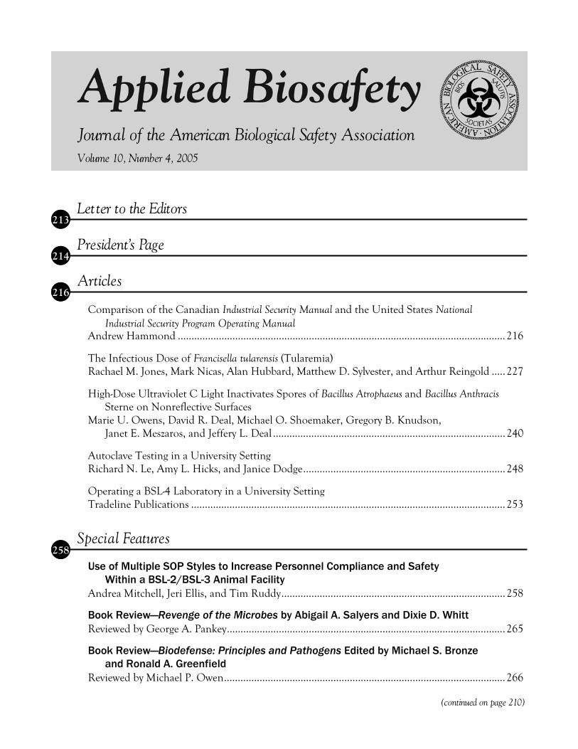
Letter to the Editors
President’s Page
Articles
Comparison of the Canadian Industrial Security Manual and the United States National Industrial Security Program Operating Manual
Andrew Hammond ........................................................................................................................216
The Infectious Dose of Francisella tularensis (Tularemia) Rachael M. Jones, Mark Nicas, Alan Hubbard, Matthew D. Sylvester, and Arthur Reingold .....227
High-Dose Ultraviolet C Light Inactivates Spores of Bacillus Atrophaeus and Bacillus AnthracisSterne on Nonreflective Surfaces
Marie U. Owens, David R. Deal, Michael O. Shoemaker, Gregory B. Knudson, Janet E. Meszaros, and Jeffery L. Deal .....................................................................................240
Autoclave Testing in a University Setting Richard N. Le, Amy L. Hicks, and Janice Dodge..........................................................................248
Operating a BSL-4 Laboratory in a University Setting Tradeline Publications ...................................................................................................................253
Specia l Features
Use of Multiple SOP Styles to Increase Personnel Compliance and Safety Within a BSL-2/BSL-3 Animal Facility
Andrea Mitchell, Jeri Ellis, and Tim Ruddy..................................................................................258
Book Review—Revenge of the Microbes by Abigail A. Salyers and Dixie D. Whitt Reviewed by George A. Pankey......................................................................................................265
Book Review—Biodefense: Principles and Pathogens Edited by Michael S. Bronze and Ronald A. Greenfield
Reviewed by Michael P. Owen.......................................................................................................266
(continued on page 210)
Journa l of the American Biologica l Safety Association
Volume 10, Number 4, 2005
258
213
214
216

210
Ask the Experts—HEPA Filtered Supply Air for BSL-3 Laboratories? John H. Keene ................................................................................................................................268
Biosafety Tips—Lymphocytic Choriomeningitis Virus—A Hazard in Rodent Animal Colonies Karen B. Byers ................................................................................................................................270
ABSA News
2005 ABSA Conference Photos .................................................................................................273
2005 ABSA Service Award Recipients......................................................................................274
New ABSA Members for 2006 ...................................................................................................277
2005 ABSA Conference Sponsors .............................................................................................279
Calendar of Events .......................................................................................................................280
ABSA Journal Subscription Information ...................................................................................281
ABSA Anthology Books Information and Order Form.............................................................282
ABSA Chapters, Affiliates, and Affiliated Organizations ........................................................283
(continued from page 209)
About the Cover
Fransicella tularensis is the causative agent of tularemia. Exposure to F. tularensis has resulted in numerous laboratory acquired infections, some of which may have been due to aerosol exposures. Read more about infectious dose modeling on pages 227-239, “The Infectious Dose of Francisella tularensis (Tularemia)” by Rachael M. Jones, Mark Nicas, Alan Hubbard, Matthew D. Sylvester, and Arthur Reingold. One natural reservoir for this zoonotic disease is the rabbit. It can be transmitted to humans by handling infected blood or tissue, or consuming undercooked infected meat. The tick, an arthropod vector, can trans-mit the disease through its bite. The symptoms developed depend on the type of exposure route. Images from the CDC Public Health Image Library are: D. variabilis tick photo, taken by Andrew Brooks of CDC; Tularemia lesion on the dorsal skin of right hand photo, taken by Dr. Brachman of the CDC; and F. tularensis colony characteristics when grown on Chocolate, Martin Lewis or Thayer-Martin medium at 48-72 hours, courtesy of Larry Stauffer, Oregon State Public Health Laboratory.
273

Applied Biosafety: Journal of the American Biological Safety Association (ISSN 1535-6760) is published quarterly by the American Biological Safety Association (ABSA). ABSA members receive the journal as a benefit of membership. An additional annual subscription for members is $60. Nonmembers and institutions/libraries may subscribe at the annual rates of $92 and $122 respectively. Single issue rates are as follows: members $18; nonmembers $28; and institutions/libraries $35.
Authorization to Copy: No part of this publication may be reproduced, stored in a retrieval system, or transmitted in any form or by any means, electronic, electrostatic, magnetic tape, photocopying, recording, or otherwise, without permission in writing from the copyright holder.
Change of Address: A change of address notice should be sent at least 6 weeks in advance to the ABSA National Office to ensure that all mailings, including the journal and newsletter, will reach you. ABSA is not responsible for misrouted mail as a result of insufficient notification of an address change. Undelivered copies resulting from an insufficient address change notification will not be replaced, but issues may be purchased at the single issue price as detailed above.
ABSA National Office American Biological Safety Association 1202 Allanson Road Mundelein, IL 60060-3808, USA 847-949-1517 / Fax 847-566-4580 E-mail: [email protected] Web Site: www.absa.org
ABSA Journal Editorial Board Co-Editors Barbara Johnson, Department of Defense, Arlington, VA Karen B. Byers, Dana Farber Cancer Institute, Boston, MA
Associate Editor Elizabeth Gilman Duane, Wyeth, Cambridge, MA Lynn Harding, Biosafety Consultant, Chattanooga, TN
Assistant Editors Richard Fink, Wyeth BioPharma, Andover, MA John H. Keene, Biohaztec Associates, Inc., Midlothian, VA Thomas A. Kost, GlaxoSmithKline, Research Triangle Park, NC Ed Krisiunas, WNWN International, Burlington, CT
International Editors Allan Bennett, European Biological Safety Association (EBSA),
United Kingdom Maureen Best, International Biosafety Working Group (IBWG),
Canada Otto Doblhoff-Dier, European Biological Safety Association (EBSA),
Austria Betty Kupskay, ABSA Canada, Canada Ai Ee Ling, Asia Pacific Biosafety Association (APBA), Singapore Leila Oda, Associação Nacional de Biossegurança (ANBio), Brazil
Production Editor Karen D. Savage
Editorial Board Matthew J. Bankowski, ViroMed (LabCorp), Minnetonka, MN Franklin R. Champlin, Mississippi State University, Mississippi
State, MS Mary L. Cipriano, Abbott Laboratories, Abbott Park, IL Robert P. Ellis, Colorado State University, Fort Collins, CO Glenn A. Funk, Lawrence Livermore National Laboratory,
Livermore, CA Raymond W. Hackney, Jr., University of North Carolina, Chapel
Hill, NC Philip Hagan, Georgetown University, Washington, DC Robert J. Hawley, Midwest Research Institute, Frederick, MD Richard Henkel, Centers for Disease Control and Prevention,
Atlanta, GA Debra L. Hunt, Duke University, Durham, NC Peter C. Iwen, University of Nebraska Medical Center, Omaha, NE John H. Keene, Biohaztec Associates, Inc., Midlothian, VA Paul Michael Kivistik, University of Nevada, Reno, NV Joseph P. Kozlovac, USDA-ARS, Beltsville, MD Jens H. Kuhn, Harvard Medical School, Southborough, MA Margy S. Lambert, University of Wisconsin, Madison, WI R. Thomas Leonard, University of Virginia, Charlottesville, VA Paul J. Meechan, Merck Research Laboratories, West Point, PA Mark Nicas, University of California, Berkeley, CA Beryl J. Packer, Iowa State University, Ames, IA Tim Ravita, Constella Health Sciences, Atlanta, GA Richard Rebar, GlaxoSmithKline R&D, King of Prussia, PA Jonathan Y. Richmond, Jonathan Richmond & Associates, Inc.,
Southport, NC Deanna S. Robbins, Department of Veterans Affairs, Baltimore, MD Richard J. Shaughnessy, University of Tulsa, Tulsa, OK Allan Showler, USDA-ARS, Weslaco, TX Cecil R. Smith, Ohio State University, Columbus, OH Gerard J. Spahn, The Salk Institute, La Jolla, CA Donald Vesley, University of Minnesota, Minneapolis, MN Catherine L. Wilhelmsen, United States Army Medical Research
Institute of Infectious Diseases (USAMRIID), Fort Detrick, MD Linda B. Wolfe, Whitehead Institute for Biomedical Research,
Cambridge, MA Jeffrey D. Wolt, Iowa State University, Ames, IA Alan G. Woodard, International Environmental Health Alliance,
Gansevoort, NY
Advertising Rates Rates 1x 2x 4x Outside back cover $800 $720 $680 Inside front cover $600 $540 $510 Inside back cover $600 $540 $510 Full page $500 $450 $425 1/2 page $300 $270 $255 1/4 page $200 $180 $170
Color rates: $350 for first color (after black) and $300 each additional color. 15% discount for agencies (orders must be supplied on agency letterhead).
Mechanical Requirements Width Height Outside back cover (full bleed) 8-1/2” 11” Inside front cover (full bleed) 8-1/2” 11” Inside back cover (full bleed) 8-1/2” 11” Full page 7” 10” 1/2 page - horizontal 7” 4-7/8” 1/2 page - vertical 3-3/8” 10” 1/4 page 3-7/8” 4-7/8”
Trim size—8-1/2” x 11” Film—133 line screen, right reading, emulsion side down, color separated
Submission Deadlines February 1 for Spring issue May 1 for Summer issue August 1 for Fall issue November 1 for Winter issue
The publication of any advertisement by this journal is not an endorsement of the advertiser or of the products or services advertised. ABSA is not responsible for any claims made in any advertisement.

212
Vision
ABSA, the leader in the profession of biological safety.
Mission Statement
The American Biological Safety Association is dedicated to expanding biological safety awareness to prevent adverse occupational and environmental impact from biohazards.
Goals
• Expand professional and public awareness of biological safety through effective communication. • Participate in the development of biological safety and biosecurity standards, guidelines, and
regulations. • Develop ABSA as the recognized resource for professional and scientific expertise in biological
safety and biosecurity. • Advance biological safety as a scientific discipline through education, research, and professional
development.
ABSA Officers
President Glenn A. Funk, Lawrence Livermore National Laboratory, Livermore, CA
President-Elect Robert J. Hawley, Midwest Research Institute, Frederick, MD
Secretary Rosamond Rutledge-Burns, National Institute Standards & Technology, Gaithersburg, MD
TreasurerLeslie Delpin, University of Connecticut, Storrs, CT
Past-President Elizabeth Gilman Duane, Wyeth, Cambridge, MA
Council Members Robert P. Ellis, Colorado State University, Fort Collins, CO Joseph P. Kozlovac, USDA Agricultural Research Service, Beltsville, MD Patricia Olinger, Pharmacia Corporation, Kalamazoo, MI Chris Thompson, Greenfield, IN
Executive Director Edward J. Stygar, Jr.
213
Applied Biosafety, 10(4) p. 213 © ABSA 2005
Letter to the Editors
Let me commend Allan Bennett et al. on valu-able piece of research and an illuminating article entitled “Development of Particle Tracer Techniques to Measure the Effectiveness of High Containment Laboratories” in Applied Biosafety (Volume 10, Num-ber 3, 2005). The facts and observations presented support some common engineering assumptions and challenge others. Both results are valuable. Definition of Laboratory Protection Factor and the test methods described advance our ability to discuss effective pressurization. The result that con-tainment correlates more closely to infiltrating air flow than to pressure difference is intriguing. I ex-pect it surprises many engineers as much as it does me. The explanation offered is that the air flowing inward through the door catches contaminants and prevents their escape while the door is open and a person walks through it. The authors themselves seem to find it unlikely that such low air velocity captures contaminants. (The air velocity reported through the open door is 0.14 m s-1 and lower: much less than the velocity of the swinging door or the walking person.) Perhaps it is appropriate to propose another ex-planation for the results. Consider the effect that the
infiltrating air flow has on contaminant concentra-tions while the door is closed before and after entry or exit. Just inside the door, contaminated air is con-tinuously replaced by the clean infiltrating air. This lowers the concentration near the door. When entry or exit occurs, certainly some air leaves the room, but this air is cleaner than it would be with a lower infiltrating air flow rate, so less contaminant leaves the room. After the entry or exit is complete and the door is closed, some quantity of contaminant lingers outside the room. A portion of it is picked up by the particle counters, but presumably, some of it is drawn back into the room by the on-going infiltra-tion. In short, the supposition is that infiltrating air flow has a continuous cleaning effect near the door, and that this on-going effect (rather than several sec-onds of infiltration at very low velocity during entry or exit) increases the Laboratory Protection Factor. Do the authors have more information that will help us choose one mechanism or the other to ex-plain the results? In some cases, the question is moot. Containment is the issue and the mechanism may be unimportant, but there are ventilation sys-tems where the distinction is crucial.
James J. Coogan
Siemens Building Technologies, Buffalo Grove, Illinois
Editorial Note
Letters to the Editors (approximately 400 words) discuss information published in Applied Biosafety inthe past nine months or discuss topic areas of gen-eral interest in the biosafety profession. Letters can
be submitted electronically to Karen D. Savage, Production Editor, at [email protected] or by mail to ABSA National Office, Applied Biosafety, 1202 Allanson Road, Mundelein, IL 60060. Letters pub-lished in part or whole are subject to editing for clar-ity and special formatting.

214
I suspect every new organization president wres-tles with questions like “What do I want to achieve during my presidency?” and “Where do I want to lead the organization during the next year?” These are weighty issues that focus on the best interests of the membership in general and the institution in particular. They’re made especially challenging by the outstanding leadership and successes provided by recent past presidents. I have, appropriately, a very high standard to uphold. With 1,500 members representing 30 different countries and 16 Affiliates and affiliated organiza-tions, ABSA is no longer a small club of friends. We are gaining the mass to be noticed, to be listened to, to be asked for advice and help, and to make a dif-ference in the realms of science we impact. It would be easy for me to suggest that we grow the organiza-tion because as we grow larger, we grow stronger and we have a louder voice. There is truth in this, and growing ABSA is a worthy goal that we should pur-sue. However, at this point, I believe it is also impor-tant to “grow our profession”—to bring more highly qualified scientists into our ranks, to strengthen our knowledge base, and to provide additional ABSA members who will enrich our organization. I believe there are many scientists and health and safety pro-fessionals who are unaware of the challenges and rewards of biosafety as a career, or of the need for highly skilled biosafety professionals in the scientific community. To keep our profession attractive to others and respected within the scientific community, we must maintain, strengthen, and continue to demonstrate the high professional standards to which we adhere every day. Each of us must set the Gold Standard for professionalism, for technical competency, for cus-
tomer service, and for flexibility and adaptability to meet the varied and often unanticipated needs of our customers. We must not simply continue to be the best of the best; we must continually get better at what we do. As “Neutron Jack” Welch, ex-CEO of General Electric, used to say, “You can’t just talk the talk; you’ve also got to walk the walk.” One of the things we do best in ABSA is share our knowledge through networking, seminars, train-ing sessions, and professional courses. ABSA is your organization, and I urge you to use the opportunities it offers to improve your skills as a biosafety profes-sional. Meanwhile, your ABSA Council and I will continue to make ABSA better for you. The manage-ment consultants who helped us restructure our business model also helped us identify important areas for Task Force study; we held off implementing these during the first year in order to focus on the business infrastructure. This year we’ll put Task Forces to work defining the parameters of the bio-safety profession, enhancing the value and benefits of professional Registration and Certification, and determining the feasibility and usefulness of an ABSA Emergency Response capability to provide assistance during national and international emer-gencies that involve biosafety. If you have an interest in serving on one of these Task Forces, please let me know ([email protected]). One other effort soon to be underway is a re-newed push to catalog the ABSA Historical Collec-tion and transfer it to safe archival storage. Once that’s done, we can look at ways to make it accessible to members through, for example, displays at meet-ings and articles in this Journal. Ultimately, I’d like to see at least parts of the collection available online. In future columns I’ll discuss other ideas for
Glenn A. Funk
Gualala, California
Applied Biosafety, 10(4) pp. 214-215 © ABSA 2005
President’s Page

215
moving ABSA forward. As always, I greatly appreci-ate your volunteer efforts to help make ABSA what it is. We wouldn’t be here today if it weren’t for the active and positive participation of our many tal-ented members. As President, my door is open to
you. Please send me e-mails with your ideas and sug-gestions. We’ll talk about them, I’ll take the great ideas to Council, and together we’ll continue to make ABSA better and stronger.
G. A. Funk
Corrections and Clarifications
Special Features, Ask the Experts: “USDA BSL-3 Facility Requirements: What’s the Concern?” authorship attribution in the Table of Contents is incorrect (Volume 10, Number 3, page 133). It should be: John H. Keene.
EPA Pesticide Program Update from EPA’s Office of Pesticide Programs 10/11/05: EPA Approves New Non-Chemical Control for Corn Rootworm www.epa.gov/pesticides
After an intensive, multi-year scientific analysis, EPA has approved applications submitted by Mycogen Seeds (c/o Dow AgroSciences, LLC) and Pioneer Hi-Bred International, Inc. for the use of a new corn plant-incorporated protectant (PIP) designed to control corn rootworm. Corn rootworm is a widespread and destructive insect pest responsible for the single largest use of conventional insecticides in the United States. The new product is the second PIP to offer protection against corn rootworm and is expected to result in a further reduction of chemical insecticide use by growers. The new corn plant-incorporated protectant, Event DAS-59122-7 Corn, produces its own insecticide within the corn plant derived from Bacillus thuringiensis (Bt), a naturally occurring soil bacterium. The Bt proteins used in this product, called Cry34Ab1 and Cry35Ab1 (Cry 34/35), control corn rootworm. To reduce the likelihood of corn rootworm developing resistance to Bt, EPA is requiring Mycogen and Pioneer to ensure that buffer zones within the planted acreage be planted with corn that is not protected from corn rootworm to serve as a “refuge.” The insect populations in the refuges will help prevent resistance development when they cross-breed with insects in the Bt fields. This resistance management strategy was developed as a condition of the registration, and EPA will require routine monitoring and documentation that these measures are followed. The reduction in chemical pesticide use will benefit the environment directly and can mean less chemical exposure to people who apply pesticides to corn. The availability of multiple corn rootworm-protected corn products will also increase grower choice and price competition, resulting in lower seed prices for consumers and higher adoption rates. The product provides yet another way to combat corn rootworm, as well as indirect benefits such as energy savings resulting from reduced chemical insecticide use. As with similar products, EPA has approved Cry 34/35 for time-limited use, which will be subject to reevaluation in five years. For more information on EPA’s regulation of biopesticide products, see www/epa.gov/pesticides/biopesticides/.

216
Abstract
Because of the potential for use as a bioterrorism agent or bioweapon, many governments have imposed strict regulations regarding the possession, use, and transfer of “select” biological agents. Consequently, much of the information surrounding the possession and use of these agents is potentially classified, and those contractors and their employees who require access to this information must receive Facility (contractor) and Personnel (employees) clearances. Both Canada and the United States (U.S.) have produced industrial secu-rity manuals—the Industrial Security Manual (ISM) (Canadian and International Industrial Security Direc-torate, 2004) and the National Industrial Security Program Operating Manual (NISPOM) (Defense Technical Information Center, 1995)—for use by cleared government contractors. These documents set forth the requirements, restrictions, and other safe-guards that are necessary to prevent unauthorized dis-closure of classified information and assets provided to or produced by private government contractors. This article compares and contrasts the requirements set forth in the ISM and the NISPOM. The results of this comparison present a valuable security management tool for private organizations that wish to engage in classified work for the Canadian, U.S., or both govern-ments.
Introduction
As a result of the October 2001 anthrax letter attacks, both the United States and Canada enacted new laws imposing additional restrictions on certain hazardous biological agents and toxins. The U.S. enacted the Public Health Security and Bioterrorism Preparedness and Response Act of 2002 (Public Law 107-188) (U.S. Government Printing Office, 2002) and Canada passed the Public Safety Act, 2002 (Parliament of Canada, 2002). Because of the poten-tial for “select” biological agents and toxins being used as bioterrorism agents or in a bioweapons pro-gram, both Acts impose strict regulations regarding their possession, use, and transfer. Consequently, much of the information surrounding the possession and use of these agents is potentially classified (or confidential), and those organizations and their em-ployees who require access to this information must receive Facility (organization) and Personnel (employee) clearances. Both Canada and the U.S. have produced industrial security manuals—Industrial Security Manual (ISM) (Canadian and International Industrial Security Directorate, 2004) and the Na-tional Industrial Security Program Operating Manual(NISPOM) (Defense Technical Information Center, 1995)—for use by cleared government contractors. These documents set forth the requirements, restric-tions, and other safeguards that are necessary to pre-vent unauthorized disclosure of classified informa-tion (and assets) provided to or produced by private
Applied Biosafety, 10(4) pp. 216-226 © ABSA 2005
Article
Andrew Hammond
Constella Health Sciences, Atlanta, Georgia
Comparison of the Canadian Industrial Security Manual and the United States NationalIndustrial Security Program Operating Manual

217
government contractors and to control the author-ized disclosure of classified information (and assets) released by the governments to their contractors.
United States—National Industrial Security Program Operating Manual
Security Classifications An original classification decision at any level can be made only by a U.S. Government official who has been delegated this authority in writing. Contractors may make derivative classification deci-sions based on the guidance provided by the Con-tract Security Classification Specification that is is-sued with each classified contract. Derivative classifi-cation is the act of classifying a specific item of infor-mation or material on the basis of an original classi-fication decision already made by an authorized original classification authority. The source of au-thority for derivative classification ordinarily consists of a previously classified document or a classification guide issued by an original classification authority.
Top Secret Top secret information or material is that infor-mation or material whose unauthorized disclosure could be reasonably expected to cause exceptionally grave damage to the national security that the original classification authority is able to identify or describe. Examples include armed hostilities against the United States or its allies, disruption of foreign rela-tions vitally affecting the national security, and the disclosure of scientific or technological develop-ments vital to national security.
Secret Secret information or material is that informa-tion or material whose unauthorized disclosure could be reasonably expected to cause serious damageto the national security that the original classifica-tion authority is able to identify or describe. Exam-ples of serious damage include significant impair-ment of a program or policy directly related to the national security and compromise of significant sci-entific or technological developments relating to na-tional security.
Confidential Confidential information or material is that in-formation or material whose unauthorized disclo-sure could be reasonably expected to cause damage to the national security that the original classification authority is able to identify or describe. Examples include documents relating to clearance or assign-ment of personnel who will have knowledge of, or access to, classified information or materials or de-tails pertaining to features of routes and schedules of shipments of confidential materials.
Facility Security
Facility Clearances A facility security clearance (FCL) is an adminis-trative determination that a facility is eligible for ac-cess to classified information at the same or lower classification category as the clearance being granted. Contractors are eligible for custody of classified ma-terial, if they have an FCL and storage capability ap-proved by the Cognizant Security Agency (CSA). A procuring activity of the Government or cleared contractor may request a facility clearance for a contractor or prospective contractor/ subcontractor when a definite, classified procure-ment need has been established. Also, the contractor must be organized and existing under the laws of any of the 50 states, the District of Columbia, or Puerto Rico, and be located in the U.S. and its territorial areas or possessions.
Meetings Classified disclosure at a meeting (e.g., confer-ence, seminar, symposium, exhibit, convention, training course, or other such gathering) which serves a government purpose and at which adequate security measures have been provided in advance may be conducted by a cleared contractor provided the meeting is authorized by a Government Agency that has agreed to assume security jurisdiction. The Government Agency must approve security arrange-ments, announcements, attendees, and the location of the meeting. (Classified meetings shall be held only at a Federal Government installation or a cleared contractor facility where adequate physical security and procedural controls have been ap-
A. Hammond

218
proved.) Contractors wishing to conduct classified meetings shall submit their requests to the Govern-ment Agency having principal interest in the subject matter of each meeting.
Personnel Security
Security Officers The Facility Security Officer (FSO) shall be a U.S. citizen employee appointed by the contractor who is cleared as part of the facility clearance. The FSO will supervise and direct security measures nec-essary for implementing the NISPOM and related Federal requirements for classified information. The senior management official and the FSO must always be cleared to the level of the Facility Clearance (FCL). Other officials, as determined by the CSA, must be granted a Personnel Clearance (PCL) or be excluded from classified access.
Personnel Clearances An industrial personnel security clearance is an administrative determination that an industrial em-ployee is eligible for access to classified information. This determination is based on investigation and review of available personal data and a finding that access is clearly consistent with national interests. An individual may be processed for a personnel security clearance only when employed by a cleared contractor in a job requiring access to classified in-formation. As an exception, a candidate for employ-ment may be processed for a PCL provided a written commitment for employment that prescribes a fixed date for employment within the ensuing 180 days has been made by the contractor, and the candidate has accepted the employment offer in writing. Under rare circumstances, a non-U.S. citizen may be issued a Limited Access Authorization for access to classified information. Specific criteria and limitations are provided in the NISPOM. Contractors have no authority to grant, deny, or revoke personnel clearances for their employees. This authority is reserved by the U.S. Government.
Subcontracting Before a prime contractor may release, disclose classified information to a subcontractor, or cause
classified information to be generated by a subcon-tractor, he or she must determine the security re-quirements of the subcontract and determine clear-ance status of prospective subcontractors. The prime contractor shall verify the clearance status and safe-guarding capability of the subcontractor from the CSA. If a prospective subcontractor does not have the appropriate FCL or safeguarding capability, the prime contractor shall request the CSA of the sub-contractor to initiate the necessary action. The prime contractor shall ensure that a Con-tract Security Classification Specification is incorpo-rated in each classified subcontract. The contractor shall also review the security requirements during the different stages of the subcontract and provide the subcontractor with applicable changes in these security requirements. Upon completion of the sub-contract, the subcontractor may retain classified ma-terial received or generated under the subcontract for a 2-year period, provided the prime contractor or GCA does not advise to the contrary.
Education, Training, and Briefings Contractors shall provide all cleared employees with security training and briefings commensurate with their involvement with classified information. Contractors shall also be responsible for ensuring that the FSO, and others performing security duties, complete security training deemed appropriate by the CSA. (Training, if required, should be com-pleted within 1 year of appointment to the position of FSO.) The contractor is responsible for providing all cleared employees with some form of security education and training at least annually. The SF 312 is an agreement between the United States and an individual who is cleared for access to classified information. An employee issued an initial PCL must execute an SF 312 prior to being granted access to classified information. The employee must also receive an initial security briefing that includes a Threat Awareness Briefing, a Defensive Security Briefing, an overview of the security classification system, employee reporting obligations and require-ments, and security procedures and duties applicable to the employee’s job. Contractors shall debrief cleared employees at the time of termination (discharge, resignation, or
Comparison of the Canadian Manual and the U.S. Manual

219
retirement); when an employee’s PCL is terminated, suspended, or revoked, and upon termination of the FCL.
Visits The contractor must determine that the visit is necessary and that the purpose of the visit cannot be achieved without access to, or disclosure of, classi-fied information. All classified visits require advance notification to, and approval of, the organization being visited. In urgent cases, visit information may be furnished by telephone provided that it is fol-lowed up in writing. The contractor shall issue a Visit Authorization Letter (VAL) to the organization being visited that shall include the following: • Contractor’s name, address, and telephone num-ber, assigned CAGE Code, and certification of the level of the FCL • Name, date, place of birth, and citizenship of the employee intending to visit • Certification of the proposed visitor’s PCL and any special access authorizations required for the visit• Name of person(s) to be visited • Purpose and sufficient justification for the visit to allow for a determination of the necessity of the visit• Date or period during which the VAL is to be valid Contractors shall maintain a record of all visi-tors to their facility who have been approved for ac-cess to classified information.
Document Security
General Marketing All classified material shall be marked on the face of the document to show the name and address of the facility responsible for its preparation and the date of preparation. The highest level of classified information contained in a document is its overall marking. The overall marking shall be conspicuously marked or stamped at the top and bottom on the outside of the front cover, on the title page, on the first page, and on the outside of the back cover. Inte-rior pages of classified documents shall be marked at
the top and bottom with the highest classification of the information appearing thereon or marked UN-CLASSIFIED if all the information on the page is UNCLASSIFIED. The major components of com-plex documents are likely to be used separately. Therefore, each major component shall be marked as a separate document. Also, each section, part, paragraph, or similar portion of a classified docu-ment shall be marked to show the highest level of its classification, or that the portion is unclassified. Un-classified subjects and titles shall be selected for clas-sified documents, if possible. If a classified subject or title must be used, it shall be marked with the appro-priate symbol—(TS), (S), or (C)—placed immediately following and to the right of the item. All classified information shall be marked to reflect the source of the classification and declassifi-cation instructions. This required information shall be placed on the cover, first page, title page, or in another prominent position.
General Storage Cognizant security officials shall work to meet appropriate security needs according to the intent of the NISPOM and at an acceptable cost. TOP SECRET material shall be stored in a GSA-approved security container, an approved vault, or an approved Closed Area. Supplemental protec-tion is required. SECRET material shall be stored in the same manner as TOP SECRET material without supple-mental protection. CONFIDENTIAL material shall be stored in the same manner as TOP SECRET or SECRET material except that no supplemental protection is required.
Reproduction Contractors shall establish a reproduction con-trol system to ensure that reproduction of classified material is held to the minimum consistent with contractual and operational requirements. Classified reproduction shall be accomplished by authorized employees knowledgeable about the procedures for classified reproduction. The use of technology that prevents, discourages, or detects the unauthorized reproduction of classified documents is encouraged.
A. Hammond
220
All reproductions of classified material shall be conspicuously marked with the same classification markings as the material being reproduced.
Domestic Transmission Standards (Outside of Facility) Top Secret • Written authorization of the Government Con-tracting Activity (GCA) • Sealed, opaque inner and outer covers with the inner cover being a wrapper or envelope plainly marked with the assigned classification and ad-dresses of both sender and addressee • A receipt that identifies the sender, the ad-dressee, and the document shall be attached to or enclosed in the inner cover • Via: a. Defense Courier Service (DCS), if author-
ized by GCA b. A designated courier or escort cleared for
access to TOP SECRET information c. By electrical means over CSA-approved se-
cured communications security circuits pro-vided such transmission conforms with the NISPOM, the telecommunications security provisions of the contract, or is authorized by the GCA
Secret • Sealed, opaque inner and outer covers with the inner cover being a wrapper or envelope plainly marked with the assigned classification and ad-dresses of both sender and addressee • A receipt that identifies the sender, the ad-dressee, and the document shall be attached to or enclosed in the inner cover • Via: a. TOP SECRET methods b. USPS Express or Registered mail c. A cleared “Commercial Carrier” d. A cleared commercial messenger service en-
gaged in the intracity/local area delivery (same day delivery only) of classified material
e. A commercial delivery company approved by the CSA
f. Other methods as directed, in writing, by the GCA
Confidential • Packaged by SECRECT material methods except that a receipt is required only if the sender deems it necessary • Via: a. SECRET methods b. USPS Certified mail
International Transmission Standards Top Secret • Domestic requirements • Via: a. Defense Courier Service b. Department of State Courier System c. Courier service authorized by GCA
Secret and Confidential • Domestic requirements • Via: a. Registered mail through U.S. Army, Navy,
or Air Force postal facilities b. Appropriately cleared contractor employee c. U.S. civil service employee or military person
designated by the GCA d. U.S. and Canadian registered mail with reg-
istered mail receipt to and from Canada and via a U.S. or Canadian government activity
e. As authorized by the GCA
Destruction Contractors shall destroy classified material in their possession as soon as possible after it has served the purpose for which it was intended. Classified material may be destroyed by burning, shredding, pulping, melting, mutilation, chemical decomposition, or pulverizing. Pulpers, pulverizers, or shedders may be used only for the destruction of paper products. Residue shall be inspected during each destruction to ensure that classified informa-tion cannot be reconstructed. Crosscut shredders shall be designed to produce residue particle size not exceeding 1/32 inch in width by 1/2 inch in length. Public destruction facilities may be used only with the approval of the CSA, and classified material removed from a cleared facility for destruction shall be destroyed on the same day it is removed. Destruction shall be performed only by appropri-
Comparison of the Canadian Manual and the U.S. Manual

221
A. Hammond
ately cleared employees of the contractor. For de-struction of TOP SECRET material, two persons are required. For destruction of SECRET and CONFI-DENTIAL material, one person is required. Destruction records that indicate the date of destruction, identify the material destroyed, and are signed by the individuals designated to destroy and witness the destruction are required for TOP SE-CRET material.
Information System Security
Information systems (IS) that are used to cap-ture, create, store, process, or distribute classified information must be properly managed to protect against unauthorized disclosure of classified informa-tion and loss of data integrity, and to ensure the availability of the data and system. Protection requires a balanced approach includ-ing IS security features to include. but not limited to, administrative, operational, physical, computer, communications, and personnel controls. Protective measures commensurate with the classification of the information, the threat, and the operational re-quirements associated with the environment of the IS are required. The requirements outlined in the NISPOM ap-ply to all information systems processing classified information. Additional requirements for high-risk systems and data are covered in the NISPOM Supple-ment. The CSA is the Designated Accrediting/ Approving Authority (DAA) responsible for accredit-ing information systems used to process classified information in industry. A formal certification and accreditation (C&A) occurs after the protection measures have been implemented and any required IS protection documentation has been approved. Certification validates that the protection measures described in the System Security Plan (SSP) have been implemented on the system and that the pro-tection measures are functioning properly. Accredi-tation is the approval by the CSA for the system to process classified information.
Canada—Industrial Security Manual
Security Classifications The originator of the information and assets determines the classification level.
Top Secret TOP SECRET refers to information and assets related to the national interest that may qualify for an exemption or exclusion under the Access to In-formation Act or Privacy Act and that the compro-mise of which would reasonably be expected to cause exceptionally grave injury to the national interest.
Secret SECRET refers to information and assets related to the national interest that may qualify for an ex-emption or exclusion under the Access to Informa-tion Act or Privacy Act and that the compromise of which would reasonably be expected to cause serious injury to the national interest.
Confidential CONFIDENTIAL refers to information and assets related to the national interest that may qual-ify for an exemption or exclusion under the Access to Information Act or Privacy Act and that the com-promise of which would reasonably be expected to cause injury to the national interest.
Protected “C” PROTECTED “C” refers to information and assets related to other than the national interest that may qualify for an exemption or exclusion under the Access to Information Act or Privacy Act that could reasonably be presumed to cause extremely serious in-jury, such as loss of life, if compromised.
Protected “B” PROTECTED “B” refers to information and assets related to other than the national interest that may qualify for an exemption or exclusion under the Access to Information Act or Privacy Act that could reasonably be expected to cause serious injury if com-promised.

222
Protected “A” PROTECTED “A” refers to information and assets related to other than the national interest that may qualify for an exemption or exclusion under the Access to Information Act or Privacy Act that could reasonably be presumed to cause injury if compro-mised.
Facility Security
Facility Clearances A Facility Security Clearance is an administrative determination that an organization is eligible, from a security viewpoint, for access to CLASSIFIED and PROTECTED information and assets of the same or lower classification level as the clearance being granted. There are three types of Facility Security Clear-ances each of which may be authorized at the classifi-cation level of CONFIDENTIAL, SECRET, or TOP SECRET:1. Personnel Assigned (PA). This is the most basic type of Facility Security Clearance which involves security screening of the organization’s Key Senior Officials and employees. There is NO requirement to evaluate the physical security status of the organi-zation’s facilities. The organization is not authorized to possess or store CLASSIFIED information and assets. 2. Document Safeguarding Capability (D.Sc.). In addi-tion to the security screening of the organization’s Key Senior Officials and employees, the physical se-curity of the organization’s facilities is assessed to ensure safeguarding requirements are met. The or-ganization is authorized to possess and store CLAS-SIFIED information and assets. 3. Production (PROD). This includes all of the ele-ments of a Document Safeguarding Facility Security Clearance. In addition, the security of the manufac-turing, repairing, modifying, or otherwise working on CLASSIFIED components or items is assessed to ensure government security requirements are met. A Designated Organization Screening (at the PRO-TECTED level) is an administrative determination that an organization is eligible, from a security view-point, for access to PROTECTED information and assets of the same or lower level as the clearance be-
ing granted. The three types of Designated Organiza-tion Screening are equivalent to the three types of Facility Security Clearances except they pertain only to PROTECTED information and assets. Each of the three types may be authorized at one of the fol-lowing levels: PROTECTED “A,” PROTECTED “B,” or PROTECTED “C.” An organization is eligible to obtain an organiza-tion security screening/clearance only if it is spon-sored by an authorized sponsor in support of an ex-isting or impending contract or bid solicitation which cal ls for access to CLASSI-FIED/PROTECTED information, assets, and/or certain restricted work sites.
Meetings
(No provisions are established within the Cana-dian Industrial Security Manual.)
Personnel Security
Security Officers All organizations that require a Designated Or-ganization Screening or a Facility Security Clearance shall appoint a Company Security Officer. The Com-pany Security Officer shall be appointed by the Chief Executive Officer (CEO) or the designated Key Senior Official (KSO) of the organization. The CSO must be a Canadian citizen employee, report to a designated KSO, and be security screened or cleared to the Reliability Status level or Facility Security Clearance level of the facility. The appointment of the Company Security Officer must be approved by the Canadian and International Industrial Security Directorate (CIISD). When a facility-cleared Canadian parent organi-zation owns one or more cleared subsidiaries in Can-ada, a Corporate Company Security Officer (CCSO) should be appointed to oversee government indus-trial security matters for the entire corporation.
Personnel Clearances Personnel Security Screening must be carried out according to the highest sensitivity level of infor-mation and assets that will be accessed during the
Comparison of the Canadian Manual and the U.S. Manual

223
A. Hammond
contracting process and/or required for access to restricted work sites. Access to PROTECTED infor-mation, assets, and restricted work sites requires that an individual has Reliability Status, and access to CLASSIFIED information, assets, and/or restricted work sites requires a Security Clearance at the appro-priate level of sensitivity. Only individuals employed or under a contract to commence employment within 60 days by a pri-vate sector organization on a contract/subcontract requiring access to CLASSIFIED/PROTECTED information, assets, and/or certain restricted work sites may be security screened. Non-Canadian citi-zens may be security cleared with access limitations. The limitations include denying access to CLASSI-FIED/PROTECTED information and assets which are not of Canadian origin, do not come from the country of which the person is a citizen, or are not releasable to his/her nation of origin. Contractors have no authority to deny or revoke Personnel Security Clearances for employees. This authority is reserved by the Canadian Government. The contractor may suspend the access of an individ-ual, while notifying CIISD of the circumstances.
Subcontracting Contractors shall subcontract work only to com-panies holding a current Designated Organization Screening or a Facility Security Clearance of the type and at the level appropriate to the work to be per-formed under the subcontract. CIISD approval of the subcontractor must be obtained before award of the subcontract and the Designated Organization Screening or Facility Security Clearance for the pro-posed subcontractor(s) must be verified by CIISD before issue of bid solicitation documents. Contrac-tors shall not assign a subcontract to organizations located outside of Canada without the prior written approval of CIISD and the Public Works and Gov-ernment Services Canada (PWGSC) contracting au-thority. The prime contractor shall ensure the security safeguarding of work placed with subcontractors.
Education, Training, and Briefings A major objective of the Company Security Officer in conducting a Security Education Program
involves working closely with management, from the top down, to ensure proper company security. Man-agers and supervisors at all levels are responsible not only for their own personal security measures, but also for ensuring that proper security procedures are followed by all employees in the organization. An initial security briefing, reinforced by an ongoing Se-curity Education and Awareness Program, is essential to the maintenance of an effective security program. Upon receiving a Personnel Security Clearance an employee acknowledges his or her responsibilities by reading and signing the Security Screening Cer-tificate and Briefing Form, TBS/SCT 330-47 Rev. 2002/06. A briefing from the Company Security Officer, which details the individual’s specific re-sponsibilities and duties relative to security in the facility, must be presented. (New employees, even though not yet security-screened and therefore pro-hibited from access to CLASSIFIED information and assets, should be given a security briefing appro-priate to their duties.)
Visits A Visit Clearance Request (VCR) (submitted to CIISD via a Request for Visit form) is required when a security-cleared individual must visit a gov-ernment/commercial organization in Canada or abroad, for the purpose of having access to CLASSI-FIED information and assets or where access to the installation is restricted in the interest of national security. Visitors must not proceed with CLASSI-FIED visits without prior visit clearance authoriza-tion from CIISD. The host organization shall deny access to CLASSIFIED information and assets or access to certain restricted work sites until the visi-tors’ Personnel Security Clearance level and their need-to-know have been verified and confirmed by the CIISD through official visit protocol. Submission of a VCR initiates verification by CIISD that confirms: • The organization requesting the visit has an Fa-cility Security Clearance to the required level • Each of the proposed visitors has a valid Person-nel Security Clearance to the required level • Foreign disclosure limitations are identified and strictly observed Visit Clearance Request is approved when the

224
Comparison of the Canadian Manual and the U.S. Manual
requesting organization is notified by CIISD. Visi-tors must not proceed on CLASSIFIED visits with-out prior visit clearance authorization. Visit Clearance Request (VCR) requires strict lead-times imposed by the authorities of foreign na-tions. Every effort must be made to ensure that lead-times are observed, as failure to do so will likely re-sult in rejection of the RFV. Organizations shall maintain a record of all indi-viduals who visit the facility for the purpose of hav-ing access to CLASSIFIED information. This record shall be separate from the record of unclassified vis-its.
Document Security
General Marketing All documents shall be marked on the outside of both the front and back covers with the highest level of classification and loose documents shall be marked on every sheet. Security markings should include the applicable classification/protection and the date or event at which declassification or down-grading is to occur. All covering or transmittal letters or forms or circulation slips must be marked to show the highest level of classification or protection of the attachments. For TOP SECRET information, mark the classi-fication in the upper right corner of each document page and show the total number of pages on each page of the document. For SECRET information, mark the classifica-tion in the upper right corner of each document page. For CONFIDENTIAL information, mark the classification in the upper right corner of the face of the document. For PROTECTED information, mark the word “PROTECTED” in the upper right corner of the face of the document and, where required, with the letter “A,” “B,” or “C” to indicate the level of protec-tion.
General Storage PROTECTED B and PROTECTED C informa-tion and assets and all CLASSIFIED information must be stored in an approved security container.
PROTECTED A information and assets shall be stored in a locked container. CLASSIFIED or PROTECTED information and assets may be stored on open shelving in a se-cure room, only after inspection and approval by CIISD and only to the level approved by CIISD. Also, CLASSIFIED and PROTECTED information and assets shall not be stored in the same container as negotiable or attractive assets.
Reproduction Reproduction of CLASSIFIED information shall be done only with the authorization of the Company Security Officer or an authorized Alter-nate Company Security Officer. Reproductions must be marked, registered, and accounted for in the same manner as for the originals. Reproductions of PROTECTED information must be marked in the same manner as the originals. TOP SECRET and PROTECTED C information shall NEVER be re-produced without written authorization from CIISD.
Domestic Transmission Standards (Outside of Facility) Top Secret • Documents must be double enveloped (gum sealed, heavy duty) and sealed with government ap-proved security tape. • A self-addressed receipt is enclosed in the inner envelope or wrapping and the inner envelope or wrapping is closed with an approved security tape. • Inner envelope or wrapping must bear the secu-rity marking and the recipient’s address. • Shipment must be recorded prior to leaving a Security Zone and the recipient must be notified in advance of shipment. • Documents are sent via a security-cleared/reliability-checked individual employed by the dispatching/receiving Facility Security Cleared Canadian organization.
Secret, Confidential, and Protected “C” • Documents must be double enveloped (gum sealed, heavy duty) and sealed with government ap-proved security tape. • A self-addressed receipt is enclosed in the inner

envelope or wrapping and the inner envelope or wrapping is closed with an approved security tape. • Inner envelope or wrapping must bear the secu-rity marking and the recipient’s address. • Via: a. Priority courier b. Registered mail c. A security-cleared/reliability-checked individ-
ual employed by the dispatching/receiving Facility Security Cleared Canadian organiza-tion
Protected “A” and “B” • Single, gum-sealed, heavy duty envelope • Via: a. First class mail b. An individual employed with the organiza-
tion c. Classified/Protected “C” methods
International Transmission Standards Top Secret, Secret, Confidential, and Protected “C” • Double enveloped (gum sealed, heavy duty) and sealed with government approved security tape • Via CIISD
Protected “B” • Single, gum sealed, heavy-duty envelope • Via CIISD
Protected “A” • Single, gum sealed, heavy-duty envelope • Via first class mail, priority courier, or registered mail
Destruction Unless otherwise specified, TOP SECRET, and PROTECTED “C” information and assets must be returned to CIISD for disposal. Unless otherwise specified, SECRET, CONFI-DENTIAL, and PROTECTED “A” and “B” infor-mation and assets of Canadian origin may be de-stroyed by the organization with the approval of CIISD. CLASSIFIED and PROTECTED information and assets which have been authorized for destruc-tion must be disposed of in accordance with the following:
• It must be destroyed only by approved destruc-tion equipment, or at a facility authorized by CIISD. • Information awaiting destruction or in transit to destruction must be safeguarded in the manner pre-scribed for the most highly CLASSIFIED and PRO-TECTED information asset involved. • CLASSIFIED and PROTECTED informa-tion/assets awaiting destruction must be kept sepa-rate from other information/assets awaiting destruc-tion. • An employee with a proper security clearance or with Reliability Status, as applicable, must be present to monitor the destruction of CLASSIFIED and PROTECTED information, respectively. • Surplus copies and waste that could reveal CLASSIFIED and PROTECTED information must be protected to the appropriate level and should be promptly destroyed.
Information System Security
The ISM establishes operational standards in Canadian industry for the safeguarding of Govern-ment information electronically processed, stored, or transmitted. This also applies to the safeguarding of technology assets. The administrative, organiza-tional, physical, and personnel security standards as documented in the ISM also apply to the informa-tion technology environment. The Government Security Policy requires that the degree of safeguarding provided by industry be commensurate with the level of the information and assets and the associated threats and risks. The con-tracting authority is responsible for ensuring that the requirements of the Government Security Policy are met and that the security standards are applied by the private sector contractor. The security standards contained in the Government Security Policy, Infor-mation Technology Standards, are the minimum standards for security in the private sector. Assess-ments, advice, and guidance regarding these stan-dards are available from the Canadian and Interna-tional Industrial Services Directorate (CIISD) of Public Works and Government Services Canada (PWGSC). The prime contractor’s Information Technology Facility(s) must be approved by CIISD prior to proc-essing government information.
A. Hammond

226
Conclusions
“It’s important to be responsible here and to be par-ticularly careful after 9/11 that we’re not giving our enemies information or materials that would make their job easier.” (Chui, 2003)
John H. Marburger III, Director, Office of Science & Technology Policy
(and science adviser to President George W. Bush)
To no surprise, the anthrax letter attacks of 2001 led directly to national policy changes since they specifically targeted both lawmakers and media personnel at their workplaces. To better protect their citizens, the United States and Canadian gov-ernments established controls not only over the pos-session and use of hazardous biological agents, but also over the information pertaining to their posses-sion and use. Legislation is now in place that forbids the disclosure of information that may identify which biological agents are possessed, who possesses that agent(s) and where, and any safeguard and secu-rity measures used to protect unauthorized access to the agent(s). Because of the genuine threat of bioter-rorism, biodefense research has become a vital and necessary component of an overall national security program. The United States alone has committed billions of dollars towards biodefense research and development. To protect biodefense information and assets, organizations working on projects deemed to be “classified” (for the sake of national security) must follow precise requirements, restric-tions, and safeguards established by their federal gov-ernment. For Canada and the United States, these requirements are conveyed in the Canadian Industrial Security Manual (ISM) and the U.S. National Industrial Security Program Operating Manual (NISPOM). These manuals provide guidance in implementing a uni-
form and cost-effective security system, thus allowing an organization to focus mainly on research rather than the burden of developing and implementing security procedures. Without these standards and consistent security policies and practices the poten-tial for compromise leading to a serious national security threat is enormous.
References
Canadian and International Industrial Security Di-rectorate. (2004). Industrial security manual. Available at www.ciisd.gc.ca/ism/text/preface-e.asp. Accessed online 2004.
Chui, G. (2003). Security concerns imperil research: Restrictions shackle scientists, some say. The Mercury News, March 3, 2003. Available at www. mercurynews.com/mld/mercurynews/news/ 5303757.htm?1c. Accessed online 2004.
Defense Technical Information Center. (2004). Na-tional industrial security program operating manual (DoD 5220.22-M). Available at www.dtic.mil/whs/ directives/corres/html/522022m.htm. Accessed online 2004.
Parliament of Canada. (2004). Public Safety Act, 2002. Available at www.parl.gc.ca/37/3/parlbus/ chambus/house/bills/summaries/c7-e.pdf. Accessed online 2004.
U.S. Government Printing Office. (2004). Public Health Security and Bioterrorism Preparedness and Re-sponse Act of 2002. Available at frwebgate.access.gpo. gov/cgi-bin/getdoc.cgi?dbname=107_cong_public_ laws&docid=f:publ188.107.pdf. Accessed online 2004.
Comparison of the Canadian Manual and the U.S. Manual
Abstract
Quantitatively estimating an individual’s risk of infection by an airborne pathogen requires knowledge of the expected dose and the pathogen’s infectious dose. Based on our review of the published literature on tularemia, we conclude that the infectious dose of Fran-cisella tularensis varies among individuals, but that a substantial proportion of the population can be infected by a single bacillus. We also conclude that infection can be initiated by inhaling bacilli carried on respirable parti-cles (diameters less than 10 μm) or nonrespirable parti-cles (diameters between 10 μm and 100 μm). Regres-sion analyses based on two-parameter Weibull and log-normal models of human inhalation dose-infection data aggregated across three studies indicate that approxi-mately 30% of individuals who inhale a single F. tu-larensis bacillus will develop tularemia. Further, when the organism is carried on particles with diameters on the order of 1 μm, it is estimated that the deposition of a single bacillus produces infection in 40% to 50% of individuals; thus, when F. tularensis is carried on respir-able particles, the estimated ID50 via inhalation is close to one deposited bacillus. These results are consistent with separate analyses using nonparametric methods and with experimental animal models in which infection is observed after injection of a single bacillus. The risk of person-to-person transmission of tularemia is gener-ally considered negligible, perhaps due to a low concen-tration of F. tularensis in respiratory fluids. However, viable F. tularensis bacilli are present in human respira-tory fluids, and can be carried in inspirable particles (diameters less than 100 μm) which are emitted during coughs and sneezes.
Introduction
Francisella tularensis (formerly termed Pasteurella tularensis and Bacterium tularensis) is a bacterium that causes a spectrum of clinical illnesses termed “tularemia.” F. tularensis is a candidate agent for bioterrorism because it can be weaponized readily and is considered to have a low airborne infectious dose (Dennis et al., 2001; Franz et al., 1999). The World Health Organization (1970) has estimated that aerosol dispersal of 50 kg of F. tularensis over a metropolitan area with approximately 5 million in-habitants would result in 250,000 incapacitating casualties, including 19,000 deaths. Infection by the respiratory route has been demonstrated in Macaca mulatta using both respirable particles (diameters less than 10 m) and inspirable, but nonrespirable, parti-cles (diameters between 10 m and 100 m) (Day & Berendt, 1972). Naturally occurring respiratory in-fection has been documented in Scandinavian farm workers exposed when handling hay contaminated by voles and their waste products (Dahlstrand, Ringertz & Zetterberg, 1971; Syjala, Kujala, Myllyla, & Sandstrom, 1996). Historically, laboratory person-nel have become infected by bacterium-containing aerosols generated during normal laboratory proce-dures and accidents (Ledingham & Fraser, 1923/1924; Overholt et al., 1961; Van Metre, Jr. & Kadull, 1959). Perhaps due to the low infectious dose by inhalation, implementation of careful han-dling procedures for F. tularensis in clinical laborato-ries has not entirely eliminated the potential for in-fection (Shapiro & Schwartz, 2002). Secondary (person-to-person) transmission of F.
Applied Biosafety, 10(4) pp. 227-239 © ABSA 2005
Article
Rachael M. Jones, Mark Nicas, Alan Hubbard, Matthew D. Sylvester, and Arthur Reingold
University of California—Berkeley, Berkeley, California
The Infectious Dose of Francisella tularensis (Tularemia)

228
tularensis is generally considered improbable. For example, the Centers for Disease Control and Pre-vention (2004) states: “Tularemia is not known to be spread from person-to-person.” Similarly, the Work-ing Group on Civilian Biodefense (Dennis et al., 2001) concludes: “Isolation is not recommended for tularemia patients given the lack of human-to-human transmission.” In 1951, however, Fillmore reported the case of a nurse’s aide who developed symptoms of tularemia 3 to 4 weeks after attending a patient with pleuropulmonary tularemia; the nurse’s aide had no contact with domestic or wild animals. In determining procedures to control airborne infection, it is useful to quantitatively estimate infec-tion risk, which depends on the pathogen’s infec-tious dose, the concentration of the pathogen in air, and the duration of exposure. Variability in host susceptibility is captured, in part, by interindividual variability in a deterministic infectious dose. Given an appreciation of the potential intensity of airborne exposure to F. tularensis and of the pathogen’s infec-tious inhalation dose, one can evaluate existing bio-safety protocols in laboratory and clinical settings. In this paper, we argue that the infectious dose of F. tularensis for a substantial portion of the population is on the order of one bacillus. We also argue that person-to-person transmission of tularemia is theo-retically possible given low infectious dose values overall, and the presence of the bacillus in the spu-tum of some infected patients. However, the risk of secondary airborne infection may typically be low due to low pathogen concentrations in respiratory fluid and small aerosol volumes emitted in coughs commonly described as “nonproductive.”
Background on Tularemia
F. tularensis is a Gram-negative coccobacillus, with diameter ranging from 0.2 to 0.7 m (Evans, 1985). The organism is rickettsial in that it cannot replicate outside a host cell, and it is pathogenic af-ter being phagocytized by macrophages (Sjostedt, Tarnvik, & Sandstrom, 1996). Although originally described in ground squirrels in Tulare County, California, in 1911 by McCoy, the organism was renamed for Edward Francis who described the clinical and epidemiologic features of the disease
(Francis, 1927; Francis 1983). F. tularensis is found globally in mammals and arthropod vectors, and two strains produce infection in humans. Type A is asso-ciated with illness in North America; Type B is less virulent and is associated with illness in Europe (Reinjes et al., 2002). Type A has been traditionally considered for use as a biological weapon (Conlan et al., 2003), and the Schu strain of Type A, isolated from a finger ulcer (Bell, Owen, & Larson, 1955), was frequently used in experimental work until the late 1960s. At that time, investigators began using the live vaccine strain (LVS) of F. tularensis for experi-mental work because it is less virulent in humans yet is virulent in mice (Conlan et al., 2003). Note that the terms “infectivity” and “virulence” are distinct. Infectivity signifies the ability of a patho-gen to penetrate into host tissue and multiply. Infec-tivity can be quantified by the metric of infectious dose, that is, the number of viable organisms (colony-forming units) that must penetrate into host tissue to initiate an infection, where infection is as-sessed by serology and clinical indicators. However, in nonhuman mammalian studies, the infectivity of F. tularensis is typically reported as the number of organisms that are lethal to 50% of exposed animals, termed the LD50. Virulence refers to the intensity of the disease produced by pathogen infection in a given host species (Black, 2002); the term is also used to compare the intensity of disease produced by a given pathogen in different host species. For F. tularensis, there is evidence that virulence is influ-enced by the dose of organisms received. For exam-ple, among Macaca mulatta receiving Schu S-4 strain organisms carried on 2.1 m diameter particles by inhalation, five inhaled bacilli infected 6/6 hosts but caused death in only 1/6 hosts, whereas higher in-haled doses caused death in a progressively greater proportion of animals (McCrumb, 1961). Infection with F. tularensis can occur through ingestion, dermal contact, and inhalation of the or-ganism, and produces an array of clinical features (Centers for Disease Control and Prevention, 2003; Dennis et al., 2001). Ingestion of F. tularensis typi-cally produces oropharnygeal tularemia (pharyngitis and cervical adenitis) (Reintjes et al., 2002). Dermal contact, arthropod bites, and intracutaneous inocu-lation often produce an ulcerated lesion at the site
The Infectious Dose of Francisella tularensis (Tularemia)

229
of contact and/or swelling of the regional lymph nodes, although some individuals exposed through these routes can present with fever and other signs indicative of systemic infection (Dennis et al., 2001; Evans, 1985; Saslaw et al., 1961), including pulmo-nary involvement (bronchopneumonia and hilar adenopathy) (Miller & Bates, 1969). Inhalation of F. tularensis also produces systemic disease in humans and may produce pneumonia (McCrumb, 1961; Overholt et al., 1961), oval lesions in the lungs (Overholt & Tigertt, 1960), and/or bronchial changes (Syrjala et al., 1986). Although it has been demonstrated experimentally that humans can de-velop tularemia through inhalation of F. tularensis(McCrumb, 1961; Sawyer et al., 1966), rapid disease onset and delay in examination make it difficult to determine in some cases if pulmonary involvement precedes or follows systemic infection. Cough fre-quently occurs in patients with and without objec-tive pulmonary involvement (Dennis et al., 2001; Saslaw et al., 1961). Although McCrumb (1961) re-ports that patients exhibited a lack of sputum pro-duction and nonproductive cough, case reports indi-cate that some patients exhibit increased mucous and sputum production, and productive cough (Cluxton, Jr., Cliffton & Worley, 1948; Syrjala et al., 1986). If untreated, pulmonary tularemia resulting from the type A strain has a case fatality proportion of 40% to 60% (McCrumb, 1961). Mortality due to all clinical manifestations of tularemia has been approximately 5%, although in the United States, treatment with streptomycin and gentamicin has reduced the overall case fatality proportion to below 2% (Dennis et al., 2001). The type B strain found in Europe is rarely fatal (Dennis et al., 2001).
Experimental Airborne Infection
Study Descriptions Airborne transmission of F. tularensis has been demonstrated using investigator-generated aerosol and respiratory aerosol emitted by infected animals. Work with investigator-generated aerosols com-menced at Fort Detrick, Maryland in the mid-1940s, when infection of mice by “clouds” of F. tularensiswas assessed (Rosebury, 1947). Animals were ex-
posed in a stainless steel chamber to aerosol gener-ated by a Chicago atomizer from cultures suspended in water; the reported mass median diameter of the aerosol particles was less than 1 m. The inhalation dose was estimated based on the animal’s breathing rate (related to weight), duration of exposure, and the viable F. tularensis aerosol concentration as meas-ured by impinger sampling of chamber air. The re-ported inhalation doses assumed that 100% of in-haled particles were retained in the respiratory tract. For mice, the doses ranged from 14 to 4,500 bacilli. Only one of thirty (1/30) mice receiving a dose of 14 organisms died, while all 59 mice exposed to doses of 330 or more organisms died. All animals that did not die within 16 days subsequent to exposure were autopsied and found by gross evaluation and spleen culture to be negative for F. tularensis. It was esti-mated that 70 organisms would produce mortality in 50% of exposed mice, with 95% confidence limits of 2 to 2,063 organisms. The wide confidence interval was attributed to the large variability in recovery of aerosolized organisms. Rosebury (1947) also reports the results of Henderson, which were conveyed in a personal communication. Working at Porton Down in the United Kingdom, Henderson found that the 50% lethal dose in mice exposed via inhalation was 12 organisms. Hood (1961) exposed guinea pigs to aged F. tu-larensis (Schu D strain) aerosols generated with a Henderson apparatus and stored in a rotating stainless steel drum. Of the aerosols generated in the Henderson apparatus, approximately 98% of the dried particles had diameters less than 1 m, and 90% of the droplets emerging from the spray appara-tus had diameters less than 10 m (Henderson, 1952). The inhaled dose was estimated based on the animal’s breathing rate (related to weight), exposure duration, the viable F. tularensis aerosol concentra-tion as measured by impinger sampling near the guinea pigs, and an estimated inhaled particle reten-tion factor of 0.55. When bacterial suspensions aged 6 to 30 days were aerosolized and held in the drum for 3 seconds before the animals were exposed, the LD50 was 1 to 4 organisms. The investigators found that there was no significant loss of infectivity when aerosols were aged for 20 minutes in the drum, but significant infectivity was lost when aerosols were
R. M. Jones, et al.

230
aged for 20 hours. It is unclear, however, what por-tion of the decreased infectivity was due to organism die-off versus deposition on the drum walls. A similar study was undertaken by Sawyer et al. (1966), who exposed Macaca mulatta and human volunteers to aerosols of the F. tularensis Schu-S4 strain, generated with a two-fluid nozzle and stored in a spherical static chamber. The concentration of aerosol particles was measured by a total collector, and an impinger was used to determine the number of particles with diameters of 5 m or less; it was estimated that 65% of viable organisms were con-tained in particles with diameters of 5 m or less. Caged monkeys were placed in the test chamber for 3 or 10 minutes; human subjects were exposed through a facemask for ten 1-liter breaths in 60 sec-onds. The inhaled dose was defined as the product of the duration of exposure, respiratory minute vol-ume, and the concentration of viable organisms in particles with diameters 5 m or less; the fraction of organisms retained was not considered. It was found that an inhaled dose of 80 to 180 viable organisms from an aerosol aged for 60 minutes infected three of four (3/4) humans and seven of eight (7/8) mon-keys. In another series of experiments, it was found that among human subjects who inhaled 150 viable organisms, two of four (2/4) became infected when the aerosol was aged 30 minutes, and three of four (3/4) became infected when the aerosol was aged 60 minutes. Only results for aerosols aged 60 minutes or less are reported here because aerosols aged for 120 and 180 minutes showed substantially decreased infectivity for humans and M. mulatta. One limitation of the Sawyer et al. study was that the dose estimate was based on particles with diameters less than 5 m. Day and Brendt (1972) noted that all particles with diameters greater than 5 m contained F. tularensis bacilli, and that while these particles may not penetrate to the pulmonary region of M. mulatta, infection could result from deposition in the upper respiratory tract. This obser-vation suggests that the inhaled doses were larger than reported by Sawyer et al. On the other hand, Sawyer et al. did not adjust the inhaled dose for the fraction of organisms that deposited in the respira-tory tract, which signifies that the retained respirable dose was less than the dose reported.
Low dose infectivity of F. tularensis aerosols in vaccinated and nonvaccinated men has been re-ported by two investigators (Table 1). Saslaw et al. (1961) exposed men via a facemask to an aerosol with an average particle diameter of 0.7 m. The subjects were instructed to inhale through their nose and exhale through their mouths. Sixteen of the twenty (16/20) nonvaccinated men became infected after inhaling doses of 10 to 52 F. tularensis bacilli. The four men who did not develop infection had inhaled 10 to 45 organisms. The lowest doses pro-ducing infection among nonvaccinated and vacci-nated men were 10 and 13 bacilli, respectively. We note that for particles with an aerodynamic diameter of 0.7 m, the approximate deposition fraction in the human pulmonary region is 0.2 (Hinds, 1999). Therefore, 10 to 52 inhaled F. tularensis bacilli repre-sent approximately 2 to 10 deposited bacilli. McCrumb (1961) reported that among nonvacci-nated individuals exposed to 20, 200, and 2,000 F. tularensis bacilli, all individuals (4/4, 4/4, and 2/2, in the respective dose groups) became infected; among 12 vaccinated individuals exposed to 20 or-ganisms, four (33%) were infected. Unfortunately, McCrumb provided little information on the meth-odology used in these studies and did not account for the particle deposition fraction.
Statistical Analysis of Aggregate Data To examine the infectious dose statistically, we began by pooling the inhalation dose-infection re-sults obtained by McCrumb (1961) and Saslaw et al. (1961) for nonvaccinated individuals, and by Sawyer et al. (1966) for aerosols aged 60 minutes or less (Table 1). For this analysis, none of the inhaled doses were adjusted for the deposition fraction of the challenge particles in the respiratory tract, which is to say that the true deposited doses were less than the reported inhaled doses. We alternatively fit a discrete nonparametric maximum likelihood cumu-lative probability distribution and two continuous cumulative probability distributions (lognormal and Weibull) to the data to estimate the probability of remaining uninfected after being exposed to a fixed dose. These fitted probability distributions describe the survival distribution; the complement of the sur-vival distribution is the cumulative infection distri-
The Infectious Dose of Francisella tularensis (Tularemia)

231
bution (i.e., the proportion of individuals infected with increasing dose). The survival data structure can be considered “current status.” For each dose, one knows whether the dose given was either less than a subject’s mini-mum infectious dose (i.e., the subject was not in-fected) or greater than the minimum infectious dose (i.e., the subject was infected). The nonparametric maximum likelihood estimate of the survival func-tion for current status data has been referred to as the pooled adjacent violator (PAV); the analysis in-volves a nonparametric, monotonically decreasing regression on the proportion of subjects left unin-
fected at each dose (Kalbfleisch & Prentice, 2002). Inference can be derived from nonparametric boot-strapping. Because the data are sparse and thus the non-parametric estimates are highly variable, we also ex-plored parametric regressions by fitting both a two-parameter Weibull model and a two-parameter log-normal model to the data. The Weibull survival function has the form:
Eq. 1 S(d) = exp(— [ d] )
where S(d) is the probability of not being infected at dose d, and where > 0 and > 0 are scale and
R. M. Jones, et al.
Estimated Bacilli Dose Response #Infected/#Exposed Reference
10 1/2 Saslaw, et al., 1961
12 0/1 Saslaw, et al., 1961
13 1/1 Saslaw, et al., 1961
14 1/1 Saslaw, et al., 1961
15 1/1 Saslaw, et al., 1961
16 1/1 Saslaw, et al., 1961
18 1/1 Saslaw, et al., 1961
20 5/6 Saslaw, et al., 1961;
McCrumb, 1961 23 2/2 Saslaw, et al., 1961
25 1/1 Saslaw, et al., 1961
30 1/1 Saslaw, et al., 1961
45 0/1 Saslaw, et al., 1961
46 2/2 Saslaw, et al., 1961
48 1/1 Saslaw, et al., 1961
50 1/1 Saslaw, et al., 1961
52 1/1 Saslaw, et al., 1961
150 5/8 Sawyer, et al., 1966
200 4/4 McCrumb, 1961
350 2/4 Sawyer, et al., 1966
750 4/4 Sawyer, et al., 1966 2,000 2/2 McCrumb, 1961
Table 1 Infection dose-response data in human subjects used to determine low dose infectivity.

232
shape parameters, respectively. The lognormal sur-vival function has the form:
Eq. 2
where GM and GSD denote the geometric mean (median value) and geometric standard deviation, respectively, and (z) denotes the cumulative stan-dard normal distribution evaluated for the argument z. For improved readability, all fits are displayed graphically as the probability of infection, which is one minus the probability of survival. We note that the infectious dose models explored in this analysis are “deterministic” or
“threshold” models. It is assumed that if the host receives a threshold number of organisms or more, infection is certain to occur, whereas if the host re-ceives fewer than the threshold number of organ-isms, infection is certain not to occur. Variability in host susceptibility is reflected by interindividual vari-ability in the threshold dose value, and the ID50 is that deposited dose which will infect 50% of the population with certainty. In contrast, a purely “probabilistic” model assumes that a single organism can successfully infect the host with probability psuccess, such that the probability of infection is 1 —(1 — psuccess)D, where D is the deposited number of organisms. If the value of psuccess is the same across individual hosts, the ID50 is that deposited dose which produces a 50% likelihood of infection in all
The Infectious Dose of Francisella tularensis (Tularemia)
Figure 1 Estimated cumulative infection probability of humans experimentally exposed to increasing inhaled doses of F. tularensis based on the Table 1 data, via a nonparametric maximum likelihood analysis (black line with dashed lines representing the 95% CI), a lognormal parametric regression, and a Weibull parametric regression. The Table 1 data points are indicated by the diamonds.

233
hosts. A probabilistic model can also account for variability in the value of psuccess between individual hosts. Figure 1 displays the data listed in Table 1, the best fit curves for the lognormal and Weibull model, and the PAV fit along with the 95% pointwise confi-dence interval for the PAV fit. Over the range of data (inhaled doses of 10 and greater), the log-normal, Weibull, and PAV fits are consistent (the lognormal and Weibull fits are within the 95% con-fidence interval for the PAV fit). The lognormal and Weibull models allow extrapolation to inhaled doses below 10 bacilli. The lognormal model parameter estimates (GM = 3.71, GSD = 6.01) indicate that infection risk given inhalation of one bacillus is 23%, while the Weibull model parameter estimates ( = 0.191, = 0.412) indicate that infection risk given inhalation of one bacillus is 40%. Overall, the lognormal and Weibull models suggest that 20% to 40% of the population would be infected due to in-haling a single F. tularensis bacillus. The PAV fit indicates that inhaling 10 bacilli would infect approximately 30% of the population; unfortunately, this nonparametric model does not permit estimating the percent infected below the inhaled doses for which data are available. However, it does not make sense that 10 bacilli is the mini-mum inhalation infectious dose. If inhaling 10 or-ganisms produces infection with 30% probability, it is reasonable that inhaling 9 organisms should pro-duce infection with some probability less than 30% but greater than zero, unless infection is a multihit process requiring a minimum of 10 organisms. In regard to the latter idea, we note that the infectivity of just one F. tularensis organism is supported by ani-mal data. In mice, guinea pigs, and rabbits, the in-tracutaneous and intraperitoneal injection of a sin-gle F. tularensis bacterium is capable of producing lethal disease (Bell, Owen, & Larson, 1955; Downs et al., 1947; Emel’yanova, 1961; Rosebury, 1947). The distributions depicted in Figure 1 are some-what confused by aggregating the results from aero-sol exposure studies involving different or unknown particle size distributions. McCrumb (1961) did not specify the particle size distribution or describe the aerosol generating equipment, while Sawyer et al. (1966) considered the dose to be only those organ-
isms present in particles of diameter 5 m or less. Because particle size is inversely related to infectivity (Day & Berendt, 1972), we applied the same statisti-cal analyses to the data of Saslaw et al. (1961) for which the aerosol size distribution was described as homogeneous with an average particle diameter of 0.7 m. We also decreased Saslaw’s reported inhaled doses to account for a deposition fraction of 0.3 in the respiratory tract (Hinds, 1999). Figure 2 plots the Saslaw et al. (1961) data from Table 1, and the cu-mulative infection probability as a function of the deposited dose based on fitted lognormal and Weibull models; the 95% confidence intervals for the cumulative probability functions are also shown. The lognormal model parameter estimates are GM = 1.48 and GSD = 5.75, and the Weibull model pa-rameter estimates are = 0.427 and = 0.458. Given the deposition of one bacillus, the lognormal model parameter estimates indicate that infection risk is 41%, while the Weibull model parameter esti-mates indicate that infection risk is 49%. Overall, the parametric models suggest that 40% to 50% of the population would be infected by the deposition of a single F. tularensis bacillus. Figures 3 and 4 show that the 95% confidence intervals for the lognormal and Weibull cumulative probability functions, re-spectively, fall within the 95% confidence interval of the PAV cumulative probability function. Therefore, the lognormal and Weibull probability estimates are consistent with the PAV estimates in the dose range of three bacilli or more.
Person-to-Person Transmission
Based in part on the observation that surgeons did not become ill after incising or excising suppu-rating lymph nodes from tularemia patients, Francis concluded in 1927 that tularemia is not transmitted from person-to-person. This conclusion has been reiterated frequently in the tularemia literature. However, it is inconsistent with the report by Fill-more (1951) of tularemia in a health care worker who had treated a tularemia patient, and with reports of droplet spray transmission resulting from experimental animals “sneezing” at persons (Aagaard, 1944; Francis, 1983; Ledingham & Fraser, 1923/1924). Further, airborne transmission between
R. M. Jones, et al.

234
mice was demonstrated experimentally by Owen and Bucker (1956), who found that ten of twenty-eight (10/28) healthy mice strapped into tubes in near nose-to-nose contact with moribund mice suc-cumbed to infection. For secondary airborne trans-mission to occur, viable organisms must be emitted from the infected animal and remain viable until reaching target tissues in the susceptible host. In this segment of the paper, we evaluate the feasibility that these events can occur and cause person-to-person transmission of tularemia. Viable F. tularensis bacilli are present in the hu-man body, including respiratory fluids. Emel’yanova (1961) compared strains of F. tularensis isolated from
the abscesses of two tularemia patients to strains iso-lated from well water which had caused a tularemia outbreak; all strains had the same biological proper-ties and were lethal to mice, rats, guinea pigs, and rabbits in the same doses and at the same intervals. In addition, there are several reports that bacilli from throat swabs and/or sputum of tularemia pa-tients with and without pulmonary involvement were fatal to experimental animals (Johnson, 1944; Larson, 1945). In a more extensive evaluation, gas-tric, pharyngeal, and sputum specimens were col-lected from patients with laboratory-acquired tulare-mia during the first 3 weeks of their illness; these specimens were inoculated into guinea pigs or cul-
The Infectious Dose of Francisella tularensis (Tularemia)
Figure 2 Estimated cumulative infection probability of humans experimentally exposed to increasing inhaled doses of F. tularensis, based on the Saslaw et al. (1961) data adjusted for particle deposition, via a lognormal parametric regression and a Weibull parametric regression, including 95% confidence intervals. The Saslaw, et al. (1961) data points are indicated by the diamonds.

235
tured on glucose-cysteine blood agar plates. A total of 14/16 sputum specimens, 18/32 pharyngeal specimens, and 22/31gastric specimens either killed the guinea pigs or produced cultures of F. tularensis(Overholt et al., 1961). Unfortunately, concentra-tion data for viable F. tularensis bacilli in respiratory fluids have not been reported. The presence of F. tularensis in respiratory fluids implies that the organism will be present in the aero-sol of respiratory fluids emitted during coughs and sneezes. The numbers and size distribution of respi-ratory aerosol particles have been summarized by Nicas, et al. (2005). Aerosolized F. tularensis remain viable in air for a sufficient time to be inhaled by health care workers or family members attending a
tularemia patient. As described previously, Hood (1961) observed that F. tularensis aerosols retained a diminished, but still lethal, infectivity in guinea pigs when aged 20 hours. In humans, Sawyer et al. (1966) found that the inhaled dose of viable cells required to produce infection increased when the aerosol aged more than 120 minutes, but the severity of clinical illness and the incubation period did not vary with the age of the aerosol. Ambient tempera-ture and relative humidity influence the viability of airborne F. tularensis, although the relationships are not linear. Peak recovery of viable airborne F. tularen-sis was observed between -7o C and 3o C (Ehrlich & Miller, 1973), and when disseminated from a wet state, survival of the organism in air was greatest at
R. M. Jones, et al.
Figure 3 Estimated cumulative infection probability of humans experimentally exposed to increasing inhaled doses of F. tularensis, based on the Saslaw et al. (1961) data adjusted for particle deposition, comparing the lognormal parametric regression to the nonparametric PAV regression, including 95% confidence intervals.

236
The Infectious Dose of Francisella tularensis (Tularemia)
high relative humidity (Cox & Goldberg, 1972). Cough is frequently associated with tularemia, and increased sputum production has been observed in some patients. Coughing emits many particles that quickly attain diameters less than 10 m; these particles can penetrate to and deposit in the alveolar region. Coughing also emits many particles that quickly attain diameters in the 10 m to 100 mrange; these particles can be inspired and deposit in the upper respiratory tract (Nicas, et al., 2004). Ex-perimental evidence indicates that F. tularensis re-mains virulent in the human host and retains viabil-ity in air for prolonged periods of time. Coupled with an infectious dose of one bacillus, these condi-tions indicate that airborne person-to-person trans-
mission of tularemia is possible. The overall lack of reported person-to-person transmission may be due to a low concentration of F. tularensis in respiratory fluids, such that the airborne concentration of the pathogen is usually low and the infection risk by in-halation is small.
Conclusion
Overall, we judge there is adequate evidence that infection can be produced by the respiratory tract deposition of a single F. tularensis bacillus. Human experimental work concerning inhalation infection by F. tularensis is limited to inhaled doses of 10 or-ganisms or greater. However, regression parameter
Figure 4 Estimated cumulative infection probability of humans experimentally exposed to increasing inhaled doses of F. tularensis, based on the Saslaw et al. (1961) data adjusted for particle deposition, comparing the Weibull parametric regression to the nonparametric PAV regression, including 95% confidence intervals.

237
estimates for the two-parameter lognormal and Weibull models indicate that the deposition of a single bacillus carried on a respirable particle will produce infection in 40% to 50% of individuals. This prediction is consistent with the observation that a single F. tularensis bacillus can initiate infec-tion in experimental animals exposed via injection (Bell, Owen & Larson, 1955; Downs et al., 1947; Emel’yanova, 1961; Rosebury, 1947). Our review of the published literature indicates that airborne tularemia transmission in the labora-tory has been frequent, and that person-to-person tularemia transmission is possible. In particular, Fill-more (1951) reported the case of a nurse’s aide who developed symptoms of tularemia 3 to 4 weeks after attending a case of pleuropulmonry tularemia, and virulent bacilli have been recovered from respiratory fluids of tularemia patients (Johnson, 1944; Larson, 1945; Overholt et al., 1961). Coughing, which is associated with tularemia, emits many particles of respiratory fluid that quickly attain diameters less than 100 m; these particles can be inspired and deposit in the alveolar region or upper respiratory tract depending on particle diameter. The risk of person-to-person airborne infection is a multivariable function involving the pathogen concentration in respiratory fluid, the expiratory event rate, the size and volume distribution of parti-cles emitted per respiratory event, the receptor’s breathing rate and exposure duration, and the recep-tor’s location in the room relative to the source case (Nicas, et al., 2005). Given a specified infectious dose distribution for F. tularensis, and the concentra-tion of bacilli in respiratory fluids, it is possible to estimate tularemia infection risk due to bacilli in emitted respiratory aerosol. Similarly, the risk of air-borne infection in the laboratory involves the patho-gen concentration in the materials being handled, the size and volume distribution of the aerosolized particles, the aforementioned receptor parameters, and the nature of the pathogen’s infectious dose. A quantitative estimate of infection risk, even if uncer-tain, serves to inform biosafety officers in their deci-sion making about infection control procedures.
Acknowledgements
This work was supported by a collaborative agreement with the Association of Schools of Public Health, S2148-22/23. The opinions expressed are solely those of the authors and do not necessarily reflect the views of the funding agency.
References
Aagaard, G. N. (1944). Involvement of the heart in tularemia. Minnesota Medicine, 27, 115-117.
Bell, J. F., Owen, C. R., & Larson, C. L. (1955). Virulence of Bacterium tularense I: A study of the virulence of Bacterium tularense in mice, guinea pigs, and rabbits. Journal of Infectious Diseases, 97, 162-176.
Black, J. (2002). Microbiology: Principles and explora-tions. New York: John Wiley and Sons, Inc.
Centers for Disease Control and Prevention. (2003). Key facts about tularemia. Retrieved August 11, 2004 from www.cdc.gov/agent/tularemia/facts.asp.
Cluxton, Jr., H. E., Cliffton, E. E., & Worley, J. A. (1948). Primary tularemia of the lungs masquerading as other forms of lung pathology. The Journal of the Arkansas Medical Society, 44, 266-275.
Conlan, J. W., Chen, W., Shen, H., Webb, A., & KuoLee, R. (2003). Experimental tularemia in mice challenged by aerosol or intradermally with virulent strains of Francisella tularensis: Bacteriologic and histopathologic studies. Microbial Pathogensis, 34,239-248.
Cox, C. S., & Goldberg, L. J. (1972). Aerosol sur-vival of Pasteruella tularensis and influence of relative humidity. Applied Microbiology, 23, 1-3.
Dahlstrand, S., Ringertz, O., & Zetterberg, B. (1971). Airborne tularemia in Sweden. Scandinavian Journal of Infectious Diseases, 3, 7-16.
R. M. Jones, et al.
238
Day, W. C., & Berendt, R. F. (1972). Experimental tularemia in Macaca mulatta: Relationship of aerosol particle size to infectivity of airborne Pasteurella tu-larensis. Infection and Immunity, 5, 77-82.
Dennis, D. T., Inglesby, T. V., Henderson, D. A., Bartlett, J. G., Ascher, M. S., Eitzen, E., Fine, A.D., Friedlander, A. M., Hauer, J., Layton, M., Lilli-bridge, S. R., McDade, J. E., Osterholm, M. T., O’Toole, T. O., Parker, G., Perl, T. M., Russell, P. K., & Tonat, K. (2001). Tularemia as a biological weapon: Medical and public health management. JAMA, 285, 2763-2773.
Downs, C. M., Coriell, L. L., Pinchot, G. B., Mau-menee, E., Klauber, A., Chapman, S. S., & Owen, B. (1947). Studies on tularemia I. The comparative susceptibility of various laboratory animals. Journal of Immunology, 56, 217-228.
Ehrlich, R., & Miller, S. (1973). Survival of airborne Pasteurella tularensis at different atmospheric tempera-tures. Applied Microbiology, 25, 369-372.
Emel’yanova, O. S. (1961). The virulence of strains of Pasteurella tularensis isolated from man. Journal of Microbiology, Epidemiology and Immunobiology, 32, 853-858.
Evans, M. E. (1985). Francisella tularensis. Infection Control, 6, 381-383.
Fillmore, A. J. (1951). Tularemia with report of six cases. Arizona Medicine, 8, 27-33.
Francis, E. (1927). Tularemia. The Atlantic Medical Journal, 30, 337-344.
Francis, E. (1983). Landmark article April 25, 1925: Tularemia by Edward Francis. JAMA, 250, 3216-3224.
Franz, D. R., Jahrling, P. B., Friedlander, A. M., McClain, D. J., Hoover, D. L., Byrne, W. R., Pavlin, J. A., Christopher, G. W., & Eitzen, E. M. (1999). Clinical recognition and management of patients
exposed to biological warfare agents. In J. Lederberg (Ed.), Biological weapons: Limiting the threat (pp. 38-80). Cambridge, MA: MIT Press.
Henderson, D. W. (1952). An apparatus for the study of airborne infection. Journal of Hygiene, 50, 53-68.
Hinds, W. C. (1999). Aerosol technology (2nd ed.). New York: John Wiley and Sons, Inc.
Hood, A. M. (1961). Infectivity of Pasteurella tularen-sis clouds. Journal of Hygiene, 54, 497-504.
Johnson, H. N. (1944). Isolation of Bacterium tu-larense from the sputum of an atypical case of human tularemia. Journal of Laboratory and Clinical Medicine,29, 903-905.
Kalbfleisch, J. D., & Prentice, R. L. (2002). The statis-tical analysis of failure time data (2nd ed.). New Jersey: Wiley-Interscience.
Larson, C. L. (1945). Isolation of Pasteurella tularensisfrom sputum: A report of successful isolations from 3 cases without respiratory symptoms. Public Health Reports, 60, 1049-1053.
Ledingham, J. C. G., & Fraser, F. R. (1923/1924). Tularemia in man from laboratory infection. The Quarterly Journal of Medicine, 17, 365-382.
McCrumb, F. R. (1961). Aerosol infection of man with Pasteurella tularensis. Bacteriological Reviews, 25,262-267.
Miller, R. P., & Bates, J. H. (1969). Pleuropulmon-ary tularemia: A review of 29 patients. American Re-view of Respiratory Disease, 99, 31-41.
Nicas, M., Nazaroff, W. W., & Hubbard, A. (2005). Toward understanding the risk of secondary air-borne infection: Emission of respirable pathogens. Journal of Occupational and Environmental Hygiene, 2,143-154.
The Infectious Dose of Francisella tularensis (Tularemia)

239
Overholt, E. L., & Tigertt, W. D. (1960). Roent-genographic manifestations of pulmonary tularemia. Radiology, 74, 758-765.
Overholt, E. L., Tigertt, W. D., Kadull, P. J., Ward, M. K., Charkes, N. D., Rene, R. M., Salzman, T. E., & Stephens, M. (1961). An analysis of forty-two cases of laboratory acquired tularemia. American Jour-nal of Medicine, 30, 785-806.
Owen, C. R., & Buker, E. O. (1956). Factors in-volved in the transmission of Pasteurella tularensisfrom inoculated animals to healthy cage mates. Jour-nal of Infectious Diseases, 99, 227-233.
Reintjes, R., Dedushaj, I., Gjini, A., Jorgensen, T. R., Cotter, B., Lieftucht, A., D’Ancona, F., Dennis, D. T., Kosoy, M. A., Mulliqi-Osmani, G., Grunow, R., Kalaveshi, A., Gashi, L., & Humolli, I. (2002). Tularemia outbreak investigation in Kosovo: Case control and environmental studies. Emerging Infec-tious Diseases, 8, 69-73.
Rosebury, T. (1947). Experimental airborne infection.Baltimore, MD: The Williams and Wilkins Com-pany.
Saslaw, S., Eigelsbach, H. T., Prior, J. A., Wilson, H. E., & Carhart, S. (1961). Tularemia vaccine study II. Respiratory challenge. Archives of Internal Medicine,107, 702-714.
Sawyer, W. D., Jemski, J. V., Hogge, Jr., A. L., Eigels-bach, H. T., Wolfe, E. K., Dangerfield, H. G., Gochenour, Jr., W. S., & Crozier, D. (1966). Effect of aerosol age on the infectivity of airborne Pas-teurella tularensis for Macaca mulatta and man. Journal of Bacteriology, 91, 2180-2184.
Shapiro, D. S., & Schwartz, D. R. (2002). Exposure of laboratory workers to Francisella tularensis despite a bioterrorism procedure. Journal of Clinical Microbiol-ogy, 40, 2278-2281.
Sjostedt, A., Tarnvik, A., & Sandstrom, G. (1996). Francisella tularensis: Host-parasite interaction. FEMS Immunology and Medical Microbiology, 13, 181-184.
Syrjala, H., Kujala, P., Myllyla, V., & Salminen, A. (1985). Airborne transmission of tularemia in farm-ers. Scandinavian Journal of Infectious Diseases, 17, 371-375.
Syrjala, H., Sutinen, S., Jokinen, K., Nieminen, P., Touuponen, T., & Salminen, A. (1986). Bronchial changes in airborne tularemia. The Journal of Laryngol-ogy and Otology, 100, 1169-1176.
Van Metre, Jr., T. E., & Kadull, P. J. (1959). Labora-tory-acquired tularemia in vaccinated individuals: A report of 65 cases. Annals of Internal Medicine, 50,621-632.
World Health Organization. (1970). Health aspects of chemical and biological weapons. Geneva: World Health Organization.
R. M. Jones, et al.
Safety Library Reference
The National Academies Press provides, free of charge, Prudent Practices in the Laboratory: Handling and Disposal of Chemicals, 1995, in a searchable, printable format at:
http://www.nap.edu/openbook/0309052297/html/index.html

240
Applied Biosafety, 10(4) pp. 240-247 © ABSA 2005
Article
Introduction
Ultraviolet light (UV) is frequently used in lim-ited clinical settings to reduce the risk of nosocomial infections. The World Health Organization Global Solar UV Index (UVI) divides UV into A (315-400 nm), B (280-315 nm), and C (100-280 nm) bands. Light in the C band (UV-C) is listed in the UVI as the most harmful to living organisms because of its propensity to damage DNA and RNA. It is also the least relevant in the natural setting since it is com-pletely filtered by the atmosphere and does not reach Earth’s surface in levels measurable with com-mercially available equipment. Often small surface areas or airflow in high-risk areas are treated with UV-C to decrease infectious microorganism populations. Other means of decon-tamination are sometimes employed when a large area has been contaminated. Gaseous disinfection with ethylene oxide, chlorine dioxide, or formalde-hyde is costly, hazardous to workers and the environ-ment, and requires prolonged evacuation of the treatment area (Rehork et al., 1990). Liquid disinfec-tants must be manually applied and removed and may damage exposed materials such as electrical de-vices. Ionizing radiation will kill in adequate doses but is hazardous to workers, difficult to contain, and not practical for general working space disinfection (Rehork et al., 1990).
The possibility of using UV-C (254 nanometer range) to decontaminate or sterilize work areas and to avoid the problems listed above led to the devel-opment by two of the authors (JLD, DRD) of the Ultraviolet Area Sterilizer (UVAS). The device is unique in that it generates intense levels of UV-C and then utilizes measured UV-C intensities re-flected from the walls, ceilings, floors, or other treated areas to calculate the operation time to de-liver the programmed lethal dose for infectious mi-croorganisms. UV-C has been found to be highly effective against a wide spectrum of microorganisms (Banrud & Moan, 1999; Druce et al., 1995; Inamoto et al., 1979; Knudson, 1985). The development of a method to deliver a lethal and predictable UV-C dose can greatly increase the potential uses for UV-C in decontamination. Since the biological activity of UV-C is not limited to microorganisms, the UVAS has multiple safety features including remote con-trols, motion sensors, and audible voice warnings. These safety features were active during the course of this study. The ability of the UVAS device to deliver lethal doses of UV-C to bacterial spores on nonreflective surfaces was evaluated by comparing the susceptibili-ties of Bacillus atrophaeus (formerly named B. subtilisvar. niger, and B. globigii) and Bacillus anthracis Sterne spores to incremental UV-C doses. Additionally, the susceptibility of Bacillus atrophaeus spores in the pres-
Marie U. Owens1, David R. Deal2, Michael O. Shoemaker3, Gregory B. Knudson3,Janet E. Meszaros4, and Jeffery L. Deal2
1College of Charleston, Charleston, South Carolina; 2UVAS-LLC, Charleston, South Carolina; 3Armed Forces Radiobiology Research Institute, Bethesda, Maryland; and 4Steris Corporation, Mentor, Ohio
High-Dose Ultraviolet C Light Inactivates Spores of Bacillus Atrophaeus and BacillusAnthracis Sterne on Nonreflective Surfaces

ence of silica-containing powder to simulate a bioter-rorism attack weapon was tested to see if this modifi-cation altered the efficacy of UV-C deactivation. This investigation has shown that spore viability of both Bacillus atrophaeus as well as B. anthracis Sterne was significantly and reproducibly reduced by 3-5 logs under extreme contamination levels follow-ing dosimetric UV-C exposure. Complete kill can be achieved when the spore contamination level is lower. These findings are consistent with those of Nicholson and Galeano (2003) and Knudson (1986). Spores of Bacillus atrophaeus in 1%-2% silica were likewise susceptible to killing by UV-C. How-ever, the presence of gross particulate matter such as visible powder containing extremely high concentra-tions of spores significantly inhibits spore suscepti-bility to UV-C inactivation.
Materials and Methods
Bacterial Spore Suspensions/Preparations B. atrophaeus 93-PBA-1 spore mixture (provided
by the Armed Forces Radiobiology Research Insti-tute (AFRRI), Bethesda, MD) contained 1%-2% sil-ica by weight and had an initial concentration of 2.5 x 10¹¹ Colony Forming Units (CFU)/g. This spore mix was diluted in 50% ethanol to achieve concen-trations of 109, 105, 104, and 103 CFU/ml. The dry, free-flowing silica spore mixture closely simulates a weapons-grade product used by bioterrorists. B. an-thracis Sterne spores were produced at AFRRI using an inoculum from a live-spore veterinary vaccine (Colorado Serum Company, Denver, CO) as previ-ously described (Elliott et al., 2002). The B. anthracisSterne spores were washed twice in deionized sterile water and examined by phase-contrast microscopy to confirm that the refractile spore suspension was free of vegetative cells. The B. atrophaeus spores ATCC 9372 (Steris Corp., Mentor, OH) were at initial con-centrations of 3.0 x 10⁹ CFU/g and 2.5 x 10¹⁰
CFU/ml, respectively. Both of these spore products were suspended in 50% ethanol and used at a con-centration of 109, 105, and 104 CFU/ml.
Test Surfaces Aluminum plates, which measured 1276 cm2,were painted with high heat-stable, flat-black enamel (7778-822, Rust-Oleum, Vernon Hills, IL). These
test surfaces were autoclaved prior to being evenly spread with suspensions of B. atrophaeus ATCC 9372 and B. anthracis Sterne spores in ethanol. The dark surface was intended to minimize reflectance and, therefore, measured primarily the effect of di-rect UV-C exposure. For the test using the dry, free-flowing B. atrophaeus (93-PBA-1) spore powder, ster-ile 90 mm Petri plates were used.
Spore Distribution Two milliliters of the liquid spore suspensions in 50% ethanol were uniformly distributed on the metal test and control surfaces using sterile, plastic, cell spreaders. An additional 5 ml of 50% ethanol was used to facilitate the even distribution over the entire surface area. Test surfaces were air dried for a minimum of 2 hours before UV-C exposure. Dry B. atrophaeus (93-PBA-1) spore powder was spread in the base of sterile 90 mm Petri plates by adding a uniform dry measure to 16 dishes. Each dish was gently rotated and tilted by hand to achieve a relatively uniform distribution of the powder. Ex-cess spore powder was removed into a beaker by in-verting and tapping the plates. To reduce shadowing effects during UV-C exposure, the powder was wiped from the sides and corners of the dishes with sterile cotton swabs. The mass of spores remaining in each plate was determined by weighing each plate before and after addition of spores. Each plate received 20 to 50 mg of spore powder.
Exposure The UVAS device employs 14 medium-pressure mercury bulbs (product #TUV 115W HHO, Philips Corporation, Sommerset, NJ) with a combined power output of approximately 3,000 micro-watts/cm2 at 1 meter. The device was used to expose test surfaces in a room measuring 25 x 35 ft. For the flat-black metal surfaces, cumulative UV-C doses were measured using two National Institute of Stan-dards and Technology-calibrated dosimeters (PMA2100, Solar Light Company, Philadelphia, PA) placed on either side of the test surfaces and were reported as the average between both devices. For the Petri plates containing dry spore powder, a single PMA2100 dosimeter was placed in the center of the plate array as shown in Figure 1.
M. U. Owens, et al.

242
Spore Recovery and Culture Media Two methods were used to recover spores from the metal test surfaces following each UV-C expo-sure. Rodac 45-mm contact culture plates containing trypticase soy agar with lecithin and polysorbate 80 (TSALP, PML Microbiologicals, Mississauga, ON, Canada) were used to recover viable spores from 28.3 cm2 sample areas of the test surfaces by direct contact with the metal plates. Swab Dilution Sam-plers (MT0010025, Millipore Corp., Billerica, MA) were used for recovery of 16-cm2 sample areas of the test surfaces. For each test surface, a minimum of three contact plates and one swab is used to obtain spore recovery at each targeted dose. The recovered spores adhering to the swabs were eluted in the Mil-lipore buffer, subsequently diluted, and plated in triplicate as 150- l aliquots onto 90 mm trypticase
soy agar plates (TSA, BD Diagnostic Systems, Sparks, MD). Plates from both recovery methods were incu-bated at 35oC for 15 hours before colonies were counted. Average plate counts were used to calculate the number of viable spores remaining on the metal plates and were plotted as a function of UV-C dose to generate inactivation curves (Figures 1-3). Spores in dry powder that were dispensed within the Petri plates were recovered by washing the Petri plates three times with sterile water (5 ml final vol-ume) followed by recovering any material adhering to the bottom of the plate with a sterile rubber cell spreader. The suspensions were vortexed, diluted, inoculated as 150 l aliquots onto TSA plates in trip-licate, and incubated at 35oC for 15 hours before colonies were counted. Average plate counts were
High-Dose Ultraviolet C Light Inactivates Spores
025
050
075
010
0012
5015
0017
5020
00100
101
102
103
104
105
106
107
108
109
1010
UV-C Dose (milliJoules/cm2)
Rec
over
edB
. atr
opha
eus
spor
es (C
FU/ 1
276
cm2 )
Figure 1 Recovery of B. atrophaeus spores containing ~1-2% silica after exposure to UV-C
Incremental doses of UV-C were administered to a total surface inoculum of 6.2 x 10⁸ CFU/1276 cm2.Following each dose, spores were recovered using Millipore swabs and Rodac contact plates. The average number of colonies were reported and used in calculations to determine the total viable spore count over the entire test surface. The curve represents the best-fit of a single exponential decay equation to the data.

243
used to calculate the number of viable spores re-maining in each exposure plate and plotted as a function of UV-C dose (Figure 4).
Calculations
Total Population per Aluminum Plate Surface Area: Swab Dilution Samplers:
= Mean Plate Count x Dilution Factor Vol. Plated (0.150 ml) x Swab Dilution Sampler Vol. (18 ml) Aluminum Plate Surface Area Swabbed (16 cm2) x Aluminum Plate Total Surface Area (1276 cm2)
Rodac Contact Plates: = Mean Plate Count Aluminum Plate Surface Area Contacted (28.3 cm2) x Aluminum Plate Total Sur-face Area (1276 cm2)
Results
Inactivation of B. atrophaeus (93-PBA-1) Spores with Silica At extremely high spore-inoculum levels, contact plates could not provide population data from the control, unexposed plates due to the high number of colonies, but the swabbing method gave a viable spore count of 6.2 x10⁸ CFU existed over the entire plate surface. The swab method was also the only procedure that could be used for recovery studies at lower UV-C doses of 100-600 milliJoules/cm2 for the same reason. Results from the swab method indi-cated a 3-log reduction could be obtained at these UV-C exposure levels. At higher UV-C doses be-tween 600-1,800 milliJoules/cm2, a 3-4-log reduction was demonstrated with the swab recovery method and a 5-log reduction was obtained using the contact plates (Figure 1). For the test plates spread with less concentrated spore suspensions (theoretically 105, 104, and 103
M. U. Owens, et al.
Photo 1 UVAS prototype in test room with sensor and test plates containing a thin layer of dry B. atrophaeus spore powder
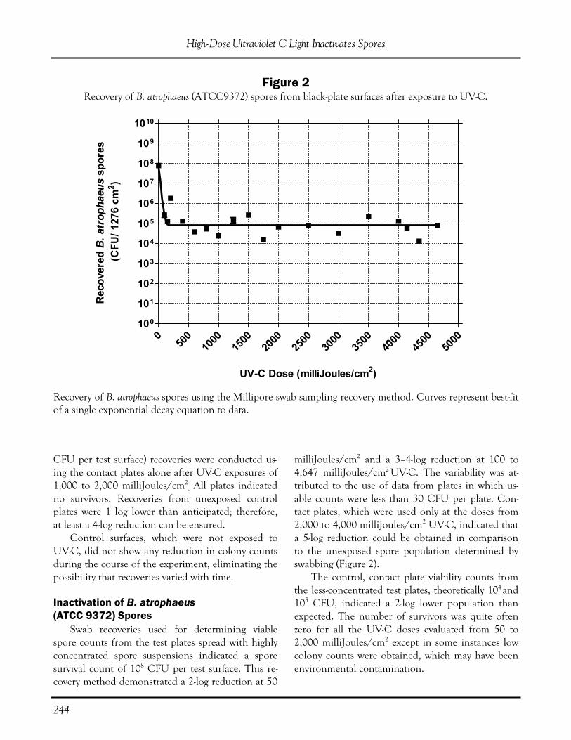
244
CFU per test surface) recoveries were conducted us-ing the contact plates alone after UV-C exposures of 1,000 to 2,000 milliJoules/cm2
. All plates indicated no survivors. Recoveries from unexposed control plates were 1 log lower than anticipated; therefore, at least a 4-log reduction can be ensured. Control surfaces, which were not exposed to UV-C, did not show any reduction in colony counts during the course of the experiment, eliminating the possibility that recoveries varied with time.
Inactivation of B. atrophaeus (ATCC 9372) Spores Swab recoveries used for determining viable spore counts from the test plates spread with highly concentrated spore suspensions indicated a spore survival count of 108 CFU per test surface. This re-covery method demonstrated a 2-log reduction at 50
milliJoules/cm2 and a 3–4-log reduction at 100 to 4,647 milliJoules/cm2 UV-C. The variability was at-tributed to the use of data from plates in which us-able counts were less than 30 CFU per plate. Con-tact plates, which were used only at the doses from 2,000 to 4,000 milliJoules/cm2 UV-C, indicated that a 5-log reduction could be obtained in comparison to the unexposed spore population determined by swabbing (Figure 2). The control, contact plate viability counts from the less-concentrated test plates, theoretically 104 and105 CFU, indicated a 2-log lower population than expected. The number of survivors was quite often zero for all the UV-C doses evaluated from 50 to 2,000 milliJoules/cm2 except in some instances low colony counts were obtained, which may have been environmental contamination.
High-Dose Ultraviolet C Light Inactivates Spores
050
010
0015
0020
0025
0030
0035
0040
0045
0050
00100
101
102
103
104
105
106
107
108
109
1010
UV-C Dose (milliJoules/cm2)
Rec
over
edB
. atr
opha
eus
spor
es (C
FU/ 1
276
cm2 )
Figure 2 Recovery of B. atrophaeus (ATCC9372) spores from black-plate surfaces after exposure to UV-C.
Recovery of B. atrophaeus spores using the Millipore swab sampling recovery method. Curves represent best-fit of a single exponential decay equation to data.

245
B. anthracis Sterne Spores Millipore swab recoveries from nonirradiated test surfaces indicated 3.6 x 10⁸ CFU. Swab recover-ies from UV-C doses of 150 and 1,000 milli-Joules/cm2 resulted in a three and four log reduc-tion respectively. Contact plates used for recoveries on the test surface exposed to UV-C doses of 2,000 to 4,000 milliJoules/cm2 demonstrated a 4 log reduc-tion (Figure 3). Test surfaces spread with the diluted spore in-oculum had 104 CFU per test surface and showed total kill when exposed to 50 to 1,000 milli-Joules/cm2.
Inactivation of Dry Spore Powder The last evaluation used dry, free-flowing B. atro-phaeus spore powder in open Petri plates, with spore counts of 108 to 109 CFU per plates. A 1-log reduc-
tion was obtained from duplicated plates after expo-sures of 10,000 or 16,000 milliJoules/cm2 (Figure 4).
Discussion
This investigation has demonstrated that UV-C generated by the UVAS in the absence of visible par-ticulate matter can be delivered at lethal doses on nonreflective, non-porous surfaces for partial spore reduction even when the contamination levels are extremely high and for total spore reduction in the presence of less-concentrated spore populations. A 3-5-log reduction can be assured following UV-C expo-sure to contamination levels simulating those used in bioterrorist weapons—108 to109 CFU/1276 cm2 or 105 to 106 CFU/cm2—using doses of one hundred to several thousand milliJoules/cm2 (Figures 1, 2, and 3). In situations where the bioburden levels are more
M. U. Owens, et al.
050
010
0015
0020
0025
0030
0035
0040
0045
0050
00100
101
102
103
104
105
106
107
108
109
1010
UV-C Dose (milliJoules/cm2)
Rec
over
edB
. ant
hrac
is s
pore
s (C
FU/ 1
276
cm2 )
Figure 3 Recovery of B. anthracis Sterne spores from black plate surfaces after exposure to UV-C.
Recovery of B. anthracis Sterne spores using the Millipore swab sampling recovery method. Curves represent best-fit of a single exponential decay equation to data.

246
representative of the contamination in areas such as operating/emergency rooms ( 102 CFU/cm2), UV-C is capable of completely deactivating the entire popu-lation at lower doses, most likely less than 100 milli-Joules/cm2. Test surfaces contaminated with high numbers of spores that are subsequently spread across the surface area as may occur during preclean-ing were readily decontaminated or sterilized with adequate doses of UV-C. Spores at contamination levels of 105 to 106
CFU/cm2 and applied in powder dense enough to be visible to the naked eye indicated a 1-log reduc-tion after UV-C doses of 1,000 to 16,000 milli-Joules/cm2 (Figure 4). These findings emphasize the need for precleaning contaminated surfaces soiled with gross material. Use of a precleaning step, such
as HEPA-vacuuming or damp wiping, for heavily contaminated surfaces in the presence of visible soil, followed by UV-C exposure, should effectively de-contaminate the area or surface. This is substanti-ated by the data in which total kill was demonstrated from surfaces contaminated with less-concentrated spore suspensions in the absence of visible powder. The presence of 1%-2% silica does not impede the germicidal effect of UV-C since the lethality was similar to that observed in the absence of silica. This finding demonstrates that the efficacy of UV-C is not altered in the presence of low concentrations of particulate matter or soil that could be present after precleaning. Prior studies in animal laboratory settings using UV light showed a significant reduction of bacterial
High-Dose Ultraviolet C Light Inactivates Spores
The curve represents the best-fit of a single exponential decay equation to the data.
020
0040
0060
0080
0010
000
1200
014
000
1600
018
000
2000
0100
101
102
103
104
105
106
107
108
109
1010
UV-C Dose (milliJoules/cm2)
Rec
over
edB
. atr
opha
eus
spor
es (C
FU/ 9
0 m
m P
etri
pla
te)
Figure 4 Recovery of a visible layer of dry B. atrophaeus spores containing
~1% silica on Petri plates after exposure to UV-C.
247
loads, and the addition of a chemical disinfectant followed by UV-C treatment was “particularly suc-cessful, reducing bacterial loads to extremely low lev-els” (Dix et al., 1992). In this study where the organ-isms were spread on the test surface without deactiva-tion or removal by any cleaning agent, a significant spore reduction was observed after UV-C irradiation. A recent study by Nicholson and Galeano (2003) found “the data indicate that standard UV treat-ments that are effective against B. subtilis spores are likely also sufficient to inactivate B. anthracis spores, and spores of standard B. subtilis strains could relia-bly be used as a biodosimetry model for the UV inac-tivation of B. anthracis spores” Our investigations have confirmed this finding. Previous experience with the UVAS device sug-gested smooth materials that reflected UV-C may be more readily decontaminated than rough, nonreflec-tive materials. Data obtained through this investiga-tion will be useful in planning surface decontamina-tion for many environmental applications, since de-termining the required decontamination doses should be based upon information obtained using the least reflective test surfaces, thus avoiding under-exposure. From these experimental findings one may rea-sonably conclude that the UVAS device, or other UV-C generating devices, could decontaminate areas in which surface contamination was 102 CFU/cm2
and could be used to decontaminate extremely con-centrated surfaces as long as a precleaning step was instituted.
Author’s Note The B. anthracis Sterne spores were washed twice in deionized sterile water and examined by phase-contrast microscopy to confirm that the refractile spore suspension was free of vegetative cells.
References
Banrud, H., & Moan, J. (1999). [Use of short wave ultraviolet radiation for disinfection in operating rooms]. Tidsskr Nor Laegeforen, 119, 2670-2673.
Dix, J., Nolan, G., Schlick, H., & Elliot, H. B. (1992). The efficiency of ultraviolet irradiation and chemical disinfection as a means of reducing bacte-rial contamination of animal rooms. Fifth FELASA Symposium: Welfare and Science.
Druce, J. D., Jardine, D., Locarnini, S. A., & Birch, C. J. (1995). Susceptibility of HIV to inactivation by disinfectants and ultraviolet light. Journal of Hospital Infection, 30, 167-180.
Elliott, T. B., Brook, I., Harding, R. A., Bouhaouala, S. S., Shoemaker, M. O., Knudson, G. B. (2002). Antimicrobial therapy for Bacillus anthracis-induced polymicrobial infection in 60Co gamma-irradiated mice. Antimicrobial Agents and Chemotherapy, 46,3463-3471.
Inamoto, H., Ino, Y., Jinnouchi, M., Sata, K., Wada, T., Inamoto, N., & Osawa, A. (1979). Dialyzing room disinfection with ultra-violet irradiation. Jour-nal of Dialysis, 3, 191-205.
Knudson, G. B. (1985). Photoreactivation of UV-irradiated Legionella pneumophila and other Legionella species. Applied Environmental Microbiology, 49, 975-980.
Knudson, G. B. (1986). Photoreactivation of ultra-violet-irradiated plasmid-bearing and plasmid-free strains of Bacillus anthracis. Applied. Environmental Microbiology, 52, 444-449.
Nicholson, W. L., & Galeano, B. (2003). UV resis-tance of Bacillus anthracis spores revisited: Valida-tion of Bacillus subtilis spores as UV surrogates for spores of B. anthracis Sterne. Appied Environmental Microbiology, 69, 1327-1330.
Rehork, B., Martiny, H., Weist, K., & Ruden, H. (1990). [Performing surface and room disinfection in the hospital]. Offentl Gesundheitswes, 52, 36-45.
M. U. Owens, et al.

248
Applied Biosafety, 10(4) pp. 248-252 © ABSA 2005
Article
Abstract
Potentially infectious wastes, if not properly treated, could expose both humans and the environment to untreated microbes and toxins, and create a potential for illness. Therefore, all pathogenic materials used in research must be destroyed by heat or chemical treat-ment prior to disposal as biomedical waste. Addition-ally, bacterial waste from nonpathogenic bacteria car-rying antibiotic resistance must be treated before place-ment in the waste stream to avoid transmission of the antibiotic-resistant trait. In biomedical laboratories, glassware, beakers, test tubes, and other contaminated research material are usually treated by autoclaving be-fore placing them into the waste stream or recycling for continued use in the laboratory.
Introduction
Florida Administrative Code (F.A.C.) Chapter 64E-16, under the Biomedical Waste Code, requires an infectious waste treatment facility to maintain records of temperature and dwell time when wastes are rendered noninfectious by gas or steam sterilization. The F.A.C. further requires biological culture testing to assure proper sterilization of all autoclaved materials. Since this University is not a treatment facility but a generator facility, the University is not required to follow these records and testing requirements. However, in the best interest of the University’s environmental and research goals, the Department of Environmental Health & Safety (EH&S) is voluntarily complying with the sterilization aspect of the F.A.C. As a service, EH&S conducts annual sterilization testing for all departmental autoclave steam cycles
used for biological waste and materials decontamina-tion. This testing is to ensure that the autoclave cy-cles used are sterilizing all microbial waste.
Background on Steam Sterilization Testing
Researchers who work with potentially infec-tious materials are at a higher risk of exposure, espe-cially when exposed to untreated infectious waste. Infections may be transmitted through several differ-ent routes, including direct contact with untreated infectious waste, indirect contact with contaminated instruments or environmental surfaces, or inhalation of airborne contaminants. Infection via any of these routes requires that all five of the following condi-tions be present to form “the chain of infection”: 1. A pathogen with sufficient infectivity and num-bers to cause infection 2. A reservoir or source that allows the pathogen to survive and multiply 3. A mode of transmission from the source to the host4. A portal through which the pathogen may enter the host 5. A susceptible host (CDC, 2003) Effective control measures are intended to break one or more of these “links” in the chain, thereby reducing the risk of, or completely preventing, expo-sure to potentially infectious materials. A key compo-nent in this link-breaking process is the proper use of steam sterilizers (autoclaves), which serve as an essential step toward eliminating the “pathogen links” in the chain of infection (DePaola, 2003; Quality America, Inc., 2001). The Occupational Safety and Health Association
Richard N. Le, Amy L. Hicks, and Janice Dodge
Florida State University, Tallahassee, Florida
Autoclave Testing in a University Setting
(OSHA) relies on guidelines published by the Cen-ters for Disease Control and Prevention (CDC) as a widely recognized and accepted standard to be fol-lowed by employers in carrying out their responsibili-ties under the Occupational Safety and Health Act. The CDC and OSHA recommend the use of Bio-logical Indicators (BI) for monitoring steam steriliza-tion cycles in autoclaves. The CDC states [for medi-cal autoclaves]...“proper functioning of sterilization cycles should be verified by the periodic use (at least weekly) of biological indicators (i.e., spore tests). Heat-sensitive chemical indicators (e.g., those that change color after exposure to heat) alone do not ensure the adequacy of sterilization cy-cle…” (CDC, 2004). In addition, F.A.C. 64E-16.007 states that a treatment facility that disposes of biomedical waste must utilize steam sterilization, incineration, or an alternative process approved by the Department of Health prior to disposal. Treatment shall occur within 30 days of collection from the generator. Ad-ditionally, steam treatment facility units should be evaluated for effectiveness with spores of Geobacillus stearothermophiluss at least once every 7 days for permitted treatment facilities, or once every 40 hours of opera-tion for generators that treat their own biomedical waste. Sterilizer manufacturers also recognize the im-portance of routine testing of sterilizers and auto-claves. They recommend that a biological spore test indicator be used weekly in a representative load for sterilization assurance (Ritter; Tilton et al., 2004).
Background on PROSPORE2 Biological Indicators
PROSPORE2 (HealthLink) is a self-contained biological indicator for validating and monitoring steam sterilization of solids. It consists of a paper disc carrier containing Geobacillus stearothermophilus (ATCC 7953) spores. The disc is enclosed in a plastic tube along with a glass vial containing growth media for the bacterial spores. Bromocresol purple has been added as a pH indicator to detect spore growth. The outgrowth of spores decreases pH, causing a color change from purple to yellow following a 24-hour incubation period.
A PROSPORE2 biological indicator is placed inside the autoclave and a specific cycle is selected. When the cycle is complete, the PROSPORE2 indi-cator is sealed by firmly depressing the cap. The glass ampoule of media is crushed inoculating the Geoba-cillus stearothermophilus disc. Then the indicator is incubated at 55o-60oC for a 24-hour time period along with an untreated PROSPORE2 indicator that serves as a control to ensure spore viability. Once the minimum incubation time of 24 hours has been achieved, both the control and autoclaved PROSPORE2 indicator are examined. If the auto-claved PROSPORE2 indicator retains its purple color, then the sterilization cycle is adequate and the result is recorded as passing. A failed sterilization cycle is indicated by turbidity or a change in color toward yellow, indicating spore growth due to a change in pH. The control indicator should exhibit turbidity and/or a color change to yellow after incu-bation to ensure the viability of the biological indica-tors. If the control indicator does not show signs of growth, the test is considered invalid and all the re-sults are unacceptable and not considered.
Evaluation of Autoclaves
PROSPORE2 biological indicators were used to test the steam sterilization cycles of several depart-mental autoclaves. A total of three runs were con-ducted for each autoclave. For each autoclave, a test trial run using a PROSPORE2 biological indicator was run to see if the autoclaves were capable of ster-ilization without any load. The biological indicator in the test trial was placed in a horizontal position, as recommended by the manufacturer, in various locations within the autoclave chamber. This was done to verify the effectiveness of steam sterilization in different areas within the autoclave. Following the test trial run, two additional runs were performed in which PROSPORE2 biological indicators were placed in a representative load or challenge load to be autoclaved. The challenge load test consisted of placing a PROSPORE2 biological indicator in the center of an autoclave bag contain-ing empty plastic pipette tip boxes. This bag was left partially open to allow for steam penetration and sterilization.
R. N. Le, et al.

250
For the results, a “PASS” indicated that the PROSPORE2 biological indicator retained its purple color signifying an adequate sterilization cycle. A “FAIL” resulted in the PROSPORE2 biological indi-cator having turbidity or a color change toward yellow for that sterilization cycle. Untreated control PROSPORE2 biological indicators were incubated with each autoclaved ran PROSPORE2 biological indicator. The following results were compiled over a 3-month testing period for several departmental auto-claves. These results are for the challenge load test-ing only. Challenges were performed using the tem-peratures preset by the research groups using the autoclaves.
Test Findings
Each test trial run resulted in a passing test for every autoclave. Based on this result, it was con-cluded that the autoclave was functioning properly.
An adequate amount of steam and heat were pro-duced to sterilize any unobstructed contents such as glassware. However, problems occurred with the challenge load testing. Almost every challenge load test failed to sterilize the biological indicator. There-fore, the testing procedure was modified in an effort to sterilize the enclosed biological indicator. The challenge load volume was reduced by approximately half its original volume and the bag’s opening was increased. In addition, approximately 250 mL of water was added directly to the autoclave bag to gen-erate additional steam within the bag to achieve ster-ilization. Biological indicators were also given to two sepa-rate departments to autoclave with their normal bio-hazardous waste load. One was autoclaved for 15 minutes at 121°C gravity cycle and the other load was autoclaved at 121°C for 35 minutes on a liquid setting. Since the biological indicators were placed in actual bags of biohazardous waste, these tests were true representatives of what laboratories were gener-
Autoclave Testing in a University Setting
Autoclave Cycle Pass Fail
Amsco 3021-S-A (Amsco)
G15/D30 L60
Amsco 3041 (Amsco)
All settings (Liquid and Gravity)Addition of water to bag
Consolidated (Consolidated)
G60/D15 G30/D15
Primus Autoclaves-A (Primus)
G20/D15 (Room 511) G15/D1 (Room 312)
Amsco 3021-S-B All liquid cycles
Amsco Scientific (Amsco)
G30/D30 (132°C) L30 L15
Amsco 3021-S-C (Amsco)
G15/D30 (132°C) L30 L15
Primus Autoclave(B) G15
Steris, Stage 1 (Amsco)
G30/D15 G30/D15 (addition of water)
Table 1
(“L”=liquid cycle, “G”=gravity cycle; and “D”=Dry time. Unless otherwise indicated, the standard 121°C and 15 psi were used at each autoclave)

251
ating. Unfortunately, both of these tests failed. Based on the information in the preceding table, it was evident that the results varied greatly with each autoclave due to the type of autoclave and dif-ferences in cycle times and temperatures. However, it can be shown that the shorter-timed cycles failed consistently for each challenge load tested. The ex-ceptions to this were the results generated from the Amsco 3041 autoclave and the Amsco Scientific autoclave. For the Amsco 3041 autoclave, the 250 mL of water added to the autoclave bag helped with the sterilization of the materials, resulting in a pass-ing test for the challenge load. The Amsco Scientific autoclave was able to sterilize the PROSPORE2 bio-logical indicator using a 30-minute cycle because of the higher temperature setting. However, the bags used for this autoclave were not compatible with the higher temperature, resulting in complete deteriora-tion of the bags after autoclaving.
Additional Findings
It was observed that not all autoclaves had clear instructions available detailing proper usage and that no user log was present to record the users, cycle times, settings, and autoclave contents. In addition, it was unknown if the temperature sensors on each autoclave were properly calibrated due to inadequate record keeping concerning general maintenance on several autoclaves. However, each autoclave chamber temperature was verified during the runs using a high-low thermometer that was placed in the auto-clave with each challenge load run. There was good consistency between the high-low thermometer tem-perature readings and the temperature indicated on the autoclaves. In addition, no records were kept to indicate when general or safety maintenance was last con-ducted on several autoclaves. Maintenance should be done yearly or as recommended by the autoclave manufacturer.
Recommendations
In keeping with the CDC and OSHA recom-mendations and using F.A.C. 64E-16.007 as a guide-
line, EH&S will use biological indicators for validat-ing the steam sterilization cycles on an annual basis. EH&S recommends that the following adjustments to policies and practices be implemented in order to better achieve sterilization and ensure proper main-tenance for each autoclave:
1. Readjustment of Cycle Times The standard 121°C, 15-psi, and 15-minute dwell time are adequate for sterilization of clean items or smaller loads, but were found to be inade-quate for large bulky loads of biohazardous waste. For this type of waste, it is recommended that the sterilization time be increased to a minimum of 60 minutes, while still maintaining the standard tem-perature and pressure. This setting has proven effec-tive for sterilizing larger loads. The addition of water is not recommended because results were not consis-tent enough for this to be considered an effective option. It is also recommended not to leave the auto-clave bag open for actual autoclaving of materials even though this procedure was used in the test pro-cedure. The bag should be secured loosely with a rubberband or closed loosely with a small opening (at least 1 inch in diameter). This will minimize an individual’s risk of becoming exposed to hot materi-als within the bag. It is suggested that a “kill cycle” be set on each autoclave intended specifically for sterilization of contaminated dry waste loads. The Amsco Scientific autoclave did yield a pass-ing result due to a higher temperature setting. How-ever, the autoclave bags used could not withstand the higher temperature and the bags deteriorated during the procedure. This is a safety concern be-cause the contents within the bag could spill onto the user exposing him or her to the extremely hot contents. To use this autoclave at the higher tem-perature settings, it is important to use an autoclave bag that is compatible with the 132°C temperature. In addition, the autoclaves should be retested using the new autoclave bags.
2. Proper Autoclave Use The autoclave bag should never be over-packed or sealed too tightly. It is also important to make sure that the contents of the autoclave bag are kept at a minimum. An over-packed bag will insulate mi-
R. N. Le, et al.
252
crobes at the center of the bag compromising effec-tive sterilization for the core contents (Churchill, 2003). It is the responsibility of each department to en-sure that all users understand and know how to use the autoclave correctly. EH&S recommends that proper personal protective equipment be worn when unloading materials from the autoclave. Any ex-posed skin should be covered when reaching into the autoclave after operation to prevent burns. Heat-resistant, elbow-length gloves and other personal protective equipment are necessary to prevent burns from occurring when removing hot items. In addi-tion, a shallow pan or container should be used when autoclaving bags of biohazardous waste to pre-vent potential spills of this material onto the user.
3. Record Keeping The use of an autoclave logbook is recom-mended for each autoclave. Prior to autoclaving any items, users must fill in all information requested in the autoclave logbook. The logbook should be lo-cated adjacent to the autoclave and be maintained by the department. Information that should be part of the logbook includes: user’s name, cycle time, cy-cle setting, materials being autoclaved, contact num-ber, time in, time out, and verification results.
4. Safety Maintenance General maintenance should be conducted on an annual basis or as recommended by the manufac-turer. Specifically, the safety valve should be checked and replaced as required. This information should be posted on the autoclave or maintained in a main-tenance logbook for easy reference. In addition, a quick check of the autoclave should be conducted prior to each use to ensure that all parts are properly functioning (e.g., door closes and is sealed properly, the rack is in place, correct settings are being used, the interior is clean, etc.). Autoclaves should be cleaned after every use and the work area should be disinfected as needed. Appropriate cleaning proto-cols should be obtained from each manufacturer.
5. Training Each autoclave user should be trained on the proper use of the autoclave. The cycle settings should be posted next to the autoclave to inform each user of the types of settings available, such as gravity and liquid cycles, the temperatures for each cycle, and run times. A written sterilization proce-dure should be kept near each autoclave and a stan-dard operating procedure should be developed. It should include the appropriate sterilization times for liquid and dry waste goods, identification of stan-dard treatment containers and proper load place-ment procedures, personal protection equipment required for removing materials from the autoclave, instructions for loading and unloading the auto-clave, and instructions on cleaning and maintaining the autoclave.
References
Centers for Disease Control and Prevention. (2003). Guidelines for infection control in dental healthcare settings-2003. MMWR, December 19/52(RR17), 1-98.
DePaola, L. G. (2003). Infection control and the prevention of disease transmission in the dental of-fice: Back to the basics. The Infection Control FO-RUM, 1(2).
HealthLink. Prospore and Prosure Technical Bulletin 1. Frequency of Use for Biological Indicators.
Quality America, Inc. (2001). Break the chain of in-fection. OSHA Watch, January/February, 3(1).
Ritter©—Installation and Operation Manual M9/M11 UltraClaveTM Steam Sterilizer. Sterilizer Manu-facture Monitoring Recommendations.
Tilton, G., & Kauffman, M. (2004). Sterilization: A review of the basics. STERIS Corporation white pa-pers, document number M2712EN. www.steris. com/resources/resources_wp.cfm#. June.
Autoclave Testing in a University Setting

253
The Viral Immunology Center at Georgia State University (GSU) is recognized as the national re-source facility for research related to the early diag-nosis and effective treatment of dangerous viral dis-eases, most notably B virus (Cercopithecine herpesvi-rus 1), herpesviridae. Funded by the National Insti-tutes of Health, the Center operates the National B Virus Resource Laboratory aimed at learning more about the deadly alpha herpes virus and preventing its transmission to laboratory workers. The GSU Center, located in downtown Atlanta, was the first BSL-4 facility in the country operating in a university setting. Under the direction of Julia Hil-liard, the goal of the laboratory’s research is not only to diagnose viral diseases, but also to study the agents and use their infrastructure to design vaccines. The B Virus laboratory had been located at the Southwest Foundation for Biomedical Research in San Antonio, Texas, and operations were proceeding well in the BSL-4 environment. However, space con-straints limited the size of the staff to 10 people and restricted the amount of work that could be done. Research officials at Georgia State University called upon Hilliard to move her facility to down-town Atlanta in 1997, noting the University’s prox-imity to Emory University, the Centers for Disease Control, and the Medical College of Georgia. The retrofitted lab at the University offers three times the amount of space that Hilliard was using at the Texas facility. “The thrill of working in a rich milieu and hav-ing a new facility was enough to engage my interest
in moving the laboratory,” recalls Hilliard. “We now know what it’s like to build a BSL-4 laboratory in a highly concentrated downtown area. We were told this would not be a big problem because the residents were already aware that maximum-containment laboratories were operating in the city.” Maximum-containment BSL-3 and -4 suites are necessary due to the nature of the alpha herpes vi-rus, or B Virus, which is transmitted by the Macaca species of monkeys often used in biomedical re-search. Although there is a low frequency of infec-tion among people, the virus is considered a severe occupational hazard for laboratory workers as a re-sult of its lethal consequences if not diagnosed quickly after infection. “The virus causes death in 80 percent of the in-fected individuals (after it literally attacks the cervical spinal cord),” says Hilliard. “There have been five fatalities in the last 12 years at four different institu-tions in four states. The survivors are our first intro-duction to what happens when you put a pathogen like B virus into the human population. The reason we work with this agent is because we know that rapid identification means we can successfully treat the individuals.” The cost of moving to Atlanta was approximately $200,000, which included expenses for shipping the laboratory equipment, moving staff, and flying back and forth between the new lab and the Texas lab to verify that the performance standards of the new facility matched those of the well established lab. The cost to renovate the space to accommodate the
Applied Biosafety, 10(4) pp. 253-257 © ABSA 2005
Article
Tradeline Publications
Orinda, California
Operating a BSL-4 Laboratory in a University Setting: Georgia State University Lab Studies Deadly Alpha Herpes Virus

254
Center was approximately $1 million, not including the daily maintenance costs of the hot lab. Both laboratories had to be operated concurrently in or-der to preserve the integrity of the testing until Hil-liard’s team determined that the new Center could function efficiently on its own. The architectural firm of Lord, Aeck and Sar-gent designed the first phase of the Center, while construction of the BSL-4 lab was supervised by the architectural firm of HOK in cooperation with The Baker Co., which customized the 32-linear-foot glovebox cabinet.
Designing a BSL-4 Laboratory
The Viral Immunology Center, housed in the University’s six-floor, 500,000-sf natural sciences building, features a departmental animal facility in the basement and BSL-3 and -4 laboratories on the third floor, including a specialized BSL-4 for small animal models of disease. The Center, which occu-pies about 5,000 sf and employs 30 people, also in-cludes two support BSL-2 laboratories, a biochemis-try lab, robotics facility, offices, a fermentation lab, and shared departmental resources, including elec-tron and confocal microscopy areas, a sequencing core, and a FACS-analysis core for cell sorting. The activities that take place in the Natural Sci-ence Center are of particular interest when design-ing a BSL-4 laboratory in a university setting. This building typically houses the departments of biology, chemistry, biochemistry, physics, and anatomy in addition to the BSL-4 lab. Research activities that are not part of the viral laboratories must also be taken into consideration when deciding where to locate the BSL-4 suites. Other activities to consider include teaching sessions and seminars. Providing access to the laboratories in a manner that protects visitors is critical to the efficient opera-tion of the facility. Serious consideration is given to the type of barriers that are erected between com-mon access hallways and restricted areas to ensure that lay visitors and research supporters can view the work taking place without being exposed to poten-tial risks. Access is provided via keypad entry for stu-dents, faculty from other disciplines, visiting scien-tists, security and maintenance personnel, and inves-
tigators. The BSL-3 and -4 laboratories have a mag-netic card access so that entry and exit into the facili-ties can be tracked by a computer. With the addi-tional restrictions imposed by the new Homeland Security Program, all individuals who work within the BSL-3 and -4 labs must first have federal security clearance after submission of extensive documenta-tion to the FBI. The facilities must also meet Clini-cal Laboratory Improvement Amendments and Se-lect Agent inspection requirements to carry out the research and diagnostic missions of the laboratory.
Key Components of a Maximum- Containment Facility
Each day Hilliard’s facility receives up to 20 boxes from all over the world from individuals who may have been exposed to the secretions of a Macaca monkey. Minimizing the transport of materials going to high-containment areas is critical. When the sam-ples arrive, they are triaged to determine what tests must be done so the results can be sent to the con-cerned individuals as quickly as possible. The packages, transported through an isolated hallway that is sectioned off from other areas of the facility, are opened in the Level 2 unpackaging room, attached to the BSL-3 and -4 suites. Each of these areas can be accessed only by computer-controlled keypad entry. Any materials that leave the BSL-4 facility undergo an intensive search for evi-dence of any live virus, using virus plaque assays to verify that all virus has been inactivated. If the assay is negative, the chain of custody for removal begins. Items that leave the BSL-4 laboratory must go through two sets of autoclaves or two sets of cross-linked glutaraldehyde dunk baths. Dunk baths en-able chemical sterilization of the outside wall of sub-mersed containers. This allows a live virus to be re-moved from the cabinet for ultra-low freezing and storage within the BSL-4 and passage of inactivated materials to the Level 2 labs. Live virus never leave the suite. The disposal system consists of two redundant 150-gallon tanks that are released into a boiler-decontamination unit. A chilling mechanism cools the water, which is released into the public system
Operating a BSL-4 Laboratory in a University Setting
255
once it is steam disinfected and verified as inacti-vated. A drain is situated under the tanks to contain leakage, should that occur. The specialized HVAC unit dedicated to the independent air system of the BSL-3 and -4 suites is located on the roof of the Natural Sciences Center, while the decontamination facility is positioned un-der the BSL-4 laboratory. Computers are used to analyze temperatures, airflow, and the rooftop ex-haust systems, which are actively engaged. In case of power failures, a natural gas backup system is in place to maintain air purification, negative pressure, and equipment power. It is critical to be able to monitor the tempera-ture and air supply for each of the high-containment suites by computer on site or from remote locations by the extensively trained and certified engineer re-quired to maintain the facility. The outer hallways are kept at negative pressure assessed by magnahel-ics, and with each step further into the biocontain-ment areas the negative pressure gradient increases according to the specifications outlined in the CDC’s Biosafety in Microbiological and Biomedical Laboratories. “The goal of the BSL-3 rooms is to isolate viruses from small samples. In order to process the samples, pull out the virus, and identify unusual agents, we have to have incubators that are fed five percent car-bon dioxide and 95 percent air,” explains Hilliard. “We can’t bring tanks in and out of these rooms, so we have a separate alcove which houses the gas tanks feeding the incubators in the BSL-3 and BSL-4 labs.” It is challenging to keep the BSL-3 laboratories stocked and tidy at the same time, keeping in mind that many of the items that come into the labs are not going to leave until they are thoroughly melted down by repeated autoclaving. Disabled equipment cannot be removed from the laboratory for mainte-nance or repair. Tools for minor repairs are decon-taminated with gas sterilization before they are re-moved from the maximum-containment suites.
Keeping the Scientists Safe
Necessary precautions are taken to protect the researchers while they are working in the laborato-ries. Scientists usually work in a cabinet or a suit lab,
which relies on the standard biocontainment gear, in the BSL-4 facility. Working in a cabinet lab is ex-tremely challenging, but considered safer than a suit lab because the individual uses large glove ports and is separated from the pathogen by thick stainless steel. The cabinet lab at the Center is 32-feet long and can accommodate three or four researchers at a time. A nearby animal section houses mice, rats, and rabbits, and a screen built into the cabinet allows investigators to visualize materials analyzed by an inverted, phase-contrast microscope. Working in the cabinet lab can sometimes lead to fatigue because scientists must do all of their work using arm-length gloves to maneuver materials within the stainless steel cabinet while working in a line with other researchers. Working in the suit lab allows scientists to use the traditional lab layout and to move around more freely. However, breathing the positive-pressure air in the suit can also cause fa-tigue. Barrier protection masks are worn at all times in the BSL-3 and -4 labs. Only 10 people on Hilliard’s staff are trained to work in the BSL-3 lab, while only five of these can enter the BSL-4 facility. “The masks are basically a reminder for us not to put our double-gloved hands up to our faces. If we relied on them as filtration or personal protection devices, we would go to something different,” says Hilliard. “All agents are handled in a Class 2 cabinet in the BSL-3, so there is no time at which an agent is uncapped in the room surrounding the cabinet.”
Important Considerations
Careful consideration must be given to the in-stallation of all systems and how they will interact. The HVAC system at GSU had been repaired at an annual cost of $12,000 every year since Hilliard’s lab moved into the facility. A new HVAC system specifi-cally engineered after analysis of previous failures was installed at a cost of $250,000 and 2003 marked its first year of operation. “This is an existing building that we modified. It wasn’t meant to be under constant negative pressure and that has caused substantial stress on the walls and ceiling,” says Hilliard. “Careful consideration of the systems that are linked is very important. We are
Tradeline Publications

256
right in the middle of downtown on a rooftop so there are many issues to address.” Cabinet labs should be designed so they are er-gonomically comfortable for researchers of varying physical stature. The cabinet labs at the Center were designed largely with the input of the former associ-ate director, who is more than 6-feet tall, creating a challenge for other lab staff to do their work. Smaller gloves, extenders, and a few other modifica-tions enable the other investigators to work relatively comfortably. It is also crucial to properly position the labora-tories in a manner that is most conducive to the type of work being done. For example, a West Nile BSL-3 is located adjacent to the robotics facilities that ser-vice the sera and viral culture laboratories. The BSL-2 labs have standard virology for lower containment agents like HSV-1 and HSV-2. The sum total of the work performed in these labs is to take core resources of the BSL-3 and -4 labs, com-bine them with the research missions, maintain a high-containment robotics lab for high-throughput virus identification, as well as serologic screening, perform microarray analysis, and be flexible enough to look at new agents as they come to the scientists’ attention. “All of our work would be for naught if we didn’t have the supportive BSL-2 labs,” says Hilliard. The robotics unit can perform multiple tests and process thousands of sera daily. The viral culture robot can plate samples and screen them at multiple times during the day for the presence of a virus. This allows mass processing of thousands of diagnostic samples. If a disturbance in cell culture is noted, a pager is sounded and an investigator is called to the room for further analyses and to notify the institu-tion submitting the sample for analysis. “In the BSL-4 laboratory, the goal of our work is the production of large amounts of virus and con-taining the supply very securely,” says Hilliard. “We can then look at drug sensitivities and how the virus
behaves in cells in order to understand the pathogen and how to control it.”
Biography
Julia Hilliard is director of the Viral Immunol-ogy Center at Georgia State University in the De-partment of Biology. She is also the Georgia Re-search Alliance Eminent Scholar in Molecular Bio-technology and Director of the National B Virus Resource Center for global diagnostic resources. She has worked with the BSL-4, newly classified Select Agent, B Virus (Cercopithecine herpesvirus 1) for 23 years, providing diagnostic resources to the biomedi-cal community for the last 16 years. This report is based on a presentation given by Julia Hilliard at Tradeline’s International Conference on Biocontainment Facilities in May 2003. For more information, please contact: Julia Hil-liard, Director, Viral Immunology Center, Georgia State University, Box 4118, Atlanta, GA 30302-4118, 404-651-0811, [email protected]. Reprinted with Permission © December 2003 from TradelineInc.com, a registered product of Tradeline Inc., a provider of leading-edge resources to facilities planning and management through conferences, publications, and the Internet community. Visit www.TradelineInc.com for more information.
Resources
Biosafety in Microbiological and Biomedical Laboratories: www.cdc.gov/od/ohs/biosfty/bmbl4/bmbl4toc.htm Clinical Laboratory Improvement Amendments:
www.cms.hhs.gov/clia/ Select Agents:
www.cdc.gov/od/sap/docs/salist.pdf Viral Immunology Center:
www.gsu.edu/~wwwvir/index.html
Operating a BSL-4 Laboratory in a University Setting

257
Tradeline Publications
Figure 1 The entry area for the maximum-containment labs includes a pass-through shower. Scientists
prepare for their work by donning either a suit lab or working in the cabinet lab.
(Photo courtesy of Julia Hilliard.)
Figure 2 Materials that leave the BSL-4 lab go through
two sets of autoclaves or two sets of cross-linked glutaraldehyde dunk baths. A decontamination facility is situated under the lab. The disposal system consists of two redundant 150-gallon
tanks released into a boiler-decontamination unit. (Photo courtesy of Julia Hilliard.)
Figure 3 When pathogen samples arrive, the packages are transported through an isolated hallway
that is cordoned off from other areas of the facility. They are opened in a Level 2 unpackaging room, which is attached to the BSL-3 and -4 labs. (Photo courtesy of Julia Hilliard.)

258
Special Feature
Abstract
Biohazard training and compliance of husbandry and research staff are often complicated at the Univer-sity of Arizona by the sporadic nature of the work within the BSL-2/BSL-3 suite. Projects may last for a month and then not be repeated for a significant period of time. Failure to adhere to approved safety measures and techniques for specific rooms/projects over time increased the need for clear standard operating proce-dures (SOPs) that were easily accessed and utilized. SOPs available only in the employee lounge are of little use when a question arises while changing cages within a BSL-2 animal room. To this end, new SOP formats and display locations were developed and instituted to increase awareness and comprehension of techniques used within the BSL-2/BSL-3 suite. Changes included the placement of plain text as well as picture-enhanced SOPs in common areas such as the employee lounge and entrance to the biohazard suite, and within the ani-mal rooms. The introduction of SOPs containing color photographs has been instrumental in increasing compli-ance of in-room procedures by both native English speakers and those who do not speak English as a first language. Overall, the use of multiple styles of SOPs has been a huge success. This process has helped to reduce confusion and increase compliance by both the husbandry and research staffs working within the Uni-versity Animal Care facilities.
Introduction
In 2002, a review of University Animal Care (UAC) husbandry and research staff procedures within the biohazard suite at the Central Animal Facility identified several areas of concern. Even though SOPs formalized in the late 1990s were GLP-compliant, most husbandry technicians did not have a good understanding of the SOPs or biosafety basics and were using techniques and procedures that could potentially cause the spread of contamination to personnel and/or the environment. Less than optimal processes were immediately halted and all husbandry staff were retrained based on existing ver-sions of SOPs. Fortunately, the agents in use were primarily ABSL-2, and appropriate personal protec-tive equipment (PPE) was being worn when the ani-mals were handled. Research staff used appropriate technique when working with their animals, but had difficulty packaging contaminated cages appropri-ately for autoclaving out of the suite, often necessi-tating the unpacking and repacking of cages.
Data Gathering
Staff Interviews UAC husbandry and research technicians were surveyed to establish the cause of the breakdown in technique and noncompliance with UAC SOPs. Numerous barriers were identified:
Andrea Mitchell, Jeri Ellis, and Tim Ruddy
University of Arizona Animal Care, Tucson, Arizona
Use of Multiple SOP Styles to Increase Personnel Compliance and Safety Within a BSL-2/BSL-3 Animal Facility

259
A. Mitchell, et al.
Complex or Unique Procedures. Husbandry technicians frequently used practices appropriate for sterile animal rooms instead of the required prac-tices needed for biohazard rooms. As the bulk of their workload involved handling “clean” animals, they found it counter-intuitive to handle the “dirty” animals differently. The mindset that the animals must be protected from the technician and the envi-ronment was ingrained and using techniques to pro-tect personnel, not animals, was reported as “feeling wrong.”
Individual Variations Among Studies. Proce-dural differences based on the agent used confused the husbandry technicians. Often the techniques used with one study were applied to the next, with-out regard to the differences in agent or route of infection.
Training. Husbandry technicians were often trained by the last person to work in the animal room, with minimal emphasis on SOPs. Incorrect techniques were passed from one technician to the next, creating the “we’ve always done it this way” mindset. A heavy reliance was placed on expecting the technician to memorize and retain verbal infor-mation rather than to consult written documenta-tion.
Sporadic Nature of the Studies. The sporadic nature of the biohazard studies hindered the process of knowledge “ownership” by the husbandry techni-cians. Often a study would last several weeks and not be repeated for 6 months to a year. It was found that husbandry technicians were trying to rely on mem-ory rather than written documentation to refresh their memories about required processes.
Imprecise and Tedious SOPs. A review of exist-ing husbandry SOPs showed that there were process inaccuracies, as well as contradictory statements among SOPs. Husbandry technicians complained that reading SOPs was tedious and time-consuming. The inaccuracies caused confusion, often resulting in the technician abandoning the SOP and relying on memory to complete a task.
Location of SOPs (distance to animal room).Husbandry technicians reported distance from the animal room to the employee lounge, where the GLP-compliant SOPs were kept, as an issue in the use of SOPs. They related that if a question arose
while in the animal room, they did not want to waste time by removing their PPE, leaving the room, and searching out the appropriate SOP. Instead, they would rely on memory and check the SOP after their animal room work had been completed.
Language. Some husbandry and research staff identified difficulty comprehending written instruc-tions as one barrier to bagging contaminated caging appropriately. The individuals experiencing the most difficulty were those for whom English was not their primary language.
Communication. Instructions from research staff as to PPE and husbandry procedures used for their agent at times varied from the information in-cluded in the Institutional Animal Care and Use Committee (IACUC)-approved protocol.
Institutional Input Next, the IACUC-approved protocols were re-viewed for agents and species, PPE and equipment needed, and procedures involved in the study. This information was then compared to existing GLP-compliant UAC husbandry SOPs and the differ-ences between the two were discussed with the Uni-versity Biosafety Officer to facilitate changes needed in the SOPs to increase compliance with known in-dustry standards for the agents used.
Materials and Methods
SOPs Based on the data gathered from the IACUC protocols and interviews with researchers, UAC hus-bandry staff, research staff, and the University Insti-tutional Biosafety Officer, it was apparent that the UAC biohazard SOPs needed revamping. The infor-mation presented in the SOPs, as well as the loca-tion and format of the SOPs, needed to be ad-dressed. It was found that new husbandry staff needed the in-depth presentation of the GLP-compliant SOPs during initial training and for occa-sional review, whereas experienced staff had no need to read hundreds of pages of SOPs every time they were assigned to a biohazard animal room for the week. To this end, all biohazard SOPs were reviewed and revised. Careful attention was paid to ensuring

260
Use of Multiple SOP Styles to Increase Personnel Compliance and Safety
that interconnecting SOPs “spoke” to each other with accuracy. Two non-GLP-compliant SOP for-mats were developed to distill the information pre-sented in the GLP-compliant SOPs to an abbreviated format for use by experienced, trained husbandry staff members. SOP placement was also revised:
Employee Lounge SOP Book. University Ani-mal Care maintains master electronic copies of GLP-compliant standard operating procedures which are available for review on a computer located in the employee lounge. Pictures were added to those SOPs where a photo could illustrate a complex issue, in-crease comprehension, and reduce the wordiness inherent in complex SOPs. To increase accessibility specifically to the biohazard SOPs, photocopies were organized into a three-ring binder and placed in the employee lounge.
Entrance to the Biohazard Suite SOP Binder.The biohazard suite is on a different floor from the employee lounge. Because of this, another three-ring binder containing photocopies of the GLP-compliant husbandry biohazard SOPs was placed in the entry airlock to the biohazard suite. SOPs were placed in plastic sleeves with closure flaps to keep the pages clean and facilitate updating the binder without spending excessive dollars to laminate the pages. This location allows the husbandry techni-cians to review procedures prior to entering the bio-hazard suite.
Animal Room. Several changes were imple-mented at the animal room level: • A biohazard questionnaire was designed and implemented to address several of the issues identi-fied during the data-gathering phase. The question-naire (shown in abbreviated format in Figure 1) is completed by the PI before a new study begins (new agent or change in procedure). The Animal Facility Supervisor compares the information presented on the questionnaire with the listed IACUC protocol to determine if there are any differences in the PPE and procedures listed on the documents. Discrepan-cies are addressed prior to the start of the study. The Animal Facility Supervisor also searches the Web to gain third-party insight into the hazards associated with the agent. The multi-page questionnaire is con-verted into a single page SOP (Figure 2) for posting on the outside of the animal room door for the
duration of the study. This SOP provides several key pieces of information that the husbandry techni-cian needs prior to entering the animal room: agent used, species used, contaminated caging/dead ani-mal/trash-processing procedures, contact name, and phone number. This SOP varies in format and length from the GLP-compliant SOPs in an attempt to increase comprehension and compliance by using a pared down, bullet-point presentation of informa-tion. The purpose of this SOP is to quickly answer the question “What do I need to do in this room?” leaving the “How do I do this task?” to the more ex-tensive GLP-compliant SOP format. As an example, Figure 2 instructs the husbandry technician to auto-clave the soiled cages. This translates roughly into 15 individual GLP-compliant SOPs and 25+ pages of material for the naïve husbandry technician to read. The experienced husbandry technician needs to know only “Do I autoclave the dirty caging?” The naïve technician needs to know how to put on the PPE, change the cages, bag the cages, operate the autoclave, etc. • A photo-enhanced SOP (shown in abbreviated format in Figure 3) was designed to increase compli-ance by research and husbandry staff when bagging dirty caging for autoclaving. Including pictures with text has increased procedural compliance while de-creasing the amount of caging that must be reproc-essed. The laminated SOP is posted on each bio-safety cabinet within the animal facility. This SOP also varies in format from the GLP-compliant SOPs in an attempt to increase comprehension and com-pliance by using bright, colorful, digital pictures to illuminate a process that is vital to ensure steriliza-tion of contaminated caging. Autoclave package size limitations and autoclave operational idiosyncrasies increase the need for compliance to departmental SOPs. • Three-ring binders containing the GLP-compliant SOPs pertaining specifically to that room were placed in the BSL-3 anterooms. These rooms vary radically in equipment, procedures, and person-nel risk from the BSL-2 rooms and each other. The additional reference material has been a key compo-nent in training new personnel. Once again, SOPs were placed in plastic sleeves with closure flaps to facilitate updating the information.

261
A. Mitchell, et al.
Training to the SOPs After the GLP-compliant SOPs were revised, the additional SOP designs formalized, and both styles put in place, departmental training packets were up-dated to reflect the changes in procedures and proc-esses. Husbandry staff members were retrained and a “one size fits all” mindset was discouraged. Emphasis was placed on recognizing the differences in proce-dures in their daily routine. Husbandry staff mem-bers are now formally trained using all styles of bio-hazard SOPs and are fully aware of where to find the information they need to work safely in the animal rooms.
Conclusion
Compliance with standard operating procedures is a crucial step in ensuring the safety of husbandry
and research technicians working in areas housing animals exposed to biohazards. It is vital to identify barriers to comprehension of SOPs by the techni-cians. Physical location, language, and accuracy are all important factors to consider when reviewing SOPs. Is the SOP in an area where the technician can review it when actually performing the task? If not, is it possible to place the information where it will be readily accessible? Is it important that all of the information is presented in its entirety or can the SOP be abbreviated and the format changed to present a more readily comprehended and con-sumed product? Will using pictures reduce confu-sion and wordiness? Do all interrelated SOPs speak to one another accurately? By answering these ques-tions, University Animal Care was able to produce stronger SOPs and a safer working environment for its technicians and research staff.
IMPORTANT ................. IMPORTANT......................IMPORTANT...............IMPORTANT
A change of address notice should be sent at least 6 weeks in advance to the ABSA National Office to ensure that all mailings, including the journal, will reach you. ABSA is not responsible for misrouted mail as a result of insufficient notification of an address change. Undelivered copies resulting from an insufficient
address change notification will not be replaced, but single issues may be purchased at the single issue price.
CHANGE OF ADDRESS FORM
Name
Old Address
Old Phone Number
NEW ADDRESS
CITY STATE ZIP +4
NEW PHONE NUMBER
E-MAIL ADDRESS
Effective Date

FIGURE 1UAC Biohazard Information Questionnaire
This worksheet has been prepared to aid in the establishment of investigator specific Standard Operating Procedures (SOPs) for biohazard studies. Completing this worksheet will better enable UAC to care for your biohazardous animals and assist you in meeting your research goals.
This form must be filled out and submitted to the UAC Facility Supervisor prior to initiation of animal experiments.
P.I. Office Phone # Research Tech:
Lab Phone # Weekend/Holiday Contact Name and Phone #:
Species used: Protocol #:
Agent used: Please check all that apply and list agent (s). (Note: This form must be filled out in addition to the Authorization Form for Radioactive Material.)
Carcinogenic agent(s): Radioactive agent(s): Human Infectious Agent(s): Animal Infectious Agent(s):
Study size and duration: How many cages of animals do you expect to have in use at a time? How long will the animals be in the room? If using a radioactive compound, can the animals be housed in a general animal room?
Husbandry Tasks. Please check one:
UAC staff will do all husbandry tasks for the room. This includes changing the cages, removing dead animals, servicing the cages by adding food and water as needed and changing flooded cages.
Investigator will perform all husbandry tasks. (Note-This includes packaging the used caging/bedding/trash for autoclaving.)
Investigator and UAC staff will share husbandry tasks. Please explain:
Diets: Please check one
All mice will be fed Teklad NIH-31 diet All rats will be fed Teklad 4% Mouse/Rat diet Other, Please explain:
Protective Equipment Needed: All personnel entering this room need to wear the following: Please check all that apply:
Bonnet N95 Respirator Eye protection Yellow gown Jump suit Shoe covers Gloves Other:
All personnel handling the animals in this room need to use the following when opening the cages: Biosafety Cabinet Glove Box Chemical Fume Hood Other:
Check all that apply:
All animal cages and bedding must be autoclaved after use in the animal room. All animal bedding must be disposed of in yellow radioactive bags and the label filled out. All trash must be autoclaved prior to disposal from the animal room. Trash may be disposed in the red barrels without autoclaving. All animal carcasses must be autoclaved prior to disposal. All animal carcasses must be disposed of in yellow radioactive bags and the label filled out. Animal carcasses may be disposed of in the red barrels without autoclaving. Other:

FIGURE 2
UAC Biohazard SOP (Completed Sample)
P.I. John Jones, DVM, PhD P.I. E-mail: [email protected] Office Phone # 555-1234
Research Technician: Jane Smith E-mail: [email protected] Fax # 555-2345
Lab Phone # 555-3456 Weekend/Holiday Contact Name and Phone #: Jane 555-9023
Species used: Mouse Protocol #: 04-555
Agent used: Human and Animal Infectious Agent: Cryptosporidium parvum
Study size and duration: Approximately 10-12 cages will be in the room for 7-10 days.
Husbandry Tasks.
UAC staff will do all husbandry tasks for the room. This includes changing the cages, removing dead animals, servicing the cages by adding food and water as needed and changing flooded cages.
Diets: All mice will be fed Teklad NIH-31 non-irradiated mouse chow.
Protective Equipment Needed:
All personnel entering this room need to wear the following: Please check all that apply:
Bonnet N95 Respirator Yellow gown Impervious shoe covers x 2 Gloves x 2
All personnel handling the animals in this room need to use the following when opening the cages: Biosafety cabinet
Check all that apply:
All animal cages, bedding and trash must be autoclaved after use in the animal room.
Animal carcasses may be disposed of in the red barrels without autoclaving.
Personnel must ungown in the ante room.
The PI will be responsible for terminating all moribund animals.
Notification of Death: Death slips will be filled out and mailed as usual. Investigators will be notified immediately upon finding dead animals in order for them to harvest tissues or necropsy.
P.I. UAC Supv. Signature Date Signature Date

264
FIGURE 3
Bagging animal caging using a biosaf ety cabinet with a dump station:
3. Turn the biosafety cabinet on.
2. Line the dump station barrel with a red, or orange bi ohazard bag – see door SOP.
4. Clean cabinet with Clidox. Spray directl y on all surfaces except “ceiling” and wi pe down with paper towels. Follow the same process with alcohol.
3
5. Place clean cages, water bottles, etc. i n the biosafety cabinet if changing cages.
5
1. Gown up before entering the ani mal room. See door SOP for specific PPE worn.
1
2
4
Use of Multiple SOP Styles to Increase Personnel Compliance and Safety
Join a Committee
Have you ever considered joining a committee? When you choose to serve on a volunteer committee, you open up a world of possibilities for networking, professional growth, and career opportunities while serving your profession. Volunteer member groups are the backbone of the association because they: • Serve as a forum for exchange of information • Advance the science in all specialties of biosafety • Develop guidelines and standards • Provide education and training • Link ABSA to many other institutions You should explore committees in areas of the profession where you are active or have an interest. There is a great variety; you can be sure to find one of interest to you. Please review the list of committees and identify those areas in which you would like to participate or contact the chair of the committee (http://www.absa.org/abocommittees.html) that interests you to find out more information about the committee’s goals. You are also invited to attend the committee’s meeting during our national conference or at any other time (all committee meetings are open).

265
Revenge of the Microbes
By Abigail A. Salyers and Dixie D. Whitt Washington, DC: ASM Press for Microbiology 186 pp, $ 29.95 ISBN: 1-55581-298-8
This book, for “the general public about antibi-otics and resistance to them,” is the authors’ first attempt at such a publication. They are both well-known scientists and microbiology educators who have written textbooks on the subject. As would be expected, some of the material in Revenge of the Microbes is a little too advanced for the “general public”; however, for students of medicine, and members of ABSA, this is an excellent little book that makes a strong attempt to orient the reader to the problems of antimicrobial resistance and the potential etiologies. It is particularly refresh-ing that potential contributors to the problem are not lambasted. Discussions on the use and abuse of antibiotics by physicians, veterinarians, farmers, and industry are presented in a balanced fashion. The dentists somehow escaped discussion, although they are big antimicrobial users. I especially enjoyed the historical coverage and the issues to ponder that occur at the end of each of the 11 chapters. Following is my evaluation of each chapter, in parentheses, after each title: 1. Magic Bullets, Miracle Drugs (Excellent) 2. A Brief Look at the History of Antibiotics (Excellent) 3. Bacteria Reveal Their Adaptability, Threatening the
Brief Reign of Antibiotics (Simple and accurate) 4. Antibiotic-Resistant Bacteria in the News (The re-
porters get their due.) 5. Antibiotics that Inhibit Bacterial Cell Wall Synthesis
(Clearly detailed) 6. Antibiotics that Inhibit the Synthesis of Bacterial Pro-
teins (Good, but eperezolid has not “hit the mar-ket” yet)
7. Fluoroquinolones, Sulfa Drugs, and Antituberculosis Drugs (Treatment specifics of antibiotic choices and side effects are not data-based and material ventures too far from the principles under dis-cussion.)
8. Bacterial Promiscuity: How Bacterial Sex Contributes to Development of Resistance (Beautifully written)
9. The Looming Crisis in Antibiotic Availability (Amust read)
10. Antiseptics and Disinfectants (A practical and sim-ple summary)
11. Antiviral, Antifungal, and Antiprotozoal Compounds(Shows that bacteria are not the only antimicro-bial-resistant microorganisms)
The authors’ folksy writing style makes reading the book very pleasant. It reminded me of many of the authors in the United Kingdom whose writing style keeps one interested and awake.
Revenge of the Microbes is appropriately illus-trated and has an adequate suggested reading list. I did not find the “structures of antimicrobial agents mentioned in the text” in Appendix I or Appendix II regarding “how clinical laboratories measure resis-tance” to be necessary or appropriate for the in-tended audience. However, this book should be re-quired reading for all medical, veterinary, nursing, and pharmacy students as well as those interested in biosafety.
Reviewed by George A. Pankey
Ochsner Clinic Foundation, New Orleans, Louisiana
Book Review
Special Feature
266
Special Feature
Biodefense: Principles and Pathogens
Edited by Michael S. Bronze and Ronald A. Greenfield Wymondham, United Kingdom: Horizon Bioscience 838 pp., $335, hardcover ISBN: 1-9094933-12-2
Disclaimer: The opinions and conclusions ex-pressed in this article are solely the views of the re-viewer and do not necessarily reflect those of the U.S. Food and Drug Administration. The 2001 anthrax attacks on the East Coast of the United States, the 1984 release of Salmonella bac-teria in Oregon, the multiple attempts by the Aum Shinrikyo cult in Japan to use biological weapons, and the regularly-reported threats of Al-Qa’ida and other terrorist movements attempting to use biologi-cal agents have demonstrated that the public health sector needs an increased sense of urgency to ade-quately prepare for bioterrorism events. One core component of biodefense is continu-ing education. Physicians, scientists, government officials, and other groups involved in public health need to be constantly aware of current developments in disease pathology, treatments, detection, biologi-cal safety, and biosecurity to provide an effective re-sponse to bioterrorism attacks. Michael S. Bronze and Ronald A. Greenfield, University of Oklahoma Health Sciences Center in Oklahoma City, Okla-homa, have edited a book about biodefense that as-sists with this challenge. One of the notable aspects of this text is that its information is current, includ-ing many references published in 2005. Biodefense: Principles and Pathogens’ 23 chapters are detailed review articles and the book is actually
two different texts in the same volume. The first seven chapters are a general discussion on biode-fense, including a history of biological weapons, pub-lic health preparedness, public policy and legal issues surrounding terrorism in the U.S., hospital prepar-edness and infection control, surveillance and detec-tion methods, and psychosocial issues. The remaining 16 chapters are detailed presenta-tions about biological agents and biological toxins. Most of these chapters have sections on the agent’s history, microbiology, epidemiology, pathogenesis, veterinary manifestations, clinical manifestations in humans, diagnosis, treatment, prognosis, preven-tion, and research issues. Substances from all three CDC Bioterrorism Agent Categories A, B, C are presented. Chapters about food safety, water safety, agroterrorism, and emerging infectious diseases such as multi-drug resistant tuberculosis and severe acute respiratory syndrome are also included, along with a timely discussion on highly pathogenic avian influ-enza.
Biodefense: Principles and Pathogens is almost en-tirely text. Although there are few images and tables, those that are included summarize many of the book’s important topics. The limited graphics is not necessarily a disadvantage because Biodefense: Princi-ples and Pathogens is basically one large review article. As a result of this format, the book contains more information than tightly-packed chromatin in an eukaryotic cell. If the reader wants to see more graphics, the excellent citations and lists of refer-ences should minimize any effort to locate original publications containing the desired graphics. The first section presents many excellent points on preparedness. For example, the text compares nations such as the United Kingdom and Israel that
Reviewed by Michael P. Owen
U.S. Food and Drug Administration Pacific Regional Laboratory Northwest, Bothell, Washington
Book Review

267
M. P. Owen
have national bioterrorism response plans in place to the United States which has 50 different plans, one for each state. Additionally, when discussing the Strategic National Stockpile of pharmaceuticals and vaccines, the text cautions that most communities will be on their own for at least 72 hours until these supplies can be distributed. It is a sad irony that a few months after this book was published, the U.S. was challenged by these issues during the strikes of Hurricane Katrina and Hurricane Rita. As a result of those two storms, adjusting the responsibilities of federal, state, and local agencies when responding to natural disasters and terrorist attacks and planning for the degree of self-sufficiency in disaster response each must assume will likely be often discussed top-ics over the next several years. The chapter on surveillance and detection is well written and lists methods that are both state-of-the-art and in development. A longer discussion about the CDC Laboratory Response Network (LRN) would have been helpful; however, this subject is addressed in detail on the CDC web site <www.bt.cdc.gov> and in Biological Weapons Defense: Principles and Mechanisms for Infectious Diseases Counter-Bioterrorism by Lindler et al (Humana Press, December 2004). The chapters on the agents are comprehensive. Because of their depth and detail, physicians and scientists will probably find them more useful than the general public. As in the first part of the book, the references continue to be very current and easily retrievable. The clinical information is written at a level of detail similar to the PDR Guide to Biological and Chemical Warfare Response: Diagnosis, Treatment, Prevention by Sifton et al (Thomson PDR, February 2002) and is oriented toward physicians and other healthcare personnel. The microbiology sections are in-depth summaries of current knowledge. The re-search issues presented at the end of each agent’s chapter list several new treatment strategies in devel-opment. The first half of the book could have been ex-
panded. These chapters offer only an overview of bioterrorism preparedness; however, they do provide a solid launching pad for further investigation into this topic, and the long lists of references make find-ing information on this subject easier. The food safety, water safety, and agroterrorism chapters would have been more useful if they were longer, too. However, this observation is not actually a weakness of the book. As mentioned above, the text is meant to be a review of biodefense. The rela-tively small size of these sections is most likely the result of most efforts in biodefense and infec-tious diseases being focused on higher-level threats. Longer reviews will likely appear when research ef-forts in these areas are increased. The main disadvantage of Biodefense: Principles and Pathogens is the cost, which is higher than many biodefense and biology books available today. In-cluding an electronic version of the book on a CD-ROM, similar to Biological Weapons Defense: Principles and Mechanisms for Infectious Diseases Counter-Bioterrorism, would have been a helpful addition. An-other option would have been access to a Web site that has book updates similar to Molecular Cloning: A Laboratory Manual by Sambrook and Russell (Cold Spring Harbor Laboratory Press, January 2001). Again, the lists of references at the end of each chap-ter are quite valuable, saving readers a great deal of time on literature searches. Biodefense: Principles and Pathogens is a good primer on current issues in biodefense. Although it will be most beneficial to physicians and researchers, the text will also be helpful to biological safety pro-fessionals, especially when performing risk assess-ments. It will be a nice addition to any biodefense, infectious disease, or biological safety library.
Acknowledgement
I would like to thank Karen B. Byers for provid-ing the opportunity to review the text and submit a review for this journal.

268
Do you have a biosafety question and you’re not sure who to ask? Send your questions to the “Ask the Experts” column and I’ll get them answered for you. Drawing from my own experience or that of other experts in the field, we’ll try to compile a thor-ough and comprehensive answer to your question. Please e-mail your questions to jkeene@biohaztec. com or to Co-Editor Barbara Johnson at [email protected] or Co-Editor Karen B. Byers at [email protected].
HEPA-Filtered Supply Air for BSL-3 Laboratories?
Question I have observed that NIH Design and Policy Guidelines (http://orf.od.nih.gov/policy/index.htm) do not appear to require HEPA filtration of air sup-ply at the BSL-3 level. Isn’t there a possibility that microorganisms could be drawn into the intake air and potentially affect the outcome of the science being conducted in a BSL-3 lab (or any other lab)? If so, to satisfy the requirement for “flexibility,” wouldn’t it be more prudent to suggest that designers provide Air Handling Units (AHUs) with the capacity of adding HEPA filters? If I’m not mistaken, the source of the contami-nation in the air supply during the Legionnaire’s incident turned out to be carried on airborne dust from a construction site in close proximity to the air intakes of the hotel. Are the 30% prefilters and the 95% filters that the guidelines require for supply air adequate to prevent a similar incident?
Response The question of HEPA filtration of supply air for containment laboratories is being asked more frequently as the construction of more containment laboratories is being considered. The intake air in most facilities is filtered by a prefilter and a 95% fil-ter (not HEPA). This level of filtration is sufficient for both the general building and the containment laboratory. HEPA filtering of the air delivered to the BSL-3 laboratory is not necessary to protect the work since all open manipulations of infectious materials are performed in a biosafety cabinet or other physi-cal containment devices. Moreover, these are leaky labs and air is drawn through the door from areas outside of the laboratory anyway. HEPA filtration of the air in these containment labs was considered because of concern about poten-tial “backflow” of air from the lab to the supply sys-tem. This is not a valid concern for several reasons: 1. The lab is not generally contaminated since the work is contained. 2. In modern buildings control systems should en-sure that the lab would not go positive in case of an exhaust fan failure. 3. If for some reason the controls failed and the lab did go positive, the air would take the route of least resistance—the spaces around the doors and not the supply system which has a built-in resistance in the building filter system, and which, if it is still run-ning, would be pushing air into the room so it would have to escape through another route. 4. For any possible release, a highly unlikely se-quence of events would have to occur, i.e., a spill in the laboratory at the same time that the exhaust
Special Feature
John H. Keene
Biohaztec Associates, Midlothian, Virginia
Ask the Experts

269
fans, primary and backup, failed as well as the supply fan failing at the same time. In addition, installing HEPA filters because of a concern about contamination of the supply duct work in a containment lab should be reviewed care-fully. HEPA filtration of clean rooms is performed to reduce the particulate count in these rooms, not necessarily to remove infectious particles. The filters are placed so that the supply air blows against the filter and against the seal of the filter to the housing, thus ensuring, when properly installed, the contin-ued seal of the filter. If one assumes that there is a possibility of contamination of the ducts (not a real-istic assumption given the above discussion), one would want to install the HEPA filter so that it would seal in the direction of the potentially con-taminated air flow, which would be toward the sup-ply air duct. However, with the supply air blowing in the opposite direction the majority of the time, it would be difficult, if not impossible, to ensure that
the filter seal was not disrupted by the push of air against it. Secondly, if it is assumed that the labora-tory air is contaminated when it passes back through the filter, then the filter must be capable of being decontaminated and tested—a daunting task in itself. Consequently, the HEPA filtration of supply air in a BSL-3 laboratory is neither necessary nor desirable. Regarding the Legionella question: The outbreak resulted from outside air contamination, but not necessarily from construction dust. In fact, the out-break resulted from Legionella contamination of the cooling tower water that was being aerosolized. While contamination of the intake air with Le-gionella or any other infectious agent might affect the personnel in a containment facility, as it has in many instances, it would not affect the work since good microbiological practice assumes that air in the labo-ratory is never sterile. In summary, the work in BSL-3 labs is not “clean room” work and does not have to be HEPA filtered to keep out potential pathogens.
J. H. Keene
Fact Sheets on Terrorist Attacks
The National Academies is preparing, in cooperation with the Department of Homeland Security, fact sheets on four types of terrorist attacks. Drawing on our many reviewed publications, the expertise of our members, and the knowledge of other esteemed authorities, the fact sheets will provide reliable, objective information. Go to: www.nae.edu/nae/pubundcom.nsf/weblinks/CGOZ-642P3W?OpenDocument. They are being designed primarily for reporters as part of the project News and Terrorism: Communicating in a Crisis, though they will be helpful to anyone looking for a clear explanation of the fundamentals of science, engineering, and health related to such attacks. These fact sheets are a product of the National Research Council Division on Earth and Life Studies. Biological Attack (pdf file, 277 KB)—Where do biological agents originate? What’s the difference between “infectious” and “contagious”? How long after exposure will symptoms appear? Chemical Attack (pdf file, 72 KB)—What are the different origins of toxic chemicals that could be used? How do chemical toxicities vary? What are the practical steps to take if there’s a chemical release? Radiological Attack (pdf file, 68 KB)—What are radiological dispersal devices, a.k.a. “dirty bombs”? How are they different from nuclear bombs? What are their physical and psychological health effects? Nuclear Attack (pdf file, 192 KB) NEW!—What is radioactive fallout, and how is it dangerous? What are the short-term and long-term effects of radiation exposure? What is the likely size of a nuclear explosion from an attack by terrorists?

270
Biosafety Tips brings you practical approaches to biosafety or “news you can use.” If you are looking for a useful and sensible solution to a biocontain-ment problem or perhaps a reference to help con-vince a skeptical researcher of the need for caution, this is the place to look. In this column I will share some biosafety insights for managing a variety of workplace situations. I welcome feedback or sugges-tions for future topics. Please e-mail any comments or suggestions to [email protected] or to Co-Editor Barbara Johnson at barbara_johnson@ verizon.net.
Lymphocytic Choriomeningitis Virus— A Hazard in Rodent Animal Colonies
In April 2005, a woman died of a stroke and after a thorough work-up, her organs were donated to four recipients. In May, state authorities notified the CDC that three of the transplant recipients had died, and the fourth was ill. Extensive testing re-vealed that lymphocytic choriomeningitis virus (LCMV) was present in the transplanted organs. Re-view of the organ donor’s history indicated that she had a new pet hamster, and the hamster was positive for LCMV infection. Although the donor had a sub-clinical (asymptomatic) infection, the immunosup-pressed organ recipients developed fatal or serious infections from the infected organs (CDC, 2005). To prevent disease transmission from pet rodents, CDC has posted advice at: www.cdc.gov/ncidod/ dvrd/spb/mnpages/dispages/lcmv/owners.htm. Wild, or house, mice are a far more common source of LCMV. LCMV is a rodent-borne Arenavi-rus, and about 5% of adults living in urban popula-tions have antibodies indicating previous exposure
to LCMV (CDC, 2005). Subclinical LCMV infec-tion in a healthy adult may go unnoticed, but the virus may also produce a flu-like illness, or aseptic meningitis. LCMV is not spread from person to per-son, except that it may spread vertically from a preg-nant woman to a developing fetus. Exposures during the first or second trimester are a serious risk to the fetus (CDC, 2005). To protect the general public from the risk of contracting LCMV from wild ro-dents, the CDC has posted practical guidelines on the Web and provided detailed information in the publication Interim Guidance for Minimizing Risk for Human Lymphocytic Choriomeningitis Virus Infection Associated with Rodents (CDC, 2005).
Case Study 1
In the occupational setting, animal facilities are carefully designed to prevent entry by wild mice and colonies are routinely screened for LCMV. In addi-tion to the zoonotic risk, infection with LCMV can affect a wide range of research results (National Re-search Council, 1991). The following case study de-scribes the chain of events that lead to an LCMV outbreak at a research institute in 1989 (Dykewicz et al., 1992). • In 1964, a cancer research institute developed a proprietary cancer cell line. This institute routinely injected the cell line into rodents to induce tumors in order to study metastasis. • In 1988, the institute began replacing the ham-ster animal model with nude mice. • A lapse in the routine serological monitoring of rodent colony health occurred between August 1988 and March 1989. When the monitoring resumed, the oldest sentinel hamsters were positive for LCMV.
Special Feature
Karen B. Byers
Dana Farber Cancer Institute, Boston, Massachusetts
Biosafety Tips
271
• In May 1989, an animal care worker who had never worked with hamsters was hospitalized with aseptic meningitis. With the information that the sentinel animals had LCMV antibodies, the insti-tute’s management requested testing to “rule out” LCMV. The hospitalized staff member was diag-nosed with LCMV. • How could this infection have occurred? The proprietary cell line was tested, and it was positive for LCMV. Other staff members began reporting a range of complaints, so the CDC was alerted and an investigation followed.
Results of CDC Investigation Eighty-two out of 90 staff members consented to serological monitoring for LCMV antibodies. Seven were positive, indicating previous exposure to LCMV, and one was a “probable” previous infec-tion. The control group included 145 local blood donors with only one sample positive for LCMV (Dykewicz et al., 1992). Frozen aliquots of the pro-prietary cell line were thawed and it was discovered that aliquots as far back as 1975 were seropositive for LCMV. In addition, 13 out of 70 other cell lines passaged in animals were also positive. So why did the outbreak occur in 1988, if the cells had been infected for a long time? A review of all the factors the eight seropositive staff members had in common revealed no association with needle-sticks or sharps injuries, bites or scratches, rural resi-dence, pets, or noncompliance with the use of per-sonal protective equipment. The facility had a policy of requiring gloves and a surgical mask. Only one fact stood out—the eight seropositive staff members had more contact with nude mice (median, 10 hours per week) compared to seronegative staff (median, 1 hour per week) (Dykewicz et al.,1992). Nude mice are hairless, lack a thymus, and have an impaired immune system. They can become per-sistently infected with LCMV and excrete it in high titer; this characteristic is aptly called “viruria.” (Dykewicz et al., 1992). LCMV infection of nude mice does not produce symptoms in the mice and cannot be detected by direct serological monitoring since nude mice do not produce an antibody re-sponse. Apparently, the 80-fold increase in the use of nude mice resulted in a build-up of the viral reser-
voir in this facility and led to seven infections with two hospitalizations and one “probable” infection. The article goes on to describe the institute’s effort to contact former staff members and the many recipients of various cell lines to inform them of the LCMV infection. If the facility administration had not requested testing of the staff member hospital-ized with aseptic meningitis, this outbreak might not have been recognized. Even if an animal colony is considered pathogen-free, the use of cell lines pas-saged in animals in another facility puts the colony, and the staff, at risk.
Case Study 2
An example similar to the one above was pre-sented at the 2004 ABSA conference (Braun, 2004). In 1998, an animal technician developed a persistent high fever of unknown origin and “worrisome” symptoms. Several months later, three sentinel ani-mals in the room where the technician worked were positive for LCMV infection. All of the other techni-cians, veterinarians, and investigators tested nega-tive, but the animal technician who was ill tested positive. The source was a human cell line obtained from another institution that had been passaged in nude mice for 10 to 12 years without reports of con-tamination. Fortunately, that outbreak was limited to a single case. The research community must understand the importance of compliance with a cell line screening policy, since once a study has been approved, it is very difficult to monitor compliance. When incorpo-rated into training programs, case studies such as the ones described above help researchers understand that noncompliance results in risks to themselves, the animal care staff, and their research results.
Additional LCMV Resources
Today, animal care staff and animal researchers receive training on zoonotic risks, including LCMV. For example, the University of California-Irvine web site has the following statement under “rodent health” at: www.rgs.uci.edu/ular/policies/ rodentcolonyhealth.htm.
K. B. Byers

272
The overwhelming significant feature of LCMV is its zoonotic potential. It can be predicted that T cell-deficient mice will am-plify LCMV infection. The polytropic na-ture of LCMV and its wide host range allow this virus to readily infect transplantable tumors and cell lines, which can serve as a source of contamination for mouse colonies. • Organotropism—kidney, salivary gland, lymphohematopoietic cells • A zoonosis • Interference with research—immunology, oncology, physiology
Well-established procedures for using immuno-competent sentinel animals are described at the University of Washington web site at: h t t p : //dep t s .wa sh i ng ton . e du/c ompme d/ rodenthealth/#sentinels.
Roughly analogous to the “canary in the coal mine,” sentinel rodents monitor the pathogen status of the investigator’s rodents. Every rack in a room is monitored. As you are facing a rack, the cage containing the two sentinels usually resides in the lower right-hand position. Every time an investiga-tor’s rodent cage is changed (usually weekly), about a tablespoon of soiled bedding from that cage is transferred to the sentinel cage. In this way, sentinels are exposed to what-ever pathogens may be present in the urine, feces, fur, saliva, dander, etc. from 100% of the cages on the rack. Because of this, inves-tigators must not handle or move sentinels or sentinel cages. PCR testing for many zoonotic pathogens is also
commercially available; this allows direct testing of immunocompromised strains. Charles River Labora-tory has posted an explanation of test methods available at: www.criver.com/research_models_and_ services/research_animal_diagnostics/LAD_DS_
MolecularW.pdf and www.criver.com/research_ m o d e l s _ a n d _ s e r v i c e s / r e s e a r c h _ a n i m a l _ diagnostics/LAD_DS_InVivoBiosafetyW.pdf.
References
Braun, A. (2004). Case report: Transmission of lym-phocytic choriomeningitis virus from laboratory mice to an animal technician. 47th Annual Biologi-cal Safety Conference, San Antonio, Texas.
Centers for Disease Control Special and Prevention Pathogens Branch. (2005). Information for pet owners: Reducing the risk of becoming infected with LCMV from pet rodents. Available at: www.cdc.gov/ncidod/dvrd/ spb/mnpages/dispages/lcmv/owners.htm. Accessed online 2005.
Centers for Disease Control and Prevention. (2005). Lymphocytic choriomeningitis virus infection in or-gan transplant recipients. Morbidity and Mortality Weekly Report, 54(21), 537-539.
Centers for Disease Control and Prevention. (2005). Interim guidance for minimizing risk for human lymphocytic choriomeningitis virus infection associ-ated with rodents. Morbidity and Mortality Weekly Re-port, 54(30), 747-749.
Dykewicz, C. A., Dato, V. M., Fisher-Hock, S. P., Howarth, M. V., Perez-Oronoz, G. I., Ostroff, S. M., Gary, H., Shonberger, L. B., & McCormick, J. B. (1992). Lymphocytic choriomeningitis outbreak asso-ciated with nude mice in a research institute. Journal of the American Medical Association, 267, 1349-1353.
National Research Council. (1991). A companion guide to infectious diseases of mice and rats. Washington, DC: National Academy Press.
Biosafety Tips

273
2005 ABSA Conference Photos
ABSA News
Affiliate Relations Exhibit Booth Conference attendees visiting the Exhibit Hall
Betsy Gilman Duane receives Past- President Award from Glenn Funk
Conference attendees at the Scientific Program

274
Award Presentations from the ABSA Conference, October 2005
Arnold G. Wedum Distinguished Achievement Award David G. Stuart, PhD The Baker Company Sanford, Maine The award shall be given to an individual for outstanding contributions to biological safety through teaching, research, service or leadership.
David Stuart has had a long and highly success-ful career at the Baker Company. He received his Doctorate in Microbiology, and is a Charter Mem-ber of ABSA. Through his distinguished career he has served on the original Steering Committee of ABSA, the Technical Review and Nominating Com-mittees, and the Journal Editorial Board. He has mentored numerous colleagues in the certification profession regarding microbiology, de-
contamination and biocontainment and has been a constant source of reliable and timely information to the Controlled Environment Test Association (CETA), ABSA, and the National Sanitation Foun-dation (NSF). He has had a profound influence in the area of standards writing as a participant in NSF committees. He is the voice on many panels that send the clear message that standards be developed based on scientific fact by ensuring participants pro-vide research results to support their positions on standards development. He continues to make con-tributions to biosafety on international and national levels by teaching courses including “Advanced Cer-tification” in the United States, Russia, and Hong Kong. He is noted by his colleagues and fellow bio-safety professionals as an individual that leads by example. He is a dedicated researcher and teacher in the field of contamination control with an inspira-tional work ethic.
Richard C. Knudsen Memorial Publication Award Janet E. Meszaros, MS Steris Corporation Mentor, Ohio The award shall be given, when merited, to the author(s) of an article that reports a significant contri-bution in scientific investigation and/or health and safety in areas of interest to Richard Knudsen during his ca-reer. The award recipient need not be a member of the American Biological Safety Association.
Research Article: Meszaros, J. E., Antloga, K., Justi, C., Plesnicher, C., & McDonnell, G. (2005). Area Fumigation with Hydrogen Peroxide Vapor. Applied Biosafety, 10(2), 91-100. Ms. Meszaros, the senior author of the article selected for the Richard Knudsen Memorial Publica-tion Award, earned her BS and MS from Cleveland State University. She is a Senior Scientist at STERIS
2005 ABSA Service Award Recipients
ABSA News
David G. Stuart, PhD, accepting the Arnold G. Wedum Distinguished Achievement Award
at the 2005 ABSA Conference in Vancouver.

275
Corp. where she has worked for over a decade, as well as serving the academic community as a Science Instructor at Cuyahoga Community College—also for over a decade. This original research article was selected for its contribution of knowledge to the field of biosafety. Results of this research quantitatively identified the sterilizing capabilities of hydrogen peroxide vapor across various classes of resilient organisms that were applied to numerous common laboratory surfaces over varied exposure times.
Robert I. Gross Memorial Award Cassandra Kelly, BS New York State Department of Health Albany, New York Awarded to a student in recognition of academic achievement in biological safety. Cassandra Kelly is a Research Scientist and the Biosafety Level 3 Supervisor in the Biodefense Labo-ratory at the Wadsworth Center, New York State Department of Health (NYSDOH). Cassandra re-ceived a BS in 1997 from Carleton University and is currently pursuing a PhD in the Biomedical Sciences Department at the State University of New York, Albany. Prior to joining the NYSDOH Biodefense Laboratory in 2002, Ms. Kelly worked at the Cana-dian Science Center for Human and Animal Health conducting research in prion diseases, hepatitis vi-ruses and emerging blood borne pathogens. Ms. Kelly is involved in the development of new diagnos-tics for select agents as well as training of laboratori-ans, first responders and members of the law en-forcement communities in New York State.
Abstract: Effects of bacillus spore decontamina-tion methods on forensic evidence: an evaluation of decontamination methods: vaporized hydrogen per-oxide, formaldehyde, and autoclaving. Many environmental samples submitted to the New York State Biodefense Laboratory for biothreat testing are often requested to be transferred, follow-ing select agent testing, to forensic laboratories in or-der to proceed with criminal prosecutions. Even after the presence of potential bioterrorism agents have been ruled-out, forensic laboratories may be unwilling to accept samples because they are restricted to work-ing in biosafety level l or 2 facilities. In order for sam-
ples to be analyzed safely and expeditiously to the fo-rensic laboratories, all traces of infectious or toxic bio-logical material must first be removed or inactivated. In collaboration with the New York State Police Forensics Investigation Center, a study has been conducted to determine which decontamination method(s) can render a letter/white powder sample free from infectious biological agents while at the same time preserving the integrity of forensic evi-dence. Several letters seeded with Bacillus spores, rep-resentative inks, drug surrogates, human hair or DNA, and fingerprints were exposed to three com-mon decontamination methods: vaporized hydrogen peroxide, formaldehyde and steam autoclaving. Subse-quent microbiological testing to insure spore inactiva-tion and concomitant forensic analysis to evaluate the integrity of trace evidence has determined which de-contamination method will be most useful to safely release and transfer suspect biothreat samples to fo-rensic laboratories for additional testing.
John H. Richardson Special Recognition Award J. Patrick Condreay, PhD GlaxoSmithKline Research Triangle Park, North Carolina The award shall be given to an individual in recognition of a specific contribution that has enhanced the American Biological Safety Association and/or the profession of biological safety. Patrick Condreay received his undergraduate degree in biochemistry from Rice University and his PhD in microbiology from the University of Texas at Austin. For over 20 years he has pursued a career in research, studying the molecular biology of different microorganisms. Since first becoming an instructor at an ABSA-Eagleson Spring Seminar Series in 1998—where he helped teach the course “Viral Vectors and Biosafety Considerations”—he has helped develop and taught 13 additional courses on similar topics. His ability to convey complex scientific topics that pose increasing challenges to biosafety professionals and his sus-tained commitment has made him one of the most prolific and sought-after teachers promoting bio-safety for ABSA in the last decade. By the close of the Vancouver conference, he will have increased his
2005 ABSA Service Award Recipients

276
number of ABSA or ABSA-related teaching assign-ments to 16 classes in 7 years, in topics of extreme relevance and importance to ABSA members and biosafety professionals around the globe.
Everett Hanel, Jr. Presidential Award Robert J. Hawley, PhD Midwest Research Institute Frederick, Maryland The award shall be given to an individual for out-standing contributions to the American Biological Safety Association by promoting the field of biological safety and by fostering the high professional standards of the Association’s membership. Bob Hawley is a Senior Advisor, for the Midwest Research Institute in Frederick, Maryland, and was previously employed at the U.S. Army Medical Re-search Institute of Infectious Diseases at Fort Detrick, Maryland, as Biosafety Officer, and later as Chief of the Safety and Radiation Protection Office. He holds
a PhD in Microbiology from the College of Medicine and Dentistry of New Jersey; an MS in Microbiology from the Catholic University of America, and a BS in Biology from Pennsylvania Military College.
He has authored numerous journal articles and textbook chapters on biosafety and microbiology, and further promotes biosafety by teaching pre-conference courses, serving on ABSA committees and participating in IBCs and IACUCs. He is a Reg-istered Biological Safety Professional serving as the Chair of the Biological Safety Examination Develop-ment Committee with the National Registry of Mi-crobiology, and actively encourages biosafety practi-tioners to pursue registration and certification as Biosafety Professionals. He has expressed his com-mitment to the biosafety profession in many ways to include being elected President-Elect of ABSA, Presi-dent of ChABSA, Editorial Review Board member for Applied Biosafety, and as an active promoter of biosafety in ASM.
Thanks to the Elizabeth R. Griffin Research Foundation
The ABSA Council would like to thank the Elizabeth R. Griffin Research Foundation for its continued support of ABSA activities and the field of biosafety. Through the generous support of the Foundation, ABSA was fortunate to have Benjamin J. Weigler, DVM, MPH, PhD, as the Elizabeth R. Griffin Research Foundation Lecturer at the 2005 Conference in Vancouver. Dr. Weigler is the Director of Animal Health Resources at the Fred Hutchinson Cancer Research Center. His presentation was titled “A National Survey of Occupationally Acquired Zoonotic Diseases in Laboratory Animal Workers.” He has published an article on this research. See Weigler, B. J., Di Giacomo, R. F., & Alexander, S. (2005, April). A national survey of laboratory animal workers concerning occupational risks for zoonotic diseases.[Journal Article]. Comparative Medicine, 55(2), 183-191. ABSA is also developing an animal biosafety training DVD with the support of the Foundation. We are grateful to have the opportunity to work with the Elizabeth R. Griffin Research Foundation in pursuit of safe science.
2005 ABSA Service Award Recipients

277
Natalie Barnett Sandia National Laboratories Albuquerque, NM
Malcolm Barth Loyola University—Chicago Chicago, IL
Shelley Belford Animal Resources Centre, Univ. Health Network Toronto, Ontario, Canada
Arlisa Benford University of Houston Houston, TX
Bill Biebesheimer Steris Corp. Rosemount, MN
Asa Bjorndal Swedish Inst. for Infectious Disease Control Solna Sweden
Sylvie Blondelle The Burnham Institute La Jolla, CA
Mudu Ler Bok San Kowa Lab Engineering Pte. Ltd. Singapore, Singapore
Jean-Luc Boudreau SFBC Anapharm Ste-Foy, Quebec, Canada
Tiffany A. Brigner Colorado Department of Agriculture Denver, CO
Neil Chin BC Centre for Disease Control Vancouver, British Columbia, Canada
Benton Daw East Carolina University Greenville, NC
Neville Debattista U of M Mosta, Malta
Tina M. Ennis United States Army APO, AE
Rowelle Enriquez UCLA Environment Health & Safety Los Angeles, CA
Jean Francois University of British Columbia Vancouver, British Columbia, Canada
Sabrina Frank-DeBose Constella Group Snellville, GA
Daniel Frasier Cornerstone Commissioning, Inc. Boxford, MA
Claire Fritz Steris Corporation Lakewood, CO
Joshua Jenkins Constella Health Sciences Stockbridge, GA
New ABSA Members for 2006
ABSA News

278
New ABSA Members for 2006
Stephane Karlen Swiss Federal Institute of Technology Lausanne, VD, Switzerland
William R. Lonergan SAIC-Frederick, Inc. Frederick, MD
Bruce K. McDowell Lawrence Livermore National Laboratory Livermore, CA
Paul Mehta, MD Constella Group, LLC Lawrenceville, GA
Janet E. Meszaros Steris Corp. Mentor, OH
Thomas Miller Constella Health Sciences Atlanta, GA
Matt Mitchell Steris Corp. Mentor, OH
Michael Penn Boston University Boston, MA
Emily E. Pullins USDA APHIS BRS Riverdale, MD
Emily Ranken MITCambridge, MA
Jamie Stalker, MD Argonne National Laboratory Argonne, IL
Clarence Stanley, Jr. Department of Army Ft. Gordon, GA
Jan Vleck Western Institutional Review Board Olympia, WA
Michael Wheatley City of Hope Duarte, CA
New Student Members
Donato Aceto Sandia National Laboratories Albuquerque, NM
Changhua Chen University of Iowa Hospitals and Clinics Iowa City, IA
New Corporate Memberships
UAB-Comite de Bioseguretat Bellaterra, Barcelona Pere Ysern Comas Sebastian Calero Garnica Josep Santalo Pedro
New Members of Existing Corporations
Mike Brueggerhoff Siemens Building Technologies Norcross, GA
2005 ABSA Conference Sponsors
ABSA News
We would like to thank the following sponsors of the 2005 ABSA Conference in Vancouver.
Featured Sponsor
Elizabeth R. Griffin Research Foundation—www.ergriffinresearch.org The Elizabeth R. Griffin Research Foundation is a 501(c)(3) non-profit foundation dedicated to the support of professional scientific and educational organizations that endeavor toward the common good of humankind. This includes, but is not lim-ited to, supporting research that aims at the solution of human health and societal problems and support-ing worker safety training in dealing with non-human primates and other animal subjects. The Foundation has an expressive interest in research of the macaque-borne B virus and the prevention of human exposure to B virus.
Bronze Sponsors
Germfree, Inc.—www.germfree.com Germfree Laboratories, Inc. was founded in 1962. From its inception, Germfree’s primary objec-tive has been to manufacture biological safety equip-ment for the scientific research and healthcare fields. Germfree systems have been purchased by more than 5,000 institutions and companies in the U.S. and 60 other countries worldwide. Our equipment has even gone in space.
NuAire, Inc.—www.nuaire.com NuAire has been universally recognized as a leader for more than 30 years in providing labora-tory professionals with reliable products such as bio-logical safety cabinets, CO2 incubators, Laminar Airflow equipment, animal facility products, and ultra-low temperature freezers for the most demand-ing environments.
The Baker Company—www.bakerco.com The Baker Company manufactures safety cabi-nets, clean benches, and fume hoods. Baker cabinets are designed for dependability, high performance and low life cycle costs.
Certek, Inc.—www.certekinc.com Certek builds modular BL3 laboratories, safemod/safescan filters for hazardous exhausts, for-maldehyde generators/neutralizers and other con-taminations control equipment.
National Select Agent Registry (NSAR) Sponsored by CDC and APHIS—www.cri-solutions.com The National Select Agent Registry (NSAR) is a system jointly developed by APHIS and CDC to al-low Entities that possess or wish to possess Select Agents to register or amend their registration online—eliminating the need to use cumbersome paper forms. NSAR will be available for use in De-cember 2005.
Siemens—www.sbt.siemens.com As a leading provider of building controls, fire safety and security system solutions, Siemens Build-ing Technologies, Inc., makes buildings comfortable, safe, productive and less costly to operate. The com-pany focuses on improving the performance of its customers’ buildings, so that its customers can focus on improving their business performance. With U.S. headquarters in Buffalo Grove, Illinois, Siemens Building Technologies employs 7,500 people and provides a full range of services and solutions from more than 100 locations coast-to-coast. Worldwide, the company has 29,000 employees and operates in more than 42 countries.
H.E.P.A. Filter Services Inc./Design Filtration Inc.—www.hepafilterservices.com www.designfiltration.com

280
Calendar of Events
ABSA News
October 15-18, 2006 American Biological Safety Association (ABSA) 49th Annual Conference Marriott Copley Hotel, Boston, Massachusetts Contact: Phone: 847-949-1517, Fax: 847-566-4580, E-mail: [email protected], Web Site: www.absa.org
October 7-10, 2007 American Biological Safety Association (ABSA) 50th Annual Conference Opryland Hotel, Nashville, Tennessee Contact: Phone: 847-949-1517, Fax: 847-566-4580, E-mail: [email protected], Web Site: www.absa.org
October 19-22, 2008 American Biological Safety Association (ABSA) 51st Annual Conference John Ascuaga’s Nugget, Reno, Nevada Contact: Phone: 847-949-1517, Fax: 847-566-4580, E-mail: [email protected], Web Site: www.absa.org
ABSA ITEMS FOR SALE
Pin $5.00 Watch $30.00
Men’s Women’s
Total Enclosed: $ Charge my: VISA MasterCard American Express
Card #:
Exp. Date: Signature:
Ship to Name:
Address:
City: State: Zip:
Mail to: ABSA, 1202 Allanson Road, Mundelein, Illinois 60060-3808 or fax your credit card order to 847-566-4580. Please allow two to three weeks for delivery.

Applied Biosafety is the Journal of the American BiologicalSafety Association and publishes articles on the research, theory,and applied practice of biological safety. An expert panel of InternationalEditors representing ABSA Canada, Associação Nacional de Biossegurança(ANBio), Asia Pacific Biosafety Association (APBA), European BiologicalSafety Association (EBSA), and the International Biosafety Working Group(IBWG) contribute a global perspective on biosafety and biosecurity. As aservice to the scientific community, online subscriptions are available at cost –$25/year.
A free preview of Applied Biosafety, Volume 10, Number 1, is atwww.absa.org/resabj.html; for userID, type in “abj273” with password “8f9T74”

ABSA Announces Professional Publications for Purchase
Name __________________________________________ Member ID# ___________________________________________
Address _____________________________________________________________________________________________________
City/State/Zip ________________________________________________________________________________________________
Total ___________________________ ❑ Check enclosed ❑ Please charge my: ❑ Visa ❑ MasterCard ❑ AmEx
Card # ________________________ Exp. Date ________________________ Signature ____________________________
Mail or fax this order form to:
American Biological Safety Association (ABSA)1202 Allanson Road, Mundelein, IL 60060-3808
Phone: 847-949-1517 • Fax: 847-566-4580
NOTE: Please allow 2-3 weeks for delivery. If using a credit card, please mail or fax the form as we will need a signature on file.We do not accept purchase orders.
Anthology of Biosafety I:Perspectives onLaboratory Design
❑ Member: $41.00❑ Nonmember: $55.00
Anthology of Biosafety II:Facility DesignConsiderations
❑ Member: $42.00❑ Nonmember: $57.00
ADD POSTAGE/HANDLING: U.S. – $3.00 per book; Canada – $5.00 per book; Foreign – $15.00 per book
Anthology of Biosafety III:Application of Principles
❑ Member: $49.00❑ Nonmember: $64.00
Anthology of Biosafety IV:Issues in Public Health
❑ Member: $42.00❑ Nonmember: $55.00
Anthology of Biosafety V:BSL-4 Laboratories
❑ Member: $53.00❑ Nonmember: $67.00
Anthology of Biosafety VI:Arthropod Borne Diseases
❑ Member: $39.00❑ Nonmember: $52.00
Anthology of Biosafety VII:Biosafety Level 3
❑ Member: $44.00❑ Nonmember: $57.00
Anthology of Biosafety VIII:Evolving Issues inContainment
❑ Member: $36.00❑ Nonmember: $49.00

www.anbio.org.br
Please visit our ABSA Chapters, Affiliates, and Affiliated Organizations for more biosafety information and news.
www.socalbionet.org
[email protected] www.duke.edu/~alder002 www.chabsa.org
Japanese Biological Safety Association (JBSA) (Note: JBSA affiliate status is in process.)
www.mabsa.org www.sebsa.net www.ebsa.be
www.absa-canada.org


Guidelines for Submissions Authors will be acknowledged of submission receipt by the ABSA National Office. Final decisions regarding publication are made by the Editorial staff and reviewers. The following are the submission guidelines.
Submission Categories
Articles, Reviews, and Summary Articles—Reviews may focus on the theory, practice, and overarching areas relevant to biological safety, biosecurity or related areas. Articles must include an abstract not to exceed 250 words summarizing the main topic of the article. Typically articles do not exceed 20 pages in total length. Reports—Articles that focus on the results of research are appropriate for this section. Brief reports should include informa-tion on the research design, methods, and results. An abstract not to exceed 250 words must also be included. Articles vary in length. Viewpoints—Short articles focusing on personal experiences may be submitted to this section. Articles vary in length. Book and Video Reviews—Reviews of books and media (videos/CD/other) of interest to biological safety or biosecurity may be submitted at any time. Reviews typically do not exceed 1-2 pages in length. Books or media which authors wish to have considered for review may be sent directly to the ABSA National Office. Commentary/Editorial—Brief comments on submissions published in Applied Biosafety, issues critical to the profession and practice of biological safety, or letters to the Editor may be submitted to this section. Individuals may be invited by the Edi-tors to submit a guest editorial article. Presentations—Articles that recount or summarize information relevant to the field of biological or biosecurity that has been presented at a conference. Presentation articles vary in length.
Other Requirements
1. Send an electronic submission or one (1) typeset copy with a disk of each submission to: Editor, Applied Bio-safety: Journal of the American Biological Safety Association, c/o ABSA, 1202 Allanson Road, Mundelein, IL 60060-3808, USA. Submissions that are under consideration by another periodical or publisher or submissions that have been previously published must be identified as such, and previous citing must be disclosed.
2. Submission guidance: Format for 8-1/2” x 11” paper using 1” margins, double-spacing, and full-justification. References, footnotes, table captions, and quotations should be single-spaced, a guide to references can be found at
www.absa.org. Use Times New Roman, Arial, AvantGarde, Helvetica, or Universal font in 12 point. Use high resolution laser printing if submission is made in hard copy media. Primary headings should be flush left, bolded, and have the first letter of all main words capitalized throughout the
submission. Secondary headings should be flush left, italicized, and have the first letter of all main words capitalized.
3. Submissions should follow ASM guidelines regarding fundamental style and ethics--refer to Applied and Environmental Micro-biology at http://aem.asm.org.
4. The Attention Authors Form may serve as a cover sheet with the full name(s) and degree(s) of the author(s), professional affiliations, and the return mailing address of the author to whom correspondence can be sent. Authors’ names, posi-tions, titles, and places of employment should not appear in the body of the paper to facilitate the blind review process.
5. Tables, charts, photographs (at least 3-1/2” x 5”) or diagrams must be computer-generated or professional quality and submitted as camera ready artwork. Tables, charts, or diagrams should be submitted on a separate page, referenced back to the text in a vertical (portrait) format including any legend, label, or number associated with them. Refer to each as Table 1, Table 2, etc., centered above the table. Captions should be single-spaced.
6. It is the author’s responsibility to secure written permission from the original copyright holder to use quotations of over 300 words from one source or use adaptation of tables or figures from copyrighted sources. A copy of the copyright holder’s written permission must be provided to the Editor immediately upon acceptance of the submission for publica-tion. The author(s) bear full responsibility for the accuracy of all results, references, quotations, and materials accompany-ing their submissions.
7. In the event a diskette it used, it should be prepared on either an IBM or IBM-compatible computer. All submissions should be formatted using either: Microsoft Word, Microsoft Publisher, or WordPerfect. ASC II files are also acceptable.

Attention Authors Please complete the following information and include with your submission.
Name
Degrees/Credentials
Affiliation
Address
Phone Numbers Work Home (opt.)
E-mail Address
Type of Submission (check one): Report Article, Review, Summary Article Viewpoint Book Review Video Review Presentation Commentary/Editorial
Title of Submission
Checklist
Electronic submission or one (1) hard copy (typeset using 8-1/2” x 11” paper with 1” margins).
Electronic submission or one (1) hard copy original of tables, figures, and/or illustrations.
Submission follows ASM guidelines regarding fundamental style and ethics.
Abstract of approximately 250 words (for Articles, Reviews, and Summary Articles only).
This Attention Authors form.
Author’s Signature Date
Please send the completed form along with your submission to: Editor, Applied Biosafety: Journal of the American Biological Safety Association, 1202 Allanson Road, Mundelein, IL 60060-3808, USA. Electronic submissions may be e-mailed to: Production Editor, Karen D. Savage, at [email protected]. If you have formatting, processing, or general questions, please contact Ms. Savage at the ABSA National Office Monday through Friday between 9:00 a.m. and 5:00 p.m. Central Time at 847-949-1517.

Learn more about Tradeline Inc and register today at:
http://www.tradelineinc.com/bio2006Questions? Call 925.254.1744 x12
Register through March 4th 2005 and SAVE $150 US
Biocontainment Facilities 2006 ConferenceDesign • Construction • Operation
March 27-28, 2006Renaissance Vinoy Beach and Golf ResortSt. Petersburg, Florida
Here you’ll get the details on:
• Developing efficient BSL-3 solutions for animal facilities
• Sorting out certification, validation, commissioning and design guideline issues
• Getting the design and construction details right
• Benchmarking costs for new construction upgrades and conversions
• Planning for BSL facilities operations and maintenance
• New lab types - Bioaerosol, Bird Flu, Advanced Imaging in BSL environments
Credit: CUH2A/George Ely
Biocontainment Facilities 2006Design • Construction • Operation
Tradeline Inc.
Courtesy of Smith Carter Architects and Engineers Inc.