Journal of MAR Oncology (Volume 3 Issue 6)
15
Citation: Diana Toledano-Cuevas MD “Radiation Ttherapy for gastric MALT lymphomas. A Retrospective Study” MAR Oncology 3.6 www.medicalandresearch.com (pg. 1) Research Article Journal of MAR Oncology (Volume 3 Issue 6) Radiation Therapy for gastric MALT lymphomas. A Retrospective Study Valero Saldaña Luis Manuel 2 , Marco Reséndiz Chavelas MD 1 , Ramiro Espinoza-Zamora MD 2 Valentín Lozano Zavaleta MD 2 , Nidia Paulina Zapata Canto MD 2 , Alejandro Sosa Espinoza 2 , Víctor Itai Urbalejo Ceniceros MD 2 , Celia López González; Diana Toledano-Cuevas MD 1 1. Department of Radiotherapy, National Cancer Institute. Mexico. 2. Department of Hematology, National Cancer Institute. Mexico. Corresponding Author: Diana Toledano-Cuevas MD, Department of Radiotherapy, National Cancer Institute. Mexico. Copy Right: © 2022 Diana Toledano-Cuevas MD, This is an open access article distributed under the Creative Commons Attribution License, which permits unrestricted use, distribution, and reproduction in any medium, provided the original work is properly cited. Received Date: April 06, 2022 Published Date: May 01, 2022 Abstract Background and Purpose: Although rare, gastric mucosa associated lymphoid tissue (MALT) lymphomas have an exceptionally high cure rate with Helicobacter pylori (H. pylori) eradication treatment. Nevertheless, there is a proportion of cases that show antibiotic resistance or larger tumor burden that require radiation therapy (RT). The purpose of this study is to describe our 10- year experience in treating gastric MALT lymphomas with RT and a review of pertinent literature. Materials and Methods: A retrospective study was carried out at the National Cancer Institute, Mexico from January of 2008 to December 2018. Patients included were diagnosed with gastric MALT lymphomas after endoscopic biopsy.
-
Upload
khangminh22 -
Category
Documents
-
view
3 -
download
0
Transcript of Journal of MAR Oncology (Volume 3 Issue 6)

Citation: Diana Toledano-Cuevas MD “Radiation Ttherapy for gastric MALT lymphomas. A Retrospective Study” MAR Oncology 3.6
www.medicalandresearch.com (pg. 1)
Research Article Journal of MAR Oncology (Volume 3 Issue 6)
Radiation Therapy for gastric MALT lymphomas. A Retrospective Study
Valero Saldaña Luis Manuel2, Marco Reséndiz Chavelas MD 1, Ramiro Espinoza-Zamora MD 2 Valentín
Lozano Zavaleta MD 2, Nidia Paulina Zapata Canto MD 2, Alejandro Sosa Espinoza 2, Víctor Itai
Urbalejo Ceniceros MD 2, Celia López González; Diana Toledano-Cuevas MD 1
1. Department of Radiotherapy, National Cancer Institute. Mexico.
2. Department of Hematology, National Cancer Institute. Mexico.
Corresponding Author: Diana Toledano-Cuevas MD, Department of Radiotherapy, National Cancer
Institute. Mexico.
Copy Right: © 2022 Diana Toledano-Cuevas MD, This is an open access article distributed under the Creative
Commons Attribution License, which permits unrestricted use, distribution, and reproduction in any medium,
provided the original work is properly cited.
Received Date: April 06, 2022
Published Date: May 01, 2022
Abstract
Background and Purpose: Although rare, gastric mucosa associated lymphoid tissue (MALT)
lymphomas have an exceptionally high cure rate with Helicobacter pylori (H. pylori) eradication
treatment. Nevertheless, there is a proportion of cases that show antibiotic resistance or larger
tumor burden that require radiation therapy (RT). The purpose of this study is to describe our 10-
year experience in treating gastric MALT lymphomas with RT and a review of pertinent literature.
Materials and Methods: A retrospective study was carried out at the National Cancer Institute,
Mexico from January of 2008 to December 2018. Patients included were diagnosed with gastric
MALT lymphomas after endoscopic biopsy.

Journal of MAR Oncology (Volume 3 Issue 6)
Citation: Diana Toledano-Cuevas MD “Radiation Ttherapy for gastric MALT lymphomas. A Retrospective Study” MAR Oncology 3.6
www.medicalandresearch.com (pg. 2)
Introduction
Extranodal lymphomas of the mucosa-associated lymphoid tissue (MALT) is a rare entity complying for
8% of all B cell Non-Hodgkin lymphomas (1,2,3). Specifically, gastric MALT lymphomas represent 5% of
all gastric malignancies and around 50% of all gastric lymphomas (4,5,6). According to national data,
they constitute less than 2.4% of all malignancies in Mexico (6).
Despite its low frequency and good treatment response, some gastric MALT lymphomas required
complementary treatment. Usually they have an intrinsic potential for local control and overall survival
with non-invasive therapies. After antibiotic treatment failure, radiation therapy is an excellent option
for achieving great outcomes with mild to moderate post-treatment consequences (7,8,9,10,11,12).
Efforts must not be undermined for treating this disease.
Here we present our 10-year experience in treating gastric MALT lymphomas with radiotherapy through
an observational and retrospective analysis and a review of pertinent literature to evaluate overall
survival, relapse free survival and shown toxicities.
Materials & Methods
Study Design
This was an observational retrospective study, developed at the National Cancer Institute in Mexico. All
patients included in this study had gastric MALT lymphomas confirmed with pathology under
endoscopic biopsy. They also had undergone radiation therapy to the local site with or without any type
of prior treatment, whether it had been antibiotic, chemotherapy or a combination of both. These
Results: We found 7 patients treated with RT. Prior to RT all patients received chemotherapy and
two of them failed to antibiotic eradication. Radiation therapy with four-field 3D-CRT technique
included whole stomach as primary site. Average dose prescribed was 30 Gy ranging from 20-40
Gy. After RT, an overall survival (OS) at 5 and 10 years were 100% and 43% respectively. Relapse
free survival (RFS) at 5 and 10 years were 86% and 57% respectively. Overall toxicity was
sporadic, (gastrointestinal and hematologic) presenting mild to moderate severity and exceptional
tolerance.
Conclusions: Our experience achieved a great rate of cure with more than acceptable
posttreatment toxicity. Radiation therapy is remarkably effective even after chemotherapy and H.
pylori eradication failure.
Journal of MAR Oncology (Volume 3 Issue 6)
Citation: Diana Toledano-Cuevas MD “Radiation Ttherapy for gastric MALT lymphomas. A Retrospective Study” MAR Oncology 3.6
www.medicalandresearch.com (pg. 3)
patients were considered from January 2008 to December 2018. Patients excluded from the study were
those with any kind of response (partial or complete) upon completion of antibiotic and/or chemotherapy
treatment. Given that this study was retrospective and without any kind of clinical or comparative
intervention it didn`t need approval by the bioethics committee of the National Cancer Institute.
Data Collection and Outcomes
Data collection was obtained from electronic clinical records and patients` charts (age, sex, Karnofsky
Score, ECOG score, Helicobacter pylori infection, Lugano-Ann Arbor staging and IPI criteria. Analysis of
radiation therapy treatment was taken from the ECLIPSE (Varian Medical Systems, Palo Alto, CA)
planning system.
Statistical Methods
All statistics were analyzed using IBM SPSS Statistics 25.0. Our observational study analyzed two
primary outcomes: Overall Survival (OS) was defined as the time patients remained alive since the start
of radiotherapy through the length of the study; and Relapse Free Survival (RFS) defined as the time
patients were free of any kind of relapse, local or at distance. Both OS and RFS were calculated with the
Kaplan-Meier method. A confidence interval of 95% was used for conclusions. As secondary outcomes
we evaluated patient toxicity. We also recorded pertinent restriction doses. The study follow-up was 87
months.
Radiation Therapy Treatment
All patients were planned and treated in the supine position with arms raised towards the head. They
were prepared with an empty stomach for at least 2 hours before the simulation and every given fraction.
Image acquisition was performed with computed tomography with a minimum of 5 mm per slice.
According to the International Commission on Radiation Units and Measures (ICRU 50), nomenclature,
gross, clinical, and planning target volumes were considered.
Several linear accelerators were used for treating our patients, all with 6 MV of energy and with 3D CRT
four-field technique. Conventional fractionation (between 1.8 and 2 Gy per fraction) was the standard.
Patients were scheduled to receive five fractions per week (from Monday to Friday).
The organs at risk restrictions were evaluated with the Quantitative Analyses of Normal Tissue Effects
in the Clinic (QUANTEC) initiative (13,14,15). Final dose and conformality (16) were reviewed in the
ECLIPSE planning system.
Journal of MAR Oncology (Volume 3 Issue 6)
Citation: Diana Toledano-Cuevas MD “Radiation Ttherapy for gastric MALT lymphomas. A Retrospective Study” MAR Oncology 3.6
www.medicalandresearch.com (pg. 4)
Toxicity
Upon every follow-up consults, potential toxicity symptoms were asked to the patient like dyspepsia or
acid reflux. Renal, hepatic injuries, as bone marrow depletion was also taken into account (17). Every
toxicity was evaluated with the Radiation Therapy Oncology Group (RTOG) criteria (18).
Results
Patient characteristics
Between January of 2008 to December of 2018, 7 patients diagnosed with gastric MALT lymphoma were
found to be treated with radiation therapy, mostly after chemotherapy or antibiotic failure. Just one
female patient (14.28%) was included in our group. This finding differs from normal gender distribution
in gastric MALT lymphomas. The median age of diagnosis was 55 years old, with an age span between
26 and 83 years old. About 57% of patients were less than 60 years old with a favorable performance
status (Karnofsky from 80 to 90%, ECOG score from 0-1).
Clinical stage was classified with the Lugano modification of the Ann Arbor staging system (7,19). Early
stages were the most common. The IIE stage represented more than a half (56.5%). Our patients had a
good prognosis overall. More than half of them had a low or low-intermediate International Prognostic
Index (IPI) score (20).
Detection of H. pylori was only found in 28.57% of all patients. This concurs with the indolent evolution
in which this disease behaves after antibiotic resistance is demonstrated (21). All characteristics are
described in table 1.
Characteristics Total (%)
Gender
Male 6 (85%)
Female 1 (15%)
Age
Median (Range) 55 years (26 - 83 years)
≤ 60 years 4 (57%)
> 60 years 3 (43%)
Karnofsky Score
90 2 (28.5%)
80 5 (71.5%)
≤ 70 0
ECOG score

Journal of MAR Oncology (Volume 3 Issue 6)
Citation: Diana Toledano-Cuevas MD “Radiation Ttherapy for gastric MALT lymphomas. A Retrospective Study” MAR Oncology 3.6
www.medicalandresearch.com (pg. 5)
0 4 (57%)
1 2 (28.5%)
2 1 (14.5%)
3 or higher 0
Helicobacter pylori
Positive 2 (28.5%)
Negative 5 (71.5%)
Lugano-Ann Arbor staging
IE 1 (14.5%)
IIE 4 (56.5%)
IV 1 (14.5%)
IVb 1 (14.5%)
IPI criteria
Low (0-1) 4 (56.5%)
Low-Intermediate (2-3) 1 (14.5%)
High-Intermediate (4-5) 2 (29%)
High (≥ 6) 0
Table 1. Patient baseline characteristics.
Treatment characteristics and follow-up
The majority of patients (85.71%) received chemotherapy prior to radiotherapy. Regimes were
heterogeneous (CHOP, COP, R-CHOP, and ICE). Antibiotic treatment for H. pylori eradication was given
to 28.57% of patients. Only one patient received both treatments before radiotherapy.
Consolidation radiotherapy was the most common indication related to the treatment. Only one patient
had local bulky disease that needed radiotherapy, and another one had disease progression. All patients
received radiotherapy only to the primary site (22,23,24,25,26,27).
The total RT dose was diverse. Prescribed doses ranged from 20 Gy and 40 Gy, choosing a lesser dose
for a 26-year-old patient with a low IPI score. On the other hand, the total dose of 40 Gy was prescribed
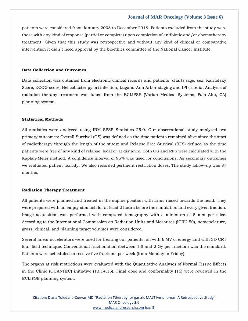
when bulky disease was found. The median dose among the group was 30 Gy. Figure 1 describes the
doses employed and time to finish radiation therapy.
Journal of MAR Oncology (Volume 3 Issue 6)
Citation: Diana Toledano-Cuevas MD “Radiation Ttherapy for gastric MALT lymphomas. A Retrospective Study” MAR Oncology 3.6
www.medicalandresearch.com (pg. 6)
Figure 1. Total dose / Protraction
The maximum time of follow-up was 87 months and a median of 30 months. Average follow up time was
reduced due to one patient who achieved complete response. Median treatment protraction was 22 days,
spanning from 11 to 53 days. The greater protraction was again due to the same patient with poor
adherence to follow-up.
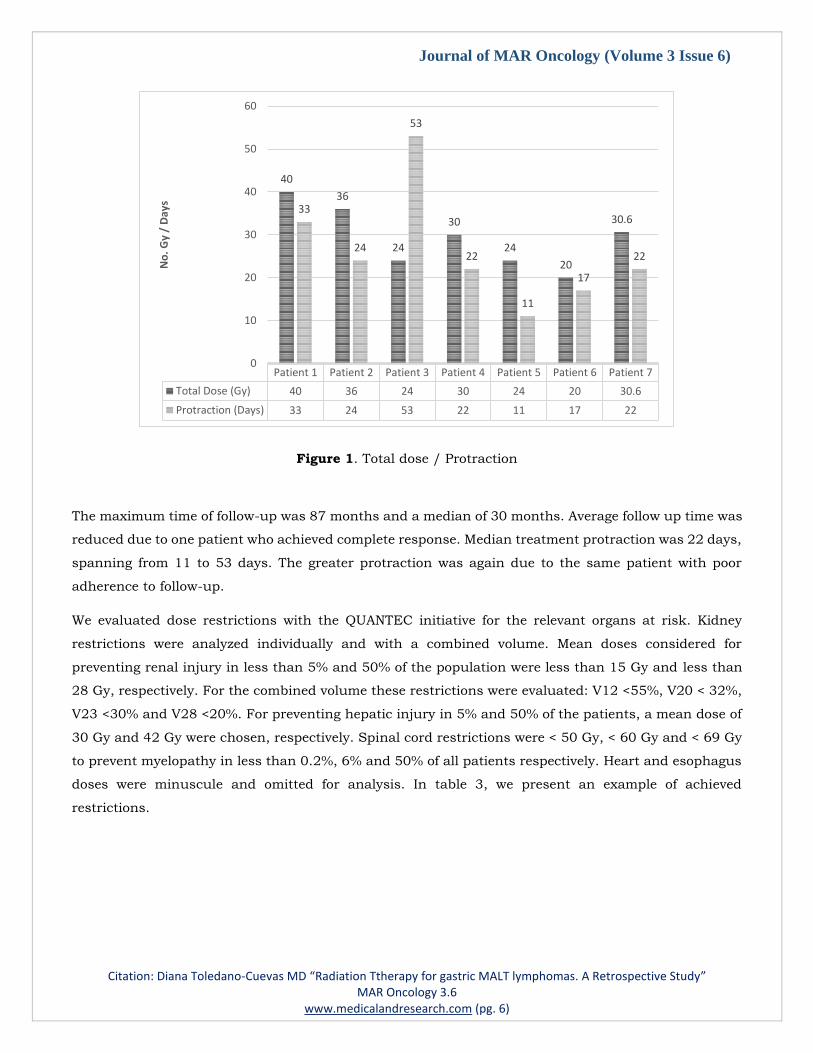
We evaluated dose restrictions with the QUANTEC initiative for the relevant organs at risk. Kidney
restrictions were analyzed individually and with a combined volume. Mean doses considered for
preventing renal injury in less than 5% and 50% of the population were less than 15 Gy and less than
28 Gy, respectively. For the combined volume these restrictions were evaluated: V12 <55%, V20 < 32%,
V23 <30% and V28 <20%. For preventing hepatic injury in 5% and 50% of the patients, a mean dose of
30 Gy and 42 Gy were chosen, respectively. Spinal cord restrictions were < 50 Gy, < 60 Gy and < 69 Gy
to prevent myelopathy in less than 0.2%, 6% and 50% of all patients respectively. Heart and esophagus
doses were minuscule and omitted for analysis. In table 3, we present an example of achieved
restrictions.
Patient 1 Patient 2 Patient 3 Patient 4 Patient 5 Patient 6 Patient 7
Total Dose (Gy) 40 36 24 30 24 20 30.6
Protraction (Days) 33 24 53 22 11 17 22
40
36
24
30
24
20
30.633
24
53
22
11
17
22
0
10
20
30
40
50
60
No
. G
y /
Day
s
Journal of MAR Oncology (Volume 3 Issue 6)
Citation: Diana Toledano-Cuevas MD “Radiation Ttherapy for gastric MALT lymphomas. A Retrospective Study” MAR Oncology 3.6
www.medicalandresearch.com (pg. 7)
Organs at
risk
Dose
restrictions Clinical outcome
Actual dose
received
Kidney
Mean dose
<15 Gy
Renal injury in less
than 5% of treated
patients
Right
Kidney
5.4 Gy
Left
Kidney
6.9 Gy
Mean dose
< 28 Gy
Renal injury in less
than 50% of treated
patients
Right
Kidney
(N/A)
Left
Kidney
(N/A)
Kidneys
(Combined
volume)
V12 <55% Renal injury in less
than 5% of treated
patients
15.17%
V20 <32% 2.68%
V23 <30% N/A
V28 <20% N/A
Liver
Mean Dose
<30 Gy
Radio-induced
hepatic injury
(Classic) in less than
5% of treated
patients
15.45 Gy
Mean Dose
< 42 Gy
Radio-induced
hepatic injury
(Classic) in less than
50% of treated
patients
N/A
Spinal cord
Max Dose
<50 Gy
Myelopathy in less
than 0.2% of treated
patients
11.82 Gy
Max Dose
<60 Gy
Myelopathy in less
than 6% of treated
patients
N/A
Max Dose
<69 Gy
Myelopathy in less
than 50% of treated
patients
N/A
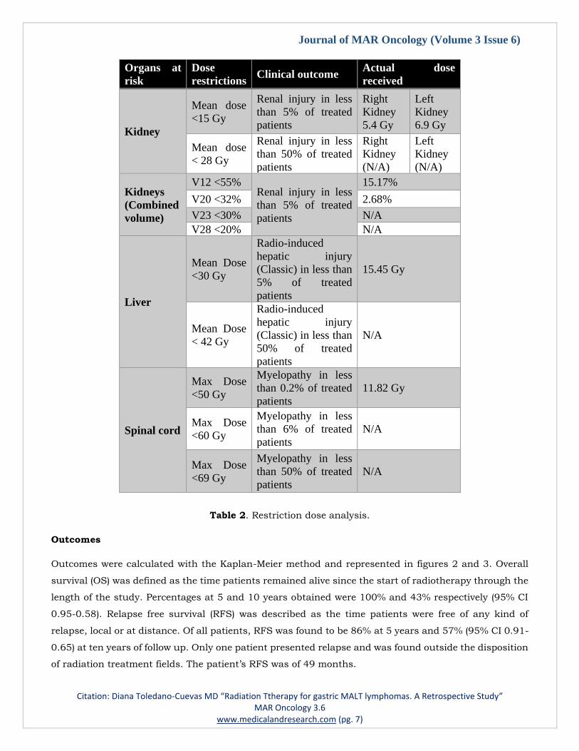
Table 2. Restriction dose analysis.
Outcomes
Outcomes were calculated with the Kaplan-Meier method and represented in figures 2 and 3. Overall
survival (OS) was defined as the time patients remained alive since the start of radiotherapy through the
length of the study. Percentages at 5 and 10 years obtained were 100% and 43% respectively (95% CI
0.95-0.58). Relapse free survival (RFS) was described as the time patients were free of any kind of
relapse, local or at distance. Of all patients, RFS was found to be 86% at 5 years and 57% (95% CI 0.91-
0.65) at ten years of follow up. Only one patient presented relapse and was found outside the disposition
of radiation treatment fields. The patient’s RFS was of 49 months.

Journal of MAR Oncology (Volume 3 Issue 6)
Citation: Diana Toledano-Cuevas MD “Radiation Ttherapy for gastric MALT lymphomas. A Retrospective Study” MAR Oncology 3.6
www.medicalandresearch.com (pg. 8)
Figure 2. Kaplan-Meier method for Overall Survival.
Figure 3. Kaplan-Meier method for Relapse Free Survival.
Complete response was observed in 57% of patients. Furthermore, 28.5% had partial response. There
is growing evidence that defines the optimal time for evaluating response in gastric MALT lymphomas
(28), although it is yet inconclusive. We established the best time to be from the start date of RT up to
documentation of complete response. Our average time for CR was of 29 days and for any response of
30.3 days.
0%
20%
40%
60%
80%
100%
120%
1 2 3 4 5 6 7 8 9 10
OS
pro
bab
ility
(%
)
Time after radiotherapy (years)
Overall survival
0%
20%
40%
60%
80%
100%
120%
1 2 3 4 5 6 7 8 9 10
RFS
Pro
bab
ility
(%
)
Time after radiotherapy (years)
Relapse free survival
Journal of MAR Oncology (Volume 3 Issue 6)
Citation: Diana Toledano-Cuevas MD “Radiation Ttherapy for gastric MALT lymphomas. A Retrospective Study” MAR Oncology 3.6
www.medicalandresearch.com (pg. 9)
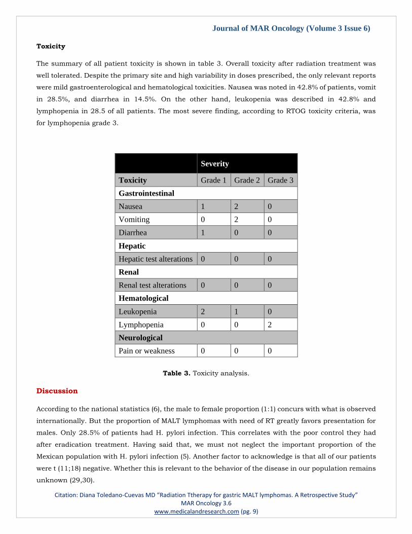
Toxicity
The summary of all patient toxicity is shown in table 3. Overall toxicity after radiation treatment was
well tolerated. Despite the primary site and high variability in doses prescribed, the only relevant reports
were mild gastroenterological and hematological toxicities. Nausea was noted in 42.8% of patients, vomit
in 28.5%, and diarrhea in 14.5%. On the other hand, leukopenia was described in 42.8% and
lymphopenia in 28.5 of all patients. The most severe finding, according to RTOG toxicity criteria, was
for lymphopenia grade 3.
Severity
Toxicity Grade 1 Grade 2 Grade 3
Gastrointestinal
Nausea 1 2 0
Vomiting 0 2 0
Diarrhea 1 0 0
Hepatic
Hepatic test alterations 0 0 0
Renal
Renal test alterations 0 0 0
Hematological
Leukopenia 2 1 0
Lymphopenia 0 0 2
Neurological
Pain or weakness 0 0 0
Table 3. Toxicity analysis.
Discussion
According to the national statistics (6), the male to female proportion (1:1) concurs with what is observed
internationally. But the proportion of MALT lymphomas with need of RT greatly favors presentation for
males. Only 28.5% of patients had H. pylori infection. This correlates with the poor control they had
after eradication treatment. Having said that, we must not neglect the important proportion of the
Mexican population with H. pylori infection (5). Another factor to acknowledge is that all of our patients
were t (11;18) negative. Whether this is relevant to the behavior of the disease in our population remains
unknown (29,30).

Journal of MAR Oncology (Volume 3 Issue 6)
Citation: Diana Toledano-Cuevas MD “Radiation Ttherapy for gastric MALT lymphomas. A Retrospective Study” MAR Oncology 3.6
www.medicalandresearch.com (pg. 10)
The cardinal symptom found was dyspepsia. None of the patients referred gastrointestinal bleeding or
‘B’ symptoms. At diagnosis, the majority of patients had a low risk. More than half (57%) had less than
60 years of age All patients presented a good performance status (KPS 80-90% and ECOG 1-2). Early
clinical stages were also the most frequent in 71%. Clinical stage IIE was the most common. IPI was also
low in 56.5% and low-intermediate in 14.5%.
Treatment before RT was heterogeneous. Of all patients, 85.7% of them received a diversity of
chemotherapy regimens with poor tumor response. Only patients with H. pylori detection received
antibiotics as first-line treatment (31). We consider these findings to be explained because a great
proportion of patients were treated when Rituximab was unavailable in our country.
Radiation therapy indication was mostly for consolidation (85.7%). Age, tumor size, and tumor burden
were considered for determining total doses. They ranged from 20 to 40 Gy, and the most common dose
was 30 Gy. Average number of fractions was 14 ranging from 10 to 20. All patients received conventional
fractionation. Overall, protraction was satisfactory, with an average of 26 days. It is noteworthy that the
patient who showed the longest protraction was due to health complications not related to RT.
Through all ten years, we obtained remarkable OS and RFS. Relapse was found in only one patient. This
patient was the same one who needed radiation therapy for bulky disease and even had the group's
worst prognosis. This relapse presented outside of the RT fields. A 4-year RFS was achieved despite poor
baseline prognosis.
After RT treatment toxicity was well tolerated. No renal, hepatic or neurologic injury was presented. Most
common toxicities were gastrointestinal and hematologic, being mild and moderate. Nausea was found
in 42.8% of patients. Vomit was presented in 28.5%. Of hematological toxicities, leukopenia was the
most common in 42.8%, followed by lymphopenia in 29%.
The present study has certain limitations inherent to an observational, single-center investigation. Our
limited sample size and patients lost during follow-up can be explained by the prevalence of the disease
and the long distances some of our patients have to travel (around 520 miles / 10 hours) to receive any
kind of treatment or attending for follow ups. Other important factors such as the available technology
through the timespan (2D-CRT to 3D-CRT evolution), and a lack of homogeneity in treatment algorithms
in the early years were not controlled for in the present study.
Conclusion
Our findings over 10 years suggest that rather than multiple chemotherapy regimens, radiotherapy is
the next best option after antibiotic failure, H. pylori naïve, or bulky disease. We found a OS at 5 and
10 years of 100% and 43% respectively and a RFS at 5 and 10 years of 86% and 57% respectively. At
least 57% of all patients treated with radiotherapy had CR.

Journal of MAR Oncology (Volume 3 Issue 6)
Citation: Diana Toledano-Cuevas MD “Radiation Ttherapy for gastric MALT lymphomas. A Retrospective Study” MAR Oncology 3.6
www.medicalandresearch.com (pg. 11)
Toxicity was very well tolerated showing only mild gastrointestinal symptoms. The only moderate toxicity
found was lymphopenia. All toxicities were well controlled without complications.
Although our data has its limitations it contributes to the evidence (39) that RT obtains a high rate of
OS and CR even at low doses < 30 Gy given with 3D-CRT. Further larger scaled studies are needed to
clarify the optimal dose, safety profile, and the efficacy of this technique with other modalities.
Reference
1. Filip, Petruta Violeta, et al. “MALT Lymphoma: Epidemiology, Clinical Diagnosis and Treatment.”
Journal of Medicine and Life, vol. 11, no. 3, 2018, pp. 187–193., doi:10.25122/jml-2018-0035.
2.- Halperin, E. C., Brady, L. W., & Pérez, C. A. (2019). Perez and Bradys principles and practice of
radiation oncology. Philadelphia: Wolters Kluwer.
3.- De_Vita, V. T., Lawrence, T. S., & Rosenberg, S. A. (2015). Cancer: principles & practice of oncology.
Philadelphia: Wolters Kluwer Health.
4.- Filip, P. V., Cuciureanu, D., Diaconu, L. S., Vladareanu, A. M., & Pop, C. S. (2018). MALT lymphoma:
epidemiology, clinical diagnosis and treatment. Journal of Medicine and Life, 11(3), 187–193. doi:
10.25122/jml-2018-0035
5.- Bosques-Padilla, F., Remes-Troche, J., González-Huezo, M., Pérez-Pérez, G., Torres-López, J., Abdo-
Francis, J., . . . Velasco, J. V. (2018). IV consenso mexicano sobre Helicobacter pylori. Revista De
Gastroenterología De México, 83(3), 325-341. doi: 10.1016/j.rgmx.2018.05.003
6.- Rizo Ríos, P., Sierra, M. I., Vázquez, G., Cano, M., Meneses, A., & Mohar, A. (2007). Registro
Hospitalario de Cáncer: Compendio de Cáncer 2000 - 2004. Cancerología, 203–287.
7.-Zelenetz, A. D., Gordon, L. I., Abramson, J. S., Bartlett, N. L., Caimi, P. F., Chang, J. F., & Chavez,
J. C. (2019, June 18). NCCN Clinical Practice Guidelines in Oncology (NCCN Guidelines) B-Cell
Lymphomas Versión 4.2019 - June 18, 2019. Retrieved October 16, 2019, from
https://www.nccn.org/professionals/physician_gls/pdf/b-cell.pdf.
8.- Zucca, E., & Dreyling, M. (2010). Gastric marginal zone lymphoma of MALT type: ESMO Clinical
Practice Guidelines for diagnosis, treatment and follow-up. Annals of Oncology, 21(Supplement 5), v175–
v176. doi: 10.1093/annonc/mdq182
9.- Gong, E. J. (2019). Radiation Therapy for the Treatment of Gastric Mucosa-Associated Lymphoid
Tissue Lymphoma. The Korean Journal of Gastroenterology, 73(1), 1. doi: 10.4166/kjg.2019.73.1.1

Journal of MAR Oncology (Volume 3 Issue 6)
Citation: Diana Toledano-Cuevas MD “Radiation Ttherapy for gastric MALT lymphomas. A Retrospective Study” MAR Oncology 3.6
www.medicalandresearch.com (pg. 12)
10.- Kim, S.-W. (2013). Clinical outcomes of radiation therapy for early-stage gastric mucosa-associated
lymphoid tissue lymphoma. World Journal of Gastroenterology, 19(36), 6062. doi: 10.3748/wjg.
v19.i36.6062
11.- Ghorbal, L., et al. “Place De La Radiothérapie Pour Les Lymphomes Du Mucosa-Associated
Lymphoid Tissue Gastriques : Résultats d’Une Étude Rétrospective.” Cancer/Radiothérapie, vol. 22, no.
8, 2018, pp. 763–766., doi:10.1016/j.canrad.2018.02.001.
12.- Wirth, A., Gospodarowicz, M., Aleman, B. M. P., Bressel, M., Ng, A., Chao, M., … Zucca, E. (2013).
Long-term outcome for gastric marginal zone lymphoma treated with radiotherapy: a retrospective,
multi-centre, International Extranodal Lymphoma Study Group study. Annals of Oncology, 24(5), 1344–
1351. doi: 10.1093/annonc/mds623
13.- Bentzen, S. M., Constine, L. S., Deasy, J. O., Eisbruch, A., Jackson, A., Marks, L. B., … Yorke, E.
D. (2010). Quantitative Analyses of Normal Tissue Effects in the Clinic (QUANTEC): an introduction to
the scientific issues. International journal of radiation oncology, biology, physics, 76(3 Suppl), S3–S9.
doi: 10.1016/j.ijrobp.2009.09.040
14.- Marks, L. B., Yorke, E. D., Jackson, A., Haken, R. K. T., Constine, L. S., Eisbruch, A., … Deasy, J.
O. (2010). Use of Normal Tissue Complication Probability Models in the Clinic. International Journal of
Radiation Oncology*Biology*Physics, 76(3). doi: 10.1016/j.ijrobp.2009.07.1754
15.- Jackson, A., Marks, L. B., Bentzen, S. M., Eisbruch, A., Yorke, E. D., Haken, R. K. T., … Deasy, J.
O. (2009). The Lessons of QUANTEC (Quantitative Analysis of Normal Tissue Effects in the Clinic):
Recommendations for Reporting and Gathering Data on Dose-Volume Dependencies of Treatment
Outcome. IFMBE Proceedings World 38.- Zucca, E., & Dreyling, M. (2010). Gastric marginal zone
lymphoma of MALT type: ESMO Clinical Practice Guidelines for diagnosis, treatment and follow-up.
Annals of Oncology, 21(Supplement 5), v175–v176. doi: 10.1093/annonc/mdq182 Congress on Medical
Physics and Biomedical Engineering, September 7 - 12, 2009, Munich, Germany, 1075–1075. doi:
10.1007/978-3-642-03474-9_303
16.- Lim, H. W., Kim, T. H., Choi, I. J., Kim, C. G., Lee, J. Y., Cho, S. J., … Kim, D. Y. (2016). Radiation
therapy for gastric mucosa-associated lymphoid tissue lymphoma: dose-volumetric analysis and its
clinical implications. Radiation Oncology Journal, 34(3), 193–201. doi: 10.3857/roj.2016.01865
17.- Zhang, Andrew, et al. “Vertebral Body Irradiation during Chemoradiation Therapy for Esophageal
Cancer Contributes to Acute Bone Marrow Toxicity.” Journal of Gastrointestinal Oncology, vol. 10, no.
3, 2019, pp. 513–522., doi:10.21037/jgo.2019.01.20.
18.- RTOG. (n.d.). Retrieved from
https://www.rtog.org/ResearchAssociates/AdverseEventReporting/CooperativeGroupCommonToxicity
Criteria.aspx.

Journal of MAR Oncology (Volume 3 Issue 6)
Citation: Diana Toledano-Cuevas MD “Radiation Ttherapy for gastric MALT lymphomas. A Retrospective Study” MAR Oncology 3.6
www.medicalandresearch.com (pg. 13)
19.- Juárez-Salcedo, Luis Miguel, et al. “Primary Gastric Lymphoma, Epidemiology, Clinical Diagnosis,
and Treatment.” Cancer Control, vol. 25, no. 1, 2018, p. 107327481877825.,
doi:10.1177/1073274818778256.
20.- Thieblemont, Catherine, et al. “A MALT Lymphoma Prognostic Index.” Blood, vol. 130, no. 12, 2017,
pp. 1409–1417., doi:10.1182/blood-2017-03-771915.
21.- Gisbert, J. P., & Calvet, X. (2011). Review article: common misconceptions in the management of
Helicobacter pylori-associated gastric MALT-lymphoma. Alimentary Pharmacology & Therapeutics,
34(9), 1047–1062. doi: 10.1111/j.1365-2036.2011. 04839.x
22.- Nam, T.-K., Ahn, J.-S., Choi, Y.-D., Jeong, J.-U., Kim, Y.-H., Yoon, M. S., … Chung, W.-K. (2014).
The Role of Radiotherapy in the Treatment of Gastric Mucosa-Associated Lymphoid Tissue Lymphoma.
Cancer Research and Treatment, 46(1), 33–40. doi: 10.4143/crt.2014.46.1.33
23.- Nakamura, Shotaro, et al. “Long-Term Clinical Outcome of Gastric MALT Lymphoma after
Eradication OfHelicobacter Pylori: a Multicentre Cohort Follow-up Study of 420 Patients in Japan.” Gut,
vol. 61, no. 4, 2011, pp. 507–513., doi:10.1136/gutjnl-2011-300495.
24.- Kim, S. W., Lim, D. H., Ahn, Y. C., Kim, W. S., Kim, S. J., Ko, Y. H., & Kim, K. M. (2013). Clinical
outcomes of radiation therapy for early-stage gastric mucosa-associated lymphoid tissue lymphoma.
World journal of gastroenterology, 19(36), 6062–6068. doi:10.3748/wjg. v19.i36.6062
25.- Delibegovic, E. (2016). An Aggressive Form of MALT Lymphoma of the Stomach with Pancreas
Infiltration. Medical Archives, 70(3), 235. doi: 10.5455/medarh.2016.70.235-237
26.- Pinnix, C. C., Reed, V., & Dabaja, B. (2012). Gastric MALT Lymphoma Treated with Primary
Radiotherapy in the Setting of Autoimmune Disease. Journal of the National Comprehensive Cancer
Network, 10(7), 815–819. doi: 10.6004/jnccn.2012.0085
27.- Kobayashi, Y. (2019). JSH practical guidelines for hematological malignancies, 2018: II. Lymphoma-
2. Marginal zone lymphoma (MALT lymphoma/extranodal marginal zone lymphoma of mucosa-
associated lymphoid tissue and splenic marginal zone lymphoma). International Journal of Hematology,
110(4), 393–405. doi: 10.1007/s12185-019-02719-6
28.- Bertoni, F., Rossi, D., & Zucca, E. (2018). Recent advances in understanding the biology of marginal
zone lymphoma. F1000Research, 7, 406. doi: 10.12688/f1000research.13826.1
29.- Troppan, Katharina, et al. “Molecular Pathogenesis of MALT Lymphoma.” Gastroenterology
Research and Practice, vol. 2015, 2015, pp. 1–10., doi:10.1155/2015/102656.

Journal of MAR Oncology (Volume 3 Issue 6)
Citation: Diana Toledano-Cuevas MD “Radiation Ttherapy for gastric MALT lymphomas. A Retrospective Study” MAR Oncology 3.6
www.medicalandresearch.com (pg. 14)
30.- Du, Ming-Qing. “MALT Lymphoma: Genetic Abnormalities, Immunological Stimulation and
Molecular Mechanism.” Best Practice & Research Clinical Haematology, vol. 30, no. 1-2, 2017, pp. 13–
23., doi:10.1016/j.beha.2016.09.002.
31.- Follows, G. A., Ardeshna, K. M., Barrington, S. F., Culligan, D. J., Hoskin, P. J., Linch, D., …
Wimperis, J. Z. (2014). Guidelines for the first line management of classical Hodgkin lymphoma. British
Journal of Haematology, 166(1), 34–49. doi: 10.1111/bjh.12878
32.- Swerdlow, S. H., Campo, E., Pileri, S. A., Harris, N. L., Stein, H., Siebert, R., … Jaffe, E. S. (2016).
The 2016 revision of the World Health Organization classification of lymphoid neoplasms. Blood,
127(20), 2375–2390. doi: 10.1182/blood-2016-01-643569
33.- Dogan, A. (2019). Extranodal Marginal Zone Lymphoma of Mucosa-Associated Lymphoid Tissue.
Encyclopedia of Pathology, 1–5. doi: 10.1007/978-3-319-28845-1_3898-1
34.- Sagaert, X., Cutsem, E. V., Hertogh, G. D., Geboes, K., & Tousseyn, T. (2010). Gastric MALT
lymphoma: a model of chronic inflammation-induced tumor development. Nature Reviews
Gastroenterology & Hepatology, 7(6), 336–346. doi: 10.1038/nrgastro.2010.58
35.- Marcelis, L., Tousseyn, T., & Sagaert, X. (2019). MALT Lymphoma as a Model of Chronic
Inflammation-Induced Gastric Tumor Development. Current Topics in Microbiology and Immunology
Molecular Mechanisms of Inflammation: Induction, Resolution and Escape by Helicobacter Pylori, 77–
106. doi: 10.1007/978-3-030-15138-6_4
36.- Kuo, Sung-Hsin, et al. “The B-Cell-Activating Factor Signalling Pathway Is Associated With
Helicobacter Pylori independence in Gastric Mucosa-Associated Lymphoid Tissue Lymphoma without
t(11;18)(q21;q21).” The Journal of Pathology, vol. 241, no. 3, 2016, pp. 420–433.,
doi:10.1002/path.4852.
37.- Kuo, S. H., Yeh, K. H., Chen, L. T., Lin, C. W., Hsu, P. N., Hsu, C., … Cheng, A. L. (2014).
Helicobacter pylori-related diffuse large B-cell lymphoma of the stomach: a distinct entity with lower
aggressiveness and higher chemosensitivity. Blood cancer journal, 4(6), e220. doi:10.1038/bcj.2014.40
38.- Ohkubo, Yu, et al. “Radiotherapy for Localized Gastric Mucosa–Associated Lymphoid Tissue
Lymphoma: Long-Term Outcomes over 10 Years.” Journal of Radiation Research, vol. 58, no. 4, 2017,
pp. 537–542., doi:10.1093/jrr/rrw044.
39.- Pinnix, Chelsea C., et al. “Outcomes After Reduced-Dose Intensity Modulated Radiation Therapy
for Gastric Mucosa-Associated Lymphoid Tissue (MALT) Lymphoma.” International Journal of Radiation
Oncology*Biology*Physics, vol. 104, no. 2, 2019, pp. 447–455., doi:10.1016/j.ijrobp.2019.02.002.
Journal of MAR Oncology (Volume 3 Issue 6)
Citation: Diana Toledano-Cuevas MD “Radiation Ttherapy for gastric MALT lymphomas. A Retrospective Study” MAR Oncology 3.6
www.medicalandresearch.com (pg. 15)