JOINTS OF HUMAN BODY (BRIEF COURSE) - Белорусский ...
50
H. А. PASIUK, О. L. ZHARIKOVA, F. А. АRDEN JOINTS OF HUMAN BODY (BRIEF COURSE) Minsk BSMU 2021
-
Upload
khangminh22 -
Category
Documents
-
view
1 -
download
0
Transcript of JOINTS OF HUMAN BODY (BRIEF COURSE) - Белорусский ...

H. А. PASIUK, О. L. ZHARIKOVA, F. А. АRDEN
JOINTS OF HUMAN BODY
(BRIEF COURSE)
Minsk BSMU 2021


МИНИСТЕРСТВО ЗДРАВООХРАНЕНИЯ РЕСПУБЛИКИ БЕЛАРУСЬ
БЕЛОРУССКИЙ ГОСУДАРСТВЕННЫЙ МЕДИЦИНСКИЙ УНИВЕРСИТЕТ
КАФЕДРА НОРМАЛЬНОЙ АНАТОМИИ
А. А. ПАСЮК, О. Л. ЖАРИКОВА, Ф. А. АРДЕН
СОЕДИНЕНИЯ ТЕЛА ЧЕЛОВЕКА
(КРАТКИЙ КУРС)
JOINTS OF HUMAN BODY
(BRIEF COURSE)
Учебно-методическое пособие
Минск БГМУ 2021

2
УДК 611.7(075.8)-054.6
ББК 28.70я73
П19
Рекомендовано Научно-методическим советом университета в качестве
учебно-методического пособия 21.10.2020 г., протокол № 12
Р е ц е н з е н т ы: каф. нормальной физиологии; канд. мед. наук, доц. П. В. Маркауцан;
канд. филол. наук, доц. М. Н. Петрова
Пасюк, А. А.
П19 Соединения тела человека (краткий курс) = Joints of human body (brief course) :
учебно-методическое пособие / А. А. Пасюк, О. Л. Жарикова, Ф. А. Арден. – Минск :
БГМУ, 2021. – 47 с.
ISBN 978-985-21-0735-8.
Содержит общие сведения о соединениях скелета человека и краткую характеристику отдельных
суставов.
Предназначено для студентов медицинского факультета иностранных учащихся 1–2-го курсов, обу-
чающихся на английском языке. Предлагаемые материалы могут быть использованы при прохождении
курса травматологии и ортопедии.
УДК 611.7(075.8)-054.6
ББК 28.70я73
ISBN 978-985-21-0735-8 © Пасюк А. А., Жарикова О. Л., Арден Ф. А., 2021
© УО «Белорусский государственный
медицинский университет», 2021

3
GENERAL CHARACTERISTICS OF JOINTS
The human skeletal system consists of more than 200 hundred bones connected
to each other by numerous articulations, or joints (more than 230), which are
supported by approximately 1000 ligaments. The joints tightly binding the elements
of the skeleton provide its stability, weight-bearing support for the body and
protection of the vital organs. The less stable joints that allow motion between
the bones ensure a wide range of body movements.
A joint (Lat. junctura) is a place of junction between two or more bones, a bone
and a cartilage, or parts of the same (immature) bone. Joints can be classified based
on their structure and function.
Functional classification of joints considers amount of movements permitted.
Accordingly, three categories of joints are distinguished: synarthrosis (Lat. syn —
means together, arthro — means joint) is an immovable joint; amphiarthrosis (Lat.
amfe — means of both kinds) is a slightly movable joint; and diarthrosis (Lat. di —
means two) is a freely movable joint. Function of the joint depends on its general
structural plan and specific structural features (e.g., strength of supporting ligaments,
congruency of articulating surfaces in the synovial joints).
Structurally joints are classified based on 1) absence or 2) presence of space
between the articulating bones. The first group is solid joints; the second group is
synovial joints.
Solid joints (continuous joints) are subdivided into 3 groups according to
the type of connective tissue that binds the articulating bones: fibrous joints,
cartilaginous joints, and bony joints (fusions).
1. Fibrous joints (often called syndesmoses), in which the bones are joined by
fibrous connective tissue, are classified as follows:
● Syndesmoses (Lat. desmos — ligament) are connections by longer or shorter
fibrous bands; they include:
– Interosseous ligament, such as in the tibiofibular syndesmosis, or ligamentum
flavum between adjacent vertebral arches;
– Interosseous membrane, e.g., between the bones of the forearm or leg bones;
– Gomphosis (syn. socket joint), which is the dentoalveolar syndesmosis,
formed by short collagen fibers called the periodontal ligament;
● Sutures which are connections with minimal amount of fibrous tissue
between the skull bones, and are formed by intramembranous ossification; based on
the shape and position of connecting bones edges several kinds of sutures are
distinguished: plane, limbous (squamous), serrate, and denticulate sutures,
schindylesis (“wedge-and-groove” suture). Ossification of the fibrous tissue
(i.e. replacing it by bones) in sutures with age results in synostosis.
Most fibrous joints are immovable or only slightly movable (synarthroses and
amphiarthroses). The amount of movement depends on the length of fibers between
the articulating bones. Thus, the longer fibers in the interosseous membranes allow
a larger amount of movement, than in other kinds of fibrous joints.
4
2. Cartilaginous joints, in which the bones are united by cartilage, are of two
types:
– Synchondrosis (syn. primary cartilaginous joints) where bones are connected
by hyaline cartilage: e.g., sternocostal synchondrosis between the 1st rib and sternum,
joints between bones of the skull base, joints between parts of a growing bone, such
as the pelvic bone, bones of the scull base, long bones (cartilaginous plates between
diaphyses and epiphyses — epiphyseal cartilage, cartilage between diaphyses and
apophyses);
– Symphysis where bones are connected by fibrocartilage: e.g., intervertebral
discs and pubic symphysis.
Among the cartilaginous joints, the synchondroses are immovable joints
(synarthroses), while symphyses are slightly movable joints (amphiarthroses) that
provide strength with flexibility.
3. Synostosis (bony union) is a result of substitution of fibrous tissue or
cartilage by bony tissue: e.g., fusion of sacral vertebrae in the sacrum, ossification of
cranial sutures, fusion between the diaphysis and epiphysis in long bones.
Synovial joints (Syn. diarthroses, discontinuous joints) are formed by bones
bound together by the articular capsule and separated within the joint cavity by
a narrow space filled with synovial fluid. Synovial joints are the most common type
among joints of the body, which usually provide free movement between the bones
(predominantly diarthroses).
Basic features of each joint are presence of the following:
– Articular surfaces covered with articular (hyaline) cartilage;
– Articular (joint) capsule that attaches along the borders of the articular
surfaces and encloses the joint cavity: it’s made of the outer fibrous layer and inner
synovial membrane;
– Articular cavity, a potential space limited by the articular surfaces and
capsule, filled with a small amount of synovial fluid;
– Synovial fluid, viscous slippery liquid produced by the synovial membrane;
lubricates and nourishes articular cartilages;
– Nerves and vessels.
Accessory structures of a joint are:
1) Accessory ligaments, fibrous bands reinforcing a joint:
– capsular ligaments, thickenings of the fibrous capsule;
– extracapsular ligaments, separated from the fibrous capsule;
– intracapsular (intra-articular) ligaments, lying within the fibrous capsule and
covered with a synovial membrane;
2) Articular fibrocartilage that adjusts articular surfaces for better fit:
– articular disc (oval or round shape);
– meniscus (crescent, C-shape);
– articular labrum (annular shape, makes deeper an articular surface);
3) Synovial folds and fat pads — synovial structures filled with fat tissue; fill
spaces formed when bones move and the joint cavity changes its shape;

5
Synovial bursae and synovial sheaths are structures associated with muscle
tendons but in some joints may be connected to the articular cavity:
4) Synovial bursa — a flattened fibrous sac lined by a synovial membrane;
reduces friction between moving structures: between tendons or ligaments and bones
(e.g., suprapatellar bursa of the knee joint), or between skin and bones;
5) Synovial sheath — an elongated synovial sac that covers a tendon passing
across a bony surface (e.g., synovial sheath of the long head of the biceps brachii
muscle).
Classification of the synovial joints takes into account their structural features,
such as shapes of the articular surfaces, and functional properties, i.e. types of
possible movements. Generally, movements allowed at a joint depend on its shape.
Fig. 1. Examples of movement at synovial joints [1]

6
Basic types of movements of bones (Fig 1):
1. Gliding — linear motion in one plane, i.e. sliding of one bone across another
one;
2. Angular movements, which change the angle between articulating bones;
described in reference to three anatomical axes or planes by two ways: as motion in
the plane or as motion around the axis. The most common angular movements
occur:
– In the sagittal plane, around the transverse (frontal) axis: flexion — reduces
the angle between articulating bones; extension — increases the angle between
bones;
– In the coronal (frontal) plane, around the sagittal axis: abduction —
movement of a limb away from the body (or digits of the hand away from the central
digit); adduction — movement of a limb toward the body (or digits toward
the central digit);
3. Circumduction — sequential combination of angular movements at a joint,
i.e. transitioning from one axis to another (e.g., flexion, abduction, extension, and
adduction), while moving a bone so that its free (distal) end describes a circle;
4. Rotation — moving a bone around its longitudinal axis, or part of the body
around the vertical axis.
Special movements (supination-pronation, opposition-reposition, protraction-
retraction, etc.) will be described with the specific joints at which they occur.
CLASSIFICATION OF THE SYNOVIAL JOINTS
Synovial joints are divided into two groups:
1. Simple — formed by two bones;
2. Complex — formed by three or more bones; containing articular discs or
menisci that create additional articular surfaces. Two or more individual joints that
can function only simultaneously are distinguished as combination of joints.
Based on the numbers of possible movements, i.e. axes of motion allowed,
the synovial joints are divided into three categories:
– uniaxial joints that allow movement in one plane, around a single axis;
– biaxial joints that allow movements in two planes, around two axes;
– multiaxial joints that allow movements in all three anatomical planes, around
three axes.
As movements at joints are largely determined by the shape of the articular
surfaces the synovial joints are classified by function and shape as follows:
1. Uniaxial: are cylindrical joints, which can be of two types — pivot and
hinge joints:
– Pivot joints: are formed between a rounded cylindrical end (vertically
oriented) of one bone and a bony-ligamentous ring formed by another bone; allow
only rotation (around vertical axis). Examples: proximal radioulnar joints, middle
atlantoaxial joint (joint between the atlas and the dens of axis).
– Hinge joints are formed between the cylindrical end (transversally oriented) of
one bone and the trough-shaped depression of another bone; allow only flexion and

7
extension (around frontal axis, in the sagittal plane). Examples: elbow joint, ankle
joint.
2. Biaxial:
– Ellipsoid (condylar) joints are formed between ovoid convex surface of one
bone and a corresponding depression of another bone; allow angular movements —
flexion-extension, abduction-adduction (circumduction). Examples: wrist joint,
metacarpophalangeal joints.
– Bicondylar joints have two distinct ovoid convex surfaces of one bone
articulating with two concave surfaces of the other bone. The movements are
permitted around two axes as in ellipsoid joints, e.g., flexion-extension and lateral
flexion at the atlanto-occipital joint, or flexion-extension and rotation at the knee
joint.
– Saddle (sellar) joints are formed between two articular surfaces resembling
a saddle — each surface has both convex and concave areas; allow the same
movements as the ellipsoid joints but in a bigger range. An example is
the carpometacarpal joint of thumb.
3. Multiaxial (triaxial):
– Ball and socket (spheroidal, cotyloid) joints are formed between a spherical
end of one bone and a cup-shaped socket of another bone; allow all types of
movements, including flexion-extension, abduction-adduction, rotation, and
circumduction, and the greatest range of motion. Examples: shoulder and hip joints.
– Plane (arthrodial) joints are formed by bones with flat or slightly curved
articulating surfaces; allow only the sliding of opposed bones, but no rotation.
The joints that permit gliding movements in multiple directions are multiaxial, e.g.,
joints between the articular processes of the vertebrae. Those permitting only small
movements are called nonaxial, e.g., intercarpal and intertarsal joints.
When describing a synovial joint, it is convenient to follow the specific
algorithm.
Algorithm for description of a joint:
1. Articulating bones and articular surfaces.
2. Classification of a joint:
– by number of articular surfaces (simple, complex);
– by shape of articular surfaces;
– by number of axes of movement.
3. Movements allowed (specifying planes and axes of motion).
4. Specific features of capsule attachment.
5. Ligaments.
6. Other accessory structures.
7. Blood supply and innervation (for final exam).
However, it should be noted that not every joint can be described in all
the points, as well as not all joints of the body exactly fall under the above given
classification.

8
JOINTS OF VERTEBRAL COLUMN
Fibrous joints of vertebral column
Fibrous joints, syndesmoses (Fig. 2), connect the following parts of vertebrae:
● the bodies:
– anterior longitudinal ligament (lig. longitudinale anterius);
– posterior longitudinal ligament (lig. longitudinale posterius);
● the arches:
– ligamenta flava;
● the spinous processes:
– interspinous ligaments (ligg. interspinalia);
– supraspinous ligament (lig. supraspinale);
– ligamentum nuchae [nuchal ligament] (lig. nuchae);
● the transverse processes:
– intertransverse ligaments (ligg. intertransversaria).
Fig. 2. Joints of vertebral column (lateral view) [2]

9
Cartilaginous joints of vertebral column
Cartilaginous joints connect the bodies of vertebrae:
– Intervertebral symphyses (symphyses intervertebrales);
– Intervertebral disc (discus intervertebralis): anulus fibrosus and nucleus
pulposus;
– Lumbosacral symphysis (symphysis lumbosacralis)
– Sacrococcygeal joint (junctura sacrococcygeа): connects the apex of sacrum
and the base of the first coccygeal vertebra; reinforced by the sacrococcygeal
ligaments (ligg. sacrococcygea) from all sides — lateral, anterior and posterior1.
Synostoses (bony union) connect the sacral and coccygeal vertebrae that fuse
together to form the sacrum (os sacrum) and coccyx (coccyx, os coccygis).
Synovial joints of vertebral column
Zygapophysial joints (artt. zygapophysiales) (Fig. 2).
Articulating bones and surfaces: the articular facets of the superior and inferior
articular processes of the adjacent vertebrae.
Classification: Functionally combined with the joint of the opposite side. Plane.
Multiaxial.
Movements:
– flexion (bending torso forward) and extension (bending torso backward) —
around the transverse axis, in the sagittal plane;
– lateral flexion (bending torso to the sides) — around the sagittal axis, in
the frontal plane;
– rotation — around vertical axis;
– circumduction.
Ligaments: no proper ligaments.
Blood supply:
– cervical part — vertebral artery (subclavian artery);
– thoracic part — posterior intercostal arteries (thoracic aorta);
– lumbar part — lumbar arteries (abdominal aorta);
– sacral part — lateral sacral artery (internal iliac artery) and median sacral artery (abdominal
aorta).
Nerve supply: dorsal rami of cervical, thoracic, lumbar, sacral spinal nerves.
Uncovertebral joints (artt. uncovertebrales) (Fig. 3).
Articulating bones and surfaces: the uncinate processes of the C4-C7
vertebrae C4-C7; and the corresponding surfaces of the bodies of the vertebrae
above (C3-C6).
Classification: functionally combined with the joint of the opposite side. Plane.
Multiaxial.
Movements: follow the cervical spine movements and stabilize it by limiting
lateral movements.
1 “The mobility of the coccyx varies with the character of the joint. Sometimes in young adults, particularly in females,
it may possess a true joint cavity and be in fact a synovial joint” (Terminologia Anatomica, 2nd ed.)

10
Fig. 3. Left: CIV, CV vertebrae (anterior view); right: uncovertebral joint [3]
Ligaments: no proper ligaments.
Notes: the joints, 4 in number, are located anterior to the intervertebral
foramina.
Blood supply and nerve supply: see zygapophysial joints.
Median atlanto-axial joint (art. atlantoaxialis mediana) (Fig. 4, 5).
Articulating bones and surfaces: the dens of the axis (C2) and the anterior arch
(facet for dens) of atlas (C1), anteriorly, with the transverse ligament of the atlas
posteriorly.
Classification. Functionally combined with the lateral atlanto-axial joints. Pivot.
Uniaxial.
Movements: rotation of the atlas around the dens, i.e. vertical axis (turning
the head from side to side).
Ligaments:
– cruciform ligament (lig. cruciforme): consists of transverse ligament of atlas
(lig. transversum atlantis) and superior and inferior longitudinal bands (fasciculi
longitudinales superior et inferior);
– alar ligaments (ligg. allaria);
– apical ligament of dens (lig. apicis dentis);
– tectorial membrane of cervical vertebral column (membrana tectoria columna
vertebralis cervicalis).
Blood supply: vertebral artery (subclavian artery).
Nerve supply: dorsal rami of the second cervical spinal nerve.
Right/left lateral atlanto-axial joint (art. atlantoaxialis lateralis) (Fig. 4, 5).
Articulating bones and surfaces: the inferior articular surfaces of the atlas (C1)
and the superior articular facets of the axis (C2).
Classification. Functionally combined with the same joint of the opposite side
and with the median atlanto-axial joint. Plane. Multiaxial.
Movements: gliding during atlas rotation around the dens in the median atlanto-
axial joint.
Ligaments: common with the median atlanto-axial joint.
Blood supply and nerve supply: see median atlanto-axial joint.

11
Right/left atlanto-occipital joint (art. atlantooccipitalis)2 (Fig. 4, 5).
Articulating bones and surfaces: the occipital condyles of the occipital bone
and the superior articular surfaces of the atlas.
Classification. Functionally combined with the same joint of the opposite side.
Bicondilar. Biaxial.
Movements:
– flexion and extension (forward and backward head tilts) — around
the transverse axis, in the sagittal plane;
– lateral flexion (right and left head tilts) — around the sagittal axis, in
the frontal plane;
– circumduction of the head.
Ligaments:
– anterior atlanto-occipital membrane (membrana atlantooccipitalis anterior);
– posterior atlanto-occipital membrane (membrana atlantooccipitalis posterior);
– lateral atlanto-occipital ligament (lig. atlantooccipitale laterale)3.
Blood supply and nerve supply: see median atlanto-axial joint.
a b
c d
Fig. 4. Atlanto-occipital and atlanto-axial joints (posterior view) [2]:
a — nuchal ligament and posterior atlanto-occipital membrane; b — posterior longitudinal
ligament. Removed: Spinal cord; vertebral canal windowed; c — cruciform ligament of atlas.
Removed: Tectorial membrane; d — alar apical ligaments. Removed: Transverse ligament of atlas,
longitudinal fascicles
2 In Terminologia Anatomica (2nd ed.) refers to Joints of skull.
3 Reinforces the articular capsule connecting the anterolateral aspect of the transverse process of the atlas with
the jugular process of the occipital bone.

12
Fig. 5. Median atlanto-axial joint (superior view) [3]
JOINTS OF THORAX
Fibrous joints of thorax
Syndesmoses:
– External intercostal membrane (membrana intercostalis externa), connects
the costal cartilages replacing the external intercostal muscles;
– Internal intercostal membrane (membrana intercostalis interna), connects
the ribs in the segment between the vertebral column and the angle of rib replacing
the internal intercostal muscles;
Cartilaginous joints of thorax
Synchondroses:
– Sternochondral [sternocostal] synchondrosis of first rib (synchondrosis
costae primae);
– Costochondral joints (artt. costochondralеs) between the anterior ends of
the ribs and the costal cartilage;
Cartilaginous joints of sternum:
– Manubriosternal joint [Synchondrosis sterni] (symphysis manubriosternalis);
– Xiphisternal joint (symphysis xiphosternalis);
– Articulations of the false (VIII–X) ribs with the ribs above are weak
cartilaginous or fibrous joints (are not included in Terminologia Anatomica 2nd
ed.).
Synovial joints of thorax
Costovertebral joints (artt. costovertebralеs) (Fig. 6).
Include: joints of head of rib and costotransverse joints.
Joint of head of rib (art. capitis costae).
Articulating bones and surfaces: the articular facets of head of rib and
the costal facets of the vertebral bodies.
13
a
b
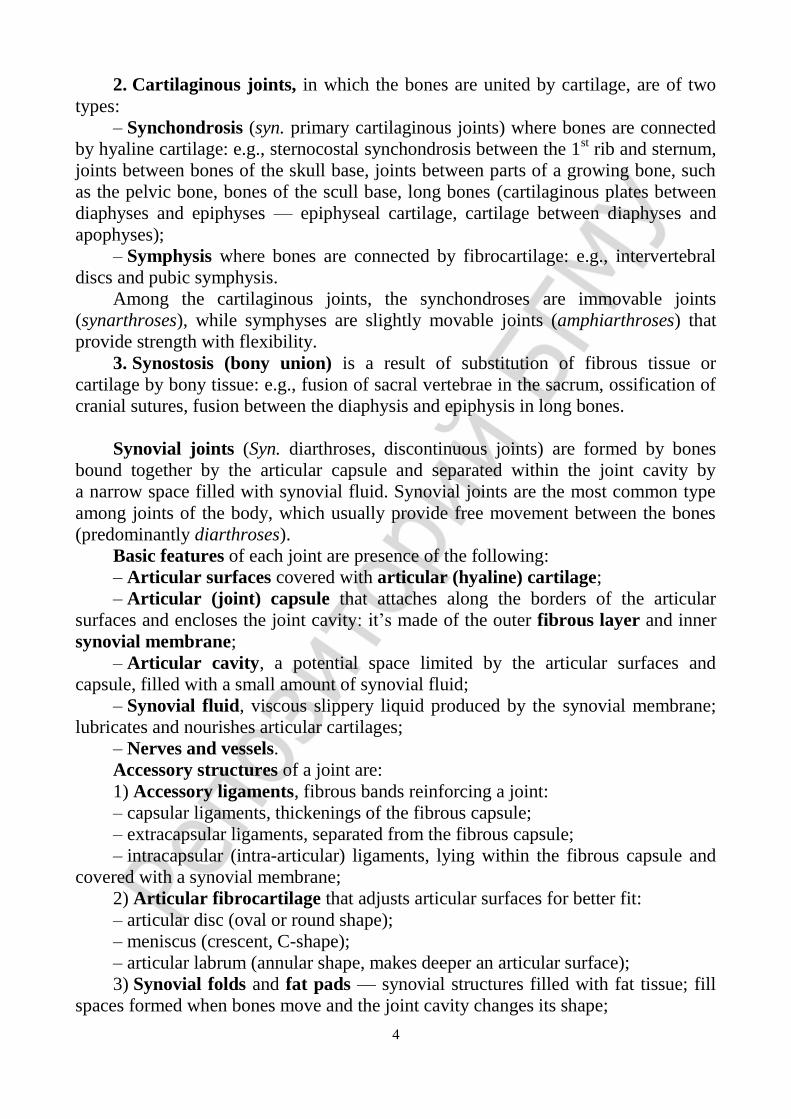
Fig. 6. Costovertebral joints [2]:
a — costotransverse joint. Superior with joints of the left rib transversely sectioned;
b — costovertebral joints. Left lateral view with the joint head of the 7th
rib opened
Classification. Functionally combined with the costotransverse and sternocostal
joints. Plane. Functionally uniaxial.
Movements: a small degree of gliding and rotation of the rib head around
an axis that passes through the neck of the rib results in elevation and depression of
the ribs that occur on respiration.

14
Ligaments:
– radiate ligament of head of rib (lig. radiatum capitis costae);
– intra-articular ligament of head of rib (lig. intraarticulare capitis costae) in
joints of the ribs II–X.
Notes: the ribs II–X articulate with the superior and inferior costal facets of
the adjacent vertebrae (I–X); the ribs I, XI, and XII articulate with single costal facets
of the corresponding vertebrae.
Blood supply: posterior intercostal arteries (thoracic aorta).
Nerve supply: dorsal rami of thoracic spinal nerves.
Costotransverse joint (art. costotransversaria).
Articulating bones and surfaces: the transverse costal facet of the thoracic
vertebra (Ι–X) and the articular facet of the tubercle of rib.
Classification. Functionally combined with the joint of head of rib and
the sternocostal joint. Plane. Nonaxial.
Movements: gliding that results in elevation and depression of the ribs.
Ligaments: costotransverse ligaments (ligg. costotransversaria)4.
Blood supply and nerve supply: see joints of head of rib.
Sternocostal [Sternochondral] joints (artt. sternocostales [sternochondrales])
(Fig. 7).
Articulating bones and surfaces: the II–VII costal cartilages and the costal
notches of the sternum.
Classification. Functionally combined with the costovertebral joints. Plane.
Nonaxial.
Movements: slight gliding motions that allow moving the ribs up and down.
Ligaments: radial sternocostal [sternochondral] ligaments (ligg. sternocostalia
radiatae), sternal membrane (membrana sterni).
Blood supply: internal thoracic artery (subclavian artery).
Nerve supply: intercostal nerves.
Interchondral joints (artt. interchondrales) (Fig. 7).
Articulating bones and surfaces: formed by the adjacent borders of the costal
cartilages of ribs from VI to IX.
Classification. Functionally combined with the sternocostal and
the costovertebral joints. Plane. Nonaxial.
Movements: gliding movements while elevating and depressing the ribs.
Ligaments: interchondral ligaments (ligg. interchondralia).
Blood supply and nerve supply: see sternocostal joints.
4 At least 3 kinds of costotransverse ligaments are distinguished based on their directions: costotransverse, lateral and
superior costotransverse ligaments.

15
Fig. 7. Sternocostal and intercostal joints [3]
JOINTS OF SKULL
Cranial fibrous joints
Cranial syndesmoses:
– Ligaments: connecting parts of the skull bones (stylohyoid ligament,
stylomandibular ligament, ligaments of the auricle, etc.).
Cranial sutures. Most of them are named after articulating bones (or parts of
bones); some have their own names:
– Coronal suture (sutura coronalis) ;
– Sagittal suture (sutura sagittalis);
– Lambdoid suture (sutura lambdoidea) ;
– Squamous suture (sutura squamosa);
– Median and transverse palatine sutures (sutura palatina mediana et
transversa).

16
Dentoalveolar syndesmosis (tooth socket joint, or gomphosis): consists of
periodontal ligament and cement.
Fontanels (in the developing skull), fibrous membranes filling gaps between
more than 2 adjacent bones of the calvaria.
Cranial cartilaginous joints
Cranial synchondroses (most of them close sooner or later in life and become
synostoses):
– spheno-occipital synchondrosis (synchondrosis sphenooccipitalis) (the closure
completes by 16–18 years, 1–2 years later in males);
– petro-occipital synchondrosis (synchondrosis petrooccipitalis);
– sphenopetrosal synchondrosis (synchondrosis sphenopetrosa);
– spheno-ethmoidal synchondrosis (synchondrosis sphenoethmoidalis) (ossifies
around 6 years);
– synchondroses between parts of the developing bones of the cranial base
(ossify soon after birth).
Cranial synovial joints
Temporomandibular joint (art. temporomandibularis) (Fig. 8, 9).
Articulating bones and surfaces: the mandibular fossa and articular tubercle of
the temporal bone; the condylar process carrying the head of mandible.
Classification. Complex, combined with the same joint of the opposite side.
Hinge (Bicondylar). Multiaxial.
Movements: – depression (lowering the mandible) and elevation (raising the mandible) —
around the transverse axis, in the sagittal plane: both joints act the same way;
– protraction (moving the mandible forward) and retraction (moving
the mandible backward) — in the horizontal plane: both joints act the same way;
– lateral movements — around the vertical axis: in one joint, on the side where
mandible moves, the head of mandible rotates on the disc; in the other joint the head
with the disc slides along the articular surface of the temporal bone.
The articular capsule is attached as follows:
– on the temporal bone: anteriorly — in front of the articular tubercle;
posteriorly – in front of the petrotympanic fissure;
– on the mandible: to the neck of mandible. The capsule is dense at the back,
therefore anterior dislocations are more common.
Ligaments:
– capsular: lateral ligament (lig. laterale) and smaller medial ligament (lig.
mediale);
– extracapsular (cranial syndesmoses): stylomandibular ligament (lig.
stylomandibulare) and sphenomandibular ligament (lig. sphenomandibulare).
Other accessory structures: the biconcave articular disc (discus articularis)
adjusts the incongruent articular surfaces, attaches to the capsule and divides
the articular cavity into two, superior and inferior, synovial cavities.

17
Fig. 8. Temporomandibular joint (lateral view) [4]
Fig. 9. Temporomandibular joint (medial view) [3]
Notes: movements in the cavities above and below the articular disc occur as in
isolated joints. Depression of the mandible includes 2 phases: the 1st phase (in
the inferior cavity) is rotation of the head under the disc around the transverse axis;
the 2nd phase (in the superior cavity), when mouth opens wide, is sliding of the head
with the articular disc forward below the articular tubercle. Movements when raising
the mandible occur in the opposite order and direction. In protraction and retraction

18
both joints act the same way: the head of mandible with the articular disc slides
below the articular tubercle forward and then returns back into the mandibular fossa.
Relatively flat articular surfaces and loose articular capsule allow an extensive
range of movements and predispose to dislocation (more common is anterior
dislocation of the head of mandible).
Blood supply: maxillary and superficial temporal arteries (external carotid artery).
Nerve supply: auriculotemporal nerve of mandibular nerve (trigeminal nerve V).
Atlantooccipital joint is described in the section “Synovial joints of vertebral
column”.
JOINTS OF UPPER LIMB
JOINTS OF PECTORAL GIRDLE
Fibrous joints of pectoral girdle
Ligaments of the scapula:
– coraco-acromial ligament;
– superior transverse scapular ligament;
– inferior transverse scapular ligament.
Synovial joints of pectoral girdle
Sternoclavicular joint (art. sternoclavicularis) (Fig. 10).
Fig. 10. Joints of the pectoral girdle (anterior view) [3]
Articulating bones and surfaces: the clavicular notch of the sternum and
sternal articular surface of the clavicle.
Classification. Complex joint. Saddle. Biaxial (functions as multiaxial).
Movements:
– elevation (moving the clavicle upwards) and depression (moving the clavicle
downwards) — around the sagittal axis, in the frontal plane;

19
– protraction (moving the clavicle forwards) and retraction (moving the clavicle
backwards) — around the vertical axis, in the horizontal plane;
– rotation — around the longitudinal axis of the clavicle5 (the greatest rotation is
achieved when the clavicle is moved posteriorly);
– circumduction.
Ligaments:
– anterior sternoclavicular ligament (lig. sternoclaviculare anterius);
– posterior sternoclavicular ligament (lig. sternoclaviculare posterius);
– interclavicular ligament (lig. inerclaviculare);
– costoclavicular ligament (lig. costoclaviculare).
Other accessory structures: the edge of the articular disc fuses with the capsule
and divides the joint into two compartments: lateral and medial cavities.
Notes: the sternoclavicular joint is the only articulation connecting the upper
limb to the axial skeleton.
Blood supply: internal thoracic artery (subclavian artery).
Nerve supply: intercostal I–II nerves.
Acromioclavicular joint (art. acromioclavicularis) (Fig. 10).
Articulating bones and surfaces: the clavicular facet of the acromion of
the scapula and the acromial facet of the clavicle.
Classification. Complex joint. Plane. Multiaxial.
Movements: limited gliding (passive) movements associated with the lateral
rotation of the scapular, particularly after the motion in the sternoclavicular joint has
reached its limit.
Ligaments:
– acromioclavicular ligament (lig. acromioclavicularis);
– coracoclavicular ligament (lig. coracoclaviculare), which consists of trapezoid
ligament (lig. trapezoideum) and conoid ligament (lig. conoideum).
Other accessory structures: the incomplete wedge-shaped articular disc
superiorly attached to the capsule (absent in some cases).
Blood supply: thoraco-acromial and dorsal scapular arteries (axillary artery); suprascapular
artery (thyrocervical trunk of subclavian artery). Nerve supply: suprascapular nerve (brachial plexus).
JOINTS OF FREE UPPER LIMB
Fibrous joints of free upper limb
Radio-ulnar syndesmoses:
– interosseous membrane of the forearm (membrana interossea antebrachii);
– oblique chord (chorda obliqua).
Synovial joints of free upper limb
Glenohumeral (shoulder) joint (art. glenohumeralis, art. humeri) (Fig. 11, 12).
5 This is indirect passive movement (no muscles that produce it) due to scapular rotation motion.

20
Articulating bones and surfaces: the head of humerus and the glenoid cavity of
the scapula, which is deepened by the glenoid labrum (labrum glenoidale).
Classification. Simple. Ball and socket. Multiaxial.
Fig. 11. Shoulder joint and fibrous joints of pectoral girdle (anterior view) [2]
Fig. 12. Shoulder joint and fibrous joints of pectoral girdle (anterior view) [2]

21
Movements:
– flexion and extension of the arm — around the transverse axis, in the sagittal
plane;
– abduction and adduction — around the sagittal axis, in the frontal plane;
– around the vertical axis — medial and lateral rotation;
– circumduction.
The articular capsule (fibrous layer) is attached to the margin of the glenoid
cavity on the scapula and to the anatomical neck on the humerus. The inferior part of
the capsule is the weakest; forms a recess when the arm is adducted.
Ligaments:
– coracohumeral ligament (lig. coracohumerale);
– glenohumeral ligaments (ligg. glenohumeralia): thickened fibrous bands of
the capsule;
– transverse humeral ligament (lig. transversum humeri): extends between
the lesser and greater tubercles.
Other accessory structures: the glenoid labrum is a fibrocartilage rim around
the glenoid cavity.
The tendon of the long head of biceps brachii passes from the supraglenoid
tubercle through the articular cavity of the shoulder joint. The synovial membrane
forms around the tendon a tubular synovial sheath, which extends beyond
the articular capsule into the intertubercular groove, where it is called
the intertubercular tendon sheath (vagina tendinis intertubercularis).
The articular cavity continues with the synovial subscapular bursa located
between the tendon of the subscapularis muscle and the neck of scapula —
subtendinous bursa of subscapularis muscle (bursa subtendinea m. subscapularis).
Notes: without rotation, the abduction of the arm in the shoulder joint is possible
only up to the horizontal level due to the contact of the greater tubercle with
the acromion and coracoacromial ligament, the laterally rotated arm allows abduction
in a bigger range.
The muscles surrounding the shoulder joint, collectively called “rotator cuff”,
help to hold the head of humerus in the glenoid cavity, additionally to the articular
capsule and ligaments.
The incongruence of the articular surfaces, i.e. big difference (almost 3 times) in
sizes of the head of humerus and the glenoid cavity, loose articular capsule permit
the greatest range of motion in the shoulder compared to any join of the body. At
the same time these features predispose to dislocations.
Blood supply: anterior and posterior circumflex humeral, circumflex scapular (axillary artery)
and suprascapular (subclavian artery) arteries.
Nerve supply: suprascapular, lateral pectoral, axillary and subscapular nerves (brachial plexus).
Elbow joint (art. cubiti) (Fig. 13, 14).
Comprises three joints:
– humeroulnar joint (art. humeroulnaris);
– humeroradial joint (art. humeroradialis);
– proximal radio-ulnar joint (art. radioulnaris proximalis).

22
Fig. 13. Elbow joint (anterior (right) and posterior (left) view) [3]
Fig. 14. Elbow joint, opened (anterior view) [2]

23
Articulating bones and surfaces: the humerus, ulna and radius; the articular
surfaces for each joint are given in the Table 1.
Classification. Complex. Biaxial. Classification for the individual joints is given
in Table 1.
Movements (for individual joints are given in the Table 1):
– flexion-extension of the forearm — around the transverse axis, in the sagittal
plane;
– medial rotation (pronation) and lateral rotation (supination) of the radius —
around the vertical axis (i.e. around the longitudinal axis of the radius). Table 1
Structural and functional characteristics of the joints composing the elbow joint
Joint name Humero-ulnar joint Humeroradial joint Proximal radio-ulnar
joint
Articular
surfaces
Trochlea of humerus and
trochlear notch of ulna
Capitulum of humerus and
articular facet of head of
radius
Articular circumference of
head of radius and radial
notch of ulna
Classification
Hinge. Uniaxial.
Functionally combined
with humeroradial joint
Ball and socket. Biaxial
(by function). Functionally
combined with
humeroulnar, proximal and
distal radio-ulnar joints
Pivot. Uuniaxial.
Functionally combined
with humeroradial and
distal radio-ulnar joint
Movements
Flexion-extension Flexion- extension; medial
rotation (pronation) and
lateral rotation (supination)
Medial rotation
(pronation) and lateral
rotation (supination)
The articular capsule is attached: to the humerus — above the coronoid and
radial fossae anteriorly, above the 2/3 of the olecranon fossa posteriorly, at
the margins of the articular surfaces medially and laterally; to the ulna — at the edge
of the trochlear notch; to the neck of the radius.
Ligaments: – ulnar collateral ligament (lig. collaterale ulnare);
– radial collateral ligament (lig. collaterale radiale);
– anular ligament of the radius (lig. annulare radii);
– quadrate ligament (lig. quadratum), the fibrous bundles connecting the distal
margin of the radial notch of ulna to the neck of radius;
Notes: pronation is turning the palm posteriorly; supination is turning the palm
anteriorly.
Blood supply: network of elbow joint:
– radial collateral and medial collateral arteries (profunda brachii artery);
– superior and inferior ulnar collateral arteries (brachial artery);
– radial recurrent artery (radial artery);
– anterior ulnar recurrent, posterior ulnar recurrent and recurrent interosseous arteries (ulnar
artery).
Nerve supply: ulnar, median, radial, musculocutaneous nerves (brachial plexus).

24
Distal radio-ulnar joint (art. radioulnaris distalis) (Fig. 15).
Articulating bones and surfaces: the articular circumference of ulna; the ulnar
notch of radius, and the articular disc of distal radio-ulnar joint (separates ulna
from the carpus).
Classification. Functionally combined with the proximal radio-ulnar joint.
Pivot. Uniaxial.
Movements: medial rotation (pronation) and lateral rotation (supination) —
around the vertical axis: the radius moves around the distal end of the ulna.
The articular capsule is attached along the edges of the articular surfaces and to
the articular disc of distal radio-ulnar joint.
Ligaments:
– dorsal radio-ulnar ligament (lig. radioulnare dorsale);
– palmar radio-ulnar ligament (lig. radioulnare palmare).
Other accessory structures: articular disc of distal radio-ulnar joint separates
the cavity of the joint above the disc from the cavity of the wrist joint below it.
Notes: the articular cavity is L-shaped in the coronal section, with the vertical
bar between the radius and ulna, and the horizontal bar between the ulna and
the articular disc.
Blood supply: palmar and dorsal carpal branches (ulnar artery); palmar and dorsal carpal
branches (anterior interosseous artery); posterior interosseous artery.
Nerve supply: median, radial nerves (brachial plexus).
Fig. 15. Distal radio-ulnar, wrist, midcarpal, carpometacarpal, intermetacarpal joints (coronal
section) [3]

25
Radiocarpal (wrist) joint (art. radiocarpea, art. radiocarpalis) (Fig. 15, 16,
17).
Articulating bones and surfaces: the carpal articular surface of the radius and
the articular disc of distal radio-ulnar joint from one side; the radial articular facets
of the scaphoid, lunate, triquetrum bones.
Classification. Complex, functionally combined with midcarpal joint. Ellipsoid.
Biaxial.
Fig. 16. Ligaments of the hand (palmar view) [3]

26
Fig. 17. Ligaments of the hand (dorsal view) [3]
Movements:
– flexion and extension — around the transverse axis;
– abduction and adduction — around the sagittal axis;
– circumduction.

27
Ligaments:
– radial collateral ligament of wrist joint (lig. collaterale radiale carpi);
– ulnar collateral ligament of wrist joint (lig. collaterale ulnare carpi);
– palmar radiocarpal ligament (lig. radiocarpeum palmare);
– dorsal radiocarpal ligament (lig. radiocarpeum dorsale);
– palmar ulnocarpal ligament (lig. ulnocarpeum palmare);
– dorsal ulnocarpal ligament (lig. ulnocarpeum dorsale).
Notes: the ulna is not part of the joint, the articular disc separates it from the carpus.
Blood supply: dorsal carpal arch (formed by branches of the radial, ulnar, anterior and
posterior interosseous arteries) and palmar carpal arch (formed by branches of the radial, ulnar, and
anterior interosseous artery).
Nerve supply: ulnar, median, radial nerves (brachial plexus).
Joints of hand (Fig. 15, 16, 17).
Intercarpal joints (artt. intercarpeae).
Include: intercarpal joints between carpal bones of the proximal row and
between carpal bones of the distal row; midcarpal joint; pisiform joint.
Intercarpal joints of the proximal and distal rows of the carpal bones.
Articulating bones and surfaces: the side surfaces of adjacent carpal bones of
either proximal row (scaphoid, lunate and triquetrum bones) or distal row (trapezium,
trapezoid, capitate, and hamate bones).
Classification. Plane. Nonaxial.
Movements: limited gliding, as the joints of the proximal row allow limited
mobility; the joints of the distal row are practically immovable.
Midcarpal joint (art. mediocarpea).
Articulating bones and surfaces: facing each other bones of the proximal and
distal rows of the carpus.
Classification. Complex. Irregular shaped (S-shaped: combination of plane
joint, in the lateral part, and condylar joint, in the medial part). Biaxial.
Movements:
– flexion (a little range) and extension — around the transverse axis, in
the sagittal plane;
– abduction and adduction (a little range) — around the sagittal axis, in
the frontal plane
Ligaments (for intercarpal and midcarpal joints):
– radiate carpal ligament (lig. radiatum carpi);
– dorsal intercarpal ligaments (ligg. intercarpea dorsalia);
– palmar intercarpal ligaments (ligg. intercarpea palmaria);
– interosseous intercarpal ligaments (ligg. intercarpea interossea).
Pisiform joint (art. ossis pisiformis).
Articulating bones and surfaces: the pisiform bone and the palmar surface of
the triquetrum bone.
28
Classification. Simple. Plane. Nonaxial.
Movements: gliding during contraction of flexor carpi ulnaris as pisiform is
a sesamoid bone for this muscle.
Ligaments (continuation of the flexor carpi ulnaris tendon):
– pisohamate ligament (lig. pisohamatum);
– pisometacarpal ligament (lig. pisometacarpeum).
Notes: intercarpal joints, except for the pisiform joint, share a common articular
cavity and augment movement at the wrist joint.
Blood supply and nerve supply: see radiocarpal joint.
Caprometacarpal joints (artt. carpometacarpeae) (Fig. 15, 16, 17).
Caprometacarpal joints II–V. Articulating bones and surfaces: the distal row of the carpal bones, except for
the trapezium and the bases of the metacarpal bones II–V.
Classification. Plane. Nonaxial.
Movements: limited sliding, with a bigger degree for the 5th metacarpal bone.
Ligaments: see below.
Notes: together with the distal row of the intercarpal joints and
the intermetacarpal joints, the caprometacarpal joints provide support for the wrist.
Carpometacarpal joint of thumb (art. carpometacarpea pollicis).
Articulating bones and surfaces: the trapezium and the basis of the 1st
metacarpal bone.
Classification. Simple. Saddle. Biaxial.
Movements:
– flexion (with opposition of the thumb to the V finger) and extension (with
reposition of the thumb) around the “transverse” axis (because of the position of
the articulating bones the real axis of movement is under the angle to the transverse
axis);
– abduction and adduction — around the sagittal axis, in the frontal plane;
– circumduction.
Ligaments:
– dorsal carpometacarpal ligaments (ligg. carpometacarpea dorsalia);
– palmar carpometacarpal ligaments (ligg. carpometacarpea palmaria).
Notes: the carpometacarpal joints, acting together with the distal intercarpal
joints and the intermetacarpal joints, provide support for the wrist. The carpometacarpal
joint of thumb has the most freedom of motion and permits the hand to grasp an object.
Blood supply: branches of radial artery, branches of the deep palmar arch and branches of
the dorsal and palmar carpal arches.
Nerve supply: ulnar, median, radial nerves (brachial plexus).
Intermetacarpal joints (artt. intrmetacarpeae) (Fig. 15, 16, 17).
Articulating bones and surfaces: the adjacent bases of the II–V metacarpal
bones.
29
Classification. Simple. Plane. Nonaxial.
Movements: limited.
Ligaments:
– dorsal metacarpal ligaments (ligg. metacarpea dorsalia);
– palmar metacarpal ligaments (ligg. metacarpea palmaria);
– interosseous metacarpal ligaments (ligg. metacarpea interossea).
Blood supply and nerve supply: see caprometacarpal joints.
Metacarpophalangeal joints (artt. metacarpophalangeae) (Fig. 16, 17).
Articulating bones and surfaces: the heads of the metacarpal bones and
the bases of the proximal phalanges.
Classification. Simple. Ellipsoid. Biaxial.
Movements:
– flexion and extension — around the transverse axis, in the sagittal plane;
– abduction and adduction — around the sagittal axis, in the frontal plane;
– circumduction.
Ligaments:
– collateral metacarpophalangeal ligaments (ligg. metacarpophalangea
collateralia);
– palmar metacarpophalangeal ligaments (ligg. metacarpophalangea palmaria);
– deep transverse metacarpal ligament (lig. metacarpeum transversum
profundum).
Blood supply: dorsal digital arteries from dorsal metacarpal arteries; proper
palmar digital arteries from common palmar digital arteries.
Nerve supply: ulnar, median, radial nerves (brachial plexus).
Interphalangeal joints of hand (artt. interphalangeae manus) (Fig. 16, 17).
Include: proximal and distal interphalangeal joints of hand, interphalangeal
joint of thumb. Articulating bones and surfaces: the heads of phalanges and the bases of more
distally located phalanges.
Classification. Simple. Hinge. Uniaxial.
Movements: flexion and extension — around the transverse axis, in the sagittal
plane.
Ligaments:
– collateral interphalangeal ligaments of hand (ligg. interphalangea collateralia
manus);
– palmar interphalangeal ligaments (ligg. interphalangea palmaria).
Blood supply and nerve supply: see metacarpophalangeal joints.

30
JOINTS OF LOWER LIMB
JOINTS OF PELVIC GIRDLE
Fibrous joints of pelvic girdle
Syndesmoses (Fig. 18):
– obturator membrane (membrana obturatoria), closes the obturator foramen;
– sacrospinous ligament (lig. sacrospinale), connects the ischial spine to
the lateral borders of the sacrum and coccyx;
– sacrotuberous ligament (lig. sacrotuberale), connects the ischial tuberosity to
the lateral borders of sacrum and coccyx;
– iliolumbar ligament (lig. iliolumbale), connects the transverse process of
the L5 vertebra to the iliac crest; important in stabilizing the lower spine and in
restricting movement in the sacroiliac joint.
Fig. 18. Joints of pelvic girdle [3]
Cartilaginous joints of pelvic girdle
Pubic symphysis (symphysis pubica) is formed by the fibrous cartilage —
interpubic disc (discus interpubicus), that connects the opposing symphysial surfaces
(facies symphysialis) of the pubic bones. The interpubic disc often has a cavity (not
lined by the synovial membrane).
Supported by ligaments:
– superior pubic ligament (lig. pubicum superius);
– inferior (arcuate) pubic ligament (lig. pubicum inferius seu lig. arcuatum
pubis).

31
Triradiate cartilage is the synchondrosis of the hip bone during growth. It is
the Y-shaped cartilaginous plate uniting the ilium, ischium, and pubis in the region of
the acetabulum. At age of 12–16 years at the place of the triradiate cartilage
the synostosis (bony union) develops.
Synovial joints of pelvic girdle
Sacro-iliac joint (art. sacroiliaca) (Fig. 18).
Articulating bones and surfaces: the congruent auricular surfaces of
the sacrum and ilium.
Classification. Simple. Plane. Nonaxial.
Movements: limited gliding and rotatory movement.
Ligaments:
– anterior sacro-iliac ligament (lig. sacroiliacum anterius);
– posterior sacro-iliac ligament (lig. sacroiliacum posterius);
– interosseous sacro-iliac ligament (lig. sacroiliacum interosseum);
– iliolumbar ligament (refers to syndesmoses of the pelvic girdle).
Blood supply: lumbar III–IV arteries (abdominal aorta); Iliolumbar, superior gluteal, lateral
sacral arteries (internal iliac artery).
Nerve supply: obturator nerve (lumbar plexus) and superior gluteal nerve (sacral plexus).
JOINTS OF FREE LOWER LIMB
Fibrous joints of free lower limb
Syndesmoses:
– interosseous membrane of leg (membrane interossea cruris) — between the
interosseous borders of the tibia and fibula.
– tibiofibular syndesmosis [inferior tibiofibular joint] (syndesmosis
tibiofibularis) — connects the fibular notch of the tibia and the distal end of
the fibula above the articular facet of lateral malleolus; supported by the anterior and
posterior tibiofibular ligaments (lig. tibiofibularis anterius et posterius) and
transverse tibiofibular ligament (lig. tibiofibularis transversum) that connects
the lateral malleolus to the tibia posteriorly from the trochlea of the talus.
Synovial joints of free lower limb
Hip joint (art. coxae) (Fig. 19, 20).
Articulating bones and surfaces: the head of the femur and the acetabulum of
the hip bone, which is widened by the acetabular labrum (labrum acetabulare) and
the transverse acetabular ligament. The articular surfaces that directly contact with
each other are the head of femur and the lunate surface of acetabulum covered by
hyaline cartilage.
Classification. Simple. Ball and socket. Multiaxial.

32
Fig. 19. Coronal section through hip joint [3]
Fig. 20. Hip joint [6]

33
Movements:
– flexion and extension — around the transverse axis, in the sagittal plane;
– abduction and adduction — around the sagittal axis, in the frontal plane;
– rotation: lateral (supination) and medial (pronation) — around the vertical
axis;
– circumduction.
The articular capsule (fibrous layer) is attached to:
– the hip bone along the margin of the acetabulum and the transverse acetabular
ligament;
– the femur: anteriorly — along the intertrochanteric line; posteriorly — medial
to the intertrochanteric crest.
Ligaments:
– iliofemoral ligament (lig. iliofemorale);
– pubofemoral ligament (lig. pubofemorale);
– ischiofemoral ligament (lig. ischiofemorale);
– zona orbicularis (zona orbicularis), capsular ligament;
– ligament of head of femur (lig. capitis femoris), intracapsular ligament
covered with the synovial membrane; extends from the transverse acetabular
ligament and from the acetabulum in the region of the acetabular notch to the fovea
of head of the femur; contains a small artery to the head of femur.
Other accessory structures: the acetabular labrum is a fibrocartilaginous ring
attached to the margin of the acetabulum. The transverse acetabular ligament
(lig. transversum acetabuli) continues with the acetabular labrum bridging across
the acetabular notch.
Notes: the ligament of head of femur and a fat-pad fill the acetabular fossa; they
decrease friction between the bones, stabilize the joint and absorb shock when
walking.
Blood supply: medial and lateral circumflex femoral arteries (deep femoral artery); acetabular
branch of obturator artery, superior and inferior gluteal arteries (internal iliac artery).
Nerve supply: femoral and obturator nerves (lumbar plexus), superior gluteal nerve and nerve
to quadratus femoris (sacral plexus).
Knee joint (art. genus) (Fig. 21, 22).
Articulating bones and surfaces. Three bones connect with each other in three
points:
– the articular surfaces of the medial and lateral condyles of the femur articulate
with the superior articular surfaces of the medial and lateral condyles of the tibia;
– the patella articulates with the patellar surface of femur.
Classification. Complex. Bicondylar (modified). Biaxial.
Movements:
– flexion and extension — around the frontal axis, in the sagittal plane: rolling
and gliding motions of the femur on the tibia; the patella slides vertically;
– rotation (when the knee is flexed) — around the vertical axis.

34
Fig. 21. Knee joint capsule, anterior and posterior view [3]
Fig. 22. Knee joint, anterior (flexed) and posterior view [3]
The articular capsule (fibrous layer) attaches: to the femur — slightly above
the patellar surface in front, along the articular surfaces laterally and posteriorly,
enclosing the intercondylar fossa; to the tibia — along the margins of the articular
35
surfaces, to the medial and lateral borders of the patella, quadriceps femoris tendon
and patellar ligament that substitute the articular capsule in front of the joint.
Ligaments. Extracapsular and capsular ligaments:
– patellar ligament (lig. patellae) — the distal part of the quadriceps femoris
tendon;
– medial and lateral patellar retinaculum (retinac. mediale et laterale patellae) —
expansions of the quadriceps femoris tendon on the sides of the patella, and lying
dipper patellofemoral and patellotibial ligaments;
– fibular collateral ligament (lig. collaterale fibulare);
– tibial collateral ligament (lig. collaterale tibiale);
– oblique popliteal ligament (lig. popliteum obliquum);
– arcuate popliteal ligament (lig. popliteum arcuatum).
Intracapsular (intra-articular) ligaments:
– anterior cruciate ligament (lig. cruciatum anterius);
– posterior cruciate ligament (lig. cruciatum posterius);
– transverse ligament of knee (lig. transversum genus); connects two menisci
anteriorly;
– ligaments attaching menisci to the femur, tibia, and patella (meniscofemoral,
meniscotibial, meniscopatellar ligaments).
Other accessory structures. Intra-articular fibrocartilages:
– lateral meniscus (meniscus lateralis);
– medial meniscus (meniscus medialis).
Synovial folds and fat pads:
– alar folds (plicae alares);
– infrapatellar synovial fold (plica synovialis infrapatellaris);
– infrapatellar fat pad, syn. corpus adiposum genus (corpus adiposum
infrapatellare), lies between the patellar ligament and the infrapatellar synovial folds.
Synovial bursae:
– suprapatellar bursa (bursa suprapatellaris), a big expansion of the synovial
membrane superior to the patella between the femur and the quadriceps femoris
tendon, continues with the articular cavity;
– subpopliteal recess [bursa] (recessus subpopliteus), the small expansion of
the synovial membrane between the lateral meniscus and the popliteal muscle,
communicates with the articular cavity;
– subcutaneous prepatellar bursa (bursa subcutanea prepatellaris), between
the patella and skin;
– subcutaneous and deep infrapatellar bursae (bursa subcutanea infrapatellaris,
bursa infrapatellaris profunda), anterior and posterior to the patellar ligament;
– other numerous bursae associated with the tendons and ligaments around
the joint.
Notes: the knee joint is the largest and most complex joint of the body.
The articular surfaces of the knee joint are incongruent: the condyles of the femur are
convex and rounded, while the condyles of the tibia are flattened. The C-shaped
menisci, which have concave upper and flat lower surfaces, wide outer and thin inner

36
margins, adapt the articular surfaces by filling the spaces along the periphery of
the condyles. The ends of the menisci are attached to the intercondylar fossae and
the eminence of the tibia.
The outer edge of the medial meniscus fuses with the fibrous capsule and
the tibial collateral ligament. The outer edge of the lateral meniscus is separated from
the fibrous capsule by the synovial membrane and the popliteal muscle, which leaves
the space between the fibrous and synovial layers of the capsule on the posterior
aspect of the lateral condyle.
Anteriorly, the space between the condyles of the femur and tibia is filled with
the synovial folds and the fat pad (described above). These structures help to
maintain the proper position of the articular surfaces and prevent their displacement
during any movement.
The fibrous layer of the articular capsule is thin but strengthened almost from all
sides by the capsular ligaments and tendons of the surrounding muscles.
The role of the extra- and intracapsular ligaments is to prevent excessed flexion
or rotation, abnormal bending to either side, and excessed sliding the femur on
the tibia forward or backward.
The knee joint is more stable, “locked”, in maximal extension when the femur is
slightly medially rotated on the tibia. To start flexion the joint should be “unlocked”
by action of the popliteus muscle rotating the femur laterally. In flexed position,
when the tibia contacts with the posterior rounded parts of the femoral condyles and
most ligaments are relaxed, the knee joint acts as ellipsoid joint and allows rotation.
Blood supply: network of the knee joint (genicular anastomosis):
– descending branch of the lateral circumflex femoral artery;
– descending genicular artery (femoral artery);
– lateral superior and inferior genicular arteries;
– medial superior and inferior genicular arteries, and middle genicular arteries (popliteal
artery);
– anterior and posterior tibial recurrent arteries (anterior tibial artery);
– circumflex fibular branch (posterior tibial artery).
Nerve supply: tibial and common fibular nerves (sacral plexus); femoral and obturator nerves
(lumbar plexus).
Superior tibiofibular joint (art. tibiofibularis).
Articulating bones and articular surfaces: the fibular articular facet on
the lateral condyle of the tibia and the articular facet of head of fibula.
Classification. Simple. Plane. Nonaxial.
Movements: very little upward movement of the fibula during dorsiflexion of
the foot.
Ligaments:
– anterior ligament of fibular head (lig. anterius capitis fibulae);
– posterior ligament of fibular head (lig. posterius capitis fibulae).
Blood supply: lateral superior and inferior genicular arteries (popliteal artery), circumflex
fibular branch (posterior tibial artery).
Nerve supply: common fibular nerve (sacral plexus).

37
Ankle joint (art. talocruralis) (Fig. 23, 24, 25).
Articulating bones and articular surfaces: the inferior articular surface and
articular facet of medial malleolus of the tibia; articular surface of lateral malleolus
of the fibula; trochlea of talus (superior facet), medial and lateral malleolar facets on
the body of talus.
Classification. Complex. Hinge. Uniaxial.
Movements: plantar flexion and dorsiflexion — around the transverse axis, in
the sagittal plane.
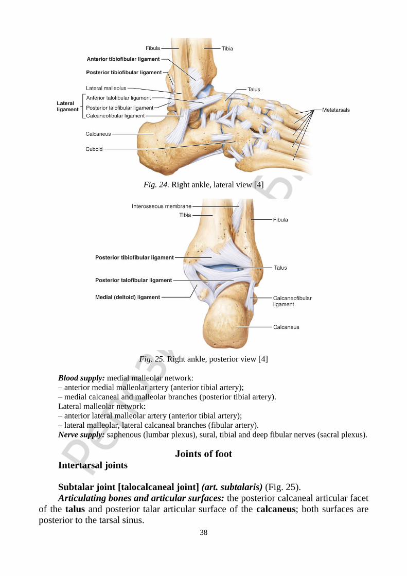
Ligaments:
● medial collateral ligament, syn. deltoid ligament (lig. collaterale mediale, seu
lig. deltoideum), consists of 4 parts:
– tibionavicular ligament, lig. tibionaviculare;
– anterior and posterior tibiotalar ligaments (lig. talotibiale anterius et
posterius);
– tibiocalcaneal ligament (lig. calcaneotibiale);
● lateral collateral ligament (lig. collaterale laterale), consists of 3 parts:
– anterior and posterior talofibular ligaments (lig. talofibulare anterius et
posterius);
– calcaneofibular ligament (lig. calcaneofibulare).
Notes: Dorsiflexion refers to the extension of the foot, i.e. the movement of
the foot upward towards the lower leg.
The ankle joint is more stable when the foot is dorsiflexed, when the wider front
of the trochlea of talus is firmly held by the malleoli; in plantar flexion the narrower
posterior portion of the trochlea moves between the malleoli and the joint becomes
less stable (allowing slight side-to-side movement of the talus).
Fig. 23. Right ankle, medial view [4]
38
Fig. 24. Right ankle, lateral view [4]
Fig. 25. Right ankle, posterior view [4]
Blood supply: medial malleolar network:
– anterior medial malleolar artery (anterior tibial artery);
– medial calcaneal and malleolar branches (posterior tibial artery).
Lateral malleolar network:
– anterior lateral malleolar artery (anterior tibial artery);
– lateral malleolar, lateral calcaneal branches (fibular artery).
Nerve supply: saphenous (lumbar plexus), sural, tibial and deep fibular nerves (sacral plexus).
Joints of foot Intertarsal joints
Subtalar joint [talocalcaneal joint] (art. subtalaris) (Fig. 25).
Articulating bones and articular surfaces: the posterior calcaneal articular facet
of the talus and posterior talar articular surface of the calcaneus; both surfaces are
posterior to the tarsal sinus.

39
Classification. Functionally combined with talocalcaneonavicular and
calcaneocuboid joints. Plane. Functionally uniaxial.
Movements: inversion and eversion — gliding and rotation around the sagittal
axis.
Ligaments:
– talocalcaneal interosseous ligament (lig. talocalcaneum interosseum);
– lateral, medial, posterior, and anterior talocalcaneal ligaments (ligg.
talocalcaneum laterale, mediale, posterior and anterior);
Comments: inversion is turning the whole sole of the foot inward; and eversion
is turning the soles outward.
The strongest talocalcaneal interosseous ligament lies within the tarsal sinus.
Blood supply: see ankle joint.
Nerve supply: sural, medial plantar and posterior tibial nerves (sacral plexus).
Talocalcaneonavicular joint (art. talocalcaneonavicularis) (Fig. 26, 27).
Articulating bones and articular surfaces. The head of talus articulates
inferiorly, anterior to the tarsal sinus, with the calcaneus (including
the sustentaculum tali) and the plantar calcaneonavicular ligament; anteriorly — with
the navicular bone.
Classification. Functionally combined with the subtalar and calcaneocuboid
joints. Ball and socket (the talonavicular articulation). Multiaxial.
Movements of the foot:
– inversion and eversion — rotation around the sagittal axis;
– abduction and adduction — around the vertical axis in the transverse plane
(specifically for the foot);
– plantarflexion and dorsiflexion (minor) — around the transverse axis, in
the sagittal plane.
Ligaments:
– plantar calcaneonavicular ligament, syn. spring ligament (lig.
calcaneonaviculare plantare);
– talonavicular ligament (lig. talonaviculare);
– plantar calcaneonavicular ligament (lig. calcaneonaviculare plantare);
– talocalcaneal interosseous ligament (lig. talocalcaneum interosseum).
Notes: the plantar calcaneonavicular ligament runs from the anterior part of
the sustentaculum tali to the plantar surface of the navicular bone and forms the part
of the articular “socket” supporting the talar head. It plays an important role in
maintaining the longitudinal arch of the foot.
Blood supply: medial and lateral malleolar networks; plantar metatarsal arteries (lateral
plantar artery — posterior tibial artery); lateral and medial tarsal arteries (dorsalis pedis artery —
anterior tibial artery).
Nerve supply: medial plantar and deep fibular nerves (sacral plexus).

40
Calcaneocuboid joint (art. calcaneocuboidea) (Fig. 26, 27).
Articulating bones and articular surfaces: the articular facets on the anterior
surface of the calcaneus and the posterior surface of the cuboid bone.
Classification. Simple. Saddle. Biaxial.
Movements (limited):
– inversion-eversion — rotation around the sagittal axis;
– contribution to other movements of the foot.
Ligaments:
– plantar and dorsal calcaneocuboid ligament (lig. calcaneocuboideum plantare
et dorsale);
Fig. 26. Ligaments of the foot, anterior view [3]

41
Fig. 27. Ligaments and tendons of the foot, plantar view [3]
Blood supply: plantar metatarsal arteries (lateral plantar artery from posterior tibial artery);
lateral tarsal artery (dorsalis pedis artery from anterior tibial artery).
Nerve supply: lateral plantar nerve, sural and deep fibular nerves (sacral plexus).

42
Transverse tarsal joint [Midtarsal, Chopart joint] (art. transversa tarsi)
(Fig. 26).
The joint is composed of two separate joints: the talonavicular part of
the talocalcaneonavicular joint and the calcaneocuboid joint.
Movements:
– flexion and extension, more extensive than in the other tarsal joints;
– inversion and eversion.
Ligaments: ● bifurcate ligament (lig. bufircatum), Y-shaped, serves for both parts of
the transverse tarsal joint, has two components:
– calcaneonavicular ligament;
– calcaneocuboid ligament;
● long plantar ligament (lig. plantare longum);
● ligaments reinforcing the talonavicular and calcaneocuboid joints (described
above).
Notes: the Chopart joint is a place where partial foot amputation can be
performed.
Cuneonavicular joint (art. cuneonavicularis) (Fig. 26).
Articulating bones and articular surfaces: the anterior surface of the navicular
bone and facing it facets of the middle, intermediate and lateral cuneiform bones.
Classification. Complex. Plane. Nonaxial.
Movements: limited gliding and rotation.
Ligaments: dorsal and plantar cuneonavicular ligaments (ligg. cuneonavicularia
dorsalia et plantaria).
Other intertarsal joints (Fig. 26):
– cuboideonavicular joint (art. cuboideonavicularis);
– cuneocuboid joint (art. cuneocuboidea);
– intercuneiform joints (artt. intercuneiformes).
These joints are formed by the side surfaces of the adjacent bones. They are
plane joints, reinforced by the dorsal, plantar and interosseous ligaments; allow
limited gliding and rotating movements.
Blood supply: plantar metatarsal arteries (lateral plantar artery from posterior tibial artery);
lateral and medial tarsal arteries (dorsalis pedis artery from anterior tibial artery).
Nerve supply: deep fibular (dorsally), medial and lateral plantar nerves (plantar surface)
(sacral plexus).
Tarsometatarsal joints [Lisfranc’s joint] (artt. tarsometatarseae) (Fig. 26).
Articulating bones and articular surfaces: the anterior articular facets of
the distal row of the tarsal bones: cuneiform (middle, intermediate, lateral) and
cuboid bones; the bases of the metatarsal bones.
Comprises three separate joints: 1) between the I metatarsal bone and medial
cuneiform bone; 2) between the II and III metatarsals and intermediate and lateral
cuneiform bones; 3) between the IV and V metatarsals and cuboid bone.

43
Classification. Complex. Plane. Nonaxial.
Movements: slight gliding movements. The joint of the I metatarsal bone allows
a wider range of movements (flexion-extension and rotation).
Ligaments:
– dorsal and plantar tarsometatarsal ligaments (ligg. tarsometatarsea dorsalia et
plantaria);
– cuneometatarsal interosseous ligaments (ligg. cuneometatarsea interossea).
Notes: the Lisfranc’s joint is the level of distal partial amputation of the foot.
The medial cuneometatarsal ligament (from the medial cuneiform to the II metatarsal
bone) being the strongest is the key of the Lisfranc’s joint.
Blood supply: deep plantar arch (lateral and medial plantar arteries); arcuate artery (dorsalis
pedis artery — anterior tibial artery).
Nerve supply: deep fibular nerve (dorsally), medial and lateral plantar nerves (plantar surface)
(sacral plexus).
Intermetatarsal joints (artt. intermetatarseae) (Fig. 26, 27).
Articulating bones and articular surfaces: the adjacent surfaces of the metatarsal
bones bases.
Classification. Simple. Plane. Nonaxial.
Movements: limited gliding.
Ligaments: dorsal, plantar, and interosseous metatarsal ligaments (ligg.
metatarsea dorsalia, plantaria, and interossea).
Blood and nerve supply: see tarsometatarsal joints.
Metatarsophalangeal joints (artt. metatarsophalangeae) (Fig. 26, 27).
Articulating bones and articular surfaces: the heads of the metatarsals and
the bases of the proximal phalanges of foot.
Classification. Simple. Ellipsoid. Biaxial.
Movements:
– flexion and extension — around the transverse axis, in the sagittal plane;
– abduction and adduction — around the vertical axis, in the transverse plane
(specifically for the foot);
– circumduction.
Ligaments:
– collateral and plantar metatarsophalangeal ligaments (ligg.
metatarsophalangea collateralia et plantaria);
– deep transverse metatarsal ligament (lig. metatarseum transversum
profundum).
Blood supply: plantar metatarsal arteries (deep plantar arch); dorsal metatarsal arteries
(arcuate artery).
Nerve supply: medial and lateral plantar nerves, superficial and deep fibular nerves (sacral
plexus).
44
Interphalangeal joints of foot (artt. interphalangeae pedis) (Fig. 27).
Include: proximal and distal interphalangeal joints of foot, interphalageal
joint of great toe.
Articulating bones and articular surfaces: the heads of phalanges and
the bases of more distally located phalanges.
Classification. Simple. Hinge. Uniaxial.
Movements: flexion and extension of the phalanges — around the transverse
axis, in the sagittal plane.
Ligaments:
– collateral interphalangeal ligaments of foot (ligg. interphalangea collateralia
pedes);
– plantar interphalangeal ligaments (ligg. interphalangea plantaria).
Blood supply: – plantar digital arteries (plantar metatarsal arteries);
– dorsal digital arteries (dorsal metatarsal arteries).
Nerve supply: medial and lateral plantar nerves, superficial and deep fibular nerves (sacral
plexus).

45
LITERATURE
1. https://upload.wikimedia.org/wikipedia/commons/e/ed/909_Types_of_Synovial_Joints.jpg
2. Atlas of anatomy / A. M. Gilroy [et al.]. Stuttgart : Thieme, 2008. 450 p.
3. Gray’s atlas of anatomy / R. L. Drake, [et al.]. 2nd ed. 2015.
4. Marieb, E. N. Human anatomy / E. N. Marieb, P. B. Wilhelm, J. Mallatt. 6th ed. Media
update, 2012.
5. Moore, K. L. Clinically oriented anatomy / K. L. Moore. Wolters Kluver, 2017. 1168 p.
6. Pocket Atlas of Human Anatomy Founded Heinz Feneis / ed. by W. Dauber. Stuttgart,
2007. 545 p.
7. Netter, F. H. Atlas of human anatomy / F. H. Netter. 5th ed., 6th ed., 7th ed. USA :
Elsevier, 2011, 2014, 2019. 578 p.
8. Sapin, M. R. Texbook of hyman anatomy. In 2 vol. / M. R. Sapin. Moscow, 2017. Vol. 1.
416 p.
9. Sapin, M. R. Texbook of hyman anatomy. In 2 vol. / M. R. Sapin. Moscow, 2017. Vol. 2.
480 p.
10. Terminologia Anatomica. 2nd ed. 2019. https://www.ifaa.net/committees/anatomical-
terminology-fipat.
11. Гайворонский, И. В. Нормальная анатомия человека. В 2 т. / И. В. Гайворонский.
Санкт-Петербург : СпецЛит, 2000.
12. Калмин, О. В. Артрология : учеб.-метод. пособие / О. В. Калмин, Т. Н. Галкина,
И. В. Бочкарева. Пенза : ИИЦ ПГУ, 2003. 68 с.
13. Кровоснабжение и иннервация суставов человека / В. Н. Андриеш [и др.].
Кишинев, 2001. 344 с.
14. Привес, М. Г. Анатомия человека / М. Г. Привес, Н. К. Лысенков, В. И. Бушкович.
Санкт-Петербург : Гиппократ, 2000.
15. Синельников, Р. Д. Атлас анатомии человека. В 4 т. / Р. Д. Синельников,
Я. Р. Синельников. Москва : Медицина, 1996.
16. Международная анатомическая терминология / под ред. Л. И. Колесникова. Москва :
Медицина, 2003. 4214 с.

46
CONTENTS
GENERAL CHARACTERISTICS OF JOINTS .......................................................... 3
JOINTS OF VERTEBRAL COLUMN ......................................................................... 8
JOINTS OF THORAX ................................................................................................ 12
JOINTS OF SKULL .................................................................................................... 15
JOINTS OF UPPER LIMB ......................................................................................... 18
JOINTS OF LOWER LIMB ....................................................................................... 30
JOINTS OF FREE LOWER LIMB ............................................................................ 31
LITERATURE ............................................................................................................ 45

47
Учебное издание
Пасюк Анна Андреевна
Жарикова Ольга Леонидовна
Арден Франка Александровна
СОЕДИНЕНИЯ ТЕЛА ЧЕЛОВЕКА (КРАТКИЙ КУРС)
JOINTS OF HUMAN BODY (BRIEF COURSE)
Учебно-методическое пособие
На английском языке
Ответственная за выпуск Н. А. Трушель
Переводчики А. А. Пасюк, О. Л. Жарикова, Ф. А. Арден
Компьютерная вёрстка Н. М. Федорцовой
Подписано в печать 27.01.21. Формат 6084/16. Бумага писчая «Xerox performer».
Ризография. Гарнитура «Times».
Усл. печ. л. 5,58. Уч.-изд. л. 3,13. Тираж 120 экз. Заказ 46.
Издатель и полиграфическое исполнение: учреждение образования
«Белорусский государственный медицинский университет».
Свидетельство о государственной регистрации издателя, изготовителя,
распространителя печатных изданий № 1/187 от 18.02.2014.
Ул. Ленинградская, 6, 220006, Минск.

48