JCCA-v58-Full-issue.pdf - Canadian Chiropractic Association
100
J Can Chiropr Assoc 2014; 58(1) 1 CANADIAN CHIROPRACTIC ASSOCIATION President Jeff Warren, BSc, DC JCCA STAFF Editor Allan Gotlib, C.M., BSc, DC Canadian Chiropractic Association, Toronto, Ontario Associate Editors Jeff Quon, DC, PhD School of Population & Public Health Faculty of Medicine, University of British Columbia Kent Stuber, DC, MSc Calgary, Alberta Assistant Editors André Bussières, DC, FCCS(C), PhD Faculty of Medicine, McGill University Département chiropratique, Université du Québec à Trois-Rivières Pierre Côté, DC, PhD University of Ontario Institute of Technology Gregory N Kawchuk, DC, PhD University of Alberta, Edmonton, Alberta Dana J. Lawrence, DC, MMedEd, MA Palmer College of Chiropractic Davenport, Iowa John J. Triano, DC, PhD Canadian Memorial Chiropractic College Production Co-ordinator Tami Ehrlich Advertising Editor, Journal of the Canadian Chiropractic Association 186 Spadina Avenue, Suite 6, Toronto, Ontario M5T 3B2 Tel: 416-585-7902 877-222-9303 Fax: 416-585-2970 Email: Dr. Allan Gotlib<[email protected]> Website: www.jcca-online.org TYPESETTING Thistle Printing Limited 35 Mobile Drive, Toronto, Ontario M4A 2P6
-
Upload
khangminh22 -
Category
Documents
-
view
2 -
download
0
Transcript of JCCA-v58-Full-issue.pdf - Canadian Chiropractic Association

J Can Chiropr Assoc 2014; 58(1) 1
CANADIAN CHIROPRACTIC ASSOCIATIONPresident Jeff Warren, BSc, DC
JCCA STAFFEditor Allan Gotlib, C.M., BSc, DC
Canadian Chiropractic Association, Toronto, Ontario
Associate Editors Jeff Quon, DC, PhD School of Population & Public Health Faculty of Medicine, University of British Columbia
Kent Stuber, DC, MSc Calgary, Alberta
Assistant Editors André Bussières, DC, FCCS(C), PhD Faculty of Medicine, McGill University Département chiropratique, Université du Québec à Trois-Rivières
Pierre Côté, DC, PhD University of Ontario Institute of Technology
Gregory N Kawchuk, DC, PhD University of Alberta, Edmonton, Alberta
Dana J. Lawrence, DC, MMedEd, MA Palmer College of Chiropractic Davenport, Iowa
John J. Triano, DC, PhD Canadian Memorial Chiropractic College
Production Co-ordinator Tami Ehrlich
Advertising Editor, Journal of the Canadian Chiropractic Association 186 Spadina Avenue, Suite 6, Toronto, Ontario M5T 3B2 Tel: 416-585-7902 877-222-9303 Fax: 416-585-2970
Email: Dr. Allan Gotlib<[email protected]> Website: www.jcca-online.org
TYPESETTING Thistle Printing Limited
35 Mobile Drive, Toronto, Ontario M4A 2P6

2 J Can Chiropr Assoc 2014; 58(1)
JCCA Journal of the Canadian Chiropractic Association
(Formerly the Canadian Chiropractic Association Journal)
Copyright Registered © by the Canadian Chiropractic Association 1961
Copyright: The Canadian Chiropractic Association, 2014
All rights reserved. Without limiting the rights under copyright above, no part of this publication may be reproduced, stored in or introduced into any retrieval system, or transmitted in any form
or by any means (electronic, mechanical, photocopying, recording or otherwise), without the prior written permission with the copyright owner and the publisher.
Published by the Canadian Chiropractic Association and issued quarterly
EDITORIAL AND EXECUTIVE OFFICES,
General Information: The Journal of the Canadian Chiropractic Association is the official quarterly publication by the Canadian Chiropractic Association. The JCCA is published quarterly by the Canadian Chiropractic Association as a medium of communication be-tween the Association and its members and is a forum for fair comment and discussion of all matters of general interest to the chiropractic profession and the Association. Readers are invited to comment and express their opinions on relevant subjects. Views and opinions in editorials and articles are not to be taken as official expression of the Association’s policy unless so stated. Publication of contributed articles does not necessarily imply endorsement in any way of the opinions expressed therein and the Journal and its publisher does not ac-cept any responsibility for them. Business correspondence should be addressed to: the Edi-tor of JCCA, 186 Spadina Avenue, Suite 6, Toronto, Canada M5T 3B2.
INDEXING SERVICES
JCCA is indexed by PubMed Central, CINAHL (Cumulative Index to Nursing and Allied Health Literature), MANTIS (formerly CHIROLARS), AMED, PASCAL, British Library Complemen-tary Medicine Index, Index to Chiropractic Literature, and selectively by SPORTDiscus.
186 SPADINA AVENUE, SUITE 6, TORONTO, CANADA M5T 3B2

J Can Chiropr Assoc 2014; 58(1) 3
ContentsJCCA Vol 58 No 1 ISSN 0008-3194 (Print) and ISSN 1715-6181 (Electronic)
Commentaries 6 Evidence-based case reports Jennifer E. Bolton, PhD, MA (Ed), FHEA, FRCC(Hon), FBCA, FFEAC
8 Creating a Chiropractic Practice-Based Research Network (PBRN): Enhancing the management of musculoskeletal care
André Bussières, DC, FCCS (C), PhD Pierre Côté, DC, PhD Simon French, BAppSc(Chiropractic), MPH, PhD Marshall Godwin, MD, MSc, FCFP Allan Gotlib, C.M., BSc, DC Ian D Graham, PhD, FCAHS Diane Grondin, DC, MHK, PhD student (University of Toronto) Cheryl Hawk, DC, PhD Charlotte Leboeuf-Yde, DC, MPH, PhD Sil Mior, DC, FCCS (C), PhD Kent Stuber, DC, MSc
Original Articles16 Detection of syringomyelia in a pediatric patient with mild scoliosis: a case report Ismat Kanga, BSc, DC
Jessica J. Wong, BSc, DC, FCCS(C) Paula J. Stern, BSc, DC, FCCS(C)
24 A comparison of quality and satisfaction experiences of patients attending chiropractic and physician offices in Ontario
Edward R. Crowther, BA, DC, MS, EdD, FCCS
39 Duplicated right crus of the diaphragm: a cadaveric case report Srinivasa Rao Sirasanagandla, MSc
Satheesha B Nayak, MSc, PhD Kumar MR Bhat, MSc, PhD Sudarshan Surendran, MSc, PhD Deepthinath Regunathan, MSc, PhD Naveen Kumar, MSc Surekha D Shetty, MSc, BAMS Jyothsna Patil, MSc
45 A delayed diagnosis of bilateral facet dislocation of the cervical spine: a case report Julie O’Shaughnessy, DC, FCCS(C), MSc
Julie-Marthe Grenier, DC, DACBR/FCCR(C) Paula J. Stern, BSc, DC, FCCS(C)
52 Chiropractic management of elbow tendinopathy following a sports related trauma Jordan A. Gliedt, DC
Clinton J. Daniels, DC, MS

4 J Can Chiropr Assoc 2014; 58(1)
ContentsJCCA Vol 58 No 1 ISSN 0008-3194 (Print) and ISSN 1715-6181 (Electronic)
58 Financial attitudes, knowledge, and habits of chiropractic students: A descriptive survey Julie Lorence, DC, MS
Dana J. Lawrence, DC, MMedEd, MA Stacie A. Salsbury, PhD, RN Christine M. Goertz, DC, PhD
66 Ross E. Baker, DC: A Canadian chiropractic survivor Douglas M. Brown, DC
76 Conservative management of idiopathic anterior atlantoaxial subluxation without neurological deficits in an 83-year-old female: A case report
Andrée-Anne Marchand, DC Jessica J. Wong, BSc, DC, FCCS(C)
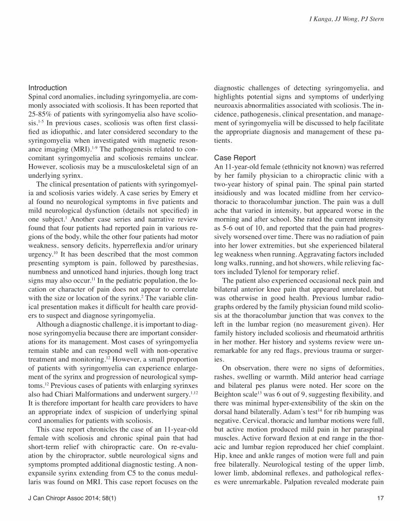
85 Pilot study of the impact that bilateral sacroiliac joint manipulation using a drop table technique has on gait parameters in asymptomatic individuals with a leg length inequality
John Ward, DC, MA, MS Ken Sorrels, DC, BA Jesse Coats, DC, BS, DAAPM, CCSP Amir Pourmoghaddam, PhD Carlos DeLeon, BS Paige Daigneault, BS
Letters to the Editor96 Carlo Ammendolia, DC, PhD
96 Marc-André Blanchette, DC, PhD candidate, Jan Hartvigsen, DC, PhD
97 Neilank K. Jha, MD, FRCS(C)
98 Mark Erwin, DC, PhD

J Can Chiropr Assoc 2014; 58(1) 5
Editorial BoardAlan H Adams DC Texas Chiropractic College Pasadena, Texas
Kelly E. Donkers Ainsworth DC, MD, FRCPC Staff Pediatric Radiologist McMaster University Hamilton, Ontario
Carlo Ammendolia DC, PhD University of Toronto
Samuel Bederman MD, PhD, FRCSC Department of Orthopedic Surgery University of California at Irvine Orange, CA
Paul Bruno DC, PhD Faculty of Kinesiology and Health Studies University of Regina
Brian Budgell DC, PhD CMCC
Jason Busse DC, PhD McMaster University
J David Cassidy DC, MSc, PhD, FCCS(C), Dr Med Sc University of Southern Denmark
Raphael K Chow MD, FRCP(C) University of Toronto
Colin M Crawford B App Sc (Chiro), FCCS(C), MSc, Grad Dip Neuro, MB BS Perth, Australia
Simon Dagenais DC, PhD Boston, Massachusetts
Martin Descarreaux DC, PhD Université du Québec à Trois-Rivières
John A. Dufton DC, MSc, MD, FRCPC Staff Radiologist University Hospital of Northern British Columbia Prince George, British Columbia
Mark Erwin DC, PhD University of Toronto
Brian Gleberzon DC, MHSc CMCC
Richard Goldford BSc, DC, MBA, FCCSS(C), FCCPOR(C) Toronto, Ontario
Bart Green DC, MSEd, DACBSP Naval Medical Center, San Diego San Diego, California
François Hains DC, FCCS(C), MSc Dorval, Québec
Scott Haldeman DC, MD, PhD, FRCP(C) University of California Irvine, California
Jill Hayden DC, PhD Dalhousie University Halifax, NS
Walter Herzog PhD University of Calgary
Thomas E Hyde BA, DC, DACBSP N Miami Beach, Florida
Claire Johnson DC, MSEd, DACBSP National University of Health Sciences Lombard, Illinois
Mohsen Kazemi RN, DC, FCCSS(C), FCCPOR(C), MSc CMCC
Clark R Konczak MSc, DC, DABCO, FCCO(C), FRCCSS(C) Victoria, BC
Deborah Kopansky-Giles DC, FCCS(C), FICC, MSc St. Michael’s Hospital Toronto, Ontario
Doug M Lawson BA, DC, MSc D’Youville College
Cynthia Long PhD Palmer Centre for Chiropractic Research Davenport, Iowa
Marion McGregor DC, PhD CMCC Toronto, Ontario
William C Meeker DC, MPH Palmer Chiropractic University System San Jose, CA
Robert D Mootz DC Associate Medical Director for Chiropractic, State of Washington Department of Labor and Industries Olympia, WA
Bernadette Murphy DC, PhD University of Ontario Institute of Technology
Martin Normand DC, PhD UQTR
Steven Passmore DC, PhD Faculty of Medicine University of Manitoba
Stephen Perle DC, MS University of Bridgeport Bridgeport, CT
Reed B Phillips DC, PhD, DACBR Southern California University of Health Sciences
Mathieu Piché DC, PhD UQTR
John J Riva DC, MSc Department of Family Medicine McMaster University Hamilton, Ontario
John Z Srbely DC, PhD University of Guelph
Igor Steiman MSc, DC, FCCS(C) CMCC
John S Stites DC, DACBR Palmer College of Chiropractic Davenport, Iowa
Donald C Sutherland DC, LLD, FICC CMCC
John A M Taylor DC, DACBR, FCCR(C) D’Youville College Buffalo, NY
Haymo Thiel DC, MSc (Orth), FCCS(C), Dip Med Ed, PhD Anglo-European College of Chiropractic Bournemouth, England
Gabrielle M van der Velde BSc, DC, FCCS(C), PhD Toronto Health Economics and Technology Assessment Collaborative University of Toronto
Marja J Verhoef PhD University of Calgary

6 J Can Chiropr Assoc 2014; 58(1)
Commentary
Professor Jennifer Bolton, PhD, MA (Ed)*
Anglo-European College of Chiropractic Bournemouth, UK
Clinicians love to read case reports because they tell a good story (and who doesn’t like a good story?), they are relevant to their day-to-day practice and, by implication, they are interesting and informative. In some cases they have quickly alerted clinicians and the general public to unsafe practice, as was the case in the use of thalidomide in early pregnancy in the 1960s, and new cases of disease, such as HIV/AIDS in the 1980s. Yet, case reports are con-
sidered very weak evidence in the hierarchy of research evidence, not least because the information they provide has not been tested by rigorous scientific means. How-ever, we might take issue with this stand in that not only are case reports informative, but they are particularly rel-evant to clinical practice from whence they came. Since the shift towards evidence-based practice (EBP) in the 1990s1, there has been continuing debate on the relevance of findings from research studies, most often based on group data, in the management of an individual patient. Increasingly, the RCT has become divorced from normal patient populations by its insistence on the inclu-sion of patients defined by narrow criteria and absence of co-morbidities. Moreover, the original definition of EBP that included clinician experience and characteristics of individual patients as well as sound research evidence in the decision-making process has been slowly hijacked over the years to the extent that ‘evidence-based prac-tice’ is now synonymous with ‘research evidence-based practice’ and perhaps more alarmingly, only that evidence generated by the hallowed RCT. We need to get back to a model of EBP that is not only inclusive of all types of evi-dence, but one that is patient-centred and that can be used in the management of an individual patient. The fault in narrow interpretations of EBP lies not with the model it-self, but with uninformed, misinformed and biased inter-pretations of it. As part of the postgraduate Masters programmes at AECC in Bournemouth, UK2 we run a distance-based course in EBP that fosters the combination of research findings with clinician experience, and the application of this synthesis in the management of an individual patient.
*Professor Jennifer E. Bolton, PhD, MA (Ed), FHEA, FRCC(Hon), FBCA, FFEAC Vice-Principal (Postgraduate and Research) http://www.aecc.ac.uk/cpdandpostgraduate Anglo-European College of Chiropractic 13-15 Parkwood Road, Bournemouth, Dorset, BH5 2DF Tel 44 (0)1202 436244 Email: [email protected] ©JCCA 2014
Evidence-based case reportsProfessor Jennifer Bolton, PhD, MA (Ed)*

J Can Chiropr Assoc 2014; 58(1) 7
J Bolton
It seems to us that this is the essence of EBP in any clin-ical discipline, including chiropractic. As part of our EBP course, students are required to produce an evidence-based case report (EBCR). This requires the student to adopt a systematic and evidence-based approach to case management. In the EBCR, the student describes a patient presentation in much the same way as in a traditional case report. Out of this, the student articulates a structured clinical question in a format that generates key terms that can be used to search the research evidence base. In the EBCR, unlike the traditional case report, the search strategy is described so that the read-er can decide whether a comprehensive search has been conducted, much in the same way as for a systematic re-view. Once the relevant evidence has been identified and appraised, it is synthesised together with the clinician’s experiential knowledge to inform a management plan for the patient. It seems to us that the EBCR encompasses all the steps of an evidence-based approach to practice, in particu-lar appreciating the role of clinician experience in EBP. Sometimes, there is no research evidence, or what there is may not be good enough so that patient management
is based entirely on clinician experience. What is not ac-ceptable however, is that when there is good research evi-dence, then this is either ignored or dismissed. EBCRs remain interesting reading for clinicians while at the same time informing clinicians of the available re-search evidence (if any), and perhaps more importantly how the research evidence can be applied to the care of an individual patient. This gap between clinical research and clinical practice is arguably as wide today as it has ever been; the EBCR is just one way of bridging this gap. Of course, the EBCR is not a new concept. EBCRs are pub-lished by the British Medical Journal3 among others. Now we need to see more EBCRs in our chiropractic journals.
References1. Sackett et al. Evidence-based practice: a new
approach to teaching the practice of medicine. JAMA. 1992;268(17):2420-5.
2. Anglo-European College of Chiropractic. http://www.aecc.ac.uk/cpd/postgrad/
3. Godlee F. Applying research evidence to individual patients. BMJ. 1998;316:1621-2.

8 J Can Chiropr Assoc 2014; 58(1)
Commentary
1 Canadian Chiropractic Research Foundation Professorship (CCRF) in Rehabilitation Epidemiology Assistant Professor, School of Physical and Occupational Therapy, McGill University, Montreal. Quebec, Canada Professor, Département chiropratique, Université du Québec à Trois-Rivières, Trois-Rivières, Québec, Canada Editor, The Canadian Chiropractic Guideline Initiative Assistant Editor, Journal of the Canadian Chiropractic Association
2 Canada Research Chair in Disability Prevention and Rehabilitation Associate Professor, Faculty of Health Sciences, University of Ontario Institute of Technology (UOIT) Director, UOIT-CMCC Centre for the Study of Disability Prevention and Rehabilitation Associate Professor of Epidemiology, Dalla Lana School of Public Health, University of Toronto Assistant Editor, Journal of the Canadian Chiropractic Association
3 Canadian Chiropractic Research Foundation (CCRF) Professorship in Rehabilitation Therapy Assistant Professor, School of Rehabilitation Therapy, Queen’s University, Kingston, Ontario, Canada. NHMRC Research Fellow, General Practice and Primary Health Care Academic Centre, University of Melbourne, Melbourne, Australia Associate Editor, Journal Chiropractic & Manual Therapies
4 Professor, Family Practice Unit Director, Primary Healthcare Research Unit Faculty of Medicine, Memorial University, NF, Canada
5 Director, Research Programs, Canadian Chiropractic Association Executive Vice-President, Canadian Chiropractic Research Foundation Editor, Journal of the Canadian Chiropractic Association
6 Professor, Department of Epidemiology and Community Medicine, University of Ottawa, Ottawa, Canada Senior Scientist, Clinical Epidemiology Program, Ottawa Hospital Research Institute, Ottawa, Canada
7 Assistant Professor, Canadian Memorial Chiropractic College, Toronto, Canada. Adjunct Assistant Professor at the University of Ontario Institute of Technology, Toronto, Canada
8 Dean of Research, Logan University, MO, United States 9 Research Director, Institut Franco Europeen de Chiropratique, Paris, France
Professor in Clinical Biomechanics, University of Southern Denmark, Odense, Denmark Visiting Professor, Université Paris Sud, Paris, France Distinguished Collaborator, Adjunct Professor, Murdoch University, Perth, Australia
10 Senior Advisor to the President at Canadian Memorial Chiropractic College Research Scientist, Department of Research, CMCC, Toronto, Canada
11 Adjunct Professor, Division of Graduate Education & Research, Canadian Memorial Chiropractic College, Calgary, Alberta, Canada Associate Editor, Journal of the Canadian Chiropractic Association
©JCCA 2014
Creating a Chiropractic Practice-Based Research Network (PBRN): Enhancing the management of musculoskeletal careAndré Bussières, DC, FCCS (C), PhD1 Pierre Côté, DC, PhD2 Simon French, BAppSc(Chiropractic), MPH, PhD3 Marshall Godwin, MD, MSc, FCFP4 Allan Gotlib, C.M., BSc, DC5 Ian D Graham, PhD, FCAHS6 Diane Grondin, DC, MHK, PhD student7 Cheryl Hawk, DC, PhD8 Charlotte Leboeuf-Yde, DC, MPH, PhD9 Silvano Mior, DC, FCCS (C), PhD10 Kent Stuber, DC, MSc11

J Can Chiropr Assoc 2014; 58(1) 9
A Bussières, P Côté, S French, M Godwin, A Gotlib, ID Graham, D Grondin, C Hawk, C Leboeuf-Yde, S Mior, K Stuber
IntroductionChiropractic is a regulated health profession currently serving approximately 10% of the Canadian population annually1 with the aim to improve the health and well-being of Canadians, primarily with musculoskeletal dis-orders. Despite available evidence for optimal manage-ment of these disorders,2-4 poor adherence to guidelines and wide variations in service delivery by clinicians have been noted across health care disciplines,3,5 including chiropractic.6,7
Efforts to embrace and enhance evidence-based prac-tice among chiropractors and develop opportunities for multi-disciplinary research collaboration have been ham-pered by a number of issues. Issues include: 1) limited research capacity in chiropractic with less than 1% of the chiropractic profession conducting research;8 2) frag-mented integration of chiropractic into the health care system that has been hampered by discrepancies among practising chiropractors, chiropractic researchers, and regulatory bodies over scope and paradigm of practice (alternative or empiricist/experiential-based vs. evidence-based practice);9,10 3) over half of chiropractors are in solo practice11 with solo providers having greater variation in accepted clinical practices;12 and 4) perceived suboptimal coordination of efforts from professional associations, regulatory boards and chiropractic teaching institutions to successfully implement evidence into practice. One strategy to address these issues is the creation of practice-based research networks (PBRNs). Primary care PBRNs bring together researchers and groups of clin-icians and practices with the goal of improving health ser-vices delivery and closing the gap between research and practice.13-16 The general aim is to stimulate the develop-ment of appropriate research that reflects the context of healthcare practice in a primary care setting.17
Do PBRNs provide an effective approach to develop and support research?While a number of approaches to assess the develop-ment and impact of primary care networks have been pro-posed,18,19 there is currently no generic and validated tool that enables meaningful comparison between different net-work models.20 Nonetheless, a growing body of research supports the role of PBRNs in promoting health care qual-ity.15,21,22 Still, a formal evaluation of the effectiveness of PBRNs in the area of musculoskeletal disorders is needed.
Perceived strengths and weaknesses of practice-based research networksA PBRN founded upon an integrated knowledge trans-lation framework and a participatory approach can: 1) promote culturally and logistically appropriate and useful research; 2) enhance recruitment capacity in research; 3) generate professional capacity and competence in stake-holder groups; 4) result in productive conflicts followed by useful negotiation; 5) increase the quality and gener-alizability of research output, and offer numerous advan-tages to clinicians over time (e.g., growth of skills and expertise, sense of empowerment, increase satisfaction, career development); 6) increase the sustainability of pro-ject goals beyond funded time frames and during gaps in external funding; and 7) create system changes and new unanticipated projects and activities.23,24 Primary care PBRNs provide a unique opportunity to engage clinicians in quality improvement activities, create an evidenced based practice culture, and improve patient care.14
PBRNs are well established in other primary health-care professions in Canada. Despite their acceptance, there are barriers that influence their sustainability. In family practice, perceived barriers that hamper participa-tion in PBRN include lack of time, inadequate training in research methods, lack of collaborators and support staff, institutional review board hurdles, and community dis-trust of research.13,23 Additional barriers that particularly face complementary and alternative health care providers include the lack of resources (e.g., funding, compensa-tion, infrastructure and partnerships/linkages), environ-mental (e.g., the nature of a clinic’s patient population) and logistical issues (e.g., the actual implementation of a research program and the applicability of research data).25
Creating a chiropractic practice-based research network in CanadaThere is a growing need to establish a formal network of Canadian chiropractors to facilitate the translation of research into practice to improve the quality and safety of patient care, primarily in the management of muscu-loskeletal conditions. In 2014, we plan to assemble key stakeholders, including academics, elected professional provincial and national leaders, clinicians, government policy advisors, insurers, and patients, to explore the fac-tors critical to establishing and implementing a Canadian chiropractic PBRN. The mission of this PBRN is to im-

10 J Can Chiropr Assoc 2014; 58(1)
Commentary
prove chiropractic health care delivery and patient health in Canada through research and quality-improvement in-itiatives. A PBRN that includes a formal collaboration be-tween patients, health professionals, elected professional provincial and national leaders and health researchers from across Canada can help bridge the gap between re-search evidence and health care practice.26,27
Targeted health conditions and strategy to improve care within the proposed PBRN
Burden of musculoskeletal disordersMusculoskeletal conditions are one important reason pa-tients consult primary care professionals including gen-eral practitioners and chiropractors.28 Musculoskeletal conditions (spinal pain, consequences of injuries, osteo-porosis, and arthritis) result in enormous social, psycho-logical, and economic burden to society.28-37 They are a leading cause of pain and disability, resulting in extensive utilization of Canadian health care resources.38-40 In Can-ada, the total economic burden of musculoskeletal condi-tions ranks second only to cardiovascular disease and are the most costly disease for women and third most costly for men.41 The total economic burden has been estimated to be about $16.4 billion when considering both indirect costs ($13.7 billion) and direct costs ($2.6 billion)41 per year. The largest component of expenditures is related to morbidity and long-term disability. The substantial bu-rden associated with musculoskeletal disorders is com-pounded by suboptimal clinical management and the risk of clinical iatrogenesis.42-44 This highlights the need for rigorous knowledge translation science in the primary care setting to improve chiropractic patient outcomes. PBRNs provide an infrastructure for the dissemination and implementation of research evidence. PBRNs are particularly useful considering the highly heterogeneous therapeutic approaches offered by chiropractors and other primary care professionals when dealing with musculo-skeletal conditions.3,5-7
How can we improve process of care and patient outcomes?Clinical Practice Guidelines (CPGs) are an important way to improve the quality and safety of healthcare through the implementation of research findings.45 The Canadian chiropractic profession has been proactive in developing
CPGs over the past two decades.46-48 However, simple dissemination of CPGs cannot overcome the various bar-riers to clinician adherence.49 Instead, their successful implementation is more likely when evidence is scientif-ically robust; clinically relevant; the context is receptive to change within sympathetic cultures; and appropriate monitoring, feedback systems and strong leadership are in place.50 Recent advances in methods to conduct know-ledge synthesis, derive evidence-based recommendations, adapt high quality guidelines, and increase the uptake of CPGs have prompted an update of the structure, methods and procedures for the development, dissemination and implementation of CPGs in chiropractic in Canada.51
One approach to improve the uptake of CPGs is access-ing PBRNs. PBRNs have the potential to increase the up-take of best practice because they “aim to share informa-tion and create new knowledge, strengthen research and communication capacity among members, and identify and implement strategies to engage decision makers more directly.”52 Currently, routinely collecting administrative and clinical outcomes in Canadian chiropractic practices is not feasible. In part this is due to limited coverage from provincial health plans and the rare use of electronic med-ical records (EMR). Establishing a PBRN can provide the structure to recruit clinicians, profile chiropractic prac-tice, identify knowledge-practice gaps, monitor practice change, and evaluate the impact of knowledge transla-tion (KT) strategies to increase uptake of evidence-based practice. Collectively, CPGs and PBRNs can provide the structure and processes to improve care delivery and pa-tient outcomes.
Relevance to national health research prioritiesThe national chiropractic research agenda is harmonious with the Canadian Institutes of Health Research’s (CIHR) mandate (CIHR is the major health research funding agency in Canada). Its mandate is to “excel, according to internationally accepted standards of scientific excel-lence, in the creation of new knowledge and its transla-tion into improved health for Canadians, more effective health services and products and a strengthened Canadian health care system.”53 This mandate is congruent with the need to develop a well-articulated national chiropractic research agenda. The agenda should include the facilita-tion of collaborative, multi-disciplinary health research designed to improve the way chiropractic services are or-

J Can Chiropr Assoc 2014; 58(1) 11
A Bussières, P Côté, S French, M Godwin, A Gotlib, ID Graham, D Grondin, C Hawk, C Leboeuf-Yde, S Mior, K Stuber
ganized, managed and delivered to improve the quality and effectiveness of care provided to Canadians.54,55 The development of this research agenda is supported by the Consortium of Canadian Chiropractic Research Centres whose main purpose is to coordinate chiropractic research capacity in Canada and facilitate the development of new chiropractic knowledge through multi-disciplinary and multi-institutional collaboration, and its dissemination to health providers and health policy makers with even-tual integration into the health care system.54 A Canadian PBRN can provide a strategic framework from which to operationalize the above agendas. A PBRN also promotes the exchange of knowledge be-tween partners of the Network. Establishing a Canadian chiropractic PBRN aligns well with CIHR’s Strategy for Patient-Oriented Research (SPOR) vision to improve health outcomes and enhance the health care experience for patients through the integration of evidence at all lev-els of the health care system, focus on patient-oriented research networks, and improve guideline development, dissemination and uptake.27 This SPOR Network will support evidence-informed transformation and delivery of more cost-effective and integrated health care to im-prove clinical, population health, health equity, and health system outcomes. The Patient-Oriented Community-Based Primary Healthcare (CBPHC) is one of eight Roadmap Signature Initiatives recently announced by CIHR.56 CBPHC Net-work is one of several networks that will be funded as part of Canada’s Strategy for SPOR. CBPHC covers a range of services across the continuum of care – primary prevention (including public health) and primary care services from health promotion and disease prevention, chronic disease diagnosis, treatment and management to rehabilitation support, home care and end-of-life care. Networks under this initiative will be expected to obtain funding from multiple sources and to engage national as-sociations, health charities, clinicians, industry, patients and the public.
Proposed approachPBRN’s have been successfully created in the US57-59, in Denmark60, and in Canada61 for more than 15 years. Re-searchers have identified the necessary components for a PBRN as infrastructure (including training in data collec-tion by a full-time coordinator), practitioner-researcher
partnership, centralized data management by the research centre, and standardized quality assurance measures.60,62,63 Other desirable elements of a PBRN infrastructure in-clude support staff, electronic medical records, multiuser databases, mentoring and development programs, mock study sections, and research training.64 The infrastructure of the proposed chiropractic PBRN will be elaborated based upon these recommendations. Furthermore, a number of procedures used for plan-ning and implementing PBRN research studies will be adapted from previous work60,65, including how to select fundable, feasible studies; compose the study team; re-cruit and select sites; and train practice staff and clin-icians. Clinicians will be involved throughout the process from identifying research questions whose answers may lead to improvements in clinical practice, recruitment of patients, and data collection.66,67 Various existing primary care PBRN-relevant toolkits proposed by the Agency for Healthcare Research Quality may also be used.68 These include: implementing the chronic care model; health lit-eracy and research toolkits, informed consent and author-ization for minimal risk research, patient safety, practice facilitation handbook and manual, state-specific health care quality information, office survey on patient safety culture, workflow assessment for health IT, and a written materials toolkit. Peterson et al. recently described a model for the de-velopment of an electronic infrastructure to support clin-ical research activities in primary care PBRNs.69 The au-thors suggest that the potential for introducing a fast and efficient infrastructure to facilitate PBRN research offers the possibility of rapid advances in a wide variety of areas including comparative effectiveness research, patient safety, event monitoring for drugs and devices, and clin-ical trials. The Canadian Memorial Chiropractic College has successfully pilot-tested an EMR system within its six outpatient clinics. In the future, a similar EMR may be implemented across participating PBRN practices to ease data collection. Types of outcome indicators used to assess the success of PBRNs include structural (organizational), process and clinical indicators.20,24 PBRN members will identify a core set of indicators felt to be most relevant to the ob-jectives of the chiropractic PBRN. Structural indicators may include the number of active clinicians/practices, a multidisciplinary membership, creating research lead-

12 J Can Chiropr Assoc 2014; 58(1)
Commentary
ers, embedding a research culture in the organization, and providing career development opportunities. Process indicators could include the degree of research aware-ness, numbers of trained members in research method, success rate in grant applications, number of collabora-tive projects and completed research projects, numbers of peer-reviewed publications and conference presentations. Clinical or quality of care outcome indicators (e.g., appro-priate x-ray utilization rate for back and neck pain) and important patient reported health outcomes (e.g., levels of pain and disability, return to work and satisfaction with care) will also be identified.
Members of the NetworkA PBRN should engage four groups including patients (citizen engagement), clinicians (knowledge-users), lead-ers and decision-makers (provincial and national leaders in the profession and decision-makers from insurance and government), and researchers including CPG developers and KT experts. i) Patients: Meaningful patient involvement can be ensured by recruiting individuals who are familiar with the diversity of the chiropractic profession and have been involved in previous chiropractic forums. Patient (public) members at ‘Level Three’ should be included, as described in the Health Council of Canada’s “Primer on Public In-volvement” (2006).70 The intent of citizen engagement is to: ‘encourage end users participation throughout the research process so that they can inform the study ques-tion and research plan, and be involved in interpreting the findings, in crafting the dissemination messages, and in applying the results’.71
ii) Clinicians: Canadian chiropractors interested and involved in clinical research will be actively engaged in various activities and projects of the PBRN. Participat-ing clinicians will be involved throughout the process from identifying research questions whose answers lead to improvements in clinical practice and patient health outcomes, recruitment of patients, and data collection.66,67 Participating in a PBRN can be rewarding in many ways. These include an opportunity to connect with likeminded and unlike minded colleagues, help the profession build the evidence base for its patients and colleagues, and al-low for an increased likelihood of successful uptake of new knowledge into practice for the benefit of patients. iii) Professional provincial and national leaders and
Government and insurance policy advisors: Leaders/de-cision makers from the thirty-six chiropractic organiza-tions in Canada should also be included to improve co-ordination of efforts toward implementing evidence into practice and to provide congruent messages to clinicians. These individuals include elected leaders and representa-tives from: national and provincial chiropractic associa-tions and regulatory boards; the professional liability in-surance group; and Canadian chiropractic academic insti-tutions. Policy advisors from insurance and government agencies could identify and provide input to challenges and knowledge-practice gaps in current policy impacting the creation or sustainability of PBRN; identify possible funding opportunities; and be informed about role of evi-dence in chiropractic practice. iv) Researchers: Researchers with expertise in quan-titative, qualitative, mixed, and advocacy/participatory approaches to research should be involved to support a range of projects. Projects can range from observational studies, through intervention studies, clinical trials, and quality of care research, to large-scale practice change interventions. Members of the Guideline Initiative (re-sponsible to develop, disseminate and implement CPGs for patients with musculoskeletal disorders among chiro-practors and supported by national and provincial profes-sional associations and regulatory boards), and scientists with academic affiliations should also be included.60
In summaryThe main goal of the proposed PBRN is to optimize pro-cess of care delivery and patient outcomes by ensuring clinical decisions are informed by evidence, patients’ val-ues and preferences, and engaged clinicians. A PBRN can create a vital link between researchers, clinicians, patients, and professional leaders. It can serve as a research and KT network. Specifically, the PBRN could become a mechan-ism to link the chiropractic community around research and best practices and identify practice-based problems requiring research (from the patient and provider perspec-tive). The PBRN could also mobilize researchers and fa-cilitate conducting clinical research on these issues. When evidence exists, the PBRN could focus on developing and promoting uptake of best practices/guidelines. Such strat-egies could address issues relevant to chiropractors and their patients, link chiropractors via databases to facilitate research and outcome measurement, and build capacity of

J Can Chiropr Assoc 2014; 58(1) 13
A Bussières, P Côté, S French, M Godwin, A Gotlib, ID Graham, D Grondin, C Hawk, C Leboeuf-Yde, S Mior, K Stuber
the chiropractic profession to participate in, conduct and use research.
Interested in becoming a member of the first Canadian Chiropractic Practice-Based Research Network? For more information, please contact Dr. André Bussières DC, PhD at: [email protected] or Ms Sareekha Singh, CCA Research Manager at: [email protected].
References1. McManus E, Mior S. Impact of provincial subsidy changes
on chiropractic utilization in Canada. Chiropr Educ. 2013; 27(1): 73.
2. Koes B, van Tulder M, Lin CW, Macedo L, McAuley J, Maher C. An updated overview of clinical guidelines for the management of non-specific low back pain in primary care. Euro Spine J. 2010; 19(12): 2075-94.
3. Hurwitz EL, Carragee EJ, van der Velde G, Carroll LJ, Nordin M, Guzman J, Peloso PM, Holm LW, Côté P, Hogg-Johnson S, Cassidy JD, Haldeman S; Bone and Joint Decade 2000-2010 Task Force on Neck Pain and Its Associated Disorders. Spine. 2008; 33(4S): S123-S52.
4. Nordin M, Carragee EJ, Hogg-Johnson S, et al. Assessment of neck pain and its associated disorders: results of the Bone and Joint Decade 2000-2010 Task Force on Neck Pain and Its Associated Disorders. Spine. 2008; 33(4 Suppl): S101-22.
5. Haldeman S, Dagenais S. A supermarket approach to the evidence-informed management of chronic low back pain. Spine J. 2008; 8(1): 1-7.
6. Walker BF, French SD, Page MJ, O’Connor DA, McKenzie JE, Beringer K, Murphy K, Keating JL, Michie S, Francis JJ, Green SE. Management of people with acute low-back pain: a survey of Australian chiropractors. Chiropractic & Manual Therapies. 2011; 19(1): 29.
7. McKenzie JE, O’Connor DA, Page MJ, Mortimer DS, French SD, Walker BF, Keating JL, Grimshaw JM, Michie S, Francis JJ, Green SE. Improving the care for people with acute low-back pain by allied health professionals (the ALIGN trial): A cluster randomised trial protocol. Implementation Sci. 2010; 5(1): 86.
8. Stuber K, Bussières A, Gotlib A. Chiropractic research capacity in Canada in 2008. J Can Chiropr Assoc. 2009; 53(2): 78-86.
9. Villanueva-Russell I. Caught in the crosshairs: Identity and cultural authority within chiropractic. Soc Sci Med. 2011; 72(11): 1826-37.
10. Biggs L, Mierau D, Hay D. Measuring philosophy: a philosophy index. J Can Chiropr Assoc. 2002; 46(3): 173-84.
11. Intellipulse. Canadian Chiropractic Resources Databank – National Report. 2011. 25 Eastbourne Crescent, Toronto, O. 2011.
12. Verstappen WHJM, ter Riet G, Dubois WI, Winkens R, Grol RPTM, van der Weijden T. Variation in test ordering behaviour of GPs: professional or context-related factors? Fam Pract. 2004; 21(4): 387-95.
13. Davis MM, Keller S, DeVoe JE, Cohen DJ. Characteristics and lessons learned from practice-based research networks (PBRNs) in the United States. J Healthc Leadersh. 2012; 4 107-16.
14. Agency for Healthcare Research and Quality. Support for Primary Care Practice-Based Research Networks (PBRNs). Available at: http://pbrn.ahrq.gov/ (accessed Sept 16 2013).
15. Abraham AJ, Knudsen HK, Roman PM. The relationship between Clinical Trial Network protocol involvement and quality of substance use disorder treatment. J Subst Abuse Treat. 2014; 46(2): 232-7.
16. Long J, Cunningham F, Braithwaite J. Bridges, brokers and boundary spanners in collaborative networks: a systematic review. BMC Health Serv Res. 2013; 13(1): 158.
17. Peckham S, Hutchison B. Developing primary care: The contribution of Primary Care Research Networks. Healthc Policy. 2012; 8(2): 56-70.
18. Evans D, Exworthy M, Peckham S, Robinson R, Day P. Primary Care Research Networks: Report to the South and West Research and Development Directorate. Southampton: Institute for Health Policy Studies, University of Southampton. 1997.
19. Fenton E, Harvey J, Sturt J. Evaluating Primary Care Research Networks. Health Serv Manage Res. 2007; 20: 162-73.
20. Bleeker JMC, Stalman WAB, van der Horst HE. Evaluating Primary Care Research Networks: A review of currently available tools. J Am Board Fam Med. 2010; 23(4): 465-75.
21. Tapp H, Hebert L, Dulin M. Comparative effectiveness of asthma interventions within a practice based research network. BMC Health Serv Res. 2011; 11(1): 188.
22. Yawn BP, Dietrich AJ, Wollan P, Bertram S, Graham D, Huff J, Kurland M, Madison S, Pace WD; TRIPPD practices. A practice-based network effectiveness study of postpartum depression screening and management. Ann Fam Med. 2012; 10(4): 320-9.
23. Bakken S, Lantigua RA, Busacca LV, Bigger JT. Barriers, enablers, and incentives for research participation: A report from the Ambulatory Care Research Network (ACRN). J Am Board Fam Med. 2009; 22(4): 436-45.
24. Jagosh J, Macaulay AC, Pluye P, Salsberg J, Bush PL, Henderson J, Sirett E, Wong G, Cargo M, Herbert CP, Seifer SD, Green LW, Greenhalgh T. Uncovering the benefits of participatory research: implications of a realist

14 J Can Chiropr Assoc 2014; 58(1)
Commentary
review for health research and practice. Milbank. 2012; 90(2): 311-46.
25. Verhoef M, Mulkins A, Kania A, Findlay-Reece B, Mior S. Identifying the barriers to conducting outcomes research in integrative health care clinic settings – a qualitative study. BMC Health Serv Res. 2010; 10(1): 14.
26. Gagliardi AR, Brouwers MC, Bhattacharyya OK. The guideline implementability research and application network (GIRAnet): an international collaborative to support knowledge exchange: study protocol. Implementation Sci. 2012; 7: 26.
27. Canadian Institutes of Health Reasearch. Startegy for Patient-Oriented Research. Frequently Asked Questions. Available at: http://www.cihr-irsc.gc.ca/e/44000.html (accessed Oct 2, 2013).
28. McBeth J, Jones K. Epidemiology of chronic musculoskeletal pain. Best Practice & Research Clinical Rheumatology. 2007; 21(3): 403-25.
29. Pizzo PA, Clark NM, Committee on Advancing Pain Research, Care, and Education. Institute of Medicine. Relieving Pain in America: A Blueprint for Transforming Prevention, Care, Education, and Research. Washington, DC: The National Academies Press Available at: http://wwwiomedu/Reports/2011/Relieving-Pain-in-America-A-Blueprint-for-Transforming-Prevention-Care-Education-Researchaspx 2011.
30. Croft P, Blyth FM, van der Windt D. The global occurrence of chronic pain: An introduction. In: Croft P, Blyth FM, van der Windt D, editors. Chronic pain epidemiology: From aetiology to public health. Oxford, England: Oxford University Press. 2010. p. 9-18.
31. Ferrari R, Russell AS. Regional musculoskeletal conditions: neck pain. Best Pract Res Clin Rheumatol. 2003; 17(1): 57-70.
32. Hogg-Johnson S, van der Velde G, Carroll LJ, Holm LW, Cassidy JD, Guzman J, Côté P, Haldeman S, Ammendolia C, Carragee E, Hurwitz E, Nordin M, Peloso P. The burden and determinants of neck pain in the general population: results of the Bone and Joint Decade 2000-2010 Task Force on Neck Pain and Its Associated Disorders. Spine. 2008; 33(4 Suppl): S39-51.
33. Asche C, Kirkness C, McAdam-Marx C, Fritz J. The societal costs of low back pain: data published between 2001 and 2007. J Pain Palliat Care Pharmacother. 2007; 21(4): 25-33.
34. Dagenais S, Caro J, Haldeman S. A systematic review of low back pain cost of illness studies in the United States and internationally. Spine J. 2008; 8(1): 8-20.
35. Mäkelä M, Heliövaara M, Sievers K, Knekt P, Maatela J, Aromaa A. Musculoskeletal disorders as determinants of disability in Finns aged 30 years or more. J Clin Epidemiol. 1993; 46(6): 549-59.
36. Holm LW, Carroll LJ, Cassidy JD, et al. The burden and determinants of neck pain in whiplash-associated
disorders after traffic collisions: results of the Bone and Joint Decade 2000-2010 Task Force on Neck Pain and Its Associated Disorders. Spine. 2008; 33(4 Suppl): S52-9.
37. Luo X, Pietrobon R, Sun S, Liu G, L. H. Estimates and patterns of direct health care expenditures among individuals with back pain in the United States. Spine. 2004; 29(1): 79-86.
38. Lagacé C, Perruccio A, DesMeules M, Badley E. The impact of arthritis on Canadians. In: Health Canada. Arthritis in Canada. An ongoing challenge. Ottawa: Health Canada. 2008.
39. Hawker G. Epidemiology of Arthritis and Osteoporosis. Toronto: Institute for Clinical Evaluative Sciences. 1988.
40. Badley E, Rasooly I, Webster GK. Relative importance of musculoskeletal disorders as a cause of chronic health problems, disability and health care utilization: finding from the 1990 Ontario health survey. J Rheumatol. 1994 1994; 21: 505-14.
41. Stokes J, Desjardins S, Perruccio A. Economic Burden. In: Health Canada. Arthritis in Canada. An ongoing challenge. Ottawa: Health Canada. 2003.
42. Haldeman S, Carroll LJ, Cassidy JD. The empowerment of people with neck pain: introduction: the Bone and Joint Decade 2000-2010 Task Force on Neck Pain and Its Associated Disorders. Spine. 2008; 33(4 Suppl): S8-s13.
43. Haldeman S, Kopansky-Giles D, Hurwitz EL, Hoy D, Mark Erwin W, Dagenais S, Kawchuk G, Strömqvist B, Walsh N. Advancements in the management of spine disorders. Best Pract Res Clin Rheumatol. 2012; 26(2): 263-80.
44. Côté P, Soklaridis S. Does early management of whiplash-associated disorders assist or impede recovery? Spine. 2011; 36(25 Suppl): S275-9.
45. Lugtenberg M, Burgers JS, Westert GP. Effects of evidence-based clinical practice guidelines on quality of care: a systematic review. Qual Saf Health Care. 2009; 18(5): 385-92.
46. The Canadian Chiropractic Association and the Canadian Federation of Chiropractic Regulatory Boards Clinical Practice Guidelines Development Initiative (The CCA/CFCRB-CPG) development, dissemination, implementation, evaluation, and revision (DevDIER) plan. J Can Chiropr Assoc. 2004; 48(1): 56-72.
47. Triano JJ. Literature syntheses for the Council on Chiropractic Guidelines and Practice Parameters: methodology. J Manipulative Physiol Ther. 2008; 31: 645-50.
48. Clinical guidelines for chiropractic practice in Canada [Glenerin Guidelines] : Proceedings of a consensus conference commissioned by the Canadian Chiropractic Association, Glenerin Inn, Mississauga, Ont., April 3 to 7, 1993. Toronto ON: Canadian Chiropractic Association. 1994.
49. Cabana MD, Rand CS, Powe NR, Wu AW, Wilson MH,

J Can Chiropr Assoc 2014; 58(1) 15
A Bussières, P Côté, S French, M Godwin, A Gotlib, ID Graham, D Grondin, C Hawk, C Leboeuf-Yde, S Mior, K Stuber
Abboud PA, Rubin HR. Why don’t physicians follow clinical practice guidelines? A framework for improvement. JAMA. 1999; 282: 1458-65.
50. McCormack B, Kitson A, Harvey G, Rycroft-Malone J, Titchen A, Seers K. Getting evidence into practice: the meaning of `context’. J Adv Nurs. 2002; 38(1): 94-104.
51. Bussières A, Stuber K. The Clinical Practice Guideline Initiative: A joint collaboration designed to improve the quality of care delivered by doctors of chiropractic. J Can Chiropr Assoc. 2013; 57(4): 279-84.
52. Creech H. Strategic Intentions: Principles for Sustainable Development Knowledge Networks. International Institute for Sustainable Development. Manitoba http://www.iisd.org/pdf/2001/networks_operating_principles.pdf. 2001.
53. Canadian Institutes of Health Research: Our mandate. Available at: http://www.cihr-irsc.gc.ca/e/7263.html (accessed Dec 28, 2013).
54. Stuber K, Bussières A, Gotlib A. Research Consortium Workshop III to advance the Canadian Chiropractic Research Agenda. J Can Chiropr Assoc. 2009; 53(1): 7-13.
55. Foundation CCR. Research Bulletin 1998-2013. Available at: http://www.canadianchiropracticresearchfoundation.com/research-bulletins.html (accessed Dec 28 2013).
56. Canadian Institutes of Health Research. Community-Based Primary Health Care (CBPHC) overview. Available at: http://www.cihr-irsc.gc.ca/e/44079.html (accessed Jan 5, 2014).
57. Hawk C. A chiropractic practice-based research network. Palmer J Res. 1995; 2: 93-5.
58. Hawk C, Evans MW, Rupert R, Ndetan H. Opportunities to integrate prevention into the chiropractic clinical encounter: a practice-based research project by the Integrated Chiropractic Outcomes Network (ICON). Top Integrative Health Care. 2011; 2(3): 1-19.
59. Evans MW, Hawk C, Ndetan H, Rupert R. Patient characteristics, screening use, and health education advice in a chiropractic practice-based research network. Top Integrative Health Care. 2012; 3(1): 1-14.
60. Axén I, Leboeuf-Yde C. Conducting practice-based projects among chiropractors: a manual. Chiropractic & Manual Therapies. 2013; 21(1): 1-11.
61. Network CPCSS. Atlantic Practice Based Research
Network (APBRN). Available at: http://www.cpcssn.ca/cpcssn/networks/apbrn-e.asp (accessed Oct 2, 2013).
62. Hawk C, Long CR, Boulanger K. Development of a practice-based research program. J Manipulative Physiol Ther. 1998; 2(3): 149-56.
63. Hawk C, Long CR, Boulanger KT. Prevalence of nonmusculoskeletal complaints in chiropractic practice. Reports from a practice-based research program. J Manipulative Physiol Ther. 2001; 24(3): 157-69.
64. Green L, White L, Barry H, Nease D, Hudson B. Infrastructure Requirements for Practice-Based Research Networks. Ann Fam Med. 2005; 3(Suppl 1): s5-11
65. Graham DG, Spano MS, Stewart TV, Staton EW, Meers A, Pace WD. Strategies for planning and launching PBRN research studies: A project of the Academy of Family Physicians National Research Network (AAFP NRN). J Am Board Fam Med. 2007; 20(2): 220-8.
66. Fagnan LJ, Davis M, Deyo RA, Werner JJ, Stange KC. Linking practice-based research networks and Clinical and Translational Science Awards: new opportunities for community engagement by academic health centers. Acad Med. 2010; 85(3): 476-83.
67. Lindbloom EJ, Ewigman BG, Hickner JM. Practice-based research networks: the laboratories of primary care research. Med Care. 2004; 42(4(Suppl):III): 45-9.
68. Quality AfHRa. Primary care practice-based research networks; Toolkits. Available at: http://pbrn.ahrq.gov/toolkits (accessed Jan 5 2014). 2013.
69. Peterson KA, Delaney BC, Arvanitis TN, et al. A model for the electronic support of Practice-Based Research Networks. Annals Fam Med. 2012; 10(6): 560-7.
70. Gauvin P, Abelson J, MacKinnon M, Watling J. Primer on public involvement. Health Council of Canada. Toronto, www.healthcouncilcanada.ca. 2006.
71. (CIHR) CIHR. Citizen Engagement. Focus 3. Research priority setting and Integrated Knowledge Translation. Available at: http://www.cihr-irsc.gc.ca/e/41746.html (Accessed Oct 2, 2013). 2013.

16 J Can Chiropr Assoc 2014; 58(1)
ISSN 0008-3194 (p)/ISSN 1715-6181 (e)/2014/16–23/$2.00/©JCCA 2014
Detection of syringomyelia in a pediatric patient with mild scoliosis: a case reportIsmat Kanga, BSc, DC1 Jessica J. Wong, BSc, DC, FCCS(C)2,3 Paula J. Stern, BSc, DC, FCCS(C)4
1 Clinical Sciences Resident, Graduate Studies, Clinical Sciences, CMCC2 Research Associate, UOIT-CMCC Centre for the Study of Disability Prevention and Rehabilitation, University of Ontario Institute of
Technology and CMCC3 Tutor, Undergraduate Education, CMCC4 Director, Graduate Studies, CMCCCorresponding author:Ismat [email protected]: (416) 482-2340 F: (416) 482-25606100 Leslie Street, Toronto, Ontario, Canada, M2H 3J1Consent: Written consent was obtained from the patient’s mother (as the patient was a minor) to use information and images from her file for this case report.©JCCA 2014
It can be challenging to detect syringomyelia in patients with scoliosis, as some cases are mildly symptomatic with little to no neurological deficits. However, a timely diagnosis of syringomyelia is needed to facilitate important treatment considerations. This case report details an 11-year-old female with mild scoliosis and a two-year history of spinal pain that had short-term symptomatic relief from chiropractic treatment. Subtle neurological signs were detected only at re-evaluation, which prompted further investigation with radiographs and subsequent magnetic resonance imaging (MRI). MRI revealed a non-expansile syrinx measuring 3 mm at its widest diameter that extended from C5 to the conus medullaris. The aim of this case is to heighten awareness of the potential diagnostic challenges in patients with syringomyelia and scoliosis. The incidence, pathogenesis, clinical presentation, and management of syringomyelia will be presented to help primary contact providers with appropriate referral and co-management of these patients. (JCCA 2014;58(1):16-23) k e y w o r d s : scoliosis, syringomyelia, syrinx, diagnosis, chiropractic, conservative management
La détection de la syringomyélie peut être difficile chez les patients atteints de scoliose, car certains cas sont légèrement symptomatiques, avec peu ou pas de déficits neurologiques. Toutefois, il faut effectuer un diagnostic rapide de la syringomyélie pour faciliter les aspects importants de traitement. Cette étude de cas présente une jeune de 11 ans atteinte de scoliose légère, avec des douleurs vertébrales depuis deux ans qui ont bénéficié d’un soulagement symptomatique à court terme à la suite d’un traitement chiropratique. Des signes neurologiques subtils ont été détectés seulement pendant un nouvel examen, ce qui a incité un examen plus approfondi à l’aide de radiographies et l’imagerie par résonance magnétique (IRM). L’IRM a révélé une syrinx non extensible mesurant au plus 3 mm de diamètre et s’étendant de C5 jusqu’au cône médullaire. Le but de cette étude est d’accroître la sensibilisation aux difficultés potentielles du diagnostic chez les patients atteints de syringomyélie et de scoliose. L’incidence, la pathogenèse, la présentation clinique et la gestion de la syringomyélie seront présentées en vue d’offrir aux fournisseurs des soins primaires des outils pour l’orientation et la cogestion appropriées de ces patients. (JCCA 2014;58(1):16-23) m o t s c l é s : scoliose, syringomyélie, syrinx, diagnostic, chiropratique, traitement conservateur
J Can Chiropr Assoc 2014; 58(1) 17
I Kanga, JJ Wong, PJ Stern
IntroductionSpinal cord anomalies, including syringomyelia, are com-monly associated with scoliosis. It has been reported that 25-85% of patients with syringomyelia also have scolio-sis.1-5 In previous cases, scoliosis was often first classi-fied as idiopathic, and later considered secondary to the syringomyelia when investigated with magnetic reson-ance imaging (MRI).1-9 The pathogenesis related to con-comitant syringomyelia and scoliosis remains unclear. However, scoliosis may be a musculoskeletal sign of an underlying syrinx. The clinical presentation of patients with syringomyel-ia and scoliosis varies widely. A case series by Emery et al found no neurological symptoms in five patients and mild neurological dysfunction (details not specified) in one subject.1 Another case series and narrative review found that four patients had reported pain in various re-gions of the body, while the other four patients had motor weakness, sensory deficits, hyperreflexia and/or urinary urgency.10 It has been described that the most common presenting symptom is pain, followed by paresthesias, numbness and unnoticed hand injuries, though long tract signs may also occur.11 In the pediatric population, the lo-cation or character of pain does not appear to correlate with the size or location of the syrinx.2 The variable clin-ical presentation makes it difficult for health care provid-ers to suspect and diagnose syringomyelia. Although a diagnostic challenge, it is important to diag-nose syringomyelia because there are important consider-ations for its management. Most cases of syringomyelia remain stable and can respond well with non-operative treatment and monitoring.12 However, a small proportion of patients with syringomyelia can experience enlarge-ment of the syrinx and progression of neurological symp-toms.12 Previous cases of patients with enlarging syrinxes also had Chiari Malformations and underwent surgery.1,12 It is therefore important for health care providers to have an appropriate index of suspicion of underlying spinal cord anomalies for patients with scoliosis. This case report chronicles the case of an 11-year-old female with scoliosis and chronic spinal pain that had short-term relief with chiropractic care. On re-evalu-ation by the chiropractor, subtle neurological signs and symptoms prompted additional diagnostic testing. A non-expansile syrinx extending from C5 to the conus medul-laris was found on MRI. This case report focuses on the
diagnostic challenges of detecting syringomyelia, and highlights potential signs and symptoms of underlying neuroaxis abnormalities associated with scoliosis. The in-cidence, pathogenesis, clinical presentation, and manage-ment of syringomyelia will be discussed to help facilitate the appropriate diagnosis and management of these pa-tients.
Case ReportAn 11-year-old female (ethnicity not known) was referred by her family physician to a chiropractic clinic with a two-year history of spinal pain. The spinal pain started insidiously and was located midline from her cervico-thoracic to thoracolumbar junction. The pain was a dull ache that varied in intensity, but appeared worse in the morning and after school. She rated the current intensity as 5-6 out of 10, and reported that the pain had progres-sively worsened over time. There was no radiation of pain into her lower extremities, but she experienced bilateral leg weakness when running. Aggravating factors included long walks, running, and hot showers, while relieving fac-tors included Tylenol for temporary relief. The patient also experienced occasional neck pain and bilateral anterior knee pain that appeared unrelated, but was otherwise in good health. Previous lumbar radio-graphs ordered by the family physician found mild scolio-sis at the thoracolumbar junction that was convex to the left in the lumbar region (no measurement given). Her family history included scoliosis and rheumatoid arthritis in her mother. Her history and systems review were un-remarkable for any red flags, previous trauma or surger-ies. On observation, there were no signs of deformities, rashes, swelling or warmth. Mild anterior head carriage and bilateral pes planus were noted. Her score on the Beighton scale13 was 6 out of 9, suggesting flexibility, and there was minimal hyper-extensibility of the skin on the dorsal hand bilaterally. Adam’s test14 for rib humping was negative. Cervical, thoracic and lumbar motions were full, but active motion produced mild pain in her paraspinal muscles. Active forward flexion at end range in the thor-acic and lumbar region reproduced her chief complaint. Hip, knee and ankle ranges of motion were full and pain free bilaterally. Neurological testing of the upper limb, lower limb, abdominal reflexes, and pathological reflex-es were unremarkable. Palpation revealed moderate pain

18 J Can Chiropr Assoc 2014; 58(1)
Detection of syringomyelia in a pediatric patient with mild scoliosis: a case report
throughout her paraspinal muscles from cervicothoracic junction to thoracolumbar junction, reproducing her chief complaint. Joint motion palpation revealed no joint restric-tions, though mild tenderness was present from T1-L2. The patient was diagnosed with nonspecific back pain and was recommended a trial of chiropractic treatment. A treatment plan was provided over 3-4 weeks with 1-2 vis-its per week and consisted of education, soft tissue ther-apy, thoracic and lumbar joint mobilizations, and rehabili-tative exercises. Specifically, the treatment included: 1) education on nature of condition, prognosis, reassurance, encouraging mobility and early return to activity; 2) soft tissue therapy to the paraspinal muscles aimed to relieve myofascial tension; 3) low velocity, low amplitude os-cillatory mobilizations to the thoracic and lumbar spine; and 4) strengthening the thoracic and lumbar region with exercises including abdominal curl, bird-dog, cat-camel,
plank, side plank, and abdominal bracing. The chiroprac-tor also recommended ongoing monitoring for any pro-gression or change in the patient-reported bilateral leg weakness with running. Over the next month, the patient was treated six times by the chiropractor and experienced mild improvement. On re-evaluation, she reported 30% improvement in pain from treatment, but the pain relief was temporary. The patient still complained of mild thoracic spinal pain and intermittent flare-ups of her low back pain. On examina-tion, lumbar motion was full in all directions, with min-imal back pain on active and passive extension. Palpation for joint motion revealed tenderness from T12 to L3 and L5. Spinous percussion was negative for sharp pain or any jump sign, but elicited moderate pain throughout the thoracic and lumbar spine, with the worst pain reported at L2-3 and L5.
1A 1B
Figure 1: AP lower (A) cervical radiograph revealed scalloping at the left lateral border of the C4 vertebral body (arrow). Lateral radiograph (B) of the cervical spine
revealed scalloping of the anterior edges of the spinolaminar at C5, C6 and C7 (arrows), considered to be likely a normal variant.

J Can Chiropr Assoc 2014; 58(1) 19
I Kanga, JJ Wong, PJ Stern
A repeat neurological examination revealed decreased sensation to crude touch in the left lateral thigh but was bilaterally symmetric in all other dermatomes. Motor strength was 5/5 bilaterally for all lower limb myotomes, though the patient still complained of bilateral leg weak-ness with running. Deep tendon reflexes were 1+ bilat-erally for Patellar reflexes (with the Jendrassik maneu-ver) and 1+ bilaterally for Achilles reflexes, which were equivalent findings to the first neurological examination. However, after Herron-Pheasant test15 was performed, Achilles reflexes became hyperreflexic at 2+ bilaterally, and motor strength had decreased to 4/5 bilaterally in the hip flexors. Based on the neurological findings, the chiropractor ordered a series of full spine radiographs to further assess the scoliosis, examine for any congen-ital anomalies, and investigate for causes of the patient’s signs and symptoms. A full spine radiograph revealed scalloping at the left lateral border of the C4 vertebral body (Figure 1A). The anterior edges of the spinolaminar line were also scalloped at C5, C6 and C7 (Figure 1B). The scalloping visualized at the spinolaminar line is likely a normal variant. How-ever, the vertebral body scalloping at C4 warranted a re-ferral for advanced imaging to rule out a space occupying lesion. Postural changes were visualized, including flat-tened cervical lordosis with a mild anterior shift in the gravitational line, minimal left lateral listing and accen-tuated thoracic kyphosis. A minimal left thoracolumbar scoliosis was visualized from T10 to T12 that measured 10° by the Cobb method (Figure 2). The lumbar lordosis was mildly accentuated with a Type 1A lumbosacral tran-sitional segment at L5. A letter documenting the patient’s response to treat-ment, findings on re-evaluation and radiographic find-ings was sent to the family physician by the chiropractor. The family physician ordered a full spine MRI to assess for any spinal cord or soft tissue anomalies. Two months later, an MRI revealed that the left lateral vertebral body scalloping at C4 was due to asymmetrical vertebral ar-teries, with a hypoplastic right artery and compensatory hyperplastic left artery (Figure 3A). The left vertebral ar-tery was at least twice the diameter of the right. There was also a prominence of the central canal indicating an early slightly expansile syrinx from C5 to the level of the conus medullaris. (Figures 3B and 3C) The syrinx measured 3 mm in its maximum dimension in the mid thoracic spine.
The patient was referred by the family physician for a pediatric neurosurgeon consult. A follow-up evaluation and repeat MRI was scheduled by the pediatric neurosur-geon in one year’s time to monitor the syrinx. The patient was advised to continue with physical therapy for symp-tomatic relief of her back pain. A recommendation was also made for a referral to a pediatric orthopedic surgeon to assess and monitor the scoliosis. A repeat MRI at one
Figure 2:
AP radiograph of the thoracic spine reveals a minimal left thoracolumbar scoliosis visualized from T10 to T12
measuring 10° via the Cobb method.

20 J Can Chiropr Assoc 2014; 58(1)
Detection of syringomyelia in a pediatric patient with mild scoliosis: a case report
year showed the size of her syrinx was unchanged in com-parison to her previous scan. Follow-up with the pediatric neurosurgeon a year later revealed no progression in the patients’ symptomatology and a follow up appointment in three years’ time was suggested.
Discussion
Incidence and Etiology:Syringomyelia is a term that delineates conditions of ab-normal fluid cavities within the spinal cord, while syrinx denotes the fluid-filled cavity within the spinal cord par-enchyma.1,2,4,5,9 Syrinxes can be lined with ependymal or glial cells and are thought to be filled with a derivative of cerebrospinal fluid (CSF).2 The incidence of syringo-myelia in the population was reported to be 8.4 cases per 100,000.5,10 The average age of a scoliosis diagnosis was reported to be approximately 8 years and a syrinx diag-nosis at 10 years.3 Idiopathic scoliosis is more prevalent in females, but there appears to be no clear gender pre-
dilection for cases of scoliosis and syringomyelia.8,16 In addition, previous studies suggest that syringomyelia, scoliosis and Chiari malformations tend to present con-currently.17 Diagnostic imaging of our patient also re-vealed a hypoplastic right vertebral artery, a compensa-tory hyperplastic left vertebral artery, and a transitional lumbosacral segment. To our knowledge, this is the first case report describing these congenital anomalies pre-senting concomitantly with the syringomyelia. However, it is often believed that congenital anomalies can present in clusters.18
The mechanistic relationship between syringomyelia and scoliosis is not well understood. One theory suggests that syringomyelia occurs secondary to the scoliosis. In this theory, it is proposed that there are radicular lesions and CSF imbalance at the convex side of the scoliosis, which give rise to the syrinx.1 Other authors hypothesize that asymmetric syrinx expansion affects the medial mo-tor nuclei in the anterior horn of the spinal cord.1,19 This results in a motor imbalance of the trunk muscles, initiat-
3A
3B
3C
Figure 3: Axial T2 weighted MRI (A) reveals a hyperplastic left vertebral artery (arrow) with a hypoplastic right vertebral artery. Sagittal T2 weighted MRI (B) of the cervical and thoracic spine demonstrates a syrinx extending from the C5 vertebral body to the conus medullaris (arrow). Axial T2 weighted MRI (C) demonstrates a syrinx within the
parenchyma of the spinal cord (arrow).

J Can Chiropr Assoc 2014; 58(1) 21
I Kanga, JJ Wong, PJ Stern
ing the development of a scoliosis.1,19 In comparison to scoliosis and syringomyelia, theories regarding the etiol-ogy of post-traumatic syringomyelia are distinctive and have a temporal association between trauma and syringo-myelia formation. It is thought that absorption of haema-tomas, ischemia and oedema secondary to the force dur-ing trauma are involved in the pathogenesis of post-trau-matic syrinxes.20 In our patient, the scoliosis was already present when the syrinx was found on MRI. Therefore, our case report is unable to further elucidate any of these theories related to the development of syringomyelia and scoliosis.
Assessment and Diagnosis:A thorough history and physical examination are import-ant for detecting syringomyelia in patients with scolio-sis. The history should focus on inquiring about the wide range of symptoms that may present with syringomyelia. Patients with symptomatic syrinxes have been described to initially present with pain, and then progress to dis-sociated loss of pain and temperature with preservation of light touch and proprioception.17,21 Patients with syringo-myelia and Chiari malformations often present with oc-cipital pain and headaches.5 Pes cavus, neuropathic joints and back pain may also be present.17,21,22 Neurological signs and symptoms related to syrinxes in patients with scoliosis include asymmetric abdominal and deep tendon reflexes, motor atrophy and weakness, spasticity, loss of bladder control, upper motor neuron lesions, sensory changes and wasting of intrinsic muscles of the hand.17,21,22 It is therefore important to also conduct thorough neuro-logical testing during the physical examination. An appropriate index of suspicion for neuroaxis abnor-malities in patients with scoliosis is required during initial assessments and with re-evaluations over time. During the initial assessment, it is important to rule our neuro-genic causes prior to classifying a scoliosis as idiopathic.1 Indications for advanced imaging to rule out neuroaxis abnormalities in patients with scoliosis include atypical curves such as a left thoracic curve, infantile or juvenile onset, pain, neurological deficits, sympathetic disturb-ances, rapid curve progression and males with a large curve.17,21,22 23
However, syrinxes can present with nonspecific symp-toms or without neurological deficits.6 Some of these patients experience subtle progression of neurological
symptoms that may only be detected on re-evaluation.1 Health care providers need to consider ongoing monitor-ing, evaluation and advanced imaging when suspecting neuroaxis abnormalities, including syringomyelia. Our patient had mild signs and symptoms of a syr-inx which can often be overlooked, thus making detec-tion difficult. She suffered from two years of back pain before the syrinx was detected. This may have occurred because our patient did not display characteristic signs of a syrinx such as dissociated pain and temperature loss, upper motor neuron lesions, muscle atrophy or neuro-pathic joints. Arnold-Chiari malformations are frequently found in patients with a syringomyelia and scoliosis, but was absent in our case.3 The patient also did not present with a left thoracic curve or rapidly progressing curve that have been found to be associated with neuroaxis abnor-malities.17 Contrary to reports in the literature, our patient did not present with pes cavus but rather presented with bilateral pes planus.17 She did present with unrelenting spinal pain, decreased motor strength and abnormal deep tendon reflexes that guided the chiropractor to suspect an underlying neuroaxis abnormality. The subtle neuro-logical deficits were only detected on re-evaluation by the chiropractor, who had an appropriate index of suspicion of an underlying neuroaxis abnormality.
Management:There are no guidelines to inform the optimal treatment for patients with syringomyelia. A neurosurgical and orthopedic evaluation is warranted for all patients with a syrinx and a scoliosis.24 A survey of neurosurgeons re-vealed that most favor surgical intervention when patients present with progression in motor/sensory loss, scoliosis, associated pain and/or size of the syrinx.25 For patients who are minimally symptomatic or asymptomatic, the majority suggest monitoring with neurological examina-tions and MRI every 6-12 months.25 In this case report, our patient had an early non-expansile syrinx prior to the onset of menarche with mild neurological deficits. The neurosurgeon recommended non-operative treatment for our patient, with follow-up imaging at regular intervals to monitor for expansion and curve progression. Conservative management is considered the first-line treatment for syrinxes that are not progressive and in ab-sence of surgical indicators. Most minimally symptom-atic or asymptomatic syrinxes remain stable in the short-

22 J Can Chiropr Assoc 2014; 58(1)
Detection of syringomyelia in a pediatric patient with mild scoliosis: a case report
term, and monitoring for symptom and curve progression is suggested for these cases.12 Some syrinxes appear to undergo spontaneous reduction without treatment. In a retrospective review of 27 patients with scoliosis and syringomyelia, syrinxes spontaneously reduced in size by 50% in 14 patients.16 Most authors suggest that syringo-myelia has a slow and benign course, but both rapid neurological progression and spontaneous resolution have also been reported.2,12 Ongoing neurological tests and re-evaluation should be included in the management of patients who are being monitored. It is not clear which conservative interventions should be used in providing symptomatic relief for patients with syrinxes and no neurological symptoms. A narrative re-view by Roy et al indicated that 10 of 16 cases were treated conservatively, but did not provide the details of the con-servative interventions.10 A review of the chiropractic lit-erature revealed five cases of patients with post-traumatic syringomyelia whose symptoms were treated with chiro-practic care.26-29 The patients in three of the reports were primarily treated with spinal manipulation at the level of the syrinx with no adverse events. Of these, two case re-ports included lifestyle recommendations, and one case report included a Chiropractic Biophysics protocol.28,29 On the other hand, Busse et al used low force techniques to the spine, stretching, intermittent traction and soft tis-sue therapy in a patient with post-traumatic syringomyel-ia.26 Busse et al cautioned that for cases of post-traumatic syringomyelia, high-velocity, low-amplitude spinal ma-nipulation should be considered an absolute contraindica-tion in the area of the syrinx.26 It was suggested that there is potential for rupture of the syrinx and damage to the parenchyma of the spinal cord.26
In our case, the chiropractor used a trial of joint mo-bilizations, soft tissue therapy, and exercises to gauge the patient’s response to treatment. This was in light of the patient’s subjective weakness, and progressive nature of the chronic pain, even though the syrinx was not yet de-tected. The patient reported 30% improvement in spin-al pain with four weeks of treatment, but the relief was short-term. There were no adverse events to the conserva-tive treatment reported by our patient. To our knowledge, this is the first case report in the chiropractic literature to describe conservative interventions for relief of spinal pain related to an atraumatic syrinx. Further research is needed to determine the effectiveness of these conserva-
tive interventions for the management of syrinxes with minimal neurological deficits.
Summary:Although syringomyelia can be difficult to detect in pa-tients with scoliosis, a timely diagnosis is important to allow for appropriate management. This case report de-tailed an 11-year-old female with mild scoliosis, and a two-year history of spinal pain. Subtle neurological signs attributed to her syringomyelia were detected only on re-evaluation by the chiropractor. The chiropractor had an appropriate index of clinical suspicion for an underlying neuroaxis abnormality, which prompted radiographs and MRI. Imaging of the patient in this case revealed a non-expansile syrinx spanning from C5 to the conus medul-laris. Ongoing monitoring of the minimally progressive syrinx was used to manage this patient. This case report aids in the diagnosis and management of syringomyelia by primary contact providers, including chiropractors.
References1. Emery E, Redondo A, Rey A. Syringomyelia and Arnold
Chiari in scoliosis initially classified as idiopathic: experience with 25 patients. Eur Spine J. 1997;6(3):158-62.
2. Magge SN, Smyth MD, Governale LS, Goumnerova L, Madsen J, Munro B, et al. Idiopathic syrinx in the pediatric population: a combined center experience. J Neurosurgery: Pediatrics. 2011;7(1):30-6.
3. Kontio K, Davidson D, Letts M. Management of scoliosis and syringomyelia in children. J Pediatr Orthop. 2002;22(6):771-9.
4. Guinto G, Abdo M, Arechiga N, Zepeda E. Different types of syringomyelia and their management: Part I. Contemporary Neurosurgery. 2009;31(20):1-7.
5. Sharma M, Coppa N, Sandhu FA. Syringomyelia: A Review. Seminars in Spine Surgery. 2006;18(3):180-4.
6. Hanieh A, Sutherland A, Foster B, Cundy P. Syringomyelia in children with primary scoliosis. Childs Nerv Syst. 2000;16(4):200-2.
7. Diab M. Physical examination in adolescent idiopathic scoliosis. Neurosurg Clin N Am. 2007;18(2):229-36.
8. Arlet V, Reddi V. Adolescent idiopathic scoliosis. Neurosurg Clin N Am. 2007;18(2):255-9.
9. Samuelsson L, Lindell D, Kogler H. Spinal cord and brain stem anomalies in scoliosis. MR screening of 26 cases. Acta Orthop Scand. 1991;62(5):403-6.
10. Roy AK, Slimack NP, Ganju A. Idiopathic syringomyelia: retrospective case series, comprehensive review, and update on management. Neurosurgical Focus. 2011;31(6):E15.

J Can Chiropr Assoc 2014; 58(1) 23
I Kanga, JJ Wong, PJ Stern
11. Mallucci CL, Stacey RJ, Miles JB, Williams B. Idiopathic syringomyelia and the importance of occult arachnoid webs, pouches and cysts. Br J Neurosurg. 1997;11(4):306-9.
12. Singhal A, Bowen-Roberts T, Steinbok P, Cochrane D, Byrne AT, Kerr JM. Natural history of untreated syringomyelia in pediatric patients. Neurosurgical Focus. 2011;31(6):E13.
13. Beighton P, Solomon L, Soskolne CL. Articular mobility in an African population. Ann Rheu Dis. 1973;32(5):413-8.
14. Reamy BV, Slakey JB. Adolescent idiopathic scoliosis: review and current concepts. Am Fam Physician. 2001;64(1):111-6.
15. Herron LD, Pheasant HC. Prone knee-flexion provocative testing for lumbar disc protrusion. Spine. 1980;5(1):65-7.
16. Tokunaga M, Minami S, Isobe K, Moriya H, Kitahara H, Nakata Y. Natural history of scoliosis in children with syringomyelia. J Bone Joint Surg Br. 2001;83(3):371-6.
17. Akhtar OH, Rowe DE. Syringomyelia-associated scoliosis with and without the Chiari I malformation. J Am Acad Orthop Surg. 2008;16(7):407-17.
18. Solomon BD. VACTERL/VATER Association. Orphanet J Rare Diseases. BioMed. 2011;16;6(1):56.
19. Huebert HT, MacKinnon WB. Syringomyelia and scoliosis. J Bone Joint Surg Br. 1969;51(2):338-43.
20. Brodbelt AR, Stoodley MA. Post-traumatic syringomyelia: a review. J Clinical Neuroscience. 2003;10(4):401-8.
21. Farley FA, Song KM, Birch JG, Browne R. Syringomyelia and scoliosis in children. J Pediatr Orthop. 1995;15(2):187-92.
22. Ouellet JA, LaPlaza J, Erickson MA, Birch JG, Burke S, Browne R. Sagittal plane deformity in the thoracic spine: a clue to the presence of syringomyelia as a cause of scoliosis. Spine. 2003;28(18):2147-51.
23. Lee RS, Reed DW, Saifuddin A. The correlation between coronal balance and neuroaxial abnormalities detected on MRI in adolescent idiopathic scoliosis. Eur Spine J. 2012;21(6):1106-10.
24. Strahle J, Muraszko KM, Kapurch J, BapuraJ JR, Garton HJL, Maher CO. Natural history of Chiari malformation Type I following decision for conservative treatment. J Neurosurgery: Pediatrics. 2011;8(2):214-21.
25. Haroun RI, Guarnieri M, Meadow JJ, Kraut M, Carson BS. Current opinions for the treatment of syringomyelia and chiari malformations: survey of the Pediatric Section of the American Association of Neurological Surgeons. Pediatr Neurosurg. 2000;33(6):311-7.
26. Busse JW, Hsu W, Kim P. Diagnosing post-traumatic syringomyelia prior to manipulation. JNMS. 1999;7(3):107-11.
27. Murphy DR. Post-traumatic syringomyelia: absolute contraindication to manipulation? A report of two cases. JNMS. 2000;8(2):54-8.
28. Patel SN, Kettner NW, Osbourne CA. Myelopathy: A report of two cases. J Manip Physiol Thera. 2005;28(7):539-46.
29. Haas JW, Harrison DE, Harrison DD, Bymers B. Conservative treatment of a patient with syringomyelia using chiropractic biophysics protocols. J Manip Physiol Thera. 2005 Jul;28(6):452.e1-452.e7.

24 J Can Chiropr Assoc 2014; 58(1)
ISSN 0008-3194 (p)/ISSN 1715-6181 (e)/2014/24–38/$2.00/©JCCA 2014
A comparison of quality and satisfaction experiences of patients attending chiropractic and physician offices in OntarioEdward R. Crowther, BA, DC, MS, EdD, FCCS*
* Associate Professor, Division of Chiropractic, School of Health and Medicine International Medical University, No. 126 Jalan Jalil Perkasa 19, Bukit Jalil 57000 Kuala Lumpur, Malaysia [email protected] 60129504264
Disclaimers: NilInstitution Where Work Was Completed:Department of Doctoral Studies, D’Youville College320 Porter Avenue, Buffalo, New York, USA 14201Sources of Support: The author received no funding support for the completion of this study.©JCCA 2014
Introduction: Improving the quality of healthcare is a common goal of consumers, providers, payer groups, and governments. There is evidence that patient satisfaction influences the perceptions of the quality of care received. Methods: This exploratory, qualitative study described and analyzed, the similarities and differences in satisfaction and dissatisfaction experiences of patients attending physicians (social justice) and chiropractors (market justice) for healthcare services in Niagara Region, Ontario. Using inductive content analysis the satisfaction and dissatisfaction experiences were themed to develop groups, categories, and sub-categories of quality judgments of care experiences. Results: Study participants experienced both satisfying and dissatisfying critical incidents in the areas of standards of practice, professional and practice attributes, time management, and treatment outcomes. Cost was not a marked source of satisfaction or dissatisfaction. Conclusion: Patients may be more capable of generating quality judgments on the technical aspects
Introduction : L’amélioration de la qualité des soins de santé est un objectif commun pour les consommateurs, les fournisseurs de soins, les payeurs et les gouvernements. Il est prouvé que la satisfaction des patients influe sur la perception de la qualité des soins reçus. Méthodologie : Cette étude exploratoire et qualitative décrit et analyse les similitudes et les différences dans les expériences de satisfaction et d’insatisfaction des patients traités par des médecins (justice sociale) et des chiropraticiens (justice de marché) pour les services de soins de santé dans la région de Niagara, en Ontario. À l’aide d’analyses de contenu par raisonnement inductif, les expériences de satisfaction et d’insatisfaction ont été regroupées, catégorisées et sous-catégorisées par thème pour l’évaluation de la qualité de l’expérience de soins. Résultats : Les participants à l’étude ont connu des incidents critiques à la fois satisfaisants et insatisfaisants dans les domaines des normes de la pratique, des attributs professionnels et cliniques, de la gestion du temps, et des résultats du traitement. Le coût ne constituait pas une source importante de satisfaction ou d’insatisfaction. Conclusion : Les patients sont peut-être davantage en mesure de passer des jugements de qualité sur les

J Can Chiropr Assoc 2014; 58(1) 25
ER Crowther
IntroductionMuch of our conceptualization of healthcare quality has come from the work of Donabedian.1 Published in 1980, Donabedian’s Explorations in Quality Assessment and Monitoring brought together broad acknowledgements of early notions of healthcare quality. These included safety, accessibility, coordination of service delivery within and across systems, interpersonal skills of health profession-als, the technical abilities of health services providers, and cost. From these Donabedian developed a Unifying Model of Quality that defined healthcare as the manage-ment by a practitioner of a clearly definable episode of illness in a patient. This management, or “module of care” is characterized by three components; technical care, or the application of science and technology of healthcare to an episode of illness; the social and psychological man-agement of the patient and; amenities, those things that contribute to the comfort, promptness, courtesy, privacy and acceptability of healthcare. Donabedian expanded his Unifying Model to include other components. While insufficient quantity of health-care services is a well-recognized concern, excess care delivery that provides no benefit or increases the risk of harm, is associated with poor quality. Cost remains in-extricably linked to quantity; as costs increase, the quan-tity of healthcare services decrease. Conversely, low-cost, or free healthcare services increase utilization and risk of harm from care that is useless or precludes the delivery of effective care. Three activities of healthcare delivery are considered to be linked to quality. Accessibility is achieved when care is easy to initiate and maintain. Fi-nancial, spatial, social and psychological factors contrib-ute to the ease or difficulty in accessing care. Effective coordination of care is achieved when there remain no in-terruptions in the delivery of successive modules of care within and across health disciplines and health systems.
Continuity is achieved with preservation of the orderly and reasonable evolution of care. Figure 1 considers the components and relationship of Donabedian’s Unifying Model of Quality. While Donabedian considered healthcare quality to be “whatever you want it to be” he considered that the patient was solely responsible for rating the attributes of quality of care.2 The collective summation and balancing of these attributes of care is considered patient satisfaction and is a reflection of the quality of care delivered. Satisfaction and quality are inextricably linked and interchangeable.
of medical and chiropractic care, particularly treatment outcomes and standards of practice, than previously thought. (JCCA 2014;58(1):24-38) k e y w o r d s : quality, satisfaction, chiropractic care
aspects techniques des soins médicaux et chiropratiques, en particulier sur les résultats de traitement et les normes de la pratique, que l’on pensait auparavant. (JCCA 2014;58(1):24-38) m o t s c l é s : qualité, satisfaction, soins chiropratiques
Figure 1
Inter-relationship of the Components of Donabedian’s Unifying Model of Quality.

26 J Can Chiropr Assoc 2014; 58(1)
A comparison of quality and satisfaction experiences of patients attending chiropractic and physician offices in Ontario
Donabedian’s Unifying Model has formed the basis for the development of a number of quality improve-ment initiatives in healthcare. In the United States, the Committee on the Quality of Health Care in America generated six aims for improvement in health services; safety, effectiveness, patient-centeredness, timeliness, ef-ficiency, and equity.3 In Canada, the “Romanow Report” considered threats to health care delivery including ac-cessibility, coordination, cost and quality.4 The concepts of quality in medicine and population health occupy a sig-nificant portion of the literature on healthcare quality. This is not the case in chiropractic. There remains a paucity of research exploring the chiropractic patient’s concept of quality. A number of studies of have con-sidered satisfaction with chiropractic and medical care in diagnostic related conditions such as low back pain5, 6,7,8, asthma9, and management of their conditions in general10. Quantitative satisfaction studies suggest that patients are satisfied with the interpersonal and psychosocial manage-ment of their problems through concern for their condi-tion, advice for self-management, explanation of treat-ment and accessibility to care. They were least satisfied with cost. The direct comparison of quality in the delivery of medical and chiropractic services in Canada is difficult. The delivery of medical care in Canada occurs within a social justice context where access to basic medical care is considered a right.11 As there is no limit to healthcare service consumption when cost is removed government “planned rationing” limits access to services. This ra-tioning is consistent with current complaints with the Canadian healthcare system concerning access to a diag-nostic services and interventions.4 Conversely, chiroprac-tic services are generally delivered in Canada within a market justice system. Subject to the laws of supply and demand, equilibrium is achieved when the capacity to pay for chiropractic services meets the ability of chiropractors to provide those services at a price. In Ontario, both professions have been impacted in their ability to provide high quality care. For medicine this includes a lack of investment by governments in health care infrastructure and training sufficient number of physicians.12,13 For chiropractors it has been a chronic overproduction of chiropractors for the marketplace, de-creased utilization of chiropractic services and competi-tion from other allied health professions.14
Against this contrasting backdrop of social and market justice delivery of medical and chiropractic services in Ontario, and within the theoretical framework of Don-abedian’s Unifying Model of Quality, this exploratory, qualitative study describes and analyzes the similarities and differences in satisfaction and dissatisfaction experi-ences of patients attending primary care physicians (so-cial justice) and chiropractors (market justice) for health-care services in Niagara Region, Ontario. Using induct-ive content analysis the satisfaction and dissatisfaction experiences are themed to develop groups and categor-ies of quality judgments of care experiences of patients. These groups and categories are considered in the light of Donabedian’s framework of technical skill, interpersonal skills, amenities, cost, accessibility, continuity and co-ordination.
Methods
Selection and Description of ParticipantsRecruitment of patient study participants and data collec-tion took place in 20 chiropractic offices in the Region of Niagara, Ontario. To insure the greatest exposure to potential study participants, only practitioners in full-time practice (greater than 15 hours per week) and who treat-ed in excess of 35 patients per week for greater than five years were invited to participate.15
Potential chiropractors were selected from the College of Chiropractors of Ontario Search Option webpage by location.16 The CCO database yielded 152 chiropractors registered in the Niagara Region. Of these, 43 were not considered eligible for the study for a variety of reasons including suspensions, revoked licenses, resignations, ac-tive but non-practicing status, and inactive and deceased status. Fourteen chiropractors were considered to be in-eligible due to potential conflict of interest with the re-searcher (ERC). Seventeen of the chiropractors were not eligible for inclusion as they had been in practice less than five years. Of the remaining 92 practitioners, 18 agreed to participate in the study. Two additional chiropractors were recruited from the adjacent Hamilton Region to par-ticipate in the study.
Population and SampleWomen and men aged 21 or older attending for chiroprac-tic treatment at one of the 20 participating chiropractic of-

J Can Chiropr Assoc 2014; 58(1) 27
ER Crowther
fices were asked to participate in the study. No interviews were conducted at primary care physician offices. The sample was a convenience sample of 200 patients attending for chiropractic treatment. Inclusion criteria required subjects to be aged 21 years or older; attended both a chiropractor and a family physician at least twice in the preceding year for examination or treatment and; consented to participate in the study.
Data Collection MethodsPatients who met the inclusion criteria and wished to par-ticipate in the study were given a Consent Form to review and sign. To avoid congestion and time delays in the daily flow of care delivery and impact perceptions of satisfac-tion, the remainder of data collection took place prior to or following the delivery of the chiropractic treatment. Basic demographic data was collected including age, gender, number of years as a chiropractic patient with most current practitioner, number of years as a medical patient with most current physician, and total average, annual out-of-pocket cost estimates for both chiropractic and medical visits. The researcher (ERC) conducted a brief interview with each study participant using Flanagan’s Critical Incident Technique.17 Widely used in business, education, military and healthcare settings, Critical Incident Technique (CIT) and related criteria is a systematic, inductive, open-end-ed procedure for eliciting verbal or written information from respondents.18, 19 An incident is any observable hu-man activity that is sufficiently complete to permit infer-ences and predictions to be made. A critical incident must satisfy five criteria: is the actual incident reported; was it observed by the reporter (study participant); were all relevant factors in the situation given; has the reporter (study participant) made a definite judgment regarding the criticalness of the incident and; has the reporter (study participant) made it clear just why she or he believes the incident was critical? Criteria One through Three address the validity of the experience. The remaining two criteria identify observed behavior that was significant and mean-ingful to the aim of the activity under study, and to gener-ate explicit reasons for those judgments. Five pre-determined, adapted, semi-structured ques-tions were posed to each participant; think of a time when, as a chiropractic/physician patient, you had a satisfying/dissatisfying care experience; when did the incident hap-
pen; exactly what happened; what specific circumstances led up to this care experience and; what resulted that made you feel that the care experience was satisfying/dis-satisfying?20 This was repeated until the study participant was interviewed concerning a satisfying chiropractic care experience, dissatisfying chiropractic care experience, a satisfying medical care experience, and a dissatisfying medical care experience. The interviews were digitally recorded for transcription and content analysis. To stan-dardize and facilitate all aspects of the data collection and analysis processes, twenty test interviews were con-ducted, recorded, transcribed and reviewed prior to the experimental maneuverer.
Human Rights Protection Full Institutional Review Board approval was re-ceived. This study employed methodology to insure the confidentiality of study participants and the anonymity of their data but allow for withdrawal from the study up to 72 hours after participation. To insure study participant privacy and confidentiality all interviews were conducted in a private setting within the chiropractic offices.
Treatment of DataRecorded and transcribed interviews of study participants were reviewed and consensus achieved by two separate reviewers against the criteria to determine if the experi-ences were Critical Incidents.17 A third reviewer (ERC) resolved disagreements between reviewers. Interviews considered not to be critical incidents were excluded from further analysis. This study employed inductive content analysis as developed by Strauss.21 Interviews of satisfying and dis-satisfying experiences of patients attending physicians and chiropractors were grouped separately for content an-alysis. The data was reviewed through careful and repeat-ed readings to identify dimensions or themes that were meaningful to the study participants. Further reading and analysis lead to a sorting of themes and dimensions into major groups. Successive clustering processes were con-ducted until categories and sub-categories within groups were identified. A label that articulated and broadly de-fined the satisfying and dissatisfying groups, categories, and sub-categories was generated. To confirm label valid-ity, each reviewer involved in the earlier consensus was asked to sort thirty incidents according to groups and cat-

28 J Can Chiropr Assoc 2014; 58(1)
A comparison of quality and satisfaction experiences of patients attending chiropractic and physician offices in Ontario
egory labels. Inter-rater agreement between the review-ers and the researcher was calculated. Validity was estab-lished at 80%. Descriptive statistics were used to calculate means and percentages to describe the study group and the differ-ences in out-of-pocket costs and years of attendance at chiropractors and physicians. The n’s of each domain, group, category, and sub-category were analyzed using descriptive statistics to describe the differences between the two groups. The qualitative differences between the chiropractors and physician domains, groups, categories and sub-categories were explored. A relative strength of differences scale was created to more effectively describe the levels of differences be-tween the percentages and n’s of the groups within the Satisfying and Dissatisfying Domains, Groups, Cat-egories and Sub-categories. It consisted of four relative strength levels; 0 – 4% difference represented no differ-ences between groups; 5 – 9% difference represented minimal differences between groups; 10 – 14% difference represented moderate differences between groups and; 15% or greater difference represented marked differences between groups.
Results
Study Group DescriptionIn all, 197 participants were recruited from 20 participat-ing chiropractors. Of these 62% (n=122) were female; 38% (n = 75) were male. The mean age of the study par-ticipants was 55.0 years (SD + 16.1). Study participants, on average, had been patients of their family physicians for 15. 4 years (SD = 11.4), compared to 10.3 years (SD = 9.1) for their chiropractors. When study participants at-tended their family physicians they did so, on average, 3.9 (SD = 2.8) times per year. This is significantly lower than the attendance at their chiropractors. On average, study participants attended their chiropractor 20.9 (SD = 19.4) times per year. The mean annual cost for all study participants at-tending chiropractors was $355.70 (SD = $310.48). Sixty study participants incurred no costs for chiropractic ser-vices as visits were fully covered by a variety of insurers. Ten study participants incurred annual costs ranging from $20 to $120 at their physician’s for services charges.
Domain DevelopmentIn all, 197 study participants participated in the study pro-viding for 394 satisfying interviews. Ten interviews were excluded as they did not meet the criteria for a satisfy-ing critical incident: five each for satisfying physician and satisfying chiropractic. The total n of the Satisfying Domain was reduced to 384, or 192 for each of the satis-fying physician and satisfying chiropractic. There were 394 dissatisfying interviews in total. Ten interviews were excluded having not met the criteria for a dissatisfying critical incident: five each for dissatisfying physician and dissatisfying chiropractic. The total n of the Dissatisfying Domain was reduced to 384, or 192 for each of the dis-satisfying physician and dissatisfying chiropractic. The collection of satisfying and dissatisfying critical incidents were termed “domains”, a reflection of the highest taxo-nomic level.
Group Development Within The Satisfying and Dissatisfying DomainsEach critical incident transcript was reviewed using in-ductive content analysis. Six distinct, identical groups became clear within each of the Satisfying and Dissatis-fying Domains. For the Satisfying Domain these includ-ed Satisfying Time Management, Satisfying Treatment Outcomes, Satisfying Standards of Practice, Satisfying Professional and Practice Attributes, Satisfying Cost, and Satisfying Gestalt Experiences. For the Dissatisfying Domain, this included Dissatisfying Time Management, Dissatisfying Treatment Outcomes, Dissatisfying Stan-dards of Practice, Dissatisfying Professional and Practice Attributes, Dissatisfying Cost, and Dissatisfying Gestalt Experiences. A number of interviews were gestalt in na-ture. Study participants had a general sense of whether they were satisfied, or dissatisfied, with their health care professional based on overall, general actions of their practitioners on every visit.
Category Development Within Satisfying GroupsWithin the Satisfying Domain, each Group was further reviewed to identify discrete categories. The satisfying groups, categories, labels and descriptions are found in Table 1. The frequency and percentages of the n’s of the Satis-fying Groups and Categories and the relative strengths of differences are found in Table 2.

J Can Chiropr Assoc 2014; 58(1) 29
ER Crowther
Table 1: Groups, Categories, Labels and Descriptions of the Satisfying Domains
Satisfying DomainSatisfying Groups Satisfying CategoriesSatisfying Standards of Practice
A: Communicate a Diagnosis – establishing a timely and appropriate diagnosisB: Timely/Appropriate Referral – ability of HCP to refer to a medical specialist or other HCPC: Treatment – ability of HCP to provide timely and effective treatmentD: Education and Reassurance – education and comfort provided by HCP concerning their healthE: Managing Multiple Health Concerns – ability of HCP to manage multiple complaints simultaneouslyF: Holism – willingness of HCP to practice or endorse a holistic approach to health
Satisfying Time Management A: Care Outside Hours – outside office hours or home visitsB: Office Wait Times – wait times in office prior to seeing HCPC: Time With HCP – visit time spent with HCPD: Office Contact to Appointment Time – ease or time required to contact HCP and book appointment
Satisfying Treatment Outcomes
A: Positive Response to Treatment – satisfaction with improvement in symptoms and/or functionB: Full Resolution of Complaint – complete recovery in response to treatment
Satisfying Professional and Practice Attributes
A: Professional Attributes – personal characteristic of HCP – caring, professional, smart, compassionate, trustworthy, kind, ethical, warm, re-assuring
B: Accepting New Patients – willingness of HCP to accept as new patientC: Heroic – lifesaving HCP conduct or management of extraordinary life threatening eventD: Practice Attributes – general office environment – sense of friendliness, warmthE: Advocacy – effort of HCP to assist patient through complex health systems
Satisfying Cost A: Cost – satisfaction with costs associated with careSatisfying Gestalt Experiences B: Gestalt Experiences – delivery of an overall satisfying experience on each and every visit
Table 2: Categories, Differences, and Relative Strengths of Differences of the Satisfying Experience Domains
Physician-MD Chiropractor Difference of Differences Relative Strengthn % n % n %
Standards of Practice (n=134) 84 63 50 37 A: Communicate a Diagnosis 33 25 11 8 22 17 Marked-MD B: Timely/Appropriate Referral 33 25 2 2 31 23 Marked-MD C: Treatment 1 <1 13 10 12 9 Minimum-DC D: Education and Reassurance 11 8 13 10 2 2 None E: Managing Multiple Health Concerns 2 2 9 9 7 5 None F: Holism 4 3 2 2 2 1 NoneSatisfying Time Management (n=89) 42 47 47 53 A: Care Outside Office Hours 1 1 13 15 12 11 Moderate-DC B: Office Wait Times 13 15 8 6 6 7 Minimal-MD C: Time With HCP 13 15 9 10 4 5 Minimal-MD D: Office Contact to Appointment 15 17 18 20 2 3 NoneTreatment Outcomes (n=74) 3 4 71 94 A: Positive Response to Treatment 2 3 57 77 55 74 Marked-DC B: Full Resolution of Complaint 1 1 14 19 13 18 Marked-DCSatisfying Professional/Practice Attributes (n=73) 49 68 23 32 A: Professional Attributes 32 44 14 19 18 25 Marked-MD B: Accepting New Patients 7 10 0 0 7 10 Moderate-MD C: Heroic 4 6 1 1 3 5 Minimal-MD D: Practice Attributes 3 4 5 7 2 3 None E: Advocacy 3 4 3 4 0 0 NoneSatisfying Gestalt Experiences (n=13) 13 100 0 0 A: Gestalt Experiences 13 100 0 0 100 0 Moderate-MDSatisfying Cost (n=2) 1 50 1 50 0 0 A: Cost 1 50 1 50 0 0 None

30 J Can Chiropr Assoc 2014; 58(1)
A comparison of quality and satisfaction experiences of patients attending chiropractic and physician offices in Ontario
Category Development Within Dissatisfying GroupsWithin the Dissatisfying Domain, each Group underwent further content analysis into categories. In some instances these categories were similar to categories found within groups in the Satisfying Domain Groups. In some instan-ces additional, new categories emerged within each group not present in Satisfying Domain Groups. The Dissatisfy-ing groups, categories, subcategories, labels and descrip-tions are found in Table 3. The frequency, percentages and relative strengths of the Dissatisfying Groups, Categories and Sub-categories are found in Table 4. The validity of category labeling was challenged. Each of the two reviewers involved in the inclusion and exclu-sion of the interviews was asked to sort a series of critical incident interviews according to category, and sub-cat-egory labels. Thirty satisfying critical incident transcrip-
tions (15 physician, 15 chiropractic) and thirty dissatis-fying critical incident transcriptions (15 physician, 15 chiropractic) were allocated to Reviewer Number One and Number Two. Reviewer Number One correctly al-located 86% (n = 26) of the critical incidents to their re-spective categories and sub-categories. Reviewer Number Two completed a similar task correctly allocating 83% (n = 25) of the critical incidents to their respective categories and sub-categories. A pre-determined level of acceptabil-ity was considered to be 80%.
Calculations of Relative Strengths of DifferencesThe relative strength of differences between n’s of phys-ician and chiropractic satisfying and dissatisfying cat-egories, groups and sub-groups was calculated using four relative strength levels as outlined in the methods. Results are highlighted in Table 2 and Table 4.
Table 3: Groups, Categories, Sub-categories, Labels and Descriptions of the Dissatisfying Domain
Dissatisfying DomainDissatisfying Groups Dissatisfying CategoriesDissatisfying Time Management A: Office Wait Times – wait times in office prior to seeing HCP
B: Appointment Booking Difficulties – busy signals, extend phone holds, no answer, failure to return calls C: Time With HCP – insufficient time spent with HCPD: Office Contact to Appointment Time – ease or time required to contact HCP and book appointmentE: Booking Errors – incorrect recording or communication of appointment timesF: Hours of Convenience – difficulty of HCP office hours to facilitate attendance
Dissatisfying Professional and Practice Attributes
A: Dissatisfying Practice Attributes
Sub-categoriesi: Accessibility-physical or financial barriers to careii: Access to HCP of Choice – transfer of care to other HCPs iii: Test Result Callbacks – HCP practice of informing only when tests positiveiv: Comfort – limited amenities, coat racks, magazines, decorv: Staff Attributes – miserable, impedes access to HCP
B: Professional Attributes – miserable, disagreeable, disrespectful, disinterested, reluctantC: Advocacy – unwillingness of HCP to assist patient through complex health systems
Dissatisfying Treatment Outcomes A: Protracted Recovery Times – poor recovery timelines in response to careB: Aggravation of Presenting Complaints – exacerbation of complaints in response to careC: No or Incorrect Treatment – failure of HCP to provide patient care preferenceD: No Benefit – treatment which provided no benefitE: Iatrogenic Complaints – new health concerns caused by treatment
Dissatisfying Gestalt Experiences A: Gestalt Experiences – delivery of an overall dissatisfying experienceDissatisfying Standards of Practice A: Incorrect Diagnosis – inability to establish correct diagnosis
B: Failure to Diagnose – no diagnosisC: Failure to Refer – HCP unwilling or unable to refer to specialist of other HCPD: Record Keeping – poor or no evidence of record keepingE: Delayed Diagnosis – delay in establishing a diagnosis
Dissatisfying Cost A: Cost – burdensome cost

J Can Chiropr Assoc 2014; 58(1) 31
ER Crowther
DiscussionThe study participants in this research roughly mirror that which is known about utilization of chiropractic services in Ontario. This study population consisted of 62% fe-male and 38% male in keeping with increased utilization among female patients. The mean age was 55.0 years (SD = 16.1) and consistent with utilization by older individ-uals. Large differences were seen in annual utilization. Study participants attended their physicians on average 3.9 (SD = 2.8) but their chiropractors 20.9 (SD = 19.4). This likely represents the nature of chiropractic practice where patients are often managed for chronic conditions and for supportive and wellness care.
In a market justice system cost is expected to be a sig-nificant constraint in utilization. Interestingly 60 partici-pants incurred no costs associated with attending their chiropractors having expenses covered by insurance car-riers or other agencies. The remainder paid, on average, anywhere from $200 to greater than $1,800 annually. Half of the study participants paid less than $200 annu-ally. Cost was considered by only three study participants to be dissatisfying. One participant voiced concern that the fees charged by their chiropractor were not consistent with the level of training required to become a chiroprac-tor and therefore undervalued. For physicians, where the assumption a social market would keep fees hidden, ten
Table 4: Categories, Sub-categories, Differences, and Relative Strengths of Differences of the Dissatisfying Experience Domains
Physician-MD Chiropractor Difference of Differences Relative Strengthn % n % n %
Dissatisfying Time Management (n=100) 67 67 33 33 A: Office Wait Times 25 25 15 15 10 10 Moderate-MD B: Appointment Booking Errors 14 14 2 2 12 12 Moderate-MD C: Time With HCP 12 12 5 5 7 7 Minimal-MD D: Office Contact to Appointment Time 10 10 3 3 7 7 Minimal-MD E: Booking Errors 2 2 4 4 2 2 None F: Hours of Convenience 4 4 4 4 0 0 NoneDissatisfying Professional/Practice Attributes (n=88) 46 52 42 48 A: Practice Attributes 25 28 14 16 2 2 None i: Accessibility 3 6 13 26 10 20 Marked-DC ii: Access to HCP of Choice 11 22 3 6 8 16 Marked-MD iii: Test Result Callbacks 6 12 0 0 6 12 Moderate-MD iv: Comfort 1 1 5 10 4 9 Minimal-DC v: Staff Attributes 4 8 4 8 0 0 None B: Professional Attributes 12 14 14 16 2 2 None C: Advocacy 3 3 3 3 0 0 NoneDissatisfying Treatment Outcomes (n=77) 22 71 55 29 A: Protracted Recovery Times 1 1 14 18 13 17 Marked-DC B: Aggravation of Presenting Complaints 1 1 20 25 19 24 Marked-DC C: No or Incorrect Treatment 14 18 3 4 11 14 Moderate-MD D: No Benefit 4 3 15 13 11 8 Minimal-DC E: Side Effects to Treatment 2 3 5 6 3 3 NoneDissatisfying Gestalt Experiences (n=66) 14 21 52 79 A: Gestalt Experiences 14 21 52 79 38 58 Marked-DCDissatisfying Standards of Practice (n=47) 42 89 5 11 A: Incorrect Diagnosis 14 30 2 4 12 26 Marked-MD B: Failure to Diagnose 10 21 2 4 8 17 Marked-MD C: Failure to Refer 9 19 0 0 9 19 Marked-MD D: Record Keeping 5 11 0 0 5 11 Moderate-MD E: Delayed Diagnosis 4 9 1 2 3 7 Minimal-MDDissatisfying Cost (n=3) 0 0 3 100 A: Cost 0 0 3 100 3 100 Marked-DC

32 J Can Chiropr Assoc 2014; 58(1)
A comparison of quality and satisfaction experiences of patients attending chiropractic and physician offices in Ontario
participants cited paying between $20 and $120 for “ad-ministrative fees” to cover future requests such as sick leave notes, form completion and file management. No participant cited the “free” cost of health care as a satisfy-ing incident for either chiropractic or medical services. Within the chiropractic experience a mixed model existed with participants having complete, partial or no coverage for costs. For some attending physicians unexpected out of pocket costs did occur. When the satisfying critical incidents underwent in-ductive analysis a number of categories emerged. These included standards of practice, satisfying time manage-ment, treatment outcomes, satisfying professional and practice attributes, satisfying gestalt experiences and cost. Not surprisingly, the corollary is that the dissatis-fying incidents would mirror the categories from satis-fying domain. Indeed that was the case where critical incidents were themed around the similar categories of standards of practice, satisfying time management, treat-ment outcomes, satisfying professional and practice at-tributes, satisfying gestalt experiences and cost. It is clear that study participants experienced both similar satisfying and dissatisfying experiences at both their physician and chiropractors around time management, professional and practice attributes, treatment outcomes, standards of prac-tice, gestalt experiences, and in some cases cost. When applying Donabedian’s framework on the qual-ity of care that includes technical care, interpersonal care amenities, cost, quantity, continuity and coordination a number of observations are made.
Technical Component of Care QualityConsidering first the technical component of a module of care, the most prominent judgment on care quality is treat-ment outcomes. Almost exclusively, and overwhelmingly, study participants considered high quality chiropractic care to include either a full resolution of their complaints (marked difference) or a positive response to treatment (marked difference). Poor quality care was a result of pro-tracted recovery times (marked differences), aggravation of presenting complaints (marked difference), care that provided no benefit (minimal difference), or carried with it other iatrogenic side effects. Almost absent among study participants was any judgment on high quality treatment outcomes from their physicians. In their absence, however, were a number of
poor quality judgments of physicians when they had no or incorrect treatment available (moderate difference). It may not be unreasonable to suggest that, for the most part, study participants have little expectation of their family physicians to address their immediate health concerns in a positive or negative way from treatment they are likely to deliver. The opposite for chiropractic practitioners is clear. Patients expect high quality intervention delivery and are unsatisfied when treatment fails to meet their ex-pectations. In the context of Donabedian’s framework, where care should maximize benefit and minimize risk, the study participant responses are surprising. Almost half of the satisfying experiences related to a reduction or resolution of symptoms while half of the dissatisfy-ing experiences are related to a failure to respond or re-solve symptoms, an exacerbation of presenting complaint or new iatrogenic complaints. Indeed, when it comes to chiropractic care study participants appear to have a more acute awareness of the quality of the technical compon-ents of care in ameliorating or aggravating their pain-re-lated conditions. The second group that is firmly anchored in the tech-nical component of care is Standards of Practice. Com-prised of the key competencies of professional practice including ability to diagnosis, communicate a diagnosis, provide treatment options, initiate timely and appropriate referrals, maintain appropriate records, it is dominated by both high quality and poor quality judgments of care de-livered by physicians and, less so by chiropractors. When high quality judgments are awarded for physician care in this group, it is primarily for the ability of the physician to generate a diagnosis (marked difference) and to make timely and appropriate referrals (marked difference). When poor quality judgments are offered they are over-whelmingly for physicians in every category including delayed diagnosis (minimal difference), failure to diag-nosis (marked difference), generating incorrect diagnoses (marked difference), failing to refer (marked difference), and keeping inadequate records (moderate difference). Study participants clearly expect a high degree of com-petence in their physician to establish a diagnosis and make a timely and appropriate referral. They provide poor quality judgments when they fail to generate an accurate diagnosis in reasonable time, fail to refer, and fail to keep adequate records. For chiropractors, high quality judgments are award-

J Can Chiropr Assoc 2014; 58(1) 33
ER Crowther
ed for variety in treatment options (minimal difference) and ability to manage multiple health concerns (minimal difference). Few study participants provided poor qual-ity judgments for failure to diagnosis, make timely and appropriate referrals, and keeping adequate records for chiropractic experiences. It might appear that study par-ticipants see a greater responsibility of their physicians to diagnose, refer, and keep adequate records. Chiroprac-tors, trained and regulated to be primary contact practi-tioners, are required to adhere to similar standards of practice activities as physicians in the areas of diagnosis, referral, and record keeping. This tends not to be a source of satisfaction and dissatisfaction and of expectation of study participants vis-à-vis their chiropractors. Considering both the Treatment Outcomes and Stan-dards of Practice Groups within the technical components of care a number of things becomes clearer. Donabedian considered that by and large overall judgments of care quality were based on a patient’s perception of quality of care from the interpersonal and amenities domains. He considered that few patients had the capacity to rate the technical quality of care. For physician judgments in this study, participants were most satisfied with aspects of diagnosis and a referral to another provider for additional assessment and treatment. They were dissatisfied when, in their view, these expectations were not met. For qual-ity judgments concerning chiropractic care, in general, study participants were keenly aware of the success or failure of the interventions. As pain appeared to be the primary outcome, study participants were provided with a convenient benchmark to assess the high quality, or poor quality of treatment outcomes. Combined, these quality judgments represent almost 50% of all critical incidents reported in this study. There is some suggestion that pa-tients may make significantly more quality judgments concerning technical quality than originally considered by Donabedian.
Interpersonal Component of Care QualityThe second component of Donabedian’s framework con-sidered the interpersonal aspects of care. Most quality judgments in this study are found in the Professional and Practice Attributes. Once again, the overall frequencies of responses are primarily physician in nature. From an interpersonal perspective, study participants generated more high quality judgments in the area of professional
attributes (moderate difference) for their physicians. They were more likely, and often, to describe their physician as caring, compassionate, competent, kind, ethical, and available than their chiropractor (marked difference). By virtue of scope of practice physicians are more likely able to engage in heroic, life saving acts (minimal differences). When study participants confirmed poor quality judg-ments on their physicians it was on descriptions of pro-fessional attributes such as miserable, disagreeable, reluc-tant, drug pusher, disrespectful, disinterested, and a failure to advocate on their behalf (no difference). They expect their physicians to portray the requisite professional at-tributes and interpersonal skills and are dissatisfied when they fail to meet their quality expectations. With the ex-ception of some interest in high quality judgments around personal attributes such as kindness, compassionate, and dedicated quality judgments, attributes to chiropractors in this component of care are limited. Paradoxically study participants are more likely to raise concerns over profes-sional attributes of chiropractors describing them as lack-ing initiative, providing therapies of convenience to the chiropractor, intellectually condescending, prone to over treatment and overbilling and having ethical conflicts of interest around marketing and sales (no difference).
Amenities Component of Care QualityThe third component of a module of care is amenities. Such amenities as warm and welcoming office environ-ments were proposed for both chiropractors and physician office environments but these were limited. Chiropractors were most likely to garner poor quality judgments on amenities with concerns over décor, climate control, and lack of simple office conveniences such as coat racks (no difference).
CostCost and quantity are considered to be inter-related. Cost as a factor influencing quality of care was almost a non-factor in this study. Few study participants had any quality judgments to pass on cost. Most surprisingly, few quality judgments were passed on cost and chiropractic services. While 25% of study participants incurred no personal costs associated with their consumption of chiropractic services, the remainder paid, on average $350.00 annu-ally for care. No study participant considered that the cost of chiropractic care was economically burdensome. There

34 J Can Chiropr Assoc 2014; 58(1)
A comparison of quality and satisfaction experiences of patients attending chiropractic and physician offices in Ontario
were however some other indicators of cost, supply and quality. Seven study participants considered that simply having their physician accept them as a patient was a high quality judgment. This might be considered a reflection of a system that has trained too few physicians for the popu-lation. It might also simply, and most probably, reflect a local variation is physician supply. Although cost did not appear to represent a significant barrier to access to care for the chiropractic group this is reported with limitations given that this study, by its na-ture, sampled those study participants with the financial capacity to attend for chiropractic care. No study partici-pant voiced that the quality of their chiropractic care was compromised by cost and the ability to attend as frequent-ly as they wished. Cost for care for physicians, naturally, within a social justice system, was not a source of poor quality judgments.
Accessibility, Continuity, CoordinationOverlapping the three components of technical, inter-personal, amenities and cost are accessibility, continu-ity, and coordination. Care is considered to be accessible when it is easy to initiate, and maintain with limited fi-nancial, spatial, social, and psychological factors are fac-tors that enhance or detract from accessibility. For chiro-practors, a number of poor quality judgments were cited around what might be considered to be physical access to care. This included a lack of disability access ramps and doorways, poor office maintenance, and snow removal. Outside of parking issues, physical access was not a qual-ity issue for physicians. Physicians were most likely to be plagued with poor quality judgments over access to their HCP of Choice. Given the emergence of Family Health Teams that, by design, recruit other health professionals such as nurse practitioners, it is more likely that study par-ticipants might encounter a circumstance where the phys-ician may not see them. This is compounded by a med-ical training system that places clerks and residents in the family physician and family health team offices. While it may not be unreasonable to think that study participants might be buoyed with the notion of their physician being involved in medical education, no study participant con-sidered that this enhanced the quality of their experiences. This is less likely to occur in the chiropractic realm. Only two chiropractic study sites incorporated other health pro-fessionals. These two sites were the principle source of
dissatisfaction where study participants were treated by physiotherapists or, on occasion, junior chiropractors. Given that chiropractic education programs have been un-able to develop community-training program for students, it is unlikely that study participants would have a quality concern in this regard. Coordination is considered to be the process by which the elements of care are linked in overall design. Effective coordination is characterized by the lack of interruption in needed care, and the maintenance of the relatedness between successive sequences of care. The most frequent source of quality judgments concerning coordination can be considered the Time Management Group. For chiro-practors, most quality judgments were generated around the ease at which the office could be contacted and ap-pointments booked. Study participants were also enthusi-astic about the willingness of the chiropractor to provide care outside of published office hours or their willingness to perform home visits (moderate difference). An appre-ciable number of poor quality judgments were raised con-cerning office wait times in chiropractic offices. The re-mainder of the categories in this group generated greater numbers of quality judgments regarding physician inter-action. High quality judgments were awarded for ease of booking appointments, office wait times (minimal differ-ences), and time spent with their physician (minimal dif-ferences). While study participants awarded high quality judgments under these circumstances, they also awarded considerable poor quality judgments when physicians failed to make the booking of appointments an easy pro-cess (moderate difference), experienced delays in contact to appointment time (minimal differences), created what would be considered unrealistic office wait times (mod-erate differences) and spent too little time with patients (minimal differences). Study participants clearly gener-ated more quality judgments around time management and coordination of the continuum of care, at least within the family physicians office. Other coordination quality issues were raised. Results of this study suggest that study participants do not con-sider timely and appropriate referrals to be a primary pro-fessional responsibility of their chiropractors. Most chiro-practic care was delivered sequentially over time by a single chiropractor and did not raise concerns from study participants. For physicians, the potential for poor quality judgments around coordination of care is greater. Study

J Can Chiropr Assoc 2014; 58(1) 35
ER Crowther
participants did consider a number of poor quality judg-ments around failure to refer (marked difference). Still, these were eclipsed by high quality judgments around the physician’s ability to make timely and appropriate refer-rals (marked difference) and coordinate care across other health care services and facilities and between other pro-viders and specialists. Indeed, some of these stories were extraordinary. Study participants described their phys-ician as the “quarterback who took charge”, or “sprung into action in a way I had never seen before”, and “made sure I got everything I needed right away” when they were faced with serious health threats. This is in stark contrast to an ongoing cultural awareness of a health system com-promised by dangerous wait times and shortages of phys-ician specialists. No study participant provided poor qual-ity judgments that could be considered indictments of the system in this regard. On the contrary, study participants had high praise for their physician’s ability to coordinate their care. Finally, continuity remains the preservation of past findings, evaluations, and decisions and their use that promotes stability the overall objectives and methods of management. Outcomes of effective coordination and on-going continuity of care are considered by Donabedian to be accurate diagnosis, appropriate management, and enhanced patient satisfaction. Outside of concerns over record keeping (moderate difference), primarily a poor quality judgment of physicians, continuity did not appear to be a significant source of quality judgments.
Market Justice ImplicationsIn this study it was considered that chiropractic services, not covered by a government health-funding plan, might behave as a market justice commodity. Physician service costs covered under government plans should behave as a social justice commodity. While this study did not expressly set out to determine if the differences seen be-tween the quality judgments of physicians and chiroprac-tors were directly related to the requirement to pay for care, it was anticipated that it would provide some infor-mation for consideration for future research. Cost, which should be a consideration in any discus-sion around health services delivery, remarkably, generat-ed almost no satisfying or dissatisfying critical incidents; almost to the point where it might be considered a non-issue. Only three of 192 dissatisfying chiropractic critical
incidents chronicled concerns over out of pocket cost. No study participant voiced concerns that cost represented a potential barrier to access or created a burdensome finan-cial situation. This may be due to several reasons. First is the potential selection bias of study participants who must have attended their chiropractor for a minimum of one year to be eligible to participate in the study. The in-clusion requirements may not sample those study partici-pants for who cost may represent a potential barrier to access or be burdensome. Second are the overall costs. An analysis of annual costs paid for by study participants for chiropractic services suggests that 54% (n = 105) paid less than $200 per year for chiropractic care. Another 21% (n = 42) paid between $201 and $400. Another 10% (n = 19) paid between $401 and $600 annually. In all, 85% (n = 166) of study participants paid $600 or less an-nually for chiropractic services. This may not represent a sufficient cost to create dissatisfying critical incidents. Still, no study participant voiced satisfaction over the low cost of chiropractic treatment. In market justice environments it might be expected that some measure of enhanced service be provided to position the competitor more strategically in the market-place. This does not appear to be the case. Chiropractic dissatisfying experiences were prevalent in the categor-ies of accessibility and comfort. These critical inci-dents included such concerns as limited parking, lack of wheelchair ramps, heavy doors that impeded access, lack of snow clearing, poor climate control, and absence of simple amenities such as coat racks. No such similar critical incidents were described concerning physician of-fices. This may be reflective of the 50% decreased earning capacity that Ontario chiropractors witnessed over the ten years from 1993 through 200314. There may simply not be the financial resources available to continue to provide high quality practice facilities. One might expect other aspects of chiropractic care to be enhanced in a market justice environment. Since study participants are paying for time with their chiro-practor one might expect this to be reflected in the Time Management Group. In the Satisfying Group, study par-ticipants were more likely to be satisfied with office wait times and time spent with their physician than with their chiropractors. Study participants were more likely to be satisfied with the ability of their chiropractors to book ap-pointments. There is no clear indication that chiroprac-

36 J Can Chiropr Assoc 2014; 58(1)
A comparison of quality and satisfaction experiences of patients attending chiropractic and physician offices in Ontario
tors provide extra time with patients or limit office wait times as a service strategy within a market justice system. The patterns of satisfying and dissatisfying chiropractic experiences were similar to those of physicians.
Study ImplicationsThe results of this study have implications for practice for both physicians and chiropractors in the Niagara Region and potentially generalizable to other regions. For physicians, many poor quality judgments were passed in the Standards of Practice Categories. Study par-ticipants described that their physicians were often unable to diagnosis their problems, generated an incorrect diag-nosis, or failed to diagnosis at all. It must be remembered that this remains the study participant’s perception of their physician’s diagnostic abilities, not a confirmation of the inability to generate a diagnosis. Physicians must be seen to provide an adequate explanation around diag-nostic challenges and conundrums to insure that patients are given some confidence in their diagnostic abilities. Similarly physicians must provide adequate explanations of why referrals to specialists are typically not required. What may be self-evident to the physician around a lack of need for referral that requires no explanation may be seen as a failure to explain or refer by the patient. Phys-icians must be seen to be actively engaged in the record keeping process and conduct a regular review of clinical records to insure that patients have confidence in the pres-ervation of their clinical data. Many patients used to their physicians employing pen and paper records may find that electronic medical records provide little confidence for completeness. Practice implications for chiropractors are significant. Results of this study suggest that while patients are par-ticularly satisfied concerning the outcomes of treatment, a large number of study participants reported a lack of re-sponse to care, protracted recovery, aggravation of com-plaints, and the emergence of new complaints following treatment. This suggests a greater negative response to care than what is currently thought and chiropractic prac-titioners should be aware of this in their day-to-day prac-tice. Chiropractors should also be sensitive to criticisms over accessibility issues, amenities, and professional at-tributes as voiced by their patients.
Implications for ManagementBoth physicians and chiropractors perform aspects of practice management of varying quality. Both health disciplines experienced difficulties with time manage-ment and, overall, quality assurance and quality improve-ment. While some issues of time management may be due to patient volumes and, under some circumstances, short-ages of medical practitioners, most time management issues are a product of ineffective or no-existent process management. All time management categories, from ease of appointment booking, office contact to appointment time, office wait times, time with HCP, booking errors, and hours of convenience are outcomes of poor time man-agement. This represented a large source of poor quality judgments for both physicians and chiropractors. More effective time management methods would address many of these quality issues. The fact that many of the dissatisfying critical incidents occur speaks to the lack of any quality improvement and quality assurance programs in any practitioner’s offices. It is not unreasonable to think that such issues as poor climate control, poor snow clearance, lack of coat racks, dissatisfaction with time management, pain and discom-fort from treatment, lack of advocacy, and access to HCP of choice would not be identified and addressed if even basic quality assurance/quality improvement initiatives were put in place.
Implications for TrainingThe results of this study have implications for under-graduate and continuing education for both physicians and chiropractors. For undergraduate education, curricula should be reviewed and changes implemented that reflect enhanced training, skills, and knowledge around quality management. Health practitioners, partially on the basis of proprietorship, find themselves in the position of being responsible for the quality of care delivered in their prac-tices. Future practitioners must acquire the training prior to graduation to insure they have the capacity to monitor the quality of care in their practice settings and respond to same. For professional associations, regulatory agen-cies, and post-graduate academic departments, continu-ing education programs should be developed to provide theoretical and practical training around quality.

J Can Chiropr Assoc 2014; 58(1) 37
ER Crowther
Recommendations for Future ResearchThe quality judgments provided by patients in this study are from patients who attend both physicians and chiro-practors. The results are not necessarily generalizable to patients who attend just physicians. In some ways the quality judgments of participants concerning their phys-icians may be influenced in some manner because they attend chiropractors. Still, the information and tested methodology from this study can be used as a platform for further explorations into quality in both physician’s and chiropractor’s offices. For chiropractors, the results of this study suggest that further study is required in a number of areas. First is in the matter of treatment outcomes. Donabedian considered that the highest measure of healthcare care quality is care that provides the greatest benefit for the lowest risk. A high number of study participants reported no benefit from care, protracted recovery times, aggravation of pre-senting complaints, and side effects from treatment. The risks of serious injury from chiropractic treatment have been well documented but the results of this qualitative study suggest the potential for a much broader, previ-ously unrecognized consequence of chiropractic treat-ment. Second is the self-perception of chiropractors and how they see their role and identity as defined by training versus the perception of their patients. Chiropractors are trained as primary contact practitioners with a respon-sibility to diagnosis and refer as required. Results of this study suggested that chiropractic patients see their chiro-practors as “pain technicians” rewarding their practition-ers with high quality judgments when pain is managed ef-fectively, and awarding poor quality judgments when pain complaints are not addressed. Study participants provided few quality judgments around diagnosis and referral by their chiropractors. Instead, study participants generated a large number of quality judgments around their phys-ician’s activities in this regard. This is an unanticipated observation and requires some future consideration.
Limitations of the StudyA number of study limitations exist. The first is the gener-alizability of the results of this study to other jurisdictions. The results of this study are not necessarily generalizable to other regions within and outside of Ontario. Different payment systems, cultural differences and practitioner availability, among other factors, make generalizability
difficult. Second, critical incidents around costs and as-sociated results are limited. Only those individuals who met inclusion criteria, including ongoing chiropractic care greater than one year were included. By design, this creates a bias towards those individuals who can afford ongoing care. Third, the results may only be generalizable to those individuals who attend both a chiropractor and physician. There may be some inherent difference in qual-ity and satisfaction perspectives of patients who see both chiropractors and physicians over patients who see just physicians. And finally, there are those limitations associ-ated with qualitative, inductive studies. While this study did address issues of multiple coding, in part through the use of the label validity process there always remains the possibility of bias and subjectivity in the theming process. Respondent validation exercises were not designed into the process over concerns of demands on participants time.22
References1. Donabedian. A. Explorations in quality assessment and
monitoring. Ann Arbor, Michigan: Health Administration Press; 1980.
2. Donabedian A. Reflections on the effectiveness of quality assurance. In Palmer RH, Donabedian A, Povar GS. editors. Striving for quality in healthcare: an inquiry into policy and practice. Ann Arbor, Michigan: Health Administration Press; 1991.
3. Institute of Medicine. Crossing the quality chasm: a new health system for the 21st century. Washington, D.C.: National Academy Press; 2001.
4. Romanow RJ. Building on values: the future of healthcare in Canada. National Library of Canada; 2002.
5. Coulter ID, Hurwitz EL, Adams A, Genoves B, Hays R, Shekelle P. Patients using chiropractors in North America: who are they and why are they in chiropractic care. Spine. 2002; 291-297.
6. Cherkin DC, MacCormack FA. Patient evaluations of low back pain care from physicians and chiropractors. Western J Med. 1989;351-355.
7. Carey TS, Garrett J, Jackman A, McLaughlin C, Fryer J, Smucker DR. (2010). The outcome ad costs of care for acute low back pain among primary care practitioners, chiropractors, and orthopedic surgeons. NEJM. 2010. 913-917.
8. Hertzman-Miller RP, Morgenstern H, Hurwitz EL, Fei Y, Adams AH, Harber P, et al. Comparing satisfaction of low back pain patients randomized to receive medical or chiropractic care: results from the UCLA low-back pain study. Am J Public Health. 2002; 92: 1628-1633.

38 J Can Chiropr Assoc 2014; 58(1)
A comparison of quality and satisfaction experiences of patients attending chiropractic and physician offices in Ontario
9. Balon J, Aker PD, Crowther ER, Danielson C, Cox PG, O’Shaughnessy D, et al. (1998). A comparison of active and simulated chiropractic manipulation as adjunctive treatment for childhood asthma. NEJM. 1998; 339:1013-1020.
10. Gaumer G. Factors associated with patient satisfaction with chiropractic care: survey and review of the literature. J Manip Physiol Thera. 2005; 29: 455-462.
11. Darr K. Ethics in health services management. Baltimore: Health Professions Press. 1991.
12 Organization for Health Economic Co-operation and Development. 1999 Health Data. Paris. 2000.
13. Kral B. 2010. Physician Human Resources in Ontario: the Crisis Continues. Available from http://www.oma.org/Shortage/Data/01crisis.asp.
14 Mior SA, Laporte A. Economic and resource status of the chiropractic profession in Ontario, Canada: a challenge or an opportunity. J Manip Physiol Thera. 2008: 31; 104-114.
15. Canadian Chiropractic Protective Association. Available from http://www.ccpaonline.ca. 2009.
16. College of Chiropractors of Ontario. Available from www.cco.on.ca. 2010.
17. Flanagan JC. The critical incident technique. Psychological Bulletin. 1954; 51: 327-357.
18. Norman IJ, Redfern SJ, Tomalin DA, Oliver S. Developing Flanagan’s critical incident technique to illicit indicator’s of high and low quality nursing care from patients and their nurses. J Adv Nursing. 1992: 17; 590-600.
19. Fivers G, Fitzpatrick R. (2004). The Critical Incident Technique Bibliography. Available from http://www.apa.org/pubs/databases/psycinfo/cit-intro.pdf. 2004.
20. Bitner MJ, Booms BH, Tetreault MS. The Service Encounter: diagnosing favorable and unfavorable incidents. J Marketing. 1990; 54; 71-84.
21. Strauss AL. Quantitative Analysis for Social Scientists. New York: Cambridge University Press. 1987.
22. Barbpur RS. Checklists for improving rigour in qualitative research: a case of the tail wagging the dog. Br Med J. 2001: 322; 1115-1117.

J Can Chiropr Assoc 2014; 58(1) 39
ISSN 0008-3194 (p)/ISSN 1715-6181 (e)/2014/39–44/$2.00/©JCCA 2014
Duplicated right crus of the diaphragm: a cadaveric case reportSrinivasa Rao Sirasanagandla, MSc1 Dr. Satheesha B Nayak, MSc, PhD1 Dr. Kumar MR Bhat, MSc, PhD2 Dr. Sudarshan Surendran, MSc, PhD1 Dr. Deepthinath Regunathan, MSc, PhD1 Naveen Kumar, MSc1 Dr. Surekha D Shetty, MSc, BAMS1 Jyothsna Patil, MSc1
1 Department of Anatomy, Melaka Manipal Medical College, Manipal University, Madhav Nagar, Manipal, Karnataka, India2 Department of Anatomy, Kasturba Medical College, Manipal University, Manipal, Karnataka, IndiaCorresponding author:Dr. Satheesha B Nayak MSc, PhD Professor and Head, Department of Anatomy Melaka Manipal Medical College (Manipal Campus) International Centre for Health Sciences Manipal University Madhav Nagar, Manipal Udupi District Karnataka State, INDIA. 576 104 Telephone: +91 820 2922519
+91 9844009059©JCCA 2014
The lumbar part of the diaphragm arises from the lumbar vertebrae by right and left crura. The duplication of crura of the diaphragm is rarely reported in the past. During regular dissection classes to the medical students, we came across a case of duplicated right crus of the diaphragm. The right crus of the diaphragm was duplicated completely and presented two separate crura; medial right crus & lateral right crus. The medial right crus was attached to the anterolateral surfaces of the superior three lumbar vertebral bodies and intervertebral discs and merged with the anterior longitudinal ligament. The lateral right crus attached
La partie lombaire du diaphragme se divise en pilier droit et pilier gauche qui s’attachent directement aux vertèbres lombaires. La duplication des piliers du diaphragme est rarement signalée dans le passé. Pendant les cours réguliers de dissection avec les étudiants en médecine, nous avons constaté un cas de duplication du pilier droit du diaphragme. Le pilier droit du diaphragme a été dupliqué complètement et se présentait comme deux piliers distincts : pilier médial droit et pilier latéral droit. Le pilier médial droit était toujours attaché aux surfaces antérolatérales des trois corps vertébraux lombaires supérieures et aux disques intervertébraux, et se confondait avec le ligament longitudinal antérieur. Le pilier latéral droit était attaché seulement au disque intervertébral entre la troisième et la quatrième vertèbre lombaire. Ces deux

40 J Can Chiropr Assoc 2014; 58(1)
Duplicated right crus of the diaphragm
IntroductionThe diaphragm is a musculoaponeurotic sheet forming a partition between the thoracic and abdominal cavities. The muscle fibres of the diaphragm arise from the cir-cumference of the inferior thoracic aperture. Although it is a continuous structure, the muscle is considered to have three parts, sternal, costal and lumbar based on the region of their attachment. Its lumbar part arises from the lum-bocostal arches (arcuate ligaments) and from the lumbar vertebrae by two pillars or crura. Near the vertebral at-tachment, the crura are tendinous in structure and merge with the anterior longitudinal ligament. The right crus is stronger, broader and longer than the left, and origin-ates from the anterolateal surface of the first three lumbar vertebral bodies and intervertebral discs (IVDs). The left crus arises from similar surfaces of the upper two lumbar vertebrae and the intervening IVD. The medial margins of the two crura meet in the midline and form the ill-de-fined median arcuate ligament and form the boundaries of the aortic hiatus.1 Although the diaphragm is studied as a respiratory muscle, currently it is considered to have two distinct functional parts; the costal diaphragm with major respiratory role and crural diaphragm with minor respiratory role.2,3 The latter is said to contribute greatly to the gastroesophageal functions, such as swallowing, vomiting, and also acts as a gastroesophageal reflux bar-
rier.4 The esophageal hiatus is an elliptical opening in the muscular part of the diaphragm, situated at the level of the T10 vertebral body. Several studies have reported that the formation of the hiatus gets contribution from the muscle fibres of both right and left crura.5 Studies have also con-firmed the crucial role of the crural diaphragm in pre-venting the development of gastroesophageal reflux.6 Any surgical or pathological process that affects the structural integrity of the wall of the esophageal hiatus will interfere with the mechanics of the gastroesophageal junction.7
Consequently a good knowledge of the structural varia-tions of the diaphragmatic crura becomes crucial to our understanding of gastrointestinal physiology. The small triangular region situated in the posterior mediastinum, inferiorly, bordered anteriorly by the two diaphragmatic crura, is referred to as the retrocrural space. As this space is subjected to pathologic processes, the anatomic varia-tions of the crural diaphragm are clinically important for diagnostic procedures involving this anatomic compart-ment.8 In this report, we present a rare case of a duplicated right diaphragmatic crus forming an accessory retroster-nal space and discuss the clinical significance of this ana-tomical variant.
Case reportDuring regular dissection classes for medical students, we
only to the intervertebral disc between the third and fourth lumbar vertebrae. These two crura bordered a retrocrural space in the inferior posterior mediastinum. The greater and lesser splanchnic nerves entered the abdomen by passing through this space. No duplication was observed in the left crus. The muscle fibres of medial right crus contributed to the formation of the esophageal opening. Knowledge of variations in the diaphragmatic crural anatomy is useful in the diagnosis of disease processes in the retrocrural space and also might help while performing the surgical repair of gastroesophageal reflux disease. (JCCA 2014;58(1):39-44) k e y w o r d s : diaphragm, crura, hiatus, lumbar, gastroesophageal reflux, retrocrural space
piliers étaient accolés à un espace inframédiastinal dans le médiastin postérieur inférieur. Les nerfs splanchniques (grands et petits) entraient dans le ventre en passant par cet espace. Aucune duplication du pilier gauche n’a été observée. Les fibres musculaires du pilier médial droit ont contribué à la formation de l’ouverture de l’œsophage. La connaissance des variations de l’anatomie du diaphragme crural est utile pour le diagnostic des processus pathologiques dans l’espace inframédiastinal et peut aussi être utile lors d’une réparation chirurgicale du reflux gastro-œsophagien pathologique. (JCCA 2014;58(1):39-44) m o t s c l é s : diaphragme, piliers, hiatus, lombaire, reflux gastro-œsophagien, espace inframédiastinal

J Can Chiropr Assoc 2014; 58(1) 41
SR Sirasanagandla, SB Nayak, KMR Bhat, S Surendran, D Regunathan, N Kumar, SD Shetty, J Patil
identified duplicated right crus of the diaphragm in an ap-proximately 55-years-old male cadaver of South Indian origin. The lumbar part of diaphragm had right and left crura. The right crus of the diaphragm was completely duplicated and presented two separate crura; medial right crus & lateral right crus (Figures 1 & 2). The medial right crus was attached to the anterolateral surface of the bodies
and intervertebral discs of the upper three lumbar verte-brae and blended with the anterior longitudinal ligament. The lateral right was crus attached only to the interverte-bral disc between the third and fourth lumbar vertebrae (Figure 1 & 2). The two crura are widely separated from each other. These two crura bordered an additional retro-crural space, which is situated in the lower part of the
Figure 1:
Dissection of posterior abdominal wall showing the duplication of right crus of the diaphragm into medial right crus (MRC) and lateral right crus (LRC). Note the emergence of splanchnic nerve (SN) between the MRC and LRC. (SM: stomach, PM: psoas major, AA:
abdominal aorta, QL: quadratus lumborum, DM: diaphragm, CT: central tendon)
Figure 2:
Closer view showing the duplicated right crus of the diaphragm (DM). (SM: stomach, PM: psoas major, AA: abdominal aorta, SN: splanchnic nerve, MRC: medial right crus, LRC: lateral right crus, CT: central tendon)

42 J Can Chiropr Assoc 2014; 58(1)
Duplicated right crus of the diaphragm
posterior mediastinum. The greater and lesser splanchnic nerves entered the abdomen by passing through this space (Figure 1 & 2). The esophageal hiatus was formed by the contribution from the medial right crus (Figure 3). No duplication was observed on the left side. The left crus arose from the anterolateral surface of the bodies and intervertebral discs of the upper two lumbar vertebrae.
DiscussionDevelopment of diaphragm occurs between the 3rd to 8th weeks of intrauterine life. It mainly develops from the four components; septum transversum, pleuroperitoneal membranes, dorsal mesentery of the esophagus and mus-cular ingrowth from the lateral body walls.9 Bochdalek hernia, Morgagni’s hernia, and hiatal hernias and agenesis are the commonly reported congenital anomalies of the diaphragm. However, occurrence of accessory diaphragm and anomalies affecting the crura alone (duplication) are very rare. These rare anomalies are usually asymptomatic and are found incidentally during imaging. It has been demonstrated that the formation of accessory diaphragm is due to the improper timing in the interaction of the lung buds and septum transversum. The duplication of right crus might be a result of lack of proper timing in the interaction of the lung buds and dorsal mesentery of the esophagus as cura of the diaphragm mainly come from the dorsal mesentery.10,11
Most studies demonstrate that the vertebral attachments of the diaphragmatic crura usually extend from L1to L3 vertebrae on the right side, L1 to L2 on the left side.1 How-ever, the attachment of the right crus can extend down to the lower border of L4.12 In a study by Ahmad et al, the left crus attachment had extended down to the lower border of L3.13 In the present case the medial right crus attached to the bodies and intervertebral discs of the upper three lum-bar vertebrae and blended with the anterior longitudinal ligament, but the additional right crus was attached only to the intervertebral disc between L3 and L4. Though the duplication of the right crus of the diaphragm has been re-ported, information about the frequency of its occurrence is scanty in the scientific literature.14
Loukas et al. have studied the various morphologic-al patterns of circumferential muscle fibers forming the esophageal hiatus and classified them into six groups. The most common type of esophageal hiatus was formed by the muscular contributions arising solely from the right crus, the Type I (45%). Type II (20%) formed by the equal muscular contributions from the right and left crura. Type III (15%) formed by the right and left muscular contribu-tions arose from the right crus with an additional band from the left crus. In, Type IV (10%) the right and left muscular contributions arose from the right crus along with two additional (anterior and posterior) bands com-ing from the left crus. In Type V (5%), the hiatus received
Figure 3:
Dissection of posterior abdominal wall showing the esophageal opening (EO) formed by the medial right
crus of the diaphragm. (MRC: medial right crus, LRC: lateral right crus, CT: central tendon)

J Can Chiropr Assoc 2014; 58(1) 43
SR Sirasanagandla, SB Nayak, KMR Bhat, S Surendran, D Regunathan, N Kumar, SD Shetty, J Patil
contributions arising solely from the left crus. In Type VI (5%), the right and left muscular contributions originated from the left crus with two additional bands, one from the right crus and one from the left crus.3 Earlier, stud-ies conducted on morphological patterns of muscle fibres forming the hiatus have showed that type 1 was the pre-dominant.3,15,16 Contrary to these studies, one study has shown that the type 1 was observed only in 10% of study subjects.17 In the present study the muscular fibres of the hiatus received contribution from only the medial right crus, similar to type 1. Muscular tumors namely leiomyosarcomas and rhab-domyosarcomas; lipomas and desmoids are the primary neoplasms that have been reported to occur in the dia-phragmatic crura. The intrathoracic malignancies such as pleural mesothelioma and metastatic lung or esophageal malignancies may spread and cause subsequent invasion of diaphragmatic crura.18,19 The knowledge of variations of the diaphragmatic crura is very useful during the diag-nosis and treatment of the malignancies of the crura. It has been described that thickening of the crura can be used as an indicator for diaphragmatic injury in the setting of trauma.20,21 The knowledge of anatomic variants of the crura may also be important while setting of trauma. The retrocrural space is situated in the inferior part of the posterior mediastinum bordered by the right and left crura. The contents of this space may be subjected to the various pathologic processes, including lipoma, lymph-angioma, vascular abnormalities like aortic aneurysm, hematoma, azygos and hemiazygos continuation of the inferior vena cava, and abscesses.8 Knowledge of varia-tions in the diaphragmatic crural anatomy may be import-ant as it facilitates diagnosis of disease processes in retro-crural region. Usually, the thoracic sympathetic trunk passes behind the medial arcuate ligament. Sometimes, it passes through the diaphragmatic crura to become the lumbar sympathet-ic trunk. The medial branches of the lower thoracic sym-pathetic ganglia; the greater and lesser splanchnic nerves, enter the abdomen by piercing the diaphragmatic crura and finally relay in the celiac ganglia and contribute in the formation of celiac plexus. In the present case, the right crus of the diaphragm was duplicated completely and the splanchnic nerves entered the abdominal cavity by pass-ing through the space between the two right crura. Aware-ness of variant anatomy of splanchnic nerves in the retro-
crural region is clinically important while performing imaging-guided techniques for percutaneous blockade of the celiac plexus.8
There is anecdotal evidence which indicates that chiro-practic spinal manipulative therapies of the thoracolumbar spine may have a beneficial effect on conditions such as gastroesophageal reflux disease, irritable bowel and even duodenal ulcers22,23 but the scientific evidence to support this contention is lacking. The greater and lesser splanch-nic nerves which originate in the lower thoracic spinal cord, pass through the diaphragmatic crura and supply the stomach and small intestine and may play a role in this therapeutic effect. It is possible that spinal manipu-lative therapy of the thoracolumbar spine could help to alleviate entrapments of the greater and lesser splanchnic nerves as they pass through the muscular crura of the dia-phragm and, in this way, promote normal gastrointestinal function. Conversely, abnormalities of the diaphragmatic crura, as described in the present paper, may have a detri-mental effect on the splanchnic nerves which normally pass through them.
References1. Standring Susan, Gray’s Anatomy. 40th edition; Churchill
Livingstone Elsevier. Spain 2008; pp 1007-1009.2. De Troyer A, Sampson M, MacKlem PT. Diaphragm: two
muscles. Science. 1981;213: 237-38.3. Loukas M, Wartmann Ch T, Tubbs RS, Apaydin N,
Louis Jr RS, Gupta AA, Jordan R. Morphologic variation of the diaphragmatic crura: a correlation with pathologic processes of the esophageal hiatus?. Folia Morphol. 2008; 67:273-279.
4. De Troyer A, Sampson M, MacKlem PT. Action of the costal and crural parts of the diaphragm on the rib motor neuron nucleus demonstrated by retro grade cage in dog. J Appl Physiol. 1982;53:30-39.
5. Skandalakis JE (ed). Surgical anatomy, the embryologic and anatomic basis of modern medicine. Chapter 8: diaphragm. Paschalidis Medical Publication, Athens 2005; pp. 367-372.
6. Mittal RK. The crural diaphragm, an external lower esophageal sphincter, a definitive study. Gastroenterology. 1993;105:740-47.
7. Apaydin N, Uz A, Elhan A, Loukas M, Tubbs RS. Does an anatomical sphincter exist in the distal esophagus? Surg Radiol Anat. 2008;30:11-16.
8. Restrepo CS, Eraso A, Ocazionez D, Lemos J, Martinez S, Lemos DF. The diaphragmatic crura and retrocrural space: normal imaging appearance, variants, and pathologic conditions. Radio Graphics. 2008;28:1289-1305.

44 J Can Chiropr Assoc 2014; 58(1)
Duplicated right crus of the diaphragm
9. Moore KL, Persaud TVN. The developing human. In: Chapter 9. Body cavities, mesenteries and diaphragm. 7th ed. Elsevier, USA 2003; pp 192-197.
10. Oh KS, Newman B, Bender TM, Bowen A. Radiologic evaluation of the diaphragm. Radiol Clin North Am. 1988;26:355-364.
11. Smrek M, Vidiscak M. Accessory diaphragm: review of 31 cases in the literature. Eur J Pediatr Surg. 1995;5:253.
12. Mazhar S, Mogotlane RA, Ahmad F. Crura of the diaphragm: variation in their anatomy. J Anatomy. 1988;188:499-500.
13. Ahmad I, Kaukab N, Ikram M, Hussain A. Anatomical variations of diaphragmatic crura. J Rawalpindi Medical College (JRMQ). 2011;15:120-122.
14. Bergman RA, Thompson SA, Afifi AK, Saadeh FA. Compendium of Human Anatomic Variation. Munich, Urban and Schwarzenberg 1988.
15. Botros KG, Bondok AA, Gabr OM, el-Eishi HI, State FA. Anatomical variations in the formation of the human esophageal hiatus. Anat Anz. 1900;171:193-199.
16. Shehata R. The crura of the diaphragm and their nerve supply. Acta Anat (Basel). 1966;63:49-54.
17. Botha GS. The gastro-esophageal region in infants; observations on the anatomy, with special reference to the closing mechanism and partial thoracic stomach. Arch Dis Chil. 1958;33:78-94.
18. Panicek DM, Benson CB, Gottlieb RH, Heitzman ER. The diaphragm: anatomic, pathologic, and radiologic considerations. RadioGraphics. 1988;8:385-425.
19. Yeh HC, Halton KP, Gray CE. Anatomic variations and abnormalities in the diaphragm seen with US. RadioGraphics. 1990;10:1019-1030.
20. Leung JC, Nance ML, Schwab CW, Miller WT Jr. Thickening of the diaphragm: a new computed tomography sign of diaphragm injury. J Thorac Imaging. 1999;14:126-129.
21. Larici AR, Gotway MB, Litt HI, et al. Helical CT with sagittal and coronal reconstructions: accuracy for detection of diaphragmatic injury. AJR Am J Roentgenol. 2002;179:451-457.
22. Leboef-Yde C, Ahlefeldt G, Lidefelt P, Rosenbaum A, Thurnherr T. The types and frequencies of improved nonmusculoskeletal symptoms reported after chiropractic spinal manipulative therapy. J Manip Physiol Ther. 1999;22:559-564.
23. Haines G, Haines F, Gescarreaux M. Gastroesophageal reflux disease, spinal manipulative therapy and ischemic compression: A preliminary study. J Am Chiropr Assoc. 2007;44:7-19.

J Can Chiropr Assoc 2014; 58(1) 45
ISSN 0008-3194 (p)/ISSN 1715-6181 (e)/2014/45–51/$2.00/©JCCA 2014
A delayed diagnosis of bilateral facet dislocation of the cervical spine: a case reportJulie O’Shaughnessy, DC, FCCS(C), MSca Julie-Marthe Grenier, DC, DACBR/FCCR(C)a Paula J. Stern, BSc, DC, FCCS(C)b
a Full time professor, Département de chiropratique, Université du Québec à Trois-Rivières (UQTR), Trois-Rivières, Québec, Canada.b Director, CMCC Graduate Studies program, Canadian Memorial Chiropractic College, Toronto, Ontario, Canada.Address correspondence : Julie O’Shaughnessy, Département de Chiropratique, UQTR, 3351 boul. des Forges, C.P. 500, Trois-Rivières, QC, Canada G9A 5H7. E-mail: Julie.O’[email protected] interests: The authors declare that they have no competing interests.©JCCA 2014
Objective: To review the case of a patient suffering from bilateral facet dislocation of the cervical spine. Clinical features: A 53-year-old male was involved in a car accident and was transported to the hospital. Cervical radiographs were taken at the emergency department and interpreted as normal. Four days later, he consulted a chiropractor where radiographs of the cervical spine were repeated. The examination revealed bilateral cervical facet joint dislocation at C5-C6 as well as a fracture involving the spinous process and laminae of C6. Intervention and outcome: The patient was referred to the hospital and underwent surgery. Conclusion: Patients involved in motor vehicle accidents often consult chiropractors for neck pain treatment. A high index of suspicion due to significant history and physical examination findings should guide the clinician in determining the need for reviewing the initial radiographs (if taken and available) or request repeat studies, regardless of the initial imaging status. (JCCA 2014;58(1):45-51) k e y w o r d s : dislocation, cervical spine, radiographs
Objectif : Examiner le cas d’un patient souffrant d’une dislocation facettaire bilatérale de la colonne cervicale. Caractéristiques cliniques : Un homme de 53 ans a été transporté à l’hôpital à la suite d’un accident de voiture. Les radiographies cervicales prises à l’urgence ont été jugés normales. Quatre jours plus tard, les radiographies de la colonne cervicale ont été répétées chez un chiropraticien. L’examen a révélé une dislocation facettaire bilatérale à C5-C6, ainsi qu’une fracture impliquant l’apophyse épineuse et les lames de C6. Intervention et résultat : Le patient a été envoyé à l’hôpital où il a subi une intervention chirurgicale. Conclusion : Les patients impliqués dans des accidents de véhicules automobiles consultent souvent les chiropraticiens pour le traitement des douleurs cervicales. Un indice élevé de suspicion dû à l’histoire importante et aux résultats de l’examen physique doit pousser le clinicien à déterminer la nécessité de réviser les radiographies initiales (si disponibles) ou à demander la répétition des examens, peu importe l’interprétation initiale de l’imagerie. (JCCA 2014;58(1):45-51) m o t s c l é s : dislocation, colonne cervicale, radiographies

46 J Can Chiropr Assoc 2014; 58(1)
A delayed diagnosis of bilateral facet dislocation of the cervical spine: a case report
IntroductionCervical injuries, such as bilateral facet dislocation, are part of a spectrum of cervical spine flexion/distraction-type injuries. Many definitions exist for these injuries and efforts have been made recently to standardize the nomenclature to avoid confusion during interprofessional communications, as well as to facilitate documentation and research. According to the Subaxial Cervical Injury Description System (SCIDS), bilateral facet dislocation is defined as a disruption of both facet joints in which the inferior articular processes of the cranial vertebra have translated anterosuperiorly over the superior articular processes of the caudal vertebra.1 The lesion may be as-sociated with fracture of the facet joint complex. Perched facets is a subtype of dislocation where there is complete loss of apposition of the articular surfaces, but the tip of the inferior articular process only abuts, without ex-tending past, the superior articular process.1 For the pur-pose of this paper, facet dislocations (uni or bilateral) will refer to an injury where there is less than full apposition of facet articular surfaces, regardless of the subtypes and individual variations. Cervical dislocations are more frequent in males (4:1) with the median age being 29 years. The most common causes of cervical dislocations are motor vehicle acci-dents, diving accidents, and falls.2,3 According to vari-ous studies, the most common levels for dislocations are C4-C5, C5-C6, and C6-C7.4-9 The lower cervical spine is particularly at risk because of its increased mobility (as compared to the upper thoracic spine), as well as its more horizontally oriented and smaller dimension superior facets. In addition, the transition from kyphotic to lordotic curve increases the stress on the cervical region, contrib-uting to the risk of dislocation. Hyperflexion of the cervical spine has traditionally been described as the mechanism for bilateral facet dis-locations. Recent studies question this association and suggest the buckling phenomenon as the principal mech-anism of injury, at least when occurring in the context of sports injury. Buckling movement of the cervical spine would be caused by force vectors with significant com-pressive forces leading to flexion of the lower cervical spine and extension of the upper cervical spine, resulting in separation of the facets at the fulcrum point.2,4,8 Regard-less of the exact mechanism, the amount of force exerted to separate the facet joints inevitably causes extensive
soft tissue injury and cervical spine ligamentous instabil-ity. A study using magnetic resonance imaging (MRI) described the extent of the ligamentous damage seen in 30 patients with bilateral facet dislocation. Almost every patient (97%) demonstrated a disruption of the posterior column ligament complex, mainly the supraspinous and interspinous ligaments. The intervertebral disc and liga-mentum flavum showed disruption in 90% of the subjects. It is important to note that 63.3% of patients also had a facet fracture.10
Furthermore, the forces exerted during the injury also create tremendous stresses on the vasculature. Vertebral artery injury following any type of cervical trauma is not rare.11-14 It is most commonly seen with dislocations or when a fracture involves the transverse foramen, espe-cially if bony fragments are present.11,12 The incidence of vertebral artery injury in patients with dislocation or frac-ture has been found to be quite important, even though the range is quite large, between 21% to 75% of patients.11,13,14 The vertebral arteries may be damaged in both unilateral and bilateral facet dislocations, but are more commonly associated with unilateral facet dislocation. Signs and symptoms of bilateral vertebral artery injury may not be apparent immediately. Delays of a few hours to a few days have been reported. Patients who are stable initially may suddenly deteriorate very rapidly. This injury dramatic-ally increases the risk of thrombus formation, leading to posterior circulation stroke symptoms.11,12,15
We present a case of a 53-year-old male with a bilateral cervical dislocation. The objective of this case report is to demonstrate common characteristics of a rare condition in a patient that may present to chiropractors or other health care professionals.
Case reportA 53-year-old man presented to a chiropractic clinic com-plaining of neck pain that began four days earlier follow-ing a motor vehicle accident. The patient was driving at approximately 80-90 km/h when he lost control of his vehicle and rolled over several times. He was transported to the hospital by ambulance where cervical and thor-acic radiographs were taken and interpreted as normal. He was discharged from the hospital with a prescription for muscle relaxants, anti-inflammatory medication, and sick leave recommendation for a week. The initial pain was located between the mid-cervical and upper thoracic

J Can Chiropr Assoc 2014; 58(1) 47
J O’Shaughnessy, JM Grenier, PJ Stern
spine (approximately between C4 and T4) and was ac-companied by occasional pain and a sensation of numb-ness along the right C6 dermatome. All ranges of neck movement aggravated the symptoms, while non-steroidal anti-inflammatory medication and muscle relaxants pro-vided relief. Past medical history and systems review were unremarkable. Upon examination, pronounced cervical spine antal-gia, in left lateral flexion and anterior translation, was ob-served. The patient also had a decreased lordosis, with a noticeable protuberance at the mid-cervical spine. A mod-erate decrease in right lateral flexion and right rotation as-sociated with muscular spasms (bilaterally) at the cervical paraspinal and scalenus muscles was present. Cervical distraction decreased pain and numbness in the right arm, whereas compression in extension and compression in lateral flexion increased the symptoms. Upper and lower extremity neurological examination (i.e. deep tendon re-flexes [DTR], sensation [light touch and sharp/pain], mo-tor testing and pathologic reflexes [clonus, Hoffman and plantar reflex]) was not performed. Static palpation of the cervical spine revealed multiple trigger points in the right scalenus and bilateral erector spinae muscles, while mo-tion palpation illustrated an articular restriction of C7 and T1. The chiropractor took two radiographs of the cervical spine in his clinic: antero-posterior (AP) cervical with open mouth (Figure 1) and lateral (Figure 2) projections. The radiographs revealed severe anterior intervertebral disc space narrowing with 25% anterolisthesis of the ver-tebral body of C5 upon C6. Both pairs of intervertebral facets at C5-6 were discontinuous. A complete loss of ap-position of the articular surfaces, with the most inferior tip of the C5 facets balancing on the most superior tip of the C6 facets, was observed (i.e. perched facets). This finding was indicative of a bilateral facet dislocation. A vertical radiolucency representing a fracture of the spin-ous process of C6 with extension into both laminae was also present. The anterior soft tissues appeared widened, especially in the retropharyngeal and retrotracheal por-tions. The cervical spine exhibited an acute kyphosis with the apex at C5-6, resulting in marked anterior translation of the head and upper cervical spine. Although the mid cervical region was superimposed with the mandible on the AP view, the facet joint spaces were visible and ap-peared widened. The uncinate processes at C5-6 could not
be evaluated. The spinous processes were rotated to the right above the C5 level yielding a doubling of the facet silhouette on the lateral view. Mild-to-moderate degen-erative disc disease was present at C3-4, C4-5, and C6-7, with associated osteophytes and disc space narrowing. The chiropractor immediately referred the patient to the hospital where the neurological status was assessed and advanced imaging performed. The orthopaedic team confirmed the bilateral facet dislocation and a cervical reduction were performed. Specific details regarding the surgical procedure and rehabilitation were not available. A telephone conversation, one year after the surgery, re-vealed that the patient had returned to work and did not report any residual symptoms.
Figure 1:
Antero-posterior (AP) view of the cervical spine with open-mouth. This does not represent a standard radiographic view. Separate films for the upper and
lower cervical spine are preferred. The rotation of the spinous processes above the level of the dislocation is
shown by the dotted line.

48 J Can Chiropr Assoc 2014; 58(1)
A delayed diagnosis of bilateral facet dislocation of the cervical spine: a case report
Discussion
Clinical presentationWhen considering the severe mechanism of injury that produces cervical dislocation, it is unlikely for patients to present without signs or symptoms of neurological injur-ies.16,17 Occasionally, however, signs may be more subtle, as in this particular case. It is estimated that 90% of pa-
tients with interfacetal dislocation will present with symp-toms, and between 50% to 90% will suffer from a spinal cord injury.2,18 One review of surgical cases has shown that, among patients with neurological injuries, approxi-mately 40% had a partial cord lesion, 40% had a complete spinal cord injury, and 20% had sustained injury to the nerve root only.19 The clinical signs of spinal cord injury in the upper extremities may include loss of sensation or radiculopathy, motor weakness, decreased deep tendon reflexes, and/or the presence of a pathological reflex (e.g. clonus and Hoffman). In the lower extremities, signs of an upper motor neuron lesion may also be observed. Gait can also be abnormal, particularly with “heel-toe” walking. In more extreme cases, a loss of vital functions, paraly-sis, or death can occur. The vertebral level of the disloca-tion may have a direct impact on the location and type of symptoms. The amount of displacement of the vertebra appears also in direct linear relationship with the level of neurological impairment.6 Studies are, however, contra-dictory on the impact of the pre-injury spinal canal diam-eter. A larger canal is sometimes found to be protective against spinal cord injuries, while other studies showed no relationship.20-22
Once a chiropractor has identified cervical dislocation, whether it be unilateral or bilateral, his or her manage-ment options are limited to arranging for safe transporta-tion of the patient to the hospital for rapid evaluation and management by the emergency team.19 Obviously, spinal manipulation or any manual therapies are contraindicated. A complete neurological evaluation should always be per-formed and reported to the medical professional, who can then evaluate for neurological stability. Early manage-ment is always best, as delays complicate the treatment and decrease the possibility of satisfactory outcomes.9
Radiological featuresMissed cervical injuries are not a rare occurrence. It is estimated that between 5% to 30% go unrecognized.23,24 Facet dislocations are among the most frequently missed conditions and they are often categorized as «neglected». Such injuries are defined as “injuries not treated in a time-ly fashion and found late when options are limited”.25 It has been shown that this discrepancy can be attributed to the low sensitivity of plain film radiography (three views) to bony injuries. In fact, the sensitivity of radiography in detecting bony injuries is estimated to be approximately
Figure 2:
Neutral lateral cervical view demonstrating a break in the posterior vertebral body line, an acute kyphosis with separation of the spinous processes (double arrow), the anterior slippage of the C5 vertebral body on C6 as well as the complete separation of the articular surface of the
facets indicating bilateral facet dislocation at C5-C6. There is also an oblique radiolucent line in the posterior elements indicating a fracture of the C6 spinous process
and laminae (bold arrow).

J Can Chiropr Assoc 2014; 58(1) 49
J O’Shaughnessy, JM Grenier, PJ Stern
only 50%, compared to computed tomography (CT).26-28 Radiography remains accepted as a screening tool for pa-tients with blunt trauma; however, its limitations should be understood. An important factor limiting the sensitiv-ity of radiographs is poor image quality. More than 30% of radiographs are deemed inadequate for diagnosis, and in some studies this number can reach up to 80%.29 Some of the reported issues include poor visualization of crucial anatomy such as the cranio-cervical or cervico-thoracic regions, improper positioning in ambulatory patients, artifacts caused by immobilization devices, and/or failure to obtain the proper radiographic series.30,31 Flexion-ex-tension studies have not been shown to add information or to increase the sensitivity to detecting fracture, especially since no definite criteria for their interpretation exist. In the same vein, oblique radiographs have not been found to convey additional information, are challenging to ob-tain, and few radiologists are comfortable with their inter-pretation.32-35
Clinical guidelines have been developed to help prac-titioners determine if radiography is necessary for a pa-tient with neck trauma. The Canadian C-Spine Rule for Radiography in Alert and Stable Trauma Patients was developed in the late 1990s.36 This clinical algorithm works by establishing the presence of risk factors for cer-vical injuries. It has been shown to have a sensitivity of almost 100% for detecting acute cervical spine injury in the emergency department setting, with a potential or-dering radiograph rate of 58.2%.36 Many patients do not require radiographic assessment of the cervical spine after trauma, especially when no signs or symptoms are present. Asymptomatic patients after trauma are defined as those who are neurologically normal, have a normal level of alertness, are not intoxicated, do not have neck pain or midline tenderness, and do not have an associated injury that is distracting. In the present case, the protocol for ordering cervical radiographs was in accordance with the Canadian C-Spine Rule.36 The patient demonstrated numbness along the C6 dermatome. He was also involved in a high-risk rollover accident, was unable to rotate his neck 45 degrees, and demonstrated midline cervical spine tenderness. Although the patient reported that his previous films from the hospital were read as normal, given these clinical findings, as well as the presence of a palpable pro-tuberance in the patient’s neck, the clinician repeated the radiographs. However, in this present case, an incomplete
cervical series was obtained. A neutral lateral view (from base of the skull to T1), a complete AP cervical view dem-onstrating C3 to T1, and a separate AP open mouth view would have been more appropriate. For high-risk patients, such as those demonstrating frank neurological signs and symptoms, have an altered mental state, and/or those with multiple injuries, CT imaging should always be preferred to plain film radiography; and should also be performed if plain film radiographs are not of acceptable quality.3,37,38
TreatmentTreatment options are multiple and depend on the type of injury, the delay in presentation, the type of signs and symptoms present, as well as the associated injuries. Pri-ority is given to the decompression of neurological struc-tures and reperfusion of the tissue first, followed by the restoration of mechanical integrity.39,40 This can be per-formed with axial traction and/or manipulation or during surgery.41 Surgical protocols vary greatly, and different approaches, such as anterior, posterior, or combined have been described in the literature. No consensus exists in the literature however, and decisions are likely to be motiv-ated by the neurologic status of the patient, interpretation of a disc herniation, unilateral or bilateral nature of the in-jury, as well as surgeon training and experience.42,43 Cer-vical facet dislocations include a wide array of bony and disco-ligamentous injuries, making it difficult to compare treatment outcomes and treatment protocols.44 For ex-ample, the presence of an associated disc herniation may prevent attempts at closed reductions.21,41 It seems that the earlier reduction leads to a better prognosis, whereas a herniated disc noted on post reduction might have an in-creased risk for a deterioration of neurological status.19,45 Fractures of a facet also seem to decrease the chance of a successful closed reduction.46
ConclusionChiropractors often see trauma patients after they have been examined and “cleared” by other health profession-als. Regardless of the situation, every patient requires a thorough history and physical examination. Additional or repeat imaging may be necessary and chiropractors should not rely on another professional’s decision or in-terpretation to determine its need. If timely or feasible, chiropractors should also always attempt to review the previous films and report regardless of their source and

50 J Can Chiropr Assoc 2014; 58(1)
A delayed diagnosis of bilateral facet dislocation of the cervical spine: a case report
date. In the case of cervical facet dislocation, the patient should be immobilised and safely transported to the emer-gency department for orthopaedic and/or neurological management.
References1. Bono CM, Schoenfeld A, Gupta G, Harrop JS, Anderson P,
Patel AA, et al. Reliability and reproducibility of subaxial cervical njury description system: A standardized nomenclature schema. Spine. 2011;36(17):E1140-E4
2. Panjabi M, Simpson A, Ivancic P, Pearson A, Tominaga Y, Yue J. Cervical facet joint kinematics during bilateral facet dislocation. European Spine J. 2007;16:1680-8.
3. Saltzherr TP, Fung Kon Jin PHP, Beenen LFM, Vandertop WP, Goslings JC. Diagnostic imaging of cervical spine injuries following blunt trauma: A review of the literature and practical guideline. Injury. 2009;40(8):795-800.
4. Ebraheim N, Patil V, Liu J, Haman S, Yeasting R. Morphometric analyses of the cervical superior facets and implications for facet dislocation. International Orthopaedics. 2008;32:97-101.
5. O’Brien P, Schweigel J, Thompson W. Dislocation of the lower cervical spine. J Trauma. 1982;22:710-4.
6. O’Connor PA, McCormack O, Noël J, McCormack D, O’Byrne J. Anterior displacement correlates with neurological impairment in cervical facet dislocations. International Orthopaedics. 2003;27(3):190-3.
7. Tator C, Carson J, Edmonds V. Spinal injuries in ice hockey. Clinical sports medicine. 1998;17(1):183-94.
8. Kuster D, Gibson A, Abboud R, Drew T. Mechanisms of cervical spine injury in rugby union: a systematic review of the literature. Br J Sports Med. 2012;46:550-4.
9. Basu S, Malik F, Ghosh J, Tikoo A. Delayed presentation of cervical facet dislocations. J Orthopaedic Surg. 2011;19(3):331-5.
10. Carrino JA, Manton GL, Morrison WB, Vaccaro AR, Schweitzer ME, Flanders AE. Posterior longitudinal ligament status in cervical spine bilateral facet dislocations. Skeletal Radiology. 2006;35(7):510-4.
11. Inamasu J, Guiot BH. Vertebral artery injury after blunt cervical trauma: an update. Surgical Neurology. 2006;65:238-46.
12. Biffl W, Moore E, Elliott J, Ray C, Offner P, Franciose R, et al. The devasting potential of blunt arterial injuries. Annals Surg. 2000;231(5):672-81.
13. Mueller C-A, Peters I, Podlogar M, Kovacs A, Urbach H, Schaller K, et al. Vertebral artery injuries following cervical spine trauma: a prospective observational study. European Spine J. 2011;20(12):2202-9.
14. Willis BK, Greiner F, Orrison WW, Benzel EC. The incidence of vertebral artery injury after midcervical spine fracture or subluxation. Neurosurgery. 1994;34(3):435-41.
15. Yoshihara H, VanderHeiden T, Harasaki Y, Beauchamp K, Stahel P. Fatal outcome after brain stem infarction related to bilateral vertebral artery occlusion – case report of a detrimental complication of cervical spine trauma. Patient Safety in Surgery. 2011;5:18.
16. Beyer CA, Cabanela ME, Berquist TH. Unilateral facet dislocations and fracture-dislocations of the cervical spine. J Bone Jt Surg (Br). 1991;73(6):977-81.
17. Kim SW, Ciccarelli JM, Fedder IL. Bilateral cervical facet dislocation without neurological injury. Orthopedics. [Case Reports]. 2004;27(12):1297-8.
18. Carrino J, Manton G, Morrison W, Vaccaro A, Schweitzer M, Landers A. Posterior longitudinal ligament status in cervical spine bilateral facet dislocation. Skeletal Radiology. 2006;35:510-4.
19. Rizzolo SJ, Vaccaro AR, Cotler JM. Cervical spine trauma. Spine. [Review]. 1994;19(20):2288-98.
20. Lintner D, Knight R, Cullen J. The neurological sequelae of cervical spine facet injuries. Spine (Phila Pa 1976). 1993;18(6):725-9.
21. Eismont F, Arena M, Green B. Extrusion of an intervertebral disc associated with traumatic subluxation or dislocation of cervical facets. J Bone Jt Surg [Am]. 1991;73:1555-60.
22. Ivancic PCP, Pearson AMMD, Tominaga YMDP, Simpson AKBA, Yue JJMD, Panjabi MMP. Mechanism of cervical spinal cord injury during bilateral facet dislocation. Spine (Phila Pa 1976). 2007;32(22):2467-73.
23. Kahn A, Leggon R, Lindsey R. Cervical facet dislocation: management following delayed diagnosis. Orthopedics. 1998;21(10):1089-91.
24. Platzer P, Hauswirth N, Jaindl M, Chatwani S, Vecsei V, Gaebler C. Delayed or missed diagnosis of cervical spine injuries. J Trauma. 2006;61(1):150-5.
25. Sengupta DK. Neglected spinal injuries. Clinical orthopaedics and related research. [Review]. 2005(431):93-103.
26. Holmes J, Akkinepalli R. Computed tomography versus plain film radiography to screen for cervical spine injury: a meta-analysis. J Trauma. 2005;58(5):902-5.
27. Jones C, Jazayeri F. Evolving standards of practice for cervical spine imaging in trauma: a retrospective review. Australasian Radiology. 2007;51:420-5.
28. Mehta S, Goss B, Gibson A, Athanasiov A, Williams R. Computed tomographic artifact suggesting cervical facet subluxation. Spine (Phila Pa 1976). 2011;36(15):E1138-E041.
29. McCulloch P, France J, Jones D, Krantz W, Nguyen T, Chambers C, et al. Helical computed tomography alone compared with plain radiographs with adjunct computed tomography to evaluate the cervical spine after high energy trauma. J Bone Jt Surg [Am]. 2005;87(11):2388-94.
30. Mower W, Hoffman J, Pollack C, Zucker M, Browne B, Wolfson A. Screening cervical spine radiography in blunt trauma. Annals Emerg Med. 2001;38:1-7.

J Can Chiropr Assoc 2014; 58(1) 51
J O’Shaughnessy, JM Grenier, PJ Stern
31. El-Khoury G, Kathol M, Daniel W. Imaging of acute injuries of the cervical spine: Value of plain radiography, CT and MR imaging. Am J Roentgenology. 1995;164:43-50.
32. Pollack C, Hendey G, Martin D, Hoffman J, Mower W. Use of flexion-extension radiographs of the cervical spine in blunt trauma. Annals Emerg Med. 2001;38:8-11.
33. Knopp R, Parker J, Tashjian J, Ganz W. Defining radiographic criteria for flexion-extension studies of the cervical spine. Annals Emerg Med. 2001;38:31-.
34. Freemyer B, Knopp R, Piche J, Wales L, Williams J. Comparison of five-view and three-view cervical spine series in the evaluation of patients with cervical trauma. Annals Emerg Med. 1989;18(8):818-21.
35. Silva C, Doria A, Traubici J, Moineddin R, Davila J, Shroff M. Do additional views improve the diagnostic performance of cervical spine radiography in pediatric trauma. Am J Roentgenology. 2010;194(2):500-8.
36. Stiell IG, Wells GA, Vandemheen KL, Clement CM, Lesiuk H, De Maio VJ, et al. The Canadian C-spine rule for radiography in alert and stable trauma patients. JAMA: J Am Med Assoc. [Multicenter StudyResearch Support, Non-U.S. Gov’t]. 2001;286(15):1841-8.
37. Van Goethem JWM, Maes M, Özsarlak Ö, van den Hauwe L, Parizel PM. Imaging in spinal trauma. European Radiology. 2005;15(3):582-90.
38. Parizel P, van der Zijden T, Gaudino S, Spaepen M, Voormolen M, Venstermans C, et al. Trauma of the spine
and spinal cord: imaging strategies. European Spine J. 2010;19(0):8-17.
39. Newton D, England M, Doll H, Gardner BP. The case for early treatment of dislocations of the cervical spine with cord involvement sustained playing rugby. J Bone Jt Surg (Br). 2011;93-B(12):1646-52.
40. O’Dowd J. Basic principles of management for cervical spine trauma. European Spine J. 2010;19(0):18-22.
41. Lauweryns P. Role of conservative treatment of cervical spine injuries. European Spine J. 2010;19(0):23-6.
42. Nassr A, Lee J, Dvorak M, Harrop J, Dailey A, Shaffrey C, et al. Variations in surgical treatment of cervical facet dislocations. Spine (Phila Pa 1976). 2008;33(7):E188-E93.
43. Lee JY, Nassr A, Eck JC, Vaccaro AR. Controversies in the treatment of cervical spine dislocations. The Spine J. 2009;9(5):418-23.
44. Dvorak M, Fisher C, Aarabi B, Harris M, Hurbert R, Rampersaud R, et al. Clinical outcomes of 90 isolated unilateral facet fractures, subluxations, and dislocations treated surgically and nonoperatively. Spine (Phila Pa 1976). 2007;32(26):3007-13.
45. Hadley MN, Fitzpatrick BC, Sonntag VK, Browner CM. Facet fracture-dislocation injuries of the cervical spine. Neurosurgery. 1992;30(5):661-6.
46. Surgeons CoN. Guidelines for management of acute cervical spinal injuries. Introduction. Neurosurgery. 2002;50(3 Suppl):S1.

52 J Can Chiropr Assoc 2014; 58(1)
ISSN 0008-3194 (p)/ISSN 1715-6181 (e)/2014/52–57/$2.00/©JCCA 2014
Chiropractic management of elbow tendinopathy following a sports related traumaJordan A. Gliedt, DC* Clinton J. Daniels, DC, MS**
* Chiropractic Physician, Private Practice Chandler, AZ, USA
** Chiropractic Physician, Private Practice Brentwood, MO, USA
Corresponding Author: Jordan A. Gliedt, DC Chiropractic Physician, Private Practice 655 S. Dobson Rd., A205 Chandler, AZ 85224 Phone: 480.845.0568 [email protected]©JCCA 2014
Objective: This report describes chiropractic management of a case of sub-acute elbow pain and swelling with Active Release Technique® and acupuncture. Case presentation: A 41-year-old male presented to a chiropractic clinic with a primary complaint of elbow pain and swelling following a fall while playing basketball five weeks prior. Intervention and Outcome: Treatment consisted of two sessions of needle acupuncture and one treatment of Active Release Techniques® (ART) applied to the left elbow region. Conclusions: The patient’s outcomes indicated a quick resolution of subjective complaints and objective findings with the chosen treatment. Further research is needed to demonstrate safety, clinical effectiveness, and cost effectiveness when compared to other treatments. (JCCA 2014;58(1):52-57) k e y w o r d s : chiropractic, acupuncture, Active Release Techniques (ART)®, elbow pain
Objectif : Ce rapport décrit la gestion chiropratique d’un cas de douleur subaiguë et de gonflement au coude grâce à la technique Active Release TechniqueMD et à l’acupuncture. Exposé de cas : Un homme de 41 ans s’est présenté à une clinique de chiropratique se plaignant principalement de douleurs et de gonflement au coude à la suite d’une chute lors d’un match de basketball cinq semaines auparavant. Intervention et résultat : Le traitement consistait en deux séances d’acupuncture et d’un traitement par la technique Active Release TechniqueMD (ART) appliquée à la région du coude gauche. Conclusions : Les résultats indiquent une résolution rapide des plaintes subjectives du patient et des constatations objectives grâce au traitement choisi. D’autres recherches s’imposent pour démontrer l’innocuité, l’efficacité clinique et la rentabilité de cette méthode par rapport à d’autres traitements. (JCCA 2014;58(1):52-57) m o t s c l é s : chiropratique, acupuncture, Active Release Techniques (ART)MD, douleur au coude

J Can Chiropr Assoc 2014; 58(1) 53
JA Gliedt, CJ Daniels
IntroductionForearm extensor tendinopathy is commonly seen be-tween the fourth and fifth decades of life, with higher in-cidence in men than women, and more commonly of the dominant arm.1,2 The onset of symptoms predominately arises from repetitive movement with wrist extension and alternating pronation and supination of the forearm. It is likely to be a self-limiting pathology and approximately 80% of patients newly diagnosed report improvement at one year.1,2 An estimated 4-25% of patients are non-responsive to conservative management and will require surgical intervention.1,2 Recognized poor prognostic fac-tors for non-operative care include manual labor, dom-inant arm involvement, long duration of symptoms with high baseline pain levels, and poor coping strategies.3
Pathology of the extensor tendon is thought to origin-ate with the extensor carpi radialis brevis (ECRB) and may additionally incorporate the extensor carpi radialis longus (ECRL), and extensor carpi ulnaris (ECU) mus-cles. Upon suffering a tendinopathy injury, these tendons appear to undergo a degenerative process characterized by immature fibroblasts, the appearance of nonfunctional vascular buds and the presence of disorganized collagen.4
The purpose of this report is to describe the success-ful chiropractic management of a case of elbow pain and swelling. This case appears to be the result of a sports related trauma as opposed to an overuse mechanism.
CaseA 41-year old-Caucasian male presented with a primary complaint of generalized left elbow pain and swelling. The complaint began approximately five weeks prior fol-lowing a fall while playing basketball. The patient de-scribed tripping and falling forward, landing on his left elbow, pointing to his left olecranon process. Medical consultation was sought at an orthopedic medical phys-ician’s office a few days following the injury. At that time, plain film radiographs were performed on the left elbow, revealing no abnormal findings and no treatment was rendered. Despite self-administered massage and use of non-specified over-the-counter non-steroidal anti-inflam-matory medication, he continued to experience pain and swelling around the left elbow joint. He denied any sensa-tion of numbness, tingling, burning, night time pain and/or muscles weakness. The patient described his elbow pain as a dull ache with the pain rated 5/10 on a numerical
pain rating scale. The patient further described his com-plaint as constantly “bothersome” on a daily basis and preventing him from engaging in pain-free recreational athletic activities, particularly basketball. The patient expressed that all activities that engaged his left elbow intensified his pain. The patient additionally noted a sec-ondary complaint of minimal pain in the mid-back that began soon after the described fall. The patient failed to describe this complaint on a numerical pain rating scale. Review of past medical, health and family histories re-vealed no previous history of related complaints, no addi-tional co-morbidities or additional items of note. Physical examination revealed postural forward rota-tion of the shoulders bilaterally, a moderate decrease in left elbow extension due to pain, a moderate restriction in movement with thoracic spine flexion, extension and rotation bilaterally, and normal upper extremity deep ten-don reflexes and muscle strength testing. Static palpation of the left elbow region revealed multiple areas of tender-ness in the common wrist extensor muscles just distal to their insertion site. Static palpation of the thoracic spine revealed hypertonic erector spinae musculature bilateral-ly in the regions of T3-T8. Motion palpation of the thor-acic spine revealed a lack of motion in the sagittal plane at the T3-4 and T6-7 vertebral motor units. Provocative testing revealed pain at the left lateral epicondyle upon resisted wrist extension (Cozen Test), and was unremark-able for resisted wrist flexion, varus and valgus stress testing of the left elbow. Marked swelling about the left elbow was noted particularly at the olecranon process and common wrist extensor musculature. He was diagnosed with sub-acute left elbow tendinopathy and thoracic spine segmental dysfunction secondary to a low-impact trauma. Upon completion of examination, four acupuncture needles were superficially inserted into palpated areas of tenderness, just distal to the insertion site of the common wrist extensor muscles, for fifteen minutes. High velocity, low amplitude (HVLA) spinal manipulation was applied to the thoracic spine, with treatment area based upon re-stricted movements found in the examination. A home exercise program consisting of Brugger’s exercises (A description of Brugger’s exercises is beyond the scope of this paper and is described elsewhere)5 were given to ad-dress postural dysfunction. The patient reported immedi-ate elbow pain relief with moderately reduced swelling noted following acupuncture treatment. At the one week

54 J Can Chiropr Assoc 2014; 58(1)
Chiropractic elbow pain and swelling
follow-up session, the patient reported his swelling to have drastically reduced. The patient rated his elbow pain at a 1/10 on a numerical pain rating scale. He indicated that he had not yet attempted to engage in recreational athletic activities since the prior visit, but felt like he could do so with minimal pain. Physical examination revealed min-imal swelling and hypertonicity of the left wrist extensor muscles. Static palpation of the left elbow region revealed multiple areas of tenderness in the common wrist extensor muscles just distal to their insertion site. Four acupuncture needles were superficially inserted into the left elbow just distal to the insertion site of the common extensor muscles for fifteen minutes. Additional treatment was administered consisting of the Active Release Techniques (ART)® wrist extensor group protocol, incorporating the extensor carpi ulnaris, extensor digiti minimi, and extensor digitorum muscles. Status post-treatment left elbow swelling and left wrist extensor muscle hypertonicity was decreased and the patient reported no pain. The patient was instructed to re-turn for follow-up upon return of swelling or on as needed basis. Further treatment was not administered to the thor-acic spinal region due to a lack of related subjective and objective findings at time of the follow-up visit. The patient was contacted via telephone for follow-up at one week and six weeks post-treatment and reported no symptoms and full function of the left elbow. He reported a return to normal recreational athletic activities including basketball.
DiscussionThe differential diagnosis in lateral elbow pain includes lateral epicondylitis, radial tunnel syndrome, occult frac-ture, lateral synovial plica, injury to the lateral collat-eral ligament, and radiohumeral joint disease (synovitis, osteoarthritis).4 A thorough workup of the patient with lateral elbow pain should include provocative testing, orthopedic maneuvers, neurological examination, neuro-dynamic testing, and when clinically indicated diagnostic imaging. Provocative testing of elbow injuries should include Mill’s, Cozen’s, Varus/Valgus and Milking Maneuver. Mill’s test is performed by palpating the lateral epi-condyle and passively pronating the patient’s forearm, flexing the wrist fully, and extending the elbow. Pain over the lateral epicondyle of the humerus indicates a positive test for lateral epicondylitis.6 Cozen’s test is perfomed by
asking the patient to actively make a fist, pronate the fore-arm, and radially deviate and extend the wrist while the examiner resists the motion. A sudden severe pain in the area of the lateral epicondyle of the humerus is a positive sign for lateral epicondylitis.6 Valgus and varus testing of the elbow alternatively stresses the medial and lateral collateral ligaments respectively. The patient’s elbow is flexed to 20 to 30 and stabilized with the examiners hand, a varus force is applied to test the lateral collateral liga-ment or a valgus force is applied to test the medial collat-eral ligament.6 Milking Maneuver is an additional test for the medial collateral ligament. It consists of the patient sitting with the elbow flexed to 90 degrees or more and the forearm supinated. The examiner grasps the patient’s thumb under the forearm and pulls it imparting a valgus stress to the elbow. Reproduction of symptoms indicates a positive test and a partial tear of the medial collateral liga-ment.6 If the examiner suspects neurological involvement it would be wise to continue provocative movements with neurodynamic testing.7
Therapeutic modalities for lateral elbow pain vary widely and lack definitive evidence.8 Non-steroidal anti-inflammatory drugs (NSAIDs) and corticosteroid injec-tions have traditionally been used for the management of these patients. However, they have not been shown to be more effective than watchful waiting in the long-term.9,10 Studies using NSAIDs to treat individuals who have a ten-dinopathy show minimal, if any, improvement in pain.11 Based on a meta-analysis of physical interventions for lateral epicondylitis, exercise, manipulation techniques (including cross-friction massage), and acupuncture have shown significant short-term relief.9 The same meta-an-alysis indicated that forearm strapping, taping, laser ther-apy, extracorporeal shock wave therapy, electromagnetic field and ionization, ultrasound, and phonophoresis either did not demonstrate a significant effect or showed an in-consistent effect on outcomes.9
A combination of acupuncture, ART® and spinal ma-nipulation seemed to have been effective at resolving the patient’s complaints and allowing him to return to normal activities. Traditional acupuncture theory acknowledges the notion of qi, described as life force or energy. There-fore, treatments seek to recognize energetic imbalances and attempt to restore the identified disharmonies.12 Acu-puncture treatments consist of the stimulation of specific points located on any of twelve main “meridians” which

J Can Chiropr Assoc 2014; 58(1) 55
JA Gliedt, CJ Daniels
control the flow of qi throughout the body. Traditional theory holds that restoration of energy flow subsequently encourages healing and decreases symptoms. For local-ized problem areas, a technique known as “surround the dragon” is commonly implemented. In this case, the “sur-round the dragon” technique was utilized. (Figure 1) This technique is performed by first palpating the area, particu-larly local “ah shi” or tender points, and stimulating the sites of tenderness. Studies have attempted to understand the physiological mechanisms to explain the benefits ap-preciated following acupuncture. Multiple theories on the analgesic mechanism of action have been concluded, in-cluding such theories as: release of endogenous opioids, stimulation of descending anti-nociceptive pathways, re-lease of inhibitory neurotransmitters such as norepineph-rine and serotonin, release of beta-endorphins, deactiva-tion of multiple limbic areas subserving pain association, modulating the hypothalamic-limbic system, activation of the pain neuromatrix, and placebo.13-15 Acupuncture has also been shown to initiate other systemic behaviors such as regulating central and peripheral blood distribu-tion and microcirculation.16-18 However, the mechanisms of these actions are yet to be fully understood.
ART® is described as a hands-on touch and case-man-agement system that allows a practitioner to diagnose and treat soft-tissue injuries. This therapy is based on the ob-servation that the anatomy of the forearm has traversing tissues situated at oblique angles to one another that are prone to reactive changes producing adhesions, fibrosis and local edema and thus pain and tenderness.19, 20 During ART® therapy, the clinician applies a combination of deep digital tension at the area of tenderness and the patient act-ively moves the tissue through the adhesion site from a shortened to a lengthened position.19 It is performed by ap-plying a specific contact to the effected tissue and taking the tissue from a shortened position to a fully lengthened position while contact passes longitudinally along the soft tissue fibers.21 In this case, in order to treat the wrist exten-sor group, the clinician applies proximal tension distal to the lateral epicondyle while the patient extends the elbow and pronates and flexes the wrist.21 (Figure 2) This proced-ure is conducted several times until the practitioner sub-
Figure 1.
Four acupuncture needles were superficially inserted into the common wrist extensor musculature region just distal to the lateral epicondyle. Two sessions of
acupuncture were completed in this fashion for fifteen minute duration.
Figure 2.
One session of Active Release Techniques® wrist common extensor group protocol was performed. In order to treat the wrist extensor group, the clinician applies proximal tension distal to the lateral epicondyle while the patient extends the elbow and pronates and flexes the wrist.25

56 J Can Chiropr Assoc 2014; 58(1)
Chiropractic elbow pain and swelling
jectively determines the tissue is moving properly and the adhesions are no longer palpated.20 To date, there is little data regarding outcomes of ART® management of soft-tissue injury and no models describing the mechanism of action of this therapy has been established.22
HVLA spinal manipulation is an intervention com-monly administered by chiropractors with the goal of restoring proper joint function and decreasing pain. In this case, HVLA manipulation was administered to the thoracic spine. Spinal manipulation is performed by ap-plying a small thrust of specific magnitude in a controlled fashion to a targeted spinal joint. Although the mechan-ism of spinal manipulation has yet to be fully understood, data suggests the use of spinal manipulation as an option for the management of different types of spine related disorders, particularly of cervical and lumbar origin.23-25 According to a 2010 report by Bronfort et al, spinal ma-nipulation is considered inconclusive, yet favorable, for the management of mid-back pain.23
Although some studies have been conducted concern-ing acupuncture and manual therapies, such as Active Re-lease Techniques (ART)®, for the management of certain musculoskeletal conditions, there is a lack of data regard-ing their use in sub-acute pain and swelling following low-grade trauma. In this case, the patient reported a sub-stantial decrease in subjective symptoms and objective findings following one visit and full relief of symptoms following the second visit. No adverse effects were re-ported and the patient was able to gain sustained resolu-tion of his condition within approximately one week of initial presentation to a chiropractic office. This case of-fers support for the use of chiropractic related therapies for the management of sub-acute elbow pain and swelling following a low-grade sports related trauma. To the best of our knowledge this is the first report describing a com-bination of acupuncture needling and active release tech-nique for the treatment of elbow pain and tendinopathy.
LimitationsA fault of this study is a lack of objective outcome meas-urements throughout the case. The treatment was multi-modal and there is no way to determine the extent to which any individual treatment modality may or may not have contributed to the perceived beneficial outcome. The findings from one case may not necessarily be applicable to others.
ConclusionA case is presented with sub-acute elbow pain and swell-ing following a sports related trauma. The patient’s clin-ical outcomes indicated a quick resolution of subjective complaints and objective findings with the chosen treat-ment. Further research in the form of additional case re-ports, case series, and clinical trials need to be performed to demonstrate the safety, clinical efficacy, and cost ef-fectiveness of Active Release Techniques and acupunc-ture when compared to other treatments.
ConsentWritten informed consent was obtained from the patient for publication of this case report. A copy of written con-sent is available for review by the Editor-in-Chief of this journal.
Authors’ ContributionsJG cared for the patient, performed the literature review, and prepared the manuscript. CD performed the literature review and assisted in preparation of the manuscript. Both author’s read and approved the final manuscript.
References1. Verhaar JA. Tennis elbow. Anatomical, epidemiological
and therapeutics aspects. International Orthopaedics. 1994; 18(5):263-267.
2. Hamilton PG. The prevalence of humeral epicondylitis: a survey in general practice. J Royal College of General Practitioners. 1986;36(291):464-465.
3. Haahr JP, Anderson JH. Prognostic factors in lateral epicondylitis: a randomized trial with one-year follow-up in 266 new cases treated with minimal occupational intervention or usual approach in general practice. Rheumatology (Oxford, England). 2003;42(10):1216-1225.
4. Villaseñor-Ovies P, Vargas A, Chiapas-Gasca K, Canoso JJ, Hernández-Díaz C, Saavedra MÁ, Navarro-Zarza JE, Kalish RA. Clinical anatomy of the elbow and shoulder. Reumatol Clin. 2012;8(S2):13-24.
5. Liebenson, C. Rehabilitation of the Spine: A Practitioner’s Manual. 2nd Ed. Lippincott, Williams & Wilkins; Baltimore: 2007.
6. Magee D. Orthopedic Physical Assessment. 5th Ed. Elsevier; St.Louis: 2008.
7. Shacklock, M. Clinical Neurodynamics: A new system of musculoskeletal treatment. Elsevier, Butterworth, Heinemann; London: 2005.
8. Longo UG, Franceschetti E, Rizzello G, Petrillo S, Denaro V. Elbow tendinopathy. Muscle, Ligaments and Tendons Journal. 2012;2(2):115-120.

J Can Chiropr Assoc 2014; 58(1) 57
JA Gliedt, CJ Daniels
9. Bisset L, Paungmali A, Vincenzino B, Beller E. A systematic review and meta-analysis of clinical trials on physical interventions for lateral epicondylalgia. Br J Sports Med. 2005;39:411-22.
10. Bisset L, Beller E, Jull G, Brooks P, Darnell R, Vicenzino B. Mobilization with movement and exercise, corticosteroid injection, or wait and see for tennis elbow: randomized trial. BMJ Clinical Research. 2006;333(7575):939.
11. Magra M, Muffulli N. Nonsteroidal anti-inflammatory drugs in tendinopathy. Clin J Sports Med. 2006;16:1-3.
12. Council of Colleges of Acupuncture and Oriental Medicine. Frequently Asked Questions. (Accessed 2 May 2013). <http://www.ccaom.org/faqs.asp#1>
13. Biella G, Sotgiu ML, Pellegata G, Paulesu E, Castiglioni I, Fazio F. Acupuncture produces central activations in pain regions. Neuroimage. 2001;14:60-66.
14. Wu MT, Sheen JM, Chuang KH, Yang P, Chin SL, Tsai CY, Chen CJ, Liao JR, Lai PH, Chu KA, Pan HB, Yang CF. Neuronal specificity of acupuncture response: a fMRI study with electroacupuncture. Neuroimage. 2002;16:1028-1037.
15. Pomeranz B, Stux G. Scientific Bases of Acupuncture. Springer Verlag; Berlin: 1989. pp. 1-199.
16. Pomeranz B. Edited by Stux G. Springer Verlag; Berlin: 1987. Acupuncture Research Related to Pain, Drug Addiction and Nerve Regeneration, Scientific Bases of Acupuncture; pp. 35-52.
17. Chen YS, Yao CH, Chen TH, Lin JG, Hsieh CL, Lin CC, Lao CJ, Tsai CC. Effect of acupuncture stimulation on peripheral nerve regeneration using silicone rubber chambers. Am J Chin Med. 2001;29:377-385.
18. Sheng-Hsiung Hsiao, Li-Jen Tsai. A neurovascular transmission model for acupuncture-induced nitric
oxide. J Acupuncture and Meridian Studies – September 2008;1(1): 42-50. DOI: 10.1016/S2005-2901(09)60006-6).
19. Schiottz-Christensen B, Mooney V, Azxad S, Selstad D, Gulick J, Bracker M. The role of active release manual therapy for upper extremity overuse syndromes – a preliminary report. J Occup Rehab. 1999;9(3)201-211.
20. Mooney V. Overuse syndromes of the upper extremity: rational and effective treatment. J Musculoskeletal Med. 1998;15(8):11-18.
21. Leahy PM. Active Release Techniques ®Soft Tissue Management System for the Upper Extremity – 2nd Edition. 2008. pp. 7, 20, 158.
22. Howitt SD. Lateral epicondylosis: a case study of conservative care utilizing ART and rehabilitation. J Can Chiro Assoc. 2006;50(3):182-189.
23. Bronfort G, Haas M, Evans R, Leininger B, Triano J. Effectiveness of manual therapies: the UK evidence report. Chiropr Osteopat. 2010 Feb 25;18:3. doi: 10.1186/1746-1340-18-3.
24. Bronfort G, Evans R, Anderson AV, Svendsen KH, Bracha Y, Grimm RH. Spinal manipulation, medication, or home exercise with advice for acute and subacute neck pain: a randomized trial. Ann Intern Med. 2012 Jan 3;156(1 Pt 1):1-10. doi: 10.1059/0003-4819-156-1-201201030-00002.
25. Chou R, Qaseem A, Snow V, Casey D, Cross JT Jr, Shekelle P, Owens DK.; Clinical Efficacy Assessment Subcommittee of the American College of Physicians; American College of Physicians; American Pain Society Low Back Pain Guidelines Panel. Diagnosis and treatment of low back pain: a joint clinical practice guideline from the American College of Physicians and the American Pain Society. Ann Intern Med. 2007 Oct 2;147(7):478-91.

58 J Can Chiropr Assoc 2014; 58(1)
ISSN 0008-3194 (p)/ISSN 1715-6181 (e)/2014/58–65/$2.00/©JCCA 2014
Financial attitudes, knowledge, and habits of chiropractic students: A descriptive surveyJulie Lorence, DC, MS1 Dana J. Lawrence, DC, MMedEd, MA2# Stacie A. Salsbury, PhD, RN3 Christine M. Goertz, DC, PhD4
1 Doctoral student, Kinesiology and Community Health, University of Illinois, Huff Hall, 1206 S. Fourth Street, Champaign, IL 61820 [email protected]
2 Senior Director, Center for Teaching and Learning, Interim Senior Director for Continuing Education and Events, Palmer College of Chiropractic, 1000 Brady Street, Davenport, IA 52803
# Corresponding author [email protected]
3 Clinical Project Manager, Palmer Center for Chiropractic Research, Palmer College of Chiropractic. 741 Brady Street, Davenport, IA 52803 [email protected]
4 Vice Chancellor for Research and Health Policy, Palmer Center for Chiropractic Research, Palmer College of Chiropractic. 741 Brady Street, Davenport, IA 52803 [email protected]
©JCCA 2014
Objective: Our purpose was to describe the financial knowledge, habits and attitudes of chiropractic students. Methods: We designed a cross-sectional survey to measure basic financial knowledge, current financial habits, risk tolerance, and beliefs about future income among 250 students enrolled in business courses at one US chiropractic college. Descriptive statistical analyses were performed. Results: We received 57 questionnaires (23% response rate). Most respondents would accumulate over $125,000 in student loan debt by graduation. Financial knowledge was low (mean 77%). Most respondents (72%) scored as average financial risk takers. Chiropractic students reported recommended short-term habits such as having checking accounts (90%) and health insurance (63%) or paying monthly bills (88%) and credit cards (60%). Few saved money for unplanned expenses (39%) or long-term goals (26%), kept written budgets (32%), or had retirement accounts (19%).
Objectif : Notre but était de décrire les connaissances, les habitudes et les attitudes en matière de finances des étudiants en chiropratique. Méthodologie : Nous avons conçu une étude transversale pour mesurer les connaissances financières de base, les habitudes financières actuelles, la tolérance au risque, et les opinions sur les revenus futurs d’un groupe de 250 étudiants inscrits à des cours de commerce dans un collège de chiropratique aux États-Unis. Des analyses statistiques descriptives ont été effectuées. Résultats : Nous avons reçu 57 questionnaires (taux de réponse de 23 %). La plupart des répondants accumuleraient plus de 125 000 $ de dette en prêts aux étudiants avant d’obtenir leur diplôme. Le taux des connaissances financières est faible (moyenne de 77 %). La plupart des répondants (72 %) ont obtenu une note moyenne comme preneurs de risque financier. En ce qui concerne les habitudes recommandées à court terme, les étudiants en chiropratique ont signalé avoir des comptes chèques (90 %) et une assurance maladie (63 %), ou payer les factures (88 %) et les cartes de crédit (60 %) tous les mois. Peu disent économiser de l’argent pour des dépenses imprévues (39 %) ou pour des objectifs à long terme (26 %), maintenir des budgets écrits (32 %), ou avoir des comptes de retraite (19 %).

J Can Chiropr Assoc 2014; 58(1) 59
J Lorence, DJ Lawrence, SA Salsbury, CM Goertz
IntroductionStudents in doctor of chiropractic (DC) programs face many financial barriers to establishing successful clinical practices after graduation. Most new DC graduates carry with their diplomas significant student loan debt.1 More than 60% of DCs are self-employed, which may require additional commercial loans on top of student loan debt to begin practice.2 New DCs may be underprepared to balance the demands of being a health practitioner, busi-ness owner, financial planner, and office manager simul-taneously.3 While no reliable statistics on business failure rates exist for new chiropractic practices, 56% of all new small businesses fail within 4 years.4
The United States (US) Bureau of Labor Statistics (BLS) estimated the number of DC jobs as 52,600 in 2010 and projects employment for DCs to grow 28% between the years of 2010-2020, a faster than average rate for healthcare professions.5 Yet, consumer demand for chiro-practic services has shifted making the success of new chiropractic businesses more uncertain than in the past.6,7 The number of people seeking chiropractic care may have reached its pinnacle in the 1990s as evidenced by a 2.5% decrease in the chiropractic utilization rate.8
The average income for DCs has changed accordingly. Chiropractic practice surveys instituted in the 1960s by the American Chiropractic Association (ACA) suggest average incomes of DCs doubled from 1980 to 1989, with net incomes rising from $43,000 to $101,000, but then fell to $86,500 by 1997.9 In 2010, the Chiropractic Eco-nomics annual salary surveys reported the average salary of DC respondents at slightly over $87,000.2,10 In contrast, the BLS reported a median annual wage for DCs at just over $67,000 in 2010.5
The US economic climate in the 2000s followed one
of the worst recessions and periods of job loss since the 1930s, leading consumers to re-evaluate their financial behaviors.11 Many small businesses were hit hard due to the lack of financial planning for a decreased credit mar-ket in the rapidly changing, and potentially permanent, reorganization of consumer financial behaviors and prior-ities.12 The healthcare industry is more resilient to changes in the economy compared to other industries.13 However, the financial success of chiropractic practices often rests on out-of-pocket payments14,15 or health insurance cover-age16. Thus, economic downturns may affect chiropractic practices more strongly than they do conventional med-ical practices. Chiropractic students enter the profession within a context of a competitive healthcare market, possibly sat-urated consumer demand for chiropractic services, and tightened salary prospects. While enrollment in chiro-practic colleges has dropped over the past decade17, US chiropractic colleges graduate over 2,500 new chiro-practors annually18. Therefore, chiropractic students will require strong business acumen to assure financially successful clinical practices. However, a recent survey of the business training of practicing DCs noted sig-nificant gaps in their existing knowledge and perceived need for additional education on topics such as account-ing, finance, human resources, managerial decision making and other key components of successful practice management.3 Similarly, a survey of DC students at two chiropractic colleges revealed self-perceived knowledge deficits in personal finance, practice management, and long-term investment strategies.19 The purposes of this study were to pilot test a survey instrument to measure financial health among chiropractic students and to de-scribe the financial attitudes, knowledge, and habits of
Conclusion: These chiropractic students demonstrated inadequate financial literacy and did not engage in many recommended financial habits. (JCCA 2014;(58(1):58-65) k e y w o r d s : chiropractic education; economics; training support; student loans
Conclusion : Ces étudiants en chiropratique ont fait preuve de connaissances financières insuffisantes et d’un manque d’engagement dans de nombreuses habitudes financières recommandées. (JCCA 2014;(58(1):58-65) m o t s c l é s : formation en chiropratique, économie, soutien à la formation, prêts étudiants

60 J Can Chiropr Assoc 2014; 58(1)
Financial survey of chiropractic students
DC students enrolled in business courses at one chiro-practic college.
MethodsWe conducted a cross-sectional survey to identify finan-cial knowledge, money management habits, financial risk tolerance and beliefs about potential practice income among chiropractic students at the Palmer College of Chiropractic (PCC)-Davenport. The PCC Institutional Review Board approved this study. We received permis-sion to distribute the survey from the director of the busi-ness curriculum and from each course instructor. Partici-pant consent was assumed upon return of the completed questionnaire.
Survey DevelopmentWe developed an 81-item self-report questionnaire from existing tools2,19,20,21,22 and pre-tested the instrument with research staff. Demographics included age, gender, and trimester. We adapted a 31-item multiple-choice question-naire designed by the Federal Reserve to test the finan-cial knowledge of young adult and adult consumers.20 We chose this instrument for its on-line availability, readabil-ity levels, and previous use in adult surveys of financial literacy.20 We formulated questions on expected income, anticipated practice expenses, and the length of time in practice to reach financial success from the Chiropractic Economics annual salary survey.2 We measured financial risk taking attitudes on a 10-item domain-specific psycho-metric scale that identified the likelihood of engaging in both positive and negative financial behaviors.21 This in-strument rated behaviors on a 5-point Likert-type scale in which 1=extremely unlikely, 2=likely, 3=unsure, 4=like-ly, and 5=extremely likely.21 The Rutgers Financial Fit-ness Quiz22 identified current money management practi-ces such as developing a spending plan, setting financial goals, accumulating emergency savings, and drafting a will. The 20-item instrument is measured on a frequency scale with levels: 5=always, 4=usually, 3=sometimes, 2=seldom, and 1=never and scored as an aggregate.22 As is common in personal financial planning, we classified short-term financial practices as those that are ongoing or achievable under 1 year, mid-term financial practices as targeted for completion in 1 to 5 years, and long-term financial practices as those accomplished in 5 years or more.23
Survey SampleA convenience sample of 250 chiropractic students en-rolled in business classes at PCC-Davenport during tri-mesters 1, 7, 8, 9, and 10 in December 2011 were eligible to participate. Students in trimesters 2-6 were ineligible because no business classes are required in the DC cur-riculum. No additional inclusion or exclusion criteria were applied.
Survey ImplementationThe lead author presented each business class with an ex-planation of the study goals, estimated time (45 minutes) to complete the survey, assurance of confidentiality, and return instructions. A packet containing a cover letter, the survey instrument and a campus-addressed return envel-ope was distributed to each student present in class. Stu-dents returned completed surveys through campus mail. No identifiers were included on the survey form and no attempt was made to follow-up with students who chose not to return the survey.
Statistical AnalysisSurvey responses were entered and analyzed using SPSS 18.0 (Chicago, IL). Values are reported as number and percentage unless otherwise noted. Results from the Fed-eral Reserve financial knowledge items are reported in aggregate by totaling the correct responses divided by the number possible resulting in a percentage correct. Responses to the Rutgers Financial Fitness Quiz were collapsed so always and usually are reported together, as well as seldom and never. Responses to the risk-taking behavior items extremely likely and likely were collapsed as were the responses unlikely and extremely unlikely due to the small number of responses in certain categories. Results that do not add up to 100% are due to missing data or rounding.
ResultsFifty-seven of 250 eligible DC students completed the survey, resulting in a 23% response rate. Approximately 59% of respondents were male and the mean age was 27.1 years (range 22 to 42 years) (Table 1). These demograph-ics are comparable to the average age of 26.4 years and 64% male at the institution. Current levels of non-student loan and student loan debt are reported in Table 2. Only 5% reported they would

J Can Chiropr Assoc 2014; 58(1) 61
J Lorence, DJ Lawrence, SA Salsbury, CM Goertz
have no student loan debt at graduation. In contrast, 5% estimated they will owe less than $100,000, 15% between $100,000-125,000, 19% between $125,000-150,000, 26% between $150,000-175,000, and 28% estimated they will owe more than $175,000 in student loan debt. In total, 74% will owe more than $125,000 in student loans after chiropractic college. A majority (77%) of respondents did not plan to start a practice within 1 year of graduation (Table 2). Of the respondents who planned to start a chiropractic practice within 1 year (13/57), only one planned to start the prac-tice with personal savings. The remainder anticipated additional debt from a commercial lender (n=8) or a par-ent, family, or friend (n=2). A majority (65%) correctly identified the median range of income for a chiropractor (between $67,000 and $85,000) reported in the literature (Table 3). Only 21% identified this salary range as their personal definition of financial success. Most participants (72%) defined per-sonal financial success as an income of $90,000 or higher. When asked how many years it would take to achieve this salary level, 49% believed it would take them less than 5 years to reach this salary level, while 84% believed it would take 8 years or less (Table 2). Respondents reported conservative attitudes towards financial risk, with 46% self-described as extremely low, very low or low risk takers, 40% as average risk takers, and 12% as high, very high or extremely high risk takers. Qualitative financial risk-taking descriptors were consist-ent with these categorizations. When asked to describe risk in a financial context, 51% chose the terms danger or uncertainty, while 25% selected opportunity or thrill. When asked how they feel after a large financial decision, 74% reported being somewhat optimistic or very optimis-tic. The domain-specific questionnaire also suggested re-spondents considered themselves average financial risk takers (Table 4). DC students preferred salaried positions to commission-only jobs. Most would be unlikely to in-vest in a business with a high chance of failure and few reported the likelihood of gambling, impulsive spending, co-signing loans, or loaning money to others. The Rutgers Financial Fitness Quiz results are pre-sented in Table 5. The mean score on this instrument was 77.6 (SD=10.12). Most DC students engaged in recom-mended short-range financial behaviors including having
Table 1. Demographic characteristics (N=57)
Demographic N (%) or Mean(SD)Age, in years 27.1 (4.3)
Range 22-42 yearsSex Male 34 (59.6) Female 23 (40.4)Trimester One 7 (12.3) Seven 10 (17.5) Eight 15 (26.3) Nine 9 (15.8) Ten 16 (28.1)
Table 2.
Current personal debt and expected practice financesVariable N (%)Current non-student loan debt None 19 (33.3) <$5,000 11 (19.3) $ 5,000-$10,000 6 (10.5) $10,000-$15,000 2 (3.5) $15,000-$20,000 1 (1.8)Estimated student loan after graduation None 3 (5.3) <$50,000 1 (1.8) $ 50,000-$100,000 2 (3.5) $100,000-$125,000 9 (15.8) $125,000-$150,000 11 (19.3) $150,000-$175,000 15 (26.3) >$175,000 16 (28.1)Plans to open practice in 1 year Yes 13 (22.8) No 44 (77.2)Plans to fund practice start-up personal savings 1 (9.1) borrow from lender 8 (72.7) borrow from parent/family/friend 1 (9.1) other 1 (9.1)Time to reach financial success < 3 years 6 (10.5) 3- 5 years 22 (38.6) 5- 8 years 20 (35.1) 8-12 years 7 (12.3) 12-15 years 5 (1.8) >15 years 0 (0)

62 J Can Chiropr Assoc 2014; 58(1)
Financial survey of chiropractic students
Table 5. Recommended financial behavior participation among chiropractic students (N=57)
Variable Always or Usually1 n (%)
Sometimes n (%)
Seldom or Never n (%)
Checking account 51 (89.5) 2 (3.5) 1 (1.9)Enough rent/bill money 49 (87.5) 4 (7.0) 0 (0.0)Emergency money 22 (38.6) 19 (33.3) 13 (22.8)Pay credit card in full 34 (59.6) 7 (12.3) 11 (19.3)<20% take home pay consumer debt 27 (47.4) 10 (17.5) 13 (22.8)Health insurance 35 (63.2) 6 (10.5) 12 (21.1)3-month emergency fund 17 (29.9) 10 (17.5) 27 (47.4)Retirement account 11 (19.3) 6 (10.5) 36 (63.2)Written budget 18 (31.6) 10 (17.5) 28 (45.7)Financial recordkeeping 35 (61.4) 13 (22.8) 6 (10.6)Comparison shop major purchases 41 (72.0) 11 (19.3) 2 (3.7)Avoid impulse purchases 31 (54.4) 17 (29.8) 5 (9.4)Written short-term financial goals 8 (14.0) 8 (14.0) 38 (66.7)Save long-term financial goals 15 (26.3) 19 (33.3) 19 (33.3)Net worth calculated annually 7 (12.3) 10 (17.5) 37 (65.0)Tax bracket known 14 (24.6) 3 (5.3) 35 (67.3)Diversified investments 16 (28.1) 3 (5.3) 34 (59.7)Periodic saving plan adjustments 27 (47.4) 12 (21.1) 12 (21.1)Investment yield vs. inflation 13 (22.8) 16 (28.1) 19 (33.4)Current will 4 (4.8) 3 (5.3) 45 (78.9)
1Columns that do not add to 100% are due to rounding.
Table 3. Estimated DC earnings and personal definition of financial success (N=57)
Income Range
Average DC
Salary Estimate1
n (%)
Salary Level Defined as Financial Success n (%)
<$50,000 1 (1.8) 1 (1.8) $ 50,000 – 70,000 14 (24.6) 3 (3.5) $ 70,000 – 90,000 23 (40.4) 9 (15.8) $ 90,000 – 120,000 12 (21.1) 16 (28.1) $120,000 – 150,000 2 (3.5) 11 (19.3) $150,000 – 175,000 1 (1.8) 5 (8.8) $175,000 – 225,000 2 (3.5) 2 (3.5)>$225,000 0 (0) 7 (12.3)
1Columns that do not add to 100% are due to rounding.
Table 4.Risky financial behavior participation among chiropractic students (N=57)
VariableExtremely Likely to Likely1 n (%)
Unsure n (%)
Unlikely to Extremely Unlikely
n (%)Employment in commission-only job 7 (12.3) 11 (19.3) 36 (63.1)Invest in business with high-failure risk 1 (1.8) 6 (10.5) 47 (82.5)Co-sign loans 1 (1.8) 9 (15.8) 44 (77.2)Loan a friend 1-month salary 1 (1.8) 12 (21.1) 40 (70.1)10% blue chip stock investments 10 (17.6) 18 (31.6) 25 (43.9)10% high risk stock 4 (7.0) 10 (17.5) 39 (68.4)10% government bonds 9 (15.8) 16 (28.1) 29 (59.0)Impulsive spending habit 4 (7.0) 5 (8.8) 45 (79.0)Gamble 1-day pay at track 1 (1.8) 3 (5.3) 50 (87.7)Gamble 1-day pay at slots 0 (0.0) 5 (8.8) 48 (84.2)
1Columns that do not add to 100% are due to rounding.

J Can Chiropr Assoc 2014; 58(1) 63
J Lorence, DJ Lawrence, SA Salsbury, CM Goertz
a checking account, enough money for monthly bills, or health insurance; keeping financial records; paying credit cards in full; and avoiding impulse purchases. Fewer re-spondents engaged in medium-range financial habits such as spending less than 20% of take home pay on consumer debt, having money to cover unplanned expenses such as a car repair, or saving a 3-month emergency fund. Long-range financial habits, such as owing diversified invest-ments and retirement accounts, saving for financial goals such as a house or children’s educations, or having a cur-rent will were uncommon. Few respondents reported a written budget or short-term financial goals, calculated their net worth, or knew their tax bracket.
DiscussionOur survey builds upon a previous assessment of the fi-nancial attitudes and knowledge of chiropractic students19 and extends our understanding of DC student perceptions about their current financial health and economic pros-pects from a chiropractic practice. Our results suggest DC students enrolled at one chiropractic college have unreal-istic future salary expectations, high levels of actual and planned loan acquisition, an underestimation of their own risk tolerance, low levels of basic financial knowledge, and poor current money management skills. While most respondents correctly identified the aver-age DC salary as less than $80,000 annually, nearly 72% equated financial success as an annual income of $90,000 to greater than $150,000. Our findings are similar to those recently reported by Gliedt et al24 of a survey of DC students at another mid-western chiropractic col-lege in which most respondents anticipated salaries in the range of $40,000-$100,000 within 1 year of gradua-tion, $60,000-$500,000 within 5 years of graduation, and $100,000-$500,000 within 10 years of graduation. In a study of non-practicing chiropractors, 70% disagreed with the statement “salary surveys are realistically aligned with the real world of chiropractic practice”.7 Chiropractic stu-dents who believe they will earn more than the average DC may be heading to a similar disappointment in regards to future income. Nearly 80% of our respondents did not plan to open a chiropractic practice within 1 year of graduation. This finding is consistent with a recent study of DC student post-graduation plans in which only 17% of the sample planned to open a solo practice immediately following
graduation.24 Respondents reported they were extremely unlikely to take employment in a commission-only job, which chiropractic practice is to some extent. Over 70% of respondents said they were extremely unlikely to in-vest in business with a high likelihood of failure. These findings suggest a possible mismatch between DC stu-dents’ understanding of chiropractic practice manage-ment and the actual risks involved in running a successful chiropractic clinic. For example, a recent study reported attrition rates among chiropractors in California rose from 10% for graduates licensed in 1970 to 20-25% for those licensed in 1992-1998.6 Concurrently, fewer people used chiropractic services and reimbursement rates fell, lead-ing to increased competition among chiropractors.6 The authors hypothesized this attrition also was influenced by increased tuition at chiropractic colleges, which outpaced inflation by 414% during this timeframe.25 Similarly, Mirtz, Herbert, and Wyatt surveyed chiropractors who no longer practice and determined most believed overhead expenses and student loans were a factor in practice suc-cess.7
Chiropractic students may underestimate the finan-cial risk associated with student loans. While 86% of respondents scored as low-to-average risk takers, 74% reported they would owe more than $125,000 in student loans. The median student load debt for professional de-gree students is $80,000.25 While the reported student loan debt ($136,000) for medical or osteopathic degree students is similar to the DC students in our study25, em-ployment prospects of the respective professions differ. Medical or osteopathic college graduates enter residency programs with defined salary and benefit compensation packages. Post-residency median salaries for medical professionals greatly exceed salaries averaged by DCs.5,26 Many DC graduates are not likely to achieve their finan-cial goals given their high level of student load debt paired with low post-graduation financial prospects. Chiroprac-tic students may rationalize student loan debt into a dif-ferent risk category than other financial liabilities due to the lower interest rates, although this should not be the case. Chiropractors have the highest rate of student loan default of any health profession among borrowers from the federal Health Education Assistance Loan program.27 A student loan default will affect credit scores and does not disappear from credit reports for 7 years.28 Unlike other types of commercial debt, borrowers typically can-

64 J Can Chiropr Assoc 2014; 58(1)
Financial survey of chiropractic students
not be discharged student loan debt through bankruptcy and such loans are rarely, if ever, forgiven.28
We used the Federal Reserve instrument to assess students’ knowledge of basic financial concepts.20 Re-spondents’ average score on this quiz was 77%, or a grade of “C”. These scores suggest these DC students do not demonstrate mastery of basic financial concepts. Respondents reported good short-term personal finan-cial behavior, but a high percentage of respondents did not engage in mid- and long-range financial behaviors critical to small business success, such as using a writ-ten budget, saving money for small financial emergencies and long-term financial goals, and understanding taxes. Chiropractic colleges may consider assessing students’ financial literacy and behaviors early in the chiropractic curriculum to raise students’ awareness of their own lim-ited knowledge and practical skills. In addition, colleges might teach foundational financial literacy concepts prior to introducing business management concepts to their stu-dents.
Study LimitationsSampling biases and coverage errors are the primary lim-itations of this study. The sample size to achieve a 95% sampling error with + 0.05 standard error of measure was 152 respondents. Our sample was 57 respondents. Stu-dents who did not complete the survey may report dif-ferent financial habits and attitudes than those who re-sponded. Respondent characteristics were similar to the demographics of the DC student population at the insti-tution. Repeated distributions or reminders may have in-creased the response rate. Coverage and sampling errors were possible. Students not enrolled in business classes and those enrolled in other chiropractic colleges may re-port different financial habits or knowledge for those de-scribed here. Follow-up surveys of chiropractic students across chiropractic colleges would be informative. Additional limitations were due to the survey instru-ment itself. Huston notes the challenges of measuring fi-nancial literacy across testing circumstances.29 Previous financial research with chiropractic students did not use some survey components such as the Federal Reserve questions designed for high school students or the finan-cial risk items. These items may not be valid for the cur-rent sample. Some questions would benefit from better clarity, such as specification of gross or net income in the
questions regarding financial success. Additional items regarding student load debt, such as items about parental student load debt and default rates would be informative.
ConclusionThis pilot study suggests significant gaps may exist in the financial knowledge, attitudes and practices of chiroprac-tic students. Students’ perception of expected profession-al income and preferred income sources poorly related with traditional chiropractic practice models. Overesti-mating business income may lead to financial liabilities, including unpaid student loan, additional commercial debt, stressful work environments, and possible business closure. Furthermore, these DC students did not engage in financial habits critical to the success of a small business. A larger multi-site study is needed to understand better the financial knowledge, attitudes and habits of chiroprac-tic students. Chiropractic students may require a broader foundation of basic financial knowledge and the develop-ment of personal financial skills to support the practice of chiropractic as a financially viable option.
AcknowledgementsWe thank Lance Corber of the Palmer Center for Chiro-practic Research for data management services. We ap-preciate Dr. Rita Nafziger, formerly of the Palmer College of Chiropractic, for approving the conduct of this survey and the teachers who allowed distribution of this survey in their business classes. We are grateful to colleagues who pre-tested our instrument, and to those students who anonymously contributed their time and effort to com-plete this survey.
References1. Herman D. Financial success for the new private practice
chiropractor. Lethbridge Undergraduate Res J. Available at http://www.lurj.org/article.php/vol4n2/chiropractor.xml, Accessed February 27, 2013.
2. Chiropractic Economics 13th annual salary and expense survey 2011 Jun 3 [cited 2011 Jul 19] Available from http://www.chiroeco.com/chiro-pdfs/SalaryExpenceSurvey.pdf.
3. Henson S, Pressley M, Korfmann S. Business training and education needs of chiropractors. J Chiropr Educ. 2008;22:145-51.
4. Knaup A. Survival and longevity in the Business Employment Dynamics data. Monthly Labour Review. 2005. May:50-56.

J Can Chiropr Assoc 2014; 58(1) 65
J Lorence, DJ Lawrence, SA Salsbury, CM Goertz
5. Bureau of Labor Statistics Occupational Outlook Handbook, 2010-2011 2011 Jul 19 Available from http://www.http://www.bls.gov/oco/ocos071.htmbls.gov/oco/ocos071.htm.
6. Foreman SM, Stahl MJ. The attrition rate of licensed chiropractors in California: an exploratory ecological investigation of time-trend data. Chiropr Osteop. 2010;18:24.
7. Mirtz TA, Hebert JJ, Wyatt LH. Attitudes of non-practicing chiropractors: a pilot survey concerning factors related to attrition. Chiropr Osteop. 2010;18:29.
8. Tindle HA, Davis RB, Phillips RS, Eisenberg DM. Trends in use of complementary and alternative medicine by US adults: 1997-2002. Altern Ther Health Med. 2005;11:42-9.
9. The chiropractic practice – now, and then. ACA Survey reveals dramatic changes over last 20 years. Dynamic Chiropr. 2011.
10. Erickson S. Are you back in black? Chiropr Econ. 2010:32-48.
11. Glick R, Lansing KJ. US household deleveraging and future consumption growth. FRBSF Economic Letter. 2009;16.
12. Sahin A, Kitao S, Cororaton A, Laiu S. Why small businesses were hit harder by the recent recession. FRBNY Current Issues Econ Finance. 17:201.
13. Wood C. Employment in health care: a crutch for the ailing economy during the 2007-09 recession. Monthly Labor Review. 2011:13-18.
14. Bridevaux IP. A survey of patients’ out-of-pocket payments for complementary and alternative medicine therapies. Comple Ther Med. 2004;12:48-50.
15. Barnes P, Bloom B, Nahin RL. Complementary and alternative medicine use among adults and children. National Health Statistics Reports. 2008; December 10:12:1-24.
16. Lafferty WE. Insurance coverage and subsequent utilization of complementary and alternative medical providers. Am J Managed Care. 2006;12:397-404.
17. Fuller A. Chiropractic colleges seek legitimacy amid
financial owes. http://chronicle.com/article/Chiropractic-Colleges-Seek/131538/. Accessed July 1, 2013.
18. Bezold C, Rowley WR, Bettles C. The future of chiropractic revisited: 2005 to 2015. Alexandria, VA; Institute for Alternative Futures, 2005.
19. Zhang J, Rupert R, Nosco D, Tepe R. Financial experience, knowledge, and attitudes among chiropractic students in college. J Chiropr Educ. 2003;17:120-5.
20. Federal Reserve. Economic literacy test. http://www.federalreserveeducation.org/resources/detail.cfm?r_id=023fb526-ea9b-4164-9721-21b090cf33bd) Accessed July 1, 2013.
21. Weber E, Blais AR, Betz NE. A domain-specific risk-attitude scale: measuring risk perceptions and riskbehaviors. J Behavioral Decision Making. 2002;15:263-90.
22. O’Neill B, Xiao JJ. Financial fitness quiz: a tool for analyzing financial behavior. Consumer Interests Annual. 2003;49:1-3.
23. The Free Dictionary – Farlex Financial Dictionary (2012). http://financial-dictionary.thefreedictionary.com/. Accessed July 1, 2013.
24. Gliedt JA, Briggs S, Williams JSM, Smith DP, Blampied J. Background, expectations and beliefs of a chiropractic student population: a cross-sectional survey. J Chiropr Educ. 2012;26:146-160.
25. Student Loans. http://www.finaid.org/loans/. Accessed July 19, 2011.
26. Weida NA, Phillips RL, Bazemore AW. Does graduate medical education also follow green? JAMA Intern Med. 2010;170:389-390.
27. Defaulted Borrowers, Health Education Assistance Loans, 2011. http://bhrp.hrsa.gov/scholarshipslaons/heal/defaulters/index.html. Accessed July 1, 2013
28. Federal Student Aid. http://studentaid.ed.gov/. Accessed July 1, 2013.
29. Huston SJ. Measuring financial literacy. J Consumer Affairs. 2010;44:296-316.

66 J Can Chiropr Assoc 2014; 58(1)
ISSN 0008-3194 (p)/ISSN 1715-6181 (e)/2014/66–75/$2.00/©JCCA 2014
Ross E. Baker, DC: A Canadian chiropractic survivorDouglas M. Brown, DC*
*President, Canadian Chiropractic Historical Association281 Ridgewood RoadToronto, ON M1C 2X3Home: 416-284-1168Email: [email protected]©JCCA 2014
This paper is an historical biography of a fortunate man. It begins with a glimpse of Ross E. Baker’s origins in south-western Ontario, watches him going to school and working in Hamilton before joining the Canadian Army and shipping off to Europe to fight in the Second World War. At War’s end, the article picks up Dr. Baker as he comes home, starts a family, becomes a chiropractor and sustains a viable practice. Now in the twilight of life, the good doctor is last seen content with his retirement, spending days at his cottage property, reviewing his memoirs and reflecting on the tumult, terror and eventual triumph of the D-Day landing at Normandy. (JCCA 2014; 58(1):66-75) k e y w o r d s : Baker, history, chiropractor
Ce document fait le récit biographique d’un homme heureux. L’article commence par un aperçu des origines de Ross E. Baker dans le sud-ouest de l’Ontario, l’école qu’il a fréquentée, son travail à Hamilton avant sa conscription dans l’Armée canadienne et son départ vers l’Europe pour participer à la Deuxième Guerre mondiale. En rentrant, à la fin de la guerre, le Dr Baker fonde une famille et devient un chiropraticien réussi. Au crépuscule de sa vie, on voit le bon docteur content à la retraite passer ses jours dans sa maison de campagne en évoquant son passé et en réfléchissant sur le tumulte, la terreur et le triomphe final du débarquement du jour J en Normandie. (JCCA 2014; 58(1):66-75) m o t s c l é s : Baker, histoire, chiropraticien
Early YearsJohn and Susan Jane Baker arrived on the shores of Can-ada from England, in 18711 and settled into what would become their homestead; a 100 acre, mixed-produce farm, west of Ancaster, ON, off Hwy 2, between Hamilton and Brantford.2
Ross was born there to Keith and Annie (nee Pickard) Baker, December 14, 1920. The eldest of four children, Ross began his primary education at Easter 1927, walk-ing almost two miles along rough roads to a one-room
schoolhouse at Alberton. In 1934, he rode a bicycle seven miles back and forth daily, to the small town of Lynden, north-west of the farm, for a year of secondary education called, “Continuation School.” That accomplished, Ross worked on the family farm, earning a dollar a day at harvest time and fifty cents plough-ing and planting fields, feeding livestock and mucking out barns. He also sold vegetables, fruit and chickens at the Hamilton markets in the summer and peddled pork and veal during the winter. Ross remembers that in 1939 his

J Can Chiropr Assoc 2014; 58(1) 67
DM Brown
father traded a Jersey cow for a 1924 Chevrolet touring car with side curtains. Later he milked that cow when do-ing chores on that farm. [Interview, R. Baker by the au-thor, June 28, 2007]
Preparing for War World War II began September 3, 1939, with Britain, France, Australia, New Zealand South Africa and Canada declaring war on Germany, subsequent to the Nazi inva-sion of Poland.3 In 1940 Ross was living with an aunt in Hamilton and taking a “War Emergency Course” at the Wentworth Technical School and was paid seven dollars a week to learn tool and dye making and machine shop operations. Half the classes were in theory and math-ematics (algebra, geometry and trigonometry); the other half was practical work in the machine shop. Finishing the program, Ross was hired by the United Carr Fastener Company to set-up and repair equipment throughout the plant, which manufactured small items such as buttons and zippers for military uniforms. One of his jobs was to take raw steel and transform it into small, bomb deton-ators. Ross’ position was deemed to be “essential,” render-ing him unacceptable for military service, so he drove to Toronto, because he was unknown there, and in Sep-tember 1942, gained acceptance into the Royal Canadian
Artillery Corps as a Gunner. Here Baker got basic train-ing before being shipped off to Brampton and then Peta-wawa, in south-eastern Ontario, where his mathematical knowledge gained him classification as a Field Artillery Surveyor. His task was to take coordinates supplied by the Observation Point Officer regarding the position of the target and use them to “zero in” the four guns under his watch, onto their targets. These complex calculations could take hours, using nothing but a pen, paper and flash-light at night. By August 1943, Baker had risen up the ranks to be-come Lance-Bombardier (L-Bdr) Baker and his regiment was moved to Halifax, Nova Scotia, where it boarded the fully loaded Queen Mary, sailing to the Firth of Fourth at Greenock, Scotland, in four days. [Baker R.E. My life as I remember it, Jan. 1993, p. 8. Unpublished.] In Great Britain, Ross got leave to visit attractions such as London, Windsor Castle, Glasgow, Edinburgh and Stonehenge. His last posting was to Christchurch, as a member of the Royal Canadian Artillery 14th Field Regiment.
Entering the FrayOn D-Day, June 6, 1944, the 14th Artillery Regiment en-tered the English Channel with the first wave of the 3rd Canadian Infantry Division, spearheading the invasion of Juno Beach (code name for Bernières-sur-Mer, in Nor-
Figure 1Juno Beach Landing, June 6 1944.
Figure 2Ross Baker in Trafalgar Square, London, England.

68 J Can Chiropr Assoc 2014; 58(1)
Ross E. Baker, DC: A Canadian chiropractic survivor
mandy, France).4 Large landing ships, carrying tanks and artillery (LSTs) rode in between the smaller infantry boats, then circled back out to wait until the beach was cleared before landing. Baker’s vehicle on the LST was an Observation Point, Sherman Command tank, which hit a mine coming to shore, putting the engine out of com-mission and compelling the crew to swim in. The Channel was storm-tossed, Baker had been sea-sick for hours and now he was famished. With shells raining down, instead of running for cover, the first thing he did was sit on the beach, pull a chocolate bar out of his pack and devour it. “And then the guns went off. Ours were… 105 mm guns mounted on tank chassis, so they were really mobile.” The tanks rolled along the beach to the outside of town “and within a half hour, two of the guns were blown to smithereens, killing nine of our boys and wounding four others.”5
Baker’s regiment fought its way up the coast from Ber-nières, France to Antwerp, Belgium and on to Bremen, Germany. His surveyor duties could take him 12 miles beyond infantry outposts along with surveillance units. Baker downplays the role of locating the enemy as “in-teresting,” yet his crew had to bulldoze their way through roads blocked by mounds of debris left in the wake of ad-vancing armies, endure snipers, hand grenades, mortars, sleepless nights and the terrible stench of death. On sev-eral occasions they were bombed by “friendly” aircraft as well as German planes. Once, Baker waited under a tree for a misplaced Canadian plane to attack. Luckily it was waved off at the last moment. In his off hours Baker operated his own business. One of the first men to be wounded on D-Day was the regi-ment’s barber. A soldier on the front lines “liberated” some equipment and Ross took over. He charged a guil-der (a coin equivalent to one Canadian dollar) per haircut and made enough to be able to keep almost all the funds in his pay book. A second job was mending and pressing his buddies’ uniforms.
Coming Home The Allied Armed Forces announced their victory over Germany on May 8, 1945, naming it VE (Victory in Eur-ope) Day.6 Ross arrived home from war November 26, 1945, and rushed into the arms of his fiancée, Margaret Neville. Margaret’s parents had both lived in Ontario for several generations. Her father, Arthur Wellington Nev-
Figure 3Margaret (née Neville) Baker.
ille, was named after the first Duke of Wellington who commanded the Anglo-allied army that defeated Napo-leon at Waterloo, in 1813. Margaret was reared on a farm in Canboro Town-ship. When Ross was overseas she obtained a public school teaching certificate from the Hamilton Normal School and was now in charge of the Green Road one room schoolhouse in Caledonia. In mid December they impetuously decided to get married before Christmas and December 22, 1945, Margaret and Ross were joined in holy matrimony by Rev. Ross Crosby in the Dunnville United Church. [Baker MA. My memories, 1997, pp. 69-70. Unpublished] In January 1946, Baker went back to work for United Carr Fastener, however the company was now departmentalized. Baker soon became bored, left to obtain his Grade 13 Diploma, and decided to become a chiropractor. Margaret had understood the value of chiropractic since childhood. Her Aunt Jean and Uncle Jud Neville were farming near Smithville. One cold day, a Holstein cow fell on the ice and couldn’t get up. Dr. Phillip, a chiropractor from Hamilton happened by and offered to lend a hand. Asking for a sponge ball and a mallet, “he used them to put the cow’s bones in place and the ani-

J Can Chiropr Assoc 2014; 58(1) 69
DM Brown
mal walked back to the barn. Later, Aunt Jean developed a kidney disease…Dr. Philip treated her chiropractically and she got better.” September 12, 1930, Uncle Jud sold his Smithville holdings and he and Jean headed for the Universal Chiro-practic College (UCC) in Pittsburgh, Pennsylvania. Es-tablished in Davenport, Iowa, in 1910, the UCC was an offspring of the Palmer School of Chiropractic and “had its beginnings through disagreement with the parent insti-tution on matters related to chiropractic philosophy and education.” In 1918, with the stresses of World War I cur-tailing enrolment and looming financial disaster, the UCC amalgamated with the Pittsburgh College of Chiroprac-tic, transferring its name, location and student body from Davenport to Pittsburgh.7
By 1930 the UCC was offering two sessions: One of 18 months given in two calendar years; the other 24 months, over three years. The Nevilles opted for the 18 month program. Whereas Jean had graduated from the Toronto Normal School in 1911, Jud only had a grade eight educa-tion but was allowed to enrol providing he could keep up with the class. Jean studied with her husband and upon graduation, Jud outshone his wife, with the highest marks in the class. The Nevilles opened an office in Lake Wales, Florida, and because they were also licensed in Ontario, could adjust relatives and acquaintances on home-made tables, during return visits. Ross’ family had also benefited from chiropractic care. His mother suffered from sciatica and was suc-cessfully treated in Dundas, ON, by Thomas MacRae, a 1920 graduate of the Toronto College of Chiropractic and father of John E. MacRae (CMCC 1960). Ross had his first adjustment for low back problems in 1939, from William H. Cooper in Brantford, ON. Dr. Cooper, a 1912 graduate of the International College of Chiropractic Spondylotherapy, Detroit, Michigan, was the first of four generations of chiropractors to practice there. [Interview, S. Cooper-Latimer, by author, Feb. 26, 2013] Subsequent-ly, Ross obtained relief from neck pain and headaches, through a Dr. Banks, in Hamilton.
Canadian Memorial Chiropractic College (CMCC)Baker credits WW II veterans with being the impetus be-hind CMCC’s rapid expansion. The College opened in Toronto, at 252 Bloor Street West, September 18, 1945 and 30 applicants were expected. By December that
year 115 students had enrolled and a year later the for-mer 10,000 square foot Meadonia Hotel was packed with 243 restless souls, 81 percent of whom had served in the military.8 This necessitated the 1957 construction of the three-storey “Henderson Building.” Attached to the back of the original hotel, it increased CMCC’s usable space to 30,000 square feet. The war vets also improved the quality of CMCC’s education. By 1947 the College was teaching mammal-ian dissection, but it “did not possess the legal standing to perform human dissection,” as required by the Ontario Board of Regents. At this juncture CMCC’s Branch 450 of the Canadian Legion used its political contacts to influ-ence the addition of human dissection to the curriculum. On April 6, 1950, an Order-in-Council was approved by the Cabinet of Premier Leslie Frost (PC), listing CMCC as one of six Ontario establishments eligible to receive cadavers for dissection.9
September 2, 1947, Ross commenced his studies and five days later his responsibilities were compounded as Margaret gave birth to their first child, Donald Ross. Col-lege veterans qualified for monthly allowances from the Department of Veterans Affairs. These stipends paid the cost of their tuition and books, plus a modest sum for liv-ing expenses. As usual, Ross sought extra income, work-ing part time for Eaton’s department store throughout his four year course and cutting hair in the CMCC washrooms for 25 cents; half the going rate of 50 cents. In December, the Baker’s were renting rooms at Eglint-on Avenue and Keele Street but by the spring of 1948 they had saved enough money to purchase a lot nearby, at 52 Lonborough Avenue for $630. Ross designed a storey and three quarters home, so they could rent the upstairs and live on the main floor. Post-war building supplies were hard to find and bricks almost impossible. Starting con-struction in June, Ross used cinder blocks until the end of August, when he located a load of sub-standard bricks in Rochester, NY, and was able to cap the window open-ings and brick the front of the house. With the assistance of Margaret, an assortment of relatives, and classmates who chipped in on Saturdays, they closed the building in, obtained a mortgage and took possession in January 1949. Ross figures the total cost, not including his labour, was $12,000, a substantial sum for that era. May 27, 1949, Margaret gave birth to their second child, Murray Allan. Ross was now swamped with basic

70 J Can Chiropr Assoc 2014; 58(1)
Ross E. Baker, DC: A Canadian chiropractic survivor
science and clinical subjects. While studying radiology his own spine was x-rayed, revealing the origins of his neck and low back symptoms to be compression fractures of the first thoracic and fifth lumbar vertebrae, probably from accidents that occurred in the war or growing up on the farm. May 16, the Class of 1951 graduated in the Eaton Auditorium. Dr. Ross Edgar Baker was quoted as saying he was “quite satisfied” with his education and “felt confident in treating patients.”
Building a Practice In June 1951, Dr. Baker received his license to prac-tice from the Ontario Board of Directors of Chiropractic. Ross immediately installed an office in the basement of his home in Toronto and accepted a full-time position as a machinist at A.V. Roe (Avro) in Malton, ON, building military and commercial aircraft. One was the futuristic “Avro Arrow.” In the fall, Baker fashioned a second fa-cility in Aurora’s Town Hall, on Yonge Street, practicing on Wednesday nights and on Saturdays, when Avro was shut. In January 1954, he left Avro, joining Earl Sawyer (CMCC 1950) at his clinics in Cooksville and Hamilton. In October 1957, Ross bid adieu to Dr. Sawyer, open-ing a new clinic in Clarkson, ON. Having recently sold his Aurora office to Steven Armata (CMCC 1956), Ross retained solo practices in Clarkson and Toronto until his retirement in 1986. Dr. Baker claims he never had a large practice but we know there was a period when he oper-ated out of four offices and in Hamilton, he and Earl Saw-yer, alternating with Robert Thurlow (CMCC 1952), saw 75 to 100 patients a day. At home, he was available for drop-ins and willing to make house calls, dragging heavy modalities with him. In the early 1950’s banking policies were restrictive with respect to lending money to chiropractors but by November 1954, a group in Ontario had counteracted this by incorporating their own branch of the Credit Union National Association (CUNA). By the 1970s the On-tario Chiropractic Credit Union (OCCU) had purchased a building in Dundas, ON and hired its own staff. This is when Baker joined the Supervisory Committee, meeting monthly to oversee its conduct. As of 1980, the OCCU had assets of $1.6 million, but was no longer necessary as chiropractors were being solicited by traditional finan-cial institutions. Therefore the decision was taken to retire the OCCU, amalgamating its members and assets with
CUNA in Hamilton.10 As Secretary, Baker was one of the signatories of those archival documents. In May 1955, Dr. Baker had returned to the College to take the James W. Parker (PSC 1946) practice manage-ment seminar. One of the pillars of Dr. Parker’s think-ing centred around the teachings of Thurman G. Fleet (Texas CC 1935) and what he called “Concept Therapy.” Dr. Fleet’s premise was that disease can be caused by physical objects, mental thoughts or spiritual acts. His work has been described as a precursor to “cognitive be-haviour therapy,” or “mental health and chiropractic,” and is “viewed as an early, well-organized form of holistic health care.”11
Concept Therapy became an important part of the Bakers’ lives. In the fall of 1955 Ross and Margaret took the basic Doctor’s training together in Toronto, meeting Dr. Fleet for the first time. Over a span of 15 years they journeyed to cities in Canada and the United States, com-pleting all seven additional phases and visiting the Insti-tute’s “Ranch,” north of San Antonio, TX, where they got to know Fleet and his acolytes, Rev. E.L. Crump and Katherine Calhoun, personally.
Raising the Baker/Neville Tribe Donald and Murray attended Ross’ graduation with their mother, in 1951. In 1952 Margaret gave birth to a third son, Norman Keith, then James David (1953), Rob-
Figure 452 Lonborough Ave, Toronto.

J Can Chiropr Assoc 2014; 58(1) 71
DM Brown
ert Earl (1955), and her first daughter, Jean Margaret (1958). The last arrivals were Katherine Darlene (1959), Melody-Ann (1961) and Thomas Arthur Scott (1963). To put it mildly, with nine healthy children and an open door policy, the Lonborough residence was over-populat-ed. Lakefront real estate in Clarkson was scarce, prices prohibitive and transportation to schools inadequate, causing Ross to apply for an extension to the back of his Toronto dwelling in the spring of 1963. Flooding rains delayed the process still Ross succeeded in pouring the footings and putting up the walls during the summer. With used blocks, joists, floor and roof boards, doors, trim, chimney tile and a fireplace flue obtained from homes be-ing torn down in the area, Ross was able to close the addi-tion by December. Unfortunately, it was a cold winter and the gas furnace recently purchased to replace the original coal one, was inadequate to heat the new area, damaging the footings and cracking a wall. Undaunted, by spring the fireplace was in, the plastering done and the addition complete. Margaret and Ross were brought up in rural commun-ities under similar circumstances. Both households were industrious, gregarious, church-going and musically gift-ed. Margaret’s relatives gathered frequently to sing, dance and play a variety of instruments. She was a polished vocalist, pianist and organist; Ross played the harmonica, guitar and violin. When first they met, Margaret was sing-
ing in the Dunnville Church Choir. Moving to Toronto, they joined St. Cuthbert’s United Church. Ross became superintendant of the seniors’ Sunday School and Mar-garet assisted with the church nursery before assuming control of the children’s Sunday School. Together they were associated with St. Cuthbert’s for 45 years. During primary and secondary school the Baker kids participated in numerous extracurricular activities and went on to higher education, immersing themselves in diverse subjects such as accountancy, pedagogy, early childhood care, homeopathy, reflexology, carpentry, elec-tronics, the environment, computer technology and busi-ness management.
Roughing It August 1955, the Bakers spent their first family holi-day camping in Algonquin Park. The water was too cold for swimming, there was frost on the tent the first mor-ning and Margaret, who was a month away from giving birth to her fifth son, was severely chilled, abruptly end-ing this vacation. A couple of years later the family started spending holidays at Ivy Bromfield’s cottage, on Bartlett Lake, just north of Sprucedale, on Hwy 518. Ivy, who was a proponent of Concept Therapy, named her summer home “Metanoia,” which is Greek in origin and has come to mean “a transformative change of heart, especially a spiritual conversion.”12 Ross and his boys helped to open up the land and expand the cabin into a four bedroom bungalow, where a pack of “conceptologists” could com-mune on holidays and weekends. In 1968, Ivy sold Metanoia to Ross along with a parcel of land which he sub- divided into four, one acre, lake-front plots. In 1972, he erected a small cottage called “Hillside,” that could be heated in the winter. With the exception of table saws, Metanoia and Hillside were as-sembled without power tools. Ross retired in 1986, and “needing something to do” he began constructing “Hill-view,” a structure of comparable size and configuration to his home in Toronto. The first step was to cut down more trees and send them to the local mill, ensuring that lumber would be ready for the spring of 1987. This time, a planer/joiner, router and lathe were “a blessing,” because much of the interior was trimmed in solid oak. This edifice, in-cluding landscaping and a two car garage, was completed in 1993.
Figure 5Metanoia.

72 J Can Chiropr Assoc 2014; 58(1)
Ross E. Baker, DC: A Canadian chiropractic survivor
World War II ReduxRoss Baker was destined to travel. Whether by foot, bi-cycle, car, train, boat or plane, Ross has been on the go for over nine decades. It is true that recreational pursuits, professional inquisitiveness and military obligations had exposed him to urban civilization in North America and Western Europe, but it wasn’t until 1969 that Ross was able to revisit the dreadful atrocities he encountered dur-ing the Second World War and could appreciate Contin-ental culture. May 19, 1969, Margaret, Ross, and 80 of his army comrades, flew to Amsterdam, Holland, on the first leg of a two week journey to commemorate the 25th anniver-sary of their D-Day landing at Normandy, France. Here they “were treated like royalty” before being bussed to the Canadian War Cemetery at Groesbeek, where 2,331 of the 5,000 Canadian soldiers who gave their lives freeing the Netherlands, are buried.13 The next stop was Dieppe, a small port on the French coast between Le Havre and Boulogne. Ross was “shocked to see what our soldiers had to face on that awful day, August 19, 1942.” The fighting only lasted nine hours but it was a major disaster with 900 Canadians killed and almost 2,000 captured.14 Following Dieppe they drove to the beaches of Bernières-sur-Mer, where they were welcomed at the Town Hall before join-ing their Commanding Officer, Lieutenant (Lt) Garth S. Webb in a walk along the sea wall to their initial D-Day gun position. Afterward, they moved on to Paris, taking in attractions Ross had observed when he was there on furlough in 1945. Then they were off to Lucerne, Switz-erland, by train and cruised up the Rhine River, back to Amsterdam. From here they flew to London, England, another city Ross had frequented during the War. Besides London, Ross renewed friendships in outlying areas such as Wales, Birmingham and Coventry. On their last night in London, this weary band of gypsies joined for a fare-well dinner, before returning to Canada. In mid May 1972, the Bakers and 38 army personnel left on a second overseas trip. Flying once more to Am-sterdam, they travelled by train to Arnhem, where the al-lies had been thwarted in their quest to capture the bridge over the Rhine before the winter of 1944. Our troops were decimated through the killing or capture of 7,600 soldiers, delaying their march into Germany six months. A book and movie about this battle are titled “A Bridge too Far.”15
Continuing by bus, the crew stopped at the mediev-
al, walled city of Rothenberg, Germany, where they ran into one of its historic, monthly festivals.16 Driving on, they landed in Munich, Germany, dining at the top of the Olympic Tower, three months before the September 5 massacre of 11 Israeli athletes by eight Palestinian re-bels. This tragedy would seriously undermine the Euro-pean peace accord these war vets had risked their lives to create. Now they left for Salzburg, Vienna and Inns-bruck in Austria, before circling back to Munich, where the group split up to go in different directions. Margaret and Ross proceeded to Scandinavia by flying to Frankfurt, Germany, then on to Copenhagen, Denmark, to end up in Bergen, Norway. The trip from Bergen necessitated driv-ing down a precipitous and dangerous, multiple hair-pin turn road to a ferry that transported them out the fiord to Stockholm, Sweden and on to Oslo, Norway and Copen-hagen, before flying once more to Amsterdam, to catch their flight to Toronto.
The Juno Beach Memorial CentreRoss has always been devoted to the 14th Canadian Field Regiment, attending most of its reunions. June 6, 1994, Margaret and Ross, along with Lt Garth Webb and a large contingent of their unit, found themselves together in
Figure 6Ross Baker CMCC Memorial Day 2012,

J Can Chiropr Assoc 2014; 58(1) 73
DM Brown
Normandy, standing, on what had been the Juno battle-field half a century ago. Upon receiving commemorative medals from the French Government, it dawned on Webb that there was little on the Beach itself, to remind his chil-dren and grandchildren of Canada’s involvement in WW II. In 1998, Webb filled this void by forming the Juno Beach Centre Association. On June 6, 2003, when the Centre opened in Courseulles-sur-Mer, France, Ross was there along with Association President Garth Webb, two prime ministers and an assortment of politicians and dignitaries. Nevertheless, this day belonged to the 1,000 war veterans in attendance and to 13,000 individual men, women and children whose donations contributed sub-stantially to this $10 million project. The 1,427 square metre learning centre resembles a stylized maple leaf from the air and recognizes the sacrifices of all Canadians who served during World War II. With this in mind, there are external kiosks on site, inscribed with the names of thousands of these noble young men and women.17
AnecdotesBorn in modest circumstances, Ross was raised in an atmosphere of family and friends and still seeks their
companionship. For example, in June 1995, Ross was among 200 descendants of John and Susan Jane Baker who were present in Ancaster, to observe the 100th an-niversary of the clan’s first reunion,18 and in August 2013, Ross was driving through Sprucedale when he noticed ac-tivity at the United Church. Despite being in a hurry, he simply had to stop and say hello to several parishioners he hadn’t seen in a few years. Margaret and Ross were as energetic as they were pro-lific and a large part of their lives was consumed by work. Dr. Baker’s profession was demanding. It entailed driving to Clarkson in the early morning and adjusting patients all day, to return home in the evening, where he would gulp down his supper and straighten more spines in his basement office. Before going to bed, Ross methodically planned his activities for the morrow, making the best use of all waking hours. Margaret’s concerns were even more onerous. The mere thought of raising nine children and looking after an occasionally cantankerous husband is ex-hausting. Weekends were strenuous but provided a change of pace. The Bakers would pile into their 1962 Ford Fal-con Station Wagon and head for Bartlett Lake where they struggled to enhance their large cottage property, but also relaxed; swimming, fishing, reading and playing cards.
Figure 7Juno Beach Memorial Centre.

74 J Can Chiropr Assoc 2014; 58(1)
Ross E. Baker, DC: A Canadian chiropractic survivor
Figure 8Queen Elizabeth II Diamond Jubilee Medal.
Margaret and Ross’ hobby was travel. When not work-ing they seemed perpetually on the go. Excluding their European sojourns, between December 26, 1944, when the young couple left by train for Florida on their honey-moon and December 22, 1995, when the old folks cele-brated their 50th wedding anniversary by floating through the Panama Canal, the Bakers enjoyed 34 substantial voyages primarily in North America, often accompanied by one or more of their children. Ross would never describe himself as fearless but he has a reputation for courage. His boys, Donald and Mur-ray, have shared that, “Dad is brave and doesn’t worry about things over which he has no control.” [Interview, D. and M. Baker by the author, Feb 25, 2013] This may explain his detached demeanour on D-Day at Juno Beach, when he paused in the midst of chaos, to eat a candy bar. The day after (June 7, 1944), Ross disarmed a Ger-man airman who had a revolver in his hand. A year later, although unarmed and far ahead of the infantry unit to which he was assigned, Ross unearthed a nest of German soldiers and their corps commander, staying on the scene until they had surrendered. Probably Ross’ most unique trait is aggressive physic-ality, tempered by humility. In the spring of 1943, during training at Petawawa, ON, and in early 1944, at Alder-shot, UK, Ross had opportunities to become a commis-sioned officer but rejected them, because this would have delayed his going into battle. He was anxious to fight and wanted to be in the thick of it, but didn’t seek the celebrity of leadership. In 1998, when the Juno Beach Association was formed, Ross became one of two dozen supporters. They met regularly in Burlington, ON, until 2002, when an official governing board was established and Ross stepped deftly aside, dodging the spotlight again.
AccoladesCMCC is justifiably proud of its alumnus, Dr. Ross Baker, and has presented him with the following citations: Life Membership in the College; a “Hands on the Future” plaque, for a making a substantial donation to CMCC’s Capital Campaign; and a “Decade of Service” award, for faithful membership in the College’s Governors’ Club. L/B Baker had collected a total of 10 medals from Canada, France, England and the Netherlands for servi-ces he rendered during World War Two, yet it appeared as though his exploits above and beyond the call of duty
were long forgotten. That assumption was premature. In January 2013, Ross E. Baker received by mail, a medal and this letter from David Johnston, Governor General of Canada.
On behalf of Her Majesty Queen Elizabeth II, I am pleased to award you the Queen Elizabeth II Dia-mond Jubilee Medal, created to mark the 60th anni-versary of Her Majesty’s accession to the Throne. In granting you this honour, I thank you for your dedicated service to your peers, to your com-munity and to Canada. The contributions you have made to our nation are most commendable and de-serve our praise and admiration. I wish to convey to you the heartfelt congratu-lations of your fellow Canadians, to which I add my own.

J Can Chiropr Assoc 2014; 58(1) 75
DM Brown
References1 Aziz L. Party of a century. Canadian Living. November
1996: 11.2 Ontario back road atlas. Oshawa, ON: MapArt Publishing
Corp., 2012: 17.3 The history place – World War II in Europe timeline.
Accessed March 18, 2013, from http://www.historyplace.com/worldwar2/timeline/ww2time.com.
4 D-Day: The allied invasion of Normandy – Canada – CBC News. Accessed March 18, 2013, from http://www.cbc.ca/canada/story/2009/06/02//f-dday/history.htm.
5 The memory project: Ross Baker. Accessed May 1, 2013, from http://www.the memoryproject.com/stories/951: ross-baker/
6 The history place – World War II in Europe timeline. Accessed March 18, 2013.
7 UCC Quarterly Bulletin, 1931; 4(1): 1. Special Collections and Archives, Palmer College of Chiropractic.
8 CMCC Cornerstone Yearbooks. 1949, 1950. CMCC Archives.
9 Zoltai S. The body in question; part 2 human dissection in
anatomy education. Canadian Chiropractor July/August. 2010: 28, 30.
10 Wingfield RM. To light a candle… the origins of the Ontario Chiropractic Credit Union 1954-1980. 2009: 4. Unpublished, CMCC Archives.
11 Joseph C. Keating and George T. Fleet, Jr. Thurman Fleet, DC, and the early years of the Concept Therapy Institute. Presentation to the Association for the History of Chiropractic, Texas Chiropractic College (1977).
12 http://www.merriam-webster.com/dictionary/metanoia.13 http://www.magma.ca/~triplb/NL-Groesbeek-Canadian-
War-Cemetary.htm14 http://www.thecanadianencyclopedia.com/articles/dieppe-
raid15 http://www.answers.com/topic/battle-of-arnhem16 http://www.tompgalvin.com/places/de/bayern/rothenberg.
htm17 http://www.junobeach.org/Centre/english/Press18 Aziz L. Party of a century. Canadian Living, November
1996: 11.

76 J Can Chiropr Assoc 2014; 58(1)
ISSN 0008-3194 (p)/ISSN 1715-6181 (e)/2014/76–84/$2.00/©JCCA 2014
Conservative management of idiopathic anterior atlantoaxial subluxation without neurological deficits in an 83-year-old female: A case reportAndrée-Anne Marchand, DCa Jessica J. Wong, BSc, DC, FCCS(C)b,c
a Clinical Sciences Resident, Canadian Memorial Chiropractic College, Toronto, Ontario, Canada.b Research Associate, UOIT-CMCC Centre for the Study of Disability Prevention and Rehabilitation,
University of Ontario Institute of Technology and Canadian Memorial Chiropractic College, Toronto, Ontario, Canada.c Tutor, Undergraduate Education, CMCC, Toronto, Ontario, Canada.Corresponding Author: Andrée-Anne Marchand, Canadian Memorial Chiropractic College, Toronto, Ontario, Canada; Telephone: 416-482-2340 ext. 312; Email: [email protected] of Interest: NoneConsent: Patient gave written consent to use file and images for the purpose of this case report.©JCCA 2014
Atlantoaxial subluxation that is not related to traumatic, congenital, or rheumatological conditions is rare and can be a diagnostic challenge. This case report details a case of anterior atlantoaxial subluxation in an 83-year-old female without history of trauma, congenital, or rheumatological conditions. She presented to the chiropractor with insidious neck pain and headaches, without neurological deficits. Radiographs revealed a widened atlantodental space (measuring 6 mm) indicating anterior atlantoaxial subluxation and potential sagittal atlantoaxial instability. Prompt detection and appropriate conservative management resulted in favourable long-term outcome at 13-months follow-up. Conservative management included education, mobilizations, soft tissue therapy, monitoring for neurological progression, and co-management with the family physician. The purpose of this case report is to heighten awareness of the clinical presentation of idiopathic anterior atlantoaxial subluxation without neurological deficits. Discussion will focus on the incidence, mechanism, clinical presentation, and
La subluxation atloïdo axoïdienne qui n’est pas liée à des conditions traumatiques, congénitales ou rhumatologiques est rare et peut présenter un défi sur le plan du diagnostic. Cette étude de cas décrit un cas de subluxation atloïdo axoïdienne antérieure chez une femme de 83 ans sans antécédents de pathologies traumatiques, congénitales ou rhumatismales. Elle s’est présentée chez le chiropraticien avec des douleurs cervicales insidieuses et des maux de tête, sans déficits neurologiques. Les radiographies ont révélé un espace atlanto-dental élargi (6 mm) indiquant une subluxation atloïdo axoïdienne antérieure et la possibilité d’une instabilité atloïdo axoïdienne sagittale. La détection rapide et un traitement conservateur approprié ont mené à un résultat favorable à long terme, avec un suivi après 13 mois. Le traitement conservateur comprend la sensibilisation, les mobilisations, le traitement des tissus mous, le suivi de la progression neurologique, et la cogestion avec le médecin de famille. Cette étude de cas vise à la sensibilisation de la présentation clinique d’une subluxation atloïdo axoïdienne antérieure idiopathique sans déficits neurologiques. La discussion portera sur l’incidence, le mécanisme, la présentation clinique

J Can Chiropr Assoc 2014; 58(1) 77
AA Marchand, JJ Wong
IntroductionAnterior atlantoaxial subluxation is characterized by a radiographic distance of more than 3 mm between the anterior aspect of the dens and the posterior aspect of the anterior arch of the atlas.1 The atlantoaxial subluxa-tion can be considered stable/fixed or unstable/dynamic, which is often assessed with flexion and extension cer-vical radiographs. Stable or fixed atlantoaxial subluxation can involve significant widening of the joint space that does not change between the two views. Conversely, un-stable anterior atlantoaxial subluxation is diagnosed when the anterior atlantoaxial diameter differs ≥2 mm between flexion and extension radiographs.1 Unstable anterior at-lantoaxial subluxation, a form of atlantoaxial instability, can result in compression of the spinal cord or vertebral arteries.2 Common causes of atlantoaxial instability are rheumatological (e.g. rheumatoid arthritis), congenital (e.g. Down Syndrome, os odontoideum), and traumatic conditions (e.g. dens fracture).3-7 Atlantoaxial instability that is not related to any predisposing condition is rare and can be considered idiopathic. The clinical presentation of atlantoaxial instability var-ies widely. Severe cases can present with progressive my-elopathy, vertebrobasilar insufficiency, or quadriplegia.8 Less severe signs and symptoms include neck pain, ap-prehension with neck movement, headaches, intolerance to prolonged static posture, and increased muscle tone.9-13 Mild cases can present without neurological deficits and mimic the clinical presentation of mechanical neck pain or cervicogenic headaches. It can therefore be difficult to detect atlantoaxial instability in these patients, particular-ly in those without associated rheumatological, congen-ital or traumatic conditions. Although atlantoaxial instability may be a diagnostic challenge, timely diagnosis is needed to facilitate import-
ant considerations for its management. Surgical fusion is often required for severe or progressive neurological deficits, or vertical translocation.14 Conservative manage-ment may be considered for those with surgical indica-tors. However, there is a reported paucity of high qual-ity literature on conservative management for mild cases without neurological deficits.14 This makes it difficult for primary contact providers and patients to make decisions around the management of atlantoaxial instability in mild cases without neurological compromise. This case report details anterior atlantoaxial subluxa-tion in an 83-year-old patient with no history of trauma, rheumatological, or congenital conditions. It was a diag-nostic challenge, as the patient presented to the chiroprac-tor with neck pain and headaches, no neurological defi-cits, and subtle non-mechanical symptoms. Subsequent radiographs revealed a widenened atlantodental space that measured 6 mm, indicating anterior atlantoaxial subluxation and potential sagittal atlantoaxial instability. The patient achieved favourable long-term outcome with conservative management by the chiropractor and co-management with the family physician. The incidence, mechanism, clinical presentation, treatment considera-tions, and limitations of this case report will be discussed.
Case ReportAn 83 year-old retired female presented to a chiropractic clinic with bilateral neck pain and headaches that were worse on the right. The pain started eight weeks ago of insidious onset with an intensity that fluctuated during the day. At the time of the encounter, the pain intensity was rated a 5/10 on a verbal numeric pain rating scale. The neck pain was felt in the bilateral suboccipital and trapez-ius region, while the headaches were felt in the bilateral temporal region and vertex of the head. The neck pain pre-
conservative management of a complex case of anterior atlantoaxial subluxation. (JCCA 2014;58(1):76-84) k e y w o r d s : atlantoaxial subluxation, upper cervical, atraumatic, idiopathic, conservative management
et le traitement conservateur d’un cas complexe de subluxation atloïdo axoïdienne antérieure. (JCCA 2014;58(1):76-84) m o t s c l é s : subluxation atloïdo axoïdienne, cervicale supérieure, non traumatique, idiopathique, traitement conservateur

78 J Can Chiropr Assoc 2014; 58(1)
Conservative management of idiopathic anterior atlantoaxial subluxation without neurological deficits in an 83-year-old female
viously traveled down her right lateral arm, but was now minimal. Aggravating factors included neck extension for both neck pain and headaches. Coughing or straining did not aggravate the headaches. Self-administered massage was relieving, and the pain had improved slightly over time. There were no associated symptoms or red flags. The patient felt the neck pain and headaches when mov-ing in bed, but they were not interfering with her sleep. Her medical history was remarkable for high blood pres-sure and high cholesterol, which were being controlled by medications (i.e. Amlodipine and Lovastatin). Her health was otherwise unremarkable, though her family history was unknown. The patient did not smoke or drink alcohol and would perform simple stretches at home on a daily basis. She previously worked as a restaurant owner but had retired. No previous neck pain, headaches, allergies, trauma, or surgeries were reported. Systems review was unremarkable. The patient saw her family physician three weeks ago for this complaint and was provided education and reassurance that the pain would resolve. On examination, the patient was 125 lbs and 5’1”, blood pressure was 150/100, and all other vital signs were unremarkable. Mild anterior head carriage, rounding of shoulders, and a small bruise on the right lateral arm (at-tributed to self-massage) was noted. Cervical motion was full in flexion but produced mild right neck pain. Cervical motion was decreased by 50% in all other directions, and produced neck pain on extension and bilateral lateral flex-ion. Right Kemp’s (i.e. passive ispilateral rotation, exten-sion, and lateral flexion of the cervical spine) was positive for neck pain, while orthopedic tests for nerve root irrita-tion and compression were negative. Palpation revealed tight and tender sternocleidomastoids, suboccipitals, tra-pezius and levator scapulae bilaterally, worse on the right. Upper limb neurological examination was bilaterally present and symmetric for sensation and 5/5 for mo-tor strength bilaterally. Deep tendon reflexes were 1+ for right biceps and 2+ for left biceps, 1+ bilaterally for brachioradialis, and 0 bilaterally for triceps. Hoffman’s (i.e. flicking of the distal end of the third finger in flexion) and cranial nerve screen were unremarkable. The head-ache could not be reproduced during the examination, but the patient felt the headache at the vertex of her head im-mediately after the examination was completed. The chiropractor suspected a resolving right C5 rad-iculopathy, cervicogenic headaches and bilateral mech-
anical neck pain. For symptomatic relief, a trial of treat-ment for 3-4 weeks consisting of education, soft tissue therapy, cervical and upper thoracic joint low-velocity, low-amplitude mobilizations, and exercises was recom-mended. High-velocity, low-amplitude manipulation of the cervical spine was not recommended at this time. The chiropractor advised monitoring for neurological progres-sion, and a visit to the family physician to reassess her blood pressure and medication use. The chiropractor sent a letter to the family physician outlining the examination results and plan of management. The patient had five chiropractic treatments over three weeks. During this time, the patient saw her family phys-ician, who increased her dose of hypertensive medica-tion. On re-evaluation with the chiropractor, the patient noted 50% improvement in the intensity and frequency of symptoms. She now experienced the neck pain and headaches in the morning and when lying down, and they improved towards the evening. She described her head as feeling heavy upon waking in the morning, which lasted a few minutes. Although the patient reported improvement, the non-mechanical symptoms warranted further investi-gation and the chiropractor ordered cervical radiographs. The cervical radiographs (Figures 1A, 1B, and 1C) re-vealed: 1) atlantodental space measured 6 mm, indicat-ing anterior atlantoaxial subluxation and potential sagittal atlantoaxial instability, with borderline spinal stenosis at C1; 2) degenerative disc disease from C3-7 with central stenosis at C4 and C5; 3) moderate uncovertebral arth-rosis at C3-4, C4-5, C5-6 and C6-7; 4) postural altera-tions; 5) moderate osteopenia; and 6) arteriosclerosis of aortic knob. The radiologist recommended a neurological examination for cervical spondylotic myelopathy and an orthopedic consultation. The chiropractor recommended a visit to the family physician, suggesting flexion/extension views to assess whether the anterior atlantoaxial subluxation was un-stable and an orthopedic referral. The chiropractor also performed a lower limb neurological examination, which was unremarkable. Subsequently, the family physician suggested continuing with chiropractic treatment given the improvement, and monitoring for any neurological progression. The family physician decided that flexion/extension radiographs and an orthopedic referral may be considered later if the patient did not improve with treat-ment. The patient’s blood pressure was 130/100, and was

J Can Chiropr Assoc 2014; 58(1) 79
AA Marchand, JJ Wong
Figure 1B: Anterior Posterior Open Mouth Cervical RadiographMild joint space narrowing is noted in the right C1-2
articulation.
Figure 1A: Lateral Cervical Radiograph
Bone density is moderately diminished, but vertebral body heights were well maintained and no congenital
bony anomaly was visualized. Cervical lordosis is mildly reversed with a large atlantodental space measuring 6
mm. The space available for spinal cord (SAC) measures 15 mm at C1. Disc narrowing with bone spurring is
noted from C3-7. The sagittal spinal canal is narrowed measuring 12 mm at C4 and 9 mm at C5. The facet joints
and prevertebral soft tissue are unremarkable.
Figure 1C: Anterior Posterior Lower Cervical Radiograph
Uncinate blunting is noted at C4, C5, C6 and C7. The facet joints are unremarkable. Calcific plaques are noted
at the aortic knob.

80 J Can Chiropr Assoc 2014; 58(1)
Conservative management of idiopathic anterior atlantoaxial subluxation without neurological deficits in an 83-year-old female
being controlled by medication. However, the patient still experienced neck pain and headaches at the time, and de-cided to continue with chiropractic treatments. The patient continued with four chiropractic treat-ments over six weeks, with emphasis on neck strength-ening exercises. The cervical joint mobilizations were now performed only in a neutral cervical spine position, and included shearing of the cervical joints segmentally in lateral flexion and extension. Soft tissue therapy to tight neck muscles and home exercises were continued. The neck strengthening exercises consisted of isometric exercises in flexion, extension and bilateral lateral flex-ion. Gentle exercises such as shoulder rolls and chin tucks were also prescribed for the patient. Emphasis was given to perform these exercises in the morning when her head felt heavy, since she noted relief with exercises. On re-evaluation after six weeks by the chiroprac-tor, the patient reported 90% resolution of the headache and neck pain, and no difficulty performing normal ac-tivities. The patient was able to self-manage with daily exercises and no longer had any pain or restricted cervical motion. Minimal pain was produced on palpation of the suboccipital muscles, but the examination was otherwise unremarkable. The patient was discharged from treatment and was informed to return to the family physician and chiropractor if symptoms regressed. Two months later, a follow-up phone call made by the chiropractor found that the patient no longer had any symptoms and was still performing her neck strength-ening exercises daily. The patient maintained complete resolution of symptoms at 13 months follow-up and was satisfied with her chiropractic care.
Discussion
Incidence:Atlantoaxial subluxation involves a widened atlantoaxial joint that can be stable or unstable with movement.1 Un-stable or dynamic anterior atlantoaxial subluxation (i.e. with instability) is characterized by a difference of at least 2 mm in the anterior atlantoaxial diameter between flex-ion and extension radiographs.1 Atlantoaxial instability is most commonly reported in patients with trauma or pre-existing conditions such as rheumatoid arthritis or Down syndrome. Up to 50% of patients with rheumatoid arth-ritis for more than seven years report atlantoaxial instabil-
ity.3 Atlantoaxial instability affects 10-20% of individuals with Down Syndrome.4 Traumatic atlantoaxial instability reportedly occurs in approximately 35% of cases with type II odontoid fractures, and less than 5% of cervical trauma cases without concomitant fractures.15, 16 Atlanto-axial instability in absence of trauma or these pre-existing conditions appears rare and has been reported in only two case reports and one retrospective case series.17-19 To our best knowledge, no data exists regarding the prevalence of atlantoaxial instability without predisposing factors in the general population.
Mechanism:The pathogenesis of atlantoaxial instability in certain traumatic, rheumatological, and congenital conditions has been described in previous literature. In rheumatoid arthritis, the instability is secondary to destruction of ar-ticular and ligamentous structures from chronic synovial inflammation.20 In Down Syndrome, the phenotypic fea-ture of generalized ligamentous laxity is responsible for craniocervical instability and dislocation.21 The develop-ment of atlantoaxial instability after traumatic conditions is the result of a fracture of the atlas or odontoid process, and/or traumatic rupture of transverse or alar ligaments.6
However, the etiology of atlantoaxial instability in absence of these conditions is not well understood. The few studies describing this occurrence attributed osteo-arthritis at the C1-C2 joints in the elderly population as the cause of atlantoaxial instability.18,19,22 A retrospective analysis of 108 subjects with atlantoaxial instability and moderate-to-severe degeneration at C1-C2 joints found that only 40% of cases had head or neck trauma.19 The remaining cases had no known underlying conditions that could have resulted in atlantoaxial instability. The authors suggested that degenerative hypertrophy and weakening of periodontoid and periarticular ligaments may have contributed to the instability in these cases.19 However, it is important to note that case reports and retrospective analyses are unable to determine causal relationships. Our patient had only mild degeneration at the C1-C2 articulation. Our patient also did not have any preceding trauma or underlying rheumatological or congenital con-ditions that could have been attributed to the atlantoaxial instability. It can be noted that she did have moderate-to-severe degeneration in the lower cervical spine. However, cervical degeneration was likely an unrelated finding to

J Can Chiropr Assoc 2014; 58(1) 81
AA Marchand, JJ Wong
her neck pain and headaches. The Bone and Joint Decade 2000-2010 Task Force on Neck Pain and Its Associated Disorders found no admissible evidence suggesting that cervical degeneration was a risk factor or prognostic fac-tor for neck pain and associated disorders.23,24 Our case report sheds light on the potential for symptoms related to atlantoaxial instability in the absence of severe degenera-tion at the C1-C2 joints, though its mechanism remains unclear.
Clinical Presentation:The clinical presentation of atlantoaxial instability can be highly variable and lead to diagnostic challenges. Some cases are reported to be asymptomatic until the spinal cord is irreversibly compromised at a posterior atlanto-dental interval of 14 mm or less.25 Signs and symptoms can include occipital neuralgia (through compression of the greater occipital nerve), headaches, neck pain, loss of cervical motion, and progressive sensory and motor dys-function in the upper and lower extremities.18,19,26 More severe clinical presentation includes radiculopathy, my-elopathy, quadriplegia, and, in extreme instances, sudden death.8
The clinical presentation of atlantoaxial instability in patients without trauma or predisposing conditions ap-pears to be milder or have slowly progressive neurologic-al deficits. A report of two cases described subjects with idiopathic atlantoaxial instability (measured as 8 mm at-lantodental interval) who presented with cervico-occipital pain and no neurological deficits.18 One subject reported radiation of pain to the right parietal and occipital aspects of the scalp, upper trapezius, and shoulder. Another sub-ject reported radiation of pain to the left parieto-occipital and retro-auricular areas. Both subjects had decreased cer-vical motion in all directions except flexion and extension and had moderate C1-C2 degeneration on radiographs.18 In the retrospective analysis of 108 subjects with cranio-vertebral instability attributed to upper cervical degenera-tion, all subjects presented with neck pain, restricted cer-vical motion, and cervical muscle spasm.19 These subjects also presented with sensory deficits that were mild and slowly progressive.19
Our patient experienced symptoms similar to the mild-er clinical presentation reported in the literature. Recent onset of headache and bilateral neck pain characterized our patient’s chief complaint. The patient also reported
subtle non-mechanical patterns of pain, including aggra-vation of pain when lying down and resting in bed, and improvement of pain during the day. The feeling of heavi-ness of the head was also atypical of mechanical neck pain and headaches. It is important for primary contact provid-ers, including chiropractors, to be aware of the clinical picture of atlantoaxial instability. A retrospective review of 847 chiropractic patient charts with radiographs found 0.6% of patients had atlantoaxial instability.27 Therefore, this condition, though rare, may present to chiropractic clinics. An appropriate clinical index of suspicion for underlying pathology prompted the chiropractor in this case to investigate further with radiographs.
Management:Generally accepted indications for surgical intervention include intractable pain, severe neurologic deficits, or vertical translocation with compromise of the vertebral artery.14 Moreover, surgical indicators for atlantoaxial instability related to rheumatoid arthritis include atlan-toaxial impaction, cord stenosis, and intractable pain un-responsive to conservative treatment that is affecting daily activities.20,28 In a retrospective analysis of 108 cases with slowly progressive sensory deficits from idiopathic at-lantoaxial instability, all cases received surgical fusion.19 This suggests that the presence of progressive neurologic-al deficits may also be an indication for surgery. The decision between conservative and surgical man-agement for mild cases of atlantoaxial instability without neurological deficits is less clear. Conservative man-agement has been suggested for mild complaints and to achieve temporary relief for neurological deficits.26 How-ever, there is a paucity of high quality studies examining the effectiveness of conservative versus surgical manage-ment for atlantoaxial instability. A systematic review by Wolfs et al assessed the neurologic outcome and survival time of patients with rheumatoid cervical spine subluxa-tion after surgical or conservative treatment.14 The sys-tematic review found that neurologic outcomes after surgery were superior to conservative treatment in all pa-tients with some neurological deficits. However, surgical and conservative management yielded similar outcomes in patients who had no neurological deficits. All included studies had high risks of bias, and generalizability was limited to those with rheumatoid arthritis.14 This suggests, in part, that conservative management can be considered

82 J Can Chiropr Assoc 2014; 58(1)
Conservative management of idiopathic anterior atlantoaxial subluxation without neurological deficits in an 83-year-old female
in patients without neurological deficits from atlantoaxial instability of an idiopathic origin. Little is known about which interventions should be used in conservative management of atlantoaxial instabil-ity. Kauppi et al detailed the use of a custom made stiff collar in a small case series of patients with unstable anter-ior atlantoaxial subluxation due to rheumatoid arthritis.29 It was suggested that a collar can be used in mild forms of atlantoaxial instability, particularly for those without atlantoaxial subluxation in the neutral position.29 Kauppi et al also investigated a course of multidisciplinary treat-ment (given by a rheumatologist and physiotherapist) for adult patients with rheumatoid atlantoaxial instability.30 The treatment involved education, isometric neck exer-cises, relaxation exercises, massage, collars, ergonomics, and active disease-modifying medication. Cervical pain was substantially reduced post-intervention and this was maintained at 12 months.30 Specific to idiopathic atlan-toaxial instability, one case report documents the use of medication and a soft collar, while another documents the outcomes of no intervention.18 Both cases reported stable clinical and neurological presentation at 18 months fol-low-up.18 It is not clear which conservative intervention is superior to one another based on the current literature. In our case, the decisions around conservative manage-ment were based on clinical reasoning and limited litera-ture in this area. The chiropractor aimed to provide soft tissue therapy for tight musculature, joint mobilizations in neutral spine positions for pain relief, and strength-ening exercises for stabilization. The chiropractor avoid-ed using end range positions and spinal manipulation to be cautious, particularly with unknown etiology for the patient’s atlantoaxial instability. Conditions involving ligamentous laxity and potential anatomic subluxation or dislocation have been reported as absolute contraindica-tions to high-velocity thrust procedures in anatomical re-gions of involvement.31 These conservative interventions resulted in favourable long term outcome for this patient at 13 months follow-up, and may be studied further in future research.
Limitations:There are limitations to this case report. The anterior at-lantoaxial subluxation was not further assessed with flex-ion/extension radiographs to determine if atlantoaxial in-stability was present. It was unknown whether the patient
suffered from a stable or unstable anterior atlantoaxial subluxation. Therefore, the results of this case report are specific to anterior atlantoaxial subluxation, and may dif-fer from cases with atlantoaxial instability. There were also other variables that may have led to the patient’s headaches. First, the patient may have been experiencing hypertensive headaches, since she had elevated blood pressure when she first presented to the chiropractor. However, this is not likely the case for a number of rea-sons. The patient had previous episodes of elevated blood pressure, but this was the first time she experienced these headaches and neck pain. The headaches and neck pain also remained after the blood pressure decreased with a new dosage of antihypertensive medication from the family physician. The headaches were not aggravated by coughing or sneezing, which is one of the criteria listed for hypertensive headaches by the International Classfica-tion of Headache Disorders second edition.32
Moreover, current literature suggests that individuals with higher systolic blood pressure (unless above 180) were up to 40 percent less likely to have headaches com-pared to those with healthier blood pressure readings.33 The patient’s blood pressure was 150/100 and therefore not associated with an increased likelihood for head-aches. Second, the patient may have been experiencing headaches associated with neck pain, such as cervico-genic headaches. However, her headaches presented in a non-mechanical nature, so this was unlikely the case. For instance, the headaches were worse in the morning and with lying down, and improved towards the evening. Moreover, the headaches could not be reproduced during physical examination or with palpation of the neck. Lastly, the patient had multilevel uncovertebral osteo-arthrosis from C3 to C7. In 2008, the Bone and Joint Dec-ade 2000-2010 Task Force on Neck Pain (NPTF) found three scientifically admissible studies examining the re-lationship between degeneration and neck pain preva-lence.34 Two of the studies did not find any significant difference between degree of neck pain and radiographic evidence of cervical spine degeneration in females.35,36 One study showed increasing prevalence of neck pain with increasing grade of atlanto-odontoid osteoarthro-sis.37 However, the patient in our case had only mild joint space narrowing in the right C1-2 articulation. Moreover, the NPTF did not find any admissible studies examining the role of degenerative changes as a prognostic factor

J Can Chiropr Assoc 2014; 58(1) 83
AA Marchand, JJ Wong
for neck pain in the general population.24 Future stud-ies with confirmed atlantoaxial instability are needed to examine the effectiveness of conservative interventions for cases with no or minimal neurological deficits. Over-all, this case report highlights the critical thinking pro-cess involved in managing a complex case of suspected atlantoaxial instability without neurological deficits in an elderly patient.
Summary:Atlantoaxial subluxation of idiopathic origin is rare and can be challenging to diagnose or manage clinically. This case report highlighted the detection of idiopathic anterior atlantoaxial subluxation and potential sagittal atlantoaxial instability without neurological deficits in an elderly fe-male. Favourable long term outcome was achieved with conservative management. The case helps to heighten awareness of the clinical presentation and treatment con-siderations around idiopathic anterior atlantoaxial sub-luxation among primary contact providers.
References1. Laiho K, Soini I, Kautiainen H, Kauppi M. Can we rely
on magnetic resonance imaging when evaluating unstable atlantoaxial subluxation? Ann Rheum Dis. 2003; 62: 254-6.
2. Klimo P Jr., Kan P, Rao G, Apfelbaum R, Brockmeyer D. Os odontoideum: presentation, diagnosis, and treatment in a series of 78 patients. J Neurosurg Spine. 2008; 9: 332-42.
3. Wasserman BR, Moskovich R, Razi AE. Rheumatoid arthritis of the cervical spine – clinical considerations. Bull NYU Hosp Jt Dis. 2011; 69: 136-48.
4. Ali FE, Al-Bustan MA, Al-Busairi WA, Al-Mulla FA, Esbaita EY. Cervical spine abnormalities associated with Down syndrome. Int Orthop. 2006; 30: 284-9.
5. Nakajima K, Onomura T, Tanida Y, Ishibashi I. Factors related to the severity of myelopathy in atlantoaxial instability. Spine. 1996; 21: 1440-5.
6. Jackson RS, Banit DM, Rhyne AL, 3rd, Darden BV, 2nd. Upper cervical spine injuries. J Am Acad Orthop Surg. 2002; 10: 271-80.
7. Greene KA, Dickman CA, Marciano FF, Drabier J, Drayer BP, Sonntag VK. Transverse atlantal ligament disruption associated with odontoid fractures. Spine. 1994; 19: 2307-14.
8. Rawlins BA, Girardi FP, Boachie-Adjei O. Rheumatoid arthritis of the cervical spine. Rheum Dis Clin North Am. 1998; 24: 55-65.
9. Uhlig Y, Weber BR, Grob D, Muntener M. Fiber composition and fiber transformations in neck muscles of
patients with dysfunction of the cervical spine. J Orthop Res. 1995; 13: 240-9.
10. Olson KA, Joder D. Diagnosis and treatment of cervical spine clinical instability. J Orthop Sports Phys Ther. 2001; 31: 194-206.
11. Klein GN, Mannion AF, Panjabi MM, Dvorak J. Trapped in the neutral zone: another symptom of whiplash-associated disorder? Eur Spine J. 2001; 10: 141-8.
12. Paley D, Gillespie R. Chronic repetitive unrecognized flexion injury of the cervical spine (high jumper’s neck). Am J Sports Med. 1986; 14: 92-5.
13. Derrick LJ, Chesworth BM. Post-motor vehicle accident alar ligament laxity. J Orthop Sports Phys Ther. 1992; 16: 6-11.
14. Wolfs JF, Kloppenburg M, Fehlings MG, van Tulder MW, Boers M, Peul WC. Neurologic outcome of surgical and conservative treatment of rheumatoid cervical spine subluxation: a systematic review. Arthritis Rheum. 2009; 61: 1743-52.
15. Greene KA, Dickman CA, Marciano FF, Drabier JB, Hadley MN, Sonntag VK. Acute axis fractures. Analysis of management and outcome in 340 consecutive cases. Spine. 1997; 22: 1843-52.
16. Warner J, Shanmuganathan K, Mirvis SE, Cerva D. Magnetic resonance imaging of ligamentous injury of the cervical spine. Emergency Radiology. 1996; 3: 9-15.
17. Kafer W, Cakir B, Richter M. Osteoarthritis – a rare indication for atlantoaxial fusion. A case report and review of the literature. Acta Orthop Belg. 2004; 70: 380-5.
18. Daumen-Legre V, Lafforgue P, Champsaur P, Chagnaud C, Pham T, Kasbarian M, et al. Anteroposterior atlantoaxial subluxation in cervical spine osteoarthritis: case reports and review of the literature. J Rheumatol. 1999; 26: 687-91.
19. Goel A, Shah A, Gupta SR. Craniovertebral instability due to degenerative osteoarthritis of the atlantoaxial joints: analysis of the management of 108 cases. J Neurosurg Spine. 2010; 12: 592-601.
20. Kim DH, Hilibrand AS. Rheumatoid arthritis in the cervical spine. J Am Acad Orthop Surg. 2005; 13: 463-74.
21. Menezes AH. Specific entities affecting the craniocervical region: Down’s syndrome. Childs Nerv Syst. 2008; 24: 1165-8.
22. Ghanayem AJ, Leventhal M, Bohlman HH. Osteoarthrosis of the atlanto-axial joints. Long-term follow-up after treatment with arthrodesis. J Bone Joint Surg Am. 1996; 78: 1300-7.
23. Hogg-Johnson S, van der Velde G, Carroll LJ, Holm LW, Cassidy JD, Guzman J, et al. The burden and determinants of neck pain in the general population: results of the Bone and Joint Decade 2000-2010 Task Force on Neck Pain and Its Associated Disorders. J Manipulative Physiol Ther. 2009; 32: S46-60.
24. Carroll LJ, Hogg-Johnson S, van der Velde G,

84 J Can Chiropr Assoc 2014; 58(1)
Conservative management of idiopathic anterior atlantoaxial subluxation without neurological deficits in an 83-year-old female
Haldeman S, Holm LW, Carragee EJ, et al. Course and prognostic factors for neck pain in the general population: results of the Bone and Joint Decade 2000-2010 Task Force on Neck Pain and Its Associated Disorders. Spine. 2008; 33: S75-82.
25. Neva MH, Hakkinen A, Makinen H, Hannonen P, Kauppi M, Sokka T. High prevalence of asymptomatic cervical spine subluxation in patients with rheumatoid arthritis waiting for orthopaedic surgery. Ann Rheum Dis. 2006; 65: 884-8.
26. Ghanem I, El Hage S, Rachkidi R, Kharrat K, Dagher F, Kreichati G. Pediatric cervical spine instability. J Child Orthop. 2008; 2: 71-84.
27. Beck RW, Holt KR, Fox MA, Hurtgen-Grace KL. Radiographic anomalies that may alter chiropractic intervention strategies found in a New Zealand population. J Manipulative Physiol Ther. 2004; 27: 554-9.
28. Neo M. Treatment of upper cervical spine involvement in rheumatoid arthritis patients. Mod Rheumatol. 2008; 18: 327-35.
29. Kauppi M, Anttila P. A stiff collar for the treatment of rheumatoid atlantoaxial subluxation. Br J Rheumatol. 1996; 35: 771-4.
30. Kauppi M, Leppanen L, Heikkila S, Lahtinen T, Kautiainen H. Active conservative treatment of atlantoaxial subluxation in rheumatoid arthritis. Br J Rheumatol. 1998; 37: 417-20.
31. Haldeman S, Chapman-Smith D, Petersen DM. Guidelines
for chiropractic quality assurance and practice parameters: proceedings of the Mercy Center Consensus Conference. Gaithersburg, Md.: Aspen Publishers; 1993. 222 p.
32. The International Classification of Headache Disorders: 2nd edition. Cephalalgia. 2004; 24: 9-160.
33. Tronvik E, Stovner LJ, Hagen K, Holmen J, Zwart JA. High pulse pressure protects against headache: prospective and cross-sectional data (HUNT study). Neurology. 2008; 70: 1329-36.
34. Hogg-Johnson S, van der Velde G, Carroll LJ, Holm LW, Cassidy JD, Guzman J, et al. The burden and determinants of neck pain in the general population: results of the Bone and Joint Decade 2000-2010 Task Force on Neck Pain and Its Associated Disorders. Spine. 2008; 33: S39-51.
35. Peterson C, Bolton J, Wood AR, Humphreys BK. A cross-sectional study correlating degeneration of the cervical spine with disability and pain in United kingdom patients. Spine. 2003; 28: 129-33.
36. van der Donk J, Schouten JS, Passchier J, van Romunde LK, Valkenburg HA. The associations of neck pain with radiological abnormalities of the cervical spine and personality traits in a general population. J Rheumatol. 1991; 18: 1884-9.
37. Zapletal J, Hekster RE, Straver JS, Wilmink JT, Hermans J. Relationship between atlanto-odontoid osteoarthritis and idiopathic suboccipital neck pain. Neuroradiology. 1996; 38: 62-5.

J Can Chiropr Assoc 2014; 58(1) 85
ISSN 0008-3194 (p)/ISSN 1715-6181 (e)/2014/85–95/$2.00/©JCCA 2014
Pilot study of the impact that bilateral sacroiliac joint manipulation using a drop table technique has on gait parameters in asymptomatic individuals with a leg length inequality.John Ward, DC, MA, MS1 Ken Sorrels, DC, BA2 Jesse Coats, DC, BS, DAAPM, CCSP3 Amir Pourmoghaddam, PhD4 Carlos DeLeon, BS5 Paige Daigneault, BS5
1 Associate Professor/Research Fellow, Department of Physiology and Chemistry, Texas Chiropractic College2 Professor, Department of Technique, Principles and Therapeutics, Texas Chiropractic College3 Professor, Chairman, Department of Clinical Specialties, Texas Chiropractic College4 Memorial Bone & Joint Clinic Researcher5 TCC graduate student assistantCorresponding author: e-mail: [email protected] Texas Chiropractic College, 5912 Spencer Highway, Pasadena, TX 77505 Campus phone (281) 998-5704, Fax: (281) 487-0581Acknowledgments: This study was supported with an internal grant from TCC©JCCA 2014
Purpose: The purpose of this study was to pilot test our study procedures and estimate parameters for sample size calculations for a randomized controlled trial to determine if bilateral sacroiliac (SI) joint manipulation affects specific gait parameters in asymptomatic individuals with a leg length inequality (LLI). Methods: Twenty-one asymptomatic chiropractic students engaged in a baseline 90-second walking kinematic analysis using infrared Vicon® cameras. Following this, participants underwent a functional LLI test. Upon examination participants were classified as: left short leg, right short leg, or no short leg. Half of the participants in each short leg group were
Objectif : Le but de cette étude était de mettre à l’essai un projet pilote concernant nos procédures d’étude et d’estimer les paramètres pour le calcul de la taille de l’échantillon d’un essai contrôlé randomisé afin de déterminer si la manipulation de l’articulation sacro-iliaque bilatérale affecte les paramètres spécifiques de marche chez les personnes asymptomatiques ayant un problème d’inégalité de longueur des membres inférieurs (ILMI). Méthodologie : Vingt et un étudiants en chiropratique asymptomatiques ont pris part à une analyse cinématique de base de la marche de 90 secondes à l’aide de caméras infrarouges ViconMD, à la suite de laquelle ils ont subi un test fonctionnel d’ILMI. Après l’examen, les participants ont été catégorisés comme suit : jambe gauche courte, jambe droite courte, pas de jambe courte. La moitié des participants de chaque groupe de « jambe courte » a ensuite reçu au

86 J Can Chiropr Assoc 2014; 58(1)
Impact of bilateral sacroiliac joint manipulation using a drop table technique on gait parameters in asymptomatic individuals with LLI
IntroductionTwo types of leg length inequality (LLI) exist, anatomical and functional LLI.1,2 It has been suggested that a conse-quence of possessing a short lower limb is that it places abnormal mechanical stress on both lower limbs.1-3 The longer limb may develop greater foot pronation, and the shorter limb may be predisposed to degenerative joint changes.2,3
An anatomically short lower limb occurs when some-one is born or in some way develops a lower limb weight-bearing bone that is smaller than its contralateral counter-part.2 This can occur when individuals are born with a shorter than normal femur or tibia. A consequence of an anatomically short lower limb is that the pelvis will undergo torsion to biomechanically adapt.1 Depending on the degree of short LLI back pain,
knee pain, lower limb stress fractures, and increased rates of lower limb osteoarthritis have been reported.1,4-9 If a LLI is untreated the body quite obviously would have to adapt to the difference in limb length and that could lead to the development of a functional adaptive scoliosis.1
Another form of LLI is a functional LLI. This form of short lower limb is believed to be due to malposition of one or both innominate bones in relation to the sac-rum, resulting in a limb that is shorter than normal.10 One theoretical mechanism for this occurring is greater than normal suprapelvic muscle hypertonicity which leads to an alteration in pelvic rotation.10-13 Pelvic girdle malpos-ition like this is thought to occur when one innominate bone rotates anteriorly or posteriorly, resulting in the de-velopment of a functional LLI.10
The long-term consequence of a functionally short
then randomized to receive bilateral corrective SI joint chiropractic manipulative therapy (CMT). All participants then underwent another 90-second gait analysis. Pre- versus post-intervention gait data were then analyzed within treatment groups by an individual who was blinded to participant group status. For the primary analysis, all p-values were corrected for multiple comparisons using the Bonferroni method. Results: Within groups, no differences in measured gait parameters were statistically significant after correcting for multiple comparisons. Conclusions: The protocol of this study was acceptable to all subjects who were invited to participate. No participants refused randomization. Based on the data collected, we estimated that a larger main study would require 34 participants in each comparison group to detect a moderate effect size. (JCCA 2014;58(1):85-95) k e y w o r d s : chiropractic, manipulation, gait, biomechanics, locomotion, drop table technique, randomization
hasard un traitement chiropratique de manipulation de l’articulation sacro-iliaque bilatérale. Tous les participants ont ensuite pris part à une autre analyse de 90 secondes de la marche. Les données de marche avant et après l’intervention ont ensuite été analysées pour les groupes par une personne qui ne connaissait pas l’état des groupes de participants. Pour l’analyse principale, toutes les valeurs p ont été corrigées pour tenir compte des comparaisons multiples en utilisant la méthode de Bonferroni. Résultats : Au sein des groupes, aucune différence dans les paramètres mesurés de marche n’était statistiquement significative après la correction pour les comparaisons multiples. Conclusions : Le protocole de cette étude était acceptable pour tous les sujets invités à y participer. Aucun des participants n’a refusé la randomisation. En fonction des données recueillies, nous avons estimé qu’il faudrait, pour une étude principale plus importante, 34 participants dans chaque groupe de comparaison afin de détecter un effet d’une ampleur modeste. (JCCA 2014;58(1):85-95) m o t s c l é s : chiropratique, manipulation, démarche, biomécanique, locomotion, technique de Thompson (drop table), randomisation

J Can Chiropr Assoc 2014; 58(1) 87
J Ward, K Sorrels, J Coats, A Pourmoghaddam, C DeLeon, P Daigneault
lower limb is not clearly known.10 Theoretically, individ-uals with a significant functional LLI could experience similar symptoms as a person with an anatomical LLI. A literature review using PubMed, Index to Chiro-practic Literature, and Alt Health Watch databases using keywords “chiropractic”, “biomechanics”, and “gait” yielded only two applicable gait-related chiropractic ma-nipulative therapy (CMT) articles. Sandell et al.14 found that following CMT to the sacroiliac (SI) joint runners improved their hip extension capabilities. This interesting change, however, did not materialize into any improve-ment in running velocity post-CMT on a 30-meter sprint (p=0.572).14 Herzog, in his CMT gait biomechanics arti-cle described how corrective SI joint CMT resulted in increased gait support time and improved gait symmetry based on ground reaction force analysis over the course of a multi-week study.15 These findings add credibility to the belief that SI joint CMT may marginally alter the bio-mechanics of the lower limbs. Due to the limited research in this field more studies are warranted, particularly stud-ies utilizing state-of-the-art motion analysis technology. Chiropractors treat patients with functional LLI.16-18 The impact that SI joint CMT has on gait kinematics
should be studied further to help chiropractors better understand how they may impact gait when treating pa-tients with LLI. The overall purpose of this study was to pilot test a protocol that will be the basis for a series of larger studies aimed at measuring the impact of bilateral SI joint manipulation on gait parameters in asymptomatic individuals with a LLI. In the current study, our specific aims were to: 1) determine the feasibility of administering advanced motion analysis technology in a chiropractic re-search setting; and 2) generate point and range estimates to inform sample size estimates for a larger study of the potential effect of SI joint manipulation on improvement of gait symmetry.
MethodsThis study received ethics approval from the Texas Chiro-practic College (TCC) Human Subjects Committee.
Study Design and SettingThis was a single-blind, randomized, controlled pilot study of the immediate impact that SI joint CMT had on walking kinematics in asymptomatic individuals with a LLI. As shown in Figures 1 and 2, participants initially en-
Figure 1.
Experimental design. LLI = leg length inequality; SI = sacroiliac; CMT = chiropractic manipulative therapy; LSLM = left short leg manipulation; LSLN = left short leg no manipulation; RSLM = right short leg manipulation; RSLN =
right short leg no manipulation; NSL = no short leg.

88 J Can Chiropr Assoc 2014; 58(1)
Impact of bilateral sacroiliac joint manipulation using a drop table technique on gait parameters in asymptomatic individuals with LLI
gaged in a 90-second baseline gait analysis utilizing a Vicon® infrared camera imaging system (Vicon, Centen-nial, CO, USA). Next, they underwent a prone heel com-parison test to observe for a functionally short lower limb (Fig. 3). Study participants who possessed a short lower limb were then randomized into two groups: 1) Posterior Superior Iliac Spine (PSIS) CMT to the short leg side and ischial tuberosity CMT to the long leg side, or 2) no CMT. Next study participants underwent another 90-second gait trial. At the conclusion of that time the following
five study subgroups existed: left short leg-manipulation (LSLM), left short leg-no manipulation (LSLN), right short leg-manipulation (RSLM), right short leg-no ma-nipulation (RSLN), or no short leg (NSL) (Table 1). The LSLN, RSLN, and NSL groups were intended to serve as controls for comparison purposes.
ParticipantsAsymptomatic student volunteers were recruited with on-campus flyers and via word-of-mouth. All study ap-
Figure 2.
Illustration of a study participant and a sample computer model based on reflective marker data extraction using the Vicon® imaging system. Only the left side of the participant is marked in this diagram to avoid image clutter.
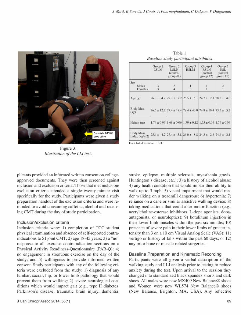
J Can Chiropr Assoc 2014; 58(1) 89
J Ward, K Sorrels, J Coats, A Pourmoghaddam, C DeLeon, P Daigneault
plicants provided an informed written consent on college-approved documents. They were then screened against inclusion and exclusion criteria. Those that met inclusion/exclusion criteria attended a single twenty-minute visit specifically for the study. Participants were given a study preparation handout of the exclusion criteria and were re-minded to avoid consuming caffeine, alcohol and receiv-ing CMT during the day of study participation.
Inclusion/exclusion criteriaInclusion criteria were: 1) completion of TCC student physical examination and absence of self-reported contra-indications to SI joint CMT; 2) age 18-45 years; 3) a “no” response to all exercise contraindication sections on a Physical Activity Readiness-Questionnaire (PAR-Q); 4) no engagement in strenuous exercise on the day of the study; and 5) willingness to provide informed written consent. Study participants with any of the following cri-teria were excluded from the study: 1) diagnosis of any lumbar, sacral, hip, or lower limb pathology that would prevent them from walking; 2) severe neurological con-ditions which would impact gait (e.g., type II diabetes, Parkinson’s disease, traumatic brain injury, dementia,
stroke, epilepsy, multiple sclerosis, myasthenia gravis, Huntington’s disease, etc.); 3) a history of alcohol abuse; 4) any health condition that would impair their ability to walk up to 3 mph; 5) visual impairment that would ren-der walking on a treadmill dangerous; 6) hypertonia; 7) reliance on a cane or similar assistive walking device; 8) taking medications that could alter motor function (e.g., acetylcholine-esterase inhibitors, L-dopa agonists, dopa-antagonists, or neuroleptics); 9) botulinum injection in their lower limb muscles within the past six months; 10) presence of severe pain in their lower limbs of greater in-tensity than 3 on a 10 cm Visual Analog Scale (VAS); 11) vertigo or history of falls within the past 60 days; or 12) any prior bone or muscle-related surgeries.
Baseline Preparation and Kinematic RecordingParticipants were all given a verbal description of the walking study and LLI analysis prior to testing to reduce anxiety during the test. Upon arrival to the session they changed into standardized black spandex shorts and dark shoes. All males wore new MX409 New Balance® shoes and Women wore new WL574 New Balance® shoes (New Balance, Brighton, MA, USA). Any reflective
Figure 3.
Illustration of the LLI test.
Table 1. Baseline study participant attributes.
Group 1 LSLM
Group 2 LSLN
(control group #1)
Group 3 RSLM
Group 4 RSLN
(control group #2)
Group 5 NSL
(control group #3)
Sex Males
Females
3 3
2 4
1 3
1 1
2 1
Age (y) 26.0 ± 4.7 29.7 ± 7.2 25.5 ± 5.1 24.7 ± 2.1 28.3 ± 4.0
Body Mass (kg) 76.6 ± 12.7 77.4 ± 18.4 78.4 ± 40.0 74.8 ± 10.4 73.5 ± 5.2
Height (m) 1.74 ± 0.06 1.68 ± 0.06 1.70 ± 0.12 1.75 ± 0.04 1.74 ± 0.04
Body Mass Index (kg/m2) 25.4 ± 4.2 27.4 ± 5.8 26.0 ± 8.8 24.3 ± 2.8 24.4 ± 2.1
Data listed as mean ± SD.

90 J Can Chiropr Assoc 2014; 58(1)
Impact of bilateral sacroiliac joint manipulation using a drop table technique on gait parameters in asymptomatic individuals with LLI
logos on the shoes were spray-painted with non-reflective paint. Prior to this study researchers purchased five pairs of male (sizes 8-12) and female (sizes 5-9) shoes in com-mon sizes. Standardized shoes were chosen as opposed to having participants walk barefoot to most closely emulate a real-world scenario. Next, trained research assistants placed 18 silver reflective markers on the participant’s lower body using double-sided marker fixing tape and surgical tape. Reflective markers were placed on the fol-lowing anatomic landmarks during this study bilaterally: ASIS, PSIS, greater trochanter, lateral epicondyle of the femur, tibial tuberosity, lateral malleolus, posterior calca-neus, top of the fifth metatarsal head, and top of the first metatarsal head (Fig. 2). Sixteen of the MoCap solutions reflective markers were 19 mm (MoCap solutions, Hun-tington Beach, CA, USA). The two PSIS markers used in this study were slightly smaller, at 14 mm. This was done in an attempt to gain better resolution by reducing the likelihood that those markers would be merged together by the Vicon® cameras considering how close the PSISs were on smaller participants with narrow hips. Prior to a participant arriving at the lab each day the Vicon® system was calibrated as suggested by the manu-facturer. Once the participant was dressed properly and all of the reflective markers were in place they stood on top of the 400 Pro series Keys® treadmill (Keys Fitness Prod-ucts, Inc., Dallas, TX, USA) for their baseline 10-second model generation. Next the participant was instructed that they would be walking as they normally would at a velocity of 1.5 mph. A research assistant started the treadmill at the same time as another researcher began recording data with the Vicon® system. The lab’s Vicon® MX system consisted of 8 infrared Bonita 0.3 megapixel cameras. Kinematic data was recorded at 100 Hz. The displacement of the 18 reflective markers over time was recorded. At the conclusion of 100 seconds the researcher operating the Vicon® computer stopped the recording and then the treadmill was stopped. The study partici-pant was not given any indication of when the treadmill would be stopped prior to the examiner finishing his com-puter data recording. Immediately after the 100 second recording was made the initial 10 seconds was clipped from the data to remove any initial steps as the partici-pant became acclimated to the treadmill upon beginning the test. Following the baseline 90 seconds of data col-lection the participant then carefully stepped off of the
treadmill. After this the research assistants removed the participant’s shoes for them. They then removed the re-flective markers on the two tibial tuberosity and two ASIS points. Prior to removing those four reflective markers the research assistants made circular pen tracings around the markers on the participant’s skin. This was done in an at-tempt to leave a guide which would aid in placing the markers back as closely as possible where they were for the 90-second post-kinematic analysis.
LLI assessmentStudy participants positioned themselves prone on a drop table with their shoes off. During positioning for the LLI test, an effort was made to ensure the participant’s whole body was in a neutral position on the table without spinal or pelvic frontal plane distortion. The treating doctor then held the participant’s ankles in a neutral position to pre-vent foot inversion or eversion. The leg length was visual-ized by comparing the inferior aspect of both compressed heels exclusively (Fig. 3). This technique was chosen as a previous study suggested that this form of LLI meas-urement demonstrated better inter-examiner reliability,19 although LLI tests in general do not have particularly high inter-rater reliability.20-23 If the examining chiroprac-tor found that the participant did have a LLI the Primary Investigator (PI) would inform him based on the pre-gen-erated randomization list if the participant was to be ma-nipulated or not. Admittedly, the pre-generated random-ization sequence was not concealed from the PI, however only two potential subjects were actively excluded by the PI. All other potential subjects were included and randomized strictly on a first-come, first-serve basis. No attempt to measure an exact LLI distance was made. If SI joint CMT was to be performed the short lower limb PSIS marker was also removed after a pen tracing was made around it on the participant’s skin. Following the intervention, or lack thereof, the study participant care-fully stood up. The treating chiropractor made no attempt to re-check the LLI manually post-CMT to confirm that a change was induced. Next the research assistants placed all of the reflective markers back where they were (the two tibial tuberosity markers, two ASIS markers, and one PSIS marker) and put the participant’s standardized shoes back on them. After the intervention phase of the study the participants walked again for their post-kinematic an-alysis.

J Can Chiropr Assoc 2014; 58(1) 91
J Ward, K Sorrels, J Coats, A Pourmoghaddam, C DeLeon, P Daigneault
Randomization and blindingParticipants were subdivided into the three following groups based on the LLI test: left short leg, right short leg, and no short leg (as shown in Fig. 1). A computer-generated randomized intervention list was created be-fore the study began. That list determined if a participant with a short lower limb would undergo CMT or not. The doctor performing the SI joint CMT was aware of which group the study participant belonged to. The researcher who analyzed the motion capture data was blinded as to group designation. He was only told that he would be pro-vided with walking data from five distinct study groups and that he needed to determine gait kinematics and if any statistically significant differences existed within groups in terms of their pre versus post gait data.
InterventionThe intervention phase of the study was performed by a chiropractor with 35 years of experience. The interven-tion involved either: 1) a hypothenar ilium apex push to the PSIS on the short leg side24 in an attempt to rotate the superior ilium anteriorly to elongate that lower limb and a hypothenar ischial tuberosity push on the long leg side, or 2) no manipulation. Bilateral SI joint CMT was chosen over unilateral short leg PSIS manipulation based on preliminary data by our lab on the lack of effectiveness of unilateral corrective CMT to improve gait symmetry of our student participants (unpublished data). All CMT con-sisted of a high-velocity low-amplitude force delivered three times in a row using a drop table (Ergostyle 2000, Chattanooga Group Inc., Hixson, TX, USA). The intent of the drop table was to try to keep the amount of force reasonably standardized. This prone form of CMT was selected to decrease the likelihood of making researchers remove more reflective surface markers than the five that were absolutely necessary to remove. One minute after receiving SI joint CMT or no CMT the study participant engaged in their walking post-kinematic analysis.
Kinematic Post-data ProcessingThe data was processed using a customized Matlab script (Mathworks, USA R2007a). The kinematic data was ana-lyzed to calculate characteristics of movement for each participant. Data for the dependent variables was aver-aged for each participant over all of their strides within each gait trial. In the current study we investigated the
changes in the functional active range of motion (in the sagittal plane) of the hip angle, knee angle, and ankle an-gle as a result of the intervention. This was performed by subtracting the minimum joint angle from the maximum joint angle for each of the aforementioned joints. In addi-tion, the double support time, percent double support time (duration both feet were on the ground in relation to the gait cycle), stance time, percent stance time (duration one foot was on the ground in relation to the gait cycle), step length, and stride length bilaterally were calculated. Approximate Entropy, a measure of gait variability, was additionally determined for each joint. In healthy in-dividuals there is a certain amount of acceptable variabil-ity that represents a normal (healthy) gait pattern. How-ever, highly variable gait patterns are typically indicative of some type of pathology or loss of coordination,25 which may render a person at risk for falling.26 Gait variability has been identified by the application of a mathematical technique called approximate entropy (ApnEn) that may reveal small changes in the gait pattern.25,27,28 Values near “0” represent a stable gait, while values near “2” repre-sent a very unstable gait.
Statistical AnalysisData was analyzed in SPSS version 19 (Release Version 19.0.1). Pre- and post-intervention gait parameter meas-urements were summarized as mean + standard devia-tions (SD) unless otherwise specified. Parametric with-in-groups, dependent variables were compared using a paired-samples t-test. Since we intended to utilize a series of t-tests we engaged in a Bonferroni adjustment to avoid type I statistical error. As a result, the alpha level of p < 0.002 was considered statistically significant for all an-alyses. For data analysis purposes both short leg groups had their data merged into one group based on ipsilat-eral short leg effects (e.g., the R lower limb data for the RSLM group and L lower limb data for the LSLM group) and contralateral long leg effects (e.g., the L lower limb data for the RSLM group and R lower limb data for the LSLM group) pre- and post-intervention. Similarly, the RSLN and LSLN groups had their data merged for com-parison purposes. The NSL group had its bilateral data values averaged together.
ResultsThis study involved twenty-one chiropractic college par-

92 J Can Chiropr Assoc 2014; 58(1)
Impact of bilateral sacroiliac joint manipulation using a drop table technique on gait parameters in asymptomatic individuals with LLI
ticipants. Just over half of our participants were female (n = 12). The mean age was = 27.2 (sd = 5.1) years, mean height was = 1.72 (sd = 0.07) m, and mean body mass was = 76.6 (sd = 18.2) kg. Only two interested subjects were excluded from this study based on exclusion criteria (one with a history of lower limb surgery, another due to existing foot drop). CMT to the SI joint resulted in no statistically significant change in functional active range of motion and other parameters of gait (Table 2a-2c). This, however, was a pilot study with only twenty-one participants and did not follow a power analysis. Few at-tributes in this study even approached statistical signifi-cance when adjusted for multiple comparisons. However, from an exploratory analysis perspective, the change in pre ipsilateral lower limb % stance time was significant at an unadjusted alpha level (p=0.031) for RSLM. In the RSLN and LSLN, our analysis demonstrated a change in the knee joint angle on the long limb side of almost 1˚ (p=0.011) and an increase in percentage of stance time by 0.4% (p=0.010) on the short limb side. This, along with the normal gait data demonstrated in Table 2c illustrates the amount of variability in gait without CMT. Addi-
tionally, there was no discernible pattern changes in the RSLM and LSLM groups.
DiscussionThe changes in walking kinematics in response to CMT in this pilot study were small. This study intentionally was a pilot study with a small sample size within each group. Subsequent larger studies should follow a power analysis. Using G*Power version 3.1.3 (Universität Kiel, Germany)29,30 we determined post-hoc that future studies should have 34 participants per study group to compare 2 groups (experimental and control). This analysis was in accordance with a desired f effect size of 0.5, α of 0.05, and power of 0.8 through a 2-group ANOVA using [stride length] as the primary outcome of interest. We do feel that we would be capable of recruiting this number of student participants with future studies if we merge right and left short leg group data together (e.g., RSLM and LSLM into one group). Our rate of eligibility for this study was 91.3% out of all applicants from our college. Recruiting outside non-student participants (general public) would also be possible as an alternative, but that would require
Table 2a. Summary of RSLM and LSLM merged group data (n=10).
Pre Ipsilateral lower limb
Post Ipsilateral lower limb t-test Pre Contralateral
lower limbPost Contralateral
lower limb t-test
Functional Active Range of Motion hip angle ˚ 39.0 ± 4.4 37.7 ± 3.1 0.258 40.3 ± 4.4 38.8 ± 2.7 0.244 knee angle ˚ 59.6 ± 3.5 58.2 ± 2.1 0.135 59.0 ± 2.5 57.9 ± 2.8 0.203 ankle angle ˚ 33.3 ± 7.1 31.5 ± 5.2 0.172 31.9 ± 5.5 31.7 ± 6.2 0.819Approximate entropy hip angle 0.21 ± 0.02 0.21 ± 0.03 0.799 0.21 ± 0.03 0.21 ± 0.04 0.972 knee angle 0.31 ± 0.06 0.31 ± 0.07 0.986 0.33 ± 0.07 0.30 ± 0.07 0.182 ankle angle 0.59 ± 0.08 0.58 ± 0.10 0.812 0.61 ± 0.09 0.61 ± 0.08 0.913Double support Double Support (s) 0.30 ± 0.02 0.31 ± 0.03 0.173 0.31 ± 0.02 0.31 ± 0.02 0.577 % Double Support 19.94 ± 1.07 20.40 ± 1.40 0.097 20.39 ± 1.12 20.47 ± 0.79 0.667Stance time stance time (s) 1.05 ± 0.05 1.06 ± 0.07 0.367 1.05 ± 0.06 1.06 ± 0.07 0.671 % stance time 69.98 ± 1.12 70.40 ± 1.13 0.031 70.30 ± 1.18 70.41 ± 1.05 0.620Step length step length (mm) 409.3 ± 46.9 413.0 ± 48.5 0.306 436.7 ± 25.8 436.3 ± 24.5 0.913Stride length stride length (mm) 1089.5 ± 50.2 1090.8 ± 59.6 0.867 1099.0 ± 62.9 1101.0 ± 69.0 0.836
Data listed as mean ± SD for group dependent variable data.

J Can Chiropr Assoc 2014; 58(1) 93
J Ward, K Sorrels, J Coats, A Pourmoghaddam, C DeLeon, P Daigneault
further resources. Additionally, a future definitive study could involve randomizing NSL participants to manipula-tion or no manipulation groups in order to constitute an additional comparison group. Our eight camera Vicon® motion analysis system worked consistently in our study. No participants were in-jured at any point. For safety reasons following the com-pletion of this experiment we have installed thin side rails on the treadmill that provide a safety bar participants can grab onto in case they lose their balance on the treadmill. We are also in the process of installing an overhead safety harness system to prevent falling. Our method of using a blinded biomechanist appeared to be effective in this pilot study. One issue we did have was the transport of the large kinematic data files we were generating (approximately 82 MB per participant). As a result, it took our offsite biomechanist many hours to transfer all of the files from our lab computer to his com-puter for analysis using a trial version of TeamViewer 8 ® software (TeamViewer Inc., Tampa, FL). For future stud-ies we will need to purchase a more robust file transfer system to handle transferring large quantities of data.
Table 2c. Summary of NSL bilateral mean data (n=3).
Pre lower limb Post lower limbFunctional Active Range of Motion hip angle ˚ 41.2 ± 3.3 41.6 ± 3.4 knee angle ˚ 62.4 ± 2.0 65.3 ± 2.8 ankle angle ˚ 31.7 ± 4.1 34.0 ± 6.2Approximate entropy hip angle 0.22 ± 0.03 0.24 ± 0.04 knee angle 0.39 ± 0.02 0.40 ± 0.02 ankle angle 0.62 ± 0.03 0.61 ± 0.05Double support Double Support (s) 0.29 ± 0.03 0.29 ± 0.04 % Double Support 19.92 ± 0.68 19.83 ± 1.08Stance time stance time (s) 1.02 ± 0.07 1.04 ± 0.11 % stance time 69.90 ± 0.74 69.84 ± 0.84Step length step length (mm) 421.0 ± 24.8 427.8 ± 37.9Stride length stride length (mm) 1052.5 ± 65.4 1068.9 ± 105.7
Data listed as mean ± SD for group dependent variable data.
Table 2b. Summary of RSLN and LSLN merged group data (n=8).
Pre Ipsilateral lower limb
Post Ipsilateral lower limb t-test Pre Contralateral
lower limbPost Contralateral
lower limb t-test
Functional Active Range of Motion hip angle ˚ 38.7 ± 3.0 39.3 ± 3.5 0.534 38.9 ± 2.9 39.9 ± 3.8 0.207 knee angle ˚ 59.5 ± 3.7 60.0 ± 4.8 0.535 59.7 ± 3.9 60.6 ± 3.7 0.011 ankle angle ˚ 33.0 ± 6.1 32.0 ± 5.0 0.456 32.5 ± 5.5 32.5 ± 4.6 0.984Approximate entropy hip angle 0.21 ± 0.03 0.22 ± 0.03 0.258 0.21 ± 0.02 0.23 ± 0.04 0.095 knee angle 0.33 ± 0.07 0.34 ± 0.07 0.303 0.33 ± 0.07 0.33 ± 0.06 0.951 ankle angle 0.61 ± 0.10 0.60 ± 0.10 0.761 0.62 ± 0.08 0.59 ± 0.08 0.239Double support Double Support (s) 0.30 ± 0.04 0.31 ± 0.04 0.183 0.30 ± 0.04 0.31 ± 0.04 0.499 % Double Support 19.99 ± 1.51 20.41 ± 1.72 0.032 20.05 ± 1.55 20.24 ± 1.38 0.361Stance time stance time (s) 1.05 ± 0.10 1.06 ± 0.09 0.677 1.06 ± 0.09 1.06 ± 0.08 0.857 % stance time 69.69 ± 1.40 70.09 ± 1.53 0.010 70.33 ± 1.33 70.51 ± 1.37 0.117Step length step length (mm) 417.2 ± 50.8 416.6 ± 47.7 0.906 427.2 ± 41.8 425.7 ± 39.5 0.728Stride length stride length (mm) 1086.9 ± 113.3 1088.0 ± 104.5 0.924 1098.9 ± 110.7 1099.1 ± 101.4 0.985
Data listed as mean ± SD for group dependent variable data.

94 J Can Chiropr Assoc 2014; 58(1)
Impact of bilateral sacroiliac joint manipulation using a drop table technique on gait parameters in asymptomatic individuals with LLI
Manual determination of LLI by chiropractors is oc-casionally performed, but it has not been shown to be as accurate as more expensive imaging methods.8,19 When choosing an ideal method of measuring LLI we, similar to field practitioners, had to consider reliability, accur-acy, magnification, radiation dose, cost, need for special equipment, convenience, and the opportunity to image an entire extremity.9 In our study design we opted to utilize a technique that we theorized would be more common amongst chiropractic general practitioners. Our original hypothesis was that we expected participants with a LLI to have a slightly asymmetric gait. Then after corrective SI joint CMT we hypothesized that the participant’s gait would be more symmetrical. This belief did not material-ize. The experience we gained through this pilot study was invaluable for the generation of subsequent larger stud-ies following a power analysis. If SI joint CMT improves gait symmetry that may directly have implications on bal-ance while walking. Further study to determine the true physiological impact of SI joint manipulation on gait is required. The main intent of this study was to determine if our design would be feasible to engage in a series of larger studies using advanced motion analysis technology. This was shown to be possible. This lab now intends to de-velop the three following walking studies involving SI joint CMT over the course of September 2012-September 2015 using large study groups: 1) normative data with a combination of healthy non-chiropractic student partici-pants (mainstream public) versus chiropractic students, 2) adult SI joint pain patient data (mainstream public), and 3) geriatric at-risk-for-fall data. As this field is developed further additional direc-tions that should be explored would be running kinematic changes in response to SI joint and/or lumbar spine CMT and changes in gait that may be induced in ataxic special populations. Also surface EMG should be used to explore if any changes found in gait are induced by alterations in motor recruitment patterns of the lower limb muscles in response to CMT.
LimitationsThis study only informs us as to the potential immediate impact SI joint CMT may have on specific gait param-eters in young, asymptomatic individuals. It is possible
that a CMT dose-response relationship may exist related to improvements in gait performance, similar to what Herzog discovered.15
One issue this study must accept is the limitation on ex-ternal validity. The population we sampled was composed of chiropractic students who regularly receive CMT. It is possible that the general public may react differently than individuals who receive CMT often. Another issue this study faced is that LLI tests do not have high levels of validity and reliability.13,19,20-24 In our study design we opted to use a test that was a modified form of a Derifield Pelvic Leg Check LLI test that only involved comparison of heel length. It is possible that our test was not ideal since a reliable and valid test does not clearly exist. No X-ray or similar imaging lower limb procedure was used to ensure that participants truly had a functionally short lower limb and not an anatomically short lower limb.2,31-35 Despite this, the primary goal of this study was to observe raw marginal change in kin-ematics pre versus post SI joint CMT (e.g., can SI joint CMT truly induce a subtle change in gait kinematics or not).
ConclusionsThere is minimal research into how spinal manipulation may augment gait. The focus of this experiment was to determine if developing a series of larger SI joint CMT biomechanics studies using motion analysis technology was possible through our study design. The findings of our study suggest larger studies are feasible and we will proceed accordingly.
Funding sources and potential conflicts of interestThis study was supported by a grant from Dr. S. M. Elliot, TCC President Emeritus. The authors report no conflicts of interest.
References1. Knutson G. Anatomic and functional leg-length inequality:
a review and recommendation for clinical decision-making. Part I, anatomic leg-length inequality: prevalence, magnitude, effects and clinical significance. Chiropr Osteop. 2005;13:11.
2. McCaw S, Bates B. Biomechanical implications of mild leg length inequality. Br J Sp Med. 1991;25(1):10-3.
3. Langer S. Structural leg shortage: a case report. I Am Podiatry Assoc. 1976;66:38-40.

J Can Chiropr Assoc 2014; 58(1) 95
J Ward, K Sorrels, J Coats, A Pourmoghaddam, C DeLeon, P Daigneault
4. Friberg O. Clinical symptoms and biomechanics of lumbar spine and hip joint in leg length inequality. Spine. 1983;8(6):643-51.
5. Sabharwal S, Kumar A. Methods for assessing leg length discrepancy. Clin Orthop Relat Res. 2008;466:2910-22.
6. Golightly Y, Allen K, Helmick C, Renner J, Salazar A, Jordan J. Relationship of limb length inequality with radiographic knee and hip osteoarthritis. Osteoarthritis Cartilage. 2007;15(7):824-9.
7. Harvey W, Yang M, Cooke T, Segal N, Lane N, Lewis C, Felson D. Associations of leg length inequality with prevalent, incident, and progressive knee osteoarthritis: a cohort study. Ann Intern Med. 2010;152(5):287-95.
8. Anderson R, Hayak R, Foggerty M. Leg length inequality and the side of low back pain. Comsig Review. 1995;4(2):33-6.
9. Reid D, Smith B. Leg length inequality: a review of etiology and management. Physio Can. 1984;36:177-82.
10. Knutson G. Anatomic and functional leg-length inequality: a review and recommendation for clinical decision-making. Part II, the functional or unloaded leg-length asymmetry. Chiropr Osteop. 2005;13:12.
11. Cooperstein R, Lisi A. Pelvic torsion: anatomic considerations, construct validity, and chiropractic examination procedures. Top Clin Chiro. 2000;7(3):38-49.
12. Giles L, Taylor J. Lumbar spine structural changes associated with leg length inequality. Spine. 1982;7(2):159-62.
13. Mannello D. Leg Length Inequality. J Manipulative Physiol Ther. 1992;15(9):576-90.
14. Sandell J, Palmgren P, Björndahl L. Effect of chiropractic treatment of hip extension ability and running velocity among young male running athletes. JCM. 2008;7:39-47.
15. Herzog W. Biomechanical studies of spinal manipulative therapy. JCCA. 1991;91:156-64.
16. Haas M, Peterson D, Panzer D, Rothman E, Solomon S, Krein R, Johansen R. Reactivity of leg alignment to articular pressure testing: evaluation of a diagnostic test using a randomized crossover clinical trial approach. J Manipulative Physiol Ther. 1993;16(4):220-7.
17. Nguyen H, Resnick D, Caldwell S, Elston E, Bishop B, Steinhouser J, et al. Interexaminer reliability of activator methods relative leg-length evaluation in the prone extended position. J Manipulative Physiol Ther. 1999;22(9):565-9.
18. Thompson J. Thompson technique reference manual, Elgin, IL 1984. Thompson Educational Workshops SM and Williams Manufacturing.
19. Walker B, Buchbinder R. Most commonly used methods of detecting subluxation and the preferred term for
its description: a survey of chiropractors in Victoria, Australia. J Manipulative Physiol Ther. 1997;20(9):583-8.
20. Fuhr A, Osterbauer P. Interexaminer reliability of relative leg-length evaluations in the prone, extended position. Chiropractic Technique. 1989;1(1):13-8.
21. Haas M, Peterson D. Interexaminer reliability of relative leg-length evaluations in the prone, extended position [letter, comment]. Chiropractic Technique. 1989;1(4):150-1.
22. DeBoer K, Harmon R, Savoie S, Tuttle C. Inter- and intra-examiner reliability of the leg-length differential measurement: a preliminary study. J Manipulative Physiol Ther. 1983;10(3):61-6.
23. Danelius D. Letter to the editor: inter- and intra-examiner reliability of leg-length measurement: a preliminary study. J Manipulative Physiol Ther. 1987;10(3):132.
24. Bergmann T, Peterson D. Chiropractic Technique: principles and procedures. 3rd ed. St. Louis: Elsevier-Mosby; 2011.
25. Myers S, Stergiou N, Pipinos I, Johanning J. Gait variability patterns are altered in healthy young individuals during the acute reperfusion phase of ischemia-reperfusion. J Surg Res. 2010;164(1):6-12.
26. Maki B. Gait changes in older adults: predictors of falls or indicators of fear. J Am Geriatr Soc. 1997;45(3):313-20.
27. Buzzi U, Stergiou N, Kurz M, Hageman P, Heidel J. Nonlinear dynamics indicates aging affects variability during gait. Clin Biomech. 2003;18:435-43.
28. Pincus S. Approximate entropy as a measure of system complexity. Proc Natl Acad Sci USA. 1991;88:2297-301.
29. Buchner A, Erdfelder E, Faul F. (1997). How to Use G*Power [WWW document]. URL http://www.psycho.uni-duesseldorf.de/aap/projects/gpower/how_to_use_gpower.html.
30. Erdfelder E, Faul F, Buchner A. GPOWER: A general power analysis program. Behav Res Meth Ins. 1996;28:1-11.
31. Clarke G. Unequal leg length: an accurate method of detection and some clinical results. Rheum Phys Med. 1972;11:385-90.
32. Beal M. The short leg problem. J Am Osteopath Assoc. 1977;76:745-51.
33. Gofton J, Trueman G. Studies in osteoarthritis of the hip. Part II: Osteoarthritis of the hip and leg-length inequality. Can Med Assoc J. 1971;104:791-9.
34. Friberg 0, Koivisto E, Wegelius C. A radiographic method for measurement of leg length inequality. Diagn Imag Clin Med. 1985;54:54-81.
35. Fisk J, Baigent M. Clinical and radiological assessment of leg length. NZ Med J. 1975;8:477-80.

96 J Can Chiropr Assoc 2014; 58(1)
Letters to the Editor
RE: Chiropractors as Primary Spine Care Providers: precedents and essential measures. JCCA. 2013;57(4):285-291. W. Mark Erwin, DC, PhD, A. Pauliina Korpela, BSc, Robert C. Jones, DC, APC
To the Editor
I would like to congratulate Dr. Mark Erwin and co-authors for their recent article in the JCCA. They have courageously highlighted the need for the profession to become focused and united in providing evidence-based spine care. This means incorporating the best available evidence into practice and being leaders and innovators in this area. Only then will we as a profession be credible and considered spine experts to the community at large. Being non operative spine experts comes with respon-sibility. It must be reflected in our language, our educa-tion, our research, and especially our practice. We need to be consistent in our message and treatment approach. We need strong leadership to bring the profession together and recognize the need for change. Our destiny is in our own hands.
Carlo Ammendolia, DC, PhD Assistant Professor, Institute of Health Policy, Management and Evaluation, Faculty of Medicine, University of Toronto Associate Scientist/Chiropractor, Rebecca MacDonald Centre for Arthritis & Autoimmune Diseases, Division of Rheumatology, Mount Sinai Hospital Associate Scientist, Institute for Work & Health, Toronto, Canada CCRF Professorship in Spine, Department of Surgery, University of Toronto Mount Sinai Hospital, 60 Murray Street, Room L2-007 Toronto, Ontario, Canada, M5T 3L9
To the Editor
We read with interest the paper “Chiropractors as Primary Spine Care Providers: precedents and essential measures”1, which is one out of several papers dealing with this issue published within the past few years2,3. We would like to comment on the paper focussing on the needs of society and feasibility of the proposed model.
In societies everywhere there is clearly a need for an increased focus on spine pain and musculoskeletal disorders. Low back pain is omnipresent and accounts for over 10% of the total “years lived with disability”.4 The associated consequences for individuals and societies everywhere are enormous in terms of lost quality of life, work absence, disability, and direct health care expenses. However, contemporary research has convincingly shown that back pain does not occur alone in most individuals, and patients with pain in more than one site experience a greater impact of their pain, have poorer prognosis in a range of domains, and respond less favourably to treatment5. Consequently, unlike dental and optical care, spine care may not have clear anatomical boundaries and one could therefore rightfully ask if the future for primary spine pain care lies with a spine care specialist or with a person who has a broader musculoskeletal focus across pain sites and conditions.
Regarding the issue of feasibility, many patients with spine pain would have to seek care from multiple providers for their multisite musculoskeletal conditions. This may not represent an effective use of the patient’s or society’s resources in particular in the primary care setting. Moreover, patients might experience difficulties in determining what is a spine related condition especially in conditions with diffuse pain patterns or radiating pain such as arm, chest or leg pain, which might lead to inappropriate care seeking, frustration, and chronicity. We suggest that the real challenge for chiropractors is integration into mainstream primary care as musculoskeletal health care providers rather than focussing exclusively on spine care. Canadian chiropractors and chiropractors in many other countries are already trained as such and 90.6% of the full time practicing chiropractors in Canada do not limit their

J Can Chiropr Assoc 2014; 58(1) 97
Letters to the Editor
treatment to the spine and include treatment of the extremities6. In addition and importantly, many of the prevention, diagnostic and treatment strategies appear to be similar between different musculoskeletal pain sites3 and prognostic factors for chronicity are also very similar5,7. Of course chiropractors are not alone in claiming the role of primary care musculoskeletal care provider. In our opinion a continued focus on research and education along with the chiropractor’s ability to integrate and function in interdisciplinary collaboration will ultimately determine the fate of the profession in this arena.
Marc-André Blanchette, DC, MScPublic Health PhD ProgramSchool of Public HealthUniversity of Montreal, Montreal, QC, Canada [email protected]
Jan Hartvigsen, DC, PhDDepartment of Sports Science and Clinical BiomechanicsUniversity of Southern Denmark Odense, DenmarkNordic Institute of Chiropractic and Clinical BiomechanicsOdense, Denmark
References1. Erwin WM, Korpela AP, Jones RC. Chiropractors as Primary
Spine Care Providers: precedents and essential measures. J Can Chiropr Assoc. Dec 2013;57:285-291.
2. Murphy DR, Justice BD, Paskowski IC, Perle SM, Schneider MJ. The establishment of a primary spine care practitioner and its benefits to health care reform in the United States. Chiropr Man Therap. 2011;19:17.
3. Murphy DR, Schneider MJ, Seaman DR, Perle SM, Nelson CF. How can chiropractic become a respected mainstream profession? The example of podiatry. Chiropractic & Osteopathy. 2008;16:10.
4. Vos T, Flaxman AD, Naghavi M, et al. Years lived with disability (YLDs) for 1160 sequelae of 289 diseases and injuries 1990-2010: a systematic analysis for the Global Burden of Disease Study 2010. Lancet. 2012;380:2163-2196.
5. Hartvigsen J, Natvig B, Ferreira M. Is it all about a pain in the back? Best Pract Res Clin Rheumatol. 2013;27:613-623.
6. Kopansky-Giles D, Papadopoulos C. Canadian Chiropractic Resources Databank (CCRD): a profile of Canadian chiropractors. J Can Chiropr Assoc. 1997;41:155-191.
7. Muller S, Thomas E, Dunn KM, Mallen CD. A prognostic approach to defining chronic pain across a range of musculoskeletal pain sites. Clinical J Pain. 2013;29:411-416.
To the Editor
I reviewed the timely article regarding the role played by chiropractors written by Dr. Mark Erwin with tremendous interest.
With the high incidence of spinal conditions in our aging population and the limited number of general practitioners and spinal specialists an opportunity exists to seize for the chiropractic community.
The majority of spinal conditions are non-surgical and are potentially managed through conservative measures by our allied health practitioners.
Dr. Erwin has provided existing models from North America and Europe that have enabled chiropractors to be at the front lines of managing selected spinal conditions which makes sense and would appear to represent an opportunity to streamline the management of non-urgent spinal pain syndromes.
I wholeheartedly agree with Dr. Erwin for the need of evidence based approaches and standardization by chiropractors to manage these conditions. I am aware that each graduating chiropractor is trained in identifying red flags, conditions that require further investigations (laboratory and imaging) and those that ultimately require timely surgical evaluation and it is clear that a closer working relationship between chiropractors and physicians/specialists would be in everyone’s best interests.
I have had occasion to give several lectures at CMCC (Canadian Memorial Chiropractic College) and in the process have interacted with the students and faculty - I am confident that the aforementioned objectives may be achieved.
Part of the Chiropractic curriculum should necessarily ensure each graduate evaluates each spinal case in an evidence based manner and manages the patient’s through a standardized approach. Ultimately primary practitioners and spinal specialists will develop additional confidence in the chiropractic community and build stronger clinical relationships and this will

98 J Can Chiropr Assoc 2014; 58(1)
potentially result in higher patient satisfaction and timely care.
As a Neurosurgeon and Fellowship trained Spine Surgeon I have had the privilege to work with pioneers like Dr. Erwin to advance the field of chiropractic care in Canada to the benefit of spinal patients. I look forward to better integration of an evidence-based, collaborative relationship with the chiropractic community for as with my physical therapy colleagues it is through an evidence-based, scientific approach that the chiropractic profession will enjoy enhanced legislative scope of practice and even better tools with which to help their patients.
Neilank K. Jha, MD, FRCS(C)Neurosurgeon, Spine SurgeonChairman, KONKUSSIONwww.konkussion.com Chairman, TELEKONKUSSIONChairman, WATCH Community Serviceswww.watchcommunity.org Editor-in-Chief, Current Research – Concussion
To the Editor in reply
I would like to thank Drs. Blanchette and Hartvigsen for their thoughtful letter with respect to the recent paper published by my colleagues Dr. Robert Jones, Anna Pauliina Korpela, BSc and me. Drs. Blanchette and Hartvigsen raise the question that unlike dental and optical (and presumably foot) care, spine care may not have clear anatomical boundaries, and perhaps the future for primary spine care may best lie within a spine care specialist or a person with broader musculoskeletal focus. The authors further suggest that many patients with spine pain may consult a number of providers for their ‘multisite MSK conditions’, a situation that would not make the most effective use of health care funds. Additionally, they suggest that the ‘real challenge’ for chiropractors is integration within mainstream primary care as MSK health providers rather than focusing exclusively on spine care. They acknowledge that chiropractors are of course not alone “claiming the role of primary care MSK care providers” and conclude with an opinion that continued focus on research and education along with the integration within the multidisciplinary collaborative approach ‘will’ ultimately determine the fate of the profession in this arena.
First, the title of our paper is “Chiropractors as Primary Spine Care Providers”. It is not “Chiropractors as ONLY Primary Spine Care Providers”. The purpose of our manuscript was to raise the question whether a chiropractor ought to be the preferred provider of spine care. A chiropractor’s education is primarily spine-based (although of course also contains a rich education in differential diagnosis with good training in other MSK-related conditions). DCs of course treat a myriad of diverse MSK problems and at no time did we suggest anything to the contrary; rather our focus was whether the DC might be the suitable ‘go to’ clinician for spinal pain. In order to address the specific question whether the DC ought to be the Primary Spine Care Provider we contrasted the evolution of optometry and other health professions that have filled such a ‘niche’ within the provision of specific healthcare needs. I think “anatomical boundaries” have nothing to do with the provision of spine care akin to the example of dental
Letters to the Editor

J Can Chiropr Assoc 2014; 58(1) 99
or optical care. The question posed by Blanchette and Hartvigsen; “[whether] the future for primary spine pain care lies with a spine care specialist or with a person who has a broader musculoskeletal focus across pain sites and conditions” fails to advance the notion that the appropriately trained DC could be the preferred spine care provider…or does it? Is the suitably trained DC not qualified in all of these areas? This was precisely the point of our manuscript; a chiropractor who is scientifically trained, evidence-based, and who practices within an integrated model with other disciplines could well be the ideal provider of spine care...but not only spine care. The discussion regarding the 90.6% of chiropractors who do not limit their practice to spine care muddies the water, as does the development of the new discipline of ‘musculoskeletal health care provider’. It seems that such a discussion devolves into one of semantics.
Professional Identity: The World Federation of Chiropractic (WFC), the Canadian and Ontario Chiropractic Associations (and many others) clearly state the chiropractor should fulfill the role of the spinal pain expert. None of these associations makes identity statements regarding broader MSK issues although MSK is often included in various definitions of chiropractic and rightly so. One definition of chiropractic listed on the WFC website (American Association of Chiropractic Colleges-1996) states; “Chiropractic is a healthcare discipline that emphasizes the inherent recuperative power of the body to heal itself without the use of drugs or surgery. The practice of chiropractic focuses on the relationship between structure (primarily the spine) and function (as coordinated by the nervous system) and how that relationship affects the preservation and restoration of health. In addition, doctors of chiropractic recognize the value and responsibility of working in cooperation with other health care practitioners when in the best interest of the patient”. Clearly, this definition emphasizes the spine within the context of the practice of chiropractic.
We chose to focus our paper with respect to the clearly stated identity statements of a host of chiropractic societies, institutions and associations. In particular, at the conclusion of the June 2005 World Federation of
Chiropractic’s 8th Biennial Congress held in Sydney, Australia, the WFC adopted the identity statement that DCs should become “The spinal health care experts in the health care system”. This conclusion reached 9-years ago, was the product of deliberation of over 100 delegates and observers from national associations in 36 countries, including both the ACA and the ICA and involved an “identity task force” and followed the recommendations of a 40-person WFC Task Force. Additionally, the most recent submission to the World Health Organization by the WFC (January 2013) suggests that the primary reasons for consulting a chiropractor are back pain (60%) and other MSK ailments such as neck pain (is this not also a form of spinal pain?), shoulder, extremity and “arthritic pain” (20%). Therefore, close to 80% of the reasons people consult chiropractors relate to some form of spinal (and related) complaint. Furthermore, this report discusses evidence and clinical trials, practice guidelines and Bone and Joint Taskforce reports concerning neck pain and related disorders. There is no discussion of other ‘broad’ MSK complaints. Again, and at the risk of appearing repetitive, we do not suggest that chiropractic only treat spinal complaints-but it appears that this is very much, where the profession’s emphasis appears to be. Furthermore, it is obvious that a host of MSK-related ailments are relevant to spinal pain and that DCs can and do treat such things.(Please see attached link from the WFC website under “identity of the profession”) http://www.wfc.org/website/
However and of particular relevance to Blanchette and Hartvigsen’s letter, despite this WFC identity statement, the chiropractic profession continues to present various professional identities. For example, the Danish Chiropractic Association (DCA) web page states that the aims of the association are (amongst others): “To unite chiropractors aimed at representing and protecting the professional, financial and social interests of the chiropractic profession”. There are further statements with respect to the mandate of the DCA such as:
• To establish guidelines for chiropractic business.
• To determine wages and working conditions for graduates in internships.
Letters to the Editor

100 J Can Chiropr Assoc 2014; 58(1)
• To co-operate with other organizations and associations on issues of mutual interest.
What is missing is any specific ‘identity’ statement. On the other hand, the American Chiropractic Association states that; “Chiropractic is a health care profession that focuses on disorders of the musculoskeletal system and the nervous system, and the effects of these disorders on general health. Chiropractic care is used most often to treat neuromusculoskeletal complaints, including but not limited to back pain, neck pain, and pain in the joints of the arms or legs, and headaches”.
It is plainly evident from these various identity statements and definitions that the chiropractic profession does not present a unified voice to the public, government, third party payers…or to itself; and this speaks to the central premise of our paper.
We agree that chiropractic ought to seek to achieve improved integration into the contemporary healthcare system and to this end, it is vital that the profession continue to invest in enhanced research and education: we make these points quite clearly and succinctly within our manuscript (pages 288-290). We specifically illustrate the success of the CCRF in Canada with the development of Chiropractic Research Chairs, the developing collaboration between the Canadian
Memorial Chiropractic College and the University of Ontario Institute of Technology. In fact, we specifically state, “Increased collaboration, an emphasis on evidence based treatment and continued efforts to broadly expand the research base will resolve many lingering obstacles” (page 289).
As illustrated by Drs. Blanchette and Hartvigsen there are hosts of other well-trained, experienced health care providers who are quite capable at the provision of broad MSK therapy-and within this context, the chiropractor is just one more.
Within the context of our manuscript and the letter by Blanchette and Hartvigsen, perhaps the most poignant question is whether the chiropractic profession ought to be a jack-of-all-trades or master of at least one (that is by definition, connected above, down inside and out)?
W. Mark Erwin, DC, PhDCCRF Professorship in Disc BiologyAssistant Professor, Divisions of Orthopaedic and Neurological Surgery, The Spine Program, University of Toronto,Toronto Western Hospital, Scientist, Toronto Western Research Institute, Associate Professor, Research, Canadian Memorial Chiropractic College
Letters to the Editor