J07 Kwok NP 2012
13
Functional anatomy of temporal organisation and domain-specificity of episodic memory retrieval Sze Chai Kwok a,n , Tim Shallice b,c , Emiliano Macaluso a a Neuroimaging Laboratory, Santa Lucia Foundation, Via Ardeatina 306, 00179 Rome, Italy b Cognitive Neuroscience Sector, SISSA, Trieste, Italy c Institute of Cognitive Neuroscience, University College London, London, UK article info Article history: Received 8 February 2012 Received in revised form 12 July 2012 Accepted 15 July 2012 Available online 2 August 2012 Keywords: What-where-when Precuneus Hierarchical structure Cinematographic material fMRI abstract Episodic memory provides information about the ‘‘when’’ of events as well as ‘‘what’’ and ‘‘where’’ they happened. Using functional imaging, we investigated the domain specificity of retrieval-related processes following encoding of complex, naturalistic events. Subjects watched a 42-min TV episode, and 24 h later, made discriminative choices of scenes from the clip during fMRI. Subjects were presented with two scenes and required to either choose the scene that happened earlier in the film (Temporal), or the scene with a correct spatial arrangement (Spatial), or the scene that had been shown (Object). We identified a retrieval network comprising the precuneus, lateral and dorsal parietal cortex, middle frontal and medial temporal areas. The precuneus and angular gyrus are associated with temporal retrieval, with precuneal activity correlating negatively with temporal distance between two happenings at encoding. A dorsal fronto-parietal network engages during spatial retrieval, while antero-medial temporal regions activate during object-related retrieval. We propose that access to episodic memory traces involves different processes depending on task requirements. These include memory-searching within an organised knowledge structure in the precuneus (Temporal task), online maintenance of spatial information in dorsal fronto-parietal cortices (Spatial task) and combining scene-related spatial and non-spatial information in the hippocampus (Object task). Our findings support the proposal of process-specific dissociations of retrieval. & 2012 Elsevier Ltd. All rights reserved. 1. Introduction Episodic memory provides information about our personal experiences of ‘‘when’’ and ‘‘where’’ events occur as well as ‘‘what’’ happens. In order to simulate the complexity of the processes involved in autobiographical memory, recent studies on episodic memory retrieval have endeavoured to employ real- life-like materials for learning. These range from photographs taken from a first-person perspective (St. Jacques, Rubin, LaBar, & Cabeza, 2008), to documentary videos of people engaged in everyday life activities (Fujii et al., 2004; Mendelsohn, Chalamish, Solomonovich, & Dudai, 2008; Mendelsohn, Furman, & Dudai, 2010), to videos showing navigation through a house (Hayes, Ryan, Schnyer, & Nadel, 2004), or navigating in virtual environ- ments (Burgess, Maguire, Spiers, & O’Keefe, 2001; Ekstrom & Bookheimer, 2007; Ekstrom, Copara, Isham, Wang, & Yonelinas, 2011; King, Hartley, Spiers, Maguire, & Burgess, 2005). A defining characteristic of episodic memories is that they allow us to relive our past as it has unfolded over extended time windows (Tulving, 1985). In order to be accessible for future retrieval, the different elements of an event have to be associatively linked into a durable memory trace (Staresina & Davachi, 2009). The organisation of temporal memory can be classified in ‘‘distance’’, ‘‘location’’, and ‘‘relative times’’ theories (Friedman, 1993). For example, distance- based explanations are dependent on processes that are correlated with the time between encoding and retrieval. A subgroup of distance-based theories, namely ‘‘chronological organisation the- ories’’, holds that representations of events are organised in the memory store by their order of occurrence. Friedman (1993) reasoned that if memory is organised according to the order of occurrence, memories laid down at adjacent points in time would prime one another (see also Estes, 1985). Behavioural findings in long-term memory recall support this prediction (Barsalou, 1988; Bruce & Van Pelt, 1989; Huttenlocher, Hedges, & Prohaska, 1988; Linton, 1986). In these studies, subjects frequently reported having thought of other events that were close to the target event in time (Friedman, 1987; Friedman & Wilkins, 1985). Similarly, serial position recall experi- ments (on a time scale of minutes) provide evidence that even when unordered recall is required, subjects show a strong unprompted tendency to recall temporally adjacent items together (e.g., Laming, 1999). These findings are consistent with the proposal that mem- ories are laid down and recalled according to the order of occurrence. Contents lists available at SciVerse ScienceDirect journal homepage: www.elsevier.com/locate/neuropsychologia Neuropsychologia 0028-3932/$ - see front matter & 2012 Elsevier Ltd. All rights reserved. http://dx.doi.org/10.1016/j.neuropsychologia.2012.07.025 n Corresponding author. Tel.: þ39 06 5150 1459; fax: þ39 06 5150 1213. E-mail address: [email protected] (S.C. Kwok). Neuropsychologia 50 (2012) 2943–2955
-
Upload
independent -
Category
Documents
-
view
4 -
download
0
Transcript of J07 Kwok NP 2012

Functional anatomy of temporal organisation and domain-specificityof episodic memory retrieval
Sze Chai Kwok a,n, Tim Shallice b,c, Emiliano Macaluso a
a Neuroimaging Laboratory, Santa Lucia Foundation, Via Ardeatina 306, 00179 Rome, Italyb Cognitive Neuroscience Sector, SISSA, Trieste, Italyc Institute of Cognitive Neuroscience, University College London, London, UK
a r t i c l e i n f o
Article history:
Received 8 February 2012
Received in revised form
12 July 2012
Accepted 15 July 2012Available online 2 August 2012
Keywords:
What-where-when
Precuneus
Hierarchical structure
Cinematographic material
fMRI
a b s t r a c t
Episodic memory provides information about the ‘‘when’’ of events as well as ‘‘what’’ and ‘‘where’’ they
happened. Using functional imaging, we investigated the domain specificity of retrieval-related
processes following encoding of complex, naturalistic events. Subjects watched a 42-min TV episode,
and 24 h later, made discriminative choices of scenes from the clip during fMRI. Subjects were
presented with two scenes and required to either choose the scene that happened earlier in the film
(Temporal), or the scene with a correct spatial arrangement (Spatial), or the scene that had been shown
(Object). We identified a retrieval network comprising the precuneus, lateral and dorsal parietal cortex,
middle frontal and medial temporal areas. The precuneus and angular gyrus are associated with
temporal retrieval, with precuneal activity correlating negatively with temporal distance between two
happenings at encoding. A dorsal fronto-parietal network engages during spatial retrieval, while
antero-medial temporal regions activate during object-related retrieval. We propose that access to
episodic memory traces involves different processes depending on task requirements. These include
memory-searching within an organised knowledge structure in the precuneus (Temporal task), online
maintenance of spatial information in dorsal fronto-parietal cortices (Spatial task) and combining
scene-related spatial and non-spatial information in the hippocampus (Object task). Our findings
support the proposal of process-specific dissociations of retrieval.
& 2012 Elsevier Ltd. All rights reserved.
1. Introduction
Episodic memory provides information about our personal
experiences of ‘‘when’’ and ‘‘where’’ events occur as well as
‘‘what’’ happens. In order to simulate the complexity of the
processes involved in autobiographical memory, recent studies
on episodic memory retrieval have endeavoured to employ real-
life-like materials for learning. These range from photographs
taken from a first-person perspective (St. Jacques, Rubin, LaBar,
& Cabeza, 2008), to documentary videos of people engaged in
everyday life activities (Fujii et al., 2004; Mendelsohn, Chalamish,
Solomonovich, & Dudai, 2008; Mendelsohn, Furman, & Dudai,
2010), to videos showing navigation through a house (Hayes,
Ryan, Schnyer, & Nadel, 2004), or navigating in virtual environ-
ments (Burgess, Maguire, Spiers, & O’Keefe, 2001; Ekstrom &
Bookheimer, 2007; Ekstrom, Copara, Isham, Wang, & Yonelinas,
2011; King, Hartley, Spiers, Maguire, & Burgess, 2005).
A defining characteristic of episodic memories is that they allow
us to relive our past as it has unfolded over extended time windows
(Tulving, 1985). In order to be accessible for future retrieval, the
different elements of an event have to be associatively linked into a
durable memory trace (Staresina & Davachi, 2009). The organisation
of temporal memory can be classified in ‘‘distance’’, ‘‘location’’, and
‘‘relative times’’ theories (Friedman, 1993). For example, distance-
based explanations are dependent on processes that are correlated
with the time between encoding and retrieval. A subgroup of
distance-based theories, namely ‘‘chronological organisation the-
ories’’, holds that representations of events are organised in the
memory store by their order of occurrence. Friedman (1993) reasoned
that if memory is organised according to the order of occurrence,
memories laid down at adjacent points in time would prime one
another (see also Estes, 1985). Behavioural findings in long-term
memory recall support this prediction (Barsalou, 1988; Bruce & Van
Pelt, 1989; Huttenlocher, Hedges, & Prohaska, 1988; Linton, 1986). In
these studies, subjects frequently reported having thought of other
events that were close to the target event in time (Friedman, 1987;
Friedman & Wilkins, 1985). Similarly, serial position recall experi-
ments (on a time scale of minutes) provide evidence that even when
unordered recall is required, subjects show a strong unprompted
tendency to recall temporally adjacent items together (e.g., Laming,
1999). These findings are consistent with the proposal that mem-
ories are laid down and recalled according to the order of
occurrence.
Contents lists available at SciVerse ScienceDirect
journal homepage: www.elsevier.com/locate/neuropsychologia
Neuropsychologia
0028-3932/$ - see front matter & 2012 Elsevier Ltd. All rights reserved.
http://dx.doi.org/10.1016/j.neuropsychologia.2012.07.025
n Corresponding author. Tel.: þ39 06 5150 1459; fax: þ39 06 5150 1213.
E-mail address: [email protected] (S.C. Kwok).
Neuropsychologia 50 (2012) 2943–2955

However, a large body of behavioural evidence gave the opposite
pattern of results. Studies on serial recall and free recall have
found that items that are near to one another in time are more
confusable (Brown & Chater, 2001; Yntema & Trask, 1963). Beha-
vioural experiments that manipulated the temporal distance between
items by increasing or decreasing the rate of presentation of items in
a list showed that temporally adjacent items tend to have their
positions recalled in the wrong order after short delays (e.g., Neath &
Crowder, 1990, 1996), and even after 24 h (Nairne, 1992). Neuropsy-
chological studies associated deficits in temporal order retrieval with
damage to the prefrontal cortex (e.g., Butters, Kaszniak, Glisky,
Eslinger, & Schacter, 1994; McAndrews & Milner, 1991; Shimamura,
Janowsky, & Squire, 1990). Specifically, Milner, Corsi, and Leonard
(1991) reported demand for temporal order retrieval was greater
when the temporal distance of a stimuli pair was shorter.
Functional neuroimaging techniques provide an additional means
to assess the neural correlates of temporal memory and the effect of
temporal distance. Behavioural measures (i.e., accuracy and RT)
provide us with the end result of a set of processes. This set is likely
to engage multiple brain regions, each of which may contribute
differentially to temporal retrieval performance. Previous fMRI studies
on temporal distance have found that the higher difficulty for items
closer in time is associated with activation of prefrontal cortex. For
example, in temporal order judgements prefrontal activations
increased with decreasing temporal distance between word pairs
[with 3 vs. 8 intervening words] (Konishi et al., 2002), between line-
drawing pictures [within vs. across lists] (Suzuki et al., 2002) or in
verbal recency judgements (Zorrilla, Aguirre, Zarahn, Cannon, &
D’Esposito, 1996).
Unlike these previous studies, in this investigation we adopted
a paradigm that employed rich stimuli entailing a large amount of
interrelated events (i.e., happenings within a TV episode). We
investigated whether the parameterised temporal distance
between encoded events led to a modulatory effect on brain
activity which can be associated with the retrieval of such
temporal information. Of particular relevance is St. Jacques et al.
(2008) study when subjects made temporal order judgements to
pairs of photographs they had personally taken. They found that
events separated by shorter temporal distance led to activations
in left prefrontal, parahippocampal, precuneus, and visual cor-
tices. Given the effect of temporal distance on retrieval perfor-
mance, St. Jacques et al. (2008)’s parametric analysis controlled
for task difficulty by taking into account subject-specific accuracy
as a potential confounding effect. However, this procedure only
copes with between-subject performance differences but not for
the critical difference between trial-types (i.e., shorter vs. longer
distances). This makes it harder to interpret their parametric
effects given that retrieval demands tend to increase with shorter
temporal distances (Christoff et al., 2001; Konishi et al., 2002).
Together with these temporal aspects, episodic memories are
characterised by complex content experiences that typically involve
multiple types of elements. According to Tulving (1972), this con-
struct can be conceptually broken down into the three elements:
‘‘when’’, ‘‘what’’ and ‘‘where’’, each of which can be assessed
behaviourally. As loss of the connections between the different
elements of an event is commonplace (Burgess & Shallice, 1996), it
is possible that processes related to the retrieval of these different
elements may be subserved by dissociable anatomical structures of a
wider retrieval network. Several previous studies made use of fMRI or
PET to disentangle the functional contributions of these elements
(Burgess et al., 2001; Ekstrom & Bookheimer, 2007; Ekstrom et al.,
2011; Fujii et al., 2004; Hayes et al., 2004; Nyberg et al., 1996). For
example, in a spatial navigation paradigm, Ekstrom and Bookheimer
(2007) had subjects play a taxi-driver game, in which they freely
searched for passengers and delivered them to specific landmark
stores. Subjects were then scanned with fMRI as they retrieved
landmarks, spatial, and temporal associations from their navigational
experience. The authors attributed perirhinal cortex activations to
landmark retrieval, hippocampal/striatal activations to temporal
order retrieval, and parahippocampal activations to spatial association
retrieval, respectively. In a subsequent study, Ekstrom et al. (2011)
dissociated brain regions involved in the retrieval of spatial and
temporal information. Again, participants first navigated a virtual city,
experiencing unique routes in a specific temporal order and learning
about the spatial layout of the city. At retrieval, subjects made
discrimination judgments either about the spatial distance between
two landmarks or about the temporal order in which they came
across the two. fMRI analyses revealed comparable hippocampal
activity during these two tasks, and confirmed greater parahippo-
campal activity during spatial retrieval, and greater prefrontal activity
during temporal order retrieval.
We aimed to address several issues with respect with these
earlier studies. First, these studies have focussed on probing
temporal order (or recency) judgements of two independent
events, which did not occur one after the other among a string
of similar events (e.g., ‘‘which store did you visit first?’’). Second,
they have not directly compared spatial (‘‘where’’) and temporal
(‘‘when’’) and object (‘‘what’’) retrieval tasks following the encod-
ing of a single experience (here, the viewing of the TV episode).
Third, the durations between encoding and retrieval in these
studies, which ranged from seconds (e.g., Ekstrom et al., 2011) to
an average of 83 min in Fujii et al. (2004), were considerably
shorter than the one-day period used in our current study.
In light of these considerations, our experiment was designed to
employ rich, semantically contiguous/continuous stimuli for encoding
(cinematic material) and to require a longer retention period (24 h).
Given the advantages of naturalistic cinematic material (e.g., Hasson,
Furman, Clark, Dudai, & Davahi, 2008), we employed a specific TV
series involving complex features characteristic of real-life-like
events. The choice of a long movie with a very large amount of
interrelated events differs from other studies that have chosen to use
short, action/goal-oriented clips (e.g., Swallow et al., 2011; Swallow,
Zacks, & Abrams, 2009). As critically, the 42-min episode contained
one hour of movie plot that related to real-world events, and
accordingly provided an almost one-to-one temporal correspondence
between the time of the events in the movie plot and the ‘‘real’’ time
experienced by the viewer. Twenty-four hours after encoding, sub-
jects were tested with a two-choice discrimination test of scenes
extracted from the film, while undergoing functional magnetic
resonance imaging. On each trial, the subject was either required to
choose the scene that happened earlier in the film (Temporal trials),
or the scene with a correct spatial arrangement when it was
contrasted with a mirror-image foil (Spatial trials), or the scene that
had been shown in the film as opposed to a novel scene (Object
trials).
This study had two main aims. First, within our paradigm we
asked whether decreasing the temporal distance between encoded
events would improve (e.g., Friedman, 1993) or weaken (e.g.,
Konishi et al., 2002) retrieval performance on temporal trials, and
so enable us to assess the effect of temporal distance on retrieval-
related brain activity. Second, we examined whether the domain-
specificity of the components of ‘‘what’’, ‘‘where’’ and ‘‘when’’ would
lead to different patterns of activation during the retrieval tasks.
2. Materials and methods
2.1. Subjects
Fifteen right-handed native Italian speakers participated in this study (mean
age: 25.9, 18–37 years; 9 females). All had normal or corrected-to-normal (contact
lenses) visual acuity and were screened by their naivety about the TV series
utilised in the study. No participants reported neurological impairments and all
S.C. Kwok et al. / Neuropsychologia 50 (2012) 2943–29552944

gave written informed consent. The study was approved by the Fondazione Santa
Lucia (Scientific Institute for Research Hospitalization and Health Care) Indepen-
dent Ethnics Committee, in accordance with the Declaration of Helsinki.
2.2. Experimental procedure
The experimental design consisted of two main phases, encoding and testing,
organised across two consecutive days. On day 1, subjects were asked to watch
one single 42-min episode of a TV series (encoding, unscanned). The following day,
they were asked to make discriminative choices, during fMRI scanning, of still
scenes extracted from the film. Before encoding (day 1), subjects were instructed
to concentrate on the film and memorise as much of it as possible. They were
made aware of the intention to test their memory of the film the following day;
however, they were not informed about what type of information they would be
tested on. Before retrieval (day 2), subjects received detailed task instructions
(Temporal, Spatial or Object trials; cf. Memory tasks section, below) with examples
of the different screen displays and familiarised themselves with using the MRI
compatible keyboard for making choices.
2.3. Stimuli
At encoding, subjects watched one episode of the American TV series ‘‘24’’. The
episode contained five concurrent storylines portraying different characters at
disparate locations (plot A: depiction of the president and his team in the White
House; plot B: interactions of inmates in a detention centre; plot C: happenings in
the office of the Counter Terrorism Unit; plot D: depiction of Agent Jack on the
move; plot E: a middleman working for the terrorists and his girlfriend). The
42-min episode represents one hour of happenings; hence, from a temporal
perspective, watching it can be viewed as mimicking ‘‘real life’’ events unfolding
over time.
For the retrieval test we generated static images from the film. These were
selected on the basis of a content analysis of the episode. The episode was first
divided into 89 epochs on the principle that each of the epochs contained a
depiction of a disparate setting. Twenty five epochs were reserved for the Spatial
trials, another 25 epochs were used for the Object trials, and the remaining
39 epochs were for the Temporal trials, with the three types of trials being ordered
in a pseudorandom manner across the 89 epochs. By this means we sought to
avoid any possible effect of repeating the presentation of the same stimulus/
picture under different task instructions. For example, seeing the same scene
twice may – upon the second presentation – result in proactive interference/
facilitation that could affect decisions in a Spatial trial, or impair reconstruction
during a Temporal trial. To avoid these potential artefacts, different stimuli/
pictures were presented in the different tasks, without any counterbalancing.
Nonetheless, the randomisation process involved in allocating epochs to trial-type
made it most unlikely any idiosyncrasies that could produce the selective patterns
of activation that we report here (cf. parametric modulation of activity in the
precuneus, see Results).
The 25 Spatial trials were generated by pairing each of the spatial target
scenes with its own mirror image; whereas the 25 Object trials were generated by
pairing each of the object target scenes with a novel scene extracted from a
different episode of the same series (hence unseen to the subjects). From the
remaining 39 epochs, 100 pairs of scenes were randomly extracted and paired-up
for the Temporal task based on two criteria: (1) the two scenes had to be extracted
from the same storyline and (2) the pairings were extracted from two different
epochs, the latter criterion thus guaranteed at least one change of settings
between the two selected scenes. This manner of pairing permitted sampling of
extensive range of temporal distances between the two chosen scenes across
Temporal trials.
2.4. Memory tasks
Subjects were scanned during the retrieval test. The retrieval test included
three experimental conditions: Temporal trials (100 trials), Spatial trials (25) and
Object trials (25) (Fig. 1 panel 1). All trials carried an identical structure consisting
of a pair of scenes, one of which was designated as the target. The left-right
positions of the target scenes were balanced across 150 trials. To minimise task-
switching requirements, the three tasks were presented in blocks of 5 consecutive
trials. By contrast, on Temporal trials, the temporal distance (i.e., the time between
the two scenes at encoding) was randomly assigned within and across blocks.
Accordingly, from the perspective of the temporal distance differential contrast
(parametric modulation, see below), our fMRI protocol conformed to the estab-
lished procedure of intermixing the different trial types (i.e., short/medium/long).
Before each block, written instructions specified what task the subject had to
perform with the forthcoming 5 trials. Each trial was presented on the screen for
5 s and then the screen was blanked for a further 2 s. Subjects were instructed to
recall events from their memory and to respond with an MRI compatible keyboard
as accurately as possible during the 5 s period. Subjects indicated the left/right
target stimulus by pressing either one of the two keys with the right hand.
Between each block, trials were separated by fixations of variable duration
(12–15 s).
Temporal trials (T). There were 100 Temporal trials. Dictated by the selection
criteria, the temporal distances of the happenings of the two scenes varied across
the 100 trials on a wide spectrum, ranging from 0.5 min apart to 31.7 min apart. At
retrieval subjects were instructed to reconstruct the order of occurrences so as to
choose the scene that had happened at an earlier time point in the film. It should
be noted that this also approximately matched to the subject’s own temporal
experience while watching the film, because of the correspondence between the
‘‘movie plot’’ time and ‘‘real’’ time.
Spatial trials (S). There were 25 Spatial trials, each of them was generated by a
target spatial scene and its mirror image. Subjects were instructed to focus on the
spatial layout of the scenes and recall which one of the two scenes had the
identical spatial arrangement as the film at encoding.
Object trials (O). There were 25 Object trials, each of them was generated by a
target object scene and a novel scene extracted from a different episode (hence
unseen by the subjects). Subjects were instructed to focus on the content of the
scenes and to identify the scene they had seen the day before. Here, the term
‘‘Object’’ was chosen as a label of the ‘‘what’’ component of ‘‘What-Where-When’’
memory tasks that have been previously used across diverse experimental
settings (e.g., Clayton & Dickinson, 1998; Tulving, 1972). However, note that the
‘‘Object’’ task could involve a wider range of elements than just ‘‘objects’’, such as
memory for settings, people, or actions, broadly representing the ‘‘what’’ element
of the ‘‘What-Where-When’’ classification.
2.5. Eye tracking
Eye position during fMRI scanning was monitored using an ASL Eye-Tracking
System with remote optics, custom-adapted for use in the scanner (Applied
Science Laboratories, Bedford, United States; Model 504, sampling rate¼60 Hz).
Good quality eye-tracking data throughout the entire scanning session were
available for 10 participants. For these subjects, we computed the frequency and
path-length of saccades made during each trial (i.e., in a 5-s window). Saccades
were identified as shifts of gaze-position of at least 1 deg, followed by at least
100 ms fixation. Median frequencies and mean path-lengths of eye movements
across subjects were then computed according to condition (T, S, O) and used as
covariates of no interest in the fMRI control analyses (see below).
2.6. Image acquisition
A Siemens Allegra (Siemens Medical Systems, Erlangen, Germany) 3T scanner
equipped for echo-planar imaging (EPI) was used to acquire functional magnetic
resonance (MR) images. A quadrature volume head coil was used for radio frequency
transmission and reception. Head movement was minimised by mild restraint and
cushioning. Thirty-two slices of functional MR images were acquired using blood
oxygenation level-dependent imaging (3�3 mm in-plane, 2.5 mm thick, 50% dis-
tance factor, repetition time¼2.08 s, echo time¼30 ms, flip angle¼70 deg,
FOV¼192 mm, acquisition order¼continuous, ascending), covering the entirety of
the cortex.
2.7. Data analysis
Data pre-processing was performed with SPM8 (Wellcome Department of
Cognitive Neurology) as implemented on MATLAB 7.4. A total of 783 fMRI volumes
for each subject were acquired in a single fMRI-session which lasted for
approximately 30 min. After having discarded the first 4 volumes, images were
realigned in order to correct for head movements. Slice-acquisition delays were
corrected using the middle slice as a reference. Images were then normalised to
the MNI EPI template, re-sampled to 2 mm isotropic voxel size and spatially
smoothed using an isotropic Gaussian kernel of 8 mm FWHM (full-width half-
maximum).
We carried out four sets of analyses. The first analysis (‘‘main analysis’’) sought
to identify brain areas that activated during retrieval in a domain-specific manner
(temporal, spatial or object). The second set of analyses (‘‘temporal distance’’)
considered specifically processes related to the retrieval of temporal information.
For this we tested for co-variation between the temporal distance of two
occurrences at encoding and brain activity during retrieval of the same events.
The third set of analyses (‘‘controls for the Spatial task’’) utilised eye-movements
data recorded in the scanner to assess the influence of overt orienting behaviour
on brain activity associated with the Spatial task. Moreover, as the behavioural
data revealed that spatial information was most difficult to retrieve (Fig. 2
panel 1), these control analyses re-assessed the effect of the Spatial task but
now including reaction times (RTs) as a covariate of no interest. Finally, the fourth
set of analyses (‘‘controls for recollection success’’) probed the issue of whether
domain-related activations were process- or content-specific by contrasting
correct vs. incorrect trials, as a function of task.
S.C. Kwok et al. / Neuropsychologia 50 (2012) 2943–2955 2945

2.7.1. Domain-specific retrievalData were analysed with SPM8 following a standard two-levels procedure
(Penny & Holmes, 2004). First-level multiple regression models (i.e., single-subject
analyses) included the 3 conditions of interest (Temporal, Spatial, Object trials),
plus Errors and movement parameters (cf. realignment pre-processing step,
above) as effect of no-interest. Each trial was modelled as an event, time-locked
to the presentation of the two scenes and with duration¼5 s. Event-related
modelling (despite the design that the T/S/O-task was blocked for 5 trials) enabled
us to discard error trials and, most importantly, to include trial-specific mod-
ulatory effects related to temporal distance (DeltaT) and reaction times (RTs), see
Fig. 1. Depictions of experimental tasks, clusters of activations and signal plots for the Temporal, Spatial and Object retrieval tasks. Panel 1: Exemplary trials of three retrieval tasks
and corresponding instructions for subjects. Panel 2: Clusters of activation (in red) and signal plots for the precuneus and the right angular gyrus that activated selectively in the
Temporal task. Panel 3: Clusters of activation (in green) and signal plots for the dorsal fronto-parietal network observed in the Spatial task. Panel 4: Clusters of activation observed in
the Object task (in blue) and for the overall effect of retrieval across the 3 tasks (in cyan), with corresponding signal plots for anterior and posterior hippocampi. Statistical thresholds
were set to p-FWE¼0.05, whole brain corrected at cluster level (cluster size estimated at p-unc.¼0.001). Effect sizes correspond to ‘‘activation vs. rest’’, in arbitrary units (a.u.). Error
bars: Standard error of the mean.
S.C. Kwok et al. / Neuropsychologia 50 (2012) 2943–29552946

also below. Time series at each voxel were high-pass filtered at 256 s and pre-
whitened by means of autoregressive model AR(1). The parameter estimates of
each subject and condition of interest were then assessed at the second-level for
random effect statistical inference. Note that because of the relatively long inter-
blocks intervals (12–15 s), the parameter estimate of each condition essentially
represents ‘‘activation vs. rest’’.
The second-level analysis consisted of a within-subjects ANOVA modelling the
three effects of interest: T, S and O conditions, considering only correct trials.
Correction for non-sphericity was used to account for possible differences in error
variance across conditions and any non-independent error terms for the repeated
measures (Friston et al., 2002). T-contrasts were used to assess the effect of each
condition vs. rest (e.g., [T40]), and – most importantly – to directly compare the
different retrieval conditions. A conjunction analysis (Nichols, Brett, Andersson,
Wager, & Poline, 2005; Price & Friston, 1997) highlighted areas activated during all
3 retrieval conditions (null-conjunction: [T40] and [S40] and [O40]; p-
FWEo0.05, whole-brain corrected at cluster level, cluster size estimated at p-
unc.¼0.001). For the identification of task-specific effects, T-contrasts compared
each condition vs. the mean of the other two conditions (e.g., [T4(SþO)/2]). For
this main contrast, the statistical threshold was set to p-FWEo0.05, whole brain
corrected at cluster level (cluster size estimated at p-unc.¼0.001). To further
ensure the specificity of these condition-specific effects, the main differential
contrast was inclusively masked with 3 additional contrasts. These were: activa-
tion for the critical condition vs. rest (e.g., [T40]) and activation for the critical
condition vs. each of the two other conditions (e.g., [T4S] and [T4O]). For these
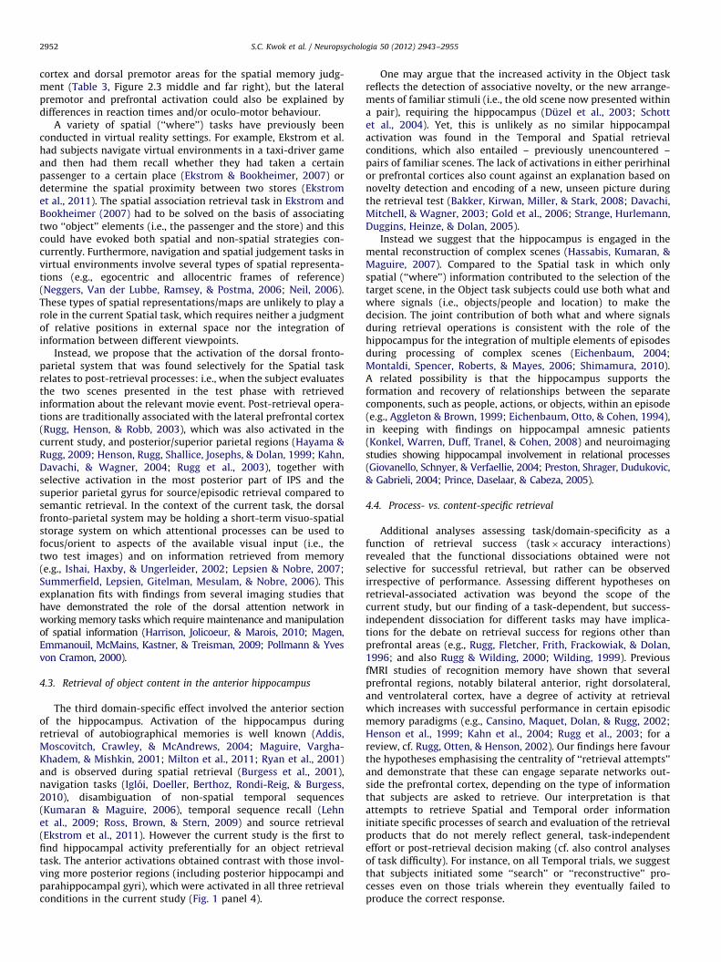
Fig. 2. Behavioural data, temporally modulated activity in the precuneus, and control analyses in the Spatial task. Panel 1: (a) Mean error rates (%) and reaction times (ms)
across the three retrieval tasks; (b) reaction times plotted against temporal distance (DeltaT) for high consistency trials of the Temporal task, showing a significant negative
correlation (p¼0.045); (c) Saccadic data obtained in 10 subjects, histograms depicting the median saccadic frequencies (1/s; on the left) and mean saccadic path-lengths
(deg; on the right) executed during the 5-s trials across tasks. Panel 2: Cluster of activation (in red) in the right precuneus modulated as a function of temporal distance (x,
y, z¼6, �70, 44; p-FWEo0.007), with the corresponding BOLD response (a.u.) plotted against DeltaT. The activation data for this plot were extracted from subject-specific
fitted-responses (first level analyses), 5 scans after the onset of the Temporal trials. Panel 3: Clusters of activation in the dorsal fronto-parietal network that activated
selectively in the Spatial task in: (a) the main analysis; (b) control analysis accounting for differences in task difficulty between conditions (reaction times as a covariate of
no interest); and (c) control analysis accounting for differential patterns of eye movements between conditions (saccadic frequency and path-lengths, in a sub-group of 10
subjects). Activations are displayed at p-unc.¼0.001. Error bars: Standard error of the mean.
S.C. Kwok et al. / Neuropsychologia 50 (2012) 2943–2955 2947

additional, not independent, masking contrasts the threshold was set to p-
unc.¼0.05. These procedures led us to identify areas specifically activated by
one of the three retrieval conditions (see also signal plots in Fig. 1).
2.7.2. Effect of temporal distance during the temporal task (DeltaT)
Behaviourally, subjects were slower (and less accurate) in trials with short
temporal distance (short DeltaT) compared to those with long temporal distance
(long DeltaT). For this analysis, we took advantage of the large pool of Temporal
trials (100 trials, vs. 25 for each of the other two tasks) further selecting a subset of
trials in which most subjects responded correctly. We called these trials ‘‘high
consistency Temporal trials’’ (T-high, as opposed to ‘‘low consistency T trials’’: T-
low). By applying a cut-off criterion to selecting trials in which at least 13 out of 15
subjects responded correctly, we obtained 67 high consistency trials for the
analysis. For these 67 trials the correlation between reaction times and DeltaT
was significant (see results section, and Fig. 2 panel 1).
We re-constructed all first-level fMRI models now considering separately T-
high and T-low trials, and including DeltaT as a trial-specific modulator of the T-
high responses (DeltaT-covariate). Moreover, because of the correlation between
RTs and DeltaT (see above), trial-specific RTs averaged across subjects were used
as an additional modulator of the T-high trials (RTs-covariate). Accordingly, any
significant co-variation between BOLD and DeltaT cannot be explained by RTs
differences (e.g., short temporal distance trials being just more difficult than long
distance trials). For completeness, these new first-level models included also the
corresponding RTs-covariates for Spatial and Object trials. The random effects
analysis consisted of a one-sample t-test assessing the significance of DeltaT-
covariate at the group level. The statistical threshold was set to p-FWE¼0.05,
considering the precuneus and the right angular gyrus (i.e., the areas activated for
Temporal trials in the main analysis, cf. Table 2) as the volume of interest (Worsley
et al., 1996).
The effect of temporal distance in the precuneus was also tested with an
additional analysis that categorically compared short vs. long trials (cf. St. Jacques
et al., 2008) and included performance at the subject-specific level, rather than
using performance consistency across subjects. We reconstructed all first-level
models, dividing the Temporal trials into ‘‘short’’ and ‘‘long’’ DeltaT trials (cf. St.
Jacques et al., 2008), and further into correct and incorrect trials. We obtained
4 conditions for the Temporal task (short/long� correct/incorrect), plus 2 for
Space (correct/incorrect) and 2 for Object (correct/incorrect). Because subjects
differed in their individual accuracy, the cut-off separating ‘‘short’’ vs. ‘‘long’’ trials
was set specifically for each subject. This ensured a well balanced number of short
and long trials for each individual. At the group level, we tested the effect of short
vs. long trials, with the aim of replicating the effect of temporal distance in the
precuneus, now using a categorical rather than parametric comparison.
2.7.3. Control analyses for the spatial task
Our main analysis showed that the Spatial task activated a large network of
brain areas including oculo-motor circuits in dorsal fronto-parietal regions.
Moreover, the behavioural data indicated that this task was more difficult than
the other two retrieval conditions (Fig. 2 panel 1). Accordingly, we ran two
additional control analyses. The first analysis consisted of a within-subject ANOVA
that was identical to the main analysis (15 subjects, 3 conditions: T, S, O), but now
including subject-specific RTs, that is, an average RT (across repetitions of the
same condition) for each subject and each condition, as a covariate of no interest.
In this way, the inherent differences in task difficulty across conditions were
accounted for. Within this we tested again for activation associated with the
Spatial task ([S4(TþO)/2], inclusively masked with (i) [S40], (ii) [S4T] and
[S4O]), but now accounting for the influence of RT differences. The second control
analysis made use of the eye-movement data recorded during fMRI. Because good
quality eye-tracking data were available only in a subgroup of subjects, this
ANOVA included 10 subjects, 3 conditions, plus subject- and condition-specific
saccadic frequency and path-length as additional covariates of no interest. Again
we tested for activations associated with the Spatial task ([S4(TþO)/2], masked
with (i) [S40], (ii) [S4T] and (iii) [S4O]), in this case excluding any contribution
of differential patterns of eye-movements between conditions. These additional,
not-independent, analyses were restricted to regions/voxels showing activation
for the Spatial task in the main analysis (cf. Tables 2 and 3).
2.7.4. Control analyses for retrieval success
In order to examine whether the domain-specific activations were due to
putative task-related retrieval processes or the retrieval of specific diagnostic
content, we investigated the effect of task and temporal distance including
incorrect trials as well as correct ones. Operationally, we associated task-related
processes to activations independently of retrieval success (i.e., showing task-
related effects for incorrect trials too), while content-components were tested as
effects specific for correct retrieval only (task by accuracy interactions). Accord-
ingly, we reconstructed all subject-specific first-level GLM including error trials
separately for each of the 3 Tasks. These now included 6 conditions given by the
crossing of Tasks (T, S, O) and Accuracy (correct, incorrect). For these additional
analyses, at the group level, we considered only Time and Space (�Accuracy),
because the Object condition had too few error trials (mean¼1.8 error/subject;
range¼0–5 errors).
3. Results
3.1. Behavioural results
Subjects performed better than chance level in all three retrieval
conditions (all pso0.001). They performed significantly better in
Object condition (error rate: 7.4771.40%) than Temporal condition
[error rate: 16.4070.75%; t (14)¼5.58, po0.001] and Spatial
condition [error rate: 32.2772.11%; t (14)¼10.28, po0.001]. On
Temporal trials subjects were more accurate than Spatial trials [t
(14)¼7.42, po0.001]. A similar pattern was observed with the
reaction times on correct trials. Subjects responded significantly
faster in Object condition [20487162 ms] than in Temporal condi-
tion [26147181ms; t (14)¼8.02, po0.001] and Spatial condition
[30387192 ms; t (14)¼9.34, po0.001], and the RTs in Temporal
condition were faster than Spatial condition [t (14)¼5.69, po0.001]
(see Fig. 2 panel 1, leftmost plot).
For the Temporal task, we assessed whether there was some
relationship between RTs (at retrieval) and the temporal distance
(at encoding) between two occurrences/scenes that subjects were
asked to judge (i.e., the DeltaT). We found a significant correlation
between RTs and DeltaT (Pearson r¼�0.25, p¼0.045), but only
when the analysis was constrained to trials that were recalled in a
reliable manner (i.e., the 67 Temporal trials correctly judged by at
least 13 out of 15 subjects). This negative correlation indicates
that subjects were faster to access/judge temporal information
stored in episodic memory, when the temporal distance between
the two events increased (Fig. 2 panel 1, central plot). This accords
with the view that memory traces are organised in some struc-
tured manner that facilitates judgements of events separated by
long temporal distances compared with short distances. As an
additional control, we tested whether there was any systematic
relationship between the absolute position of the scenes in the
film (averaging the time of the two frames) and reaction times.
This did not reveal any significant correlation (p40.1), reflecting
the specificity of the DeltaT effect, regardless of the segment’s
temporal position in the film.
With the eye-movement data available (10 subjects), subjects
made significantly more saccades (median number of saccades
per second) in the Temporal condition (1.7870.08) and in the
Spatial condition (1.8170.09) than in the Object condition
[1.5570.08; compared to Temporal: t (9)¼6.70, po0.001; com-
pared to Spatial: t (9)¼4.33, p¼0.002], and there was no
difference between Temporal and Spatial conditions [t (9)o1].
The mean path-length executed during 5-s retrieval periods (in
visual degree) was significantly larger in the Temporal condition
(46.4672.79) than in either Spatial [42.5873.48; t (9)¼3.19,
p¼0.011] or Object conditions [41.3072.31; t (9)¼7.36,
po0.001], and there was no difference between Spatial and
Object conditions [t (9)o1] (Fig. 2 panel 1, rightmost plots).
3.2. Domain-specific retrieval from episodic memory
Before testing for any domain-specific effect, we used a
conjunction analysis across the three retrieval conditions (T, S
and O) to highlight the brain regions engaged during memory
retrieval vs. rest, irrespective of retrieval task. This revealed
activation of a widespread network that included large sections
of the occipital cortex, regions in the dorsal fronto-parietal net-
work, plus motor, pre-motor and prefrontal areas bilaterally in
the frontal lobe (Table 1). Most of these activations can be
attributed to the visual stimulation, motor performance and
S.C. Kwok et al. / Neuropsychologia 50 (2012) 2943–29552948

general task-requirements. However, this analysis also revealed
that all three retrieval conditions activated the posterior part of
the hippocampus, and that was dissociated from a more anterior
region that responded selectively during object retrieval task (see
below, and Fig. 1 panel 4).
Temporal retrieval task: The direct comparison of the Temporal
task with the other two retrieval conditions revealed two clusters
of significant activation (Table 2). One cluster was located
medially and included the precuneus bilaterally. The second
cluster was on the lateral surface of the right hemisphere and
involved primarily the angular gyrus. The signal plots in Fig. 1
panel 2 show that activity in these two regions was highly specific
for the temporal retrieval task (see bar in red).
Spatial retrieval task: The fMRI analysis concerning the retrieval of
spatial memories highlighted activation of the superior parietal
gyrus, the intraparietal sulcus and frontal eye-fields in the dorsal
fronto-parietal network, the middle frontal gyrus, anterior insula,
plus regions in occipital visual cortex (Fig. 1 panel 3, and Table 2).
We performed two control analyses to assess the possible role of
task difficulty (indexed using RTs) and overt spatial behaviour
(indexed using saccade frequency and path-lengths) for the activa-
tion of this network. The analysis including RTs as a covariate of no
interest confirmed that the Spatial task activated the posterior nodes
of the dorsal fronto-partial network (superior parietal gyrus and
intraparietal sulcus) with activation also in the right superior frontal
gyrus (Table 3, and Figure 2.3 central panel). In the control analysis
including saccade frequency and path-lengths as confounding
effects, we found activation in superior parietal and superior frontal
gyrus bilaterally, plus the left intraparietal sulcus (Table 3, and
Figure 2.3 rightmost panel). Thus, the activation of dorsal fronto-
parietal regions for the Spatial retrieval task cannot be merely
explained by overall task difficulty and/or oculo-motor behaviour.
Object retrieval task: The object retrieval task was selectively
associated with the symmetrical activation the left and right
anterior hippocampus (see Fig. 1 panel 4, and Table 2), extending
to the parahippocampal cortex. Probabilistic cytoarchitectonic
maps (Amunts et al., 2005) revealed that 47.5% of the left cluster
could be assigned to the hippocampal formation, including the CA
and subiculum areas, whereas, in the right hemisphere, 32.2% of
the cluster was assigned to the hippocampal formation, with a
further 16.7% assigned to the entorhinal cortex (see Table 4). The
signal plots in Fig. 1 (panel 4) show that these activations were
selective for the Object task with spatial and temporal tasks
leading, if anything, to a de-activation of these regions. The
sagittal section in this panel highlights that the object-specific
effect was more anterior than the hippocampus activation
Table 1
Common activation across the three retrieval tasks.
Brain region Cluster Voxel
k p-FWE Z x y z
Occipital pole, L 37,502 o0.001 48 �16, �100, 4
Occipital pole, R 48 18, �102, 10
Dorsal occipital cortex, L 48 �22, �96, 12
Dorsal occipital cortex, R 48 30, �92, 24
Lateral occipital cortex, L 48 �38, �88, 10
Lateral occipital cortex, R 48 48, �74, �8
Ventral occipital cortex, L 48 �40, �76, �18
Ventral occipital cortex, R 48 38, �58, �16
Posterior hippocampus, L 48 �22, �28, �6
Posterior hippocampus, R 48 24, �28, �8
Intraparietal sulcus, L 5.01 �24, �64, 46
Intraparietal sulcus, R 6.15 32, �56, 52
Precuneus, R 5.06 8, �58, 50
Medial superior frontal gyrus, R 811 o0.001 7.38 8, 16, 56
Precentral gyrus, L 2,403 o0.001 7.31 �40, �20, 60
Superior frontal gyrus, R 5,632 o0.001 7.12 40, 0, 54
Middle frontal gyrus, R 7.16 46, 24, 22
Inferior frontal gyrus, R 6.34 48, 26, 6
Anterior insula, R 6.38 36, 24, �6
Middle frontal gyrus, L 996 o0.001 6.18 �46, 20, 24
Anterior insula, L 322 0.009 5.44 �34, 22, �4
Areas activated during all three retrieval conditions vs. rest (null-conjunction:
[T40] and [S40] and [O40]; p-FWEo0.05, whole-brain corrected at cluster
level, cluster size estimated at p-unc.¼0.001; k¼number of voxels).
Table 2
Direct comparisons between retrieval conditions.
Contrast Brain region Cluster Voxel
k p-FEW Z x y z
T4(SþO)/2 Precuneus, R 2635 o0.001 5.45 14, �60, 28
Precuneus, L 5.14 �8, �70, 26
Angular gyrus, R 343 0.0067 4.42 54, �52, 20
S4(OþT)/2 Superior parietal gyrus, L 4427 o0.001 7.10 �18, �72, 54
Intraparietal sulcus, L 5.88 �38, �42, 40
Dorsal occipital cortex, L 5.28 �30, �86, 32
Lateral occipital cortex, L 4.80 �54, �66, �2
Superior parietal gyrus, R 6292 o0.001 6.46 24, �70, 56
Intraparietal sulcus, R 6.03 44, �40, 46
Dorsal occipital cortex, R 6.20 42, �80, 26
Lateral occipital cortex, R 5.61 58, �58, �8
Superior frontal gyrus, R 2117 o0.001 6.39 28, 6, 56
Middle frontal gyrus, R 5.60 52, 10, 24
Middle frontal gyrus, L 1619 o0.001 5.48 �46, 2, 32
Superior frontal gyrus, L 5.29 �26, 4, 54
Medial sup. frontal gyrus, R 401 0.003 5.02 4, 18, 54
Inferior lingual gyrus, R 290 0.014 4.66 32, �42, �14
Inferior lingual gyrus, L 203 0.056 4.28 �30, �46, �16
Anterior insula, L 209 0.051 4.28 �36, 20, �4
O4(SþT)/2 Hippocampus, L 212 0.048 4.34 �22, �14, �20
Hippocampus, R 469 0.001 4.59 24, �6, �24
T-contrasts compared each condition vs. the mean of the other two conditions (e.g., [T4(SþO)/2]), each was inclusively masked with 3 additional contrasts, namely
activation for the critical condition vs. rest (e.g., [T40]) and activation for the critical condition vs. each of the two other conditions (e.g., [T4S] and [T4O]). The threshold
of the contrasts was set to p-FWE¼0.05, whole-brain corrected at cluster level, cluster size estimated at p-unc.¼0.001; the threshold of additional masking contrasts was
set to p-unc.¼0.05; k¼number of voxels.
S.C. Kwok et al. / Neuropsychologia 50 (2012) 2943–2955 2949

observed irrespective of retrieval condition (common activation
for T, S, and O tasks; see Table 1, and signal plot for the right
anterior hippocampus in Fig. 1 panel 4).
3.3. Effect of temporal distance (DeltaT)
Next, we turned to the issue of whether modulation of
temporal distance had any impact on functional activities within
the areas activated selectively during the Temporal task. On a
trial-by-trial basis, we assessed the relationship between BOLD
activation at retrieval and the temporal distance between the two
relevant events at encoding (DeltaT). This showed a significant
modulation of the precuneus response associated with Temporal
trials (T-high) as a function of temporal distance (x, y, z¼6, �70,
44; p-FWEo0.007). Specifically, the retrieval-related activation of
the precuneus increased with decreasing temporal distance
between the two events at encoding, providing support to the
notion of structurally-organised memory traces. It should be
noted that this analysis accounted, on a trial-by-trial basis, for
the changes of RTs as a function of temporal distance. Thus, mere
task difficulty is unlikely to explain this additional time-related
modulatory effect in the precuneus (Fig. 2 panel 2; note also that
the most difficult retrieval condition – i.e., the Spatial task –
activated this region less than the Temporal task). For complete-
ness, we also tested whether DeltaT modulated activity in Spatial-
and Object-related areas. As expected, this did not reveal any
significant effect of temporal distance in these regions.
With a non-independent additional analysis, we tested the
effect of temporal distance re-categorising all temporal trials as
‘‘short’’ or ‘‘long’’ distance trials (cf. St. Jacques et al., 2008). The
direct comparison of ‘‘short minus long’’ trials replicated the
effect of temporal distance in the precuneus, albeit only at
an uncorrected level of significance (x, y, z¼�10, �60, 48;
p-unc.o0.001). This analysis included accuracy as a factor,
allowing us to test for the interaction between distance and
accuracy (see also next section). No interaction was found in
the precuneus, even at an uncorrected level of significance.
3.4. Process- vs. content-specific retrieval
Finally, we tested whether the activations associated with the
Temporal and the Spatial tasks (cf. Fig. 1, panels 2 and 3) were
selective for correct trials or they were independent of retrieval
success. Using incorrect trials only, we replicated the activations
of the precuneus and the right angular gyrus for the Temporal
task (T4S: both p-FWEo0.05, at the whole-brain level) and the
dorsal fronto-parietal cortex for the Spatial task (S4T; including
posterior and intra-parietal cortex bilaterally and the right super-
ior frontal gyrus, all p-FWEo0.05, at the whole-brain level).
The task� accuracy interactions did not reveal any significant
activation. These results speak in favour of a process-based rather
than content-based account of our domain-specific results (see
Discussion).
4. Discussion
We obtained two main sets of findings. First, at both beha-
vioural and neural levels we found a modulatory effect on
retrieval of the parameterised temporal distance between
encoded events, in that both RTs and activity in the precuneus
showed a negative correlation with temporal distance between
two events at encoding (i.e., longer RTs and a greater activation
for shorter distances). These findings are more consistent with
search processes operating on episodic details within an orga-
nised memory structure, than with serial search between tempo-
rally organised adjacent memory traces. Second, dissociations in
the functional anatomy of domain-specific retrieval were exhib-
ited by different specific comparisons: retrieval of the temporal
order of events led to the activation of the precuneus and the
right angular gyrus. The dorsal frontal and parietal cortices were
engaged during recall of spatial information. Activations within
the hippocampal formation were found in object-based retrieval.
These task-specific effects occurred independently of retrieval
success. We discuss the implications of these patterns of activa-
tion with respect to the underlying processes that are involved
during retrieval of complex, naturalistic memories, primarily in
the context of how memory is organised temporally.
4.1. Retrieval of temporal components in the precuneus
Compared to the possible selectivity of medial temporal
structures for specific retrieval processes (e.g., Diana, Yonelinas,
& Ranganath, 2007; Hassabis, Kumaran, Vann, & Maguire, 2007),
less is known about the specific role of parietal cortex during
retrieval (cf. Cabeza & Nyberg, 2000; Nyberg et al., 2000; Vilberg
& Rugg, 2008). In a general framework of parietal functions,
activation during episodic retrieval has been associated with
attention-related processes (Ciaramelli, Grady, & Moscovitch,
2008; Wagner, Shannon, Kahn, & Buckner, 2005). However,
Sestieri, Shulman, and Corbetta (2010) reported a dissociation
between these two cognitive functions in parietal cortex (see also
Hutchinson, Uncapher, & Wagner, 2009). They found a specific
involvement of the angular gyrus, precuneus and posterior
cingulate cortex during memory-search, but of intraparietal
sulcus (IPS) and the superior parietal lobule for perceptually-
related processes. Our findings are in agreement with this
distinction showing retrieval-related activation in the precuneus
and the right angular gyrus (Fig. 1 panel 2).
Unlike the Spatial and Object tasks, which could be accom-
plished by retrieving a single ‘‘snapshot’’ of the memorised
episode, the Temporal task required the subject to access multiple
(at least two) instances of the storyline. According to chronolo-
gical organisation theory (Friedman, 1993), this can be done by
retrieving the time position of one of the two test scenes in the
film, and then scanning through the rest of the episode looking for
the second scene (i.e., serial temporal search). However, if
memory is organised in this fashion, we would expect that
Table 3
Additional control analyses for the Spatial task.
Brain region RTs controlled Saccadic data controlled
Z x y z Z x y z
Superior parietal gyrus, L 4.46 �18, �72, 54 4.10 �12, �68, 58
Superior parietal gyrus, R 3.81 28, �66, 62 4.51 22, �72, 60
Intraparietal sulcus, L 3.81 �44, �44, 46 3.69 �38, �42, 38
Intraparietal sulcus, R 4.25 52, �42, 58 – –
Middle frontal gyrus, L 3.44 �46, 0, 28
Middle frontal gyrus, R 3.34 52, 10, 28
Superior frontal gyrus, L – – 3.23 �26, 0, 52
Superior frontal gyrus, R 3.64 28, 6, 54 3.85 30, 4, 58
Lateral occipital cortex, R 3.26 58, �58, �10 3.75 62, �56, �2
Comparisons between the Spatial condition vs. the mean of the other two
conditions ([S4(TþO)/2], inclusively masked with [S40], [S4T] and [S4O])
controlled for differential RTs and eye movements between conditions (cf. also
Figure 2.1). The first control analysis included subject- and condition- specific RTs
as a covariate to account for differences in task difficulty (n¼15). The second
control analysis included subject- and condition-specific saccadic frequency and
path-length as additional covariates to exclude any contribution of differential
patterns of eye-movements (n¼10). For these additional analyses, we report
voxels at p-unc.¼0.001 that are located within the clusters showing space-specific
activation in the main analysis (cf. Table 2).
S.C. Kwok et al. / Neuropsychologia 50 (2012) 2943–29552950

memories laid down at adjacent points in time would prime one
another. Thus, when remembering some past event, it should be
easy to order events that occurred at about the same time. Here,
we found that reaction times (Fig. 2 panel 1) and activity in the
precuneus (Fig. 2 panel 2) increased with decreasing temporal
distances between the two test scenes/events. RTs could reflect
more than one process (e.g., not only retrieval times, but also
decision times which could reflect a greater uncertainty about
relative recency, when the two pictures were close in time).
However, as far as the role played by the precuneus, the key
structure in the temporal task, is concerned, these effects appear
to speak against it being involved in any form of serial search
along temporally organised memory traces, if retrieval of the
second event were to arise by scanning backward or forward from
the first on some ‘‘time-line’’. In accord with studies which
required subjects to make recency judgements of less complex
stimuli (Konishi et al., 2002; Suzuki et al., 2002), and with the
results of St. Jacques and colleagues (2008), who reported
increased precuneus activity as a function of decreasing time
lag, our data likewise speak against the precuneus having any role
in a chronological organisation process of episodic recollection
involving serial scanning through memory traces.
At least two accounts are possible for the selective modulation
of activity in the precuneus by the elapsed time between events.
The first relates to the encoding perturbation theory, a theory
originally proposed by Estes (1972, 1985) to explain findings on
short-term memory, while the second refers to the reconstructive
theory proposed by Friedman (1993, 1996, 2001, 2004).
According to the encoding perturbation theory, when an event
occurs this becomes associated with control elements at different
levels within a hierarchically organised structure. The notion has
later been elaborated and extended to explain everyday memory
phenomena in long-term memory (e.g., Anderson & Conway,
1993; Zacks, Tversky, & Iyer, 2001). On this approach the system
encodes continuous streams of observed behaviour by segment-
ing activities into events and then organising them in memory in
a basically hierarchical manner (Zacks et al., 2001), with groups of
fine-grained events clustering into larger units (Kurby & Zacks,
2008). In the present study, the observed effects of temporal
distance during retrieval may relate to the search for the two test
scenes through a hierarchical structure which holds the encoded
TV episode. When the two scenes are far apart in time (long
DeltaT), the Temporal task can be solved by searching high/
intermediate levels of the knowledge structure. By contrast, when
the two scenes are close in time (short DeltaT), they will be
associated with the same node at intermediate levels of the
structure and the search has to be continued to lower levels of
the structure. Activation of the precuneus could reflect some
aspect(s) of this search process, with increased activation when
the search involves exploring down to lower levels of the
structure. One more specific possibility is that searching the
lower levels of the hierarchy requires more of a particular sort
of process, such as creating imagery of specific scenes not
presented at retrieval (Fletcher et al., 1995; Fletcher, Shallice,
Frith, Frackowiak, & Dolan, 1996; Grasby et al., 1993; but see
Lundstrom, Ingvar, & Petersson, 2005; Roland & Seitz, 1989).
Alternatively, the precuneus may be required for the organisation
of levels per se as such an account would also be compatible with
a role of the precuneus in structuring knowledge hierarchies of
the outside world during perception and memory encoding
(Speer, Zacks, & Reynolds, 2007; Zacks et al., 2001; Zacks, Speer,
Swallow, & Maley, 2010).
A second possible account for the observed effects of temporal
distance concerns reconstructive theories of memory (Friedman,
1993; 1996; 2001; 2004). When applied to memory for personal
events (Brown, Rips & Shevell, 1985; Friedman & Wilkins, 1985),
such theories postulate the existence of a process of reconstruc-
tion that draws on a rich knowledge of social, natural, and
personal time patterns (e.g., the time of a day). In contrast to
the encoding perturbation model discussed above, there is an
explicit emphasis on the use of general time knowledge and
inferential processes at the time of recall. Reconstruction pro-
cesses are effortful operations that include retrieving contextual
details and using them to infer the order of past events (Curran &
Friedman, 2003; Skowronski, Walker, & Betz, 2003). These pro-
cesses can provide relatively high precision in the resolution of
temporal details, and are particularly likely to be employed when
past events are close in time and difficult to discern (Burt, Kemp,
Grady, & Conway, 2000; Friedman, 1993), such as those involved
in the short DeltaT trials in our study. The additional amount of
time required in short DeltaT trials in our study is to be expected
if such reconstructive-based processes are operative (Curran &
Friedman, 2003; Friedman, 1993; St. Jacques et al., 2008).
Our findings on the engagement of the precuneus in temporal
memory judgements have implications with respect to the
putative functions of other areas implicated in retrieving tem-
poral information from memory. The greater difficulty associated
with distinguishing items closer in time has been reliably
reflected in prefrontal activations in fMRI studies (e.g., Konishi
et al., 2002; St. Jacques et al., 2008; Suzuki et al., 2002; Zorrilla
et al., 1996). However, we have shown that an area, other than the
well-documented prefrontal regions, is involved in discriminating
the order of events that are closer in time. St Jacques et al. (2008)
has provided initial evidence of the role of precuneus in this
process. However, as noted in the Introduction, there is a
potential task difficulty confound in the study of St Jacques
et al. (2008), so our demonstration provides more solid evidence
of the effect of temporal distance in retrieval on the operation of
the precuneus.
4.2. Retrieval of detailed spatial content in dorsal fronto-parietal
cortex
The Spatial task elicited a widespread pattern of activation,
including parietal regions (PPC and IPS) and several premotor and
prefrontal regions (Fig. 1 panel 3). However, the activation of
some of these areas is likely not to be specifically due to a
memory process. Spatial trials are more difficult than the other
trial types, as manifested by slower reaction time and a higher
error rate (Fig. 2 panel 1). We thus ran a set of control analyses to
partial out the general effect of task difficulty (RTs) and oculo-
motor behaviour; these confirmed the role of the superior parietal
Table 4
Probabilistic localisation of the voxels belonging to the left and right hippocampal
activation clusters (Object task).
Cytoarchitectonic area Current study
Hippocampus, L Hippocampus, R
Hippocampus, CA 42.8 (12.2) 17.8 (10.4)
Hippocampus, SUB 4.7 (2.0) 14.4 (12.8)
Hippocampus, EC Nil 16.7 (11.8)
Amygdala, LB 27.8 (18.9) 20.3 (28.2)
Amygdala, SF 9.1 (10.0) Nil
For the two clusters (cf. Figure 1.4, in blue), the table reports the percentage of
voxels located within specific cytoarchitectonic areas of the medial temporal
cortex: Cornu ammonis (CA), the subicular complex, the entorhinal cortices (EC),
the laterobasal (LB) and superficial (SF) nuclear groups of the amygdala (Amunts
et al., 2005). For each of the cytoarchitectonic areas, the table also reports the
proportion of the area that was activated during the current Object retrieval task
(in parenthesis).
S.C. Kwok et al. / Neuropsychologia 50 (2012) 2943–2955 2951
cortex and dorsal premotor areas for the spatial memory judg-
ment (Table 3, Figure 2.3 middle and far right), but the lateral
premotor and prefrontal activation could also be explained by
differences in reaction times and/or oculo-motor behaviour.
A variety of spatial (‘‘where’’) tasks have previously been
conducted in virtual reality settings. For example, Ekstrom et al.
had subjects navigate virtual environments in a taxi-driver game
and then had them recall whether they had taken a certain
passenger to a certain place (Ekstrom & Bookheimer, 2007) or
determine the spatial proximity between two stores (Ekstrom
et al., 2011). The spatial association retrieval task in Ekstrom and
Bookheimer (2007) had to be solved on the basis of associating
two ‘‘object’’ elements (i.e., the passenger and the store) and this
could have evoked both spatial and non-spatial strategies con-
currently. Furthermore, navigation and spatial judgement tasks in
virtual environments involve several types of spatial representa-
tions (e.g., egocentric and allocentric frames of reference)
(Neggers, Van der Lubbe, Ramsey, & Postma, 2006; Neil, 2006).
These types of spatial representations/maps are unlikely to play a
role in the current Spatial task, which requires neither a judgment
of relative positions in external space nor the integration of
information between different viewpoints.
Instead, we propose that the activation of the dorsal fronto-
parietal system that was found selectively for the Spatial task
relates to post-retrieval processes: i.e., when the subject evaluates
the two scenes presented in the test phase with retrieved
information about the relevant movie event. Post-retrieval opera-
tions are traditionally associated with the lateral prefrontal cortex
(Rugg, Henson, & Robb, 2003), which was also activated in the
current study, and posterior/superior parietal regions (Hayama &
Rugg, 2009; Henson, Rugg, Shallice, Josephs, & Dolan, 1999; Kahn,
Davachi, & Wagner, 2004; Rugg et al., 2003), together with
selective activation in the most posterior part of IPS and the
superior parietal gyrus for source/episodic retrieval compared to
semantic retrieval. In the context of the current task, the dorsal
fronto-parietal system may be holding a short-term visuo-spatial
storage system on which attentional processes can be used to
focus/orient to aspects of the available visual input (i.e., the
two test images) and on information retrieved from memory
(e.g., Ishai, Haxby, & Ungerleider, 2002; Lepsien & Nobre, 2007;
Summerfield, Lepsien, Gitelman, Mesulam, & Nobre, 2006). This
explanation fits with findings from several imaging studies that
have demonstrated the role of the dorsal attention network in
working memory tasks which require maintenance and manipulation
of spatial information (Harrison, Jolicoeur, & Marois, 2010; Magen,
Emmanouil, McMains, Kastner, & Treisman, 2009; Pollmann & Yves
von Cramon, 2000).
4.3. Retrieval of object content in the anterior hippocampus
The third domain-specific effect involved the anterior section
of the hippocampus. Activation of the hippocampus during
retrieval of autobiographical memories is well known (Addis,
Moscovitch, Crawley, & McAndrews, 2004; Maguire, Vargha-
Khadem, & Mishkin, 2001; Milton et al., 2011; Ryan et al., 2001)
and is observed during spatial retrieval (Burgess et al., 2001),
navigation tasks (Igloi, Doeller, Berthoz, Rondi-Reig, & Burgess,
2010), disambiguation of non-spatial temporal sequences
(Kumaran & Maguire, 2006), temporal sequence recall (Lehn
et al., 2009; Ross, Brown, & Stern, 2009) and source retrieval
(Ekstrom et al., 2011). However the current study is the first to
find hippocampal activity preferentially for an object retrieval
task. The anterior activations obtained contrast with those invol-
ving more posterior regions (including posterior hippocampi and
parahippocampal gyri), which were activated in all three retrieval
conditions in the current study (Fig. 1 panel 4).
One may argue that the increased activity in the Object task
reflects the detection of associative novelty, or the new arrange-
ments of familiar stimuli (i.e., the old scene now presented within
a pair), requiring the hippocampus (Duzel et al., 2003; Schott
et al., 2004). Yet, this is unlikely as no similar hippocampal
activation was found in the Temporal and Spatial retrieval
conditions, which also entailed – previously unencountered –
pairs of familiar scenes. The lack of activations in either perirhinal
or prefrontal cortices also count against an explanation based on
novelty detection and encoding of a new, unseen picture during
the retrieval test (Bakker, Kirwan, Miller, & Stark, 2008; Davachi,
Mitchell, & Wagner, 2003; Gold et al., 2006; Strange, Hurlemann,
Duggins, Heinze, & Dolan, 2005).
Instead we suggest that the hippocampus is engaged in the
mental reconstruction of complex scenes (Hassabis, Kumaran, &
Maguire, 2007). Compared to the Spatial task in which only
spatial (‘‘where’’) information contributed to the selection of the
target scene, in the Object task subjects could use both what and
where signals (i.e., objects/people and location) to make the
decision. The joint contribution of both what and where signals
during retrieval operations is consistent with the role of the
hippocampus for the integration of multiple elements of episodes
during processing of complex scenes (Eichenbaum, 2004;
Montaldi, Spencer, Roberts, & Mayes, 2006; Shimamura, 2010).
A related possibility is that the hippocampus supports the
formation and recovery of relationships between the separate
components, such as people, actions, or objects, within an episode
(e.g., Aggleton & Brown, 1999; Eichenbaum, Otto, & Cohen, 1994),
in keeping with findings on hippocampal amnesic patients
(Konkel, Warren, Duff, Tranel, & Cohen, 2008) and neuroimaging
studies showing hippocampal involvement in relational processes
(Giovanello, Schnyer, & Verfaellie, 2004; Preston, Shrager, Dudukovic,
& Gabrieli, 2004; Prince, Daselaar, & Cabeza, 2005).
4.4. Process- vs. content-specific retrieval
Additional analyses assessing task/domain-specificity as a
function of retrieval success (task� accuracy interactions)
revealed that the functional dissociations obtained were not
selective for successful retrieval, but rather can be observed
irrespective of performance. Assessing different hypotheses on
retrieval-associated activation was beyond the scope of the
current study, but our finding of a task-dependent, but success-
independent dissociation for different tasks may have implica-
tions for the debate on retrieval success for regions other than
prefrontal areas (e.g., Rugg, Fletcher, Frith, Frackowiak, & Dolan,
1996; and also Rugg & Wilding, 2000; Wilding, 1999). Previous
fMRI studies of recognition memory have shown that several
prefrontal regions, notably bilateral anterior, right dorsolateral,
and ventrolateral cortex, have a degree of activity at retrieval
which increases with successful performance in certain episodic
memory paradigms (e.g., Cansino, Maquet, Dolan, & Rugg, 2002;
Henson et al., 1999; Kahn et al., 2004; Rugg et al., 2003; for a
review, cf. Rugg, Otten, & Henson, 2002). Our findings here favour
the hypotheses emphasising the centrality of ‘‘retrieval attempts’’
and demonstrate that these can engage separate networks out-
side the prefrontal cortex, depending on the type of information
that subjects are asked to retrieve. Our interpretation is that
attempts to retrieve Spatial and Temporal order information
initiate specific processes of search and evaluation of the retrieval
products that do not merely reflect general, task-independent
effort or post-retrieval decision making (cf. also control analyses
of task difficulty). For instance, on all Temporal trials, we suggest
that subjects initiated some ‘‘search’’ or ‘‘reconstructive’’ pro-
cesses even on those trials wherein they eventually failed to
produce the correct response.
S.C. Kwok et al. / Neuropsychologia 50 (2012) 2943–29552952

5. Conclusion
The current study suggests that memory traces of complex
naturalistic temporal events are stored in a structured, rather
than serial, manner. It also provides fMRI evidence to support a
tripartite process-specific retrieval model of episodic memory.
Activity in the precuneus is associated with temporal retrieval, a
dorsal fronto-parietal network is engaged during spatial retrieval,
while antero-medial temporal regions activate selectively during
object-related retrieval. We link this selectivity with the engage-
ment of specific retrieval processes, rather than memory content.
We propose that systems in the precuneus retrieve temporal
information by being involved in either searching within a
hierarchical knowledge structure or in reconstructing moments/
events of contextual details when considerable temporal preci-
sion is required. By contrast, decisions about spatial details utilise
operations on a visuo-spatial short-term storage system that can
maintain and compare online sensory information and signals
retrieved from episodic long-term memory; these processes
involve dorsal fronto-parietal cortex. Finally, the anterior hippo-
campus is held to be involved in object-retrieval when the
process needs to combine spatial (‘‘where’’) and non-spatial
(‘‘what’’) information. By isolating the contribution of these
regions, the present fMRI findings contribute to a theory of
dissociations between retrieval-related processes and highlights
a role of the precuneus in searching information within a
‘‘structured’’ long-term temporal memory store.
Acknowledgements
The Neuroimaging Laboratory, Santa Lucia Foundation, is
supported by The Italian Ministry of Health. The research leading
to these results has received funding from the European Research
Council under the European Union’s Seventh Framework Pro-
gramme (FP7/2007-2013)/ERC grant agreement n.242809. Thanks
are due to Steve Gazzitano, Paolo Alessandrini, Fabio Cannata and
Fabio Mollo for their assistance at various stages of this research.
References
Addis, D. R., Moscovitch, M., Crawley, A. P., & McAndrews, M. P. (2004).Recollective qualities modulate hippocampal activation during autobiographi-cal memory retrieval. Hippocampus, 14, 752–762.
Aggleton, J. P., & Brown, M. W. (1999). Episodic memory, amnesia, and thehippocampal-anterior thalamic axis. Behavioral and Brain Sciences, 22,425–444.
Amunts, K., Kedo, O., Kindler, M., Pieperhoff, P., Mohlberg, H., Shah, N. J., Habel, U.,Schneider, F., & Zilles, K. (2005). Cytoarchitectonic mapping of the humanamygdala, hippocampal region and entorhinal cortex: intersubject variabilityand probability maps. Anatomy and Embryology, 210, 343–352.
Anderson, S. J., & Conway, M. A. (1993). Investigating the structure of autobio-graphical memories. Journal of Experimental Psychology: Learning, Memory, and
Cognition, 19, 1178–1196.Bakker, A., Kirwan, C. B., Miller, M., & Stark, C. E. L. (2008). Pattern separation in the
human hippocampal CA3 and dentate gyrus. Science, 319, 1640–1642.Barsalou, L. W. (1988). The content and organisation of autobiographical mem-
ories. In: U. Neisser, & E. Winograd (Eds.), Remembering reconsidered: ecological
and traditional approaches to the study of memory (pp. 193–243). Cambridge:Cambridge University Press.
Brown, G. D. A., & Chater, N. (2001). The chronological organisation of memory:common psychological foundations for remembering and timing. In: C.M. Hoerl, & T. (Eds.), Time and memory: issues in philosophy and psychology
(pp. 77–110). Oxford: England: Oxford University Press.Brown, N. R., Rips, L. J., & Shevell, S. K. (1985). The subjective dates of natural
events in very-long-term memory. Cognitive Psychology, 17, 139–177.Bruce, D., & Van Pelt, M. (1989). Memories of a bicycle tour. Applied Cognitive
Psychology, 3, 137–156.Burgess, N., Maguire, E. A., Spiers, H. J., & O’Keefe, J. (2001). A temporoparietal and
prefrontal network for retrieving the spatial context of lifelike events. Neuro-Image, 14, 439–453.
Burgess, P. W., & Shallice, T. (1996). Confabulation and the control of recollection.Memory, 4, 359–411.
Burt, C. D. B., Kemp, S., Grady, J. M., & Conway, M. (2000). Ordering autobiogra-phical experiences. Memory, 8, 323–332.
Butters, M. A., Kaszniak, A. W., Glisky, E. L., Eslinger, P. J., & Schacter, D. L. (1994).Recency discrimination deficits in frontal lobe patients. Neuropsychology, 8,343–354.
Cabeza, R., & Nyberg, L. (2000). Imaging cognition II: An empirical review of 275PET and fMRI studies. Journal of Cognitive Neuroscience, 12, 1–47.
Cansino, S., Maquet, P., Dolan, R. J., & Rugg, M. D. (2002). Brain activity underlyingencoding and retrieval of source memory. Cerebral Cortex, 12, 1048–1056.
Christoff, K., Prabhakaran, V., Dorfman, J., Zhao, Z., Kroger, J. K., Holyoak, K. J., &Gabrieli, J. D. E. (2001). Rostrolateral prefrontal cortex involvement in rela-tional integration during reasoning. NeuroImage, 14, 1136–1149.
Ciaramelli, E., Grady, C. L., & Moscovitch, M. (2008). Top-down and bottom-upattention to memory: a hypothesis (AtoM) on the role of the posterior parietalcortex in memory retrieval. Neuropsychologia, 46, 1828–1851.
Clayton, N. S., & Dickinson, A. (1998). Episodic-like memory during cache recoveryby scrub jays. Nature, 395, 272–274.
Curran, T., & Friedman, W. J. (2003). Differentiating location- and distance-basedprocesses in memory for time: an ERP study. Psychonomic Bulletin & Review,10, 711–717.
Davachi, L., Mitchell, J. P., & Wagner, A. D. (2003). Multiple routes to memory:distinct medial temporal lobe processes build item and source memories.Proceedings of the National Academy of Sciences of the United States of America,100, 2157–2162.
Diana, R. A., Yonelinas, A. P., & Ranganath, C. (2007). Imaging recollection andfamiliarity in the medial temporal lobe: a three-component model. Trends in
Cognitive Sciences, 11, 379–386.Duzel, E., Habib, R., Rotte, M., Guderian, S., Tulving, E., & Heinze, H.-J. (2003).
Human hippocampal and parahippocampal activity during visual associativerecognition memory for spatial and nonspatial stimulus configurations. Journalof Neuroscience, 23, 9439–9444.
Eichenbaum, H. (2004). Hippocampus: cognitive processes and neural representa-tions that underlie declarative memory. Neuron, 44, 109–120.
Eichenbaum, H., Otto, T., & Cohen, N. J. (1994). Two functional components of thehippocampal memory system. Behavioral and Brain Sciences, 17, 449–518.
Ekstrom, A. D., & Bookheimer, S. Y. (2007). Spatial and temporal episodic memoryretrieval recruit dissociable functional networks in the human brain. Learning& Memory, 14, 645–654.
Ekstrom, A. D., Copara, M. S., Isham, E. A., Wang, W.-c., & Yonelinas, A. P. (2011).Dissociable networks involved in spatial and temporal order source retrieval.NeuroImage, 56, 1803–1813.
Estes, W. K. (1972). An associative basis for coding and organization in memory.In: A. W. Melton, & E. Martin (Eds.), Coding processes in human memory (pp.161–190). Washington, D.C: V. H. Winston.
Estes, W. K. (1985). Memory for temporal information. In: J. A. Michon, & J. Jackson(Eds.), Time, mind and behavior (pp. 151–168). Berlin: Springer-Verlag.
Fletcher, P. C., Frith, C. D., Baker, S. C., Shallice, T., Frackowiak, R. S. J., & Dolan, R. J.(1995). The mind’s eye—precuneus activation in memory-related imagery.NeuroImage, 2, 195–200.
Fletcher, P. C., Shallice, T., Frith, C. D., Frackowiak, R. S., & Dolan, R. J. (1996). Brainactivity during memory retrieval. The influence of imagery and semanticcueing. Brain, 119, 1587–1596.
Friedman, W. J. (1987). A follow-up to ‘‘scale effects in memory for the time ofevents’’: the earthquake study. Memory & Cognition, 15, 518–520.
Friedman, W. J. (1993). Memory for the time of past events. Psychological Bulletin,113, 44–66.
Friedman, W. J. (1996). Distance and location processes in memory for the times ofpast events. In: D. L. Medin (Ed.), The psychology of learning and motivation, Vol.35 (pp. 1–41). San Diego: Academic Press.
Friedman, W. J. (2001). Memory processes underlying humans’ chronologicalsense of the past. In: C. Hoerl, & T. McCormack (Eds.), Time and memory: issues
in philosophy and psychology (pp. 139–167). Oxford: England: Oxford Uni-versity Press.
Friedman, W. J. (2004). Time in autobiographical memory. Social Cognition, 22,591–605.
Friedman, W. J., & Wilkins, A. (1985). Scale effects in memory for the time ofevents. Memory & Cognition, 13, 168–175.
Friston, K. J., Penny, W., Phillips, C., Kiebel, S., Hinton, G., & Ashburner, J. (2002). Classicaland Bayesian inference in neuroimaging: theory. NeuroImage, 16, 465–483.
Fujii, T., Suzuki, M., Okuda, J., Ohtake, H., Tanji, K., Yamaguchi, K., Itoh, M., &Yamadori, A. (2004). Neural correlates of context memory with real-worldevents. NeuroImage, 21, 1596–1603.
Giovanello, K. S., Schnyer, D. M., & Verfaellie, M. (2004). A critical role for theanterior hippocampus in relational memory: evidence from an fMRI studycomparing associative and item recognition. Hippocampus, 14, 5–8.
Gold, J. J., Smith, C. N., Bayley, P. J., Shrager, Y., Brewer, J. B., Stark, C. E. L., Hopkins,R. O., & Squire, L. R. (2006). Item memory, source memory, and the medialtemporal lobe: concordant findings from fMRI and memory-impaired patients.Proceedings of the National Academy of Sciences, 103, 9351–9356.
Grasby, P. M., Frith, C. D., Friston, K. J., Bench, C., Frackowiak, R. S., & Dolan, R. J.(1993). Functional mapping of brain areas implicated in auditory-verbalmemory function. Brain, 116, 1–20.
Harrison, A., Jolicoeur, P., & Marois, R. (2010). ‘‘What’’ and ‘‘where’’ in theintraparietal sulcus: an fMRI study of object identity and location in visualshort-term memory. Cerebral Cortex, 20, 2478–2485.
S.C. Kwok et al. / Neuropsychologia 50 (2012) 2943–2955 2953

Hassabis, D., Kumaran, D., & Maguire, E. A. (2007). Using imagination to understand theneural basis of episodic memory. Journal of Neuroscience, 27, 14365–14374.
Hassabis, D., Kumaran, D., Vann, S. D., & Maguire, E. A. (2007). Patients withhippocampal amnesia cannot imagine new experiences. Proceedings of the
National Academy of Sciences, 104, 1726–1731.Hasson, U., Furman, O., Clark, D., Dudai, Y., & Davahi, L. (2008). Enhanced inter-
subject correlations during movie viewing correlate with successful episodicencoding. Neuron, 57, 452–462.
Hayama, H. R., & Rugg, M. D. (2009). Right dorsolateral prefrontal cortex isengaged during post-retrieval processing of both episodic and semanticinformation. Neuropsychologia, 47, 2409–2416.
Hayes, S. M., Ryan, L., Schnyer, D. M., & Nadel, L. (2004). An fMRI study of episodicmemory: retrieval of object, spatial, and temporal information. Behavioral
Neuroscience, 118, 885–896.Henson, R. N., Rugg, M. D., Shallice, T., Josephs, O., & Dolan, R. J. (1999).
Recollection and familiarity in recognition memory: an event-related func-tional magnetic resonance imaging study. Journal of Neuroscience, 19,3962–3972.
Hutchinson, J. B., Uncapher, M. R., & Wagner, A. D. (2009). Posterior parietal cortexand episodic retrieval: convergent and divergent effects of attention andmemory. Learning & Memory, 16, 343–356.
Huttenlocher, J., Hedges, L., & Prohaska, V. (1988). Hierarchical organization inordered domains: estimating the dates of events. Psychological Review, 95,471–484.
Igloi, K., Doeller, C. F., Berthoz, A., Rondi-Reig, L., & Burgess, N. (2010). Lateralizedhuman hippocampal activity predicts navigation based on sequence or placememory. Proceedings of the National Academy of Sciences, 107, 14466–14471.
Ishai, A., Haxby, J. V., & Ungerleider, L. G. (2002). Visual imagery of famous faces: effectsof memory and attention revealed by fMRI. NeuroImage, 17, 1729–1741.
Kahn, I., Davachi, L., & Wagner, A. D. (2004). Functional-neuroanatomic correlatesof recollection: implications for models of recognition memory. The Journal of
Neuroscience, 24, 4172–4180.King, J. A., Hartley, T., Spiers, H. J., Maguire, E. A., & Burgess, N. (2005). Anterior
prefrontal involvement in episodic retrieval reflects contextual interference.NeuroImage, 28, 256–267.
Konishi, S., Uchida, I., Okuaki, T., Machida, T., Shirouzu, I., & Miyashita, Y. (2002).Neural correlates of recency judgment. The Journal of Neuroscience, 22,9549–9555.
Konkel, A., Warren, D. E., Duff, M. C., Tranel, D., & Cohen, N. J. (2008). Hippocampalamnesia impairs all manner of relational memory. Frontiers in Human
Neuroscience, 2.Kumaran, D., & Maguire, E. A. (2006). The dynamics of hippocampal activation
during encoding of overlapping sequences. Neuron, 49, 617–629.Kurby, C. A., & Zacks, J. M. (2008). Segmentation in the perception and memory of
events. Trends in Cognitive Sciences, 12, 72–79.Laming, D (1999). Testing the idea of distinct storage mechanisms in memory.
International Journal of Psychology, 34, 419–426.Lehn, H., Steffenach, H.-A., van Strien, N. M., Veltman, D. J., Witter, M. P., & Haberg,
A. K. (2009). Specific role of the human hippocampus in recall of temporalsequences. Journal of Neuroscience, 29, 3475–3484.
Lepsien, J., & Nobre, A. C. (2007). Attentional modulation of object representationsin working memory. Cerebral Cortex, 17, 2072–2083.
Linton, M. (1986). Ways of searching and the contents of memory. In: D. C. Rubin(Ed.), Autobiographical memory (pp. 51–67). New York: Cambridge UniversityPress.
Lundstrom, B. N., Ingvar, M., & Petersson, K. M. (2005). The role of precuneus andleft inferior frontal cortex during source memory episodic retrieval. Neuro-Image, 27, 824–834.
Magen, H., Emmanouil, T.-A., McMains, S. A., Kastner, S., & Treisman, A. (2009).Attentional demands predict short-term memory load response in posteriorparietal cortex. Neuropsychologia, 47, 1790–1798.
Maguire, E. A., Vargha-Khadem, F., & Mishkin, M. (2001). The effects of bilateralhippocampal damage on fMRI regional activations and interactions duringmemory retrieval. Brain, 124, 1156–1170.
McAndrews, M. P., & Milner, B. (1991). The frontal cortex and memory fortemporal order. Neuropsychologia, 29, 849–859.
Mendelsohn, A., Chalamish, Y., Solomonovich, A., & Dudai, Y. (2008). Mesmerizingmemories: brain substrates of episodic memory suppression in posthypnoticamnesia. Neuron, 57, 159–170.
Mendelsohn, A., Furman, O., & Dudai, Y. (2010). Signatures of memory: braincoactivations during retrieval distinguish correct from incorrect recollection.Frontiers in behavioral neuroscience, 4, 1–12.
Milner, B., Corsi, P., & Leonard, G. (1991). Frontal-lobe contribution to recencyjudgements. Neuropsychologia, 29, 601–618.
Milton, F., Muhlert, N., Butler, C. R., Smith, A., Benattayallah, A., & Zeman, A. Z.(2011). An fMRI study of long-term everyday memory using SenseCam.Memory, 19, 733–744.
Montaldi, D., Spencer, T. J., Roberts, N., & Mayes, A. R. (2006). The neural systemthat mediates familiarity memory. Hippocampus, 16, 504–520.
Nairne, J. S. (1992). The loss of positional certainty in long-term memory.Psychological Science, 3, 199–202.
Neath, I., & Crowder, R. G. (1990). Schedules of presentation and temporaldistinctiveness in human memory. Journal of Experimental Psychology: Learn-
ing, Memory, and Cognition, 16, 316–327.Neath, I., & Crowder, R. G. (1996). Distinctiveness and very short-term serial
position. Memory, 4, 225–242.
Neggers, S. F. W., Van der Lubbe, R. H. J., Ramsey, N. F., & Postma, A. (2006).Interactions between ego- and allocentric neuronal representations of space.NeuroImage, 31, 320–331.
Neil, B. (2006). Spatial memory: how egocentric and allocentric combine. Trends inCognitive Sciences, 10, 551–557.
Nichols, T., Brett, M., Andersson, J., Wager, T., & Poline, J.-B. (2005). Validconjunction inference with the minimum statistic. NeuroImage, 25, 653–660.
Nyberg, L., McIntosh, A., Cabeza, R., Habib, R., Houle, S., & Tulving, E. (1996).General and specific brain regions involved in encoding and retrieval ofevents: what, where, and when. Proceedings of the National Academy of Sciences
of the United States of America, 93, 11280–11285.Nyberg, L., Persson, J., Habib, R., Tulving, E., McIntosh, A. R., Cabeza, R., & Houle, S.
(2000). Large scale neurocognitive networks underlying episodic memory.Journal of Cognitive Neuroscience, 12, 163–173.
Penny, W., & Holmes, A. (2004). Random effects analysis. In: R. S. Frackowiak, K.J. Friston, C. D. Frith, R. J. Dolan, C. J. Price, S. Zeki, J. Ashburner, & W. Penny (Eds.),Human brain function (2nd ed.)). San Diego, CA: Elsevier.
Pollmann, S., & Yves von Cramon, D. (2000). Object working memory andvisuospatial processing: functional neuroanatomy analyzed by event-relatedfMRI. Experimental Brain Research, 133, 12–22.
Preston, A. R., Shrager, Y., Dudukovic, N. M., & Gabrieli, J. D. E. (2004). Hippocampalcontribution to the novel use of relational information in declarative memory.Hippocampus, 14, 148–152.
Price, C. J., & Friston, K. J. (1997). Cognitive conjunction: a new approach to brainactivation experiments. NeuroImage, 5, 261–270.
Prince, S. E., Daselaar, S. M., & Cabeza, R. (2005). Neural correlates of relationalmemory: successful encoding and retrieval of semantic and perceptualassociations. The Journal of Neuroscience, 25, 1203–1210.
Roland, P. E., & Seitz, R. J. (1989). Mapping of learning and memory functions in thehuman brain. In: D. Ottoson, & W. Rostene (Eds.), Visualization of brain
functions (pp. 141–151). Basingstoke: Macmillan Press.Ross, R. S., Brown, T. I., & Stern, C. E. (2009). The retrieval of learned sequences
engages the hippocampus: evidence from fMRI. Hippocampus, 19, 790–799.Rugg, M. D., Fletcher, P. C., Frith, C. D., Frackowiak, R. S. J., & Dolan, R. J. (1996).
Differential activation of the prefrontal cortex in successful and unsuccessfulmemory retrieval. Brain, 119, 2073–2083.
Rugg, M. D., Henson, R. N. A., & Robb, W. G. K. (2003). Neural correlates of retrievalprocessing in the prefrontal cortex during recognition and exclusion tasks.Neuropsychologia, 41, 40–52.
Rugg, M. D., Otten, L. J., & Henson, R. N. (2002). The neural basis of episodicmemory: evidence from functional neuroimaging. Philosophical Transactions ofthe Royal Society of London, Series B, Biological Sciences, 357, 1097–1110.
Rugg, M. D., & Wilding, E. L. (2000). Retrieval processing and episodic memory.Trends in Cognitive Sciences, 4, 108–115.
Ryan, L., Nadel, L., Keil, K., Putnam, K., Schnyer, D., Trouard, T., & Moscovitch, M.(2001). Hippocampal complex and retrieval of recent and very remoteautobiographical memories: evidence from functional magnetic resonanceimaging in neurologically intact people. Hippocampus, 11, 707–714.
Schott, B. H., Sellner, D. B., Lauer, C.-J., Habib, R., Frey, J. U., Guderian, S.,Heinze, H.-J., & Duzel, E. (2004). Activation of midbrain structures byassociative novelty and the formation of explicit memory in humans. Learning& Memory, 11, 383–387.
Sestieri, C., Shulman, G. L., & Corbetta, M. (2010). Attention to memory and theenvironment: functional specialization and dynamic competition in humanposterior parietal cortex. Journal of Neuroscience, 30, 8445–8456.
Shimamura, A. P. (2010). Hierarchical relational binding in the medial temporallobe: the strong get stronger. Hippocampus, 20, 1206–1216.
Shimamura, A. P., Janowsky, J. S., & Squire, L. R. (1990). Memory for the temporalorder of events in patients with frontal lobe lesions and amnesic patients.Neuropsychologia, 28, 803–813.
Skowronski, J., Walker, W. R., & Betz, A. (2003). Ordering our world: an examina-tion of time in autobiographical memory. Memory, 11, 247–260.
Speer, N. K., Zacks, J. M., & Reynolds, J. R. (2007). Human brain activity time-lockedto narrative event boundaries. Psychological Science, 18, 449–455.
St. Jacques, P., Rubin, D. C., LaBar, K. S., & Cabeza, R. (2008). The short and long of it:neural correlates of temporal-order memory for autobiographical events.Journal of Cognitive Neuroscience, 20, 1327–1341.
Staresina, B. P., & Davachi, L. (2009). Mind the gap: binding experiences acrossspace and time in the human hippocampus. Neuron, 63, 267–276.
Strange, B. A., Hurlemann, R., Duggins, A., Heinze, H.-J., & Dolan, R. J. (2005).Dissociating intentional learning from relative novelty responses in the medialtemporal lobe. NeuroImage, 25, 51–62.
Summerfield, J. J., Lepsien, J., Gitelman, D. R., Mesulam, M. M., & Nobre, A. C.(2006). Orienting attention based on long-term memory experience. Neuron,49, 905–916.
Suzuki, M., Fujii, T., Tsukiura, T., Okuda, J., Umetsu, A., Nagasaka, T., Mugikura, S.,Yanagawa, I., Takahashi, S., & Yamadori, A. (2002). Neural basis of temporalcontext memory: a functional MRI study. NeuroImage, 17, 1790–1796.
Swallow, K. M., Barch, D. M., Head, D., Maley, C. J., Holder, D., & Zacks, J. M. (2011).Changes in events alter how people remember recent information. Journal ofCognitive Neuroscience, 23, 1052–1064.
Swallow, K. M., Zacks, J. M., & Abrams, R. A. (2009). Event boundaries in perceptionaffect memory encoding and updating. Journal of Experimental Psychology:
General, 138, 236–257.Tulving, E. (1972). Episodic and semantic memory. In: E. Tulving, & W. Donaldson
(Eds.), Organization of memory (pp. 381–403). New York: Academic Press.
S.C. Kwok et al. / Neuropsychologia 50 (2012) 2943–29552954

Tulving, E. (1985). Memory and consciousness. Canadian Psychology, 26, 1–12.Vilberg, K. L., & Rugg, M. D. (2008). Memory retrieval and the parietal cortex: a
review of evidence from a dual-process perspective. Neuropsychologia, 46,1787–1799.
Wagner, A. R., Shannon, B., Kahn, I., & Buckner, R. (2005). Parietal lobe contribu-tions to episodic memory retrieval. Trends in Cognitive Sciences, 9, 445–453.
Wilding, E. L. (1999). Separating retrieval strategies from retrieval success: anevent-related potential study of source memory. Neuropsychologia, 37,441–454.
Worsley, K. J., Marrett, S., Neelin, P., Vandal, A. C., Friston, K. J., & Evans, A. C.(1996). A unified statistical approach for determining significant signals inimages of cerebral activation. Human Brain Mapping, 4, 58–73.
Yntema, D. B., & Trask, F. P. (1963). Recall as a search process. Journal of VerbalLearning and Verbal Behavior, 2, 65–74.
Zacks, J. M., Braver, T. S., Sheridan, M. A., Donaldson, D. I., Snyder, A. Z., Ollinger, J. M.,Buckner, R. L., & Raichle, M. E. (2001). Human brain activity time-locked toperceptual event boundaries. Nature Neuroscience, 4, 651–655.
Zacks, J. M., Speer, N. K., Swallow, K. M., &Maley, C. J. (2010). The brain’s cutting-roomfloor: segmentation of narrative cinema. Frontiers in Human Neuroscience, 4.
Zacks, J. M., Tversky, B., & Iyer, G. (2001). Perceiving, remembering, and communicatingstructure in events. Journal of Experimental Psychology: General, 130, 29–58.
Zorrilla, L. T., Aguirre, G. K., Zarahn, E., Cannon, T. D., & D’Esposito, M. (1996).Activation of the prefrontal cortex during judgments of recency: a functionalMRI study. NeuroReport, 7, 2803–2806.
S.C. Kwok et al. / Neuropsychologia 50 (2012) 2943–2955 2955