First results on neutrinoless double beta decay of 130Te with the calorimetric CUORICINO experiment
Upload
independentCategory
view
0download
0
Isothermal Titration Calorimetric Studies on theInteraction of the Major Bovine Seminal Plasma Protein,PDC-109 with Phospholipid MembranesV. Anbazhagan, Rajeshwer S. Sankhala, Bhanu Pratap Singh, Musti J. Swamy*
School of Chemistry, University of Hyderabad, Hyderabad, Andhra Pradesh, India
Abstract
The interaction of the major bovine seminal plasma protein, PDC-109 with lipid membranes was investigated by isothermaltitration calorimetry. Binding of the protein to model membranes made up of diacyl phospholipids was found to beendothermic, with positive values of binding enthalpy and entropy, and could be analyzed in terms of a single type ofbinding sites on the protein. Enthalpies and entropies for binding to diacylphosphatidylcholine membranes increased withincrease in temperature, although a clear-cut linear dependence was not observed. The entropically driven binding processindicates that hydrophobic interactions play a major role in the overall binding process. Binding of PDC-109 withdimyristoylphosphatidylcholine membranes containing 25 mol% cholesterol showed an initial increase in the associationconstant as well as enthalpy and entropy of binding with increase in temperature, whereas the values decreased withfurther increase in temperature. The affinity of PDC-109 for phosphatidylcholine increased at higher pH, which isphysiologically relevant in view of the basic nature of the seminal plasma. Binding of PDC-109 to Lyso-PC could be bestanalysed in terms of two types of binding interactions, a high affinity interaction with Lyso-PC micelles and a low-affinityinteraction with the monomeric lipid. Enthalpy-entropy compensation was observed for the interaction of PDC-109 withphospholipid membranes, suggesting that water structure plays an important role in the binding process.
Citation: Anbazhagan V, Sankhala RS, Singh BP, Swamy MJ (2011) Isothermal Titration Calorimetric Studies on the Interaction of the Major Bovine Seminal PlasmaProtein, PDC-109 with Phospholipid Membranes. PLoS ONE 6(10): e25993. doi:10.1371/journal.pone.0025993
Editor: Petri Kursula, University of Oulu, Finland
Received March 18, 2011; Accepted September 15, 2011; Published October 14, 2011
Copyright: � 2011 Anbazhagan et al. This is an open-access article distributed under the terms of the Creative Commons Attribution License, which permitsunrestricted use, distribution, and reproduction in any medium, provided the original author and source are credited.
Funding: This work was supported by a research grant from the Department of Science and Technology (India) to MJS. VA and RSS were supported by SeniorResearch Fellowships from Council of Scientific and Industrial Research (India), and BPS was supported by a Junior Research Fellowship from University GrantsCommission (UGC) (India). We acknowledge the UGC for their support through the UPE and CAS programs, to the University of Hyderabad and School ofChemistry, respectively. The funders had no role in study design, data collection and analysis, decision to publish, or preparation of the manuscript.
Competing Interests: The authors have declared that no competing interests exist.
* E-mail: [email protected]
Introduction
The seminal plasma in mammals serves as a carrier of freshly
ejaculated spermatozoa through the female genital tract to their
final destination, the uterus. During this passage spermatozoa
undergo a series of biochemical and ultrastructural changes –
collectively referred to as capacitation – a necessary event before
they attain the ability to fertilize the egg [1,2]. It has been
established that certain seminal plasma proteins inhibit inappro-
priate acrosomal reaction whereas other proteins bind to the
surface of spermatozoa and induce their capacitation [3,4].
Among the various mammalian species proteins of bovine seminal
plasma have been studied in great detail. The major protein
fraction of bovine seminal plasma is composed of four acidic
proteins designated as BSP-A1, BSP-A2, BSP-A3 and BSP-
30 kDa, which are collectively referred to as bovine seminal
plasma proteins, or as BSP proteins [5,6]. BSP-A1 and BSP-A2 are
identical in their primary structure and differ only in the degree of
glycosylation and their mixture is also referred to as PDC-109.
Homologues of BSP protein are also present in the seminal plasma
of other mammalian species such as stallion, pig, goat etc [7–11].
These observations show that the BSP family of proteins are
widely distributed in mammalian seminal plasma, exist in several
forms in each species and may play a common biological role.
Recent studies show that spermatozoa of several mammals such as
bull, pig, rabbit etc. bind with the surface of mucosal epithelium by
sperm surface proteins, leading to the formation of oviductal
sperm reservoir, which maintains the sperm fertile and hyper
motile for longer duration, which helps in successful fertilization
[12,13].
PDC-109 is the major protein of bovine seminal plasma and is
present at 15–25 mg/mL concentration in it [14]. It is a
polypeptide with 109 amino acids and contains a 23-residue N-
terminal stretch, followed by two tandemly repeating fibronectin
type-II (Fn-II) domains [15–17]. Upon ejaculation, around 9.5
million PDC-109 molecules bind to each sperm cell [18]. This
interaction is mediated by the binding of PDC-109 to choline
phospholipids such as phosphatidylcholine (PC) and sphingomy-
elin, present on the outer leaflet of the sperm plasma membrane
[19]. Each Fn-II domain binds to one choline phospholipid
molecule on the sperm plasma membrane and stimulates
extracellular efflux of cholesterol and phospholipids (termed as
cholesterol efflux), which is an important step in sperm capacitation
[20,21]. Single crystal X-ray diffraction studies have shown that
both the choline phospholipid binding sites of PDC-109 are on the
same face of the protein molecule [22]. Biophysical studies have
shown that although PDC-109 exhibits high specificity for choline
phospholipids, it also recognizes other phospholipids such as
PLoS ONE | www.plosone.org 1 October 2011 | Volume 6 | Issue 10 | e25993
phosphatidylglycerol and phosphatidylserine, albeit with consid-
erably lower affinity [23–26]. Spin-label ESR studies indicate that,
upon binding to phosphatidylcholine membranes, PDC-109
penetrates into the hydrophobic interior of the membrane up to
the 14th C-atom of the lipid acyl chains and that cholesterol
increases the selectivity of the protein for different phospholipids
[23,24]. The higher affinity of PDC-109 for choline phospholipids
could be explained in terms of faster association and slower
dissociation rate constants for phosphatidylcholine as compared to
other phospholipids [25]. Fluorescence spectroscopic studies are
consistent with these observations and suggest that upon binding
to lipid membranes a segment of the polypeptide chain containing
Trp-90 penetrates deep into the membrane interior [27].
PDC-109 appears to be a multi-functional protein as it binds –
besides choline phospholipids – to a variety of other, structurally
unrelated molecules. Interaction of this protein with Lea
trisaccharide, present on the surface of the oviductal epithelium
in the cow, has been suggested to help in the formation of sperm
reservoir in the oviduct [28,29]. Binding of PDC-109 and the
other BSP proteins to heparin forms the basis of an affinity
chromatographic method for their purification [6,30]. In addition,
PDC-109 has been reported to recognize a number of other
biomolecules such as D-fructose, different types of collagen,
fibrinogen and apolipoprotein A-1 [31–33]. Very recent work
shows that PDC-109 exhibits chaperone-like activity against a
variety of target proteins in vitro, which could be modulated by
phospholipid binding [34,35]. These observations suggested that
PDC-109 may function as a molecular chaperone in vivo and help
in maintaining other seminal plasma proteins in a functionally
active, folded form.
The interaction of PDC-109 with membranes made up of
different phospholipids has been investigated by a variety of
biophysical techniques and some aspects of the thermodynamic
forces governing its interaction with phosphatidylcholine mem-
branes have been reported [23–27,36–38]. However, a detailed
understanding of the role of hydrophobic interaction in the
binding and how it is modulated by temperature and pH of the
surrounding environment has not been reported so far, although it
was recently reported that the binding of this protein with
palmitoleoylphosphatidylcholine (POPC) membranes is endother-
mic at low temperature and exothermic at high temperature [38].
In the present study, we have utilized isothermal titration
calorimetry (ITC) to derive detailed information on the energetics
of PDC-109 binding to lipid bilayers. The binding of PDC-109 to
membranes containing phosphatidylcholine was observed to be
stronger than to phosphatidylglycerol membranes, which is
consistent with the results obtained in our earlier SPR studies
[25]. The effect of the presence of cholesterol on the interaction of
this protein with phosphatidylcholine membranes was also
investigated as a function of temperature. ITC studies were also
performed on the interaction of PDC-109 with a single chain
phospholipid, lysophosphatidylcholine (Lyso-PC) and the data
obtained were analyzed in terms of two types of binding
interactions between the protein and the ligand. PDC-109
exhibited a stronger affinity for phosphatidylcholine at higher
pH, which could be of physiological significance in view of the
basic nature of seminal plasma.
Materials and Methods
MaterialsPhosphorylcholine chloride (Ca2+ salt), choline chloride and
tris(hydroxymethyl)amino–methane (Tris base) were obtained from
Sigma (St. Louis, MO, USA). Sephadex G-50 (superfine) and
DEAE Sephadex A-25 were purchased from Pharmacia Biotech
(Uppsala, Sweden). Dimyristoylphosphatidylcholine (DMPC),
dipalmitoylphosphatidylcholine (DPPC), dimyristoylphosphatidyl-
glycerol (DMPG), dimyristoylphosphatidylethanolamine (DMPE),
Lyso-PC and cholesterol were obtained from Avanti Polar Lipids
(Alabster, AL, USA). All other chemicals used were of the highest
purity available from local suppliers.
Purification of PDC-109. PDC-109 was purified from the
seminal plasma of healthy and reproductively active Ongole bulls
by gel filtration on Sephadex G-50, followed by affinity
chromatography on DEAE Sephadex A-25 as described earlier
[23,39] and the protein obtained was found to be pure by SDS-
PAGE [40] (see Fig. S1). Concentration of purified PDC-109 was
estimated from its A280 nm value of 2.5 for 1 mg/ml protein [39].
Turbidimetric studies. Binding of PDC-109 to
phospholipid multilamellar vesicles was investigated by
turbidimetry. Sample turbidities were monitored by measuring
the optical density at 333 nm in an Analytik Jena Spekol-1200
UV-Vis single beam spectrometer in a 1 cm pathlength cell.
Samples were prepared by taking a small, weighed quantity of the
lipid (DMPC, DMPG or DMPE) in a glass test tube, dissolving it
in ca. 50–100 mL of dichloromethane (or dichloromethane/
methanol mixture), followed by removing the solvent under a
stream of nitrogen gas. The resulting thin lipid film was vacuum
desiccated for a minimum of 4 hours to completely remove the
solvent and then hydrated with an appropriate volume of 50 mM
Tris-HCl buffer containing 150 mM NaCl, 5 mM EDTA and
0.025% NaN3, pH 7.4 (TBS-I) to yield the final desired
concentration. The samples were then vortexed and subjected to
8 freeze-thaw cycles to give a suspension of multilamellar vesicles
(MLVs). Sample turbidity was then measured for the lipid
dispersion as well as upon addition of different amounts of
PDC-109 from a high concentration stock solution.
ITC studies on the binding of PDC-109 to diacylphospholipids
The interaction of PDC-109 with different phospholipids such
as DMPC, DMPG or DPPC was investigated by isothermal
titration calorimetry using a MicroCal VP-ITC instrument
(MicroCal LLC, Northampton, MA, USA). A thin uniform layer
of the appropriate phospholipid was prepared in a glass test tube as
described above and hydrated with the appropriate buffer to yield
the desired final concentration of the phospholipid. This
suspension was subjected to bath sonication until a clear solution
was obtained, indicating the formation of unilamellar vesicles.
Solutions were degassed under vacuum prior to use in ITC
experiments. Binding of PDC-109 to phospholipid vesicles was
studied at various temperatures as indicated in Table 1. Typically,
25 consecutive injections of 5 mL aliquots of the protein at a
concentration 250 mM were added with the help of a rotator
stirrer-syringe into the calorimeter cell of 1.445 mL filled with
80 mM of the lipid in vesicular form. To minimize the contribution
of heat of dilution to the measured heat change, the protein
solution and lipid vesicles were prepared in the same buffer.
Injections were made at intervals of 3 minutes for all titrations
except for those involving DMPG, for which the interval between
successive injections was 15–20 minutes. In order to ensure proper
mixing after each injection, a constant stirring speed of 300 rpm
was maintained during the experiment. Control experiments were
performed by injecting PDC-109 solution into the buffer solution
in an identical manner and the resulting heat changes were
subtracted from the measured heats of binding. Since the first
injection is often inaccurate, a 1 or 2 mL injection was added first
Thermodynamics of PDC-109/Phospholipid Interaction
PLoS ONE | www.plosone.org 2 October 2011 | Volume 6 | Issue 10 | e25993
and the resultant point was deleted before the remaining data were
analyzed using a ‘one set of sites’ binding model as described below.
Binding of PDC-109 to Lyso-PC micelles. The interaction
between PDC-109 and Lyso-PC was also investigated by ITC at
25uC. In these experiments, 5 mL aliquots of a 10.88 mM solution
of Lyso-PC were added to the calorimeter cell containing 0.4–
0.45 mM of protein at an interval of 200 seconds. Control
titrations were performed by injecting Lyso-PC into the buffer and
the resulting blank titration data were subtracted from the titration
data obtained with PDC-109. The corrected data thus obtained
were analyzed using a ‘two sets of sites’ binding model.
Effect of cholesterol on the interaction of PDC-109 withDMPC vesicles
The effect of cholesterol on the interaction between phospha-
tidylcholine and PDC-109 was investigated by titration calorimet-
ric experiments at various temperatures. Lipid samples for these
experiments were prepared as follows. Appropriate amounts of
DMPC and cholesterol were weighed accurately into a glass test
tube to give 25 mol% of the sterol in the mixture and dissolved in
a minimum volume of dichloromethane/methanol mixture and
samples were prepared by following the procedure described
above. ITC studies were carried out at different temperatures by
placing the lipid/cholesterol mixture in the sample cell and protein
in the syringe. Each titration consisted of 20 injections (5 mL each)
of the protein (500 mM) in the syringe into the cell of 1.445 mL
filled with 76 mM of the phospholipid. Control titrations were
performed in a similar manner as described above.
Effect of pH on the interaction of PDC-109 with
phospholipid membranes. PDC-109 was dialyzed against
50 mM Tris buffer containing 150 mM NaCl and 5 mM EDTA,
at different pH (7.4–9.0) and diluted with the same buffer to a final
working concentration of 220–250 mM. To minimize the
contribution to binding heat from dilution, the protein solution
and the unilamellar vesicles were prepared in the same buffer. ITC
experiments were performed essentially as described above by
injecting the protein from the syringe into the calorimeter cell
containing ca. 90–100 mM of DMPC or DMPG. Control titrations
were performed by injecting the protein into the buffer of
appropriate pH. The data were analysed by fitting the
experimental values to the ‘one set of sites’ binding model
described above.
Analysis of ITC data. The data obtained from the above
calorimetric titrations were analyzed using the Origin ITC data
analysis software supplied by the instrument manufacturer [41].
Data obtained for the interaction of PDC-109 with
diacylphospholipids could be fit satisfactorily using the ‘one set of
sites’ binding model, whereas the binding data obtained for the
interaction of PDC-109 with Lyso-PC could be fit satisfactorily
with the ‘two sets of sites’ binding model. These two models are
briefly described below.
One set of sites binding model. For a system of one set of
identical binding sites, the total heat evolved (or absorbed) during
the binding process at the end of the ith injection, Q(i), is given by
Equation (1) [41,42]:
Q ið Þ~
nPtDHV 1zXt
nPt
z1
nKPt
{ 1zXt
nPt
z1
nKPt
� �2
{4Xt
nPt
" #12
8<:
9=;
2ð1Þ
where n is the number of binding sites, Pt is the total protein
concentration, Xt is the total ligand concentration, V is the cell
volume, K is the binding constant and DH is the binding enthalpy.
The heat corresponding to the ith injection only, DQ(i), is equal to
the difference between Q(i) and Q(i21) and is given by Equation
(2), which involves the necessary correction factor for the displaced
volume (the injection volume dVi):
DQ(i)~Q(i)zdVi
Vo
Q(i)zQ(i{1)
2
� �{Q(i{1) ð2Þ
The ITC unit measures DQ(i) value for every injection. These
values are then fitted to Equations (1) and (2) by a nonlinear least
squares method. The fit process involves initial guess of n, K and
DH which allows calculation of DQ(i) values as mentioned above
for all injections and comparing them with the corresponding
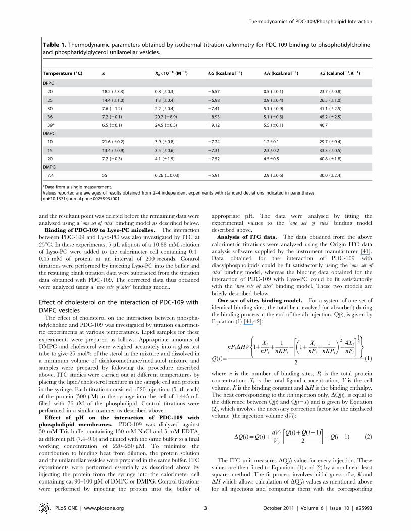
Table 1. Thermodynamic parameters obtained by isothermal titration calorimetry for PDC-109 binding to phsophotidylcholineand phosphatidylglycerol unilamellar vesicles.
Temperature (6C) n Ka61025 (M21) DG (kcal.mol21) DH (kcal.mol21) DS (cal.mol21.K21)
DPPC
20 18.2 (63.3) 0.8 (60.3) 26.57 0.5 (60.1) 23.7 (60.8)
25 14.4 (61.0) 1.3 (60.4) 26.98 0.9 (60.4) 26.5 (61.0)
30 7.6 (61.2) 2.2 (60.4) 27.41 5.1 (60.9) 41.1 (62.5)
36 7.2 (60.1) 20.7 (68.9) 28.93 5.1 (60.5) 45.2 (62.5)
39* 6.5 (60.1) 24.5 (66.5) 29.12 5.5 (60.1) 46.7
DMPC
10 21.6 (60.2) 3.9 (60.8) 27.24 1.260.1 29.7 (60.4)
15 13.4 (60.9) 3.5 (60.6) 27.31 2.360.2 33.3 (60.5)
20 7.2 (60.3) 4.1 (61.5) 27.52 4.560.5 40.8 (61.8)
DMPG
7.4 55 0.26 (60.03) 25.91 2.9 (60.6) 30.0 (62.4)
*Data from a single measurement.Values reported are averages of results obtained from 2–4 independent experiments with standard deviations indicated in parentheses.doi:10.1371/journal.pone.0025993.t001
Thermodynamics of PDC-109/Phospholipid Interaction
PLoS ONE | www.plosone.org 3 October 2011 | Volume 6 | Issue 10 | e25993

experimentally determined values. Based on this comparison the
initial guess of n, K and DH is improved and the process is repeated
till no further significant improvement in the fit can be obtained.
Two sets of sites binding model. For a system with two sets
of independent binding sites, the total heat evolved (or absorbed)
during the binding process at the end of the ith injection, Q(i), is
given by Equation (3) [42]:
Q ið Þ~PtV n1H1DH1zn2H2DH2ð Þ ð3Þ
where n1 and n2 are the number of binding sites of type 1 and type
2, and DH1 and DH2 are the corresponding enthalpies of binding.
H1 and H2 are the fractions of the type 1 and type 2 sites that are
occupied and are related to the association constants K1 and K2,
according to equation (4):
K1~H1
1{H1ð Þ X½ � K2~H2
1{H2ð Þ X½ � ð4Þ
The heat corresponding to the ith injection only, DQ(i), is equal
to the difference between Q(i) and Q(i21) and is given by
Equation (5), which involves the necessary correction factor for the
displaced volume (the injection volume dVi), according to
Equation (2).
The ITC unit measures DQ(i) value for every injection. These
values are then fitted to Equations (2) and (3) by a nonlinear least
squares method. The fit process involves initial guess of n1, n2, K1,
K2, DH1 and DH2 which allows calculation of DQ(i) values as
mentioned above for all injections and comparing them with the
corresponding experimentally determined values. Based on this
comparison the initial guess of the values of the above parameters
is improved and the process is repeated till no further significant
improvement in the fit can be obtained.
From the values of K and DH, the thermodynamic parameters,
DG and DS are calculated according to the basic thermodynamic
Equations (5) and (6):
DG~{RT ln K ð5Þ
DG~DH{TDS ð6Þ
Results
Binding and solubilization of phospholipid membranesby PDC-109
The binding of PDC-109 to multilamellar vesicles of different
phospholipids, namely DMPC, DMPG or DMPE was investigated
by turbidimetry monitoring the optical density of the samples at
333 nm as a function of protein concentration as shown in Fig. 1.
It is clear from this figure that the turbidity of DMPC vesicles
decreases abruptly at about 0.03 mM protein concentration and
then levels off (curve 1). In the case of DMPG maximum decrease
in turbidity was observed at about 0.06 mM protein concentration
(curve 2), whereas turbidity of DMPE vesicles was essentially
unaffected even at the highest concentration of PDC-109
employed (curve 3).
Thermodynamics of PDC-109 binding tophosphatidylcholine unilamellar vesicles
Thermodynamic parameters characterizing the interaction of
PDC-109 with different phospholipids were determined by ITC
measurements at various temperatures below the gel-liquid
crystalline phase transition. To minimize the effect of temperature
variation that occurs during the injection on their phase structure,
the phospholipids were taken in the calorimeter cell and PDC-109
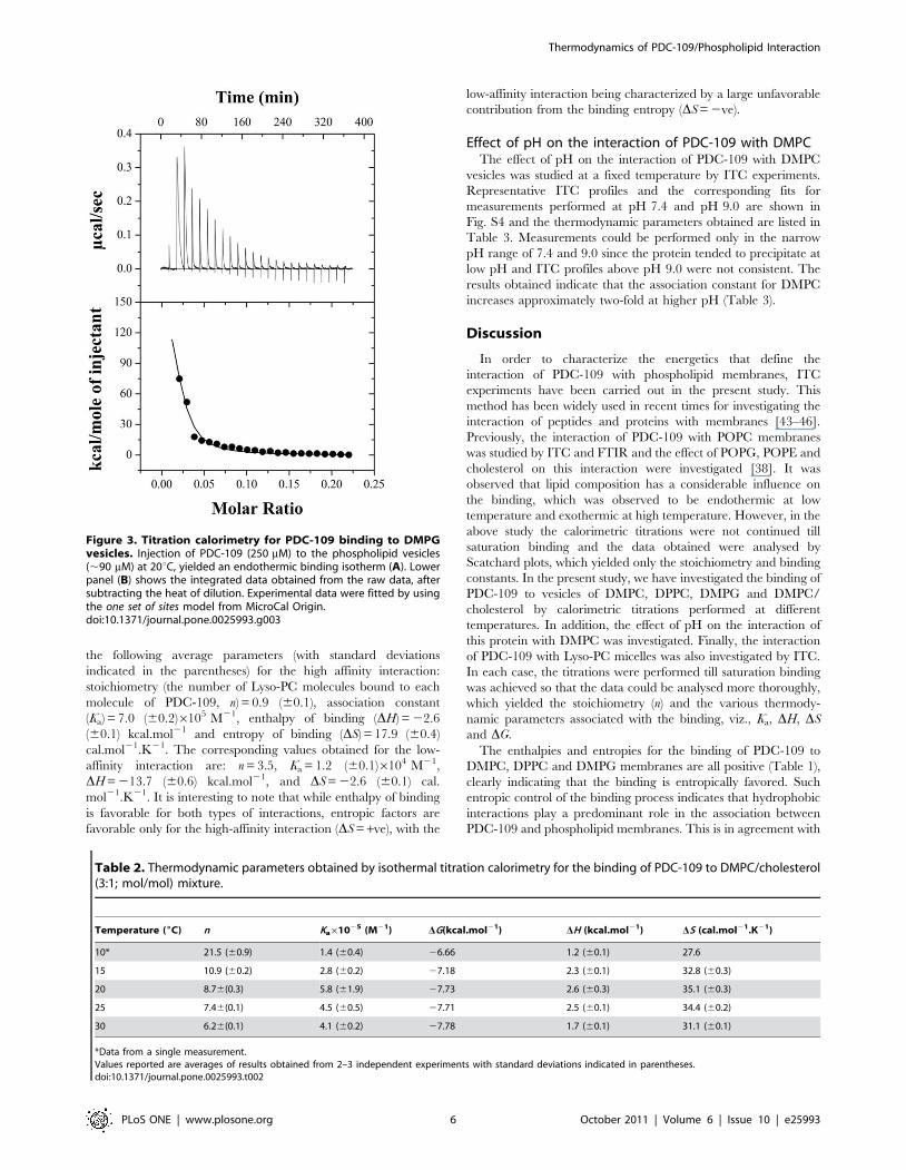
was added through the syringe. Figs. 2A and 2B show typical ITC
profiles for the binding of the protein to DMPC unilamellar
vesicles at 20uC. The titration profile in Fig. 2A shows that
injection of 5 mL aliquiots of PDC-109 into DMPC suspension
gives large endothermic heats of binding, which decrease in
magnitude with subsequent injections, showing saturation behav-
ior. The data could be best fitted by a nonlinear least squares
approach to the ‘one set of sites’ binding model, which yielded the
association constant (Ka), stoichiometry of binding (n), and the
thermodynamic parameters, enthalpy of binding (DH), entropy of
binding (DS) and free energy of binding (DG). Four independent
measurements at 20uC yielded the values of Ka, n, DH, DS, and DG
as 4.1 (61.5)6105 M21, 7.2 (60.3), 4.5 (60.5) kcal.mol21, 40.8
(61.8) cal.mol21.K21, and 27.52 kcal.mol21, respectively. These
values as well as the values obtained at other temperatures for the
PDC-109/DMPC interaction are listed in Table 1. Similar
titrations were carried out with DMPG and DPPC, and
representative ITC profiles corresponding to the titration of these
two lipids with PDC-109 are presented in Fig. 3 and Fig. S2,
respectively. Values of Ka, n, DG, DH and DS derived from the
analysis of the titration data corresponding to the binding of PDC-
109 to these lipids are also listed in Table 1. These values as well as
those given in all other tables correspond to the averages obtained
from 2–4 independent titrations and standard deviations are
Figure 1. PDC-109 induced solubilization of phospholipidmultilamellar vesicles. Solubilization was monitored by turbidimetryby measuring optical density at 333 nm. The phospholipids used are:DMPC (#), DMPG (N) and DMPE (D). See text for details.doi:10.1371/journal.pone.0025993.g001
Thermodynamics of PDC-109/Phospholipid Interaction
PLoS ONE | www.plosone.org 4 October 2011 | Volume 6 | Issue 10 | e25993

indicated in parentheses. The observed binding constant for the
interaction of PDC-109 with DMPG is smaller than the Ka values
obtained for the binding of the choline phospholipids (see Table 1),
reflecting its lower affinity for PDC-109. Thermodynamic
parameters obtained for the interaction of DMPC, DPPC and
DMPG to PDC-109 indicate that the binding is stabilized by
entropic factors, with negative contribution from enthalpy of
binding. A few titrations were also carried out with DMPC or
DPPC in the liquid crystalline phase; however, these preliminary
experiments yielded complex isotherms and a representative
isotherm corresponding to the binding of PDC-109 to DMPC
vesicles at 30uC is given in Fig. S3. These isotherms could not be
analyzed satisfactorily with the binding models available in the
Origin ITC analysis software. Hence no further experiments were
carried out in the fluid phase with these lipids.
The data presented in Table 1 show that the number of lipid
molecules corresponding to each molecule of PDC-109 decreases
with increasing temperature whereas the binding enthalpy and
entropy increase with temperature. However, the enthalpy values
did not exhibit clear linear temperature dependence; hence, it was
not possible to obtain the heat capacity changes associated with
the PDC-109/phospholipid interaction.
Effect of cholesterol on the binding of PDC-109 to DMPCmembranes
Binding isotherms for the interaction of PDC-109 with DMPC/
cholesterol (3:1; mol/mol) mixtures were also characterized by
endothermic heats of binding which decreased in magnitude with
successive injections until saturation was achieved (Fig. 2C). The
data could be best analyzed by a nonlinear least squares fit to the
‘one set of sites’ binding model in the Origin ITC data analysis
software. As summarized in Table 2, average values of Ka, n, DG,
DH and DS obtained for the interaction of PDC-109 with DMPC
membranes containing 25 mol% cholesterol at 20uC are 5.8
(61.9)6105 M21, 8.7 (60.3), 27.73 kcal.mol21, 2.6 (60.3)
kcal.mol21, and 35.1 (60.3) cal.mol21.K21, respectively. The
binding enthalpy for this interaction increases initially with
increase in temperature, but decreases with further increase in
temperature (Table 2). The ITC profiles were found to be
endothermic between 10 and 30uC for the binding of PDC-109 to
DMPC/cholesterol mixtures, in contrast to membranes containing
DMPC alone, which yielded endothermic profiles in the gel phase
and complex binding isotherms (containing both endothermic and
exothermic components) in the liquid crystalline phase.
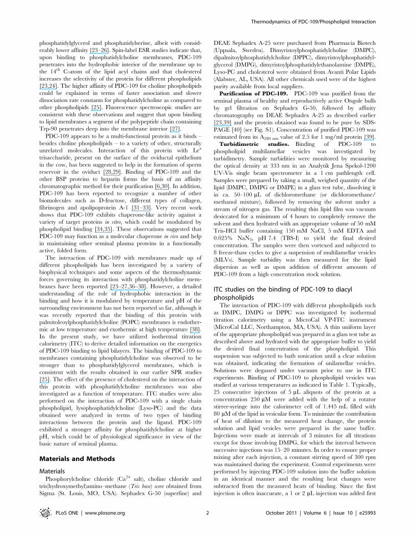
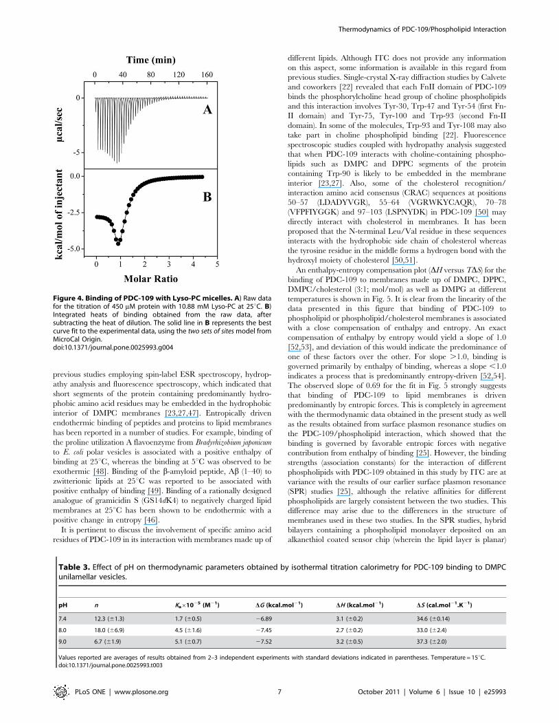
Thermodynamics of PDC-109 binding tolysophosphotidylcholine micelles
An isotherm for the binding of Lyso-PC to PDC-109 at 25uC is
shown in Fig. 4. The raw titration data shown in the upper panel
(A) could be best fitted to the ‘two sets of sites’ binding model in the
Origin software and the endothermic heats of binding corre-
sponding to the successive injections together with the fit obtained
(shown as a solid line) are given in the lower panel (B). This
analysis suggests two kinds of binding interactions between the
protein and Lyso-PC, one characterized by a higher affinity and
the other with a lower affinity. Two independent titrations yielded
Figure 2. Calorimetric titrations for the binding of PDC-109 to DMPC membranes in the gel phase in the absence and presence ofcholesterol. ITC profiles corresponding to the binding of PDC-109 to unilamellar vesicles of DMPC (A, B) and DMPC/cholesterol (3:1; mol/mol) (C, D)at 20uC are shown. Upper panels (A & C) show the raw data for the titration of phospholipid vesicles with protein and lower panels (B & D) show theintegrated heats of binding obtained from the raw data, after subtracting the heat of dilution. The solid lines in the bottom panels represent the bestcurve fits to the experimental data, using the one set of sites model from MicroCal Origin. See text for further details.doi:10.1371/journal.pone.0025993.g002
Thermodynamics of PDC-109/Phospholipid Interaction
PLoS ONE | www.plosone.org 5 October 2011 | Volume 6 | Issue 10 | e25993
the following average parameters (with standard deviations
indicated in the parentheses) for the high affinity interaction:
stoichiometry (the number of Lyso-PC molecules bound to each
molecule of PDC-109, n) = 0.9 (60.1), association constant
(Ka) = 7.0 (60.2)6105 M21, enthalpy of binding (DH) = 22.6
(60.1) kcal.mol21 and entropy of binding (DS) = 17.9 (60.4)
cal.mol21.K21. The corresponding values obtained for the low-
affinity interaction are: n = 3.5, Ka = 1.2 (60.1)6104 M21,
DH = 213.7 (60.6) kcal.mol21, and DS = 22.6 (60.1) cal.
mol21.K21. It is interesting to note that while enthalpy of binding
is favorable for both types of interactions, entropic factors are
favorable only for the high-affinity interaction (DS = +ve), with the
low-affinity interaction being characterized by a large unfavorable
contribution from the binding entropy (DS = 2ve).
Effect of pH on the interaction of PDC-109 with DMPCThe effect of pH on the interaction of PDC-109 with DMPC
vesicles was studied at a fixed temperature by ITC experiments.
Representative ITC profiles and the corresponding fits for
measurements performed at pH 7.4 and pH 9.0 are shown in
Fig. S4 and the thermodynamic parameters obtained are listed in
Table 3. Measurements could be performed only in the narrow
pH range of 7.4 and 9.0 since the protein tended to precipitate at
low pH and ITC profiles above pH 9.0 were not consistent. The
results obtained indicate that the association constant for DMPC
increases approximately two-fold at higher pH (Table 3).
Discussion
In order to characterize the energetics that define the
interaction of PDC-109 with phospholipid membranes, ITC
experiments have been carried out in the present study. This
method has been widely used in recent times for investigating the
interaction of peptides and proteins with membranes [43–46].
Previously, the interaction of PDC-109 with POPC membranes
was studied by ITC and FTIR and the effect of POPG, POPE and
cholesterol on this interaction were investigated [38]. It was
observed that lipid composition has a considerable influence on
the binding, which was observed to be endothermic at low
temperature and exothermic at high temperature. However, in the
above study the calorimetric titrations were not continued till
saturation binding and the data obtained were analysed by
Scatchard plots, which yielded only the stoichiometry and binding
constants. In the present study, we have investigated the binding of
PDC-109 to vesicles of DMPC, DPPC, DMPG and DMPC/
cholesterol by calorimetric titrations performed at different
temperatures. In addition, the effect of pH on the interaction of
this protein with DMPC was investigated. Finally, the interaction
of PDC-109 with Lyso-PC micelles was also investigated by ITC.
In each case, the titrations were performed till saturation binding
was achieved so that the data could be analysed more thoroughly,
which yielded the stoichiometry (n) and the various thermody-
namic parameters associated with the binding, viz., Ka, DH, DS
and DG.
The enthalpies and entropies for the binding of PDC-109 to
DMPC, DPPC and DMPG membranes are all positive (Table 1),
clearly indicating that the binding is entropically favored. Such
entropic control of the binding process indicates that hydrophobic
interactions play a predominant role in the association between
PDC-109 and phospholipid membranes. This is in agreement with
Figure 3. Titration calorimetry for PDC-109 binding to DMPGvesicles. Injection of PDC-109 (250 mM) to the phospholipid vesicles(,90 mM) at 20uC, yielded an endothermic binding isotherm (A). Lowerpanel (B) shows the integrated data obtained from the raw data, aftersubtracting the heat of dilution. Experimental data were fitted by usingthe one set of sites model from MicroCal Origin.doi:10.1371/journal.pone.0025993.g003
Table 2. Thermodynamic parameters obtained by isothermal titration calorimetry for the binding of PDC-109 to DMPC/cholesterol(3:1; mol/mol) mixture.
Temperature (6C) n Ka61025 (M21) DG(kcal.mol21) DH (kcal.mol21) DS (cal.mol21.K21)
10* 21.5 (60.9) 1.4 (60.4) 26.66 1.2 (60.1) 27.6
15 10.9 (60.2) 2.8 (60.2) 27.18 2.3 (60.1) 32.8 (60.3)
20 8.76(0.3) 5.8 (61.9) 27.73 2.6 (60.3) 35.1 (60.3)
25 7.46(0.1) 4.5 (60.5) 27.71 2.5 (60.1) 34.4 (60.2)
30 6.26(0.1) 4.1 (60.2) 27.78 1.7 (60.1) 31.1 (60.1)
*Data from a single measurement.Values reported are averages of results obtained from 2–3 independent experiments with standard deviations indicated in parentheses.doi:10.1371/journal.pone.0025993.t002
Thermodynamics of PDC-109/Phospholipid Interaction
PLoS ONE | www.plosone.org 6 October 2011 | Volume 6 | Issue 10 | e25993
previous studies employing spin-label ESR spectroscopy, hydrop-
athy analysis and fluorescence spectroscopy, which indicated that
short segments of the protein containing predominantly hydro-
phobic amino acid residues may be embedded in the hydrophobic
interior of DMPC membranes [23,27,47]. Entropically driven
endothermic binding of peptides and proteins to lipid membranes
has been reported in a number of studies. For example, binding of
the proline utilization A flavoenzyme from Bradyrhizobium japonicum
to E. coli polar vesicles is associated with a positive enthalpy of
binding at 25uC, whereas the binding at 5uC was observed to be
exothermic [48]. Binding of the b-amyloid peptide, Ab (1–40) to
zwitterionic lipids at 25uC was reported to be associated with
positive enthalpy of binding [49]. Binding of a rationally designed
analogue of gramicidin S (GS14dK4) to negatively charged lipid
membranes at 25uC has been shown to be endothermic with a
positive change in entropy [46].
It is pertinent to discuss the involvement of specific amino acid
residues of PDC-109 in its interaction with membranes made up of
different lipids. Although ITC does not provide any information
on this aspect, some information is available in this regard from
previous studies. Single-crystal X-ray diffraction studies by Calvete
and coworkers [22] revealed that each FnII domain of PDC-109
binds the phosphorylcholine head group of choline phospholipids
and this interaction involves Tyr-30, Trp-47 and Tyr-54 (first Fn-
II domain) and Tyr-75, Tyr-100 and Trp-93 (second Fn-II
domain). In some of the molecules, Trp-93 and Tyr-108 may also
take part in choline phospholipid binding [22]. Fluorescence
spectroscopic studies coupled with hydropathy analysis suggested
that when PDC-109 interacts with choline-containing phospho-
lipids such as DMPC and DPPC segments of the protein
containing Trp-90 is likely to be embedded in the membrane
interior [23,27]. Also, some of the cholesterol recognition/
interaction amino acid consensus (CRAC) sequences at positions
50–57 (LDADYVGR), 55–64 (VGRWKYCAQR), 70–78
(VFPFIYGGK) and 97–103 (LSPNYDK) in PDC-109 [50] may
directly interact with cholesterol in membranes. It has been
proposed that the N-terminal Leu/Val residue in these sequences
interacts with the hydrophobic side chain of cholesterol whereas
the tyrosine residue in the middle forms a hydrogen bond with the
hydroxyl moiety of cholesterol [50,51].
An enthalpy-entropy compensation plot (DH versus TDS) for the
binding of PDC-109 to membranes made up of DMPC, DPPC,
DMPC/cholesterol (3:1; mol/mol) as well as DMPG at different
temperatures is shown in Fig. 5. It is clear from the linearity of the
data presented in this figure that binding of PDC-109 to
phospholipid or phospholipid/cholesterol membranes is associated
with a close compensation of enthalpy and entropy. An exact
compensation of enthalpy by entropy would yield a slope of 1.0
[52,53], and deviation of this would indicate the predominance of
one of these factors over the other. For slope .1.0, binding is
governed primarily by enthalpy of binding, whereas a slope ,1.0
indicates a process that is predominantly entropy-driven [52,54].
The observed slope of 0.69 for the fit in Fig. 5 strongly suggests
that binding of PDC-109 to lipid membranes is driven
predominantly by entropic forces. This is completely in agreement
with the thermodynamic data obtained in the present study as well
as the results obtained from surface plasmon resonance studies on
the PDC-109/phospholipid interaction, which showed that the
binding is governed by favorable entropic forces with negative
contribution from enthalpy of binding [25]. However, the binding
strengths (association constants) for the interaction of different
phospholipids with PDC-109 obtained in this study by ITC are at
variance with the results of our earlier surface plasmon resonance
(SPR) studies [25], although the relative affinities for different
phospholipids are largely consistent between the two studies. This
difference may arise due to the differences in the structure of
membranes used in these two studies. In the SPR studies, hybrid
bilayers containing a phospholipid monolayer deposited on an
alkanethiol coated sensor chip (wherein the lipid layer is planar)
Table 3. Effect of pH on thermodynamic parameters obtained by isothermal titration calorimetry for PDC-109 binding to DMPCunilamellar vesicles.
pH n Ka61025 (M21) DG (kcal.mol21) DH (kcal.mol21) DS (cal.mol21.K21)
7.4 12.3 (61.3) 1.7 (60.5) 26.89 3.1 (60.2) 34.6 (60.14)
8.0 18.0 (66.9) 4.5 (61.6) 27.45 2.7 (60.2) 33.0 (62.4)
9.0 6.7 (61.9) 5.1 (60.7) 27.52 3.2 (60.5) 37.3 (62.0)
Values reported are averages of results obtained from 2–3 independent experiments with standard deviations indicated in parentheses. Temperature = 15uC.doi:10.1371/journal.pone.0025993.t003
Figure 4. Binding of PDC-109 with Lyso-PC micelles. A) Raw datafor the titration of 450 mM protein with 10.88 mM Lyso-PC at 25uC. B)Integrated heats of binding obtained from the raw data, aftersubtracting the heat of dilution. The solid line in B represents the bestcurve fit to the experimental data, using the two sets of sites model fromMicroCal Origin.doi:10.1371/journal.pone.0025993.g004
Thermodynamics of PDC-109/Phospholipid Interaction
PLoS ONE | www.plosone.org 7 October 2011 | Volume 6 | Issue 10 | e25993

were employed, whereas in the present study small unilamellar
vesicles, which are intrinsically highly curved, have been used.
Although complex isotherms were obtained for the binding of
PDC-109 to DMPC vesicles in the fluid phase, upon cholesterol
incorporation simple endothermic binding isotherms were ob-
tained even at temperatures corresponding to the liquid crystalline
phase of DMPC. These results are consistent with the abolition of
the gel-fluid phase transition and the rigidification of liquid
crystalline phase of DMPC membranes by cholesterol [55], since
endothermic binding isotherms were obtained with DMPC alone
only in the gel phase region where the lipid acyl chains are rigid
and tightly packed.
For phosphatidylcholines in the absence and in the presence of
cholesterol, the number of binding sites is found to decrease with
temperature, i.e., the number of lipid molecules associated with
each molecule of PDC-109 decreases as the temperature is
increased (Tables 1 and 2). It is well known that the cross-sectional
area of lipids increases with increase in temperature [56].
Assuming that the area of the protein which interacts with the
lipid membrane does not change significantly in the temperature
range studied, it is expected that the number of lipid molecules
that would be associated with the protein would decrease as the
temperature is increased. The estimated stoichiometry may also be
affected by temperature-induced alterations in the aggregation
state of PDC-109, which is known to exist as a polydisperse
aggregate in solution [57].
In the presence of excess water, Lyso-PC forms micelles that
coexist with dissolved Lyso-PC molecules. The two types of
interactions observed for the binding of PDC-109 to Lyso-PC can
therefore be interpreted as due to the binding of the protein with
the micelles and free Lyso-PC, respectively. Although the high-
affinity interaction is aided by both entropic and enthalpic
contributions, the entropic contribution is more dominant. On
the other hand the low-affinity interaction is enthalpically driven,
with negative contribution from entropy of binding. Since the
interaction of PDC-109 with different phospholipid vesicles are all
governed by positive entropic contribution at all temperatures, it is
most likely that the high affinity interaction, which also has a
significantly large positive entropy of binding, corresponds to the
binding of PDC-109 to Lyso-PC micelles. The weaker interaction
will then correspond to the association of free Lyso-PC molecules
with PDC-109.
Changing the pH of the medium is known to induce
conformational changes in the secondary and tertiary structures
of proteins [58]. Since PDC-109 is present in a slightly basic
environment in the seminal plasma, it is of interest to study the
effect of pH on its interaction with membranes containing the
major plasma membrane lipid phosphatidylcholine, for which this
protein exhibits the highest affinity. The thermodynamic data
presented in Table 3 shows that the association constant for the
binding of PDC-109 to DMPC vesicles increases approximately 2-
fold when the pH was increased from 7.4 to 9.0, which could be
physiologically relevant in view of the basic nature of the seminal
plasma of mammals.
In summary, the energetics of interaction of PDC-109 with
phospholipids has been investigated by isothermal titration
calorimetry. It has been found that the binding of PDC-109 to
gel phase lipids is endothermic in nature and is governed by a
positive entropic contribution. The binding strength of PDC-109
for choline containing phospholipids was observed to be higher
than that of other phospholipids such as DMPG and DMPE,
which is consistent with the higher specificity of PDC-109 for the
choline phospholipids. The affinity of PDC-109 for phosphatidyl-
choline was found to be higher at higher pH, which is
physiologically relevant in view of the basic nature of the seminal
plasma. Overall, the present study leads to a better understanding
of the interaction of PDC-109 with phospholipid membranes at
the molecular level, which is relevant to understanding the
interaction of PDC-109 with sperm plasma membrane during
sperm capacitation, in vivo.
Supporting Information AvailableAdditional information relevant to this article, containing
Figures S1, S2, S3, S4 is given as Supporting Information.
Supporting Information
Figure S1 SDS-PAGE of PDC-109. Lane 1, molecular weight
markers; lane 2, PDC-109. The Mr values of the standard proteins
(in kDa) are indicated on the left.
(TIF)
Figure S2 Calorimetric titration for the binding of PDC-109 to DPPC unilamellar vesicles in the gel phase at366C. Upper panel shows the raw data for the titration of
phospholipid vesicles with protein and the lower panel shows the
integrated heats of binding obtained from the raw data, after
subtracting the heats of dilution. The solid line in the lower panel
represents the best curve fit to the experimental data, using the one
set of sites model from MicroCal Origin.
(TIF)
Figure S3 Calorimetric titration for the binding of PDC-109 to DMPC unilamellar vesicles in the liquid crystal-line phase at 306C. Upper panel shows the raw data for the
titration of phospholipid vesicles with protein and the lower panel
shows the integrated heats of binding obtained from the raw data,
after subtracting the heat of dilution.
(TIF)
Figure S4 Effect of pH on the binding of PDC-109 toDMPC membranes. Representative ITC profiles are given for
titrations performed at pH 7.4 (left panel) and 9.0 (right panel).
The parameters obtained from the fits are indicated in the boxes
Figure 5. Enthalpy-entropy compensation plot. Data are shownfor the interaction of PDC-109 with phospholipid vesicles made up ofDMPC (N), DPPC (%), DMPC/cholesterol (O) and DMPG (.). The straightline corresponds to a linear least squares fit (slope = 0.69).doi:10.1371/journal.pone.0025993.g005
Thermodynamics of PDC-109/Phospholipid Interaction
PLoS ONE | www.plosone.org 8 October 2011 | Volume 6 | Issue 10 | e25993

given in the figure. The data given in Table 3 in the main
manuscript correspond to average values from 2–3 independent
titrations.
(TIF)
Acknowledgments
We are grateful to Drs. K. Babu Rao and R. Vinoo of the Lam Farm,
Guntur, Sri Venkateswara Veterinary University, for kindly providing
samples of bovine semen.
Author Contributions
Conceived and designed the experiments: VA RSS MJS. Performed the
experiments: VA RSS BPS. Analyzed the data: VA RSS MJS. Contributed
reagents/materials/analysis tools: MJS. Wrote the paper: VA RSS MJS.
References
1. Shivaji S, Scheit KH, Bhargava PM (1990) Proteins of seminal plasma. New
York: Wiley. pp 526.
2. Yanagimachi R (1994) Mammalian fertilization. In: Knobil E, Neill JD, eds. The
Physiology of Reproduction. New York: Raven Press. pp 189–317.
3. Harrison RAP (1996) Capacitation mechanisms, and the role of capacitation as
seen in eutherian mammals. Reprod Fert Dev 8: 581–594.
4. Visconti PE, Calantino-Hormer H, Moore GD, Bailey JL, Ning X, et al. (1998)
The molecular basis of sperm capacitation. J Androl 19: 242–248.
5. Manjunath P, Sairam MR, Uma J (1987) Purification of four gelatin-binding
proteins from bovine seminal plasma by affinity chromatography. Biosci Rep 7:231–238.
6. Manjunath P, Sairam MR (1987) Purification and biochemical characterizationof three major acidic proteins (BSP-A1, BSP-A2 and BSP-A3) from bovine
seminal plasma. Biochem J 241: 685–692.
7. Calvete JJ, Mann K, Schafer W, Sanz L, Reinert M, et al. (1995) Amino acid
sequence of HSP-1, a major protein of stallion seminal plasma: effect of
glycosylation on its heparin- and gelatin-binding capabilities. Biochem J 310:615–622.
8. Calvete JJ, Raida M, Gentzel M, Urbanke C, Sanz L, et al. (1997) Isolation andcharacterization of heparin and phosphorylcholine binding proteins of boar and
stallion seminal plasma. Primary structure of porcine pB1. FEBS Lett 407:201–206.
9. Villemure M, Lazure C, Manjunath P (2003) Isolation and characterization ofgelatin-binding proteins from goat seminal plasma. Reprod Biol Endocrinol 1:
39–48.
10. Boisvert M, Bergeron A, Lazure C, Manjunath P (2004) Isolation andcharacterization of gelatin-binding bison seminal vesicle secretory proteins. Biol
Reprod 70: 656–661.
11. Bergeron A, Villemure M, Lazure C, Manjunath P (2005) Isolation and
characterization of the major proteins of ram seminal plasma. Mol Reprod Dev71: 461–470.
12. Harper MJK (1994) Gamete and Zygote Transport. In: Knobil E, Neill JD, eds.The Physiology of Reproduction Raven Press: New York. pp 123–187.
13. Suarez SS (1998) The oviductal sperm reservoir in mammals. Mechanisms offormation. Biol Reprod 58: 1105–1107.
14. Scheit KH, Kemme M, Aumuller G, Seitz J, Hagendorff G, et al. (1988) Themajor protein of bull seminal plasma: biosynthesis and biological function. Biosci
Rep 8: 589–608.
15. Baker ME (1985) The PDC-109 protein from bovine seminal plasma is similar to
the gelatin-binding domain of bovine fibronectin and a kringle domain of human
tissue-type plasminogen activator. Biochem Biophys Res Commun 130:1010–1014.
16. Esch FS, Ling NC, Bohlen P, Ying SY, Guillemin R (1983) Primary structure ofPDC-109, a major protein constituent of bovine seminal plasma. Biochem
Biophys Res Commun 113: 861–867.
17. Seidah NG, Manjunath P, Rochemont J, Sairam MR, Cheretian M (1987)
Complete amino acid sequence of BSP-A3 from bovine seminal plasma,Homology to PDC-109 and to the collagen-binding domain of fibronectin.
Biochem J 243: 195–203.
18. Calvete JJ, Raida M, Sanz L, Wempe F, Scheit KH, et al. (1994) Localizationand structural characterization of an oligosaccharide O-linked to bovine PDC-
109. Quantitation of the glycoprotein in seminal plasma and on the surface ofejaculated and capacitated spermatozoa. FEBS Lett 350: 203–206.
19. Desnoyers L, Manjunath P (1992) Major proteins of bovine seminal plasmaexhibit novel interactions with phospholipids. J Biol Chem 267: 10149–10155.
20. Therien I, Moreau R, Manjunath P (1998) Major proteins of bovine seminalplasma and high-density lipoprotein induce cholesterol efflux from epididymal
sperm. Biol Reprod 59: 768–776.
21. Moreau R, Therien I, Lazure C, Manjunath P (1998) Type II domains of BSP-
A1/-A2 proteins: binding properties, lipid efflux and sperm capacitation
potential. Biochem Biophys Res Commun 246: 148–154.
22. Wah DA, Fernandez-Tornero C, Sanz L, Romero A, Calvete JJ (2002) Sperm
coating mechanism from the 1.8 A crystal structure of PDC-109-phosphoryl-choline complex. Structure 10: 505–514.
23. Ramakrishnan M, Anbazhagan V, Pratap TV, Marsh D, Swamy MJ (2001)Membrane insertion and lipid-protein interactions of bovine seminal plasma
protein, PDC-109 investigated by spin label electron spin resonance spectros-copy. Biophys J 81: 2215–2225.
24. Swamy MJ, Marsh D, Anbazhagan V, Ramakrishnan M (2002) Effect of
cholesterol on the interaction of seminal plasma protein, PDC-109 with
phosphatidylcholine membranes. FEBS Lett 528: 230–234.
25. Thomas CJ, Anbazhagan V, Ramakrishnan M, Sultan N, Surolia I, et al. (2003)
Mechanism of membrane binding by the bovine seminal plasma protein, PDC-
109. A surface plasmon resonance study. Biophys J 84: 3037–3044.
26. Greube A, Muller K, Topfer-Petersen E, Herrmann A, Muller P (2001)
Influence of the bovine seminal plasma protein PDC-109 on the physical state of
membrane. Biochemistry 40: 8326–8334.
27. Anbazhagan V, Damai RS, Paul A, Swamy MJ (2008) Interaction of the major
protein from bovine seminal plasma, PDC-109 with phospholipid membranes
and soluble ligands investigated by fluorescence approaches. Biochim Biophys
Acta 1784: 891–899.
28. Revah I, Gadella BM, Flesch FM, Colenbrander B, Suarez SS (2000)
Physiological state of bull sperm affects fucose- and mannose-binding properties.
Biol Reprod 62: 1010–1015.
29. Ignotz GG, Lo MC, Perez CL, Gwathmey TM, Suarez SS (2001)
Characterization of a fucose-binding protein from bull sperm and seminal
plasma that may be responsible for formation of oviductal sperm reservoir. Biol
Reprod 64: 1806–1811.
30. Chandonnet L, Roberts KD, Chapdelaine A, Manjunath P (1990) Identification
of heparin-binding proteins in bovine seminal plasma. Mol Reprod Dev 26:
313–318.
31. Liberda J, Kraus M, Ryslava H, Vlaskova M, Jonakova V, et al. (2001) D-
Fructose-binding proteins in bull seminal plasma. Isolation and characterization.
Folia Biol (Praha) 47: 113–119.
32. Manjunath P, Marcel YL, Uma J, Seidah NG, Cheretian M, et al. (1989)
Apolipoprotein A-1 binds to a family of bovine seminal plasma proteins. J Biol
Chem 264: 16853–16857.
33. Manjunath P, Nauc V, Bergeron A, Menard M (2002) Major proteins of bovine
seminal plasma bind to the low density lipoprotein fraction of hen’s egg yolk.
Biol Reprod 67: 1250–1258.
34. Sankhala RS, Swamy MJ (2010) The major protein of bovine seminal plasma,
PDC-109, is a molecular chaperone. Biochemistry 49: 3908–3918.
35. Sankhala RS, Damai RS, Swamy MJ (2011) Correlation of membrane binding
and hydrophobicity to the chaperone-like activity of PDC-109, the major protein
of bovine seminal plasma. PLoS ONE 6: e17330.
36. Anbazhagan V, Swamy MJ (2005) Thermodynamics of phosphorylcholine and
lysophosphatidylcholine binding to the major protein of bovine seminal plasma,
PDC-109. FEBS Lett 579: 2933–2938.
37. Swamy MJ (2004) Interaction of bovine seminal plasma proteins with model
membranes and sperm plasma membranes. Curr Sci 87: 203–211.
38. Lassiseraye D, Courtemanche L, Bergeron A, Manjunath P, Lafleur M (2008)
Binding of bovine seminal plasma protein BSP-A1/A2 to model membranes:
Lipid specificity and effect of the temperature. Biochim Biophys Acta 1778:
502–513.
39. Calvete JJ, Paloma FV, Sanz L, Romero A (1996) A procedure for the large-
scale isolation of major bovine seminal plasma proteins. Protein Expr Purif 8:
48–56.
40. Laemmli UK (1970) Cleavage of structural proteins during assembly of
bacteriophage T4. Nature 227: 680–685.
41. Wiseman T, Williston S, Brandts JF, Lin L-N (1989) Rapid measurement of
binding constants and heats of binding using a new titration calorimeter. Anal
Biochem 179: 131–137.
42. ITC Data Analysis in OriginH (1998) Tutorial Guide Version 5.0.
43. Wieprecht T, Beyermann M, Seelig J (1999) Binding of antibacterial magainin
peptides to electrically neutral membranes: Thermodynamics and structure.
Biochemistry 38: 10377–10387.
44. Arnulphi C, Jin L, Tricerri MA, Jonas A (2004) Enthalpy-driven apolipoprotein
A-I and lipid bilayer interaction indicating protein penetration upon binding.
Biochemisry 43: 12258–12264.
45. Torrecillas A, Laynez J, Menendez M, Corbalan-Garcıa S, Gomez-
Fernandez JC (2004) Calorimetric study of the interaction of the C2 domains
of classical protein kinase C isoenzymes with Ca2+ and phospholipids.
Biochemistry 43: 11727–11739.
46. Abraham T, Lewis RNAH, Hodges RS, McElhaney RN (2005) Isothermal
titration calorimetry studies of the binding of a rationally designed analogue of
Thermodynamics of PDC-109/Phospholipid Interaction
PLoS ONE | www.plosone.org 9 October 2011 | Volume 6 | Issue 10 | e25993

the antimicrobial peptide gramicidin S to phospholipids bilayer membranes.
Biochemistry 44: 2103–2112.47. Gasset M, Magdaleno L, Calvete JJ (2000) Biophysical study of the perturbation
of model membrane structure caused by seminal plasma protein PDC-109. Arch
Biochem Biophys 374: 241–247.48. Zhang W, Krishnan N, Becker DF (2006) Kinetic and thermodynamic analysis
of Bradyrhizobium japonicum PutA-membrane associations. Arch Biochem Biophys445: 174–183.
49. Lin M-S, Chiu H-M, Fan F-J, Tsai H-T, Wang SS-S, et al. (2007) Kinetics and
enthalpy measurements of interaction between b-amyloid and liposomes bysurface plasmon resonance and isothermal titration calorimetry. Colloids Surf B
58: 231–236.50. Scolari S, Muller K, Bittman R, Herrmann A, Muller P (2010) Interaction of
mammalian seminal plasma protein PDC-109 with cholesterol: implications fora putative CRAC domain. Biochemistry 49: 9027–9031.
51. Epand RM (2006) Cholesterol and the interaction of proteins with membrane
domains. Prog Lipid Res 45: 279–294.52. Sigurskjold BW, Bundle DR (1992) Thermodynamics of oligosaccharide binding
to a monoclonal antibody specific for a Salmonella O-antigen point tohydrophobic interactions in the binding site. J Biol Chem 267: 8371–8376.
53. Sultan NAM, Swamy MJ (2005) Energetics of carbohydrate binding to
Momordica charantia (bitter gourd) lectin: An isothermal titration calorimetric
study. Arch Biochem Biophys 437: 115–125.
54. Brummell DA, Sharma VP, Anand NN, Bilous D, Dubuc G, et al. (1993)
Probing the combining site of an anti-carbohydrate antibody by saturation-
mutagenesis: Role of the heavy-chain CDR3 residues. Biochemistry 32:
1180–1187.
55. Swamy MJ, Ramakrishnan M, Angerstein B, Marsh D (2000) Spin-label electron
spin resonance studies on the mode of anchoring and vertical location of the N-
acyl chain in N-acylphosphatidylethanolamines. Biochemistry 39: 12476–12484.
56. Marsh D (1990) CRC handbook of Lipid Bilayers: CRC press: Boca Raton, FL. 387
p.
57. Gasset M, Saiz JL, Sanz L, Gentzel M, Topfer-Petersen E, et al. (1997)
Conformational features and thermal stability of bovine seminal plasma protein
PDC-109 oligomers and phosphorylcholine-bound complexes. Eur J Biochem
250: 735–744.
58. Cabra V, Arreguin R, Vazquez-Duhalt R, Farres A (2006) Effect of temperature
and pH on the secondary structure and processes of oligomerization of 19 kDa
alpha-zein. Biochim Biophy Acta 1764: 1110–1118.
Thermodynamics of PDC-109/Phospholipid Interaction
PLoS ONE | www.plosone.org 10 October 2011 | Volume 6 | Issue 10 | e25993
Copyright © 2022 FDOKUMEN