Isolation of peptide transport system-6 from brain endothelial cells: therapeutic effects with...
12
Isolation of peptide transport system-6 from brain endothelial cells: therapeutic effects with antisense inhibition in Alzheimer and stroke models Dilek Dogrukol-Ak 1,2,3 , Vijaya B Kumar 2,3 , Jan S Ryerse 4 , Susan A Farr 2,3 , Sulekha Verma 2,3 , Naoko Nonaka 2,3,5,6 , Tomoya Nakamachi 5,6 , Hirokazu Ohtaki 5,6 , Michael L Niehoff 2,3 , John C Edwards 7 , Seiji Shioda 5,6 , John E Morley 2,3 and William A Banks 2,3 1 Faculty of Pharmacy, Anadolu University, Eskisehir, Turkey; 2 GRECC, Veterans Affairs Medical Center-Saint Louis, St Louis, Missouri, USA; 3 Division of Geriatrics, Department of Internal Medicine, Saint Louis University School of Medicine, St Louis, Missouri, USA; 4 Department of Pathology, Saint Louis University Health Sciences Center, St Louis, Missouri, USA; 5 Department of Oral Anatomy, Showa University School of Dentistry, Tokyo, Japan; 6 Department of 1st Anatomy, Showa University School of Medicine, Tokyo, Japan; 7 Department of Nephrology, University of North Carolina School of Medicine, Chapel Hill, North Carolina, USA By isolating for the first time ever a peptide transporter from the blood–brain barrier (BBB) and developing an antisense that selectively targets the brain-to-blood efflux component, we were able to deliver a therapeutic concentration of the neurotrophic peptide pituitary adenylate cyclase-activating polypeptide (PACAP) 27 to brain in animal models of Alzheimer’s and stroke. Efflux pumps at the BBB are major causes of BBB impermeability to peptides. PACAP is neuroprotective in vitro in femtomole amounts, but brain uptake of PACAP27 is limited by an efflux component of peptide transport system-6 (PTS-6). Here, we characterized, isolated, and sequenced this component of PTS-6, identifying it as b-F1 ATPase, and colocalized it with PACAP27 on BBB endothelial cells. Antisenses targeting the BBB inhibited PACAP27 efflux, thus increasing brain uptake of PACAP27. Treatment with antisense + PACAP27 improved cognition in a mouse model of Alzheimer’s disease and reduced infarct size after cerebral ischemia. This represents the first isolation from BBB tissue of a peptide transporter and shows that inhibition of peptide efflux pumps is a potential strategy for drug delivery to brain. Journal of Cerebral Blood Flow & Metabolism (2009) 29, 411–422; doi:10.1038/jcbfm.2008.131; published online 12 November 2008 Keywords: blood-brain barrier; antisense; PACAP; Alzheimer’s disease; stroke; peptide The blood–brain barrier (BBB) is a major obstacle to the delivery of promising therapeutics to the central nervous system. The presence of brain-to-blood saturable transport systems (efflux pumps) has emerged as a major reinforcer of BBB impermeability (Taylor, 2002; Begley, 2004; Banks, 2005). For example, pituitary adenylate cyclase-activating poly- peptide (PACAP), the newest member of the secretin/ glucagon/vasoactive intestinal polypeptide (VIP) family of regulatory peptides, is a pleuripotent peptide with neuroprotective, endocrine, and vaso- dilatory effects (Vaudry et al, 2000). In vitro, PACAP is neuroprotective in femtomole concentrations (Arimura et al, 1994). In vivo, the 38 amino-acid form of PACAP (PACAP38) reverses ischemic da- mage even when given intravenously and 24 h after 4 vessel stroke (Uchida et al, 1996). However, the physicochemical and pharmacokinetic properties of PACAP38 make it difficult to work with. Use of the C-terminal-truncated 27 amino-acid form (PACAP27) is limited by an efflux component of peptide transport system-6 (PTS-6), which decreases brain uptake by approximately 10-fold (Banks et al, 1993). Reduction in brain uptake by efflux transporters of 4- to 10-fold is enough to render many otherwise promising central nervous system therapeutics ineffectual (Taylor, 2002; Lo ¨scher and Potschka, 2002; Begley, 2004; Thomas, 2004). Inhibition of the efflux pumps should increase brain uptake of potential therapeutics and so could Received 13 August 2008; revised 8 September 2008; accepted 8 October 2008; published online 12 November 2008 Correspondence: Professor WA Banks, GRECC, Veterans Affairs Medical Center-Saint Louis, 915 N Grand Boulevard, St Louis, MO 63106, USA. E-mail: [email protected] Journal of Cerebral Blood Flow & Metabolism (2009) 29, 411–422 & 2009 ISCBFM All rights reserved 0271-678X/09 $32.00 www.jcbfm.com
Transcript of Isolation of peptide transport system-6 from brain endothelial cells: therapeutic effects with...

Isolation of peptide transport system-6 from brainendothelial cells: therapeutic effects with antisenseinhibition in Alzheimer and stroke models
Dilek Dogrukol-Ak1,2,3, Vijaya B Kumar2,3, Jan S Ryerse4, Susan A Farr2,3, Sulekha Verma2,3,Naoko Nonaka2,3,5,6, Tomoya Nakamachi5,6, Hirokazu Ohtaki5,6, Michael L Niehoff2,3,John C Edwards7, Seiji Shioda5,6, John E Morley2,3 and William A Banks2,3
1Faculty of Pharmacy, Anadolu University, Eskisehir, Turkey; 2GRECC, Veterans Affairs Medical Center-SaintLouis, St Louis, Missouri, USA; 3Division of Geriatrics, Department of Internal Medicine, Saint LouisUniversity School of Medicine, St Louis, Missouri, USA; 4Department of Pathology, Saint Louis UniversityHealth Sciences Center, St Louis, Missouri, USA; 5Department of Oral Anatomy, Showa University School ofDentistry, Tokyo, Japan; 6Department of 1st Anatomy, Showa University School of Medicine, Tokyo, Japan;7Department of Nephrology, University of North Carolina School of Medicine, Chapel Hill, North Carolina,USA
By isolating for the first time ever a peptide transporter from the blood–brain barrier (BBB) anddeveloping an antisense that selectively targets the brain-to-blood efflux component, we wereable to deliver a therapeutic concentration of the neurotrophic peptide pituitary adenylatecyclase-activating polypeptide (PACAP) 27 to brain in animal models of Alzheimer’s and stroke.Efflux pumps at the BBB are major causes of BBB impermeability to peptides. PACAP isneuroprotective in vitro in femtomole amounts, but brain uptake of PACAP27 is limited by an effluxcomponent of peptide transport system-6 (PTS-6). Here, we characterized, isolated, and sequencedthis component of PTS-6, identifying it as b-F1 ATPase, and colocalized it with PACAP27 on BBBendothelial cells. Antisenses targeting the BBB inhibited PACAP27 efflux, thus increasing brainuptake of PACAP27. Treatment with antisense + PACAP27 improved cognition in a mouse model ofAlzheimer’s disease and reduced infarct size after cerebral ischemia. This represents the firstisolation from BBB tissue of a peptide transporter and shows that inhibition of peptide efflux pumpsis a potential strategy for drug delivery to brain.Journal of Cerebral Blood Flow & Metabolism (2009) 29, 411–422; doi:10.1038/jcbfm.2008.131; published online12 November 2008
Keywords: blood-brain barrier; antisense; PACAP; Alzheimer’s disease; stroke; peptide
The blood–brain barrier (BBB) is a major obstacle tothe delivery of promising therapeutics to the centralnervous system. The presence of brain-to-bloodsaturable transport systems (efflux pumps) hasemerged as a major reinforcer of BBB impermeability(Taylor, 2002; Begley, 2004; Banks, 2005). Forexample, pituitary adenylate cyclase-activating poly-peptide (PACAP), the newest member of the secretin/glucagon/vasoactive intestinal polypeptide (VIP)family of regulatory peptides, is a pleuripotentpeptide with neuroprotective, endocrine, and vaso-dilatory effects (Vaudry et al, 2000). In vitro, PACAP
is neuroprotective in femtomole concentrations(Arimura et al, 1994). In vivo, the 38 amino-acidform of PACAP (PACAP38) reverses ischemic da-mage even when given intravenously and 24 h after 4vessel stroke (Uchida et al, 1996). However, thephysicochemical and pharmacokinetic properties ofPACAP38 make it difficult to work with. Use of theC-terminal-truncated 27 amino-acid form (PACAP27)is limited by an efflux component of peptidetransport system-6 (PTS-6), which decreases brainuptake by approximately 10-fold (Banks et al, 1993).Reduction in brain uptake by efflux transporters of4- to 10-fold is enough to render many otherwisepromising central nervous system therapeuticsineffectual (Taylor, 2002; Loscher and Potschka,2002; Begley, 2004; Thomas, 2004).
Inhibition of the efflux pumps should increasebrain uptake of potential therapeutics and so could
Received 13 August 2008; revised 8 September 2008; accepted 8October 2008; published online 12 November 2008
Correspondence: Professor WA Banks, GRECC, Veterans AffairsMedical Center-Saint Louis, 915 N Grand Boulevard, St Louis, MO63106, USA.E-mail: [email protected]
Journal of Cerebral Blood Flow & Metabolism (2009) 29, 411–422& 2009 ISCBFM All rights reserved 0271-678X/09 $32.00
www.jcbfm.com

be used as a novel central nervous system drugdelivery strategy. However, identification of mostefflux pumps for peptides are unknown and, becauseof technical difficulties, no transporter, peptide orotherwise, has ever been originally isolated fromBBB tissue. How to specifically inhibit efflux pumpsonce identified is also problematic. However, in thecase of PACAP, inhibition of its efflux pump wouldincrease retention of the endogenous PACAP pro-duced in the brain as well as increase brain retentionof the circulating endogenous and peripherallyadministered PACAP that crosses the BBB. Here,we present the first isolation of a peptide transporterfrom BBB material, identifying the efflux componentof PTS-6 as b-F1 ATPase. We developed enzymati-cally resistant antisenses that selectively inhibitedthe PTS-6 efflux pump after peripheral administra-tion. We show that inhibition of PTS-6 results in adramatic increase in PACAP27 uptake by brain,improves learning in a mouse model of Alzheimer’sdisease, and reduces infarct size after middlecerebral artery occlusion in mice. These results showthat BBB transporters can be isolated and identified,validate the inhibition of BBB efflux pumps as atherapeutic strategy, and also show how antisensescan be developed that target the BBB.
Results
We first used the in vitro BBB model of brainmicrovessels isolated from mouse brain to character-ize I-PACAP27 binding. Figure 1A shows the relationbetween incubation time and the percent-specificbinding of I-PACAP27 to mouse brain microvesselsin the range of 1.0 to 20 mins (n = 3 per time).Specific binding reached a maximum by approxi-mately 20 mins. Results fitted to a one-site bindinghyperbolic model showed that 50% of maximalbinding occurred by 3±1.2 mins. On the basis ofthis, further studies were conducted at 2.5 minsincubation times. Specific binding was temperaturedependent (Figure 1B; n = 3). A statistically signifi-cant difference occurred among the groups(F(2,6) = 15.10, P < 0.05) and specific binding of I-PACAP27 at 41C was significantly less comparedwith 251C and 371C (P < 0.05). Specific binding of I-PACAP27 was affected by pH (Figure 1C; n = 3) asshown by analysis of variance (ANOVA;F(6,14) = 6.39, P < 0.005), but the range test showedthat only the pH 6.1 value differed from the values of7.2, 7.7, and 8.1. Subsequent experiments wereconducted at pH 7.4 at room temperature for 2.5 minsincubation time.
Figure 2A shows self-inhibition of PACAP27binding by increasing concentrations of unlabeledPACAP27 (1 to 100 ng per tube; n = 2 to 3 perconcentration) in the mouse brain microvessels.The results are expressed with the specific bindingat 53 pmol/L of I-PACAP27 (no unlabeled PACAP27)set to 100% and nonspecific binding at 0% and each
Figure 1 Characterization of PACAP27 binding to brainmicrovessels. (A) The effect of incubation time on binding ofI-PACAP27 to mouse brain microvessels. Binding assays wereperformed as described in the text with 53 pM I-PACAP27 atroom temperature. Each point represented the mean oftriplicates. (B) The effect of incubation temperature on bindingof PACAP27 to mouse brain microvessels. Binding assays wereperformed as described in the text with 53 pM PACAP27 for2.5 mins. Each point represented the mean of triplicates. *41Cis statistically different from 251C and 371C at P < 0.05probability level. (C) The effect of incubation buffer pH onbinding of I-PACAP27 to mouse brain microvessels. Bindingassays were performed as described in the text with 53 pMPACAP27 for 2.5 mins and at room temperature. Each pointrepresented the mean of triplicates. *pH 6.1 is statisticallydifferent from pH values 7.2, 7.7, and 8.1 at P < 0.05.
Isolation of peptide transport system-6D Dogrukol-Ak et al
412
Journal of Cerebral Blood Flow & Metabolism (2009) 29, 411–422

data point represents two to three replicates. Aninverse relation existed between the log concentra-tion of unlabeled PACAP27 and specific binding(n = 6, r = 0.982, P < 0.001, slope =�28.1, y inter-cept = 72.9). On the basis of the results in Figure2A, an expanded dose–response curve of 9 concen-trations (0.5 to 180 nmol/L; n = 3 per concentration)was used to calculate KD and Bmax (Figure 2B). Thebinding data were analyzed for best fit comparing aone-site and two-site hyperbolic-binding model withthe Prism program. The results indicated a one-sitemodel produced the better fit with a dissociationconstant (KD) of 38.56±8.94 nmol/L and bindingmaximum (Bmax) of 87.48±8.69 fmol.
To determine the specificity of the PACAP bindingsite, mouse brain endothelial cells were incubatedwith PACAP27 in the presence of 50 nmol/L con-centration of one of the two naturally occurring
forms, PACAP27 and PACAP38, one of two PACAPantagonists, PACAP (6 to 27) and PACAP (6 to 38),or VIP; n = 6 per group. ANOVA showed a statis-tically significant effect among the groups shown inFigure 3A: F(5,30) = 11.94, P < 0.0001. The binding ofPACAP27 to cells was not affected by VIP. Thedisplacing affinity of the PACAP-related peptideswere not statistically different, but had an arithmetichierarchy of PACAP38 > PACAP27 > PACAP (6 to 38)> PACAP (6 to 27) for inhibiting PACAP27 binding tobrain microvessels.
Autoradiography of I-PACAP27 crosslinked toisolated microvessel membranes showed a promi-nent band at 55 kDa (Figure 3B). Analysis by massspectrometry showed this protein to be ATP synthaseb-subunit, also known as b-F1 ATPase.
A single injection containing the three antisenses-directed against b-F1 ATPase administered byintracerebroventricular (icv) injection as a cocktail
Figure 2 Kinetics of PACAP27 binding to brain microvessels.(A) Competitive inhibition of I-PACAP27 binding to mouse brainmicrovessels. Microvessels were incubated with I-PACAP27and increasing concentrations of unlabeled PACAP27 (1 to100 ng per tube) for 2.5 mins at room temperature as describedin ‘Materials and methods’. An inverse linear relation existedbetween the log dose of PACAP and specific binding. (B) Thevalues for PACAP27 were 87.48±8.69 fmol for Bmax and38.56±8.94 nmol/L for KD (r = 0.9708, n = 11). Kineticsanalysis was consistent with a single binding site.
Figure 3 Characterization and Isolation of PTS-6. (A) Displace-ment of I-PACAP binding by unlabeled PACAP27, PACAP38,PACAP6-27, PACAP6-38, and VIP. The figure also shows thespecificity of I-PACAP27 binding with VIP and PACAP antago-nists. Brain microvessels (30 mg protein) were incubated with I-PACAP27 with 50 nmol/L of unlabeled peptides at roomtemperature for 2.5 mins. Results are expressed as a percentageof PACAP27 binding in the absence of unlabeled hormone. *Isstatistically different from control of I-PACAP only (P < 0.05).(B) Autoradiograph of I-PACAP27 crosslinked to brain endothe-lial cells. A 55 kDa band was identified by mass spectrometry tobe b-F1 ATPase.
Isolation of peptide transport system-6D Dogrukol-Ak et al
413
Journal of Cerebral Blood Flow & Metabolism (2009) 29, 411–422

produced an inhibition in the transport of icvadministered I-PACAP27 that began within 30 minsof administration and lasted 48 h (Figure 4A):F(10,54) = 8.74, P < 0.001, n = 6 to 7 per group. Theicv antisense cocktail as assessed by t-test had noeffects on the ligands of three other BBB effluxsystems (Figure 4B, n = 8 per group for I andb-endorphin and n = 14 for Tyr-MIF-1) nor forI-PACAP38 (n = 17), but did produce a significantinhibition in I-PACAP27 efflux (t = 4.5; P < 0.005,n = 4 per group), where all ligands were alsoadministered icv 24 h after the cocktail.
We then determined whether the antisense mole-cules were effective after i.v. administration. Results
for capillary depletion (Figure 4C) showed that the32P-labeled antisenses were all taken up by thecapillary bed and crossed the BBB to enter brainparenchyma. A single injection of the antisensecocktail by tail vein 24 h before study produced afourfold increase in the uptake of I-PACAP27(Figure 4D), but not I-PACAP38, delivered by brainperfusion. The antisenses directed against preproen-kephalin, amyloid precursor protein, or the random42mer produced no statistically significant effect onuptake of I-PACAP27.
Merged images of PACAP27 and b-F1 ATPase areshown in Figure 5. Panels A–C show a capillary withexogenous PACAP27 added and panels D–F show a
Figure 4 Antisense and PACAP. (A) Effects of antisense cocktail directed against b-F1 ATPase on efflux from brain. Cocktail wasinjected into the lateral ventricle of the brain at t = 0 and I-PACAP27 was injected later at indicated times. Control received aninjection of vehicle and the 0 value received cocktail immediately before the PACAP27 injection. Inhibition of PTS-6 activity wasevident 30 mins after injection with effect lasting to 48 h. (B) Antisense cocktail injected into the lateral ventricle of the brain at t = 0had an effect 24 h later on the efflux of I-PACAP27 (I-P27) but not on three other efflux transporters nor radioactively labeledPACAP38 (I-P38). (C) Capillary depletion. Antisenses radioactively labeled with 32P were injected intravenously and their distributionbetween capillaries and brain parenchymal tissue assessed. Each of the antisenses was taken up by both the capillaries and brain.(D) Effects of various antisenses injected intravenously on the blood-to-brain uptake of I-PACAP27. Antisense to b-F1 ATPaseincreased brain retention of I-PACAP27 (I-P27) but not I-PACAP38 (I-P38) consistent with inhibition of PTS-6 efflux activity. Neitherpreproenkephalin (PPE) or amyloid precursor peptide (APP) antisenses nor a random antisense had an effect on efflux.
Isolation of peptide transport system-6D Dogrukol-Ak et al
414
Journal of Cerebral Blood Flow & Metabolism (2009) 29, 411–422

control capillary in which PACAP27 was not added.A and D show b-F1 ATPase immunoactivity (green),B and E show PACAP27 immunoactivity (red) and Cand F show colocalization in merged images. Asexpected, no PACAP27 immunoactivity was seen(panels E and F) when PACAP27 was not preincu-bated. Panel C shows that PACAP27 and b-F1 ATPaseimmunoactivities colocalize.
To evaluate the effect of the PTS-6 antisenses andPACAP on ischemia in the mouse, the neurologicdeficit score, the mortality, and the infarct volumeafter permanent middle cerebral artery occlusion(pMCAO) were determined. Two-way ANOVA forneurologic deficit score (Figure 6A) with + /�PACAPas one independent variable and + /�antisense asthe other showed a significant effect (F(1,88) = 4.87,P < 0.05)) for the antisense treatment, but no effect ofPACAP treatment or interaction. Newman–Keuls
found no significant effects. Representative imagesof the anterior surface of a series of four 2,3,5-triphenyltetrazolium chloride-stained coronalsections are shown in Figure 6B. Two-way ANOVAfor infarct volume showed a significant effect(F(1,74) = 9.09, P < 0.005) for treatment with anti-sense, but not for PACAP or interaction (Figure 6C).Newman–Keuls post-test showed the saline–salinegroup to be significantly different from the saline–antisense and the PACAP–antisense-treated groups(P < 0.05). A decrease in mortality from 20%(6 deaths out of 30 mice) for saline–saline-treatedmice to 10% (2 deaths out of 20 mice) for PACAP–antisense-treated mice 24 h after ischemia wasnot significant by Fisher’s exact test with two-sidedP-value.
To rule out the possibility that the PTS-6 anti-senses exerted their effects on ischemia by altering
Figure 5 Colocalization of b-F1 ATPase and PACAP27 in mouse brain microvessels is shown by the yellow spots in (C). Isolatedmicrovessels were immunostained for b-F1 ATPase in the presence (A–C) or in the absence (D–F) of exogenously added PACAPprotein. A and D show b-F1 ATPase labeling in green. B and E show PACAP labeling in red. C and F show the merged images.Images were taken using a Bio-Rad 1024 confocal scanning microscope and merged using the Confocal Assistant software program.Bar = 100 mm.
Isolation of peptide transport system-6D Dogrukol-Ak et al
415
Journal of Cerebral Blood Flow & Metabolism (2009) 29, 411–422
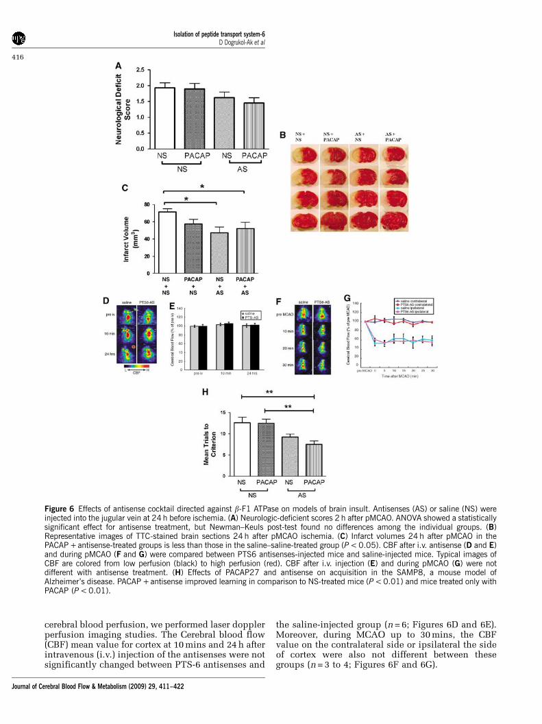
cerebral blood perfusion, we performed laser dopplerperfusion imaging studies. The Cerebral blood flow(CBF) mean value for cortex at 10 mins and 24 h afterintravenous (i.v.) injection of the antisenses were notsignificantly changed between PTS-6 antisenses and
the saline-injected group (n = 6; Figures 6D and 6E).Moreover, during MCAO up to 30 mins, the CBFvalue on the contralateral side or ipsilateral the sideof cortex were also not different between thesegroups (n = 3 to 4; Figures 6F and 6G).
Figure 6 Effects of antisense cocktail directed against b-F1 ATPase on models of brain insult. Antisenses (AS) or saline (NS) wereinjected into the jugular vein at 24 h before ischemia. (A) Neurologic-deficient scores 2 h after pMCAO. ANOVA showed a statisticallysignificant effect for antisense treatment, but Newman–Keuls post-test found no differences among the individual groups. (B)Representative images of TTC-stained brain sections 24 h after pMCAO ischemia. (C) Infarct volumes 24 h after pMCAO in thePACAP +antisense-treated groups is less than those in the saline–saline-treated group (P < 0.05). CBF after i.v. antisense (D and E)and during pMCAO (F and G) were compared between PTS6 antisenses-injected mice and saline-injected mice. Typical images ofCBF are colored from low perfusion (black) to high perfusion (red). CBF after i.v. injection (E) and during pMCAO (G) were notdifferent with antisense treatment. (H) Effects of PACAP27 and antisense on acquisition in the SAMP8, a mouse model ofAlzheimer’s disease. PACAP +antisense improved learning in comparison to NS-treated mice (P < 0.01) and mice treated only withPACAP (P < 0.01).
Isolation of peptide transport system-6D Dogrukol-Ak et al
416
Journal of Cerebral Blood Flow & Metabolism (2009) 29, 411–422

We also tested the effect of PTS-6 antisenses andPACAP on learning in 12-month-old SAMP8 mice, ananimal model of Alzheimer’s disease (Morley et al,2002). The two-way ANOVA showed a statistical effectfor antisense (F(1,27) = 43.4, P50.01)], but not forPACAP or interaction (Figure 6H). Newman–Keulsshowed that mice given antisense + PACAP learnedmore quickly than control mice or mice given onlyPACAP (P < 0.01).
Discussion
Pituitary adenylate cyclase-activating polypeptide isa potent neuroprotectant whose delivery to brain iscomplicated by the BBB. PACAP is transportedacross the BBB by PTS-6 that likely represents afamily of influx and efflux transporters (Banks et al,1993). Therapeutic use of PACAP27 in particular ishampered by a brain-to-blood efflux transporter. Ourfinding of a pH and temperature sensitive saturablebinding site for I-PACAP27 on isolated mouse brainmicrovessels is consistent with brain endothelialcells have both PACAP transporter (Uchida et al,1996) and receptor (Knutsson and Edvinsson, 2002;Nonaka et al, 2005) activities. PACAP binds to threereceptor subtypes: PAC1, VPAC1, and VPAC2 (Shioda,2000; Vaudry et al, 2000). The PAC1 receptor hasseveral splice variants and is specific for PACAP,whereas the other two receptors also bind VIP(Hasimoto et al, 1993; Spengler et al, 1993). As wefound that I-PACAP27 binding was neither displacedby VIP nor showed an inhibition pattern classic forPAC1 (Salomon et al, 1993; Rodriguez-Henche et al,1994; Salano et al, 1996; Shioda, 2000), we con-cluded that brain endothelial cells likely possess anovel-binding site.
Identification of the PACAP27-binding protein bymass spectrometry confirmed that it was not one ofthe previously identified receptors for PACAP. Theidentified protein, b-F1 ATPase, colocalized withPACAP immunoactivity on freshly isolated brainendothelial cells. b-F1 ATPase has been shown to actas either a binding protein or a transporter in othertissues for a number of other ligands unrelated toPACAP. Originally identified as an extra-membranecomponent of ATPase in mitochondria (ATPsynthase b-subunit), it was subsequently found tobe identical to the apolipoprotein A-1 receptor thattransports high-density lipoproteins at hepatocytes(Martinez et al, 2003). It is also expressed in neuronswhere it acts as a receptor for enterostatin, apentapeptide involved in feeding (Park et al, 2004).Cultured brain endothelial cells, but not aorticendothelial cells, express and secrete this proteinand expression is increased by cholesterol, insulin,and retinoic acid (Weiler-Guttler et al, 1990; Mockelet al, 1994). However, the function of b-F1 ATPase inbrain endothelial cells was unknown. Interestingly,two other lipoprotein-binding molecules have beenfound to act as efflux transporters for peptides.
P-glycoprotein is a member of the ATP-bindingcassette family, binds Apo A-1, and transports someof the small opiate peptides and cyclosporin in thebrain-to-blood direction (Begley, 2004). Low-densitylipoprotein receptor-related protein-1 acts as anefflux transporter for amyloid b protein (Deaneet al, 2004).
Inhibition of PTS-6 efflux should selectivelyincrease PACAP in the brain both by decreasing theloss of PACAP endogenous to the brain and byincreasing the retention of PACAP entering the brainfrom the blood. We showed that both of theseevents occurred in mice treated with a cocktail ofphosphorothioate antisenses directed against b-F1ATPase. Many reviews have stated that antisensemolecules cannot cross the BBB. However, thosestatements in most reviews were not referenced andwere based on untested assumptions about how theBBB and antisense would interact. Phosphorothioateoligonucleotides are enzymatically resistant (Jaegerand Banks, 2004) and many are taken up by andtransported (Banks et al, 2001, 2006) across brainendothelial cells. Peptide nucleic acids, anotherclass of antisense molecules, also effectively crossthe BBB (Tyler et al, 1999). The antisenses developedhere against PTS-6 were taken up by the capillariesand by brain tissue after the i.v. injection of their32P-labeled versions. A single icv injection of theantisenses inhibited for approximately 48 h thetransport of I-PACAP27 injected into the brain’slateral ventricle. Efflux systems for an inorganic ionefflux system (using iodide as the ligand), PTS-1(using the tetrapeptide Tyr-MIF-1 as the ligand), andP-glycoprotein (using b-endorphin as the ligand)were not affect by the antisenses (Davson andHollingsworth, 1973; Banks and Kastin, 1990; Kinget al, 2001). When injected iv, the antisensesincreased brain uptake of I-PACAP27 perfusedthrough the vasculature of the brain, whereas otherantisenses were ineffective. I-PACAP38 influx orefflux was not affected by the antisense cocktail,showing that b-F1 ATPase acts as an efflux pump forPACAP27, but not PACAP38. This is consistent withprevious pharmacokinetic work that suggested thatthe efflux systems for PACAP27 and PACAP38 weredifferent because PACAP27 efflux is not modulatedby a luteinizing hormone-releasing hormone, whereasPACAP38 efflux is modulated (Banks et al, 1993).
Pituitary adenylate cyclase-activating polypeptideis an extremely potent neuroprotectant. As noted indiscussion, femtomole concentrations are effective invitro and picomole doses are effective in vivo. Morespecifically, small doses of PACAP38 are protectiveagainst amyloid b-protein neurotoxicity in vitro(Onoue et al, 2002) and against ischemic damagein vivo (Uchida et al, 1996). Thus, we postulated thatPTS-6 inhibition could allow both endogenous andexogenous PACAP27 to exert therapeutic effects inrodent models of Alzheimer’s disease and stroke.Work based on PACAP38 indicates that very smallamounts of PACAP are needed for neuroprotection
Isolation of peptide transport system-6D Dogrukol-Ak et al
417
Journal of Cerebral Blood Flow & Metabolism (2009) 29, 411–422

(Banks et al, 1993). Thus, inhibition of PTS-6 alonecould allow enough endogenous PACAP27 to enteror remain in brain to be therapeutically effective. Ourresults in the ischemic model support this idea, astreatment with antisense alone reduced infarctsize. However, the Alzheimer’s model required thecombination of i.v. PACAP27 and antisense toimprove learning. In the stroke model, the improve-ments cannot be ascribed to increased cerebral bloodflow in either the normal or ischemic brain. Weconclude that inhibition of PTS-6 can be effectivelyachieved with peripheral administration of antisenseand that such inhibition allows endogenous andexogenous PACAP27 to attain therapeutic levels inanimal models of central nervous system disease.
In summary, we isolated the PACAP27 effluxcomponent of PTS-6 from brain endothelial cellsand identified it as b-F1 ATPase. PACAP27 and b-F1ATPase immunoactivity colocalized on brain en-dothelial cells. Inhibition of b-F1 ATPase withspecific antisenses reduced efflux of I-PACAP27 butnot other substances including I-PACAP38 frombrain and greatly increased influx into brain. Inhibi-tion of PTS-6 resulted in a therapeutic effect inmouse models of stroke and Alzheimer’s disease. Weconclude that b-F1 ATPase is the efflux componentof PTS-6 and its inhibition with peripherallyadministered antisense allows PACAP27 to exert itsneuroprotective effects in models of Alzheimer’sdisease and stroke.
Materials and methods
Radioactive Labeling of PACAP
Iodination of 5.0 mg of PACAP27 (Bachem, Torrance, CA,USA) with 131I was performed by the lactoperoxidasemethod. Iodinated PACAP27 (I-PACAP27) was purified ona C18 column by RP-HPLC. Incorporation of 131I asdetermined by acid precipitation was > 95% and specificactivity was approximately 1.82 mCi/mg.
Brain Microvessel Isolation
Cerebral microvessels were isolated from mice by amodification of a method described by Gerhart et al(1988). All reagent volumes were proportionally adjustedfor the quantity of tissue processed and unless otherwisenoted, all reagents were of cell culture quality from SigmaChemical Company (St Louis, MO, USA). All glasswarewas precoated with lactated Ringer’s solution containing1% bovine serum albumin to minimize sticking and tomaximize recovery of microvessels. Male CD-1 mice fromour in-house colony weighing 25 to 40 g were anesthetizedwith urethane (0.5 g/kg) before decapitation. This and allother animal studies were performed under protocolsapproved by the VA animal care and use committee, andin accordance with IACUC guidelines. For each micro-vessel isolation procedure, whole brains from 10 mice weredissected from surrounding structures, the pituitary and
the pineal glands discarded, and the remainder placed instock buffer (25 mmol/L HEPES, 1% dextran in minimumessential medium (Gibco Laboratories, Grand Island, NY,USA) pH 7.4) on ice. The tissue was homogenized with 10strokes in a glass tissue grinder with a teflon pestle. Thehomogenate was then filtered through a series of nylonmesh membranes (300 mm, followed by 2� 100 mm; Spec-trum Scientific Corp., Houston, TX, USA), mixed with anequal volume of 40% dextran in stock buffer andcentrifuged at 5000g for 15 mins at 41C. The myelin layerwas carefully removed. The pellet was resuspended instock buffer and filtered through a 25 mm nylon meshmembrane (Bio-Design, Carmel, NY, USA). The microves-sels were washed from the surface of the nylon mesh withstock buffer four times, collected, and centrifuged at 5000gfor 15 mins at 41C. The purity and quantity of eachpreparation was routinely checked by light microscopy.
Binding Study
Freshly isolated microvessels (30mg protein) wereresuspended in 45 mL of incubation buffer (129 mmol/LNaCl, 2.5 mmol/L KCl, 7.4 mmol/L Na2HPO4, 1.3 mmol/LKH2PO4, 0.63 mmol/L CaCl2, 0.74 mmol/L MgSO4,5.3 mmol/L glucose, 0.1 mmol/L ascorbic acid, pH 7.4)containing 1% bovine serum albumin (BSA). Microvesselprotein levels were determined with the Bio-Rad DCprotein assay kit (Hercules, CA, USA). Incubation buffer,I-PACAP27 (4 mL of 0.8 nmol/L), and any additives asindicated below were mixed with the 45 mL suspension ofcells to a final volume of 60 mL (to give a final I-PACAP27concentration of 53 pmol/L) and incubated for 2.5 mins atroom temperature unless otherwise specified. At the end ofincubation, samples were assayed for protein-bound radio-activity by vacuum filtration through Gelman Sciencesglass microfiber filters (Ann Arbor, MI, USA) pretreatedwith 0.5% polyethyleneimine. Filters were washed threetimes with 4 mL of incubation buffer at 41C. The radio-activity trapped on the filters was measured using ag-counter. Nonspecific sticking of PACAP to the filterswas determined in the absence of microvessels andaveraged approximately 20% of total binding in thepresence of microvessels. Total binding (%) was taken asthe binding in the presence of microvessels. Nonspecificbinding was defined as residual binding to microvessels inthe presence of 1 mg per tube (4.7 mmol/L) of unlabeledPACAP. Specific binding was taken as total binding minusnonspecific binding. All experiments were performed intriplicate on at least three occasions and the resultsreported as means with their standard errors.
Inhibition of Specific Binding of PACAP27
Self-inhibition was tested by adding varying amounts ofunlabeled PACAP27. Results are expressed relative to thespecific binding in the absence of unlabeled PACAP27(% control binding). The femtomole specifically boundwas calculated by dividing the percent of specific bindingby 100 and multiplying by the pmol/L (I- PACAP27 andany unlabeled PACAP27 combined) of the incubation
Isolation of peptide transport system-6D Dogrukol-Ak et al
418
Journal of Cerebral Blood Flow & Metabolism (2009) 29, 411–422

solution and by the volume. This was plotted against thenmol/L in the media and the relation fitted to a one-sitehyperbolic-binding model.
Inhibition of I-PACAP27 Binding by PACAP RelatedPeptides
The effects of 50 nmol/L of unlabeled PACAP27, PACAP38,the PACAP antagonists PACAP 6 to 27 and PACAP 6 to 38(Bachem), and VIP (Sigma) on I-PACAP27 specific bindingwas determined. Results were expressed as percent ofcontrol (I-PACAP27 only).
Isolation of PACAP27 Transporter (PTS-6)
Microvessels prepared from 10 male CD-1 mice were lysedin water for 2 h at 41C and centrifuged to obtain themembrane pellet. I-PACAP27 (105 c.p.m.) and the mem-brane fraction were incubated for 20 mins in a volume of20 mL of incubation buffer at room temperature. Disuccini-midyl suberate at a concentration of 0.05 mol/L was used tocrosslink the I-PACAP27 to membrane binding sites byincubating for 15 mins at room temperature. The cross-linked membranes were then subjected to hydrophobicfractionation using the MEM-PER kit (Pierce, Rockford, IL,USA). The hydrophobic fraction was loaded in duplicateonto a 10% Bis/Tris denaturing gel. One half of the gel wassubjected to Western transfer to a nylon membrane andstained with Coomassie blue. The other half of the gel wasair-dried and subjected to autoradiography. After visualiza-tion of the radioactive bands, the autoradiogram wasaligned to the Coomassie-stained membrane and thecorresponding band was cut out. The 55 kDa band wassent for protein identification to the Taplin Mass Spectro-metry Facility (Harvard Medical School).
Effects of Antisense Oligonucleotides to ATP Synthaseb Subunit on Brain-to-Blood Efflux
Three non-overlapping regions of the mouse ATP synthaseb-subunit were used to construct three enzymaticallyresistant phosphorothioate oligonucleotide antisense mo-lecules (Midland Certified Reagent Company, Inc. Mid-land, TX, USA): an 18mer to the 144-9 region: 50-(_P = S)TCCAATGACATTCATGAT-30; a 15mer to 328 to 332 region:50-(_P = S)CTGGTAGCCTACAGC-30; a 17mer to the 249 to353/4 region: 50-(_P = S)ATCGATCCCTTCTTGGT-30. Theeffect of these antisense oligonucleotides on PTS-6 effluxactivity was determined after both their icv and i.v.administration.
To test the effectiveness of PTS-6 antisense oligonucleo-tides after their icv administration, 2-month-old male CD-1mice kept on a 12/12 h light/dark cycle with food and waterfreely available were anesthetized on the day of study with0.15 mL of 40% urethane. The scalp was removed and ahole made into the lateral ventricle, 1.0 mm lateral and1.0 mm posterior to the bregma, with a 26-gauge needlewith a tubing guard that kept the depth of the holesconstant (3.0 to 3.5 mm). Mice received an icv injection of
1.0 mL lactated Ringer’s solution with 1% BSA containing100 ng of each of the three antisense oligonucleotides.Immediately (t = 0) or at varying times (0.5, 2, 4, 8, 18, 24,36, 48, or 72 h) after the antisense injection, mice received asecond icv injection of 1.0 mL lactated Ringer’s solutionwith 1% BSA containing 1.5 (105) c.p.m. of I-PACAP27.Mice were decapitated 10 mins after the icv injection of I-PACAP27, the brains removed, and the level of residualradioactivity in brain determined by counting in ag-counter. Mice that had been killed by an overdose ofurethane were used to determine the level of residualradioactivity in brain in the total absence of transport (R)and mice that did not receive antisense were used todetermine the level of residual activity in unimpairedtransport (N). The percent of transport was calculated withthe equation:
%T ¼ 100ðR � AÞ=ðR � NÞwhere A is the residual activity in brain in the antisense-treated mice and N is the residual activity in mice nottreated with antisense.
Specificity of the icv administered antisense oligonu-cleotides for I-PACAP27 efflux was tested by determiningtheir effects on efflux of the PTS-1 ligand Tyr-MIF-1, thep-glycoprotein ligand b-endorphin, free iodine, which is aligand for a perchlorate-sensitive active transporter and onPACAP38. Mice received an icv injection of lactatedRinger’s solution with 1% BSA either containing or notcontaining (control) the three oligonucleotide antisenses.After 24 h, the mice received a second icv injectioncontaining radioactive PACAP38, Tyr-MIF-1, b-endorphin,or free iodine were decapitated 10 mins later, and the levelof residual activity in brain determined in a g-counter. Forcomparison, I-PACAP27 transport was also studied 24 hafter the icv injections. Results were expressed as thepercent of the injected dose remaining in brain.
To test the effectiveness of PTS-6 antisenses after theiri.v. administration, 2-month-old male CD-1 mice kept on a12/12 h light/dark cycle with food and water freelyavailable were given an injection by tail vein of 0.9%saline with or without 10 mg per mouse of each of the threeantisenses. To determine the specificity of the PTS-6antisenses on I-PACAP27 efflux, three other antisenseswere also injected iv. These were a 10mer directed againstthe methionine enkephalin region of preproenkephalin (50-(_P = S)TCA TGA AGC C-30) (Banks et al, 2006), a 42merdirected at the amyloid-b mid-region of amyloid precursorprotein (50-(_P = S)GGCGCCTTTGTTCGAACCCACATCTTCAGCAAAGAACACCAG-30; Kumar et al, 2000), and arandom 40mer (50-(_P = S)GATCACGTACACATCGACACCAGTCGCCATGACTGAGCTT). After 24 h, the mice wereanesthetized with urethane and both jugular veins ex-posed. The thorax was opened, the descending thoracicaorta clamped, both jugular veins severed, and lactatedRinger’s solution containing I-PACAP27 (250,000 c.p.m./ml) perfused through the left ventricle of the heart at therate of 2 mL/min. I-PACAP38 was perfused in some micetreated with PTS-6 antisense. After 5 mins, the perfusionwas stopped and 20 mL of lactated Ringer’s solution wasinfused through the left ventricle of the heart to wash outthe vascular space of the brain. The level of radioactivity
Isolation of peptide transport system-6D Dogrukol-Ak et al
419
Journal of Cerebral Blood Flow & Metabolism (2009) 29, 411–422

was determined in the brain and in an aliquot of theperfusion fluid and results expressed as the brain/perfu-sion ratio in units of microliter per gram.
Brain and BBB Uptake of Antisense
We used the capillary depletion method to determinewhether the antisenses were taken up by the BBB (Trigueroet al, 1990; Gutierrez et al, 1993). Each antisense wasend-labeled by mixing 10 mg of the antisense with 1.5 mL of10� kinase buffer, 1.5mL of T4 polynucleotide kinase(New England Biolabs, Ipswich, MA, USA) and 10 mL of[g32P]ATP. The mixture was then incubated in a 371C waterbath for 45 mins. After incubation, the kinase wasinactivated by heating the sample to 651C for 5 mins.Labeled antisense was removed from the reaction mixtureby ethanol precipitation followed by centrifugation. Forthe initial ethanol precipitation, the labeled P-Olg wasmixed with 80 mL of water, 10 mL of 3 mol/L sodium acetate(pH = 5.0), 2 to 5 mL of Pellet Paint Co-Precipitant (Novagen,La Jolla, CA, USA) and 300 mL of cold ethanol. Thismixture was incubated at �701C overnight. The labeledantisense was separated by centrifugation at 13,000 r.p.m.for 20 mins. After removing the supernatant, the pellet(containing the labeled antisense) was resuspended in500mL of cold ethanol and subjected to centrifugation at13,000 r.p.m. for 15 mins. This step was repeated threetimes to remove any remaining unincorporated [g32P]ATP.After the final wash, the pellet was resuspended in 100 mLof water and the level of radioactivity was determined bycounting an aliquot of the sample in a scintillation counter.Mice were anesthetized and given an injection into thejugular vein of 0.2 mL saline containing approximately106 d.p.m. of the labeled antisense. At 10 mins later, arterialblood was obtained from the descending aorta, the thoraxopened and the descending thoracic aorta clamped, bothjugulars severed, and the blood washed out of the brain byinjecting 20 mL of lactated Ringer’s solution into the leftventricle of the heart in 60 secs. The brain was removedand emulsified in a glass homogenizer (8 to 10 strokes) at41C in a ninefold volume of physiologic buffer (10 mmol/LHEPES, 141 mmol/L NaCl, 4 mmol/L KCl, 2.8 mmol/LCaCl2, 1 mmol/L MgSO4, 1 mmol/L NaH2PO4, and10 mmol/L D-glucose adjusted to pH 7.4). Dextran solutionwas added to the homogenate to a final concentration of26%. An aliquot was centrifuged at 5400g for 15 mins at41C in a swinging bucket rotor. The pellet containing thebrain microvessels and the supernatant containing thebrain parenchyma were carefully separated. Results wereexpressed as capillary/serum and parenchyma/serumratios.
Colocalization of b-F1 ATPase and PACAP27 Bindingto Brain Microvessels
Microvessels prepared from 10 male, 8-week-old CD-1mice were resuspended in 100mL of incubation buffercontaining 1% BSA. The microvessels were divided into acontrol group and an experimental group of 25 mL each.PACAP27 (2.5 mg) was added to the experimental group
and the volume in both tubes was increased to a total of30 mL using the aforementioned incubation buffer. Themicrovessels were then incubated for 20 mins at roomtemperature with gentle mixing and tapping. Disuccinimi-dyl suberate (1.5 mL) at a concentration of 0.05 mol/L wasused to crosslink PACAP27 to membrane-binding sites byincubating for 10 mins at room temperature. The cross-linked membranes were then washed in 1 mL aliquots ofthe incubation buffer by inverting the tubes several timesand centrifuging at 4000 r.p.m. for 3 mins at 41C. Thesupernatant was carefully removed and the pellet washedthree more times with incubation buffer. The washed andpelleted microvessels were resuspended in 100 mL ofincubation buffer in preparation for confocal microscopyanalysis.
The treated microvessels were resuspended and 25 mLwas applied to each etched ring on glass slides (Gold SealProducts) that had been coated with poly-L-lysine solution(Sigma-Aldrich, St Louis, MO, USA) and the microvesselswere allowed to settle and attach to the slide. After60 mins, the microvessels were fixed by adding 25 mL of4% formaldehyde in phosphate-buffered saline (PBS) toeach ring for 10 mins. The preparations were then washedby adding PBS to each ring. To minimize loss ofmicrovessels during this wash and subsequent steps 50 to100mL of solution was applied to one side of the etchedring via a pipette while simultaneously removing anequivalent volume from the opposite side of the ring witha second pipette. After washing several times with PBS, thepreparations were blocked with 3% BSA in PBS for30 mins and then rinsed briefly in 0.1% BSA in PBS. Themicrovessels were then incubated with a solution of anti-PACAP27 antibody and anti-transporter antibody (diluted1:200 and 1:1000, respectively) in 0.1% BSA in PBS for16 h in a humidified sealed container at 41C. After washingwith 0.1% BSA in PBS, they were incubated in a solutionof goat anti-rabbit immunoglobulin G rhodamine red-X anddonkey anti-mouse immunoglobulin G Alexa Fluor 488(1:500 and 1:750, respectively; both from Molecular Probes,Subsidiary of Invitrogen, Carlsbad, CA, USA) for 1.5 h in asealed humidified container at room temperature. Themicrovessels were then washed several times with PBS andmounted in Fluorosave (Calbiochem, Gibbstown, CA,USA) mounting medium. The preparations were examinedwith a Bio-Rad MRC1024 confocal microscope and digitalimages processed and merged using Confocal Assistantsoftware. Negative controls consisted of omitting thePACAP27 in the initial incubations, using normal rabbitand normal mouse immunoglobulin G as primary anti-bodies and leaving out the secondary antibody.
Permanent Middle Cerebral Artery Occlusion Model
Peptide transport system-6 antisenses (10mg per mouse ofeach of the three antisenses) or saline (200 mL) wereinjected via the jugular vein 24 h before pMCAO. Micewere anesthetized with 2.0% sevoflurane in N2O/O2, afterwhich they were subjected to pMCAO by the intraluminalfilament technique by use of a monofilament nylon sutureas previously described (Ohtaki et al, 2006). PACAP27
Isolation of peptide transport system-6D Dogrukol-Ak et al
420
Journal of Cerebral Blood Flow & Metabolism (2009) 29, 411–422

(5 nmol/kg, 200mL) or saline were injected into the jugularvein immediately after pMCAO. Four groups were studied:saline-preinjection + saline-postinjection (saline–saline); sal-ine–preinjection + PACAP27 postinjection (saline-PACAP27);PTS-6 antisenses-preinjection + saline-postinjection (anti-sense-saline); PTS-6 antisenses-preinjection + PACAP27-postinjection (PTS-6 AS-PACAP27). The neurologic scorewas evaluated at 2 h after pMCAO as followed by theestablished neurologic scoring system (Hattori et al, 2000):(0 = normal; 1 = consistent forelimb asymmetry but noapparent problem in ambulation; 2 = asymmetry in ambula-tion and reduced resistance to lateral push; 3 = circlingmovement and/or seizures; and 4 = morbidly ill and neardeath. At 24 h after tMCAO, the brains were removed andwere sliced into four 2-mm coronal sections using a mousebrain matrix. Brain slices were then stained with 2% 2,3,5-triphenyltetrazolium chloride (Wako, Tokyo, Japan) at371C for 30 mins. The infarct areas were measured usingNIH Image software and the infarct volumes were calculatedby integration of the infarct areas.
Measurement of Cerebral Blood Flow
Cerebral blood flow was measured with a laser Dopplerperfusion Imager (Peri Scan PIM2; PERIMED, Stockholm,Sweden). This noninvasive procedure provides a measure-ment of blood perfusion in superficial blood vessels of thebrain without craniotomy. Mice were anesthetized withsodium pentobarbital sodium (60 mg/kg body weight).Additional doses were given as needed to maintainanesthesia. The skull was exposed by reflecting the scalpand was fixing it with cutting tape. The image of30� 30 mm area that covered the skull was obtained. Theperfusion index values (a global indicator of blood velocityand perfusion) were expressed in units of volts, and thevelocity/perfusion images were set to have a user-definedcolor scale ranging from 0 (showing black) to 10 (showingred) volts. Short- and long-term effects of i.v. infusions ofPTS-6 antisenses on CBF were determined by measuringCBF before i.v. injection and at 10 mins and 24 h afterinjection. To evaluate CBF during MCAO in PTS-6antisense and saline-treated groups, scans were performedat 24 h after injection (pre-MCAO) as baseline. The CBFwas measured every 5 mins during MCAO up to 30 mins.For each evaluation, we generated a mean perfusion indexfor the elliptic region of interest on both cortex regions overthe site supplied by the proximal segment of the MCA.Blood flow values were expressed as a percentage of theCBF baseline values.
Acquisition in the T-maze Foot Shock Avoidance
The effects on acquisition (learning) of i.v. PACAP27 andPTS-6 antisenses were tested in 12-month-old SAMP8mice in the T-maze. The SAMP8 is a spontaneous mutantthat develops age-related, amyloid-b protein-mediatedcognitive impairments (Morley et al, 2002). The T-maze isa hippocampal-dependent learning task in which theanimal must integrate multiple cues in a novel environ-ment to learn a new task and the methodology previously
described (Morley et al, 2000; Farr et al, 2000). A start boxlocated at the bottom of the start alley is separated fromthe alley by a plastic guillotine door, which preventsmovement down the alley until training begins. Anelectrifiable stainless steel rod floor runs throughout themaze to deliver a scrambled foot shock. The guillotine dooris raised and a buzzer sounds simultaneously; 5 secs laterfoot shock is applied. The goal box that is entered on thefirst trial is designated ‘incorrect’ and the foot shockcontinued until the mouse enters the other goal box, whichin all subsequent trials is designated as ‘correct’ for thatparticular mouse. At the end of each trial, the mouse isreturned to its home cage until the next trial. The intertrialinterval is 30 secs with a foot shock intensity of 0.35 mA.The buzzer intensity is 55 dB. Mice were trained until theymade one avoidance. Mice were given the antisenses (10 mgper mouse of each of the three antisenses in 200 mL) orsaline 48 h before training by tail vein injection. They werethen given PACAP27 (0.5 nmol/kg in 200mL per mouse) orsaline 24 h before training by tail vein injection.
Statistics
Means are reported with their n and standard errors. Twogroups were compared by Student’s t-test. More than twogroups were compared by ANOVA, followed by Newman–Keuls post-test.
Acknowledgements
This study was supported by VA Merit Review andR01AA12743, R01NS41863, R21DA019396, andR01NS051334.
References
Arimura A, Somogyvari-Vigh A, Weill C, Fiore RC, TatsunoI, Bay V, Brenneman DE (1994) PACAP functions as aneurotrophic factor. Ann NY Acad Sci 739:228–43
Banks WA (2005) Critical roles of efflux systems in healthand disease. In: Efflux transporters and the blood–brainbarrier (Taylor EM, ed), Nova Science Publishers:Hauppauge, NY, USA, pp 21–53
Banks WA, Farr SA, Butt W, Kumar VB, Franko MW,Morley JE (2001) Delivery across the blood-brain barrierof antisense directed againt amyloid b: reversal oflearning and memory deficits in mice overexpressingamyloid precursor protein. J Pharmacol Exp Ther297:1113–21
Banks WA, Jaeger LB, Urayama A, Kumar VB, Hileman SM,Gaskin FS, Llanza NV, Farr SA, Morley JE (2006)Preproenkephalin targeted antisenses cross the blood-brain barrier to reduce brain methionine enkephalinlevels and increase voluntary drinking. Peptides27:784–96
Banks WA, Kastin AJ (1990) Editorial review: peptidetransport systems for opiates across the blood-brainbarrier. Am J Physiol 259:E1–0
Banks WA, Kastin AJ, Komaki G, Arimura A (1993) Passageof pituitary adenylate cyclase activating polypeptide1–27
Isolation of peptide transport system-6D Dogrukol-Ak et al
421
Journal of Cerebral Blood Flow & Metabolism (2009) 29, 411–422

and pituitary adenylate cyclase activating polypeptide1–38
across the blood–brain barrier. J Pharmacol Exp Ther267:690–6
Begley DJ (2004) ABC transporters and the blood-brainbarrier. Curr Pharm Des 10:1295–312
Davson H, Hollingsworth JR (1973) Active transport of 131Iacross the blood-brain barrier. J Physiol (London)233:327–47
Deane R, Wu Z, Sagare A, Davis J, Du Yan S, Hamm K, Xu F,Parisi M, LaRue B, Hu HW, Spijkers P, Guo H, Song X,Lenting PJ, Van Nostrand WE, Zlokovic BV (2004)LRP/amyloid beta-peptide interaction mediates differ-ential brain efflux of Abeta isoforms. Neuron 43:333–44
Farr SA, Banks WA, La Scola ME, Flood JF, Morley JE(2000) Permanent and temporary inactivation of thehippocampus impairs T-maze footshock avoidanceacquisition and retention. Brain Res 872:242–9
Gerhart DZ, Broderius MA, Drewes LR (1988) Culturedhuman and canine endothelial cells from brain micro-vessels. Brain Res Bull 21:785–93
Gutierrez EG, Banks WA, Kastin AJ (1993) Murine tumornecrosis factor alpha is transported from blood to brainin the mouse. J Neuroimmunol 47:169–76
Hasimoto H, Ishihara T, Shigemoto R, Mori K, Nagata S(1993) Molecular cloning and tissue distribution of areceptor for pituitary adenylate cyclase-activating poly-peptide. Neuron 11:333–42
Hattori K, Lee H, Hurn PD, Carin BJ, Traystman RJ, DevriesAC (2000) Cognitive deficits after focal cerebral ische-mia in mice. Stroke 31:1939–44
Jaeger LB, Banks WA (2004) Antisense therapeutics and thetreatment of CNS disease. Front Biosci 9:1720–7
King M, Su W, Chang A, Zuckerman A, Pasternak GW(2001) Transport of opioids from the brain to theperiphery by P-glycoprotein: peripheral actions ofcentral drugs. Nat Neurosci 4:221–2
Knutsson M, Edvinsson L (2002) Distribution of mRNA forVIP and PACAP receptors in human cerebral arteriesand cranial ganglia. Neuroreport 13:507–9
Kumar VB, Farr SA, Flood JF, Kamlesh V, Franko M,Banks WA, Morley JE (2000) Site-directed antisenseoligonucleotide decreases the expression ofamyloid precursor protein and reverses deficits inlearning and memory in aged SAMP8 mice. Peptides21:1769–75
Loscher W, Potschka H (2002) Role of multidrug transpor-ters in pharmacoresistance to antiepileptic drugs.J Pharmacol Exp Ther 30:7–14
Martinez LO, Jacquet S, Esteve J-P, Rolland C, Cabezon E,Champagne E, Pineau T, Georgeaud V, Walker JE, TerceF, Collet X, Perret B, Barbaras R (2003) Ectopic b-chainof ATP synthase is an apolipoprotein A-1 receptor inhepatic HDL endocytosis. Nature 421:75–9
Mockel B, Zinke H, Flach R, Weiss B, Weiler-Guttler H,Gassen HG (1994) Expression of apolipoprotein A-I inporcine brain endothelium in vitro. J Neurochem62:788–98
Morley JE, Farr SA, Kumar VB, Banks WA (2002)Alzheimer’s disease through the eye of a mouse:acceptance lecture for the 2001 Gayle A. Olson andRichard D. Olson prize. Peptides 23:589–99
Morley JE, Kumar VB, Bernardo AF, Farr SA, Uezu K,Tumosa N, Flood JF (2000) b-Amyloid precursor poly-peptide in SAMP8 mice affects learning and memory.Peptides 21:1761–7
Nonaka N, Shioda S, Banks WA (2005) Effect of lipopoly-saccharide on the transport of pituitary adenylatecyclase activating polypeptide across the blood-brainbarrier. Exp Neurol 191:137–44
Ohtaki H, Nakamachi T, Dohi K, Aizawa Y, Takaki A,Hodoyama K, Yofu S, Hashimoto H, Shintani N, Baba A,Kopf M, Iwakura Y, Matsuda K, Arimura A, Shioda S(2006) Pituitary adenylate cyclase-activating polypep-tide (PACAP) decreases ischemic neuronal cell death inassociation with interleukin-6 (IL-6) signaling. Proc NatlAcad Sci USA 103:7488–93
Onoue S, Endo K, Ohshima K, Yajima T, Kashimoto K (2002)The neuropeptide PACAP attenuates beta-amyloid (1–42)-induced toxicity in PC12 cells. Peptides 23:1471–8
Park M, Lin L, Thomas S, Braymer HD, Smith PM, HarrisonDHT, York DA (2004) The F1-ATPase b-subunit is theputative enterostatin receptor. Peptides 25:2127–33
Rodriguez-Henche N, Rodriguez-Pena MS, Guijarro LG,Pietro JC (1994) Characterization of vasoactive intestinalpeptide receptors in human liver. Biochim Biophys Acta1221:193–8
Salano RM, Carmeno MJ, Carrero I, Cavallaro S, Roman F,Hueso C, Travali S, Lopez-Fraile N, Guijarro LG, PietroJC (1996) Characterization of vasoactive intestinalpeptide/pituitary adenylate cyclase-activating peptidereceptors in human being hyperplastic prostate.Endocrinology 137:2815–22
Salomon RA, Couvineau A, Rouyer-Fessard C, Voisin T,Lavalle D, Blais A, Darmoul D, Laburthe M (1993)Characterization of common VIP-PACAP receptor inhuman small intestinal epithelium. Am J Physiology264:E294–300
Shioda S (2000) Pituitary adenylate cyclase activatingpolypeptide (PACAP) and its receptors in the brain.Acta Anat Nippon 75:487–507
Spengler D, Waeber C, Pantaloni C, Holsboer F, Bockaert J,Seeburg P, Journot L (1993) Differential signal trasnduc-tion by five splice variants of the PACAP receptor.Nature 365:170–5
Taylor EM (2002) The impact of efflux transporters in thebrain on the development of drugs for CNS disorders.Clin Pharmacokinet 41:81–92
Thomas SA (2004) Anti-HIV drug distribution to thecentral nervous system. Curr Pharm Des 10:1313–24
Triguero D, Buciak J, Pardridge WM (1990) Capillarydepletion method for quantification of blood–brainbarrier transport of circulating peptides and plasmaproteins. J Neurochem 54:1882–8
Tyler BM, Jansen K, McCormick DJ, Douglas CL, Boules M,Stewart JA, Zhao L, Lacy B, Cusack B, Fauq A,Richelson E (1999) Peptide nucleic acids targeted tothe neurotensin receptor and administered i.p. cross theblood–brain barrier and specifically reduce gene ex-pression. Proc Natl Acad Sci USA 96:7053–8
Uchida D, Arimura A, Somogyvari-Vigh A, Shioda S, BanksWA (1996) Prevention of ischemia-induced death ofhippocampal neurons by pituitary adenylate cyclaseactivating polypeptide. Brain Res 736:280–6
Vaudry D, Gonzales BJ, Basille M, Yon L, Fournier A,Vaudry H (2000) Pituitary adenylate cyclase-activatingpolypeptide and its receptors: from structure to func-tion. Pharmacol Rev 52:269–324
Weiler-Guttler H, Sommerfeldt M, Papandrikopoulou A,Mischek U, Bonitz D, Frey A, Grupe M, Scheerer J, GassenHG (1990) Synthesis of apolipoprotein A-1 in pig brainmicrovascular endothelial cells. J Neurochem 54:444–50
Isolation of peptide transport system-6D Dogrukol-Ak et al
422
Journal of Cerebral Blood Flow & Metabolism (2009) 29, 411–422