Isolation and Characterization of the Novel Popeye Gene Family Expressed in Skeletal Muscle and...
12
Isolation and Characterization of the Novel Popeye Gene Family Expressed in Skeletal Muscle and Heart Birgit Andre ´ e,* Tina Hillemann,* Gania Kessler-Icekson,² Thomas Schmitt-John,‡ Harald Jockusch,‡ Hans-Henning Arnold,* and Thomas Brand* ,1 *Cell and Molecular Biology, Technical University of Braunschweig, 38106 Brunswick, Germany; ²Basil and Gerald Felsenstein Medical Research Center, Tel-Aviv University, Tel-Aviv, Israel; and ‡Developmental Biology and Molecular Pathology, University of Bielefeld, Bielefeld, Germany We identified a novel gene family in vertebrates which is preferentially expressed in developing and adult striated muscle. Three genes of the Popeye (POP) family were detected in human and mouse and two in chicken. Chromosomal mapping indicates that Pop1 and Pop3 genes are clustered on mouse chromosome 10, whereas Pop2 maps to mouse chromosome 16. We found evidence that POP1 and POP3 in chicken may also be linked and multiple transcript isoforms are generated from this locus. The POP genes encode proteins with three potential transmembrane domains that are conserved in all family members. Individual POP genes exhibit specific expression patterns during development and postnatally. Chicken POP3 and mouse Pop1 are first preferentially expressed in atrium and later also in the subepicardial compact layer of the ventricles. Chicken POP1 and mouse Pop2 are expressed in the entire heart except the outflow tract. All three Pop genes are expressed in heart and skeletal muscle of the adult mouse and lower in lung. Pop1 and Pop2 expression is upregulated in uterus of pregnant mice. Like the mouse genes, human POP genes are predominantly expressed in skeletal and cardiac muscle. The strong conservation of POP genes during evolution and their preferential expression in heart and skeletal muscle suggest that these novel proteins may have an important function in these tissues in vertebrates. © 2000 Academic Press Key Words: chick; mouse; human; heart development; subtractive hybridization; atrium; ventricle; compact layer; in situ hybridization; gene expression. INTRODUCTION The heart is the first organ to be formed in most verte- brate embryos. In the chick embryo, cells destined to form the heart arise in the epiblast lateral to the primitive streak, invaginate through the streak, and migrate rostrolaterally to form part of the lateral plate at Hamburger Hamilton (HH) stage 4 (Garcia-Martinez and Schoenwolf, 1993). The ingression pattern along the streak at HH stage 3 of these cells is colinear with their later position along the A/P axis in the tubular heart (Garcia-Martinez and Schoenwolf, 1993). At HH stage 10 in chick and E8 in mouse the bilateral heart primordia fuse at the midline to form a tubular heart. Soon after fusion the heart loops to the right and starts to contract (DeHaan, 1965). The consequence of looping is that the atrial segment which initially lies posterior resumes a more rostrodorsal position, and the ventricular and proximal outflow tract regions become positioned ventrocaudally. Several genes which are ex- pressed homogeneously within the heart tube before loop- ing become regionally restricted after this process. This has been documented for the myosin heavy chain genes, AMHC1 and VMHC1, in chicken and a- and bMyHC in the mouse embryo (Bao et al., 1999; Lyons et al., 1990). An 1 To whom correspondence should be addressed at the Depart- ment of Cell and Molecular Biology, Institute of Biochemistry and Biotechnology, Technical University of Braunschweig, Spiel- mannstrasse 7, 38106 Brunswick, Germany. Fax: 0049-531-391- 8178. E-mail: [email protected]. Developmental Biology 223, 371–382 (2000) doi:10.1006/dbio.2000.9751, available online at http://www.idealibrary.com on 0012-1606/00 $35.00 Copyright © 2000 by Academic Press All rights of reproduction in any form reserved. 371
Transcript of Isolation and Characterization of the Novel Popeye Gene Family Expressed in Skeletal Muscle and...

TU
WT
Developmental Biology 223, 371–382 (2000)doi:10.1006/dbio.2000.9751, available online at http://www.idealibrary.com on
Isolation and Characterization of the Novel PopeyeGene Family Expressed in Skeletal Muscleand Heart
Birgit Andree,* Tina Hillemann,* Gania Kessler-Icekson,†Thomas Schmitt-John,‡ Harald Jockusch,‡Hans-Henning Arnold,* and Thomas Brand*,1
*Cell and Molecular Biology, Technical University of Braunschweig, 38106 Brunswick,Germany; †Basil and Gerald Felsenstein Medical Research Center, Tel-Aviv University,
el-Aviv, Israel; and ‡Developmental Biology and Molecular Pathology,niversity of Bielefeld, Bielefeld, Germany
e identified a novel gene family in vertebrates which is preferentially expressed in developing and adult striated muscle.hree genes of the Popeye (POP) family were detected in human and mouse and two in chicken. Chromosomal mapping
indicates that Pop1 and Pop3 genes are clustered on mouse chromosome 10, whereas Pop2 maps to mouse chromosome 16.We found evidence that POP1 and POP3 in chicken may also be linked and multiple transcript isoforms are generated fromthis locus. The POP genes encode proteins with three potential transmembrane domains that are conserved in all familymembers. Individual POP genes exhibit specific expression patterns during development and postnatally. Chicken POP3and mouse Pop1 are first preferentially expressed in atrium and later also in the subepicardial compact layer of theventricles. Chicken POP1 and mouse Pop2 are expressed in the entire heart except the outflow tract. All three Pop genesare expressed in heart and skeletal muscle of the adult mouse and lower in lung. Pop1 and Pop2 expression is upregulatedin uterus of pregnant mice. Like the mouse genes, human POP genes are predominantly expressed in skeletal and cardiacmuscle. The strong conservation of POP genes during evolution and their preferential expression in heart and skeletalmuscle suggest that these novel proteins may have an important function in these tissues in vertebrates.© 2000 Academic Press
Key Words: chick; mouse; human; heart development; subtractive hybridization; atrium; ventricle; compact layer; in situhybridization; gene expression.
ci1btalpvppibA
INTRODUCTION
The heart is the first organ to be formed in most verte-brate embryos. In the chick embryo, cells destined to formthe heart arise in the epiblast lateral to the primitive streak,invaginate through the streak, and migrate rostrolaterallyto form part of the lateral plate at Hamburger Hamilton(HH) stage 4 (Garcia-Martinez and Schoenwolf, 1993). Theingression pattern along the streak at HH stage 3 of these
1 To whom correspondence should be addressed at the Depart-ment of Cell and Molecular Biology, Institute of Biochemistry andBiotechnology, Technical University of Braunschweig, Spiel-mannstrasse 7, 38106 Brunswick, Germany. Fax: 0049-531-391-
m8178. E-mail: [email protected].
0012-1606/00 $35.00Copyright © 2000 by Academic PressAll rights of reproduction in any form reserved.
ells is colinear with their later position along the A/P axisn the tubular heart (Garcia-Martinez and Schoenwolf,993). At HH stage 10 in chick and E8 in mouse theilateral heart primordia fuse at the midline to form aubular heart. Soon after fusion the heart loops to the rightnd starts to contract (DeHaan, 1965). The consequence ofooping is that the atrial segment which initially liesosterior resumes a more rostrodorsal position, and theentricular and proximal outflow tract regions becomeositioned ventrocaudally. Several genes which are ex-ressed homogeneously within the heart tube before loop-ng become regionally restricted after this process. This haseen documented for the myosin heavy chain genes,MHC1 and VMHC1, in chicken and a- and bMyHC in the
ouse embryo (Bao et al., 1999; Lyons et al., 1990). An371

1wvttg
dcndcmoapepmoveou
(
cCa[Tp[pchsSe
HDpci(acPsLcocrcaRvP
372 Andree et al.
important regulator of anteroposterior polarity is retinoicacid (RA). Ectopic application of RA results in posterioriza-tion of the tubular heart as judged by the expression ofAMHC1 in the presumptive ventricle (Yutzey et al., 1994).Conversely, RA deficiency in quail or loss-of-function mu-tation of RALDH2, the rate-limiting enzyme in RA biosyn-thesis, results in loss of posterior structures including theinflow tract of the heart (Kostetskii et al., 1999; Nieder-reither et al., 1999).
Maturation of ventricles includes the formation of thetrabecular layer within the ventricular lumen, growth andthickening of the compact layer, and growth of the inter-ventricular septum. Ablation of the genes for neuregulin orits receptors, erbB2 and erbB4, in mice leads to embryoniclethality due to impaired trabecular growth (Gassmann etal., 1995; Lee et al., 1995; Meyer and Birchmeier, 1995).Growth of the myocardial compact layer of the myocar-dium is inhibited in a surprisingly large variety of loss-of-function mutations. A thin ventricular wall with normaldevelopment of the trabecular layer has been observed innull mutations for N-myc (Moens et al., 1993), RXRa(Kastner et al., 1994; Sucov et al., 1994), WT-1 (Kreidberg etal., 1993), TEF-1 (Chen et al., 1994), gp130 (Yoshida et al.,1996), bARK-1 (Jaber et al., 1996), and p130 (LeCouter et al.,998). Despite the common phenotype of a thin ventricularall found in each of these null mutants, the time of deatharied considerably between mutants, ranging from E10.5o E16. Probably several independent regulatory pathwayshat are important for cardiac myocyte maturation androwth of the compact layer exist.Here, we describe a novel gene family identified by
ifferential cloning from a cDNA library of HH stage 3–6hicken embryos. Members of this gene family are predomi-antly expressed in heart and skeletal muscle and wereesignated Popeye (POP) genes. We cloned several POPDNAs from chicken embryo and three POP genes fromouse and human. A cDNA identical to Pop1 was previ-
usly described in the mouse, human, and chicken (Reesend Bader, 1999; Reese et al., 1999). In addition to theredominant expression in striated muscle we found weakxpression in lung and upregulated expression in uterus ofregnant mice. In the heart POP3 in chicken and Pop1 inouse are first expressed in the atrium and later in devel-
pment also in the subepicardial compact layer of theentricle. In contrast, mouse POP2 and chicken POP1 arexpressed in the entire heart with the exception of theutflow tract. The functions of these novel genes arenknown, but may be associated with membranes.
MATERIALS AND METHODS
Animals and Tissues
Fertilized White Leghorn eggs (Charles River) were incubatedand embryos were staged according to Hamburger and Hamilton(1951). ICR mice were obtained from Charles River and embryos
were dissected at various days postcoitum. For RNA preparation wCopyright © 2000 by Academic Press. All right
dissected embryos and excised tissues were placed on dry ice andwere stored at 280°C.
RNA Isolation and cDNA Library Construction
Total RNA was isolated as previous described (Schultheiss et al.,1995). Poly(A)1 mRNA was enriched using the Oligotex mRNA kitQiagen). One microgram of poly(A)1 mRNA from whole chickenembryos at HH stage 3–6 was used for constructing a plasmidcDNA library according to the manufacturer’s instruction (GibcoBRL; Superscript plasmid system for cDNA synthesis). The result-ing cDNA library contained 4 3 105 independent clones/mg cDNA.The average insert size was approximately 1.5 kb. Clones (110,592)were processed into high-density filters (Library No. 573, RZPDBerlin).
Subtraction Procedure and DifferentialHybridization
Two micrograms of poly(A)1 mRNA from E7 heart and liver ofhicken embryos was used in a PCR-based subtraction (PCR Select;lontech). The heart cDNA was used as tester and the liver cDNAs driver. The resulting subtracted cDNA pool was labeled witha-32P]dCTP in the second PCR step of the subtraction protocol.he unsubtracted liver cDNA was processed using the samerotocol without any subtraction and was also labeled with
a-32P]dCTP in the second step of the PCR. Both labeled cDNAools were hybridized onto high-density filters of the HH stage 3–6hicken embryo cDNA library (see above). By comparison of theybridization signals 246 cDNAs which hybridized only with theubtracted probe were isolated. These clones were reprobed on aouthern blot using the same probes used in the previous screen toliminate false positive clones.
Cloning of the Chicken, Murine, and Human POPcDNAs
To isolate chicken full-length cDNAs a chicken embryo (embry-onic day 10) l gt11 cDNA library (Clontech) and a chicken embryo
H stage 12–15 Lambda ZAP II cDNA library (kindly provided by. David Wilkinson and Angela Nieto) were screened using theartial chicken POP1 cDNA (clone 257) as probe. Four differentlones were isolated: POP1A (clone 9.1) is a 1.65-kb clone, which isdentical to the previously published chicken bves clone. POP1Bclone 4.2) is a partial 1.1-kb clone encoding a POP1 isoform withn alternative C-terminus of POP1A. POP1C (clone 4.1) is a 6-kblone encoding a POP1 isoform lacking the N-terminus of POP1A.OP1D (clone 15) is a 3.7-kb clone containing both POP1 and POP3equences. Screening of the chicken embryo HH stage 12–15ambda ZAP II cDNA library with an 800-bp 59 fragment fromlone 15 resulted in the identification of three POP3 cDNAs. Twof them were identical (clone 12 and 14) and were 1.5 kb. Anotherlone (clone 5) was 1.4 kb and carried a small deletion in the codingegion. Fusing the 59 sequence of POP1D with the 39 sequence oflone 12 generated the full-length sequence. The existence of suchcDNA was confirmed by RT-PCR of chicken HH stage 36 cardiacNA. The resulting fragment was sequenced in its entirety anderified the existence of the predicted full-length sequence ofOP3. The sequence of the partial chicken POP1 cDNA (clone 257)
as used to perform extensive GenBank database searches, whichs of reproduction in any form reserved.
tg
teocfP
373Characterization of the Popeye Gene Family
resulted in the identification of several mouse and human ESTclones which were used to isolate the mammalian orthologues.
The EST clones AA041984, coding for a partial Pop1 cDNA, andAA711740, coding for a partial Pop3 cDNA, were used to screen amouse E10.5 heart Uni ZAP XR cDNA library (Stratagene) in orderto obtain full-length cDNAs. The sequence of clone AA239714 wasused to amplify a 360-bp fragment from mouse heart cDNA usingprimers 59-TTCGAGTGAGCCAAGACG-39 and 59-TTCTCAGA-CTCTGGTTCC-39. The resulting PCR fragment was also used toisolate a full-length Pop2 cDNA. Screening resulted in the isolationof full-length cDNAs for Pop1 (1.6 kb), Pop2 (2 kb), and Pop3 (1.8kb). In addition four partial cDNA clones were obtained in the caseof Pop2.
In order to isolate full-length human POP clones EST cloneswith homology to mouse or chicken POP cDNAs were character-ized. Clone AI141226 was sequenced to obtain the full-lengthsequence of human POP1. For the human POP2 cDNA sequencetwo EST clones (Accession Nos. W03843 and W25440) weresequenced. W03843 had a divergent 39 end compared to mousePOP2, while clone W025440 was incomplete at the 59 end. Fusingthe 59 sequence of W03843 with the 39 sequence of W25440generated the full-length POP2 sequence. The existence of such acDNA was subsequently confirmed by RT-PCR of human cardiacRNA derived from a newborn human heart. Human POP3 cDNAsequence was generated from EST clone H84369. Sequencing of allclones was performed using the ABI Prism BigDye TerminatorCycle Sequencing system.
Northern Blot Analysis
Poly(A)1 mRNAs (2.5 mg) from different chicken and murineissues were loaded onto a 1% formaldehyde denaturing agaroseel. In the case of human material 20 mg of total RNA was loaded.
RNA was transferred onto a nylon membrane (GeneScreen) andUV-crosslinked. The membrane was hybridized with full-lengthhuman POP1 and POP3 clones and in the case of POP2 the insertof clone W03843. In the case of mouse and chicken RNA, full-length murine Pop cDNAs and probe A and B of chicken POP1Dwere employed (Fig. 4). In addition a commercial human adultmultiple tissue Northern blot (Human MTN Blot; Clontech) wasused. A human b-actin probe, a murine RPL7 probe, and a chickenGAPDH probe respectively served as loading controls.
Whole-Mount in Situ Hybridization
Whole-mount in situ hybridization and photographic documen-ation of the results obtained were as previously described (Andreet al., 1998). The following probes were used: a 600-bp 59 fragmentf clone 257 for the detection of chicken POP1, a 1.5-kb cDNA oflone 12 for the detection of chicken POP3, a 400-bp EcoRI 59ragment of mouse Pop1, and a 360-bp PCR fragment of mouseop2.
Immunofluorescence and Cell Transfection
For all three murine cDNAs C-terminal myc-epitope-taggedconstructs were generated. For this purpose fragments containingthe entire coding region were amplified using gene-specific primers
and cloned into the PCS2 vector. COS7 cells were transfected usingCopyright © 2000 by Academic Press. All right
a standard Ca–phosphate method and were fixed 2 days aftertransfection with 2% paraformaldehyde in PBS, permeabilized with0.2% Triton X-100 in PBS, and incubated for 1 h with undilutedsupernatant of myc-antibody-producing hybridoma cells. Afterseveral washes with PBS a TRITC-conjugated anti-mouse IgG(Dianova) was applied for 1 h. Cells were mounted after severalwashes with PBS and analyzed using a fluorescence microscope.
Western Blot Analysis
Cell lysates from COS7 cells nontransfected and transfectedwith myc-epitope-tagged POP constructs were prepared using RIPAbuffer. Cell lysates were separated by SDS–PAGE and blotted onHybond ECL nitrocellulose membrane (Amersham). The mem-brane was blocked, incubated with a 1:20 dilution of supernatantfrom myc-antibody-producing hybridoma cells, and washed withTBST. Secondary antibody was a peroxidase-labeled anti-mouseIgG (Vector) and detection was performed using ECL Westernblotting detection reagent (Amersham). Exposure to films wascarried out for 5 to 10 s.
In Vitro Translation Experiments
The full-length mouse Pop1 cDNA cloned in pSK Bluescriptvector was in vitro translated using the TNT Coupled ReticulocyteLysate Systems (Promega) in the absence or presence of caninepancreatic microsomal membranes (Promega) according to themanufacturer’s instructions. Translation products were labeledwith [35S]methionine (Amersham), size separated by SDS–PAGE,and visualized by autoradiography after fluorographic enhance-ment with sodium salicylate.
RT-PCR
For RT-PCR analysis of POP1 isoform expression total RNA wasisolated from various organs from several HH stage 36 chickenembryos as previously described (Schultheiss et al., 1995, 1997).cDNA was synthesized from DNase-treated total RNA using AMVreverse transcriptase. PCR was performed using the followingprimer pairs:
Product Primer Primer sequence
Productsize(bp)
POP1A a 59-CCCTACCTGAGAACTTCAGC-39 243b 59-GTGGACATTTCTGTCTAGGAGC-39
POP1D59 c 59-TAGTCCAGCCATGGGTGAAA-39 961d 59-AGTTGTCGGAATAACCAGGC-39
POP1 e 59-TTGCTCACCGTAGGATGTGC-39 419f 59-CGGTTCATCTGAGTTGATCG-39
POP1D39 g 59-TTCTGACAGGTCCAGCAGAGC-39 390h 59-CTTTCAGGCTTCTGAGAGTGC-39
POP3 i 59-CTGTCTTTGCTTGTATCAGGC-39 574j 59-ACTCCAGCTAAAGTAAGTGGC-39
Genomic Mapping
The murine Pop1, Pop2, and Pop3 genetic loci were mapped
using a T31 mouse/hamster radiation hybrid panel (Researchs of reproduction in any form reserved.

rmga(i(itbP1ParPbcheb
hpCtNail(tawma
aapaIatM(owtptlWtsC
374 Andree et al.
Genetics, Huntsville, AL). PCR was performed using the followingprimer pairs:
Product Primer sequence
Productsize(bp)
Pop1 AAC ATG AAT TCC ACG GAG TCC ATC C 228TAG AGA CAA CAT CAC TCT GAG
Pop2 CTC AAT GAC AAG CTG TTT GCC 122TCA TCT TTC TCA GAC TCT GGT TCC
Pop3 TAT CTA CTC TTT GCG CAG CAT CGG 180ATG GGG TGA TCT GGA CAA GTC TGG
PCR conditions were 4 min initial denaturation at 94°C and 35cycles of 1 min at 92°C, 1 min at 64°C (Pop1), 60°C (Pop2), or 68°C(Pop3), and 1 min at 72°C. Final extension was 5 min at 72°C.
RESULTS
The aim of this study was to identify novel transcriptsthat are preferentially expressed during chick heart devel-opment. To this end we generated a spotted high-densitycDNA library from HH stage 3–6 chick embryos. Thesefilters were hybridized with cDNA from hearts of E7 chickembryos that had been enriched for cardiac-specific tran-scripts using liver cDNA (E7 chick embryos) as driver in thesubtraction. Subsequently, the same filters were also hy-bridized to liver cDNA from E7 chick embryos. A total of 43differentially hybridizing clones were isolated and se-quenced. The majority of clones encoded known myofibril-lar and cytoskeletal proteins. Ten clones, however, repre-sented novel genes. One of these clones containing a 1.4-kbinsert showed cardiac-restricted expression in chick em-bryos and was chosen for further analysis. Rescreening ofchick cDNA libraries with this clone yielded several largercDNAs, indicating that the original isolate was incompleteat the 59 end (Fig. 4). Extensive searches in the EST databaseevealed the existence of homologous cDNAs in mouse andan and related sequences in chicken, mouse, and human
enomes. Based on the preferential expression in cardiacnd skeletal muscle (see below) we called the genes PopeyePOP). A cDNA identical to Pop1 was previously describedn the mouse, human, and chicken and was termed bvesReese and Bader, 1999; Reese et al., 1999). Altogether wedentified three POP genes each in mouse and human andwo genes in chicken (Fig. 1A). Sequence conservationetween species amounts to 70 and 86% for POP1, 79% forOP2, and 73–90% for POP3 at the amino acid level (Fig.B). Within one species POP1 was 24 and 28% identical toOP2 and POP3, respectively, while POP2 and POP3 werepproximately 50% identical. A particularly conserved coreegion (amino acids 172–266 of mouse Pop1) within theOP proteins showed 45% identity for all three and 70%etween POP2 and POP3. Thus, POP2 and POP3 are morelosely related to each other than to POP1 (Fig. 1B). No POPomologues were found in Drosophila and Caenorhabditislegans; however, a POP2 homologue was found in ze-
rafish (Accession No. AW153906) and a putative POP1Copyright © 2000 by Academic Press. All right
omologue was identified in the cephalochordate Am-hioxus floridae (Accession No. Z83270; Fig. 1D).omputer-based secondary structure modeling predicted
he existences of three transmembrane helices in the-terminal region of each family member (Sonnhammer et
l., 1998). An algorithm for the prediction of protein local-zation in eukaryotic cells suggests that POP proteins mayocalize to the endoplasmic reticulum or plasma membraneNakaki and Kenhisa, 1992). Consistent with this predic-ion, we found perinuclear distribution of POP proteinsfter transfection in COS7 cells (Figs. 2A–2C). In some casese also observed POP protein localized to the plasmaembrane. We therefore conclude that POP proteins are
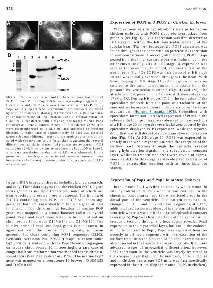
ssociated with membranes.Mouse and human POP1 and POP2 proteins contain
pproximately 360 amino acids, while POP3 has around 290mino acids, mainly due to a shorter C-terminus. Theredicted molecular weights of POP1 and POP2 are bothpproximately 41 kDa, and POP3 is approximately 37 kDa.n vitro translation (IVT) of murine POP1 cDNA revealedn apparent MW of 43 kDa on SDS–PAGE (Fig. 2D). IVT inhe presence of microsomal membranes resulted in higher
W products indicative of posttranslational modificationFig. 2D). Similarly, a protein product of approx 58 kDa wasbserved on Western blots of cell extracts of COS7 cellshich had been transfected with a C-terminally epitope-
agged POP1 construct (Fig. 2D). In addition several largerrotein species also reacted with the antibody, suggestinghat additional protein products are generated by posttrans-ational modification. No POP protein was detected on
estern blots of immunoprecipitated material from Pop1-ransfected COS7 cell supernatants (data not shown). Thisuggests that POP1 protein is not secreted, at least fromOS7 cells.
Multiple Transcripts Are Generated from theChicken POP1/POP3 Gene Locus
A total of four different chicken POP1 cDNAs, whichdiffered at both ends of the coding sequence, were isolatedfrom heart and total embryo cDNA libraries, suggestingthat multiple isoforms of POP1 exist (Fig. 3). POP1A issimilar to the murine and human POP1 cDNAs. POP1B isa partial cDNA which differs from POP1A by a variantC-terminus. POP1C is a 5.6-kb cDNA encoding a proteinthat lacks the N-terminus of POP1A, including the putativetransmembrane domains, but has a longer and differentC-terminus. POP1D is unusual in that it contains 251amino acids at the N-terminus which are identical to POP3and a POP1A-related C-terminus. We also isolated achicken POP3 cDNA (POP3A) similar in size and sequenceto the mammalian homologues and another POP3 cDNA,POP3B, that lacks the putative transmembrane domains.No POP2 cDNA was isolated from various chicken cDNAlibraries. With RT-PCR we determined the expression pat-terns of the various cDNA isoforms in the embryo. Using
primers a and b (Fig. 3A) specific for the POP1A C-terminuss of reproduction in any form reserved.

375Characterization of the Popeye Gene Family
and primers g and h specific for the 39 terminus of POP1Dwe were able to amplify these sequences in RNA from heartbut also from other organs, such as muscle, brain, stomach,kidney, lung, and spleen (Fig. 3B). In contrast, primers i andj specific for POP3, primers e and f specific for the core
FIG. 1. The vertebrate Popeye gene family. (A) Alignment of proteAmino acids shadowed in black are conserved in at least four POdomains. Alignments were done with the Clustal algorithm using tis distantly related to POP2 and POP3. (C) Predicted protein strucBoth POP1 and POP2 are similarly sized (359 and 367 aa, respectiveBlack bars mark the locations of the transmembrane domains presepartial sequence of the presumptive POP gene of Amphioxus florid
region of POP1, and primers c and d specific for the 59
Copyright © 2000 by Academic Press. All right
terminus of POP1D revealed expression mainly in the heart(Fig. 3B). Northern blot experiments using a probe fromPOP1D excluding the C-terminus (probe A) showed 1.8- and2.0-kb POP1 mRNAs that were detected only in heart (Fig.3C). Interestingly, hybridization of the same Northern blot
quences encoded by Popeye genes from chicken, mouse, and man.nes. Bars demarcate the location of presumptive transmembranesergene program. (B) Dendrogram of the Popeye gene family. POP1f vertebrate POP proteins as exemplified by the murine proteins.hile POP3 is considerably shorter and consists of 292 amino acids.all three family members. (D) Alignments of mouse Pop1 and the
in seP ge
he Lature oly), wnt inae.
with the C-terminal POP1D probe (probe B) exhibited a
s of reproduction in any form reserved.
ssdcavlcednoaaPt
376 Andree et al.
larger mRNA in several tissues, including kidney, stomach,and lung. These data suggest that the chicken POP1/3 genelocus generates multiple transcripts, some of which areheart-specific and others more widespread. The finding ofPOP1D containing both POP1 and POP3 sequences sug-gests that both are transcribed from the same gene, at leastin chicken. The chromosomal location of murine POPgenes was mapped on a mouse/hamster radiation hybridpanel. Pop1 and Pop3 were found to be colocalized onchromosome 10 between D10Mit148 and D10Mit170. Therelative order of Pop1 and Pop3 genes is not known. Inagreement with the murine mapping data, a humangenomic Pac clone containing POP1 sequences (52202;GenBank Accession No. Z95329) maps to chromosome6q21, which is syntenic with the Pop1/3-containing regionon mouse chromosome 10. Interestingly, a rare case ofcongenital heart malformation was mapped to this chromo-somal locus (Van Den Ende et al., 1996). The murine Pop2gene was mapped on chromosome 16 between D16Mit59
FIG. 2. Cellular localization and biochemical characterization ofPOP proteins. Murine Pop cDNAs were myc-epitope-tagged at theC-terminus and COS7 cells were transfected with (A) Pop1, (B)Pop2, and (C) Pop3 cDNAs. Recombinant proteins were visualizedby immunofluorescent staining of transfected cells. (D) Biochemi-cal characterization of Pop1 protein. Lane 1, cellular extract ofCOS7 cells transfected with a myc-epitope-tagged murine Pop1construct and lane 2, control extract of untransfected COS7 cellswere electrophoresed on a SDS gel and subjected to Westernblotting. A major band of approximately 58 kDa was detected(arrow). Several additional larger protein products also immunore-acted with the myc antiserum (asterisks), suggesting that severaldifferent posttranslational modified products are generated in COScells. Lanes 3–6, in vitro translation of murine Pop1 cDNA. Lane 3,a primary translation product of 43 kDa was observed. In thepresence of increasing concentrations of canine microsomal mem-branes (lanes 4–6) a major protein product of approximately 58 kDawas observed.
and D16Mit135.
Copyright © 2000 by Academic Press. All right
Expression of POP1 and POP3 in Chicken Embryos
Whole-mount in situ hybridizations were performed onchicken embryos with POP1 riboprobe synthesized fromprobe A (see Fig. 3). POP1 expression was first detected atHH stage 11 within the left ventricular segment of thetubular heart (Fig. 4A). Subsequently, POP1 expression wasfound throughout the heart with no preferential expressionin any compartment. However, after looping POP1 disap-peared from the inner curvature but was maintained in theouter curvature (Fig. 4H). At HH stage 14, expression wasseen in the myotome, notochord, and ventral half of theneural tube (Fig. 4G). POP3 was first detected at HH stage10 and was initially expressed throughout the heart. Withheart looping at HH stage 11, POP3 expression was re-stricted to the atrial compartment and absent from thepresumptive ventricular segments (Figs. 4J and 4M). Theatrial-specific expression of POP3 was still observed at stage17 (Fig. 4K). During HH stages 17–24, the formation of theepicardium proceeds from the point of attachment at thesinoventricular mesocardium to ultimately cover the entiremyocardium (Ho and Shimada, 1978). Coincident withepicardium formation increased expression of POP3 in thesubepicardial compact layer was observed. In heart sectionsof a HH stage 20 embryo the ventricular area covered by theepicardium displayed POP3 expression, while the myocar-dium that was still devoid of epicardium showed no expres-sion (Fig. 4N). At HH stage 30, POP3 was expressed in-tensely in the whole myocardium with the exception of theoutflow tract. Sections through the ventricle revealedstrong hybridization signals in the subepicardial compactlayer, while the trabeculated areas were devoid of expres-sion (Fig. 4O). At this stage we also observed expression ofPOP3 in extracardiac locations such as limbs (data notshown).
Expression of Pop1 and Pop2 in Mouse Embryos
In the mouse Pop1 was first detected by whole-mount initu hybridization at E9.5 when it was confined to theinoatrial compartment and some restricted areas in theorsal part of the ventricle. This pattern remained un-hanged in E10.5 and 11.5 embryos. Beginning at E12.5,dditional expression was observed in the ventral half of theentricle where it was limited to the subepicardial compactayer (Fig. 5). Pop2 was first detectable at E7.5 in the cardiacrescent. Sections through the head region revealed Pop2xpression in the myocardial layer, but not in the endocar-ium. In contrast to Pop1, Pop2 was expressed homoge-eously in all heart segments with the exception of theutflow tract. Between E9.5 and E10.5 Pop2 expression waslso observed in the trabeculated areas (Figs. 5F–5J). In moredvanced stages of myocardial differentiation, however,op2 expression in the ventricle was largely restricted tohe compact layer (Fig. 5K9). In summary, both in mouse
and in chicken hearts one POP gene was first specifically
expressed in the atrium (Pop1 in mouse, POP3 in chicken),s of reproduction in any form reserved.

sc
f(s A d
4A.
377Characterization of the Popeye Gene Family
while another gene was expressed throughout the heart(Pop2 in mouse and POP1 in chicken). However, later in
FIG. 3. The chicken POP1/3 locus generates multiple transcriptsvarious cDNA libraries, is depicted here. Black bars demarcate themark the identical sequence present in all four POP1 transcripts,POP1D. POP1C was isolated as an incomplete cDNA. All four POPcolored boxes are used to label the divergent sequences present inPOP1D are identical and therefore are labeled with the same colorisoforms were isolated in the case of POP3. One clone (POP3A) encDNA (POP3B) codes for a protein which lacks the transmembradepicted above each protein. Relative positions of probes A andwhole-mount in situ hybridization experiments (Fig. 6) which wererom tissues of HH stage 36 chicken embryos with primers speciprimers g and h), POP1 (primers e and f), and POP3 (primers i anequence-specific primers. (C) Northern blot analysis of poly(A)1-RN
A and probe B derived from the POP1D cDNA as depicted in Fig.
development POP genes were specifically expressed in the m
Copyright © 2000 by Academic Press. All right
ubepicardial compartment that is responsible for cardiachamber growth during the second half of fetal develop-
ation of six different cDNA transcripts, which were isolated fromtion of the three predicted transmembrane domains. Green boxesthe orange boxes mark the POP3 sequence present in POP3 and
nscripts code for proteins which differ at the C-terminus. Differentdifferent POP1 transcripts. Parts of the C-termini of POP1C andy the coding regions of the various cDNAs are depicted here. Twos a transcript with three transmembrane domains and a differentgion. Positions of PCR primers used for RT-PCR shown in B arehich were used for the Northern blot analysis shown in C andved from POP1D are shown as black bars. (B) RT-PCR using RNAr POP1A (primers a and b), POPD59 (primers c and d), POP1D39PCR products were subjected to Southern blotting using internalerived from various chicken organs at stages 29 and 36 using probe
. Isolloca
while1 trathe
. Onlcode
ne reB wderi
fic fod j).
ent (Rumyantsev, 1991).
s of reproduction in any form reserved.

wseotwtasmv
dium (ec) are devoid of POP3 expression. (N) Sagittal section
378 Andree et al.
Copyright © 2000 by Academic Press. All right
Tissue Distribution of Mouse and Human POPTranscripts in the Adult
Northern blot analysis of murine Pop genes revealed theexpression of all three Pop genes in adult skeletal andcardiac muscle (Fig. 6A). In addition, weak signals wereobserved in lung. Pop1 and Pop2 genes were also expressedin pregnant uterus, but not in nonpregnant uterus (Fig. 6B).Three Pop1 mRNA species were found in murine tissues. A4.2-kb species was present in heart and muscle, while a 2-kbPop1 mRNA species was detected in all muscle tissues andin lung. An additional 1.8-kb mRNA species was expressedin fast-twitch EDL muscle (Fig. 6B). A single 2.4-kb Pop2mRNA was expressed in heart and weakly in other muscletissues. Two mRNA species of 1.8 and 2.0 kb were detectedfor Pop3 with equal intensity in skeletal muscle and heart.A similar expression pattern was also observed for humanPOP genes (Fig. 6C). Two large 4.5- and 6-kb mRNA specieswith equal expression levels in cardiac and skeletal musclewere observed for POP1. Three mRNA species, 8.0, 3.8, and2.4 kb, were observed for POP2, which was the predomi-nant POP gene expressed in adult human heart. Two POP3mRNA species of 1.8 and 1.6 kb were predominantlyexpressed in skeletal muscle (Fig. 6C).
DISCUSSION
Early Expression of POP3 in Chicken and Pop1in Mouse Demarcates the Atrial Compartment
Popeye genes from chicken, mouse, and human sharehigh sequence conservation and more importantly highlyconserved expression patterns. In adult mouse and humanhearts the predominantly expressed Popeye gene is POP2,
hile POP1 and POP3 genes are expressed in both species atimilar or even higher levels in skeletal muscle. Similarxpression patterns are also observed in developing heartsf mouse and chicken. Mouse Pop2 and chicken POP1ranscripts are generally present throughout the heart,hile mouse Pop1 and chicken POP3 are initially restricted
o the atrium. Retinoic acid is one of the determinants fortrial-specific gene expression. Ectopically applied RA re-ults in posteriorization of the ventricle, since the atrialyosin heavy chain gene AMHC1 becomes activated in the
entricle of RA-treated embryos (Yutzey et al., 1994). More-
through the heart showing strong expression of POP3 in the atriumand in the subepicardial compact layer of the ventricle. Ventricularregions covered by epicardium (arrow) express higher levels ofPOP3 than regions still devoid of epicardium (arrowhead). Theinner curvature (*) is devoid of POP3 expression. (K) Transversesection through the myocardium of a HH stage 30 heart showinglabeling confined to the subepicardial compact layer while theepicardium is devoid of POP3 expression. Bars in (B–D) demarcate
FIG. 4. Cardiac expression of POP1 and POP3 in chicken em-bryos. (A, I, J) Ventral view; (B–D) right lateral view; (K, L) leftlateral view. (A) At HH stage 11 POP1 is expressed in the presump-tive left ventricular segment of the tubular heart. (B–D) HH stage14, HH stage 16, and HH stage 18 embryos, respectively, showingprominent myocardial POP1 expression. Additionally, weak ex-pression is seen in the neural tube, notochord, and (D) maturesomites. (E) Transverse section through the HH stage 14 embryo in(B) showing expression in the outer curvature of the ventricle. (F)Transverse section through the HH stage 16 embryo shown in (C)showing expression in the sinus venosus (sv) and the ventricle. (G)Transverse section through the HH stage 18 embryo shown in (D)showing expression in the sinus venosus and outflow tract region.In addition expression in the somites (so), ventral region of theneural tube (nt), and notochord was seen. (H) Transverse sectionthrough the HH stage 18 embryo shown in (D) with strongexpression in the outer curvature of the ventricle while expressionwas absent from the inner curvature. Arrow points to the epicardiallayer devoid of POP1 expression. (I) At HH stage 10 POP3 isexpressed throughout the tubular heart. (J) At HH stage 12 expres-sion of POP3 is confined to the sinoatrial region (arrow) and absentfrom the ventricular segments (arrowhead). (K) At HH stage 17expression of POP3 is seen exclusively in the atrium (arrow) andabsent from the ventricle (arrowhead). (L) At HH stage 30 POP3 isexpressed in the entire myocardium with the exception of theoutflow tract (arrow). (M) Transverse section through a stage HH 12embryo showing POP3 expression in the sinoatrial (sa) region of thetubular heart, while the ventricular segment (ve) and the endocar-
approximate plane of section.
s of reproduction in any form reserved.
tpm
wat
v1e
to tsecti
379Characterization of the Popeye Gene Family
over, the RA-synthetic enzyme, RALDH2, is specificallyexpressed in an overlapping pattern with AMHC1 (Xavier-Neto et al., 2000). Between HH stages 10 and 11 in chickenhe AMHC1 is expressed in the anterior (ventricular) andosterior (atrial) segments, while at HH stage 12 AMHC1RNA is limited to the atrial compartment (Bao et al.,
1999). Chicken POP3 exhibits a similar expression pattern,being expressed throughout the tubular heart at HH stage10, and becomes restricted to the atrial compartment at HHstage 11. It has been shown recently that atrium-specificexpression may be mediated by the homeobox gene Irx4,
hich is expressed in the ventricular compartment where itcts as a corepressor (Bao et al., 1999). It will be interesting
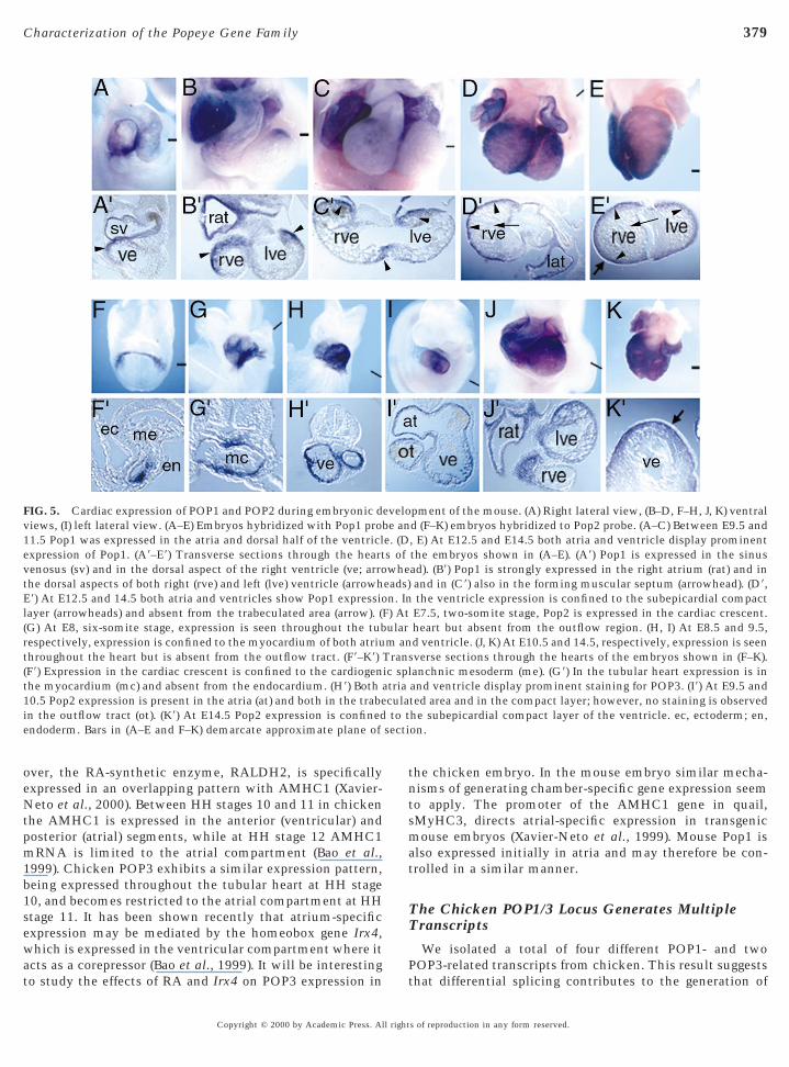
FIG. 5. Cardiac expression of POP1 and POP2 during embryonic diews, (I) left lateral view. (A–E) Embryos hybridized with Pop1 pro1.5 Pop1 was expressed in the atria and dorsal half of the ventriclxpression of Pop1. (A9–E9) Transverse sections through the heart
venosus (sv) and in the dorsal aspect of the right ventricle (ve; arrothe dorsal aspects of both right (rve) and left (lve) ventricle (arrowhE9) At E12.5 and 14.5 both atria and ventricles show Pop1 expressiolayer (arrowheads) and absent from the trabeculated area (arrow). ((G) At E8, six-somite stage, expression is seen throughout the tubrespectively, expression is confined to the myocardium of both atriuthroughout the heart but is absent from the outflow tract. (F9–K9)(F9) Expression in the cardiac crescent is confined to the cardiogenthe myocardium (mc) and absent from the endocardium. (H9) Both a10.5 Pop2 expression is present in the atria (at) and both in the trabein the outflow tract (ot). (K9) At E14.5 Pop2 expression is confinedendoderm. Bars in (A–E and F–K) demarcate approximate plane of
o study the effects of RA and Irx4 on POP3 expression in
Copyright © 2000 by Academic Press. All right
the chicken embryo. In the mouse embryo similar mecha-nisms of generating chamber-specific gene expression seemto apply. The promoter of the AMHC1 gene in quail,sMyHC3, directs atrial-specific expression in transgenicmouse embryos (Xavier-Neto et al., 1999). Mouse Pop1 isalso expressed initially in atria and may therefore be con-trolled in a similar manner.
The Chicken POP1/3 Locus Generates MultipleTranscripts
We isolated a total of four different POP1- and twoPOP3-related transcripts from chicken. This result suggests
pment of the mouse. (A) Right lateral view, (B–D, F–H, J, K) ventrald (F–K) embryos hybridized to Pop2 probe. (A–C) Between E9.5 and, E) At E12.5 and E14.5 both atria and ventricle display prominentthe embryos shown in (A–E). (A9) Pop1 is expressed in the sinusad). (B9) Pop1 is strongly expressed in the right atrium (rat) and inand in (C9) also in the forming muscular septum (arrowhead). (D9,the ventricle expression is confined to the subepicardial compactE7.5, two-somite stage, Pop2 is expressed in the cardiac crescent.heart but absent from the outflow region. (H, I) At E8.5 and 9.5,d ventricle. (J, K) At E10.5 and 14.5, respectively, expression is seenverse sections through the hearts of the embryos shown in (F–K).anchnic mesoderm (me). (G9) In the tubular heart expression is inand ventricle display prominent staining for POP3. (I9) At E9.5 andted area and in the compact layer; however, no staining is observedhe subepicardial compact layer of the ventricle. ec, ectoderm; en,on.
evelobe ane. (Ds ofwhe
eads)n. In
F) Atularm an
Transic spltriacula
that differential splicing contributes to the generation of
s of reproduction in any form reserved.

dptammbuwcPeceAwv
A
380 Andree et al.
POP1 and POP3 isoforms. Most significantly we found afusion transcript that contains POP3 and POP1 sequences.This indicates that chicken POP1 and POP3 transcripts aregenerated from the same gene locus. The fact that thecoding regions of POP1 and POP3 are separated by amaximum of 6 kb in the chicken genome is in line with thisinterpretation (T. Hillemann et al., unpublished observa-tion). In contrast to the situation in chicken we have noevidence for the presence of cDNAs containing both Pop1and Pop3 sequences in the mouse. However, Pop1 and Pop3genes are closely linked on chromosome 10 in the mousegenome. In order to clarify this point in further detail we arecurrently analyzing both the murine and the chicken POP1/POP3 genomic locus.
We found POP1 mRNA only in the myocardium and notin the epicardium. In contrast, David Bader’s group reportedexpression of bves protein, which is identical to Pop1A, inthe proepicardial organ and in migrating epicardial cellsusing antiserum raised against a specific and conservedpeptide (Reese et al., 1999). The bves protein that theydetected on a Western blot of total heart extract has a sizesimilar to the one that we found by in vitro translation inthe presence of microsomes or after transfection of mouse
FIG. 6. Expression of Popeye genes in the adult mouse and humaadult mouse tissues hybridized with Pop1, Pop2, and Pop3 probesloading control the blot was subsequently hybridized to a RPL7 proSp, spleen; St, stomach. (B) Northern blot hybridization of poly(A)1
, atria; RV, right ventricle; LV, left ventricle; Un, nonpregnant utefrom various adult human tissues was sequentially hybridized to hulung; Li, liver; M, skeletal muscle; K, kidney; Pa, pancreas. Arrowcontrol the blot was subsequently hybridized to a b-actin probe.
Pop1 cDNA into COS7 cells. One explanation for the
Copyright © 2000 by Academic Press. All right
ifference in localizing mRNA and protein may be theresence of various splice isoforms. We have evidence forhe existence of cDNAs in the chick encoding both POP1nd POP3 transcripts that lack the transmembrane do-ains. POP proteins may be secreted specifically by cardiacyocytes in the subepicardial compact layer and taken up
y the neighboring epicardial cells. It seems, however,nlikely that the protein synthesized by cardiac myocytesill be detectable only in epicardial cells and not in the
ells producing it. Preliminary data on the expression of aop1–lacZ allele generated by a knock-in approach revealedxclusive expression of the Pop1–lacZ fusion protein inardiac muscle cells. This result does not favor the hypoth-sis of a differential localization of transcript and protein (B.ndree et al., unpublished observation). Clearly, furtherork is required to understand the differences in proteinersus mRNA localization.
Expression of POP Genes in the SubepicardialCompact Layer Suggests a Role of the Epicardiumfor POP Gene Expression
In both chicken and mouse embryos we observed expres-
sue. (A) Northern blot of poly(A)1 RNA from various organs fromowhead marks the positions of the various Pop transcripts. As a, kidney; B, brain; H, heart; Lu, lung; M, skeletal muscle; Li, liver;from various muscle tissues from adult mouse. S, soleus; E, EDL;p, pregnant uterus. (C) A Northern blot containing poly(A)1 RNA
POP1, POP2, and POP3 probes. H, heart; B, brain; Pl, placenta; Lu,s mark the positions of the various POP transcripts. As a loading
n tis. Arrbe. KRNArus; Umanhead
sion of POP genes in the subepicardial compact layer. In
s of reproduction in any form reserved.

eatYucstmmta
pefmtis
D
G
G
H
K
K
K
K
381Characterization of the Popeye Gene Family
chicken the epicardial organ forms at stage 17 and epicar-dial cells migrate over the heart until stage 25 (Ho andShimada, 1978). Strikingly, we found that expression ofchicken POP3 in the ventricle strictly correlates with thepresence of an epicardial layer. Extirpation of the proepicar-dial organ in in vivo or coculture experiments of ventricularmyocardium with proepicardial cells will show whetherPOP3 expression is dependent on epicardium. We alsoobserved expression of mouse Pop1 in the subepicardialcompact layer. In the mouse the epicardium begins toappear in the region of the septum transversum region by E9(Komiyama et al., 1987). From here strands of epicardialcells migrate over the caudal aspect of the ventricular andatrial myocardium between E9.5 and 10.5. Cells derivedfrom the epicardium begin to delaminate into the subepi-cardial zone to form subepicardial mesenchyme betweenE11.5 and E12.5 (Perez-Pomares et al., 1997). These cellsundergo an epithelial-to-mesenchymal transition withinthe epicardium. Mouse Pop1 expression was not detectedbefore E12.5 in the compact layer of the ventricle. Thereforewe suggest that mouse Pop1 gene expression probably relieson signals released by the subepicardial mesenchyme cellswhich form at the time when ventricular Pop1 expressionbecomes apparent.
The epicardium most likely plays an important role ingrowth regulation of the underlying subepicardial compactlayer. Ablation of the genes coding for the cell adhesionmolecules a4-integrin and VCAM-1 results in failure of thepicardium to adhere to the myocardium. In both mutantshypoplastic ventricle was observed, which is probably due
o failure of the compact layer to expand (Kwee et al., 1995;ang et al., 1995). It can be therefore hypothesized that annknown epicardial signal may keep the underlying subepi-ardial myocytes in a proliferating and undifferentiatedtate which is essential to aquire the appropriate wallhickness of the mature ventricle. POP genes may serve asarkers for the differentiation state of the compact layeryocytes. Gain- and loss-of-function experiments will de-
ermine if the POP1 gene has a role in regulating growthnd differentiation of the subepicardial compact layer.In summary, the general conservation of POP gene ex-
ression in adult mouse and man and in mouse and chickenmbryos suggests that Popeye genes perform importantunctions during vertebrate heart development. Experi-
ents to identify the molecular mechanisms underlyinghe dynamic patterns of POP gene expression and their rolesn cardiac development and cardiac physiology, as well as inkeletal muscle, are under way.
ACKNOWLEDGMENTS
We gratefully acknowledge expert technical assistance of KerstinZander and Melanie Ronsiek. Andrea Geling is gratefully acknowl-edged for isolating full-length mouse Pop2 and Pop3 cDNA clones.The Resource Center of the German Human Genome Project at theMax-Planck-Institute for Molecular Genetics is acknowledged for
providing high-density filters and EST clones. This work wasCopyright © 2000 by Academic Press. All right
supported by Sonderforschungsbereich 271, TP A1 (T.B. andH.H.A.) and the German Israeli Foundation (T.B. and G.K.I.).Accession numbers for mouse Pop1, Pop2, and Pop3 are AF204174,AF204175, and AF204176, respectively; for human POP1, POP2,and POP3 are AF204172, AF204173, and AF204171, respectively;and for chicken POP1A through POP1D and POP3 are AF208398,AF208399, AF208400, AF208401, and AF204170, respectively.
REFERENCES
Andree, B., Duprez, D., Vorbusch, B., Arnold, H.-H., and Brand, T.(1998). BMP-2 induces ectopic expression of cardiac lineagemarkers and interferes with somite formation in chicken em-bryos. Mech. Dev. 70, 119–131.
Bao, Z. Z., Bruneau, B. G., Seidman, J. G., Seidman, C. E., andCepko, C. L. (1999). Regulation of chamber-specific gene expres-sion in the developing heart by Irx4. Science 283, 1161–1164.
Chen, Z., Friedrich, G. A., and Soriano, P. (1994). Transcriptionalenhancer factor 1 disruption by a retroviral gene trap leads toheart defects and embryonic lethality in mice. Genes Dev. 8,2293–2301.eHaan, R. (1965). Morphogenesis of the vertebrate heart. In“Organogenesis” (R. DeHaan and H. Ursprung, Eds.), pp. 377–419. Holt, Rinehart & Winston, New York.arcia-Martinez, V., and Schoenwolf, G. (1993). Primitive-streakorigin of the cardiovascular system in avian embryos. Dev. Biol.159, 706–719.assmann, M., Casagranda, F., Orioli, D., Simon, H., Lai, C., Klein,R., and Lemke, G. (1995). Aberrant neural and cardiac develop-ment in mice lacking the ErbB4 neuregulin receptor. Nature 378,390–394.o, E., and Shimada, Y. (1978). Formation of the epicardiumstudied with the scanning electron microscope. Dev. Biol. 66,579–585.
Jaber, M., Koch, W. J., Rockman, H., Smith, B., Bond, R. A., Sulik,K. K., Ross, J., Jr., Lefkowitz, R. J., Caron, M. G., and Giros, B.(1996). Essential role of beta-adrenergic receptor kinase 1 incardiac development and function. Proc. Natl. Acad. Sci. USA93, 12974–12979.
Kastner, P., Grondona, J. M., Mark, M., Gansmuller, A., LeMeur,M., Decimo, D., Vonesch, J. L., Dolle, P., and Chambon, P. (1994).Genetic analysis of RXR alpha developmental function: Conver-gence of RXR and RAR signaling pathways in heart and eyemorphogenesis. Cell 78, 987–1003.
omiyama, M., Ito, K., and Shimada, Y. (1987). Origin and devel-opment of the epicardium in the mouse embryo. Anat. Embryol.176, 183–189.ostetskii, I., Jiang, Y., Kostetskaia, E., Yuan, S., Evans, T., andZile, M. (1999). Retinoid signaling required for normal heartdevelopment regulates GATA-4 in a pathway distinct fromcardiomyocyte differentiation. Dev. Biol. 206, 206–218.reidberg, J. A., Sariola, H., Loring, J. M., Maeda, M., Pelletier, J.,Housman, D., and Jaenisch, R. (1993). WT-1 is required for earlykidney development. Cell 74, 679–691.
wee, L., Baldwin, H. S., Shen, H. M., Stewart, C. L., Buck, C.,Buck, C. A., and Labow, M. A. (1995). Defective development ofthe embryonic and extraembryonic circulatory systems in vas-cular cell adhesion molecule (VCAM-1) deficient mice. Develop-
ment 121, 489–503.s of reproduction in any form reserved.

L
L
M
M
N
N
P
R
R
R
S
S
S
S
V
X
Y
Y
Y
382 Andree et al.
LeCouter, J. E., Kablar, B., Whyte, P. F., Ying, C., and Rudnicki,M. A. (1998). Strain-dependent embryonic lethality in micelacking the retinoblastoma-related p130 gene. Development 125,4669–4679.
ee, K.-F., Simon, H., Chen, H., Bates, B., Hung, M.-C., and Hauser,C. (1995). Requirement for neuregulin receptor erbB2 in neuraland cardiac development. Nature 378, 394–398.
yons, G. E., Schiaffino, S., Sassoon, S., Barton, P., and Bucking-ham, M. (1990). Developmental regulation of myosin gene ex-pression in normal mouse cardiac muscle. J. Cell Biol. 111,2427–2436.eyer, D., and Birchmeier, C. (1995). Multiple essential functionsof neuregulin in development. Nature 378, 386–390.oens, C. B., Stanton, B. R., Parada, L. F., and Rossant, J. (1993).Defects in heart and lung development in compound heterozy-gotes for two different targeted mutations at the N-myc locus.Development 119, 485–499.akaki, K., and Kenhisa, M. (1992). A knowledge base for predict-ing protein localization sites in eukaryotic cells. Genomics 14,897–911.iederreither, K., Subbarayan, V., Dolle, P., and Chambon, P.(1999). Embryonic retinoic acid synthesis is essential for earlymouse post-implantation development. Nat. Genet. 21, 444–448.
erez-Pomares, J. M., Macias, D., Garcia-Garrido, L., and Munoz-Chapuli, R. (1997). Contribution of the primitive epicardium tothe subepicardial mesenchyme in hamster and chick embryos.Dev. Dyn. 210, 96–105.eese, D. E., and Bader, D. M. (1999). Cloning and expression ofhbves, a novel and highly conserved mRNA expressed in thedeveloping and adult heart and skeletal muscle in the human.Mamm. Genome 10, 913–915.eese, D. E., Zavaljevski, M., Streiff, N. L., and Bader, D. (1999).bves: A novel gene expressed during coronary blood vesseldevelopment. Dev. Biol. 209, 159–171.
umyantsev, P. (1991). “Growth and Hyperplasia of CardiacMuscle Cells.” Harwood Academic, London.
chultheiss, T., Burch, J., and Lassar, A. (1997). A role for bonemorphogenetic proteins in the induction of cardiac myogenesis.
Genes Dev. 11, 451–462.Copyright © 2000 by Academic Press. All right
chultheiss, T. M., Xydas, S., and Lassar, A. B. (1995). Induction ofavian cardiac myogenesis by anterior endoderm. Development121, 4203–4214.
onnhammer, E., von Heijne, G., and Krogh, A. (1998). A hiddenMarkov model for predicting transmembrane helices in proteinsequences. In “Proceedings of the Sixth International Conferenceon Intelligent Systems for Molecular Biology” (J. Glasgow, T.Littlejohn, F. Major, R. Lathrop, D. Sankoff, and C. Sensen, Eds.),pp. 175–182. AAAI Press, Menlo Park, CA.
ucov, H. M., Dyson, E., Gumeringer, C. L., Price, J., Chien, K. R.,and Evans, R. M. (1994). RXR alpha mutant mice establish agenetic basis for vitamin A signaling in heart morphogenesis.Genes Dev. 8, 1007–1018.
an Den Ende, J. J., Van Der Burgt, C. J., Jansweijer, M. C., Hamel,B. C., and Brunner, H. G. (1996). Ectrodactyly of lower limbs,congenital heart defect and characteristic facies in four unrelatedDutch patients: A new association. Clin. Dysmorphol. 5, 1–7.avier-Neto, J., Shapiro, M. D., Houghton, L., and Rosenthal, N.(2000). Sequential programs of retinoic acid synthesis in themyocardial and epicardial layers of the developing avian heart.Dev. Biol. 219, 129–141.ang, J. T., Rayburn, H., and Hynes, R. O. (1995). Cell adhesionevents mediated by alpha 4 integrins are essential in placentaland cardiac development. Development 121, 549–560.oshida, K., Taga, T., Saito, M., Suematsu, S., Kumanogoh, A.,Tanaka, T., Fujiwara, H., Hirata, M., Yamagami, T., Nakahata,T., Hirabayashi, T., Yoneda, Y., Tanaka, K., Wang, W. Z., Mori,C., Shiota, K., Yoshida, N., and Kishimoto, T. (1996). Targeteddisruption of gp130, a common signal transducer for the inter-leukin 6 family of cytokines, leads to myocardial and hemato-logical disorders. Proc. Natl. Acad. Sci. USA 93, 407–411.utzey, K. E., Rhee, J. T., and Bader, D. (1994). Expression of theatrial-specific myosin heavy chain AMHC1 and the establish-ment of anteroposterior polarity in the developing chicken heart.Development 120, 871–883.
Received for publication January 6, 2000Revised April 14, 2000
Accepted April 14, 2000
s of reproduction in any form reserved.