Iron(III) complexes of phenolate ligands as models for ...
15
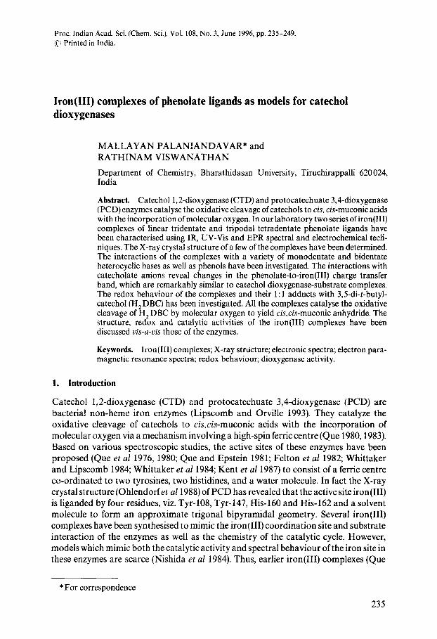
Proc. Indian Acad. Sci.(Chem. Sci.),Vol. 108,No. 3, June 1996,pp. 235-249. ~ Printed in India. Iron(Ill) complexes of phenolate ligands as models for catechol dioxygenases MALLAYAN PALANIANDAVAR* and RATHINAM VISWANATHAN Department of Chemistry, Bharathidasan University, Tiruchirappalli 620024, India Abstract. Catechol 1,2-dioxygenase (CTD) and protocatechuate 3,4-dioxygenase (PCD) enzymes catalyse the oxidative cleavage of catechols to cis, cis-muconic acids with the incorporation of molecular oxygen. In our laboratory two series ofiron(III) complexes of linear tridentate and tripodal tetradentate phenolate ligands have been characterised using IR, UV-Vis and EPR spectral and electrochemical tecfi- niques. The X-ray crystal structure of a few of the complexes have been determined The interactions of the complexes with a variety of monodentate and bidentate heterocyclic bases as well as phenols have been investigated. The interactions with catecholate anions reveal changes in the phenolate-to-iron(III) charge transfer band, which are remarkably similar to catechol dioxygenase-substrate complexes. The redox behaviour of the complexes and their 1:1 adducts with 3,5-di-t-butyl- catechol (H2DBC) has been investigated. All the complexes catalyse the oxidative cleavage of H 2DBC by molecular oxygen to yield cis, cis-muconic anhydride. The structure, redox and catalytic activities of the iron(III) complexes have been discussed vis-a-vis those of the enzymes. Keywords. Iron(Ill) complexes; X-ray structure; electronic spectra; electron para- magnetic resonance spectra; redox behaviour; dioxygenase activity. 1. Introduction Catechol 1,2-dioxygenase (CTD) and protocatechuate 3,4-dioxygenase (PCD) are bacterial non-heme iron enzymes (Lipscomb and Orville 1993). They catalyze the oxidative cleavage of catechols to cis,cis-muconic acids with the incorporation of molecular oxygen via a mechanism involving a high-spin ferric centre (Que 1980,1983). Based on various spectroscopic studies, the active sites of these enzymes have been proposed (Queet al 1976, 1980; Que and Epstein 1981; Felton et al 1982; Whittaker and Lipscomb 1984; Whittaker et al 1984; Kent et al 1987) to consist of a ferric centre co-ordinated to two tyrosines, two histidines, and a water molecule. In fact the X-ray crystal structure (Ohlendorfet a11988) of PCD has revealed that the active site iron(III) is liganded by four residues, viz. Tyr-108, Tyr-147, His-160 and His-162 and a solvent molecule to form an approximate trigonal bipyramidal geometry. Several iron(Ill) complexes have been synthesised to mimic the iron(Ill) coordination site and substrate interaction of the enzymes as well as the chemistry of the catalytic cycle. However, models which mimic both the catalytic activity and spectral behaviour of the iron site in these enzymes are scarce (Nishida et al 1984). Thus, earlier iron(Ill) complexes (Que * For correspondence 235
-
Upload
khangminh22 -
Category
Documents
-
view
0 -
download
0
Transcript of Iron(III) complexes of phenolate ligands as models for ...
Proc. Indian Acad. Sci. (Chem. Sci.), Vol. 108, No. 3, June 1996, pp. 235-249. ~ Printed in India.
Iron(Ill) complexes of phenolate ligands as models for catechol dioxygenases
MALLAYAN PALANIANDAVAR* and RATHINAM VISWANATHAN
Department of Chemistry, Bharathidasan University, Tiruchirappalli 620024, India
Abstract. Catechol 1,2-dioxygenase (CTD) and protocatechuate 3,4-dioxygenase (PCD) enzymes catalyse the oxidative cleavage of catechols to cis, cis-muconic acids with the incorporation of molecular oxygen. In our laboratory two series ofiron(III) complexes of linear tridentate and tripodal tetradentate phenolate ligands have been characterised using IR, UV-Vis and EPR spectral and electrochemical tecfi- niques. The X-ray crystal structure of a few of the complexes have been determined The interactions of the complexes with a variety of monodentate and bidentate heterocyclic bases as well as phenols have been investigated. The interactions with catecholate anions reveal changes in the phenolate-to-iron(III) charge transfer band, which are remarkably similar to catechol dioxygenase-substrate complexes. The redox behaviour of the complexes and their 1:1 adducts with 3,5-di-t-butyl- catechol (H 2 DBC) has been investigated. All the complexes catalyse the oxidative cleavage of H 2 DBC by molecular oxygen to yield cis, cis-muconic anhydride. The structure, redox and catalytic activities of the iron(III) complexes have been discussed vis-a-vis those of the enzymes.
Keywords. Iron(Ill) complexes; X-ray structure; electronic spectra; electron para- magnetic resonance spectra; redox behaviour; dioxygenase activity.
1. Introduction
Catechol 1,2-dioxygenase (CTD) and protocatechuate 3,4-dioxygenase (PCD) are bacterial non-heme iron enzymes (Lipscomb and Orville 1993). They catalyze the oxidative cleavage of catechols to cis,cis-muconic acids with the incorporation of molecular oxygen via a mechanism involving a high-spin ferric centre (Que 1980,1983). Based on various spectroscopic studies, the active sites of these enzymes have been proposed (Quee t al 1976, 1980; Que and Epstein 1981; Felton et al 1982; Whittaker and Lipscomb 1984; Whittaker et al 1984; Kent et al 1987) to consist of a ferric centre co-ordinated to two tyrosines, two histidines, and a water molecule. In fact the X-ray crystal structure (Ohlendorfet a11988) of PCD has revealed that the active site iron(III) is liganded by four residues, viz. Tyr-108, Tyr-147, His-160 and His-162 and a solvent molecule to form an approximate trigonal bipyramidal geometry. Several iron(Ill) complexes have been synthesised to mimic the iron(Ill) coordination site and substrate interaction of the enzymes as well as the chemistry of the catalytic cycle. However, models which mimic both the catalytic activity and spectral behaviour of the iron site in these enzymes are scarce (Nishida et al 1984). Thus, earlier iron(Ill) complexes (Que
* For correspondence
235
236 Mallayan Palaniandavar and Rathinam Viswanathan
Py : Im= J
Bzlm =
H
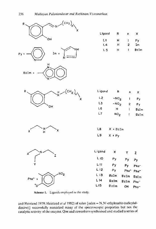
Ligond R n X
L t H I Py
L4 H 2 Im
L 5 H I Bzlm
R
×
v -OH
LJgand R n X
L2 -NO 2 I Py
L3 -NO 2 2 Py
L6 H I Bz.Em
L7 NO 2 I Bzlrn
X H L8 X = Bz lm
L9 X =Py
Y
"0" V
Scheme 1. Ligands employed in the study.
Ligond X Y Z
L I0 py py py
L I I PY PY Pho-
L I 2 PY Pho- Pho-
L 13 BzIm Bzrrn BzIm
L I 4 BzIm BzIm Pho-
L I5 Bz Im OH Pho-
and Heistand 1979; Heistand et al 1982) of salen [salen = N,N'-ethylenebis-(salicylal- dimine)] successfully mimicked many of the spectroscopic properties but not the catalytic activity of the enzyme. Que and coworkers synthesised and studied a series of
Iron-phenolate complexes as models for catechol dioxygenases 237
[Fe(L)DBC] complexes (Que et a11987; Cox and Que 1988; Cox et a11988), where L is a tetradentate tripodal ligand like TPA [TPA = tris(pyrid-2-yl- methyl)amine] or NTA (NTA = nitrilotriacetic acid) or a NTA analogue with one or two carboxylate pendants and H2DBC is 3,5-di-t-butylcatechol.
Only two of the tripodal ligand complexes (Lll, scheme 1), recently reported (Nishida et al 1984; Cox and Que 1988; Cox et al 1988) to effect oxidative cleavage, contain a coordinated phenolate and no tripodal analogue reported so far contains one/two phenolic hydroxyl groups as well as imidazole nitrogen donor(s) though dioxygenases contain two tyrosine and two imidazole functions coordinated to iron(III). Thus clearly no model reported so far possesses all the features of the dioxygenase active sites. The present paper describes our successful attempt to synthesise and study Fe(III) complexes of systematically varied tri-(Viswanathan and Palaniandavar 1995) and tetradentate tripodal ligands (Viswanathan et al 1996) containing one or two phenolate functions along with benzimidazole (bzim) or pyridine (py) moieties (scheme 1), to closely mimic the enzyme active site. The tripodal ligand systems designed for the present study are expected to generate the non-heme iron environment (Ohlendorf et al 1988) of the enzymes and encourage asymmetric coor- dination proposed (Whittaker and Lipscomb 1984) to be essential for the cleavage of H2DBC. Our effort is directed towards investigating the phenolate-to-Fe(III) CT spectra, redox and reactivity of the complexes. Our study is expected to afford improved understanding of the structure-spectra and structure-function correlations for the active site of the enzymes, verify the validity of the proposed oxidative cleavage mechanism for phenolate complexes and thereby provide an assessment of the import- ance of tyrosinase coordination in the enzyme.
2. Experimental
The synthesis and characterisation of the present iron(III) complexes and the experimental methods employed for studying them have been described previously (Viswanathan and Palaniandavar 1995; Viswanathan et al 1996).
3. Results and discussion
Based on the results of elemental analysis the formulae of the Fe(llI) complexes have been assigned. The present ligands provide a reasonable analogue to histidine and tyrosinate coordination in CTD enzyme via the bzim and phenolate moieties. Further, the tridentate ligands are varied by the saturation of the imine function to disrupt the conjugation between the functionalities and to allow more flexibility in chelation to the metal ion. The tetradentate tripodal ligands based on trimethyl amine (scheme 1) with the pendant functionalities varying from py to bzim to phenolate have been also synthesised to provide a systematic variation of the ligand Lewis basicity and charge while retaining similar coordination geometries in ternary iron(Ill) complexes with catechols. The bulky bzim ring(s) seems to be a good choice to offer steric hindrance to the added ligands or substrates as in proteins so as to closely approximate the active site in enzymes.
3.1 Structures of iron complexes
All the iron(Ill) complexes have magnetic moments in the range 5-8-5.9 BM at room temperaure, consistent with a high-spin ferric centre with five unpaired electrons. In the
238 Mallayan Palaniandavar and Rathinam Yiswanathan
EL2 EL EL1 C E 2A ~ EL
Fo
Eli El2 C C ~,A 5A
a
E5
0 Cs~ C4
27 ¢ ~ C12 C;~
CL!
0 24
b
C5 C
~ { ~ t N C 4 ~ C~
Ca
N6 E2 ~ , 1~
c c ilj~~ j 20
E ELi C 23 22
E8
12 r_s clo N~cli.~Ci2
-"e C 16 C 15
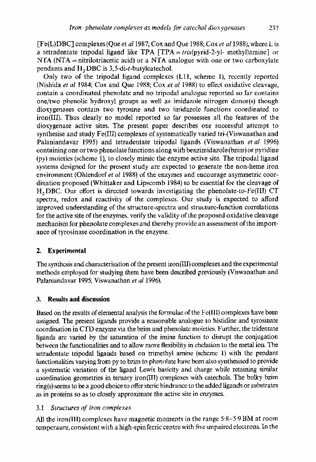
Figure 1. X-ray crystal structure of Fe(L9)C13 (a), Fe(L11)C12 (b) and Fe(L13)C13 (c). Fe(L9)CI3. contains two crystallographically independent molecules present in the asymmetric unit.
Iron-phenolate complexes as models for catechol dioxyoenases 239
X-ray crystal structures of Fe(L16)C13 (Adams et al 1990) [Lt6 = bis(benzimidazol-2- ylmethyl)methylamine] and Fe(L9)C13 (Viswanathan etal 1996) [-L9 =dipicoly- lamine] (figure 1), the homologue and pyridine analogue respectively of Fe(L8)CI 3, all the three chloride ions are coordinated. In the crystal structures of Fe(L11)C13 and [Fe(L 13)C12 ] CI (Viswanathan et al 1996), iron(Ill) is octahedrally co-ordinated (fig- ure 1). Spectral studies suggest similar structures for the other Fe(III) complexes. The chloride ions are replaced by solvent molecules or polar groups of the neighbouring ligand molecules in solution, as revealed by conductometric studies.
As evident from the above crystal structures, two cis coordination sites are open in all the present complexes for the coordination of two monodentate or one bidentate base molecule like H 2 DBC and the complexes are convenient for the investigation of the effect of adduct formation on the spectra, In fact, the hyperfine broadening observed from O ~ 7-enriched water for the 4-HBA-PCD complex (Whittaker and Lipscomb 1984; Orville and Lipscomb 1989) (4-HBA = 4-hydroxybenzoate) has shown that at least two coordina- tion sites of Fe(III) appear adjacent and accessible to exogenous ligands.
3.2 Electronic absorption spectra
The electronic spectra of all the present iron(III) phenolate complexes in methanol solution (table 1) exhibit two bands in the near UV (~ 410 sh, ~ 330 nm) and one in the visible (550-470 nm) regions. The visible band which is absent in complexes of L8, L10 and L13, which lack a phenolate function may be assigned (Cassella et al 1987) to phenolate ( rq)~ Fe m [d~(dx~)] charge transfer transition. The 400 nm band observed for all the complexes may originate (Cassella et al 1987) from the filled d-orbitals of iron(III) to the antibonding orbitals of the phenolic residue. The conjugated imine r~ ~ re* transition in the salicylaldimine residue is observed as an intense band around 330 nm.
The position and intensity of the visible band shows a remarkable dependence on the nature of the ligand. The increasing orders of its energy:Fe(L4)< Fe(L5)< Fe(L1) < Fe(L3) < Fe(L6) < Fe(L7) < Fe(L2); Fe(Lll) < Fe(L12) ~< Fe(L14) < Fe (L15) re- present the decreasing order of Lewis acidity of the ferric centre. The iron d-orbital energies are raised (Queet a11987) by the negative charge built on Fe(III) by the higher basicity of imine compared to - N H - nitrogen and of im compared to bzim, and bythe replacement of py/bzim by the second phenolate group. Further, the electron with- drawing NO z group would be expected to decrease the band energy but an increase is observed for Fe(L2)C1 z as compared to Fe(L1)C12. MOPAC calculations reveal (Viswanathan and Palaniandavar 1995) that the observed blue shift isdue to lowering in energy of the ligand orbitals on the introduction of p-NO z group.
Further, the absorptivities of the visible bands of the present complexes vary widely and do not show any regular trend. All these observations show that band positions as well as intensities are very sensitive to the iron environment; this is reminiscent of native CTD and PCD enzymes (Stewart 1989).
3.3 Adduct formation
On addition of secondary ligand molecules the position of the visible band shifts generally to higher energy with modest changes in intensity. For tridentate ligand complexes addition of N-methylimidazole (mim) shifts the visible band to higher energies with decrease in absorptivity for almost all the complexes. A similar but lesser

240 Mallayan Palaniandavar and Rathinam Viswanathan
Table 1. PhO- --* Fe(III) charge transfer spectral ' and redox potential (volts) data for Fe(III) tri- and tetradentate tripodal ligand complexes.
Complex
"~max (~:max) E1/2(V) Complex DBC adduct Fe(III)/Fe(II) DBSQ-/H2DBC Yield
Fe(L1)C12- H 20 543(400) 640(100) - 0.169 - 0.116 19.8 528sh(200)
Fe(L1)C12.H20 478(450) 693(260) - 0.330 - 0.244 42.4 415sh(1430)
Fe(L3)CI2.H20 510(300) - - - 0"260 - 0-277 - - Fe(L4)CI2"H20 580(1730) - - 0-131 0"139 - - Fe(L5)CI2 .H 20 554(1630) 567(3990) - 0"177 -- 0.121 37.0 Fe(L6)CI2.H20 508(1540) 575(1050) -0-887 -0.505 24.8 Fe(L7)Cl2.H20 503(1760) 650sh(1710) -0"319 -0.246 23.5 Fe(L8)CI 3 436sh(540) 637(440) 0.I01 0-066 13-6
450(1210) Fe(L10)C13 - - 578 b 0"078 - - 41.0
888 Fe(L11)C1 z 5 5 0 ( 1 6 5 0 ) 790(490) - 0"101 0-008 12.0
490sh(670) Fe(L12)C1.H 2 O 493(630) 850-700 c - 0"301 - 0.210 56.0
400sh(470) Fe(L13)C1 a - - 5 4 8 b - 0-002 -- 0"054 18'0
842 Fe(L14)C12 500(880) 768(360) - 0-130 - 0-130 59-0
492(510) Fe(L15)C1.H20 492(3120) 600-800 c - 0"301 - 0.240 50.0
Wiswanathan and Palaniandavar (1995); Viswanathan et a! (1996) bCox et al (1988) every broad band
shift is observed for bzim; this is expected as bzim is less basic and bulkier than mim. The addition of py also decreases the 2 ~ but enhances the absorptivity of compounds of bzim-based ligands. There is a blue shift with moderate to tremendous enhancement in absorptivity for the addition of bidentate heterocyclic bases (figure 2), as expected; however, for 2, 9-dimethyl- 1,10-phenanthroline there is only a slight enhancement, possibly because of steric hindrance from its methyl groups. The binding of phenolate (phO-) effects a large decrease in 2 r~ with increased or decreased absorptivity and for monoanionic H D B C - a visible band around 650nm with very high intensity corresponding to H D B C - ~ Fe(III) (drc) CT is observed. In contrast, for catecholate (HCAT-) no such band is discernibie and the ligand to metal charge transfer band expected for H C A T - ~ Fe(III) interaction may overlap with the visible band of the original complexes. The effect on the visible band of adding dianionic CAT 2 - is the same as that for C A T H - but the intensity is enhanced further. In contrast, the effect of addition of dianionic DBC 2- is to increase the 2 ~ appreciably with a decrease in intensity. Further, on the addition of even neutral H 2 DBC (figure 2) rather than H 2 CAT the charge transfer bands are exhibited, of course, with relatively low absorptivity. The absorptivity is enhanced on the addition of Et 3 N due to the deprotonation of H 2 DBC. This reveals the spontaneous deprotonation of H 2 DBC rather than H2CAT, to bind strongly to Lewis acidic Fe(III).
Iron-phenolate complexes as models for catechol dioxygenases 241
].00
c
0 200
c
, i t m i t
300 q00 500 EO0 700 800 850 Waveleroth (nm)
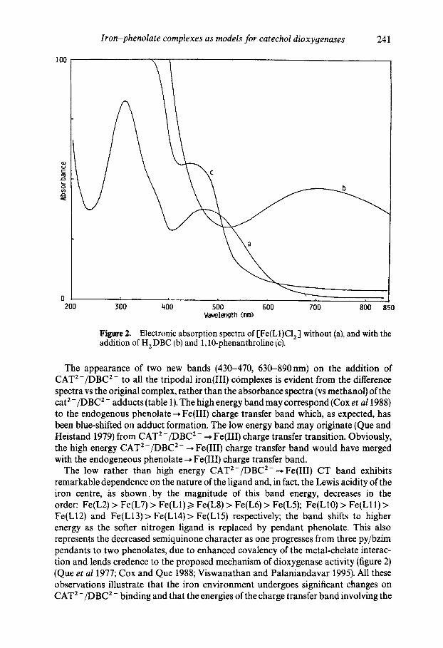
Figare 2. Electronic absorption spectra of [Fe(L1)C12 ] without (a), and with the addition of H 2 DBC (b) and 1,10-phenanthroline (c).
The appearance of two new bands (430-470, 630-890nm) on the addition of CAT2-/DBC 2- to all the tripodal iron(III) complexes is evident from the difference spectra vs the original complex, rather than the absorbance spectra (vs methanol) of the cat 2 - /DBC 2 - adducts (table 1). The high energy band may correspond (Cox et a11988) to the endogenous phenolate ~ Fe(III) charge transfer band which, as expected, has been blue-shifted on adduct formation. The low energy band may originate (Que and Heistand 1979) from CAT 2-/DBC 2- ~ Fe(IIl) charge transfer transition. Obviously, the high energy CAT 2-/DBC 2- ~ Fe(III) charge transfer band would have merged with the endogeneous phenolate--. Fe(IlI) charge transfer band.
The low rather than high energy CAT2-/DBC 2- ~Fe(III) CT band exhibits remarkable dependence on the nature of the ligand and, in fact, the Lewis acidity of the iron centre, /~s shown, by the magnitude of this band energy, decreases in the order: Fe(L2) > Fe(L7) > Fe(L1) >/Fe(L8) > Fe(L6) > Fe(L5); Fe(L10) > Fe(L11) > Fe(L12) and Fe(L13)> Fe(L14)> Fe(L15) respectively; the band shifts to higher energy as the softer nitrogen ligand is replaced by pendant phenolate. This also represents the decreased semiquinone character as one progresses from three py/bzim pendants to two phenolates, due to enhanced covalency of the metal-chelate interac- tion and lends credence to the proposed mechanism of dioxygenase activity (figure 2) (Queet al 1977; Cox and Que 1988; Viswanathan and Palaniandavar 1995). All these observations illustrate that the iron environment undergoes significant changes on CAT 2-/DBC 2- binding and that the energies of the charge transfer band involving the
18.00
1 4 . 0 0
I0.00
~" 6.000
2.00qO
-2.000
-6 .000
- I 0 . 0 0
0.450 -0 -250 I i 1 I I I
0.350 0-250 0,1,~0 0,050 - 0 " 0 5 0 -0 .150
E(V)
242 Mallayan Palaniandavar and Rathinam Viswanathan
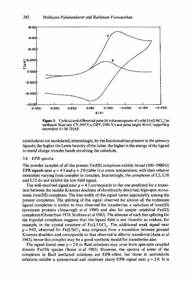
Figure 3. Cyclic (a) and differential pulse (b) voltammograms of 1 mM [Fe(L9)CI3] in methanol. Scan rate: CV, 1~05 V/s, DPV, if001 V/s and pulse height 50 mV, supporting electrolyte: 0-1 M THAP.
catecholates are modulated, interestingly, by the functionalities present in the primary ligands; the higher the Lewis basicity of the latter, the higher is the energy of the ligand to metal charge transfer bands involving the catechols.
3.4 EPR spectra
The powder samples of all the present Fe(III) complexes exhibit broad (100-5900 G) EPR signals near O = 4.3 and g = 2"0 (table 1) at room temperature, with their relative intensities varying from complex to complex. Interestingly, the complexes of L2, L10 and LI2 do not exhibit the low field signal.
The well-resolved signal near 9 = 4.3 corresponds to the one predicted for a transi- tion between the middle Kramers doublets of rhombically distorted, high-spin mono- meric iron(III) complexes. The line-width of this signal varies appreciably among the present complexes. The splitting of this signal observed for almost all the tridentate ligand complexes is similar to that observed for transferrins, a sub-class of iron(III) tyrosinate proteins (Ainscough etal 1980) and also for simple octahdral Fe(III) complexes (Oosterhuis 1974; Scullane et a11982). The absence of such fine splitting for the tidpodal complexes suggests that the ligand field is not rhombic as evident, for example, in the crystal structure of Fe(Lll)CI 2. The additional weak signal, near O= 9.05, observed for Fe(L5)C12 may originate from a transition between ground Kramers doublets and corresponds to that observed in diferric transferrin (Aasa et al 1963); hence this complex may be a good synthetic model for transferrins also.
The signal found near 9 = 2.0 in fluid solutions may arise from spin-spin coupled dimeric Fe(III) species (Borer et al 1983). However, the spectra of some of the complexes in fluid methanol solutions are EPR-silbnt, but those in acetonitrile solutions exhibit a symmetrical and relatively sharp EPR signal near O = 2-0. It is
I 0 m
0 . 0 0 0
6 . 0 0 0
4.0013
2 . 0 0 0
- 2 . 0 0 0
3.000
I i i I I
2 " 0 0 0 1 -000 O ' O 0 0 - I ' O O O - 2 ' O O O - 3 - 0 0 0
E x I0 - '
Iron-phenolate complexes as models for catechol dioxygenases 243
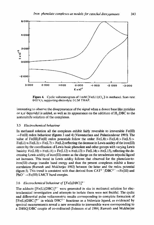
Figure 4. Cyclic voltammogram of 1 mM [Fe(L11)Cla ] in methanol. Scan rate: 0"05 V/s, supporting electrolyte: 0-1 M THAP.
interesting to observe the disappearance of the signal when a donor base like pyridine or ~,ct'-bipyridyl is added, as well as its appearance on .the addition of H2DBC to the acetonitrile solution of the complexes.
3.5 Electrochemical behaviour
In methanol solution all the complexes exhibit fairly reversible to irreversible Fe(III) Fe(II) redox behaviour (figures 3 and 4) (Viswanathan and Palaniandavar 1995). The
value of Fe(III)/Fe(II) redox potentials follow the order: Fe(L8) > Fe(IA) > Fe(L5) > Fe(L1) ~ Fe(L3) > Fe(L7) > Fe(L2) reflecting the decrease in Lewis acidity of the iron(Ill) centre by the coordination of Lewis basic phenolate and other groups with varying Lewis basicity: Fe(L10) > Fe(Lll) > Fe(L12) ~ Fe(L13) > Fe(L14) > Fe(L15), reflecting the de- creasing Lewis acidity ofiron(III) centre as the charge on the tetradentate tripodal ligand set increases. This trend in Lewis acidity follows that observed for the phenolate-to- iron(Ill) charge transfer band energy and thus the present complexes exhibit a linear correlation (Ramesh and Mukherjee 1992) between the latter and the redox potential (figure 5). This trend is consistent with that derived from CAT 2-/DBC 2- ~ Fe(III) and PhO- ~ Fe(III) LMCT band energies.
3.6 Electrochemical behaviour of [FeL(DBC)]"-
The adducts [Fe(L)(DBC)]"- were generated in situ in methanol solution for elec- trochemical investigation and attempts to isolate them were not fruitful. The cyclic and differential pulse voltammetric results corresponding to complete formation of [Fe(L)(DBC)]"- in which DBC 2- functions as a bidentate ligand, as evidenced by spectral measurements reveal a new reversible to irreversible wave corresponding to a DBSQ/DBC couple of co-ordinated (Johnson et al 1984; Ramesh and Mukherjee
244 Mallayan Palaniandavar and Rathinam Viswanathan
04 "
I , I
0.20
0.10
0"00
- 0 . 1 0
-0.20
- 0 . 3 0
- 0 . 4 0
- 0 . 5 0 16,00
I z J I ~ I
1"/.00 18.00 19.00 20-00 21-00 22,00
a)ma x x 10 - 3 ¢m - I
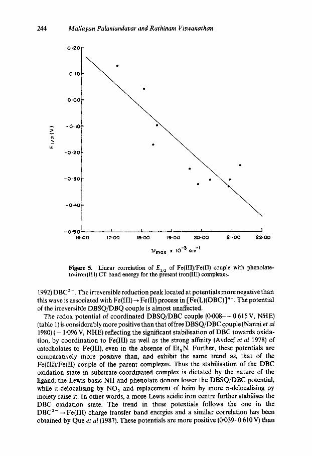
Figure 5. Linear correlation of E~/2 of Fe(III)/Fe(II) couple with phenolate- to-iron(Ill) CT band energy for the present iron(III) complexes.
1992) DBC 2 -. The irreversible reduction peak located at potentials more negative than this wave is associated with Fe(III)-* Fe(II) process in [Fe(L)(DBC)]"-. The potential of the irreversible DBSQ/DBQ couple is almost unaffected.
The redox potential of coordinated DBSQ/DBC couple (0-008--0.615 V, NHE) (table 1) is considerably more positive than that of free DBSQ/DBC couple (Nanni et al 1980) ( - 1.096 V, NHE) reflecting the significant stabilisation of DBC towards oxida- tion, by coordination to Fe(III) as well as the strong affinity (Avdeef et al 1978) of catecholates to Fe(III), even in the absence of Et3N. Further, these potentials are comparatively more positive than, and exhibit the same trend as, that of the Fe(III)/Fe(II) couple of the parent complexes. Thus the stabilisation of the DBC oxidation state in substrate-coordinated complex is dictated by the nature of the ligand; the Lewis basic NH and phenolate donors lower the DBSQ/DBC potential, while n-delocalising by NO 2 and replacement of bzim by more n-delocalising py moiety raise it. In other words, a more Lewis acidic iron centre further stabilises the DBC oxidation state. The trend in these potentials follows the one in the DBC 2---* Fe(III) charge transfer band energies and a similar correlation has been obtained by Queet al (1987). These potentials are more positive (0.039-0.610 V) than
Iron-phenolate complexes as models for catechol dioxygenases 245
those for [Fe(salen)(DBC)]- (-0-179V, NHE) (Lauffer etal 1981) and for [Fe(NTA)(DBC)] - ( - 0-024 V, NHE); while the former is unreactive (White et a11984) the latter is reactive towards dioxygen. This suggests that all the present compounds should show cleavage activity. Further, as electron transfer from chelated catecholate complex to dioxygen (Que et al 1977) is increasingly thermodynamically favourable in decreasing order of E~/2's of DBSQ/DBC couple: Fe(L4)> Fe(L1)> Fe(L5)> Fe(L2) ~ Fe(L7) > Fe(L6), the catalytic activity and hence the yield should decrease in this order (see below). For tripodal ligand complexes also a similar trend in the potentials is observed - decrease with increase in the number of coordinated phenolate groups and on replacing the more effectively n-back-bonding py by a bzim group. Similar variations in redox potentials have been associated with differing Lewis acidity of ferric centres in [Fe(L)DBC] complexes (Whittaker and Lipscomb 1984; Vis- wanathan and Palaniandavar 1995).
The E1/2's of the Fe(III)/Fe(II)-couple for almost all the DBC adducts become negative (100-500 mV) as compared to the parent complexes, suggesting (Johnson et al 1984) that DBC 2- strongly chelates to Fe(III) and that the Lewis acidity of the iron centre decreases on substrate binding. This observation is in support of the suggestion that the Fe(III) centre in the dioxygenases not only participates in the activation of the substrate but also facilitates the latter stages of the reaction. Further, the El~ 2 of the substrate coordinated Fe(III) centre reflects the reducibility of the latter to form the oxide anion (Que et al 1977; Ainscough et al 1980).
3.7 Catalytic activity of model compounds
The oxidative cleavage ofH 2 DBC was observed when it was mixed (Nishida et a11984) with the present iron(III) compounds in nitromethane solution in a molar ratio of 50:1 and kept for 4 days. The complexes act as catalysts in this reaction, because the amounts of product obtained (table 1) were more than that of the complex used.
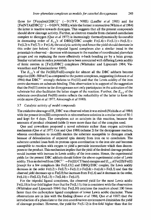
Que and coworkers proposed a novel substrate rather than oxygen activation mechanism (Que et al 1977; Cox and Que 1988) (scheme 2) for the dioxygenase reactiol~ wherein coordination to iron(Ill) renders the substrate susceptible to dioxygen attack because of delocalisation of unpaired spin density from iron on to catecholate. The. substrate catechol loses both its protons upon coordination to the iron site and becomes susceptible to reaction with oxygen to yield a peroxide intermediate which then decom- poses to the product. This mechanism implies that the yield of the desired cleavage product would increa~ with increase in Lewis acidity of the iron centre. Hence, the experimental yields for the present DBC adducts should follow the above experimental order of Lewis acidity. Thus as derived from DBC 2- --+ Fe(III) CT band energies and E~/2 of Fe(III)/Fe(II) [except for a few complexes like Fe(L13)] and DBSQ/DBC couples, the Lewis acidity decreases in the order Fe(L2) > Fe(L7) > Fe(L1) ~> Fe(L8) > Fe(L6) > Fe(L5) and thus the observed yield decreases up to Fe(L8) but increases from Fe(L6) and it decreases in the order, Fe(Lll) > Fe(L12); Fe(L13) > Fe(L14) > Fe(L15).
For the tripodal ligand complexes, the observed yield for the most Lewis acidic Fe(L10) is four-fold higher than that for Fe(L11); this is consistent with the observation (Whittaker and Lipscomb 1984) that Fe(L10) catalyzes the reaction about 100 times faster than the carboxylate ligand complexes of lower Lewis acidic character. These observations are in line with the finding (Russo et al 1986; Que et al 1987) that the introduction of a phenolate to the iron coordination environment diminishes the yield of cleavage product. However, the yield for Fe(L12) is five-fold higher than that for
246 Mallayan Palaniandavar and Rathinam Viswanathan
OH I Fe 3+ +
t - Bu
b--
t - B u
t - B u
H2° l
0 2 -
~e 5 +
t - B u
4- 0 /~ t -Bu
t- Bu O ~ 0
Fe5 + ~0 ~ ~ t - Bu
Scheme 2.
IL t - B u
Fe3+
_ t - B u
O C% o
0 f ~ \t-Bu
t - B u
Fe $+ O ~ t - Bu
Substrate activation mechanism proposed for dioxygenase enzymes.
Fe(LI1). We suggest that the rate determining step, viz. dissociation of the Fe-O (product) bond is facilitated by the very much decreased Lewis acidity of the iron centre, owing to the two coordinated phenolates. This is supported by the observation thatFe(L15) with two coordination phenolates, like Fe(L12), has almost the same yield. Thus the inclusion of two phenolate groups in the coordination sphere results in a Lewis acidity lower than that, for example, with all-N ligation but appears to be sufficiently Lewis acidic to catalyze the reaction at an adequate rate, as in the enzyme with an active site containing two coordinated phenolates. This is in line with the observation of lower yields for the iron(Ill) complexes of the above tridentate mono phenolates. The lower yields may be due to the ~till Lewis acidic five-coordinate DBC 2- adducts, which do not encourage dissociation of Fe-O(DBC) bonds and hence the semiquinone character needed for the formation of the peroxide intermediate.
Iron-phenolate complexes as models for catechol dioxygenases 247
As expected, Fe(L14) shows higher reactivity than Fe(L15). However, the turnover for Fe(L13), in spite of its Lewis acidity being higher than those for Fe(L14) and Fe(L15) and almost the same as that for Fe(L10), is low. This may be ascribed to not only the absence of phenolate coordination to sufficiently decrease the Lewis acidity as illustrated above, but also to the bulky bzim moieties which might offer steric hindrance to substrate binding. This is interesting in view of the prediction from model building (Lipscomb and Orville 1993) based on the structure of the substrate-free PCD enzyme that a single, fairly constrained binding orientation as well as a large rotation of the substrate is essential. However, Fe(L14) shows higher reactivity than Fe(LI 1) and Fe(L15), both involving comparatively low steric constraints. It appears that the bzim moiety may be involved in stabilising the reactive intermediate by charge delocalisa- tion into the fused benzo ring. The isolation and structure determination of the DBC 2 - and CAT 2- adducts may help to clarify such observations.
Thus, as expected, the Lewis acidity of the iron(Ill) centre in the complex as well as the dbc and other adducts is modulated by the nature of the donor functionalities of the phenolate ligand. Indeed bzim, py and the introduction of the NO 2 group serve to modulate the Lewis acidity and hence the yield. Thus it is possible to tune and optimize" the Lewis acidity of model compounds by incorporating one or two phenolates and other suitable donors in order to increase the turnover as in the enzymes. Such a ligand set would be expected to be close to the one actually selected by nature for the active-site iron. Thus the present results tend to emphasize the role the phenolate ligands play in modulating the Lewis acidity and determining the course of the catalytic reaction.
4. Relevance to iron oxygenases
One of the important conclusions of the present model studies is that the phenolate-to- iron(III) charge transfer band is sensitive not only to the ligand environment but also to the addends and that CAT 2-/DBC z- --* Fe(III) charge transfer band also is remark- able sensitive to the primary ligand environment. When H 2 CAT or H z DBC is added to a solution of the present iron(III) complexes, the absorption band in the region (400-500 nm) of phenolat'e-to-iron(III) charge transfer was broadened with remark- able increase in absorbance in the range 600-750nm. This behaviour strikingly resembles that of substrate complexes (Nagy and Lehrer 1912; Nozaki 1974; Bull et al 1981; Que 1983, 1985; Pyrz et al 1985) of PCD or CTD enzymes in steady state conditions; however, a negligible increase or even a slight decrease of absorption in the 400-500 nm region is observed. Since the present model compounds are coordinatively unsaturated, the binding of DBC or CAT is expected to occur without replacing the already bound donors of the primary ligand. Hence the changes in absorption between 400 and 500 nm may be ascribed to structural changes accompanying DBC binding and/or to the overlap of the high energy CAT z- ~ Fe(III) charge transfer bands with phenolate ~ Fe(III) charge transfer ones. In fact, the difference spectra of the CAT 2 - and DBC 2- adducts confirm these observations. This conclusion is in llne with the resonance Raman (Que 1989) and CD spectral studies (Bull et a11981; Que 1983, 1985) carried out on enzyme-substrate complexes. Further, the catechol binding studies have been useful not only in estimating (Viswanathan et al 1996) the A o of complexes but also in diagnosing the coordination environment of the iron site in non-heme iron enzymes which lack iron-phenolate coordination.
248 Mallayan Palaniandavar and Rathinam Viswanathan
The appearance of the DBC 2- ~ Fe(III) CT band and the DBSQ/DBC redox wave and the lowering of Fe(IIIffFe(II) redox potential on adding H: DBC, even in the absence of added base, illustrates the spontaneous deprotonation of the latter on binding to iron. This is consistent with one of the suggested functions of the iron(Ill) centre in the enzyme, viz. promoting the loss of both the protons of the substrate. This is relevant to the observation that the presence ofcatechol lowers the potential of the iron(III) centre in the enzyme and renders it difficult to reduce under biological conditions (Pyrz et al 1985).
The observation that the yield of the desired cleavage product increases with increase in Lewis acidity of the iron centre in most of the catecholate adducts of the tridentate ligand complexes reflects the coordination of the intermediate peroxide to the metal centre and provides support to the substrate activation mechanism proposed by Que and coworkers. However, for the tripodal ligand complexes the observed yields of desired cleavage product could not be illustrated solely on the basis of the Lewis acidity; it is necessary to assume that the rate-determining step in the substrate activation mechanism proposed by Que and coworkers is the dissociation of the oxygenated product from the iron centre. This is relevant to the suggestion that the slow step of the enzyme reaction is product release and not oxygen attack or ring opening. In fact, more recent studies (Salama et al 1978) of the transient kinetics of 3,4-PCD turnover of PCA have revealed the rapid formation of an enzyme-substrate complex.
The EPR spectral features of the DBC 2- adducts of the present complexes corre- spond to high-spin Fe(III) center rather than semiquinone radical. Similarly, the EPR signal observed for the enzyme-substrate complexes may possibly be due to only high- spin Fe(III) rather than to any incipient semiquinone radical. The present iron(III) complexes may be good models also for the iron tyrosinate protein lactoferrin in mimicking its tyrosinate-to-iron(III) charge transfer spectral features and the fairly negative value of El~ 2.
Acknowledgement
We thank the Council of Scientific and Industrial Research, New Delhi for a fellowship to RV and for supporting this research. We also thank Dr S Ananthapadmanabhan, Hindustan Photo Films Ltd. Ooty for C H N analysis and the Regional Sophisticated Instrumentation Centre, Indian Institute of Technology, Madras for EPR measurements.
References
Aasa R, Malmstroms B G, Saltman P and Vanngard T 1963 Biochim. Biophys. Acta 75 203 Adams H, Bailey N A, Crane J D, Fenton D E, Latour J M and Williams J M 1990 J. Chem. Soc.,
Dalton Trans. 1727 Ainscough I W, Brodie A M, Plowman J F, Bloor S J, and Loehr S J 1980 Biochemistry 19 4072 Avdeef A, Sofen S R, Bregante T C and Raymond K N 1978 J. Am. Chem. Soc. 100 5362 Borer L, Thalken L, Ceccarelli C, Glick M, Zhang J II and ReiffW M 1983a Inor9. Chem. 22 1719 Borer L, Thalken L, Zhang J II and ReiffW M 1983b Inorg. Chem. 22 3174 Bull C, Ballou D P and Otsuka S 1981 J. Biol. Chem. 256 12681 Casella L, Gullotti M, Pintar A, Messouri L, Rockenbauer A and Gyor M 1987 lnor9. Chem. 26 1031 Cox C D and Que L Jr 1988 J. Am. Chem. Soc. 110 8085 Cox D D, Benkovic S J, Bloom L M, Bradley F C, Nelson M J and Que L Jr 1988 J. Am. Chem. Soc.
110 2026
Iron-phenolate complexes as models for catechol dioxygenases 249
Felton R H, Barrow W L, May S W, Sowell A L and Geol S 1982 J. Am. Chem. Soc. 104 6132 Heistand R H II, Lauffer R B, Fikrig E and Que L Jr 1982 J. Am. Chem. Soc. 104 2789 Johnson C R, Henderson W W and Shepherd R F 1984 lnorg. Chem. 23 2754 Kent T A, Munck E, Pyrz J W, Widom J and Que L Jr 1987 lnorg. Chem. 26 1402 Lauffer R B, Heistand R H II and Que L Jr 1981 J. Am. Chem. Soc. 103 3947 Lipscomb J D and Orville A M 1993 In Metal ions in biological systems (eds) H Sigel and A Sigel (New
York: Marcel Dekker) p. 243 Nagy B and Lehrer S S 1972 Arch. Biochem. Biophys. 148 27 Nanni E J, Stallings M D and Sawyer D T 1980 J. Am. Chem. Sac. 102 4481 Nishida Y, Shimo H and Kida S 1984 J. Chem. Sot., Chem. Commun. 1611 Nozaki M 1974 In Molecular mechanisms of oxygen activation (ed.) O Hayashi (New York:
Academic) p. 135-165 Ohlendorf D H, Lipscomb J D and Weber P C 1988 Nature (London) 336 403 Oosterhuis W T 1974 Struct. Bonding (Berlin) 20 59 Orville A M, and Lipscomb J D 1989 J. Biol. Chem. 264 8791 Pyrz J W, Roe A L, Stern L J and Que L Jr 1985 J. Am. Chem. Soc. 107 614 Que L 1980 Struct. Bonding (Berlin) 40 39 Que L 1983 Adv. Inorg. Biochem, 5 167 Que L 1985 J. Chem. Educ. 12 938 Que L 1989 In Iron carriers and iron proteins (ed.) T Loehr (New York: VCH) Que L and Epstein R M 1981 Biochemistry 20 2545 Que L and Heistand R H II 1979 J. Am. Chem. Soc. 101 2219 Que L, Lipscomb J D, Zimmermann R, Munck E, Orme-Johnson W H and Orme-Johnson
N R 1976 Biochim. Biophys. Acta 452 320 Que L, Lipscomb J D, Munck E and Wood J M 1977 Biochim. Biophys. Acta 485 60 Que L, Heistand R H II, Mayer R and Roe A L 1980 Biochemistry 19 2588 Que L, Kolanczyk R C and White L S 1987 J. Am. Chem. Soc. 109 5373 Ramesh K and Mukherjee R N 1992 J. Chem. Soc., Dalton Trans. 83 Russo U, Videly M, Zarli B, Punello R and Maccarrene G 1986 lnorg. Chim. Acta 120 L1 Salama S, Stong J D, Neilands J B and Spiro T G 1978 Biochemistry 17 3781 Scullane M I, White L K and Chasteen N O 1982 J. Magn. Reson. 47 383 Stewart J J P 1989 J. Comput. Chem. 10 209 Viswanathan R and Palaniandavar M 1995 J. Chem. Soc., Dalton Trans. 1259 Viswanathan tL Palaniandavar M, Balasubramanian T and Thomas Muthiah P 1996 lnorg. Chem.
(submitted) Whittaker J W and Lipscomb J D 1984 J. Biol. Chem. 259 4487 Whittaker J W, Lipscomb J D, Kent T A and Munck 1984 J. Biol. Chem. 259 4466 White L S, Nilsson L H, Pignolet L H and Que L 1984 J. An~ Chem. Soc. 106 8312