Iron topochemistry and surface reactivity of amphibole asbestos: relations with in vitro toxicity
11
ORIGINAL PAPER Iron topochemistry and surface reactivity of amphibole asbestos: relations with in vitro toxicity Alessandro Pacella & Giovanni B. Andreozzi & Jeanine Fournier & Lorenzo Stievano & Federica Giantomassi & Guendalina Lucarini & Maria Rita Rippo & Armanda Pugnaloni Received: 30 August 2011 /Revised: 17 October 2011 /Accepted: 19 October 2011 /Published online: 6 November 2011 # Springer-Verlag 2011 Abstract Chemical reactivity of asbestos tremolite from Italy and USA localities and Union Internationale Contre le Cancer (UICC) crocidolite was studied in relation to Fe content, oxidation state, and structural coordination. Direct correlation between amount of Fe 2+ at the exposed M(1) and M(2) sites of the amphibole structure and fiber chemical reactivity was established. The in vitro toxicity of the same samples was investigated on human alveolar A549 cell line. Relationship between crystal-chemical features and cell toxicity is not straightforward. UICC crocidolite has Fe content and chemical reactivity largely higher than that of tremolite samples, but all show comparable in vitro toxic potential. Results obtained evidenced that Fe topochemistry is not a primary factor for induced cell toxicity, though it accounts for asbestos chemical reactivity (and possibly genotoxicity). Keywords Asbestos amphiboles . Fe topochemistry . Surface reactivity . Lipid peroxidation . In vitro toxicity Introduction Asbestos fibers are undoubtedly related to various respira- tory diseases: asbestosis, a non-malignant diffuse interstitial fibrosis of the lung tissue; lung cancers, particularly bronchogenic carcinomas including squamous cell carcino- mas, small- and large-cell carcinoma, and adenocarcino- mas; mesothelioma, a cancer mainly developing in the pleura and also in the peritoneum and the pericardium. The main features of such tumors are: long latency (up to 20 or 30 years after asbestos exposure), difficult diagnosis, and ability to appear even after the inhalation of an extremely low asbestos concentration [1]. Epidemiological data showed that moderate exposure to chrysotile (serpentine asbestos) presents a very low health risk compared with amphibole asbestos. This seems to be linked to the largely different solubility of asbestos fibers in the biological medium: very high for chrysotile and significantly lower for amphibole asbestos [2, 3]. The mechanism through which asbestos give rise to respiratory diseases has not been completely clarified so far. The toxic action of reactive oxygen species (ROS) is considered as the common key in these types of patholo- gies. In fact, ROS may damage all cellular biomolecules, including lipids, proteins, carbohydrates, and nucleic acids, and play an important role in asbestos-induced cytotoxicity and genotoxicity [4–8]. Notably, ROS production is mainly due to endogenous factors since they are continuously formed in several metabolic processes as oxidative stress [9–11]. In addition to endogenous factors, exogenous pathogenic factors can also enhance cellular ROS produc- A. Pacella (*) : G. B. Andreozzi Dipartimento di Scienze della Terra, Sapienza Università di Roma, Piazzale Aldo Moro 5, 00185, Rome, Italy e-mail: [email protected] J. Fournier : L. Stievano UPMC Paris VI, UMR 7197, Laboratoire de Réactivité de Surface, “Le St Raphaël”, 3, rue Galilée, Ivry-Sur-Seine, 94200, Paris, France L. Stievano Institut Charles Gerhardt-AIME, UMR CNRS 5253, Université Montpellier 2, 34095, Montpellier, France F. Giantomassi : G. Lucarini : M. R. Rippo : A. Pugnaloni Dipartimento di Scienze Cliniche e Molecolari, Universita’ Politecnica delle Marche, via Tronto n10/A, 60020, Torrette (Ancona), Italy Anal Bioanal Chem (2012) 402:871–881 DOI 10.1007/s00216-011-5525-y
-
Upload
independent -
Category
Documents
-
view
1 -
download
0
Transcript of Iron topochemistry and surface reactivity of amphibole asbestos: relations with in vitro toxicity
ORIGINAL PAPER
Iron topochemistry and surface reactivity of amphiboleasbestos: relations with in vitro toxicity
Alessandro Pacella & Giovanni B. Andreozzi & Jeanine Fournier & Lorenzo Stievano &
Federica Giantomassi & Guendalina Lucarini & Maria Rita Rippo & Armanda Pugnaloni
Received: 30 August 2011 /Revised: 17 October 2011 /Accepted: 19 October 2011 /Published online: 6 November 2011# Springer-Verlag 2011
Abstract Chemical reactivity of asbestos tremolite fromItaly and USA localities and Union Internationale Contre leCancer (UICC) crocidolite was studied in relation to Fecontent, oxidation state, and structural coordination. Directcorrelation between amount of Fe2+ at the exposed M(1)and M(2) sites of the amphibole structure and fiberchemical reactivity was established. The in vitro toxicityof the same samples was investigated on human alveolarA549 cell line. Relationship between crystal-chemicalfeatures and cell toxicity is not straightforward. UICCcrocidolite has Fe content and chemical reactivity largelyhigher than that of tremolite samples, but all showcomparable in vitro toxic potential. Results obtainedevidenced that Fe topochemistry is not a primary factorfor induced cell toxicity, though it accounts for asbestoschemical reactivity (and possibly genotoxicity).
Keywords Asbestos amphiboles . Fe topochemistry .
Surface reactivity . Lipid peroxidation . In vitro toxicity
Introduction
Asbestos fibers are undoubtedly related to various respira-tory diseases: asbestosis, a non-malignant diffuse interstitialfibrosis of the lung tissue; lung cancers, particularlybronchogenic carcinomas including squamous cell carcino-mas, small- and large-cell carcinoma, and adenocarcino-mas; mesothelioma, a cancer mainly developing in thepleura and also in the peritoneum and the pericardium. Themain features of such tumors are: long latency (up to 20 or30 years after asbestos exposure), difficult diagnosis, andability to appear even after the inhalation of an extremelylow asbestos concentration [1]. Epidemiological datashowed that moderate exposure to chrysotile (serpentineasbestos) presents a very low health risk compared withamphibole asbestos. This seems to be linked to the largelydifferent solubility of asbestos fibers in the biologicalmedium: very high for chrysotile and significantly lowerfor amphibole asbestos [2, 3].
The mechanism through which asbestos give rise torespiratory diseases has not been completely clarified so far.The toxic action of reactive oxygen species (ROS) isconsidered as the common key in these types of patholo-gies. In fact, ROS may damage all cellular biomolecules,including lipids, proteins, carbohydrates, and nucleic acids,and play an important role in asbestos-induced cytotoxicityand genotoxicity [4–8]. Notably, ROS production is mainlydue to endogenous factors since they are continuouslyformed in several metabolic processes as oxidative stress[9–11]. In addition to endogenous factors, exogenouspathogenic factors can also enhance cellular ROS produc-
A. Pacella (*) :G. B. AndreozziDipartimento di Scienze della Terra, Sapienza Università di Roma,Piazzale Aldo Moro 5,00185, Rome, Italye-mail: [email protected]
J. Fournier : L. StievanoUPMC Paris VI, UMR 7197, Laboratoire de Réactivité deSurface,“Le St Raphaël”, 3, rue Galilée, Ivry-Sur-Seine,94200, Paris, France
L. StievanoInstitut Charles Gerhardt-AIME, UMR CNRS 5253,Université Montpellier 2,34095, Montpellier, France
F. Giantomassi :G. Lucarini :M. R. Rippo :A. PugnaloniDipartimento di Scienze Cliniche e Molecolari,Universita’ Politecnica delle Marche,via Tronto n10/A,60020, Torrette (Ancona), Italy
Anal Bioanal Chem (2012) 402:871–881DOI 10.1007/s00216-011-5525-y
tion and, among these, mineral fibers may be markedlyrelevant [12–14]. In particular, the presence and structuralcoordination of Fe in the mineral fibers were proposed to beimportant factors in the generation of ROS [11, 15–18].Among the different ROS, it is necessary to distinguishbetween those with strong electrophilic character (metaloxo species, hydroxyl radicals, etc.), able to attack a greatvariety of target molecules, and those with weak electro-philic character (iron super-oxo species, various oxygenatedcomplexes of Fe3+, etc.), only able to trigger lipidperoxidation [14, 19, 20]. The hydroxyl radical (HO·) isone of the strongest electrophilic ROS and is investigated inmost reactivity studies. The production of HO· implies aninteraction of iron-containing materials with an oxygentarget such as molecular oxygen or hydrogen peroxideH2O2, according to the Fenton reaction or Haber–Weisscycle [15].
The relationships between asbestos exposure and asso-ciated diseases became clearly known around 1960 [21] andare based mainly on investigations carried out in work-places. However, the presence of asbestos in the air may bealso related to environmental causes, e.g., the widespreaddiffusion of asbestos-bearing rocks. For example, recentepidemiological studies showed the presence of asbestos-related pathologies in small villages in Turkey, Greece,Cyprus, Corsica, and New Caledonia [22]. Population ofthese localities, which lie within or near to ophiolitecomplexes, has been exposed to asbestos (in particular,tremolite and actinolite asbestos) quarried from local rocksand used as an ingredient for stucco and whitewash [22].Ophiolite complexes are fragments of oceanic lithosphereobducted onto the edges of continental plates, and haveexceptional physical and mechanical qualities such asstrength, durability, variety in appearance, and color. Suchqualities determine their large use as building and orna-mental stones. Ophiolitic complexes hosting fibrous trem-olite and actinolite have been observed in some areas of theUSA as Virginia, Maryland, Pennsylvania, and California(El Dorado County) [22]. Due to the possible health risksthat the mineral dust might have on the local residents, anasbestos exposure control plan has been instituted [22]. InItalian localities of the Western Alps and the Apennines,ophiolitic outcrops hosting chrysotile, tremolite–actinolite,and anthophylite asbestos are also abundant. In the SusaValley (Piedmont, NW Italy), high environmental hazardwas recognized in relation to recent excavations for railwaytunnels through metamorphic formations, such as serpen-tinites which contain tremolite fibers [23]. As a matter offact, fibers of tremolite from the ophiolitic outcrops of Aladi Stura (Lanzo Valley, Piedmont) showed high carcinoge-nicity and very long incubation time as a result of in vivoexperiments [24–26]. Ophiolites hosting fibrous tremolitealso outcrop in the regions of Latium [27] and Calabria
[28], where quarries of such materials have been widelyused in the past. Moreover, fibrous tremolite is present inthe soils of Lauria and Castelluccio Superiore Towns(Potenza), where pleural mesothelioma cases were observed[29].
In this work we have investigated the harmful potentialof fibrous tremolite samples coming from Italy and USA,previously carefully characterized. As positive reference, asample of Union Internationale Contre le Cancer (UICC,Johannesburg, South Africa) crocidolite was analyzed.Chemical reactivity of the fibrous samples was studied onthe basis of Fe crystal chemistry (i.e., Fe amount, oxidationstate, and structural coordination); possible relations with invitro toxicity on human alveolar A549 cell line weretentatively investigated.
Experimental
Materials
The samples studied in this work are fibrous tremolitescoming from different ophiolitic outcrops of Italy, fromnorth to south: Ala di Stura (Lanzo Valley, Piedmont), Mt.Rufeno (Acquapendente, Latium), and Castelluccio Supe-riore (Potenza, Basilicata). In addition, one Americantremolite coming from the ophiolite complex outcroppingin Maryland (Montgomery County) was studied. Moreover,a sample of UICC crocidolite, well known to be highlyharmful, was analyzed for comparison.
The detailed crystal-chemical characterization of tremo-lite samples has been previously carried out by Pacella et al.[30] by combining inductively coupled plasma-mass spec-trometry, electron microprobe analysis, scanning electronmicroscopy with microanalysis system, parallel-beam X-raypowder diffraction, 57Fe Mössbauer spectroscopy, andFourier-transform infrared spectroscopy.
Surface reactivity studies
HO· radical production
The capability of the fibrous samples to release free radicalswas evaluated by measuring the HO· radical production inthe presence of H2O2 via Fenton reaction. All tests wereperformed in a reactor at 37 °C, in the absence of light. Thereaction mixture contained 25 mg of sample (previouslyground for 1 min), 0.5 ml of sodium phosphate buffer(Na2HPO4–NaH2PO4; 99% in purity, Sigma) at pH=7.4(0.5 M), 1 ml of aqueous buffered solution of 5.5-dimethyl-1-pyrroline-N-oxide (DMPO, Aldrich, 97% in purity) as aradical trapping agent (0.1 M), and 0.5 ml of buffered H2O2
(0.6 vol.%) obtained after dilution of commercial diluition
872 A. Pacella et al.

(Prolabo, Normapur). The reactor was placed on a swingingtable in order to ensure the homogeneity of the suspension.After 5 and 30 min of incubation time, fractions of thesuspension were filtered (cellulose acetate filter, 0.22 μm)and the filtrate was transferred to a flat quartz cell for thedetection of the radical adduct [DMPO, HO]° by electronparamagnetic resonance (EPR) spectroscopy. Control meas-urements were realized in parallel using blank solutions notcontaining fibrous samples under the same experimentalconditions.
The EPR spectra of [DMPO, HO]° were obtained atroom temperature using a Brucker ESP-300E spectrometer.Analytical conditions were: magnetic field, 3,440 G;frequency field, 9.65 GHz; power, 10 mW; frequencymodulation, 100 KHz; modulation amplitude, 3.24 G andgain of 5*104; and acquisition time, 84.92 ms.
Lipid peroxidation
Lipid peroxidation was investigated by measuring theformation of malonaldehyde (MDA) and two classes ofmonoaldehydes (saturated and unsaturatedmonoaldehydes)produced by the degradation of linolenic acid (polyunsatu-rated fatty acid) in presence of the fibrous samples. All testswere performed in aerated sodium phosphate buffer(Na2HPO4–NaH2PO4; 99% in purity, Sigma) at pH 7.4(0.5 M). The sample (25 mg) was ground in an agatemortar for 1 min and then placed in a reactor with 0.5 mlof H2O2 (0.3 vol.%) and 0.5 ml of a previously purifiedlinolenic acid or 9,12,15-octadecatrienoic acid (99% inpurity, Sigma) solution. The reactor was placed on aswinging table in order to ensure the homogeneity of thesuspension. Control measurements were realized in paral-lel using blank solutions not containing fibrous samplesunder the same experimental conditions. After an incuba-tion time of 18 h, 100 μL of butylated hydroxytoluene(BHT; 99.5% in purity, Sigma) solution (0.20.2 wt.% inethanol) were added to the suspension in order to stop theperoxidation process of the fatty acid. After BHT addition,2 ml of a TBA reagent containing 0.375% (w/v) of TBA(98% in purity, Sigma) and 15% (w/v) of trichloroaceticacid (99.5% in purity, Merck) in HCl 0.25 M [31] wereadded to the mixture. Subsequently, the solution wasincubated for 15 min in water bath at 100 °C. Aftercooling to room temperature, 3 mL of 1-butanol (99.5% inpurity, Merck) were added, and the solution was centri-fuged for 10 min (8,000 rpm), in order to extract the pinkand yellow chromogens formed (TBA-MDA-TBA andTBA-monoaldehyde, respectively). The upper phase(butanol) containing colored chromogens was measuredat 1=535 nm for TBA-MDA-TBA, 1=500 nm for TBA-saturated monoaldehyde, and 1=450 nm for TBA-polyunsaturated monoaldehyde [20, 32] against the pure
1-butanol as a reference by UV-visible spectrometer(Jasco, V-550).
In vitro tests
Biological effects of the fibrous samples were tested in vitroon A549 cell lines (American Type Culture Collection,Rockville, MD, USA, number CCL-185). A549 cell line iscurrently used as a model to study cell proliferation, cellmotility and cytotoxicity exerted by different asbestosfibers on human alveolar epithelial cells [33, 34]. Theymimic cell-fiber interactions in lung type II epithelium asthey internalize asbestos fibers soon after exposure. Thecells were grown in controlled atmosphere in RPMI-1640(Gibco, USA) supplemented with 10% FBS (Gibco, USA),2 mM l-glutamine, 100 U/ml penicillin, and 100 U/mlstreptomycin (Sigma-Aldrich, Milan, Italy). Cells werepassaged every 1–3 days by digestion with 0.25% trypsin(Sigma-Aldrich, Milan, Italy, containing 0.02% EDTA).After 24 h the spent culture medium was replaced withfresh medium. Fibrous samples were added to the cellcultures at concentration of 50 μg/ml and cells were thenincubated for 24 and 48 h.
MTT assay
The cytotoxic action induced by the different fibroussamples was evaluated by 3-(4,5-dimethylthiazol-2-yl)-2,5-diphenyltetrazolium bromide (MTT) assay [35]. Thistest provides an appropriate technique to measure cellviability decrements. For MTT assay A549 cells wereseeded at a density of 3×10⁴ cells/well into 24-wellmicroplates in three replicates. After treatment, the mediumwas removed and 200 μl MTT solution (Sigma, St. Louis,USA. 5 mg/ml in RPM1-1640) and 1.8 ml RPM1-1640were added to cells. The multiwell plates were thenincubated at 37 °C for 3 h. After discarding supernatants,the formazan crystals were dissolved in 2 ml of solvent (4%HCl, 1 M in absolute isopropanol). The amount offormazan crystals is directly related to the number of viablecells. The optical density was measured at 570 nm(reference filter, 690 nm) using a spectrofluorimeter(Secomamam Anthelie light version 3.8, Contardi, Italy).Data were expressed as relative cell viability using thefollowing equation: relative viability (%) = Absorbance(Treatment)/ Absorbance (Vehicle Control)×100. Controlmonolayers were grown in culture medium with no treat-ments; their absorbance values were taken as referencevalues and considered as 100% of cell viability. Dataobtained from three assays are expressed as mean values±standard deviation. Statistical evaluations were done usingthe t test with Bonferroni’s correction for multiple compar-isons (p<0.05).
Iron topochemistry and surface reactivity of amphibole asbestos 873

Ki-67 assay
The study of rates changes of cell cycle perturbation andcell proliferation is considered an useful tool to evaluate thetoxic effect of asbestos minerals. A strong correlationbetween cell proliferative activity (S-phase fraction) andKi-67 index has been demonstrated [36, 37]. On this basis,quantitative assessment of Ki-67 protein provides anaccurate estimate of the proliferation index [38, 39]. Fordetection of Ki-67 protein cells were seeded at a density of3×104 cells/slide on two glass slides and treated withminerals at the concentration of 50 μg/ml 24 h after seedingto improve adhesion to the substrate. Monolayers werefixed in cold acetone for 10 min, permeabilized in 0.5%Triton X-100 (Sigma-Aldrich, Italy) in phosphate-bufferedsaline (PBS) for 10 min and then incubated overnight at4 °C with the MIB-1 mouse monoclonal antibody (pre-diluted, Dakocytomation, Dako, Carpinteria, USA). Thereaction was revealed using the streptoavidin-biotin perox-idase technique (Dako Envision Plus/HRP peroxidase kit,Dakocytomation, Dako, Carpinteria, USA). Section wereincubated with 3.3′ diaminobenzidine in 0.05 M Tris buffer(pH 7.6) with 0.01% hydrogen peroxide. Slides were rinsedin running tap water for 10 minutes, counterstained withMayer’s haematoxylin (Bio-Optica SPA, Milano, Italy),dehydrated in ethanol and cover-slipped with Eukittmounting medium (Electron Microscopy Sciences, Hat-field, PA). Positive controls were obtained from sections ofbreast carcinoma. Negative controls were performed bysubstituting the primary antibody with non immune serum.The immunolocalization of MIB-1 was evaluated bymorphometric investigations with light microscopy (NikonEclipse E 600). Immunopositive cells were observed in tenfields at ×200 magnification. The number of MIB-1immunopositive cells was expressed as a percentage ofthe total counted cells. Data obtained from three assays areexpressed as mean values±standard deviation. Statisticalevaluations were performed using the t test with Bonferroni’scorrection for multiple comparisons (p<0.05).
Analysis of pycnotic nuclei by fluorescence microscopy
Nuclear pycnosis, i.e., shrinkage and clumping of geneticmaterial into a dense mass, and the degraded DNA ofapoptotic cells resulting in hypodiploid DNA content areevidences of cellular stress [40]. Therefore, in this workfibers-induced toxicity was also studied by the evaluationof pycnotic and hypodiploid nuclei.
For the analysis of pycnotic nuclei, A549 cells wereseeded onto two glass slides at a density of 3×10⁴ cells/cm2
in RPMI-1640. Aderent cells were treated with 50 μg/ml ofsample for 24 or 48 h. At the end of the treatment cellswere washed in PBS, fixed in 4% formaldehyde in PBS for
5 min at room temperature (RT) and permeabilized in PBS/0.1% Triton X-100 for 10 min. After three washes in PBSat RT, a TRITC phalloidin solution (1:50 dilution in PBS,Sigma) was added for 40 min at RT. Samples were thenwashed and counterstained with Hoechst 33342 buffersolution (1:10,000 in PBS) for 15 min, washed again andanalyzed using an Eclipse E600 fluorescence microscope(Nikon, Italia) at ×400 magnification. Untreated A549 cellscultured for 24 or 48 h were used as control.
Evaluation of hypodiploid nuclei was obtained byfluorescence activated cell scanning (FACS) analysis. Cellswere treated with the fibers for 96 h; in order to evaluateapoptosis both floating and attached cells were pooled andstained with propidium iodide (PI), a DNA intercalatingagent and a fluorescentz molecule. In particular, floatingcells from the surnatant and adherent cells harvested in theappropriate manner were collected and fixed in ethanol 70% in PBS for 30 min at 4 °C. After 2 washes in PBS thespecimens were stained with propidium iodide (50 ug/ml)and 10 mg/ml RNase. Hypodiploid nuclei of apoptotic cellswere analyzed by flow cytometry (Becton DickinsonFACS, San Diego, CA). A pass filter of 585 nm was usedto collect PI fluorescence, acquiring 10000 events for eachevent. The percentage of the elements in the G1, S and G2cycle phases was also evaluated. Untreated A549 cells wereused as control.
Morphological and ultrastructural investigations
Morphological and ultrastructural investigations were per-formed to examine structural cell injuries and processed topreserve the cell contacts as monolayers. Cells were treatedwith 50 μg/ml of sample (Castelluccio Superiore and Ala diStura tremolite) for incubation time of 24 and 48 h.Untreated cells were used as a control culture. Afterincubation, monolayers were fixed in 1.5 % (v/v) glutaral-dehyde in 0.1 M cacodylate buffer (pH 7.4) for 30 min at37 °C, washed in three changes of sodium cacodylatebuffer, 5 min each, treated with 0.25% trypsin EDTA for10 min at 37 °C, and detached. Suspensions werecentrifuged and post-fixed in 1% osmium tetraoxide in0.1 M cacodylate buffer for 1 h at 4 °C, suspended in 70%agarose (GellyPhorLE, Euroclone, Milano, Italy) anddehydrated in a graded alcohol series (70%, 95%, and100%). Cells were processed through propylene oxide,embedded in araldite resin (Sigma) and polymerized at 60 °C for 48 h. Ultrathin sections were cut with an ultramicro-tome LKB NOVA, placed on copper / rhodium grids andstained with 1% (w/v) uranyl acetate and lead citrate.Morphological and ultrastructural investigations were per-formed with a transmission electron microscope (TEM) CM10 (Philips; Eindhoven, the Netherlands) operating at80 kV.
874 A. Pacella et al.
Results and discussion
Crystal chemistry and surface chemistry of fibrous samples
Results obtained on the Italian tremolites showed that theyhave distinct but relatively small Fe content (Castelluccio
Superiore<Mt. Rufeno<Ala di Stura). Notably, the samplefrom Maryland has Fe content almost double than theItalian ones [30]. Site population was assigned by combin-ing chemical, spectroscopic and structural (Rietveld refine-ment) data, and the following crystal-chemical formulaewere obtained:
Castelluccio Superiore
BðCa1:87Na0:06K0:01Mn0:01Mg0:05ÞΣ2:00C Mg4:79Fe
2þ0:19Mn0:01Fe
3þ0:05
VIAl0:01� �
Σ5:06
TSi7:99Al0:01ð ÞΣ8:00O22
O 3ð Þ OH1:97F0:02ð ÞΣ1:99
Mt:Rufeno
B Ca1:84Na0:10Mn0:01ð ÞΣ1:95C Mg4:64Fe
2þ0:20Mn0:01Fe
3þ0:06
VIAl0:08� �
Σ5:02
TSi7:99Al0:01ð ÞΣ8:00O22
O 3ð Þ OH1:97F0:03ð ÞΣ2:00
Ala di Stura
B Ca1:96Na0:02ð ÞΣ1:98C Mg4:69Fe
2þ0:25Mn0:03Fe
3þ0:03
� �Σ5:00
TSi7:99Al0:01ð ÞΣ8:00O22
Oð3Þ OH1:98F0:02ð ÞΣ2:00
Maryland
B Ca1:99Na0:01Mn0:02ð ÞΣ2:02C Mg4:48Fe
2þ0:44Mn0:02Fe
3þ0:08
� �Σ5:02
TSi7:95Al0:01ð ÞΣ7:96O22
Oð3Þ OH1:97F0:01ð ÞΣ1:98
with B, C, T, and O(3) groups filled according to the orderrecommended by Leake et al. [41].
The crystal-chemical formula retrieved for the referenceUICC crocidolite resulted to be consistent with that
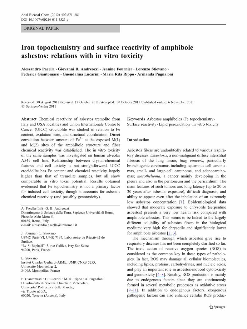
Fig. 1 Left side, SEM image oftremolite fibers from Castelluc-cio Superiore showing the pris-matic, elongated, parallel fiberstightly packed, and randomlyoriented around a commonc-axis. Right side, tremolitestructure projected along c-axis.Blue tetrahedra=T(1) and T(2);yellow octahedra=M(1); greenoctahedra=M(2); and reddots=M(4). Dashed linesembody the ideal {110}amphibole cross-section
Iron topochemistry and surface reactivity of amphibole asbestos 875

reported by Bowes and Farrow [42] and very close to thatof the ideal crocidolite (that is the mineral riebeckite,BNa2C Fe2þ3Fe3þ2ð ÞΣ5
TSi8O22
Oð3Þ OHð Þ2). UICC crocido-lite, the Fe-richest sample, has Fe2+ equally distributedover M(1) and M(3) sites, filling them almost completely.In addition, Fe3+ totally fills M(2) sites and the residuals fillM(1) and M(3) sites. On the contrary tremolite samples,which contain Fe ten times less than crocidolite, have Fe2+
equally distributed over M(1), M(2), and M(3) sites, andFe3+ only allocated at M(2) sites.
Both the site occupation and the coordination environmentof Fe in the structure play a central role in the interactionbetween fibers and biological environment, being only the Feexposed at the surface involved in biochemical reactions [9,17, 43]. Structure of both tremolite and crocidolite ismonoclinic (C2/m) and consists of double chains oftetrahedra (T(1) and T(2)) aligned along the c-axis, and astrip of octahedra (M(1), M(2), and M(3), corresponding toC group) at the center of structural “I-beam” blocks. The I-
beam blocks extend in c direction and are linked by M(4)distorted polyhedra, corresponding to B group. Moreover, itwas reported that the amphibole fibers are composed ofsmaller, parallel fibrils tightly packed and randomly orientedaround a common elongation c-axis [44, 45]. On these bases,we may consider each fibril as made of a number of I-beamblocks, exhibiting M(1), M(2), and M(4) sites on externalsurfaces (Fig. 1). Therefore, Fe2+ at M(1) and M(2) sites hashigher probability of being involved in surface reactions thanFe2+ at M(3) site. Notably, in UICC crocidolite Fe2+ contentat M(1) is ca. 2 atoms per formula unit (apfu) and Fe3+
content at M(2) sites is ca. 2 apfu, that is up to twenty timeswith respect to tremolite samples [30].
Fig. 3 Intensity of the [DMPO,HO]° radical adduct obtained from thetested samples as a function of their [M(1)+M(2)]Fe2+ content. Errorbars, ±2 SD
Fig. 2 Intensity of the [DMPO, HO]° radical adduct obtained fromthe tested samples. Mean values of two measurements, ±2 SD.Amphibole fibrous samples are ranked based on increasing Fe content
Fig. 4 Formation of MDA (% absorbance per milliliter of organicphase) observed for the tested samples. Mean values of two measure-ments, ±2 SD. Amphibole fibrous samples are ranked based onincreasing Fe content
Fig. 5 Formation of monoaldehydes (% absorbance per ml of organicphase) observed for the tested samples: (a) polyunsaturated mono-aldehydes at 450 nm; (b) saturated monoaldehydes at 500 nm. Meanvalues of two measurements, ±2 SD. Amphibole fibrous samples areranked based on increasing Fe content
876 A. Pacella et al.
Surface reactivity of fibrous samples
Surface reactivity evaluated by production of (DMPO,HO)° radicals was measured by EPR and is reported as theaverage of the signal collected after 5 and 30 min ofincubation time, subtracted by the value obtained for thecontrol. Production of HO· radicals by UICC crocidolite ismuch higher than that of tremolite samples, and thereactivity of Maryland tremolite is slightly higher than thatof the Italian ones (Fig. 2). Notably, the observedproduction of HO· radicals correlates well with the Fe2+
content at M(1) and M(2) sites (Fig. 3), comforting thehypothesis that the Fe2+ at M(1) and M(2) sites is the mostreactive species. Maryland tremolite and UICC crocidolitewere previously studied by XPS, and the oxidation state forFe at the fiber surface was obtained: results revealed thattheir surface is enriched in Fe3+ with respect to the bulk,and this Fe3+ is mainly present as iron oxyhydroxidesspecies [46]. The oxidation of Fe2+ to Fe3+ on the surface ofthe fibers is expected to inhibit, or at least delay theirreactivity [9, 47, 48].
Surface reactivity evaluated through lipid peroxidation isreported as the average value derived from duplicate testsand is expressed as absorbance per milliliter of organic
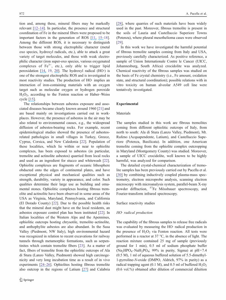
phase, subtracted by the value obtained for the control.Data referred to Castelluccio tremolite are not availablesince the available amount of this sample was insufficientto allow its analysis. The presence of MDA and mono-aldehydes observed in the organic phase indicates that allthe studied samples are active in the peroxidation process(Figs. 4 and 5). UICC crocidolite exhibits the highestreactivity in the formation of MDA; tremolite samples showcomparable behavior among them but markedly lowerreactivity (Fig. 4). Notably, among the various productsgenerated by the lipid peroxidation, MDA is the mostmutagenic and carcinogenic [49, 50]. Less systematicresults are obtained when considering the production ofmonoaldehydes and kind of correlation is absent, with Mt.Rufeno sample resulting as highly reactive as UICCcrocidolite (Fig. 5). This problematic result poses difficult-to-answer questions. Previous studies have implicated thepresence of ROS as the agent inducing the lipid perox-idation [11, 19, 51]. Our data partially support suchhypothesis, because they show that the MDA formationincreases with the production of HO· radicals, which is oneof the strongest electrophilic ROS (Fig. 6). However, nocorrelation could be found between the production of HO·radical and the generation of monoaldehydes, so theproblem is still open. It is worth noting that, in additionto ROS with strong electrophilic character (HO·, iron-oxospecies) which are able to attack a great variety ofmolecules (such as DNA), other less electrophilic species(iron super-oxo, iron-peroxo, etc.) are also capable totrigger lipid peroxidation [52]. Further investigation in thisdirection is in progress at the moment.
In vitro toxicity of fibrous samples
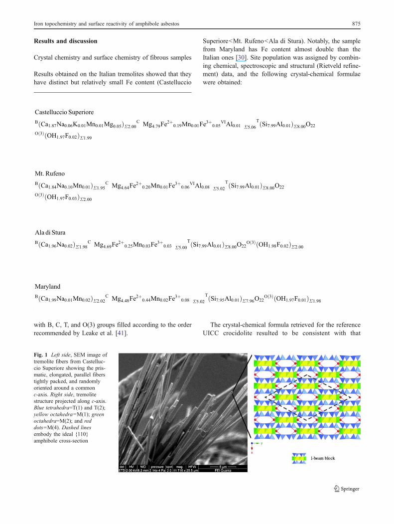
TEM images of human alveolar epithelial A549 controlcells grown in monolayers show the typical cuboidalmorphology (Fig. 7). TEM images of A549 cells put incontact with all the fibres show evidence of cell damage,such as loss of cytoplasmic organelles and superficialprotrusions. In addition, as shown in cells put in contact
Fig. 6 MDA production of the tested samples as a function of HO·radical production. Error bars, ±2 SD
a bFig. 7 TEM image of A549control cells at 24 h: (a) cellswith the typical cuboidal mor-phology; (b) higher magnifica-tion of the same cultured cellsshowing richness of peripheralcell protrusions and organelles.N nucleus, Nu nucleolus, P cellprotrusions, M mitochondria
Iron topochemistry and surface reactivity of amphibole asbestos 877

with Ala di Stura tremolite (Fig. 8), phagocytized mineralswith the typical asbestiform habit are clearly observedinside cytoplasmic vacuoles. Notably, the cell internaliza-tion of fibers is observed to be independent of the fiber size.
It is already established that asbestos involves mitochon-drial ROS production and induces apoptosis of theendothelial cells via the mitochondria-regulated (intrinsic)death pathway [53]. In the present study, results of MTTassay (Fig. 9) show significant decrements of cell viabilitywith respect to the control cells for all asbestos-treatedcells, except for Mt. Rufeno tremolite. This result indicates
the capability of the fibrous samples here tested to induce asuppression of mitochondrial functionality in differentextent, as by MTT assay the mitochondrial succinatedehydrogenase enzyme is tested. In particular, Ala di Sturatremolite results to be even more toxic than UICCcrocidolite. This result is not easily explainable as the toxicaction of our samples could be related to many factors suchas: the different capacity of fiber uptake that can affectcytoplasmic organelles; the release of Mg2+ that can havedifferent effects on lipid packing, membrane permeabilityand can counteract Ca2+ uptake by blocking the calciumuniporter [12]; the release of Ca2+ that can affect thedysregulation of mitochondrial Ca2+ homeostasis leading to
Fig. 9 MTT assay in A 549 cells. Comparison between treated andcontrol cells at 24 and 48 h. Error bars, ±2 SD. Amphibole fibroussamples are ranked based on increasing Fe content
Fig. 8 TEM image of A549 cells at 24-h treated with 50 μg/ml of Aladi Stura tremolite: (a) tremolite asbestos aggregates (black arrows)inside cytoplasmic vacuoles and lipid droplets can be observed; (b)tremolite fibers (black arrow) outside a severely damaged cell withloss of protrusions; (c) tremolite fiber inside a cell with compromisedcytoplasmic protrusions and aspects of severe damage; and (d)morphology of compromised cell with irregularly shaped protrusionsand poorness of organules. N nucleus, L lipid droplet, P cytoplasmicprotrusions
Fig. 10 Immunohistochemical Ki-67 detection at 48 h on A549 cells.Control cells: (a, b) cells treated with: (c) Mt. Rufeno tremolite, (d)Ala di Stura tremolite; (e) Castelluccio Superiore tremolite; (f) UICCcrocidolite. Magnification: (a) ×200, (b) ×400, and (c–f) ×200 (withinserts, ×400)
Fig. 11 Percent expression of Ki-67 antigen. Error bars, ±2 SD.Amphibole fibrous samples are ranked based on increasing Fe content
878 A. Pacella et al.

enhanced generation of ROS and induction of the perme-ability transition pore [12, 54].
Immunohistochemical Ki-67 detection shows markeddecrements in proliferation of fiber-treated cells comparedto control cultures for all tested samples (Fig. 10). Theobserved decline in the proliferation index seen in theasbestos-treated cells can be explained by the directcytotoxic action of the fibers, and is in agreement withprevious studies [55, 56]. The intensity of Ki-67 proteinwas evaluated with a number of positive cells: nosignificant differences are observed among the samples(Fig. 11).
Evidence of cellular stress in the asbestos-treated cellswas also obtained with the analysis of pycnotic nuclei byfluorescence microscopy. After 24 h pycnotic nuclei aremostly detectable in cells treated with Ala di Stura andMaryland tremolites (Fig. 12a). In the same specimens, adecreased number of adherent cells was observed at 48 h(Fig. 12b), which may be due to the detachment of deadcells.
Except for cells in contact with Mt. Rufeno tremolite,hypodiploid peaks are generally observed, with strongestevidence for cells treated with Ala di Stura and Marylandtremolites (Fig. 12c). In addition, cytofluorimetric cell cycleanalysis was also performed. Flow cytometry determinesthe relative cellular DNA content and enables the identifi-cation of the cell distribution during the various phases ofthe cell cycle. Four distinct phases could be recognized in aproliferating cell population: the G1-, S- (DNA synthesisphase), G2-, and M-phase (mitosis). G1 is the growth phasemarked by synthesis of various enzymes, mainly thoseneeded for DNA replication that are required in S-phase.During S phase, the amount of DNA in the cell iseffectively doubled. G2 phase is a period of rapid cellgrowth and protein synthesis during which the cell readiesitself for mitosis.
Cells treated with Ala di Stura and Maryland tremolitesshow a G1 cycle phase strongly depressed with respect tocontrol, whereas no significant difference between controland asbestos-treated cells may be observed for S and G2
a b
c d
Fig. 12 Analysis of picnoticnuclei (↑) by fluorescence mi-croscopy at: (a) 24 and (b) 48 h.Evaluation of hypodiploid nu-clei (c) and cell cycle phases (d)by FACS analysis. Standarddeviations are not shown be-cause they are relative to meanvalues obtained from twoexperiments. Amphibole fibroussamples are ranked based onincreasing Fe content
Iron topochemistry and surface reactivity of amphibole asbestos 879

cycle phases (Fig. 12d). In particular, exposure of A549cells to Ala di Stura and Maryland tremolite fibers cause aperturbation in the cells passing through the cell cycle, witha decrease in the number of cells in the G1 phase. Thesechanges were concomitant with an increase in the numberof cells containing hypodiploid nuclei. A G1 block wasevidenced by the reduced number of cells entering S phaseand the subsequent drop of cells in G2 phase (Fig. 12d).
Summarizing, results obtained from in vitro testshighlight that all tested samples, with the only exceptionof Mt. Rufeno tremolite, are responsible for significant celltoxicity. However, the toxicity of tremolite samples resultsto be not markedly different from that of UICC crocidolite.Notably, for specific tests (MTT and evaluation ofhypodiploid nuclei) Ala di Stura tremolite is found to beeven more cytotoxic than UICC crocidolite.
Conclusions
UICC crocidolite contains the highest quantity of Fe andexhibits the highest chemical reactivity; tremolite samplescontain 10 to 20 times less Fe and show fairly lowerchemical reactivity (Figs. 2, 3, 4, and 5). Moreover, thepresence of Fe2+ and Fe3+ at the exposed M(1) and M(2)sites of the amphibole structure is a key factor of Feavailability for biochemical reactions (e.g., Fenton reac-tion). On these bases, a direct correlation between Fetopochemistry and fiber chemical reactivity is established.Relationship between crystal-chemical features and in vitrocell toxicity is not found to be so straightforward: UICCcrocidolite has Fe content and chemical reactivity largelyhigher than that of tremolite samples, but all samples havecomparable in vitro toxic potential. This is an independentconfirmation that cell toxicity is due to the synergy of anumber of processes and is still an entangled matter.Focusing on Fe topochemistry, it should be strongly relatedto cell genotoxicity better than cytotoxicity, because it playsa central role in the generation of HO· and production ofMDA, with this latter well known to be highly carcinogenic[51, 52].
References
1. U.S. National Research Council (1985) Asbestiform fibres: non-occupational health risks. National Academy Press, Washington
2. Van Oss CJ, Naim JO, Costanzo PM Jr, Giese RF, Wu W, SorlingAF (1999) Impact of different asbestos species and other mineralparticles on pulmonary pathogenesis. Clay Clay Min 47:697–707
3. Taunton AE, Druschel GK, Gunter ME, Wood SA (2010)Geochemistry in the lung: reaction-path modeling and experi-mental examinations of rock-forming minerals under physiologicconditions. Am Min. doi:10.2138/am.2010.3434
4. Trubiani O, Salvolini E, Staffolani R, Di Primio R, Mazzanti L(2003) DMSO modifies structural and functional properties ofRPMI-8402 cells by promoting programmed cell death. Int JImmunopathol Pharmacol 16:253–259
5. Cardile V, Lombardo L, Belluso E, Panico A, Capella S, BalazyM (2007) Toxicity and carcinogenicity mechanisms of fibrousantigorite. Int J Environ Res Public Health 4:1–9
6. Elias Z, Poirot O, Schneider O, Amarande A-M, Danière M-C,Terzetti F, Pezerat H, Fournier J, Zalma R (1995) Cytotoxic andtransforming effects of some iron-containing minerals in Syrianhamster embryo (SHE) cells. Cancer Detect Prev 19:405–414
7. Fubini B, Otero Arean C (1999) Chemical aspects of the toxicityof invale mineral dusts. Chem Soc Rev 28:373–381
8. Kamp DW, Weitzman SA (1999) The molecular basis of asbestosinduced lung injury. Thorax 54:638–652
9. Shukla A, Gulumian M, Hei TK, Kamp D, Rahman Q, MossmanBT (2003) Serial review: role of reactive oxygen and nitrogenspecies (ROS/RNS) in lung injury and diseases. Multiple roles ofoxidants in the pathogenesis of asbestos-induced diseases. FreeRad Biol Med 34:1117–1129
10. Kusmartsev S, Gabrilovich DI (2003) Inhibition of myeloid celldifferentiation in cancer: the role of reactive oxygen species. JLeuk Biol 74:186–196
11. Halliwell B, Gutteridge JMC (1986) Oxygen free radicals and ironin relation to biology and medicine: some problems and concepts.Arch Biochem Biophys 246:501–514
12. Bergamini C, Fato R, Biagini G, Pugnaloni A, Giantomassi F,Foresti E, Lesci GI, Roveri N, Lenaz G (2007) Mitochondriachanges induced by natural and synthetic asbestos fibers: studieson isolated mitochondria. Cell Mol Biol 52(Suppl):691–700
13. Panduri V, Surapureddi S, Soberanes S, Weitzman SA, ChandelN, Kamp DW (2006) P53 mediates amosite asbestos-inducedalveolar epithelial cell mitochondria-regulated apoptosis. Am JRespir Cell Mol Biol 34:443–452
14. Pezerat H, Zalma R, Guignard J, Jaurand M-C (1989) Productionof oxygen radicals by the reduction of oxygen arising from thesurface activity of mineral fibres. In: Saracci R (ed) Non-occupational exposure to mineral fibers, vol 90. IARC ScientificPublication, Lyon, pp 100–110
15. Zalma R, Bonneau L, Guignard J, Pezerat H, Jaurand M-C (1987)Production of hydroxyl radicals by iron solid compounds. ToxicolEnviron Chem 13:171–187
16. Fubini B, Fenoglio I, Elias Z, Poirot O (2001) On the variabilityof the biological responses to silicas: effect of origin, crystallinityand state of the surface on the generation of reactive oxygenspecies and consequent morphological transformations in cells. JEnviron Pathol Toxicol Oncol 20:87–100
17. Gazzano E, Riganti C, Tomatis M, Turci F, Bosia A, Fubini B,Ghigo D (2005) Potential toxicity of nonregulated asbestiformminerals: balangeroite from the western Alps. Part 3: depletion ofantioxidant defenses. J Toxicol Environ Health 68:41–49
18. Fournier J, Guignard J, Nejjari A, Zalma R, Pezerat H (1991) In:Brown RC (ed) The role of iron in the redox surface activity offibers. Relation to carcinogenicity, mechanisms in fibre carcino-genesis. Plenum Press, New York, pp 407–414
19. Fournier J, Copin E, Dwigaj S, Chouchane S, Guignard J (1995)Peroxidation lipidique en présence de composés inorganiques.Relation avec les mécanismes du stress oxydant. C R Soc Biol189:1–15
20. Chouchane S, Guignard J, Fournier J (2000) Lipid peroxidation inthe presence of iron oxides. Toxicol Environ Chem 75:43–57
21. Wagner JC, Sleggs CA, Marchand P (1960) Diffuse pleuralmesothelioma and asbestos exposure in the Northwestern CapeProvince. Br J Ind Med 17:260–271
22. Ross M, Nolan RP (2003) History of asbestos discovery and useand asbestos-related disease in context with the occurrence of
880 A. Pacella et al.

asbestos within ophiolite complexes. Geol Soc Am Special Paper373, Bpulder
23. Ballirano P, Andreozzi GB, Belardi G (2008) Crystal chemicaland structural characterization of fibrous tremolite from SusaValley, Italy, with comments on potential harmful effects onhuman health. Am Min 93:1349–1355
24. Pacella A, Andreozzi GB, Ballirano P, Gianfagna A (2008)Crystal chemical and structural characterization of fibroustremolite from Ala di Stura (Lanzo Valley, Italy). Per Min77:51–62
25. Davis JMG, Addison J, McIntosh C, Miller BG, Niven K (1991)Variations in the carcinogeneticy of tremolite dust samples ofdiffering morphology. Ann N Y Acad Sci 643:473–490
26. Addison J, McConnel EE (2005) A review of carcinogenicitystudies of asbestos and non-asbestos tremolite and other amphib-oles. In: International symposium on the health hazard evaluationof fibrous particles associated with taconite and the adjacentduluth complex, St. Paul
27. Burragato F, Ballirano P, Fiori S, Papacchini L, Sonno M (2001)Segnalazione di tremolite asbestiforme nel Lazio. Il Cercapietre,vol. I–II, Notiziario del Gruppo Mineralogico Romano, pp 33–35
28. Punturo R, Fiannacca P, Lo Giudice A, Pezzino A, Cirrincione R,Liberi F, Piluso E (2004) Le Cave storiche della “Pietra Verde” diGimigliano e Monte Reventino (Calabria): studio petrografico egeochimico. Acc Gioen Sci Nat Catania 37:35–57
29. Pasetto R, Bruni BM, D’Antona C, De Nardo P, Di Maria G, DiStefano R, Fiorentini C, Gianfagna A, Marconi A, Paoletti L,Putzu MG, Soffritti M, Comba P (2004) Problematiche sanitariedella fibra anfibolica di Biancavilla. Aspetti epidemiologici,clinici e sperimentali. Note dell’ Istituto Superiore di Sanità 17:8–12
30. Pacella A, Andreozzi GB, Fournier J (2010) Detailed crystalchemistry and iron topochemistry of asbestos occurring in itsnatural setting: a first step to understanding its chemical reactivity.Chem Geol 277:197–206
31. Gavino VC, Miller JS, Ikharebha SO, Milo GE, Corwell DG(1981) Effect of polyunsaturated fatty acids and autoxidation onlipid peroxidation in tissue culture. J Lipid Res 22:763–769
32. Marcuse R, Johansson L (1973) Studies on the TBA test forrancidity grading: II. TBA reactivity of aldehyde classes. J Am OilChem Soc 50:387–391
33. Adiseshaiah P, Lindner DJ, Kalvakolanu DV, Reddy SP (2007)FRA-1 proto-oncogene induces lung epithelial cell invasion andanchorage-independent growth in vitro, but is insufficient topromote growth in vivo. Cancer Res 67:6204–6211
34. Pugnaloni A, Lucarini G, Giantomassi F, Lombardo L, Capella S,Belluso E, Zizzi A, Panico AM, Biagini G, Cardile V (2007) Invitro study of biofunctional indicators after exposure to asbestos-like fluoro-edenite fibres. Cell Mol Biol 53:965–980
35. Mossman BT (1983) Rapid colorimetric assay for cellular growthand survival: application to proliferation and cytotoxicity assays. JImmunol Methods 65:55–63
36. Gasparini G, Boracchi P, Verderio P, Bevilacqua P (1994) Cellkinetics in human breast cancer: comparison between theprognostic value of the cytofluorimetric S-phase fraction and thatof the antibodies to Ki-67 and PCNA antigens detected byimmunocytochemistry. Int J Cancer 57:822–829
37. Vielh P, Chevillard S, Mosseri V, Donatini B, Magdelenat H(1990) Ki67 index and S-phase fraction in human breastcarcinomas. Comparison and correlations with prognostic factors.Am J Clin Pathol 94:681–686
38. Healy E, Angus B, Lawrence CM, Rees JL (1995) Prognosticvalue of Ki67 antigen expression in basal cell carcinomas. Br JDermatol 133:737–741
39. Uboldi C, Bonacchi D, Lorenzi G, Hermanns MI, Pohl C, BaldiG, Unger RE, Kirkpatrick CJ (2009) Gold nanoparticles inducecytotoxicity in the alveolar type-II cell lines A549 and NCIH441.Part Fibre Toxicol 6:18
40. Telford WG, King LE, Fraker PJ (1992) Comparative evaluationof several DNA binding dyes in the detection of apoptosisassociated chromatin degradation by flow cytometry. Cytometry13:137–143
41. Leake BE, Woolley AR, Birch WD, Burke EAJ, Ferraris G, GriceJD, Hawthorne FC, Kisch HJ, Krivovichev VG, Schumacher JC,Stephenson NN, Withtaker EJW (2003) Nomenclature of amphiboles:additions and revisions to the InternationalMineralogical Association’samphibole nomenclature. Can Min 41:1355–1370
42. Bowes DR, Farrow CM (1997) Major and trace elementcompositions of the UICC standard asbestos sample. Am J IndMed 32:592–594
43. Fubini B, Mollo L (1995) Role of iron oxides. Toxicol Lett 82/83:951–960
44. Wylie AG (1979) Optical properties of the fibrous amphiboles.Ann N Y Acad Sci 330:600–605
45. Verkouteren JR, Wylie AG (2002) Anomalous optical propertiesof fibrous tremolite, actinolite, and ferro-actinolite. Am Min87:1090–1095
46. Fantauzzi M, Pacella A, Atzei D, Gianfagna A, Andreozzi GB,Rossi A (2010) Combined use of X-ray photoelectron andMössbauer spectroscopic techniques in the analytical character-ization of iron oxidation state in amphibole asbestos. Anal BioanalChem 396:2889–2898
47. Pezerat H, Zalma R, Guignard J (1989) Production of oxygen freeradicals by the reduction of oxygen arising from the surfaceactivity of mineral fibres. IARC Spl Publ 90:100–111
48. Bignon J, Peto J, Saracci R (1989) Non-occupational exposure tomineral fibres. Oxford University Press, Oxford
49. Marnett LJ (1999) Chemistry and biology of DNA damage bymalondialdehyde, [Review]. IARC Sci Publ (150):17–27
50. Zhang Y, Chen S, Hsu T, Santella RM (2002) Immunohistochem-ical detection of malondialdehyde–DNA adducts in human oralmucosa cells. Carcinogenesis 23:207–211
51. Chouchane S, Guignard J, Fournier J (1999) Lipid peroxidation inpresence of iron oxides. Toxicol Environ Chem 75:43–57
52. Nejjari A, Fournier J, Pezerat H, Leanderson P (1993) Mineralfibres: correlation between oxidising surface activity and DNAbase hydroxylation. Occup Environ Med 50:501–504
53. Liu G, Beri R, Mueller A, Kamp DW (2010) Molecularmechanisms of asbestos-induced lung epithelial cell apoptosis.Chem Biol Interact 188(2):309–318
54. Shukla A, Ramos-Nino M, Mossman B (2003) Cell signaling andtranscription factor activation by asbestos in lung injury anddisease. Int J Biochem Cell Biol 35:1198–1209
55. Zheng N, Sun YF, Pei DS, Liu JJ, Sun XQ, Chen JC, Cai WQ, LiW, Cao JY (2005) Anti-Ki-67 peptide nucleic acid affects theproliferation and apoptosis of human renal carcinoma cells invitro. Life Sci 76:1873–1881
56. Giantomassi F, Gualtieri AF, Santarelli L, Tomasetti M, LusvardiG, Lucarini G, Governa M, Pugnaloni A (2010) Biological effectsand comparative cytotoxicity of thermal transformed asbestos-containing materials in a human alveolar epithelial cell line.Toxicol In Vitro 24:1521–1531
Iron topochemistry and surface reactivity of amphibole asbestos 881