MM-DTI: Visualization and segmentation tool for diffusion tensor images
Upload
independentCategory
view
0download
0
Laboratory Investigation
Intratumoral injection of BCNU in ethanol (DTI-015) results in enhanced delivery to
tumor – a pharmacokinetic study
Daniel A. Hamstra2,5, Bradford A. Moffat1,5, Daniel E. Hall1,5, John M. Young7, Timothy J. Desmond6,Julie Carter7, Dennis Pietronigro7, Kirk A. Frey1,4,6, Alnawaz Rehemtulla1,2,5 and Brian D. Ross1,3,5
The 1Departments of Radiology; 2Radiation Oncology; 3Biological Chemistry; 4Neurology; The 5Center for MolecularImaging; 6The Mental Health Research Institute, The University of Michigan Medical Center, Ann Arbor, MI; 7DirectTherapeutics Inc. Redwood City, CA, USA
Key words: animal model, BCNU, brain tumor, MRI, pharmacokinetics
Summary
Solvent facilitated perfusion (SFP) has been proposed as a technique to increase the delivery of chemotherapeuticagents to tumors. SFP entails direct injection of the agent into the tumor in a water-miscible organic solvent, andbecause the solvent moves easily through both aqueous solutions and cellular membranes it drives the penetration ofthe solubilized anticancer agent throughout the tumor. To test this hypothesis, we compared the pharmacokinetics(PK) of 14C-labeled 1,3-bis-chlorethyl-1-nitrosourea (BCNU) in intra-cerebral 9L rat gliomas after intravenous (IV)infusion in 90% saline –10% ethanol or direct intratumoral (IT) injection of 14C-BCNU in 100% ethanol (DTI-015).TreatmentwithDTI-015 yielded a peak radioactive count (Cmax) for the 14C label that was 100–1000 fold higher in thetumor than in all other tissues in addition to a concentration in the tumor that was 100-fold higher than that achievedfollowing IV infusion of 14C-BCNU. Pathologic and auto-radiographic analysis of tissue sections following ITinjection of 14C-BCNU in ethanol into either tumor or normal rat brain revealed both an enhanced local volume ofdistribution and an increased concentration of BCNU delivered to tumor compared to non-tumor bearing brain. Toinvestigate the mechanism behind the SFP of BCNU to the tumor both dynamic contrast and perfusion MRI wereperformed on 9L tumors before and after treatment and demonstrated a decrease in tumor perfusion following ITinjection of DTI-015. Finally, initial PK of patient blood samples following administration of DTI-015 into relapsedhigh-grade glioma indicated a 20-fold decrease in systemic exposure to BCNU compared to IV infusion of BCNUproviding further evidence for the enhanced therapeutic ratio observed for DTI-015.
Abbreviations: AUC – area under the concentration–time curve; BCNU – 1,3-bis-chlorethyl-1-nitrosourea; CED –convection enhanced delivery; Cmax – peak concentration; CPM – counts per minute; DTI-015 – BCNU preparedin 100% ethanol; IT – intratumoral; IV – intravenous; LD10 – the dose of drug which results in death of 10% of theanimals due to drug toxicity; MRI – magnetic resonance imaging; PK – pharmacokinetics; SEM – standard error ofthe mean; SFP – solvent facilitated perfusion; t1/2 – half-life; TTR – tumor-to-tissue ratio
Introduction
High-grade malignant glioma carries a grave prognosiswith median survival typically less than 12 months fromdiagnosis even with aggressive treatment. The typicalmanagement strategy entails maximal surgical resectionfollowed by post-operative radiotherapy [1]. Intrave-nously administered BCNU has been the leading che-motherapeutic agent for treating this patient populationfor decades; however, despite activity against gliomacells both in culture and in animal models there is lim-ited clinical efficacy which has prompted numerousinvestigations into ways to enhance the pharmacoki-netics (PK) of this agent [2]. The limited efficacy ofadjuvant chemotherapy for high-grade gliomas washighlighted in a recent meta-analysis which indicatedonly a 6% increase in median survival in patients treated
with adjuvant chemotherapy (typically BCNU) follow-ing maximal surgical resection and ⁄ or radiation therapy[3].Attempts to increase this response through dose
escalation of BCNU administered systemically havebeen hampered by acute toxicity to bone marrow andgastrointestinal tract and delayed toxicity to liver andlungs [2,4–6]. Therefore, local ⁄ regional delivery ofBCNU either through intra-arterial infusion [7,8] orthrough the use of BCNU impregnated wafersimplanted in the resection cavity (Gliadel�) have beenattempted [9,10]. Unfortunately, neither of these tech-niques has offered a significant advantage over tradi-tional IV infusion of BCNU. Intra-arterial infusionresulted in a less than 4-fold increase in tumor-to-tissueratio (TTR) and was complicated by unacceptable opticand neurologic toxicity [11,12]. Following the difficulty
Journal of Neuro-Oncology (2005) 73: 225–238 � Springer 2005DOI 10.1007/s11060-004-5675-2

with local intra-arterial delivery attempts were madeusing BCNU contained within a sustained release for-mulation implanted directly into the resection cavity.Initial studies utilized relatively low dose wafers whichgave only a modest increase in survival followingtreatment of recurrent or primary glioblastoma [10,13].More recent studies have evaluated wafers containingincreased concentrations of BCNU, but these eventuallyproduced both local and systemic toxicity and the clin-ical benefit of these higher dose wafers has yet to bedemonstrated [14]. In part, this limited efficacy may bedue to the fact that Gliadel wafers are limited by bulkdiffusion of BCNU from the implant to surroundingtissues, and are, therefore, only utilizable after maximalsurgical resection and even then there is still limitedentrance of drug into the surrounding tissues, which istypically not more than a few millimeters [15]. Due tothe limited transit of drug direct intratumoral infusionof drugs in aqueous solutions (convection enhanceddelivery, CED) has been investigated to increase deliveryto the tumor while also enhancing the TTR [16]. How-ever, this technique is limited by the long time requiredfor infusion and by limited and heterogeneous distri-bution of the compounds within the tumor [17]. Despitethis fact CED is being actively pursued both in pre-clinical models [18] and in clinical investigation for pa-tients with high-grade glioma [19].Solvent facilitated perfusion (SFP) is another new
treatment technique which has been proposed to achievean enhanced TTR for chemotherapeutic agents. Thismethod utilizes a water-miscible organic solvent that canmove freely though both aqueous solutions and cellmembranes thus facilitating transit of the chemothera-peutic agent some distance from the tumor. BCNUsuspended in 100% ethanol (herein termed DTI-015) isthe first drug : solvent system to be used to study thisSFP model. It has been evaluated both in animal modelsand in patients with recurrent high-grade glioma and inboth cases DTI-015 was demonstrated to have signifi-cant anti-tumor activity [20,21]. In one preclinicalmodel, treatment of rats bearing T9 gliosarcomas withDTI-015 resulted in a >400% increase in life-span with40% of animals cured of their tumors [21]. In a separatestudy, diffusion-weighted MRI was performed followingthe injection of DTI-015 into established 9L gliosarco-mas and revealed a rapid and substantial increase in theapparent diffusion coefficient (ADC), which is a radio-logic measure of cell death [22], followed by significantregression of the tumor mass with 75% of animals freeof tumor by MRI and pathologic analysis at 30 days[23]. More importantly, changes in ADC were witnessedthroughout the tumor despite the fact that the volume ofDTI-015 injected was at most 50% of the tumor volume.These diffuse changes in ADC throughout the injectedtumors were thus an indirect suggestion of SFP of drugto the entire tumor mass. Finally, a recent phase I ⁄ IItrial was reported on 40 heavily pretreated patients withinoperable recurrences of primary brain tumors (90% ofwhich were WHO grade III or IV), 58% of whom hadbeen previously treated with nitrosoureas. Despite thispreviously treated population there was a significantbenefit in patients treated with less than or equal to the
maximal tolerated dose of DTI-015 (240 mg and 5 ml)where the median survival was 55 weeks in patients withrecurrent, inoperable GBM, as compared to a historicalcontrol of 25 weeks [20].Despite these preclinical and clinical data the local
and systemic PK of BCNU following injection of DTI-015 into intra-cerebral tumors in either experimentalanimals or clinical samples has not been reported. We,therefore, evaluated the distribution of radioactivityfollowing IT or IV injection of 14C-BCNU in Fischer344 rats bearing established 9L gliosarcomas, and uti-lized MRI measures of tumor perfusion to more fullyevaluate the PK of DTI-015 administration. Finally, theinitial pilot experience with the systemic PK of BCNUfollowing IT treatment with DTI-015 in patients withrecurrent gliomas is reported.
Materials and methods
Cell culture
Rat 9L brain tumor cells (passage 12) were obtainedfrom the Brain Tumor Research Center at the Univer-sity of California at San Francisco. The cells were grownas monolayers in 175 cm2 sterile plastic flasks in Eagle’sMEM with 10% fetal bovine serum. Cells were culturedat 37 �C in an atmosphere containing 95 ⁄ 5% air ⁄CO2
until confluent, and were harvested by trypsinization,counted, and resuspended in serum-free medium forinjection. The 9L cells were carried only until passagenumber 40 at which time cells were reactivated fromfrozen stocks.
Animals
Male Fischer 344 (F344/NHsd) rats were obtained fromHarlan Sprague Dawley (Indianapolis, IN) and wereacclimated in house, 2 per cage with food and waterad libitum for at least 1 week prior to experimentation.Animals weighing 125–150 were used for tumor cellimplantation and subsequent treatment with DTI-015 orfor injection of DTI-015 into the normal brain of non-tumor bearing mice.
Preparation of 14C-BCNU
14C-[carbonyl]-1,3-bischlorethyl-1-nitrosourea (specificactivity 53 mCi/mol) or 1,3-bis(2-chlorethyl-[2-14C])-1-nitrosourea (specific activity 205 mCi ⁄mol) wereobtained from Moravek Biochemicals Inc (Brea, CA) asa dry solid packaged under argon. The materials werereconstituted in ethanol containing unlabeled BCNU ata concentration of 67 mg ⁄ml to give the DTI-015formulation.For experiments evaluating the distribution of 14C
labeled BCNU within tumor or normal brain slices thecompound with 14C in the carbonyl position was utilizedwhere a total of 15 ll of DTI-015 was injected con-taining 1 mg of unlabeled BCNU and 135 nCi. In allother PK experiments, the compound with 14C in the
226

ethyl position was utilized with a total of 15 ll of DTI-015 injected containing 1 mg of unlabeled BCNU and1 lCi. For intravenous administration the 1,3-bis(2-chlorethyl-[2-14C])-1-nitrosourea compound was recon-stituted in ethanol containing unlabeled BCNU andfurther diluted in saline to a concentration of 1 mgBCNU (containing 1 lCi) per 900 ll ethanol/saline(10%/90%).
Induction of brain tumors
Intra-cerebral tumor implantation was performed aspreviously described (24). Male Fischer 344 rats weigh-ing between 125 and 150 g were anesthetized with aketamine ⁄ xylazine mixture administered intraperitone-ally. A small skin incision was made over the righthemisphere, and a 1 cm diameter burr hole was drilledthrough the skull using a high speed drill. Tumor cells(105) contained in a volume of 5 ll were injected into theright forebrain at a depth of 3 mm via a Hamiltonsyringe equipped with a 30 gauge needle. The area wasrinsed with 70% ethanol, and the burr hole was filledwith bone wax to minimize extra-cerebral extension ofthe tumor. The skin incision was closed with sutures,and the rats were allowed to recover from anesthesiabefore being returned to the animal quarters.
Magnetic resonance imaging
All in vivo MR experiments were performed on a VarianNMR Instruments system equipped with a 7.0 T(300 MHz proton frequency), 18.3-cm horizontal boremagnet. For MRI examination rats were anesthetizedwith 1.5% isoflurane and maintained at 37 �C inside themagnet using a heated circulating water blanket. MRI ofrat brains were initiated between 8 and 11 days after cellimplantation and repeated approximately every 2 daysusing a 32-mm-diameter quadrature radio frequencyhead coil (USA Instruments, Highland Heights, OH).Multislice axial T2 weighted images (FOV ¼ 30 · 30 mmand image matrix ¼ 128 · 128) were acquired by using afastspin echo (FSE) sequence (TR/TEeff ¼ 4000 ⁄ 45 ms,ETL ¼ 8, Thk ¼ 1 mm). The tumor boundary visualizedin each slice was electronically outlined by using imageprocessing software (MATLAB�) for calculation oftumor volume. For quantification of contrast agentuptake T1 weighted spin echo MRI was performedpre- and post intra-peritoneal injection of GdDTPA(1.0 ml of 20% Magnevist�). TR ⁄TE ¼ 1000 ⁄ 15 ms,thk =1 mm, FOV ¼ 30 · 30 mm and image matrix ¼128 · 128. For blood flow imaging, continuous arterialspin labeling (CASL) MRI was performed with a FSEbased imaging sequence. Briefly a 3 second train ofhyperbolic inversion pulses was used to invert the signalarising from inflowing blood, immediately before theinitial 90o pulse of a 16-echo, 128-phase FSE sequence(TR ⁄TEeff of 4000 ⁄ 14 ms, 2 mm slice thickness and 8transients). The images were acquired with a field 3 · 3cm field of view and the 128 · 128 acquisition matrix wassubsequently zero-filled to 256 · 256. A T1 map of theimaged slice was also acquired with a magnetizationinversion-prepared 16-echo, 64-phase encoding FSE
sequence. Five different inversion times, Ti, were usedwith 2 transients and TR ⁄TEeff of 10,000 ⁄ 14 ms. Perfu-sion images or TBF maps were calculated using theformula for continuous labeling.
TBF ¼ kT1
ðS0 � S1Þ2aS0
ð1Þ
where k is the blood/tissue partition function (assumedto be 0.9), T1 is the tissue longitudinal relaxation time,S0 and S1 are the image signals with inversion pulsesapplied to control and tag zones respectively, and a isthe efficiency of the inversion pulses. All MRI acquisi-tion matrices were zero filled to 256 · 256 so that theresulting image dimensions were identical.
Stereotactic injections of DTI-015
When tumors had reached a mean volume of65 ± 7.5 ll rats were anesthetized with 0.1–0.2 mlketamine HCl (100 mg ⁄ml) intramuscularly and placedin the stereotactic device (David Kopf Instruments,Tujunga, CA) with the bite bar set at )2.4 mm. Thescalp was opened with a scalpel and the skull wascleaned of tissue to locate the original burr hole madefor tumor cell injection. The injection needle madefrom 30 gauge stainless steel tubing (HTX-30, SmallParts Inc, Miami Lakes, FL) attached to PE-10 poly-ethylene tubing (Clay Adams, Parsippany, NJ) wasplaced in the hole just touching the surface of the brainand the scale was read. From this point the needle wasintroduced into the tumor a distance approximately thetotal depth of the tumor and then retracted to themidpoint of the tumor (as determined by diagrams ofMR images obtained 2 days previously). Using aHarvard Pump 11 and a 50 ll Hamilton syringe, 15 llof [14C]-BCNU was infused over 2 m in at a rate of7.5 ll ⁄min. The needle was left in place an additional30 s to promote diffusion of the injection into thetumor and to prevent the drawing of [14C]-BCNUalong the needle track upon withdrawal. For injectionsinto normal brains of non-tumor bearing animals,injections were made 3 mm below the dura.
Animal termination
Representative animals from each group were killed bydecapitation at the indicated times post injection and thebrains were quickly removed. For experiments requiringsectioning they were immediately mounted in anticipa-tion of coronal tissue sectioning (see below). In separateexperiments for kinetic data analysis a blood sample wasobtained prior to euthanasia. Once the animal had beenharvested the brains were removed and the tumorsteased out in its entirety and transferred to an appro-priate vial. Small slabs of ipsilateral (adjacent to thetumor) and contralateral cortex were subsequently ta-ken. An incision was made in the abdomen past thesternum and small samples of the following tissues weretaken (in the following order): lung, liver, kidney (left),spleen, bone marrow (right femur), and testis (left).
227

Frozen sections
Upon sacrifice, the forebrain was cut from the hindbrainand mounted on a small piece of gauze saturated withM-1 embedding medium (Shandon-Lipshaw, Pittsburg,PA) on a glass slide oriented for coronal sectioning. Thiswas then frozen with crushed dry ice and coated withM-1before storing at )80 �C. The tissue gauze was thawedfrom the slide and mounted on a chuck with additionalM-1 and surrounded by crushed dry ice to freeze themedia. The tissue was then placed in the cryostat (HackerInstruments, Fairfield, NJ) and allowed to equilibrate tochamber temperature before sectioning. Serial sectionsthrough the tumor were collected beginning prior to thetumor and ending just after the tumor disappeared fromthe tissue. Serial 20 lm sections were cut, placed on slides(three sections per slide), and high temperature thawmounted to prevent diffusion of the label and completelydry the sections. Slides were placed in X0ray cassetteswith radioactive standards and opposed to Hyperfilb-max (Amersham, Arlington Heights, IL) for 6 days.Films were developed in D-19 (Kodak, Rochester, NY)for 4 min, transferred to 1% acetic acid stop bath for 30 sand fixed with Rapid Fixer (Kodak) for 5 min. The filmswere washed in running tap water for 20 min, rinsed indistilled water, and air dried.
Brain radiolabel densitometry
Auto radiograms were analyzed using imaging software(Imaging Research Inc, St. Catherines, Ontario, Can-ada). Scanning coordinates on selected slides werearbitrarily chosen to be representative and the autora-diographs were scanned with s Sony XC-77 CCD cam-era. The data were captured using MCID M5+ systemwith a Matrox Genesis imaging board and saved in thesoftware format (*IM) for analysis and in *.TIF formatfor reproduction. Film density was converted to localtissue 14C concentration (nCi ⁄ g tissue) based upon theinternal standards, and the local molar concentration(mM) were based upon the injected BCNU specificactivity of 28.9 lCi ⁄mmol. Tumor and section volumeswere determined with the same system.
Histopathological evaluation of brain tissue sections
Following autoradiography, selected slides representa-tive of the site of injection of DTI-015 into the tumorsand into the brains of normal animals were stainedwith hematoxylin and eosin and submitted to Dr.Mark T. Butt (Pathology Associates International,Frederick, MD) for independent histopathologicalevaluation.
Radioactivity determinations in peripheral blood, tumorsamples and tissue samples
At the time of sacrifice of each animal the repre-sentative tissues were harvested as indicated above
and stored at )70 �C. Portions of these frozen sam-ples (approximately 50 mg for blood and 100 mg fortissues) were accurately weighed and digested in0.5 ml of ‘Solvable’ (NEF910, NEN Research Prod-ucts, Boston, MA) at room temperature for 48 h.The tissue digests were decolorized with 0.25 ml of30% H2O2 (Sigma Chemicals, St. Louis, MO), 12 mlof Cytoscint ES (ICN Biochemicals, Costa Mesa,CA) were added, and the samples were counted for1 min. No quenching was observed in non-radioactivetissue samples spiked with a known amount ofradioactivity and subjected to the same procedures.For each series of samples a background count wasobtained for 10 blank samples counted simulta-neously which was then averaged and subtractedfrom each sample’s count.
Human blood BCNU analysis
Three patients enrolled in an IRB approved prospectivetrial of DTI-015 for recurrent high-grade glioma weretreated at Evanston Hospital (Evanston, IL, 2 patients)or The University of Southern California MedicalCenter (Los Angeles, CA, 1 patient) between 11 ⁄ 13 ⁄ 01and 7 ⁄ 12 ⁄ 02. Patients were treated per the protocolpreviously described (20). Seven serial blood sampleswere collected between 5 and 360 min following injec-tion of DTI-015. Although the exact timing of eachphlebotomy was dependent upon the unique clinicalcharacteristics of the case each patient did have blooddraws that spanned at least a 120 min time period. Forthe first two patients blood samples were immediatelyextracted with ethyl-acetate and then frozen beforeanalysis. In the third patient blood samples were firstfrozen and then extracted with ethyl-acetate at theanalytic laboratory.
Statistical and pharmacokinetic calculations
Data for radioactivity in tissues was expressed as themean ± standard error of the mean (SEM ¼ standarddeviation ⁄ square-root (n)1)). The SEM for ratios wascalculated using the following formula:
SEM A/B ¼ ðSEM A/mean AÞþ ðSEM B/mean BÞ
All statistical comparisons between groups were doneusing Microsoft Excel with the Student’s t-test (two-tailed with unequal variance) except in the comparisonbetween counts in tumor following direct IT injection orIV where a single-tailed probability function was utilizedgiven the hypothesis that IT injection would result inincreased delivery of drug to tumors. Pharmacokineticcalculations were performed using ‘PK Functions forMicrosoft Excel’, a series of Add-in functions for Excelspreadsheets, designed and written by Joel I. Usansky,Atul Desai, and Diane Tang-Liu, Department of Phar-macokinetics and Drug Metabolism, Allergan, Irvine,CA.
228

Results
Injection of DTI-015 intratumorally results in increasedexposure of tumor to BCNU as compared to all othertissues
In order to assess the pharmacokinetics ofbis-[2-chlorethyl-2-14C]-nitrosourea intra-cerebral 9Ltumors were established in Fischer 344 rats. Tumorgrowth was followed by serial MRI scans (Figure 1)where approximately 12–14 days after implantation T1-weighted MRI pre-contrast injection revealed a large,isointense mass in the right cerebral hemisphere withmild compressive effect upon surrounding cerebral tis-sues. Following IP injection of GdDTPA the tumorswere briskly and uniformly enhancing consistent withtheir high-grade pathology. Once tumors had reached amean volume of 65 ll the rats were sterotactically in-jected with 15 ll of 14C-labeled BCNU with injectionguided by coordinates obtained from MRI scans per-formed the day prior to injection to assure that it oc-curred at the appropriate location and depth within thecentral mass of the tumor. Groups of 5 animals weresacrificed 1, 15, 60, 360, 1440, and 4320 min afterinjection (except for the 360 min time point which onlycontained 3 animals), and radioactivity was counted ineach tissue and expressed as counts per minute per mgof tissue (CPM/mg tissue). BCNU exerts its biologiceffect through both carbamoylation (a short-lived andreversible phenomenon) and alkylation (a long-livedand irreversible phenomenon) [25]. Due to the presenceof the radiolabel in both alkyl groups of bis-[2-chlor-ethyl-2-14C]-nitrosourea, carbamylation and alkylationare indistinguishable in this study, and no attempt wasmade to quantify either these two or other products ofBCNU.As can be seen in Figure 2a there was a rapid rise in
14C label detected in all tissues reaching a peak (Cmax)in 15 min or less except for the blood and kidneys whichdid not achieve Cmax until the 1 h time point. Notsurprisingly the time to Cmax was shorter in intra-cerebral tissues (which were adjacent to the directinjection of 14C-BCNU) where Cmax was typically seenat the 1 min post-injection time point as opposed to
systemic tissues where this peak was usually at either 15or 60 min. In all cases, following the initial peakradioactivity there was an initial shorter half-life (t1/2)over the first 3 h (Figure 2a) followed by a longer ter-minal half-life from 24 to 72 h (Figure 2b and Table 1).The half-life during the initial phase (up to 3 h afterinjection) was much greater in systemic tissues (exceptfor kidney and lung) with an average of 12.6 ± 6.9 hwhen compared to tumor and brain tissues, which hadhalf-lives between 1.8 and 2.4 h. This difference in initialt1/2 is consistent with the rapid redistribution of BCNUand metabolites from within the intra cerebral com-partment to other systemic tissues during the earlyperiod after injection. The shorter half-life of radioac-tivity within the kidney likely reflects the well-docu-mented renal excretion of BCNU and its metabolites;while the short half-life in the lung may reflect the factthat up to 10% of the alkyl portion of BCNU is expiredas CO2 within the first 24 h after treatment [26].The initial fast fall in radioactivity observed during
the first 3 h was followed by a more prolonged clearancein the period from 24 to 72 hours (Figure 2b and Ta-ble 1) with a half-life in the tumor of greater than 72 hand that in all other tissues markedly shorter(41 ± 6.9 h). The main exception to this was the bloodwhere the terminal half-life of 68 h was much closer tothat seen for the tumor and may reflect the late slowrelease of 14C-BCNU and metabolites from the tumorinto the systemic circulation.In addition to a prolonged half-life for 14C-label, it is
also clear from this first set of experiments that 14C-exposure to the tumor is much greater than that observedfor all other tissues both in terms of maximum concen-tration achieved (Cmax, P < 0.0005) and in terms of netexposure over the 72 h experiment, as expressed as thearea under the concentration vs. time curve (AUC,P < 0.014) (Table 1). In fact, themaximal concentrationachieved in the tumor as compared to all other non-cerebral tissues ranged between 100- and 2000- foldhigher giving a very high tumor-to-tissue ratio for Cmax(TTR Cmax). In comparison, the ipsilateral brain wasexposed to amaximal concentration of 14C-labled BCNUwhich although significantly higher than most other tis-sues (Cmax 176.0 vs. 14.1 CPM ⁄mg tissue) was still 40-
Figure 1. Intra-cerebral 9L gliosarcoma model. T1-weight MRI images were obtained of tumor bearing animals both pre- (a) and 20 min post-IP
administration of GdDTPA (b).
229

fold less than the tumor. Further, by 60 min followinginjection the concentration in the ipsilateral brainwas similar to other systemic tissues (C60min 14.8 vs.13.3 CPM ⁄mg tissue). The contralateral brain achieveda peak concentration (37.6 CPM ⁄mg tissue) which wasbetween that of most systemic tissues and the ispilateralbrain, but was still ~150-fold less than that seen in thetumor. In addition, similar to that seen for the ipsilateralbrain, by 60 min after injection there was a significant fallin the concentration of 14C-label in the contralateralbrain such that it was similar to that for most systemictissues (5.24 CPM ⁄mg tissue) with only the kidneys andliver having higher activity at this time point.Since BCNU exerts its activity by covalent modifi-
cation of targets within the cell, peak concentrations of
14C-label may not be the best indicator of ultimateefficacy; rather, the total exposure to 14C-BCNU andits metabolites to the tissues of interest, as expressed asthe area under the concentration vs. time curve (AUC,Table 1), may be a better PK parameter to monitor.The AUC of tumor, 1912 (CPM ⁄mg tissue ⁄min · 10)3)was 50–500 times greater than the AUC for all othertissues yielding a very high tumor-to-tissue ratio forAUC (TTR AUC). The kidneys, liver, and ipsilateralbrain all had similar AUCs between 17 and 40(CPM ⁄mg tissue ⁄min · 10)3) which were 50–100-foldless than the tumor. In contrast, the contralateralcerebral hemisphere was exposed to an AUC for 14C-label which was similar in nature to other systemictissues (lung, spleen, blood, and testes) with AUC’s
1
10
100
1000
10000
0 60 120 180 240 300 360
Time After Injection (minutes)
Time After Injection (minutes)
Rad
ioai
tcvi
ty (
CP
M/m
g t
issu
e)
TumorIpsilateral BrainContralateral BrainKidneyBloodNon-brain, non-kidney
0.1
1
10
100
1000
360 860 1360 1860 2360 2860 3360 3860 4360
dio
acti
vity
(C
PM
/mg
tis
sue)
aR
TumorIpsilateral BrainContralateral BrainKidneyBloodNon-brain, non-kidney
(a)
(b)
Figure 2. Time dependent distribution of radioactivity following IT delivery of 14C-labeled DTI-015 in the 9L intra-cerebral gliosarcoma model.
Fischer 344 rats bearing 9L gliosarcomas were treated with a 15 ll injection of 1,3-bis(2-chlorethyl-[2-14C])-1-nitrosourea (1 mg cold BCNU,
1 lCi 14C-label). Tumor, ipsilateral brain, contralateral brain, kidney, and blood levels are plotted individually. In addition, the average of all
other non-cerebral tissues are also depicted. Data are plotted as radioactivity (CPM/mg tissue) as a function of time (min) since injection. Values
represent the mean � SEM for 5 animals (except for the 360 min time point which only had 3 animals). Background for this experiment was
32.6 CPM. Note the different logarithmic scales on the y-axis. (a) Early data from the time of treatment to 360 minute after treatment. (b) Late
data from 360 min after treatment to 4320 min (72 h) after treatment.
230

between 3.4 and 7.5 (CPM ⁄mg tissue ⁄min · 10)3) andTTR AUC’s between 250 and 500.Finally, not only were the Cmax and AUC increased
in tumor following injection these values may actuallyhave underestimated the magnitude of the therapeuticratio, for at the end of the 72 h experiment (Figure 2b)there was still a significant amount of radioactivity inthe tumor (131 ± 71 CPM ⁄mg tissue). At this latetime point, in contrast, there was only minimal residualradioactivity in all other tissues including: periph-eral tissues (average for all extra cerebral tissues of0.89 ± 0.18 CPM/mg tissue) and ipsilateral (1.52 ±2.0 CPM/mg tissue) or contralateral brain (0.24 ±0.15 CPM/mg tissue).
Intratumoral injection of DTI-015 results in improvedtumor to normal tissue ratio when compared to IVinjection of BCNU
The initial PK experiments detailed above revealed anenhanced TTR for 14C-labeled BCNU and itsmetabolites both in terms of Cmax and AUC fol-lowing intratumoral injection of DTI-015. In order tocompare these results to that obtained following thestandard IV infusion of BCNU, PK evaluations of14C-BCNU were performed both 6 and 72 h followingeither direct intratumoral injection of DTI-015 or IVinfusion of BCNU (Figure 3a and b). Similar to theresults seen in the first experiment IT injection ofDTI-015 resulted in increased concentrations ofradioactivity in the tumor when compared to all othertissues both within and outside the brain at both the 6(Figure 3a) and 72-h time points (Figure 3b). Intra-venous infusion of BCNU, on the other hand, resultedin uniform radioactivity in tumor and non-tumor tis-sues at both time points (Figures 3a and b). Thisdifference in delivery of 14C-label to the tumor fol-lowing IT injection of DTI-015 resulted in a >100-fold higher concentration of label within the tumorthan that achieved following IV injection of the same
quantity of drug at the 6 (P ¼ 0.065) and 72-h(P < 0.015) time points. More importantly, althoughIT injection of 14C-BCNU did afford an enhancedexposure of tumor to the infused radioactivity it didnot significantly alter the exposure of all other tissueswhen compared to IV injection at both the 6 or 72 htime points (P > 0.5).The enhancement in exposure of tumors to
14C-label as compared to other tissues following ITinjection when compared to IV infusion is further seenwhen the tumor-to-tissue ratios (TTR) at both 6 and72 h after administration of drug are examined(P < 0.01). The increased concentration of 14C-labelwithin the tumor without a corresponding increase insystemic exposure led to TTRs following IT injection ofDTI-015 that were all greater than 10-fold with anaverage TTR of 175 ± 49 at 6-h and 97 � 23 at 72-h.Not surprisingly, after IV infusion of BCNU there wasno enhancement of delivery to the tumor compared toany other tissues with an average TTR at 6 h of1.0 ± 0.2 and at 72 h of 0.66 ± 0.14. So, there was nobeneficial therapeutic ratio obtained following IV infu-sion compared to a 100-fold therapeutic ratio obtainedfollowing IT injection of BCNU.In addition, the enhanced delivery of 14C-BCNU to
tumor is also seen when looking at the residualradioactivity evident in the tissues of interest 72-hafter treatment. As was seen for the initial IT exper-iments, there was significant residual radioactivity intumors at 72 h following IT injection(77.1 ± 51.4 CPM ⁄mg tissue) while after IV injectionthere was minimal residual radioactivity in tumortissue (0.89 ± 0.25 CPM/mg tissue). Despite the in-crease in tumor radioactivity at the 72 h time pointfollowing IT treatment there was only minimalradioactivity in all other tissues (1.4 ±0.5 CPM ⁄mg tissue) which was lower than that seenfor IV treatment (2.3 ± 0.9 CPM ⁄mg tissue) althoughthis trend approached but did not achieve statisticalsignificance (P ¼ 0.06).
Table 1. Pharmacokinetic parameters for DTI-015 injected intratumorally in established 9L intra-cerebral tumors in fischer 344 rats
Cmax Tumor to tissue
ratio for Cmax
AUC0–t(· 10 e)3) Tumor to tissue
ratio for AUC
t1/2 (h)
0–3 h
t1/2 (h)
24–72 h
Tumor 6670.0 (1347) 1912 (516) 1.8 77.7
Ipsilateral brain 176.0 (146) 38 (13.2) 28.3 (4.0) 67.6 (0.41) 2.0 50.2
Contralateral brain 37.6 (18) 178 (12.9) 5.5 (1.1) 347 (0.47) 2.4 32.0
Kidney 65.9 (16) 101 (12.6) 37.4 (6.3) 51.2 (0.44) 3.8 31.5
Liver 11.6 (1.8) 575 (12.5) 17.9 (3.5) 107 (0.47) 12.3 28.4
Lung 6.8 (2.2) 982 (12.7) 7.5 (1.4) 254 (0.45) 5.1 46.3
Spleen 3.7 (1.2) 1813 (12.7) 5.8 (0.8) 332 (0.40) 27.0 28.2
Blood 3.4 (0.54) 1944 (12.5) 6.2 (1.1) 308 (0.45) 14.9 68.0
Testes 2.7 (1.0) 2470 (12.8) 3.4 (0.5) 568 (0.41) 12.6 43.0
Pharmacokinetic parameters for 14C-labeled BCNU following IT injection of BCNU (1 mg, 1 lCi). Peak concentration (Cmax) (CPM/mg
tissue), Tumor-to-tissue ratio for Cmax (TTR Cmax), area under the curve (AUC) (CPM/mg tissue/min), and tumor-to-tissue ratio for AUC
(TTR AUC) were all calculated. In addition, the half-life of the 14C label in tissues either immediately following injection (t1/2 0-3 h) or of the late
elimination (t1/2 24–72 h) were calculated. Values in each category represent the average of 5 animals ( ± the standard error of the mean).
Background for this experiment was 32.6 CPM.
231

Intratumoral injection of DTI-015 results in an enhancedlocal volume of distribution compared to injection intonormal brain parenchyma
In order to more carefully evaluate both the intra-tu-moral and intra-cerebral distribution of BCNU follow-ing injection of DTI-015, 14C-labeled BCNU in 15 llethanol was injected either into established intra-cere-bral tumors (4 animals) or into the normal cerebralhemisphere of non-tumor bearing animals (4 animals)which were then sacrificed 1 or 6 h following injectionand whole brains with or without tumor were mountedfor sectioning. Representative images of these sections atthe 1 h time point are depicted in Figure 4a. One hourafter direct IT injection of 14C-BCNU into normal brainthere is a very small volume of distribution of the highradioactive signal of 1.5 ± 0.07 ll (mean ± range) anda relatively high concentration of BCNU within thisvolume, 10.6 ± 2.1 mM (mean ± SEM) (Figure 4b).Of note, this volume is actually smaller than the 15 llvolume injected suggesting rapid clearance of the 14C-BCNU from the site of injection within the first 1 h afterinjection.Pathologically at the sight of injection there was a
central zone of necrosis surrounded by a peripheral zone
of cell degeneration and a small outer zone with pallorconsistent with reactive edema. On the fusion images itis apparent that areas of morphology consistent withnecrosis or degeneration were limited to the small vol-ume of high radioactive signal. The radioactive signalwas largely confined to the site of injection, but in iso-lated cases there was minimal tracking of the radioactivesignal along white matter tracts (i.e. corpus callosum orexternal capsule) in which case a rim of necrosis oredema was often associated with this tract. In addition,if signal appeared to enter the lateral ventricle there wassome associated necrosis of ependymal cells. However,as can be seen in the autoradiographs despite this lim-ited tracking of 14C-BCNU into adjacent tissues thelocal volume of distribution was still very small.In contrast to what was observed in non-tumor
bearing normal brain, in the tumor bearing animalsthere was a large neoplastic mass occupying the prom-inent portion of one hemisphere (Figure 4a). The aver-age tumor volume for all animals in this experiment wasconsistent with that from our previous kinetics experi-ments (65 ± 7.5 ll). Unlike injection of DTI-015 intonormal brain, where there was very limited distributionof signal, following direct IT injection of DTI-015 intoestablished 9L tumors there was both a larger volume of
0.1
1.0
10.0
100.0
1000.0
10000.0
Tumor
IpsilateralB
rain
Contra
lateral B
rain
KidneyLive
rLung
SpleenBlood
Testes
BoneMarro
w
Ra
aoidct
tiv iy
(CPM
m/t
gi
sseu
)
0.1
1.0
10.0
100.0
1000.0
10000.0
Tumor
Ipsilateral B
rain
Contra
lateral B
rain
KidneyLive
rLung
SpleenBlood
Testes
BoneMarro
w
Ra
aoi
dct
v itiy
(CP
Mm/
tg
iss
eu
)
(a)
(b)
Figure 3. Tissue levels of radioactivity at 6 and 72 h following administration of 14C-labeled DTI-015 IT or BCNU IV. Fischer 344 rats bearing 9L
gliosarcomas were treated with 1,3-bis(2-chlorethyl-[2-14C])-1-nitrosourea via IT injection of DTI-015 (15 ll 100%EtOH, 1 mg cold BCNU, 1 lCi14C-label) or IV infusion of BCNU (900 ll 90% saline ⁄ 10%EtOH, 1 mg cold BCNU, 1 lCi 14C-label). At 6 and 72 h post-treatment animals were
sacrificed and radioactivity was determined in tissues. Data graphed are the mean for 5–6 animals � SEM. Background for this experiment was
19 CPM. (a) Radioactivity at 6 h for DTI-015 IT (black bars) and BCNU IV (gray bars) and (b) Radioactivity at 72 h for the same groups.
232
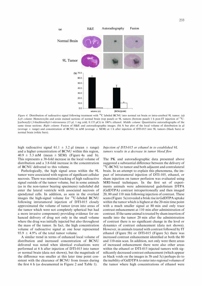
high radioactive signal 61.1 ± 3.2 ll (mean ± range)and a higher concentration of BCNU within this region,40.9 ± 5.3 mM (mean ± SEM) (Figure 4a and b).This represents a 38-fold increase in the local volume ofdistribution and a 3.8-fold increase in the concentrationof BCNU delivered to this volume.Pathologically, the high signal areas within the 9L
tumor were associated with regions of significant cellularnecrosis. There was minimal tracking of high radioactivesignal outside of the tumor volume, but in some animals(as in the non-tumor bearing specimens) radiolabel didenter the lateral ventricle with associated necrosis ofependymal cells. In addition, as seen in the overlaidimages the high-signal volume for 14C-labeled BCNUfollowing intratumoral injection of DTI-015 closelyapproximated the volume of tumor (even into areas ofthe tumor which were not completely spherical but hada more invasive component) providing evidence for en-hanced delivery of drug not only in the small volumewhere the drug was initially injected but also throughoutthe mass of the tumor. In fact, the high concentrationvolume of radioactive signal at one hour represented93.9 ± 4.9% of the total tumor volume.A similar trend in terms of enhanced local volume of
distribution and increased concentration of BCNUdelivered was noted when identical evaluations wereperformed at 6 h after injection of DTI-015 into tumoror normal brain (data not shown), but the magnitude ofthe difference was smaller at this later time point con-sistent with the clearance of BCNU from tissues duringthe first 6 h (as documented in Figure 2 and Table 1).
Injection of DTI-015 or ethanol in to established 9Ltumors results in a decrease in tumor blood flow
The PK and autoradiographic data presented abovesuggested a substantial difference between the delivery of14C-BCNU to tumor and both adjacent and contralateralbrain. In an attempt to explain this phenomena, the im-pact of intratumoral injection of DTI-105, ethanol, orsham injection on tumor perfusion was evaluated usingMRI-based techniques. In the first set of experi-ments animals were administered gadolinium DTPA(GdDTPA) contrast intraperitoneally and then imaged20, 80 and 110 min following injection of contrast. Thesescans (Figure 5a) revealed abrisk rise inGdDTPAuptakewithin the tumor which is highest at the 20-min time pointwith a much smaller signal at 80 min and only tracecontrast enhancement at 110 min after administration ofcontrast. If the same animal is treatedby sham insertion ofneedle into the tumor 20 min after the administrationof contrast there is no significant change in the level ordynamics of contrast enhancement (data not shown).However, in animals treated with contrast followed by ITethanol (Figure 5b) or DTI-015 (Figure 5c) there wasincreased contrast enhancement identified at both the 80and 110-min scan. In addition, not only were there areasof increased enhancement there were also other areaswithin the ethanol or DTI-015 injected tumors with sig-nificantly decreased contrast enhancement (which appearas black voids on the images in 5b and 5c) perhaps do tothe inability ofGdDTPA to enter into regional volumes ofthe tumor where high concentrations of ethanol were
Figure 4. Distribution of radioactive signal following treatment with 14C labeled BCNU into normal rat brain or intra-cerebral 9L tumor. (a)
Left column: Hemotoxylin and eosin stained sections of normal brain (top panel) or 9L tumors (bottom panel) 1 h post-IT injection of 14C-
[carbonyl]-1,3-bischlorethyl-1-nitrosourea (15 ll, 1 mg cold, 0.135 lCi) in 100% ethanol. Middle column: Quantitative autoradiographs of the
same tissue sections. Right column: Fusion of H&E and autoradiographic images. (b) A bar plot of the local volume of distribution in ll(average ± range) and concentration of BCNU in mM (average ± SEM) at 1 h after injection of DTI-015 into 9L tumors (black bars) or
normal brain (white bars).
233

present. In addition, when the reverse experiment wasperformed where DTI-015 or ethanol were administered20 min prior to contrast there was a corresponding de-crease in contrast uptakewithin the tumor as compared toanimals only treated with sham insertion of the needleinto the tumor again suggesting that DTI-015 or ethanoltreatment decreased tumor perfusion as demonstrated byalterations in dynamic contrast enhancement (data notshown).To further evaluate this alteration in tumor perfusion,
quantitative perfusion imaging [33] was used in intra-cerebral 9L tumors prior to and both 1 and 24 h after:sham, ethanol, or DTI-015 injection IT (Figure 5d).Prior to treatment the tumors from the 3 differentgroups had similar baseline blood flow (average45.4 ± 5.6 ml/100 g/min, P < 0.91). Sham treatmentcaused no significant change in blood flow at either the 1or 24-h time points (99.2 ± 6.6% and 112.2 ± 9.3%).In contrast, in tumors treated with IT injection of DTI-015 there was a significant decrease in tumor perfusionat the one hour post injection time point (57.7 ± 6.0%,P ¼ 0.018) which had largely normalized at the 24 h andlater time points (97.6 ± 10.0%, P ¼ 0.39). In ethanoltreated animals there was a trend toward decreasedblood flow at one hour (64.6 ± 47.9%) that did notachieve statistical significance (P ¼ 0.88). Therefore,alterations in tumor perfusion following injection ofDTI-015 confirm the results seen in dynamic contrastimages that DTI-015 administered IT caused a transientlocal decrease in tumor blood flow.
Systemic pharmacokinetics of BCNU in patients treatedwith DTI-015
The rat PK data suggested both an increase in14C-BCNU delivery to tumor and a markedly enhanced
TTR ratio for 14C-BCNU administered by IT injectionas compared to IV infusion. In order to perform aninitial evaluation of systemic exposure to BCNU fol-lowing treatment with DTI-015 three patients treated ona prospective clinical protocol for recurrent high-gradeglioma (20) had blood levels of BCNU determined atserial time points after injection of DTI-015. Followingtreatment with DTI-015 the Cmax in the blood(93.6 ± 49.3 ng/ml) was observed approximately 1 hfollowing injection (53 ± 14 min, Figures 6a and b).This peak blood concentration of BCNU occurred bothlater and was lower than that previously observed fol-lowing IV administration of BCNU to patients where apeak concentration of 2000 ng/ml was observed at40 min following the start of a 30-min IV infusion (i.e.10 min after the completion of treatment) [27]. Thisenhanced therapeutic index is further highlighted by thefact that for patients being treated with DTI-015 theaverage injected mass of BCNU (3.98 ± 0.76mg/kg) was 2-fold higher than that utilized for IVinfusion (1.71 ± 0.11 mg/kg). Finally, despite the al-tered route of administration the terminal clearance ofBCNU from the systemic circulation was similar in bothcases with the half-life observed for all time points >45min of 106 ± 14 min for DTI-015 administration and95 min for IV treatment.
Discussion
The experiments reported here reveal both the local andsystemic distribution of 14C-label following IT deliveryof 14C-BCNU in the rat 9L gliosarcoma model. Asindicated there was a >100-fold increase in theamount of radioactivity delivered to tumor tissue
Pre 20 80 110Minutes from Contrast
(a)
(b)
(c)
(d)
Control
Ethanol
DTI-015
0.0
20.0
40.0
60.0
80.0
100.0
120.0
140.0
160.0180.0
1 24Time Post Injection (hours)
Per
can
tag
e o
f S
tart
ing
Flo
w
SHAMETOHDTI-015
Figure 5. Dynamic contrast imaging and tumor blood flow following Ethanol or DTI-015 treatment of 9L tumors. T1-weighted MR images of
9L tumors either before (pre-) or 20, 80, or 110 min following IP administration of GdDTPA. Treatment was performed after the 20 min scan
and consisted of no treatment (control, a), or injection of 15 ll ethanol (b), or DTI-015 (c). In Figure 6d, quantitative perfusion imaging of 9L
tumors was performed before and both 1 and 24 h after the same three treatments. Blood flow in tumors as a percentage of baseline blood flow is
plotted for the average ± SEM of 3 animals for: Sham (black bars), ethanol (gray bars), or DTI-015 (white bars).
234

following DTI-015; in addition, despite the increaseddelivery of drug to tumor via IT injection there was noappreciable increase in the systemic exposure to 14C-BCNU or its metabolites compared to IV treatment.The dose of 1 mg of BCNU utilized by either route inthis study corresponds to 6.6–8.0 mg/kg which is similarto the 1
2 LD10 dose previously reported (6.65 mg/kg),where LD10 equals the dose that caused death due todrug toxicity in 10% of the animals [28]. In previousreports, treatment with the 1
2 LD10 dose systemicallycaused a small cytotoxic effect in this 9L gliosarcomamodel (0.2 log cell-kill), a modest change in apparentcellular water diffusion, no appreciable increase in ani-mal survival, and no systemic toxicity [28,29]. Therefore,these kinetic data suggest that the in the 9L intracerebralrat model the use of DTI-015 at the dose of 1 mg pro-vides at least a 100-fold increase in tumor exposure toBCNU compared to IV administration while providinga systemic exposure that has been documented to be welltolerated with minimal toxicity.In addition, the enhanced delivery of 14C-BCNU to
tumor compared to systemic tissues following treatmentwith DTI-015 is similar to what was previously observedby measuring DNA adducts in RIF-1 flank tumorsgrown in nude mice following either IP infusion ofBCNU or IT injection of DTI-015 where there was a164-fold increase in the DNA adducts identified in the
tumor after treatment with DTI-015 as compared to IPinfusion. In addition, despite the increased delivery ofBCNU to flank tumors, the level of adducts in othersystemic tissues (brain, lung, liver, and kidney) were notincreased above background following IT treatmentwith DTI-015 [30].The enhancement in the tumor exposure to 14C-
BCNU and its metabolites following injection of DTI-015 likely explains the therapeutic advantage previouslyreported for rats bearing 9L gliosarcoma treated withDTI-015 [23]. In that study rats received a volume ofDTI-015 up to 50% of the tumor volume or 30 ll,whichever was smaller, containing up to 2 mg ofBCNU. This treatment regimen was based on that pre-viously reported in the phase I ⁄ II trial of DTI-015 inpatients with recurrent high-grade glioma [20]. Thatstudy escalated the volume of ethanol and the dose ofBCNU utilized in parallel to achieve the maximumvalues for these two parameters. The MTD was even-tually established as 50% of the tumor volume up to5 ml and 240 mg BCNU. Therefore, when performingstudies to evaluate MRI based measures of early ther-apeutic efficacy [23] similar parameters were utilized forthe rat glioma model.Treatment with these parameters in the 9L glioma
model resulted in massive cell death (as documented bydiffusion weighted MRI), complete radiographicregression of tumor by 16 days post injection, and 75%of animals tumor free at 30 days after treatment. This issignificantly better than that previously observed usingsystemic administration of a much higher dose ofBCNU (2 · LD10 BCNU or 3.3–4.0 mg) where onlyminimal tumor regression with no complete responseswas observed and all animals still succumbed to tumorrelated death within 20 days of treatment [22,29]. Takentogether these sets of data suggest that IT injection ofDTI-015 results in a dramatic increase in tumor re-sponse to treatment compared to systemic infusion evenfor a smaller dose of drug delivered IT. Further, thecurrent PK data which reveal a greater than 100-foldincrease in delivery of 14C-BCNU to tumors by ITtreatment compared to IV infusion represent a viableexplanation of the enhanced efficacy observed in thispreclinical model of high-grade glioma.Clinically diffuse fibrillary high-grade gliomas are
known for the invasive nature of the disease with tumorcells often found on pathologic review as much as manycentimeters from the site of the tumor within whatgrossly appears to be normal brain tissue [1]. It is un-clear how the result obtained using the 9L gliosarcomamodel, where intracerebral tumors tend to grow as acontained mass with little invasion in to the surroundingbrain tissue, can be extrapolated to invasive nature ofprimary high-grade brain tumors. To date, a preclinicalmodel of DTI-015 in an infiltrative tumor model has notyet been reported to ascertain whether or not adminis-tration of DTI-015 will result in enhanced delivery ofBCNU to infiltrating tumor cells. However, the localdistribution of 14C-BCNU in tumors observed in thepresent study following direct IT injection of DTI-015would suggest transit of drug throughout the tumor. Infact, some of the non-uniform tendrils of 9L tumor
0
500
1000
1500
2000
0 100 200 300 400Time from start of infusion (min)
[B)l
m/g
n(]
UN
C
10
100
1000
10000
[]
UN
CB
(ng
/mL
)
IV IT
(a)
(b)
Figure 6. Systemic pharmacokinetics of BCNU in humans following
IV infusion of BCNU or IT delivery of DTI-015. (a) The concentration
of BCNU in peripheral blood (ng ⁄ ml) as a function of time after start
of infusion (minutes) is plotted for 3 patients treated with DTI-015
(open square, open triangle, and closed diamond) or for the average of
4 patients treated by IV infusion (x-symbols). Data for IT represent the
actual values at each time point and that for IV the mean ± SEM
taken from Cancer Treat Rep, 62: 1305–1312, (1978). (b) Peak con-
centration of BCNU in serum for the above patients.
235

appeared to have significant tracking of radioactiveBCNU suggesting that following injection of DTI-015the drug was not simply expanding to fill a sphericalvolume, but it was in fact being transported preferen-tially in tumor tissue as compared to surrounding nor-mal brain. Indeed approximately 90% of the tumorappeared to harbor the highest signal intensity ofBCNU which correlated with a concentration of 40.9 ±5.2 mM. This is much higher (>300-fold) than the IC95
(100 lM) observed in culture for BCNU in the 9L cellsused in this study (D.A.H., unpublished data).The local PK data in this animal model also confirm
the previous report of a patient with recurrent GBMtreated with DTI-015 (240 mg in 4 ml) where 60 minafter treatment stereotactic biopsies were performed andmeasurement of DNA adducts revealed an 879-fold in-crease above background in tumor tissue up to 0.9 cmfrom the site of injection and a 6-fold increase abovebackground for tissue even up to 2.4 cm from injection[31]. The combination of enhanced local concentrationsof drug and increased delivery of drug to surroundingbrain may explain the quite positive result observed inthe phase I ⁄ II trial where patients with recurrent inop-erable GBM still achieved a median survival of55 weeks, which is comparable to that for most patientsat the time of diagnosis, and was considerably betterthan matched historical controls at the same treatinginstitution where the median survival of recurrent andunresectable patients was only 25 weeks [20].Therefore, both preclinical and clinical data suggest
that solvent facilitated perfusion enhances delivery ofBCNU to tumors following IT injection. However, themechanism by which ethanol facilitates the transit ofBCNU throughout the tumor has not been fully eluci-dated. It does appear to be a factor of the organic sol-vent, for treatment of Walker 256 flank tumors withdirect injection of BCNU in either 100% ethanol or10% ethanol/90% saline gave markedly different anti-tumor response even for the same dose of BCNU [32].A number of hypotheses can be made as to the mecha-nism of the preferential delivery of 14C-BCNU to thetumor following IT treatment with DTI-015.First, 9L tumors have been documented to have a
higher cellular density and choline : ethanoloamine ratiothan surrounding normal rat brain suggesting a morelipophilic cellular mass [33]. Ethanol may, therefore,facilitate partitioning of BCNU into this lipophilic tumorbetter than BCNU in water, and this enhanced parti-tioning may be greater in tumor than in normal brainwhich is not as lipophilic. Transit of BCNUmay, thus, befacilitated in this fashion within the tumor until the sol-vent interface reaches normal brain tissue at which timethere is less of an advantage for the organic solvent anddecreased movement of the drug into surrounding brainparenchyma.A second possible contributing factor is the docu-
mented fact that 9L tumors have lower blood flow thannormal surrounding brain [34,35] which by our mea-surements was 45.4 (ml ⁄ 100 mg tissue ⁄min) in tumorvs. 138 ± 25 (ml ⁄mg tissue ⁄min) in the brain sur-rounding the tumor [36]. As a result of this decreasedblood flow within the tumor the injected BCNU would
have lower clearance in tumor as compared to normalsurrounding brain allowing a greater chance for thedrug to diffuse throughout the tumor. Similarly, oncethe drug does diffuse out of the tumor into normalbrain tissue the greater perfusion of these tissues wouldresult in enhanced clearance, and, therefore, lowerresidual concentrations of BCNU in brain tissue.A third option, which is in part supported by our
perfusion and dynamic contrast images, is that eitherBCNU, ethanol, or the combination may alter theblood flow within the tumor thus further enhancing theretention of the drug within the tumor as compared tosurrounding tissues. One possible mechanism to explainthese different responses in perfusion to DTI-015treatment is that the aberrant blood vessels which de-velop within the tumor via neoangiogenic processes maybe more susceptible to this alteration in blood flowfollowing injection of DTI-015 than mature vesselsfound within the surrounding brain parenchyma. Fi-nally, it is also may be that each of these three theoriesand perhaps other undetermined factors all contributeto both the increased local volume of delivery of BCNUto tumors and the enhanced TTR observed followingadministration of DTI-015.Finally, preliminary data are presented on the sys-
temic exposure of patients to BCNU following ITinjection of DTI-015 and are compared to previouslypublished results for IV infusion of BCNU. Despite a2-fold increase in the amount of administered BCNU,in the patients tested DTI-015 resulted in a 20-folddecrease in the Cmax for BCNU in the peripheralblood. Despite this decreased Cmax there was noapparent difference in the clearance of BCNU from thesystemic circulation as determined by the terminal half-life. It is interesting to note that the human data re-vealed a 20-fold decrease in the systemic BCNUexposure between these two modes of administrationwhile the rat PK data reported similar overall systemicexposure. One potential explanation for this differenceis that the human clinical data measured absoluteBCNU levels, while the rat PK data recorded 14Cradioactivity. Therefore, much of the late radioactivityseen in the rat model my represent inactive metabolitesor terminal adducts of BCNU and not the nativecompound.
Conclusion
The use of DTI-015 offers a 100-fold or greater in-crease in the delivery of 14C label to intra-cerebral 9Ltumor compared to IV administration without anyincrease in the systemic exposure. Further, injection of14C-BCNU in ethanol appears to provide rapid andnear complete saturation of tumors by BCNU withlittle deposition of drug into surrounding tissues,which may be in part due to alterations in tumorblood flow following injection of DTI-015. Finally, inpatients treated with DTI-015 as compared to IVinfusion of BNCU there is: a delay in the time toachieve Cmax, a decrease in systemic exposure to
236

BCNU, but a similar terminal elimination rate forBCNU. All of these preclinical and clinical data sug-gest that DTI-015 warrants further investigation as ameans to provide enhanced tumor specific delivery ofBCNU to patients with high-grade glioma.
Acknowledgements
Support for this work was provided by NIH Grants:P01CA85878, P50CA01014, andR24CA83099 and by aninstitutional grant from Direct Therapeutics Inc. toB.D.R. D.A.H. had support from a Varian MedicalSystems/RSNA Holman Pathway Research ResidentSeed Grant.
References
1. Behin A, Hoang-Xuan K, Carpentier AF, Delattre JY: Primary
brain tumours in adults. Lancet 361: 323–331, 2003
2. Dorr R, Hoff D: Carmustine. In: Dorr R, and Hoffs D (eds)
Cancer Chemotherapy Handbook. Appleton and Lange, Nor-
walk, 1994, pp. 267–275
3. Stewart LA: Chemotherapy in adult high-grade glioma: a
systematic review and meta- analysis of individual patient data
from 12 randomised trials. Lancet 359: 1011–1018, 2002
4. Hasleton PS, O’Driscoll BR, Lynch P, Webster A, Kalra SJ,
Gattamaneini HR, Woodcock AA, Poulter LW: Late BCNU
lung: a light and ultrastructural study on the delayed effect of
BCNU on the lung parenchyma. J Pathol 164: 31–36, 1991
5. O’Driscoll BR, Hasleton PS, Taylor PM, Poulter LW, Gatta-
meneni HR, Woodcock AA: Active lung fibrosis up to 17 years
after chemotherapy with carmustine (BCNU) in childhood. N
Engl J Med 323: 378–382, 1990
6. Thompson GR, Larson RE: The hepatotoxicity of 1,3-bis (2-
chloroethyl)-1-nitrosurea (BCNU) in rats. J Pharmacol Exp Ther
166: 104–112, 1969
7. Hochberg FH, Pruitt AA, Beck DO, DeBrun G, Davis K: The
rationale and methodology for intra-arterial chemotherapy with
BCNU as treatment for glioblastoma. J Neurosurg 63: 876–880,
1985
8. Hassenbusch SJ, Anderson JH, Whiting DM: Intra-arterial
chemotherapy for brain tumors. Cleve Clin J Med 57: 513–520,
1990
9. Walter KA, Tamargo RJ, Olivi A, Burger PC, Brem H:
Intratumoral chemotherapy. Neurosurgery 37: 1128–1145, 1995
10. Brem H, Piantadosi S, Burger PC, Walker M, Selker R, Vick
NA, Black K, Sisti M, Brem S, Mohr G, et al.: Placebo-
controlled trial of safety and efficacy of intraoperative con-
trolled delivery by biodegradable polymers of chemotherapy for
recurrent gliomas. The Polymer-brain Tumor Treatment
Group. Lancet 345: 1008–1012, 1995
11. Kleinschmidt-DeMasters BK: Intracarotid BCNU leukoence-
phalopathy. Cancer 57: 1276–1280, 1986
12. Miller DF, Bay JW, Lederman RJ, Purvis JD, Rogers LR,
Tomsak RL: Ocular and orbital toxicity following intracarotid
injection of BCNU (carmustine) and cisplatinum for malignant
gliomas. Ophthalmology 92: 402–406, 1985
13. Westphal M, Hilt DC, Bortey E, Delavault P, Olivares R,
Warnke PC, Whittle IR, Jaaskelainen J, Ram Z: A phase 3 trial of
local chemotherapy with biodegradable carmustine (BCNU)
wafers (Gliadel wafers) in patients with primary malignant
glioma. Neuro-oncol 5: 79–88, 2003
14. Olivi A, Grossman SA, Tatter S, Barker F, Judy K, Olsen J,
Bruce J, Hilt D, Fisher J, Piantadosi S: Dose escalation of
carmustine in surgically implanted polymers in patients with
recurrent malignant glioma: a new approaches to brain tumor
therapy CNS consortium trial. J Clin Oncol 21: 1845–1849, 2003
15. Fleming AB, Saltzman WM: Pharmacokinetics of the carmustine
implant. Clin Pharmacokinet 41: 403–419, 2002
16. Bobo RH, Laske DW, Akbasak A, Morrison PF, Dedrick RL,
Oldfield EH: Convection-enhanced delivery of macromolecules in
the brain. Proc Natl Acad Sci USA, 91: 2076–2080, 1994
17. Vavra M, Ali MJ, Kang EW, Navalitloha Y, Ebert A, Allen CV,
Groothuis DR: Comparative pharmacokinetics of 14C-sucrose in
RG-2 rat gliomas after intravenous and convection-enhanced
delivery. Neuro-oncol 6: 104–112, 2004
18. Bruce JN, Falavigna A, Johnson JP, Hall JS, Birch BD, Yoon JT,
Wu EX, Fine RL, Parsa AT: Intracerebral clysis in a rat glioma
model. Neurosurgery, 46: 683–691, 2000
19. Mardor Y, Roth Y, Lidar Z, Jonas T, Pfeffer R, Maier SE, Faibel
M, Nass D, Hadani M, Orenstein A, Cohen JS, Ram Z:
Monitoring response to convection-enhanced taxol delivery in
brain tumor patients using diffusion-weighted magnetic resonance
imaging. Cancer Res 61: 4971–4973, 2001
20. Hassenbusch SJ, Nardone EM, Levin VA, Leeds N, Pietronigro
D: Stereotactic injection of DTI-015 into recurrent malignant
gliomas: phase I ⁄ II trial. Neoplasia 5: 9–16, 2003
21. Pietronigro D, Drnovsky F, Cravioto H, Ransohoff J: DTI-015
produces cures in T9 gliosarcoma. Neoplasia 5: 17–22, 2003
22. Chenevert TL, McKeever PE, Ross BD: Monitoring early
response of experimental brain tumors to therapy using diffusion
magnetic resonance imaging. Clin Cancer Res 3: 1457–1466, 1997
23. Hall DE, Moffat BA, Stojanovska J, Johnson TD, Li Z, Hamstra
DA, Rehemtulla A, Chenevert TL, Carter J, Pietronnigro D, Ross
BD: Therapeutic Efficacy of DTI-015 using diffusion MRI as an
early surrogate marker. Clin Cancer Res 10: 7852–7859, 2004
24. Ross BD, Merkle H, Hendrich K, Staewen RS, Garwood M:
Spatially localized in vivo 1H magnetic resonance spectroscopy of
an intracerebral rat glioma. Magn Reson Med 23: 96–108, 1992
25. Colvin M, Brundrett RB, Cowens W, Jardine I, Ludlum DB: A
chemical basis for the antitumor activity of chloroethylnitrosou-
reas. Biochem Pharmacol 25: 695–699, 1976
26. DeVita VT, Denham C, Davidson JD, Oliverio VT: The
physiological disposition of the carcinostatic 1,3-bis(2-chloroeth-
yl)-1-nitrosourea (BCNU) in man and animals. Clin Pharmacol
Ther 8: 566–577, 1967
27. Levin VA, Hoffman W, Weinkam RJ: Pharmacokinetics of
BCNU in man: a preliminary study of 20 patients. Cancer Treat
Rep 62: 1305–1312, 1978
28. Ross BD, Zhao YJ, Neal ER, Stegman LD, Ercolani M, Ben-
Yoseph O, Chenevert TL: Contributions of cell kill and
posttreatment tumor growth rates to the repopulation of
intracerebral 9L tumors after chemotherapy: an MRI study.
Proc Natl Acad Sci USA, 95: 7012–7017, 1998
29. Chenevert TL, Stegman LD, Taylor JM, Robertson PL, Green-
berg HS, Rehemtulla A, Ross BD: Diffusion magnetic resonance
imaging: an early surrogate marker of therapeutic efficacy in brain
tumors. J Natl Cancer Inst 92: 2029–2036, 2000
30. Bodell WJ, Giannini DD, Singh S, Pietronigro D, Levin VA:
Formation of DNA adducts and tumor growth delay following
intratumoral administration of DTI-015. J Neurooncol 62: 251–
258, 2003
31. Bodell WJ, Giannini DD, Hassenbusch S, Levin VA: Levels of
N7-(2-hydroxyethyl)guanine as a molecular dosimeter of drug
delivery to human brain tumors. Neuro-oncol 3: 241–245, 2001
32. Simpson-Herren L, Pietronigro D: Intratumoral injection of DTI-
015 in the Walker 256 subcutaneous model. Proc Am Assoc
Cancer Res 1999, 583 pp.
33. Ross BD, Chenevert TL, Kim B, Ben-Yoseph O: Magnetic
resonance imaging and spectroscopy: application to experimental
neuro-oncology. Q Magn Reson Biol Med 1: 89–106, 1994
34. Fross RD, Warnke PC, Groothuis DR: Blood flow and blood-to-
tissue transport in 9L gliosarcomas: the role of the brain tumor
model in drug delivery research. J Neurooncol 11: 185–197, 1991
35. Steen RG, Graham MM: 31P magnetic resonance spectroscopy is
sensitive to tumor hypoxia: perfusion and oxygenation of rat 9L
237

gliosarcoma after treatment with BCNU. NMR Biomed 4:
117–124, 1991
36. Moffat B, Chenevert T, Hall D, Rehemtulla A, Ross B: Quanti-
tative perfusion imaging by continuous inversion using pulse
trained arterial spin labeling. J Magn Reson Imaging, in press
Address for offprints: Brian D. Ross, Ph.D., The Center for Molecular
Imaging, The University of Michigan Medical Center, 9403 MSRB III,
1500 East Medical Center Drive, Ann Arbor, MI 48109-0648, USA;
Tel.: +1-734-763-2099; Fax: +1-734-647-2563; E-mail: bdross@
umich.edu
238
Copyright © 2022 FDOKUMEN