The hsp90 Chaperone Complex Regulates Intracellular Localization of the Dioxin Receptor
Upload
independentCategory
view
3download
0
Interpretation of studies on the developmental reproductivetoxicology of 2,3,7,8-tetrachloro-p-dioxin in male offspring
David R Bella,g,*, Sally Clodeb, Ming Qi Fana, Alwyn Fernandesc, Paul M D Fosterd, Tao Ji-anga, George Loizoue, Alan MacNicollc, Brian G. Millerf, Martin Rosec, Lang Tranf, andShaun Whitec
a School of Biology, University of Nottingham, University Park, Nottingham NG7 2RD, UK bCovance Laboratories Ltd., Otley Road, Harrogate, North Yorkshire, HG3 1PY, UK c The Foodand Environment Research Agency, Sand Hutton, York YO41 1LZ, UK d NIEHS, PO Box 12233(MD K2-12), 111 TW Alexander Drive, Research Triangle Park, NC 27709 USA e Health & SafetyLaboratory, Harpur Hill, Buxton, Derbyshire SK17 9JN, UK f Institute of Occupational Medicine,Research Avenue North, Riccarton, Edinburgh, EH14 4AP, UK g European Chemicals Agency,P.O. Box 400, 00121 Helsinki, Finland
AbstractThere have been several studies on the maternal administration of 2,3,7,8-tetrachlorodibenzo-p-dioxin (TCDD) and effects in the reproductive tract of male offspring, subsequent to riskassessments undertaken in 2001. This review compares the methodology and results to examinekey methodological features, and consistency in reported outcomes. Maternal dosing at > 0.8 μgTCDD/kg causes lethality and weight loss, and it is difficult to distinguish between direct andindirect effects of TCDD at these dose levels. Statistically-significant effects of maternal doses of<1 μg TCDD/kg (i.e. the dose levels relevant for risk assessment) on prostate weight orepididymal sperm counts in offspring were reported in the minority of studies. Thepharmacokinetics of TCDD differs considerably between acute and chronic dosing, and with doselevel of TCDD. On the basis of body burden, TCDD had different potency at inducing adverseeffects in the only comparison study between acute and chronic dosing. Understanding of thepharmacokinetics of TCDD and relationship to adverse effects in offspring is required. Theseanalyses identify key features of TCDD developmental toxicity in male offspring, and identifydata needs for future risk assessment.
KeywordsDioxin; TCDD; developmental; reproductive; toxicity; spermatogenesis
1. Background2,3,7,8-tetrachlorodibenzo-p-dioxin (TCDD) is a potent and persistent toxicant, which isrepresentative of a family of related compounds, including polyhalogenated dibenzo-dioxins
*Correspondence to: David R. Bell, European Chemicals Agency, P.O. Box 400, 00121 Helsinki, Finland, Phone (358) 9688403, Fax(358) 968618434, [email protected]'s Disclaimer: This is a PDF file of an unedited manuscript that has been accepted for publication. As a service to ourcustomers we are providing this early version of the manuscript. The manuscript will undergo copyediting, typesetting, and review ofthe resulting proof before it is published in its final citable form. Please note that during the production process errors may bediscovered which could affect the content, and all legal disclaimers that apply to the journal pertain.
NIH Public AccessAuthor ManuscriptFood Chem Toxicol. Author manuscript; available in PMC 2011 June 1.
Published in final edited form as:Food Chem Toxicol. 2010 June ; 48(6): 1439–1447. doi:10.1016/j.fct.2010.04.005.
NIH
-PA Author Manuscript
NIH
-PA Author Manuscript
NIH
-PA Author Manuscript

and furans, and polyhalogenated biphenyls (Poland and Knutson, 1982). The high potencyof these compounds, combined with their presence in food and the environment, makes acompelling case for a robust assessment of the risk that they pose to human populations.Data on the effects of TCDD on human populations are an inadequate basis for a riskassessment (e.g. Collins et al., 2009), and so several risk assessments are based upon themost potent adverse effects of TCDD that are seen in experimental animals (COT, 2001,JECFA, 2002, SCF, 2001).
Developmental exposure of rats to as little as 64 ng TCDD/kg was found to cause toxicity inF1 male offspring (Mably et al., 1992a, b, c). Exposure of dams on gestational day (GD)15to TCDD resulted in a >60% reduction in cauda epididymal sperm levels on post-natal day(PND)63, and these reductions in epididymal sperm levels persisted to PND120 (Fig. 1A), at~50% of control values. At PND63, the decrease in sperm and spermatid numbers wasassociated with reductions in weight of the epididymis, ventral prostate and seminal vesicles(Fig. 1A). Effects on accessory sex organs by other chemicals have been causally linked toeffects on spermatogenesis through an anti-androgenic mechanism, (e.g. Mylchreest et al.,1998), and so the effect of TCDD on multiple endpoints appears to corroborate the effect onsperm levels.
However, the constellation of effects originally noted by Mably et al (1992) was notreproducible in three subsequent studies (Faqi et al., 1998, Gray et al., 1997, Wilker et al.,1996); for example, Fig. 1B summarises the results of replication at the PND62-70 timepoint with doses of <800 ng TCDD/kg. There was a statistically significant effect of TCDDdoses <800 ng/kg on F1 epididymal sperm levels in (Faqi et al., 1998) and (Gray et al.,1997), but not in (Wilker et al., 1996). The single consistent effect of TCDD in decreasingepididymal sperm levels in F1 males is the most potent adverse effect of TCDD (Mably etal., 1992a, b, c, Faqi et al., 1998) and it was consequently used as the basis of some riskassessments (COT, 2001, JECFA, 2001, SCF, 2001).
Since these risk assessments were undertaken an additional seven studies examining theeffects of developmental exposure of rats to TCDD have been published (Bell et al., 2007b,Bell et al., 2007c, Ikeda et al., 2005, Ohsako et al., 2001, Ohsako et al., 2002, Simanainen etal., 2004, Yonemoto et al., 2005). This review seeks to examine critical aspects of theconduct, design and analysis of studies on developmental exposure to TCDD, and to identifykey aspects required for interpretation of these studies. These include statistical aspects ofthe analyses, differences in measurement parameters between different laboratories, andunderstanding the differences between acute and chronic dosing regimes in terms of TCDDpharmacokinetics and body burden. The consistency of outcomes from these studies iscritically evaluated, and implications for risk assessment are considered.
2. Maternal pharmacokinetics of TCDDPharmacokinetics can explain the concentration of TCDD in a target tissue, and how thiscould be affected by variables in the published papers such as dose, dose frequency, etc.After an acute dose of TCDD to adults, there is a high initial concentration of TCDD inliver, which then redistributes to adipose tissue (Weber et al., 1993). High dose levels ofTCDD induce hepatic CYP1A2, which acts as a low-affinity, high-capacity binder ofTCDD, causing TCDD to be sequestrated in the liver (Poland et al., 1989a, b).Consequently, the ratio of the concentration of TCDD in the liver, relative to adipose tissue,is a measure of the induction of cytochrome P450 (Diliberto et al., 2001). Markedly differentdose levels of TCDD are required to attain an equivalent body burden of TCDD after acuteand chronic administration, and these different dose levels can also affect induction of
Bell et al. Page 2
Food Chem Toxicol. Author manuscript; available in PMC 2011 June 1.
NIH
-PA Author Manuscript
NIH
-PA Author Manuscript
NIH
-PA Author Manuscript

cytochromes P450. However, the relevance of these concepts to fetal exposure to TCDD hasbeen unclear until recently.
Two independent studies have compared the dosing regimen for TCDD, with either an acutedose on Gestational Day 15 (GD15) or sub-chronic administration before (~ 90 days) andduring pregnancy (Bell et al., 2007a, Hurst et al., 2000a, Hurst et al., 2000b). Fig. 2Aillustrates the difference in distribution of TCDD between acute and sub-chronic dosing,using the liver: adipose ratio of TCDD concentrations from Bell et al. (2007a). After acuteadministration of TCDD on GD15, the ratio was > 2.5 in all TCDD dose groups at GD16,but fell to <0.7 in all dose groups by GD21. By contrast, after sub-chronic dosing, there wasno difference in the liver: adipose ratio from week 10 of dosing through to GD21. Thusthese data demonstrate that there is significant redistribution of TCDD from liver toextrahepatic tissue in the five days after an acute dose, and that this redistribution is notpresent after sub-chronic dosing.
Figs. 2A-C show that acute doses of TCDD approximately 40 ng/kg cause marked hepaticsequestration of TCDD ( Fig. 2B), while the lower dose levels of TCDD used in the chronicadministration regime cause a three-fold increase in the proportion of TCDD dosedistributing to extrahepatic tissues ( Fig. 2C). This non-linearity of TCDD distribution withdose level is also seen in Hurst et al. (2000a).
A greater proportion of TCDD dose reaches the adipose tissue with lower doses of TCDD,and consequently, the apparent half-life of TCDD increases at lower doses of TCDD, shownin Fig. 2D (Bell et al., 2007a) and Fig. 2E (Hurst et al., 2000a). Both data sets show amarked increase in half-life of TCDD at doses of TCDD below 5 ng/kg/day. Fig. 2F showsthe apparent half-life of TCDD in mice (Diliberto et al., 2001) also increases at low doses.Thus increased distribution of TCDD to adipose tissue at low doses of TCDD increases theapparent half-life of TCDD.
These data show that, for the same body burden of TCDD, acute and chronic dosingregimens result in markedly different tissue-specific distribution and pharmacokinetics ofTCDD, with high acute doses showing marked hepatic sequestration of TCDD. These highacute doses have reduced delivery of TCDD to extrahepatic tissues (e.g. the fetus, milk), andare consequently less reliable for extrapolation to normal human exposure.
3. Effects of high TCDD dose on perinatal lethality and growthIt is important to choose an appropriate dose level for studies with TCDD, since the grossorgan damage arising from frank toxicity can cause secondary effects. Table 2 collates somestudies showing the effects of a high dose of TCDD (ca. 1 μg TCDD/kg on GD15, or >40 ngTCDD/kg/day) on perinatal lethality and body growth. Of studies with less than 10 littersper dose group, only one out of six studies showed an effect of TCDD on perinatal puplethality, with Nishimura et al. (2003) describing a 60% decrease in litter size. Of the fivestudies with ten or more litters per group, four detect significant decreases in perinatal pupmortality. The finding of perinatal lethality is confirmed in several other studies which havesufficient statistical power, (e.g. Bjerke and Peterson, 1994,Roman et al., 1995,Sommer etal., 1996). Thus we propose that the failure of specific studies to detect lethality in offspringafter maternal doses of >0.8–1 μg TCDD/kg is a consequence of inadequate statisticalpower, and conclude that doses of 0.8–1 μg TCDD/kg cause perinatal lethality affecting~10–20% of offspring.
The effect of ~1 μg TCDD/kg on body weight gain in offspring can be readily measured, butthis is assessed with a variety of endpoints (body weight, or body weight gain) and differingrigour of approach (weight at a single time-point, serial weights of individual animals,
Bell et al. Page 3
Food Chem Toxicol. Author manuscript; available in PMC 2011 June 1.
NIH
-PA Author Manuscript
NIH
-PA Author Manuscript
NIH
-PA Author Manuscript

failure to account for litter as a variable). Of eleven studies, seven found a significantdecrease in body weight or body weight gain (Table 2). Three of the four studies whichfound no effect on body weight gain in the offspring had six or less litters per treatmentgroup, and consequently, in-sufficient statistical power for detecting effects on body weight.
Thus high doses of TCDD during development cause increased perinatal lethality anddecreased body weight gain (Table 2). The use of these dose levels to examine effects onreproductive parameters is potentially confounded by such effects, and so it is notnecessarily possible to extrapolate from the effects seen at these high dose levels to thoseseen at lower dose levels. This review therefore concentrates on dose levels less than 1 μg ofTCDD/kg.
4. Variability in measurements of epididymal sperm levels in ratThe results of Mably et al. (1992a) show highly significant effects; for example, a t-testshows that epididymal sperm levels from control versus high-dose TCDD rats on PND120are significantly different, with P < 10−10. Moreover, Mably et al. (1992a) were able to statethat “the LOAEL can be estimated to be substantially lower than 0.064 μg TCDD/kg (thelowest dose tested)”; these high levels of significance arise in part from the low variability inepididymal sperm measurements reported by Mably et al. (1992a). However, for twelvestudies on developmental toxicity of TCDD (Bell et al., 2007b, c, Ikeda et al., 2005, Ohsakoet al., 2001, Ohsako et al., 2002, Simanainen et al., 2004, Yonemoto et al., 2005, Mably etal., 1992a, Faqi et al., 1998, Gray et al., 1997, Wilker et al., 1996, Gray et al., 1995), thecoefficient of variation in cauda epididymal sperm levels ranges from 0.089 (Mably et al.,1992a) to 0.54 (Bell et al., 2007b) at PND120, with a mean of 0.26. The study of Mably etal. (1992a) has the lowest variability in epididymal sperm count measurement. The lowvariability in sperm counts is not unique to Holtzman rats (the strain used by Mably et al.,1992a–c), since subsequent studies in Holtzman rats had coefficients of variation of 0.42(Ohsako et al., 2001) and 0.12 (Ikeda et al., 2005). It is difficult to pinpoint any specificmethodological difference that could account for the low coefficient of variation in the studyof Mably; this study used manual counting of epididymal sperm, with no mention ofoperator “blinding”, and accounts for litter effects.
Another approach to characterising variability in sperm measurements is to examine meanepididymal sperm counts from the same laboratory. For example, Ohsako et al. (2002)reported counts of 25 ± 2.9 × 10⁶ and 20 ± 1.9×10⁶ (mean ± SEM) in control groups ofPND70 males, a ~20% difference. Fig. 3C and D show that after maternal exposure toTCDD, epididymal sperm counts in F1 rats are routinely in the range of 110–140% ofcontrol values, presumably reflecting the range of normal variation in mean sperm counts.Thus mean epididymal sperm counts from rats performed in the same laboratory frequentlyshow variation of 10–30%.
5. Reproducibility of effects of developmental exposure to TCDD on F1
malesThe effect of developmental exposure to TCDD on male F1 reproductive endpoints has beenexamined in eleven studies with doses below 1 μg TCDD/kg. This review will focus oneffects at PND62-70 and PND 120+, since these are the basis of the risk assessments thathave been performed. The use of sexually mature animals is recommended for evaluatingeffects on spermatogenesis (Creasy, 2003, Lanning et al., 2002), and so data from immatureanimals is not considered further. Fig. 3A shows data examining the effect of TCDD onprostate weight at PND 62–70. With the exception of the original report, there were nosignificant effects of < 1 μg/kg TCDD on prostate weight at PND62-70 in eight other
Bell et al. Page 4
Food Chem Toxicol. Author manuscript; available in PMC 2011 June 1.
NIH
-PA Author Manuscript
NIH
-PA Author Manuscript
NIH
-PA Author Manuscript

studies. The studies have sufficient statistical power to detect an effect; for example, Bell etal. (2007c) report a 90% power for detecting a 10% decrease in ventral prostate weight,compared with the ~40% decrease in ventral prostate weight reported by Mably et al.(1992a-c).
In F1 rats at PND120+, Mably et al. (1992a-c) reported no significant effect of maternalTCDD treatment on ventral prostate weight below 1 μg TCDD/kg (Fig. 3B). In contrast, adecrease in ventral prostate weight was seen after an acute dose of 200 ng TCDD/kg(Ohsako et al., 2001), or a chronic dose approximately equivalent to an acute dose of 400 ngTCDD/kg (Ikeda et al., 2005); notably, neither of these two studies accounted for litter as asource of variation in the statistical analysis, and the failure to account for litter effects canlead to spuriously inflated estimations of significance (Weil, 1970). Bell et al. (2007c)reported a significant increase in prostate weight after chronic dosing of 8 ng TCDD/kg/day(approximately equivalent to an acute dose of 200 ng TCDD/kg). Thus after developmentalexposure to <1 μg TCDD/kg, statistically significant reductions in prostate weight have beenreported in only three studies, two of which used inappropriate statistical analysis.
Fig. 3C and D show the effect of maternal exposure to TCDD on epididymal sperm levels inF1 rats, at PND 62–70, and 120+, respectively. Faqi et al. (1998) reported a decrease inepididymal sperm at ~50 ng TCDD/kg (the chronic dose regime used was approximated toan acute dose on the basis of liver TCDD concentration), Gray et al. (1997) saw effects at200 ng TCDD/kg in adult (but not PND63) rats, and Simanainen et al. (2004) saw areduction after 300 ng TCDD/kg in line C rats, but not at lower doses (Fig. 3C, D). Themagnitude of these reductions in epididymal sperm count was less than that observed byMably et al. (1992a), and was frequently within 30% of control values, i.e. within the rangeof normal variation. Bell et al. (2007b) observed a significant increase in epididymal spermcounts of 30–38% at the top two doses of TCDD, noted that these values were notaccompanied by an effect on testicular sperm production, were within the range of historicalcontrol epididymal sperm counts, and concluded that the statistical significance of theseresults arose from random variation. Statistically significant reductions in epididymal spermnumbers have been observed in only four out of eleven studies at doses <1 μg TCDD/kg,and in only three studies at doses <300 ng TCDD/kg.
Table 1 shows measurements of developmental delay in the offspring of TCDD-exposeddams, and TCDD-induced delay in balanopreputial separation (BPS, a marker of malepuberty) has been demonstrated in each study where it has been measured. After a singledose of TCDD on GD15, Gray et al. (1997) reported a delay after a maternal dose of 200 ngTCDD/kg, Yonemoto et al. (2005) reported a significant delay after a maternal dose of 200ng TCDD/kg, and Bell et al. (2007b) found no significant effect at 50 or 200 ng TCDD/kg,only at the highest dose of 1000 ng TCDD/kg; however, in this latter experiment, the data at200 ng TCDD/kg were just above the threshold for statistical significance. After sub-chronicdosing of TCDD, Faqi et al. (1998) reported a significant delay of unspecified magnitude intheir medium and high dose groups, and Bell et al. (2007c) showed that all three dose groups(2.4, 8 and 46 ng TCDD/kg/day) caused a significant delay in BPS. Although maternalTCDD administration reduces body weight gain in offspring, the body weight of males atPND 21 or 42 did not correlate with the delay in BPS, thus excluding decreased weight gainas a cause of delayed BPS (Bell et al., 2007c). Thus delay in BPS is consistently found to bean adverse effect in the offspring of animals dosed with TCDD, and may be the mostsensitive adverse effect. The direct comparison of acute and chronic dosing of TCDD in thesame strain of rats using the same methodology (Bell et al., 2007b,2007c) provides evidencethat chronic dosing of TCDD has more potent effects in offspring. The difference in toxicitybetween acute and chronic dosing regimens has only been directly compared in one
Bell et al. Page 5
Food Chem Toxicol. Author manuscript; available in PMC 2011 June 1.
NIH
-PA Author Manuscript
NIH
-PA Author Manuscript
NIH
-PA Author Manuscript

laboratory, and repeating this experiment in an independent laboratory is essential todemonstrate reproducibility.
6. Critical periods of susceptibility for exposure to TCDDIt is known that GD16-17 is a critical period of development in the rat, when anti-androgensand phthalates exert effects on the developing male reproductive system (Carruthers andFoster, 2005, Welsh et al., 2008). Given that developmental exposure to TCDD results inadverse effects in the offspring, the developmental timing of susceptibility to TCDD isimportant, and relevant studies are summarised in Table 3. Gray et al. (1995) show thatdosing at GD15, compared to GD8, has a much greater effect on offspring. Ohsako et al.(2002) showed there were numerous significant effects in offspring from rats treated atGD15, but little effect in offspring of rats treated with TCDD on GD18 or PND2. Incontrast, Bjerke and Peterson (1994) found that both in utero and lactational exposurecaused significant effects in offspring, but in utero exposure had a larger effect thanlactational exposure on daily sperm production, day of preputial separation, and suppressionof growth. Nishimura et al. (2003) had shown that administration of TCDD to Holtzman ratson GD15 results in thyroid hyperplasia in offspring. Lactational, but not in utero, exposureto TCDD was responsible for effects in offspring (Nishimura et al., 2005). It is challengingto reconcile data showing no or minimal effect of TCDD when administered on GD18 orPND2 (Ohsako et al., 2002) with data showing that lactational exposure of pups alone canhave a constellation of effects (Bjerke et al., 1994). The experiments of Nishimura et al.(2005, 2006) do not bear directly on male reproduction, but the profound toxicity caused bylactational transfer of TCDD is likely to affect reproductive physiology. The existing dataare contradictory, and do not allow a conclusion as to when developmental exposure toTCDD causes adverse reproductive effects in offspring.
7. DiscussionMably et al. (1992c) showed that maternal administration of 160 or 400 ng TCDD/kgreduced the weight of prostate in F1 males at PND 63 (and not in adult rats), and theseeffects were highly significant. However, only two (out of 10 other studies now conducted)show a statistically significant decrease in prostate weight after maternal administration of<1 μg TCDD/kg, and these particular studies (Ikeda et al., 2005, Ohsako et al., 2001) failedto account for litter differences as a source of variation in their statistical analyses (Weil,1970). After maternal doses of <1 μg TCDD/kg, eight of eleven studies show no significantdecrease in prostate weight from control values, and so the original report (Mably et al.,1992c) is not reproducible in the majority of laboratories.
Likewise, four out of eleven studies show a decrease in epididymal sperm counts aftermaternal dosing of <1 μg TCDD/kg (Faqi et al., 1998, Gray et al., 1997, Mably et al., 1992a,Simanainen et al., 2004). With the exception of (Mably et al., 1992a), the decreases seen inepididymal sperm count at maternal doses of <1 μg TCDD/kg are less than 30% fromcontrol values. Ashby et al. (2003) highlighted the importance of historical control data ininterpreting minimalist changes in testicular sperm counts; there must be considerablecaution in interpreting differences of 20% or less in epididymal sperm levels as beingtreatment-related effects, especially when the data show an ambiguous relationship withdose (Faqi et al., 1998, Gray et al., 1997). The fact that seven of the eleven studies (Fig. 3)find no significant decrease in F1 epididymal sperm counts after maternal dosing of <1 μgTCDD/kg, coupled to small effect size and weak dose-response of effects, calls into questionwhether this effect is reproducible. With the exception of the studies of Mably et al. (1992a,b, c), the studies that found an effect of <1 μg TCDD/kg of TCDD on prostate weight didnot find an effect on epididymal sperm levels, and vice versa. The demonstration of adverse
Bell et al. Page 6
Food Chem Toxicol. Author manuscript; available in PMC 2011 June 1.
NIH
-PA Author Manuscript
NIH
-PA Author Manuscript
NIH
-PA Author Manuscript

effects both on sperm counts and on accessory sexual organs, provides a corroboration thatis causally related via mechanism of action, e.g. through anti-androgenic signalling(Howdeshell et al., 2006). Corroboration through effects on multiple endpoints for theendpoints in Fig. 1A is evanescent (Fig. 3, data not shown).
In contrast, high maternal doses of TCDD (>0.8 μg TCDD/kg) cause perinatal lethality,decreased body weight (gain) in the pups (Table 2), and other toxicities (Nishimura et al.,2003,2005,2006). After maternal doses of ~1 μg TCDD/kg, it is difficult to determine if anyeffect in offspring is a direct result of TCDD, or is instead mediated non-specifically throughtoxic effects on other organs. The use of maternal doses of 1 μg TCDD/kg for mechanisticstudies to explain effects seen at 50–200 ng/kg is to be deprecated.
It is challenging to reconcile published reports that examine when TCDD exerts adverseeffects on offspring after maternal administration, and consequently, its mechanism of action(Table 3). Some studies show that in utero exposure alone causes toxicity (Ohsako et al.,2002), whereas others show that lactational transfer alone cause toxicity in offspring (Bjerkeand Peterson, 1994,Nishimura et al., 2005). Clarification of the mode of action of TCDD isimportant for two reasons. Firstly, this information is necessary to interpret existing data.For example, Bell et al. (2007b) exposed dams to an acute dose of TCDD on GD15, andfound that the LOAEL was 1 μg TCDD/kg, with no significant effects in offspring at 50 or200 ng TCDD/kg. Chronic dosing of TCDD had a LOAEL of 2 ng TCDD/kg/day for delayin balanopreputial separation (BPS) (Bell et al., 2007c). The TCDD body burdens on GD16and 21 (Bell et al., 2007a) are roughly equivalent between a chronic dose of 2 ng/kg/day andan acute dose of 50 ng/kg/day. Given that GD16-18 is the time period when TCDD exerts itseffects on the developing embryo that lead to reproductive effects, it should follow that thereis an approximately equivalent potency between chronic and acute dosing of TCDD. In fact,chronic dosing is approximately 20-fold more potent (based on TCDD body burden) thanacute dosing of TCDD at inducing delay in BPS in F1 males. This analysis suggests thatGD16-18 is not the time when TCDD is exerting the toxic effect that leads to a delay inBPS. An alternative explanation is that TCDD exerts its toxic effects either before GD15, orafter parturition, when the body burden of TCDD would be markedly different betweenacute and chronic dosing regimens.
The second reason for understanding the timing and mechanism of action is that the riskassessments use a pharmacokinetic comparison between human and rat. One assumption isthat exposure in utero at GD16-18 mediates the effects of developmental TCDD exposure,but this assumption is questionable (Table 3). Moreover, if lactational transfer of TCDD is akey determinant of TCDD toxicity in offspring, the consequences for human risk assessmentwould be difficult to predict. Physiologically-based pharmacokinetic models of TCDDdistribution have varying predictivity (Emond et al., 2004,Aylward et al., 2005,Evans andAndersen, 2000), do not yet predict lactational transfer of TCDD, and the apparent half-lifeof TCDD in children is lower than in adults (Kreuzer et al., 1997,Kerger et al., 2006,Leunget al., 2006). Thus determining whether TCDD exerts effects in utero, or via lactation, willhave ramifications for risk assessments of dioxins that rely upon the assumption that TCDDexerts its effects in utero (COT, 2001,JECFA, 2002,SCF, 2001). This is therefore a keyuncertainty in risk assessment.
8. ConclusionsIn conclusion, there has been a failure to replicate the magnitude or variety of responsescaused by maternal doses of <1 μg TCDD/kg in the original work of Mably et al. (1992a, b,c) since the risk assessments in 2001. While there are reports of adverse effects in offspringafter maternal administration of < 1 μg TCDD/kg, these reports are in the minority for
Bell et al. Page 7
Food Chem Toxicol. Author manuscript; available in PMC 2011 June 1.
NIH
-PA Author Manuscript
NIH
-PA Author Manuscript
NIH
-PA Author Manuscript

prostate weight and epididymal sperm counts, and are frequently within the range ofhistorical variation seen in other laboratories. It is unclear why it has not been possible toreplicate these findings, despite extensive efforts. Effects on developmental milestones(BPS) are consistently found, and the potency of TCDD to induce these effects appears to bemuch greater after chronic dosing, compared with acute dosing. Maternal pharmacokineticsof TCDD vary considerably between acute and chronic dosing, and these two differingdosing regimens have been shown to impact upon the potency of TCDD at inducing adverseeffects. Thus understanding how and when TCDD operates to cause adverse effects in F1animals after low dose maternal exposure is a key research need, with consequences forcurrent risk evaluations of dioxins and dioxin-like compounds.
AcknowledgmentsPractical studies on the toxicology and disposition of TCDD were funded by a contract (T01034) from the UK FoodStandards Agency. We thank Declan Brady for excellent technical support. We thank the editor and anonymousreviewers for constructive and insightful comments.
Abbreviations
BPS Balano-Preputial Separation
CASA Computer-Assisted Sperm Analysis
GD Gestational Day
PND Post-Natal Day
TCDD 2,3,7,8-tetrachlorodibenzo-p-dioxin
ReferencesAshby J, Tinwell H, Lefevre PA, Joiner R, Haseman J. The effect on sperm production in adult
Sprague-Dawley rats exposed by gavage to bisphenol a between postnatal days 91–97. Toxicol Sci2003;74:129–138. [PubMed: 12773777]
Aylward LL, Brunet RC, Starr TB, Carrier G, Delzell E, Cheng H, Beall C. Exposure reconstructionfor the TCDD-exposed NIOSH cohort using a concentration- and age-dependent model ofelimination. Risk Anal 2005;25:945–956. [PubMed: 16268942]
Bell DR, Clode S, Fan MQ, Fernandes A, Foster PM, Jiang T, Loizou G, Macnicoll A, Miller BG,Rose M, Tran L, White S. Relationships between tissue levels of 2,3,7,8-tetrachlorodibenzo-p-dioxin (TCDD), mRNAs and toxicity in the developing male Wistar(Han) rat. Toxicol Sci 2007a;99:591–604. [PubMed: 17656490]
Bell DR, Clode S, Fan MQ, Fernandes A, Foster PM, Jiang T, Loizou G, Macnicoll A, Miller BG,Rose M, Tran L, White S. Toxicity of 2,3,7,8-tetrachlorodibenzo-p-dioxin (TCDD) in thedeveloping male Wistar(Han) rat I: No Decrease in Epididymal Sperm Count After a Single AcuteDose. Toxicol Sci 2007b;99:214–223. [PubMed: 17545212]
Bell DR, Clode S, Fan MQ, Fernandes A, Foster PM, Jiang T, Loizou G, Macnicoll A, Miller BG,Rose M, Tran L, White S. Toxicity of 2,3,7,8-tetrachlorodibenzo-p-dioxin (TCDD) in thedeveloping male Wistar(Han) rat II: chronic dosing causes developmental delay. Toxicol Sci 2007c;99:224–233. [PubMed: 17545211]
Bjerke DL, Peterson RE. Reproductive toxicity of 2,3,7,8-tetrachlorodibenzo-p-dioxin in male-rats -different effects of in-utero versus lactational exposure. Tox App Pharmacol 1994;127:241–249.
Bjerke DL, Sommer RJ, Moore RW, Peterson RE. Effects of in-utero and lactational 2,3,7,8-tetrachlorodibenzo-p-dioxin exposure on responsiveness of the male-rat reproductive-system totestosterone stimulation in adulthood. Tox App Pharmacol 1994;127:250–257.
Bell et al. Page 8
Food Chem Toxicol. Author manuscript; available in PMC 2011 June 1.
NIH
-PA Author Manuscript
NIH
-PA Author Manuscript
NIH
-PA Author Manuscript

Carruthers CM, Foster PMD. Critical window of male reproductive tract development in rats followinggestational exposure to di-n-butyl phthalate. Birth Defects Res B-Dev Reprod Toxicol2005;74:277–285. [PubMed: 15954088]
Collins JJ, Bodner K, Aylward LL, Wilken M, Bodnar CM. Mortality rates among trichlorophenolworkers with exposure to 2,3,7,8-tetrachlorodibenzo-p-dioxin. Am J Epidemiol 2009;170:501–506.[PubMed: 19561065]
COT. Comittee on Toxicity of Chemicals in Food, Consumer Products and the Environment. COT/2001/07. 2001. Statement on the tolerable daily intake for dioxins and dioxin-like polychlorinatedbiphenyls; p. COT/2001/07
Creasy DM. Evaluation of testicular toxicology: A synopsis and discussion of the recommendationsproposed by the society of toxicologic pathology. Birth Defects Res B-Dev Reprod Toxicol2003;68:408–415. [PubMed: 14745990]
Diliberto JJ, Devito MJ, Ross GD, Birnbaum LS. Subchronic exposure of [H-3]-2,3,7,8-tetrachlorodibenzo-p-dioxin (TCDD) in female B6C3F1 mice: relationship of steady-state levels todisposition and metabolism. Toxicol Sci 2001;61:241–255. [PubMed: 11353133]
Emond C, Birnbaum LS, Devito MJ. Physiologically based pharmacokinetic model for developmentalexposures to TCDD in the rat. Toxicol Sci 2004;80:115–133. [PubMed: 15056810]
Evans MV, Andersen ME. Sensitivity analysis of a physiological model for 2,3,7,8-tetrachlorodibenzo-p-dioxin (TCDD): Assessing the impact of specific model parameters onsequestration in liver and fat in the rat. Toxicol Sci 2000;54:71–80. [PubMed: 10746933]
Faqi AS, Dalsenter PR, Merker HJ, Chahoud I. Reproductive toxicity and tissue concentrations of lowdoses of 2,3,7,8-tetrachlorodibenzo-p-dioxin in male offspring rats exposed throughout pregnancyand lactation. Toxicol App Pharmacol 1998;150:383–392.
Gray LE, Kelce WR, Monosson E, Ostby JS, Birnbaum LS. Exposure to TCDD during developmentpermanently alters reproductive function in male long-evans rats and hamsters - reduced ejaculatedand epididymal sperm numbers and sex accessory-gland weights in offspring with normalandrogenic status. Toxicol App Pharmacol 1995;131:108–118.
Gray LE, Ostby JS. In-utero 2,3,7,8-tetrachlorodibenzo-p-dioxin (TCDD) alters reproductivemorphology and function in female rat offspring. Toxicol App Pharmacol 1995;133:285–294.
Gray LE, Ostby JS, Kelce WR. A dose-response analysis of the reproductive effects of a singlegestational dose of 2,3,7,8-tetrachlorodibenzo-p-dioxin in male Long Evans Hooded rat offspring.Toxicol App Pharmacol 1997;146:11–20.
Howdeshell KL, Rider CV, Wilson VS, Gray LE. Mechanisms of action of phthalate esters,individually and in combination, to induce abnormal reproductive development in male laboratoryrats. Environ Res 2008;108:168–176. [PubMed: 18949836]
Hurst CH, Devito MJ, Birnbaum LS. Tissue disposition of 2,3,7,8-tetrachlorodibenzo-p-dioxin(TCDD) in maternal and developing Long-Evans rats following subchronic exposure. Toxicol Sci2000a;57:275–283. [PubMed: 11006357]
Hurst CH, Devito MJ, Setzer RW, Birnbaum LS. Acute administration of 2,3,7,8-tetrachlorodibenzo-p-dioxin (TCDD) in pregnant Long Evans rats: Association of measured tissue concentrations withdevelopmental effects. Toxicol Sci 2000b;53:411–420. [PubMed: 10696789]
Ikeda M, Tamura M, Yamashita J, Suzuki C, Tomita T. Repeated in utero and lactational 2,3,7,8-tetrachlorodibenzo-p-dioxin exposure affects male gonads in offspring, leading to sex ratiochanges in F-2 progeny. Toxicol App Pharmacol 2005;206:351–355.
JECFA. Evaluation of certain food additives and contaminants (57th report of the Joint FAO/WHOExpert Committee on Food Additives). WHO Technical Report Series 2002;909:121–149.
Kerger BD, Leung HW, Scott P, Paustenbach DJ, Needham LL, Patterson DG, Gerthoux PM,Mocarelli P. Age- and concentration-dependent elimination half-life of 2,3,7,8-tetrachlorodibenzo-p-dioxin in Seveso children. Env Health Persp 2006;114:1596–1602.
Kreuzer PE, Csanady GA, Baur C, Kessler W, Papke O, Greim H, Filser JG. 2,3,7,8-tetrachlorodibenzo-p-dioxin (TCDD) and congeners in infants. A toxicokinetic model of humanlifetime body burden by TCDD with special emphasis on its uptake by nutrition. Arch Toxicol1997;71:383–400. [PubMed: 9195020]
Bell et al. Page 9
Food Chem Toxicol. Author manuscript; available in PMC 2011 June 1.
NIH
-PA Author Manuscript
NIH
-PA Author Manuscript
NIH
-PA Author Manuscript

Lanning LL, Creasy DM, Chapin RE, Mann PC, Barlow NJ, Regan KS, Goodman DG. Recommendedapproaches for the evaluation of testicular and epididymal toxicity. Toxicol Path 2002;30:507–520. [PubMed: 12187942]
Leung HW, Kerger BD, Paustenbach DJ. Elimination half-lives of selected polychlorinateddibenzodioxins and dibenzofurans in breast-fed human infants. J Toxicol Environ Health A2006;69:437–443. [PubMed: 16574620]
Mably TA, Bjerke DL, Moore RW, GendronFitzpatrick A, Peterson RE. In utero and lactationalexposure of male-rats to 2,3,7,8-tetrachlorodibenzo-para-dioxin. 3 effects on spermatogenesis andreproductive capability. Toxicol App Pharmacol 1992a;114:118–126.
Mably TA, Moore RW, Goy RW, Peterson RE. In utero and lactational exposure of male-rats to2,3,7,8-tetrachlorodibenzo-para-dioxin. 2 effects on sexual-behavior and the regulation ofluteinizing-hormone secretion in adulthood. Toxicol App Pharmacol 1992b;114:108–117.
Mably TA, Moore RW, Peterson RE. In utero and lactational exposure of male-rats to 2,3,7,8-tetrachlorodibenzo-para-dioxin. 1 effects on androgenic status. Toxicol App Pharmacol 1992c;114:97–107.
Mylchreest E, Cattley RC, Foster PMD. Male reproductive tract malformations in rats followinggestational and lactational exposure to di(n-butyl) phthalate: An antiandrogenic mechanism?Toxicol Sci 1998;43:47–60. [PubMed: 9629619]
Nishimura N, Yonemoto J, Miyabara Y, Sato M, Tohyama C. Rat thyroid hyperplasia induced bygestational and lactational exposure to 2,3,7,8-tetrachlorodibenzo-p-dioxin. Endocrinol2003;144:2075–2083.
Nishimura N, Yonemoto J, Nishimura H, Ikushiro S, Tohyama C. Disruption of thyroid hormonehomeostasis at weaning of Holtzman rats by lactational but not in utero exposure to 2,3,7,8-tetrachlorodibenzo-p-dioxin. Toxicol Sci 2005;85:607–614. [PubMed: 15716479]
Nishimura N, Yonemoto J, Nishimura H, Tohyama C. Localization of cytochrome P450 1A1 in aspecific region of hydronephrotic kidney of rat neonates lactationally exposed to 2,3,7,8-tetrachlorodibenzo-p-dioxin. Toxicol 2006;227:117–126.
Ohsako S, Miyabara Y, Nishimura N, Kurosawa S, Sakaue M, Ishimura R, Sato M, Takeda K, Aoki Y,Sone H, Tohyama C, Yonemoto J. Maternal exposure to a low dose of 2,3,7,8-tetrachlorodibenzo-p-dioxin (TCDD) suppressed the development of reproductive organs of male rats: Dose-dependent increase of mRNA levels of 5 alpha-reductase type 2 in contrast to decrease ofandrogen receptor in the pubertal ventral prostate. Toxicol Sci 2001;60:132–143. [PubMed:11222880]
Ohsako S, Miyabara Y, Sakaue M, Ishimura R, Kakeyama M, Izumi H, Yonemoto J, Tohyama C.Developmental stage-specific effects of perinatal 2,3,7,8-tetrachlorodibenzo-p-dioxin exposure onreproductive organs of male rat offspring. Toxicol Sci 2002;66:283–292. [PubMed: 11896295]
Poland A, Knutson JC. 2,3,7,8-Tetrachlorodibenzo-para-dioxin and related halogenated aromatic-hydrocarbons - examination of the mechanism of toxicity. Annu Rev Pharmacol Toxicol1982;22:517–554. [PubMed: 6282188]
Poland A, Teitelbaum P, Glover E. [I-125]2-Iodo-3,7,8-Trichlorodibenzo-p-dioxin-binding species inmouse-liver induced by agonists for the ah receptor - characterization and identification. MolPharmacol 1989a;36:113–120. [PubMed: 2546045]
Poland A, Teitelbaum P, Glover E, Kende A. Stimulation of in vivo hepatic uptake and invitro hepaticbinding of [i-125]2-iodo-3,7,8-trichlorodibenzo-p-dioxin by the administration of agonists for theAh receptor. Mol Pharmacol 1989b;36:121–127. [PubMed: 2546046]
Roman BL, Sommer RJ, Shinomiya K, Peterson RE. In-utero and lactational exposure of the male-ratto 2,3,7,8-tetrachlorodibenzo-p-dioxin - impaired prostate growth and development withoutinhibited androgen production. Toxicol App Pharmacol 1995;134:241–250.
Rose JQ, Ramsey JC, Wentzler TH, Hummel RA, Gehring PJ. Fate of 2,3,7,8-tetrachlorodibenzo-para-dioxin following single and repeated oral doses to rat. Toxicol App Pharmacol 1976;36:209–226.
SCF. Opinion of the Scientific Committee On Food on the risk assessment of dioxins and dioxin-likepcbs in food. Update based on new scientific information available since the adoption of the SCFopinion of 22nd November 2000. 2001 Adopted on 30 May 2001.
Bell et al. Page 10
Food Chem Toxicol. Author manuscript; available in PMC 2011 June 1.
NIH
-PA Author Manuscript
NIH
-PA Author Manuscript
NIH
-PA Author Manuscript

Simanainen U, Haavisto T, Tuomisto JT, Paranko J, Toppari J, Tuomisto J, Peterson RE, Viluksela M.Pattern of male reproductive system effects after in utero and lactational 2,3,7,8-tetrachlorodibenzo-p-dioxin (TCDD) exposure in three differentially TCDD-sensitive rat lines.Toxicol Sci 2004;80:101–108. [PubMed: 15084753]
Sommer RJ, Ippolito DL, Peterson RE. In utero and lactational exposure of the male Holtzman rat to2,3,7,8-tetrachlorodibenzo-p-dioxin: Decreased epididymal and ejaculated sperm numbers withoutalterations in sperm transit rate. Toxicol App Pharmacol 1996;140:146–153.
Weber LWD, Ernst SW, Stahl BU, Rozman K. Tissue distribution and toxicokinetics of 2,3,7,8-tetrachlorodibenzo-p-dioxin in rats after intravenous-injection. Fund App Toxicol 1993;21:523–534.
Weil CS. Selection of valid number of sampling units and a consideration of their combination intoxicological studies involving reproduction, teratogenesis or carcinogenesis. Food CosmetToxicol 1970;8:177–182. [PubMed: 5421015]
Welsh M, Saunders PTK, Fisken M, Scott HM, Hutchison GR, Smith LB, Sharpe RM. Identificationin rats of a programming window for reproductive tract masculinization, disruption of which leadsto hypospadias and cryptorchidism. J Clin Invest 2008;118:1479–1490. [PubMed: 18340380]
Wilker C, Johnson L, Safe S. Effects of developmental exposure to indole-3-carbinol or 2,3,7,8-tetrachlorodibenzo-p-dioxin on reproductive potential of male rat offspring. Toxicol AppPharmacol 1996;141:68–75.
Yonemoto J, Ichiki T, Takei T, Tohyama C. Maternal exposure to 2,3,7,8-tetrachlorodibenzo-p-dioxinand the body burden in offspring of Long-Evans rats. Environ Health Prev Med 2005;10:21–32.
Bell et al. Page 11
Food Chem Toxicol. Author manuscript; available in PMC 2011 June 1.
NIH
-PA Author Manuscript
NIH
-PA Author Manuscript
NIH
-PA Author Manuscript

Fig. 1. Male reproductive effects of TCDD after developmental administrationA. Some of the effects noted by Mably et al (1992). Holtzman rats were dosed on GD15with the indicated dose of TCDD (ng/kg), and daily sperm production (circle), rightepididymis weight (star), ventral prostate weight (square), seminal vesicle weight (diamond)and cauda epididymal sperm number (triangle) measured in the F1 males on PND 63 (opensymbols) or PND120 (closed symbols). Results are normalised to control (set as 100%), andare presented as mean and SD. * indicates P<0.05; note that at 64 ng/kg, right epididymisweight, seminal vesicles weight and ventral prostate weight are not significantly differentfrom control. B. Comparison of effects of TCDD on F1 males at PND62-70, for a maternalTCDD dose of approximately 500 ng/kg and below. Data from (Mably et al., 1992c),(Mably et al., 1992a), (Faqi et al., 1998), (Gray et al., 1997) and (1996) (Wilker et al., 1996)are compared, on the basis of stated statistically significant results. √ means that there was astatistically significant effect. No means no statistically significant effect, Not Done meansthat the measurement is not reported, and 1 refers to measurements on whole prostate, ratherthan ventral prostate weight.
Bell et al. Page 12
Food Chem Toxicol. Author manuscript; available in PMC 2011 June 1.
NIH
-PA Author Manuscript
NIH
-PA Author Manuscript
NIH
-PA Author Manuscript
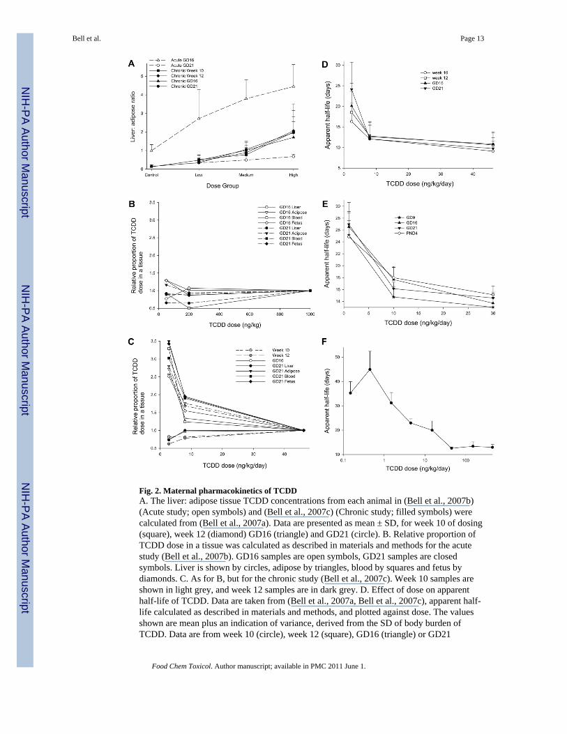
Fig. 2. Maternal pharmacokinetics of TCDDA. The liver: adipose tissue TCDD concentrations from each animal in (Bell et al., 2007b)(Acute study; open symbols) and (Bell et al., 2007c) (Chronic study; filled symbols) werecalculated from (Bell et al., 2007a). Data are presented as mean ± SD, for week 10 of dosing(square), week 12 (diamond) GD16 (triangle) and GD21 (circle). B. Relative proportion ofTCDD dose in a tissue was calculated as described in materials and methods for the acutestudy (Bell et al., 2007b). GD16 samples are open symbols, GD21 samples are closedsymbols. Liver is shown by circles, adipose by triangles, blood by squares and fetus bydiamonds. C. As for B, but for the chronic study (Bell et al., 2007c). Week 10 samples areshown in light grey, and week 12 samples are in dark grey. D. Effect of dose on apparenthalf-life of TCDD. Data are taken from (Bell et al., 2007a, Bell et al., 2007c), apparent half-life calculated as described in materials and methods, and plotted against dose. The valuesshown are mean plus an indication of variance, derived from the SD of body burden ofTCDD. Data are from week 10 (circle), week 12 (square), GD16 (triangle) or GD21
Bell et al. Page 13
Food Chem Toxicol. Author manuscript; available in PMC 2011 June 1.
NIH
-PA Author Manuscript
NIH
-PA Author Manuscript
NIH
-PA Author Manuscript

(inverted triangle). E. As for D, but using the data from (Hurst et al., 2000a). Data are fromGD9 (star), GD16 (triangle), GD21 (inverted triangle) and PND4 (circle). F. Effect of doseon apparent half-life of TCDD in mice. As for D, but the data are taken from (Diliberto etal., 2001).
Bell et al. Page 14
Food Chem Toxicol. Author manuscript; available in PMC 2011 June 1.
NIH
-PA Author Manuscript
NIH
-PA Author Manuscript
NIH
-PA Author Manuscript
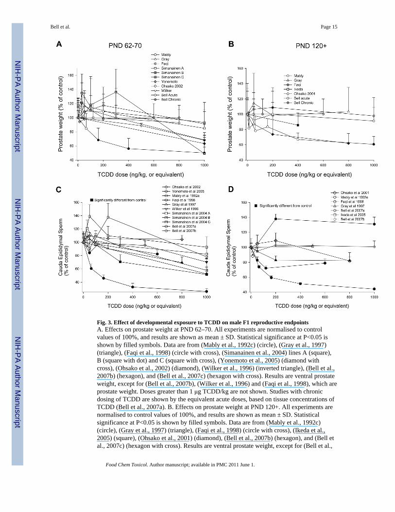
Fig. 3. Effect of developmental exposure to TCDD on male F1 reproductive endpointsA. Effects on prostate weight at PND 62–70. All experiments are normalised to controlvalues of 100%, and results are shown as mean ± SD. Statistical significance at P<0.05 isshown by filled symbols. Data are from (Mably et al., 1992c) (circle), (Gray et al., 1997)(triangle), (Faqi et al., 1998) (circle with cross), (Simanainen et al., 2004) lines A (square),B (square with dot) and C (square with cross), (Yonemoto et al., 2005) (diamond withcross), (Ohsako et al., 2002) (diamond), (Wilker et al., 1996) (inverted triangle), (Bell et al.,2007b) (hexagon), and (Bell et al., 2007c) (hexagon with cross). Results are ventral prostateweight, except for (Bell et al., 2007b), (Wilker et al., 1996) and (Faqi et al., 1998), which areprostate weight. Doses greater than 1 μg TCDD/kg are not shown. Studies with chronicdosing of TCDD are shown by the equivalent acute doses, based on tissue concentrations ofTCDD (Bell et al., 2007a). B. Effects on prostate weight at PND 120+. All experiments arenormalised to control values of 100%, and results are shown as mean ± SD. Statisticalsignificance at P<0.05 is shown by filled symbols. Data are from (Mably et al., 1992c)(circle), (Gray et al., 1997) (triangle), (Faqi et al., 1998) (circle with cross), (Ikeda et al.,2005) (square), (Ohsako et al., 2001) (diamond), (Bell et al., 2007b) (hexagon), and (Bell etal., 2007c) (hexagon with cross). Results are ventral prostate weight, except for (Bell et al.,
Bell et al. Page 15
Food Chem Toxicol. Author manuscript; available in PMC 2011 June 1.
NIH
-PA Author Manuscript
NIH
-PA Author Manuscript
NIH
-PA Author Manuscript

2007b) and (Faqi et al., 1998), which are prostate weight. C. Effects on epididymal spermcount at PND 62–70. All experiments are normalised to control values of 100%, and resultsare shown as mean ± SEM. Statistical significance at P<0.05 is shown by filled symbols.Data are from (Mably et al., 1992a) (circle), (Gray et al., 1997) (triangle), (Faqi et al., 1998)(circle with cross), (Simanainen et al., 2004) lines A (square), B (square with dot) and C(square with cross), (Yonemoto et al., 2005) (diamond with cross), (Ohsako et al., 2002)(diamond), (Wilker et al., 1996) (inverted triangle), (Bell et al., 2007b) (hexagon), and (Bellet al., 2007c) (hexagon with cross). Doses greater than 1 μg TCDD/kg are not shown.Studies with chronic dosing of TCDD are shown by the equivalent acute doses, based ontissue concentrations of TCDD (Bell et al., 2007a). D. Effects on epididymal sperm countsat PND 120+. All experiments are normalised to control values of 100%, and results areshown as mean ± SEM. Statistical significance at P<0.05 is shown by filled symbols. Dataare from (Mably et al., 1992c) (circle), (Gray et al., 1997) (triangle), (Faqi et al., 1998)(circle with cross), (Ikeda et al., 2005) (square), (Ohsako et al., 2001) (diamond), (Bell et al.,2007b) (hexagon), and (Bell et al., 2007c) (hexagon with cross).
Bell et al. Page 16
Food Chem Toxicol. Author manuscript; available in PMC 2011 June 1.
NIH
-PA Author Manuscript
NIH
-PA Author Manuscript
NIH
-PA Author Manuscript

NIH
-PA Author Manuscript
NIH
-PA Author Manuscript
NIH
-PA Author Manuscript
Bell et al. Page 17
TABLE 1
Effect of maternal TCDD dose on developmental delay.
Study Strain TCDD dosing regime Developmental delay
Faqi et al., 1998 Wistar Loading and weekly maintenancedose
“Age at preputial separation wasslightly delayed”, no statistical analysisshown (n=17–22)
Ikeda et al., 2005 Holtzman Loading and weekly maintenancedose
N.D.
Bell et al., 2007c Wistar(Han) Chronic dietary dosing for > twelveweeks
Yes, delay in BPS of 1.8, 1.9 and 4.4.days (n=18–25)
Mably et al., 1992a,Mably et al., 1992b,Mably et al., 1992c
Holtzman Single dose on GD15 Yes, testis descent and eye opening.BPS N.D. (n=9)
Bjerke and Peterson,1994
Holtzman Single dose on GD15; also in uteroand/or lactational
yes, 2.4 or 3.4 day delay in BPS (n=9–11)
Gray et al., 1995 Long Evans hooded Single dose on GD15 or GD 8 Yes, 3.6 day delay in BPS (n=6–8)
Roman et al., 1995 Holtzman Single dose on GD15 Yes, 2.1 day delay in BPS (n=30–32)
Wilker et al., 1996 Sprague-Dawley Single dose on GD15 N.D.
Sommer et al., 1996 Holtzman Single dose on GD15 Yes, 2 day delay in BPS (n=34–39)
Gray et al., 1997 Long Evans hooded Single dose on GD15 Yes, delay in eye opening, delay inBPS of 1.5, 3.1 days (n=10–12)
Ohsako et al., 2001 Holtzman Single dose on GD15 N.D.
Ohsako et al., 2002 Sprague-Dawley Single dose on GD15 or GD18 N.D.
Simanainen et al., 2004 Wistar(Han)xLong Evans crosses Single dose on GD15 N.D.
Yonemoto et al., 2005 Long Evans Single dose on GD15 Yes, delay in BPS (n=9–12)
Bell et al., 2007b Wistar(Han) Single dose on GD15 Yes, delay in BPS of 2.8 days (n=15–21)
The study, strain and TCDD dosing regimen are indicated. Indices of developmental delay are shown and effect size indicated, together with thenumber of litters (n=). Balanopreputial separation is BPS, Not Done is N.D. Studies with chronic and acute dosing are shown above and below thethick black line, respectively.
Food Chem Toxicol. Author manuscript; available in PMC 2011 June 1.

NIH
-PA Author Manuscript
NIH
-PA Author Manuscript
NIH
-PA Author Manuscript
Bell et al. Page 18
TABLE 2
Effect of high TCDD dose on perinatal pup mortality.
Study Strain TCDD dosePerinatal lethality andnumber of litters
Decreased body weight gain[statistical allowance for littereffects]
Bell et al., 2007c Wistar(Han) 46.2 ng/kg/day increase in total litter loss, andnumber of pups surviving toPND 4 reduced, n=27
From PND1-120 [yes]
Mably et al., 1992c Holtzman 1 μg/kg 8% decrease in live birth index,n=12–16
From PND 1–63 [yes]
Gray and Ostby, 1995 Long Evans hooded 1 μg/kg no effect, n=8 From PND1-22 [yes; litter means]
Wilker et al., 1996 Sprague-Dawley 1 μg/kg no effect, n=3–5 From PND1-45 [yes; litter means]
Gray et al., 1997 Long Evans hooded 0.8 μg/kg pup survival to day 22 reducedby 17%, n=10–12
From PND1-49 (except PND15)[yes; litter means]
Ohsako et al., 2001 Holtzman 0.8 μg/kg no effect, n=6 no [no]
Ohsako et al., 2002 Sprague-Dawley 1 μg/kg not stated, n=4 no significant effect measured onPND70 [no]
Nishimura et al., 2003 Holtzman 0.8 μg/kg 60% decrease in litter size, n=5 no [no]
Simanainen et al.,2004
Wistar(Han)xLon gEvans crosses
1 μg/kg not stated, n=4–7 From PND 1–49 (A), 1–35, 70 (B),1–49 (C) [yes; litter means]
Yonemoto et al., 2005 Long Evans 0.8 μg/kg no effect, n=12 From PND 28–56 [no]
Bell et al., 2007b Wistar(Han) 1 μg/kg 12% decrease in number ofpups alive at PND22, n=15
From PND1-120 [yes]
The study, strain and high dose regimen are indicated. Indices of perinatal lethality are shown and effect size indicated, together with the number oflitters (n=). The time points of decreases in body weight gain or body weight are shown (decreased body weight gain). The statistical treatment oflitter effects is shown in square brackets; yes indicates that the analysis accounted for litter effects, whereas no indicates that there was noallowance for litter effects. Studies with chronic and acute dosing are shown above and below the thick black line, respectively.
Food Chem Toxicol. Author manuscript; available in PMC 2011 June 1.

NIH
-PA Author Manuscript
NIH
-PA Author Manuscript
NIH
-PA Author Manuscript
Bell et al. Page 19
TABLE 3
Studies on developmental timing of susceptibility to TCDD
Study Strain TCDD exposure Effects
Gray et al., 1995 Long Evans hoodedrats
GD8 Decrease in body weight on PND1, ejaculated sperm count, age atBPS
GD15 Decrease in body weight on PND1-22, AnoGenital Distance(AGD), male sexual behaviour, weight of testes, cauda epididymis,seminal vesicles, testis spermatid count, cauda epididymal spermnumber, ejaculated sperm count.
Ohsako et al., 2002 Sprague-Dawley GD15 Decrease in weight of testes, epididymes, ventral prostate,urogenital complex, cauda epididymal sperm count, AGD.
GD18 Decrease in AGD
PND2 Increase in right kidney weight
Bjerke and Peterson,1994
Holtzman in utero Decrease in crown-rump length, body weight at days 1–63, AGD,age at BPS, plasma testosterone, weight of ventral prostate, seminalvesicles, testes, right caudal epididymis, glans penis, counts fortestis spermatid count, cauda epididymal sperm.
lactational Decrease in crown-rump-length, body weight at days 4–63, AGD(day 4 only), plasma testosterone, weight of ventral prostate,seminal vesicles, testes, glans penis, cauda epididymal sperm count.
in utero andlactational
Decrease in crown-rump length, body weight at days 1–63, AGD,age at BPS, weight of ventral prostate, seminal vesicles, testes, rightcaudal epididymis, glans penis, counts for testis spermatid count,cauda epididymal sperm.
Nishimura et al.,2005
Holtzman in utero no effect on pathological parameters
lactational Increase in liver weight, decrease in male thymus weight, decreasein serum thyroxine, increase in serum TSH.
in utero andlactational
Increase in liver weight, decrease in thymus weight, decrease inserum thyroxine, increase in serum TSH.
Food Chem Toxicol. Author manuscript; available in PMC 2011 June 1.
Copyright © 2022 FDOKUMEN