skripsi gambaran pengetahuan dan sikap ibu akseptor kb terhadap ...
Upload
independentCategory
view
0download
0
Interleukin 17A Promotes Hepatocellular CarcinomaMetastasis via NF-kB Induced Matrix Metalloproteinases2 and 9 ExpressionJian Li1, George Ka-Kit Lau2, Leilei Chen1,3, Sui-sui Dong1,3, Hui-Yao Lan4, Xiao-Ru Huang4, Yan Li1,3,
John M. Luk5, Yun-Fei Yuan6, Xin-yuan Guan1,3,6*
1 Department of Clinical Oncology, Li Ka Shing Faculty of Medicine, The University of Hong Kong, Hong Kong, China, 2 Department of Medicine, Li Ka Shing Faculty of
Medicine, The University of Hong Kong, Hong Kong, China, 3 State Key Laboratory for Liver Research, Li Ka Shing Faculty of Medicine, The University of Hong Kong, Hong
Kong, China, 4 Department of Medicine and Therapeutics and Li Ka Shing Institute of Health Sciences, The University of Hong Kong, Hong Kong China, 5 Department of
Pharmacology and Department of Surgery, Cancer Science Institute, Yong Loo Lin School of Medicine, National University of Singapore, Singapore, Singapore, 6 State Key
Laboratory of Oncology in Southern China, Sun Yat-sen University Cancer Center, Guangzhou, China
Abstract
Background: IL-17A is a pro-inflammatory cytokine that plays important role in inflammatory disease pathology and tumormicroenvironment. The aim of this study is to investigate the effect of IL-17A on the progression of hepatocellularcarcinoma (HCC).
Methodology and Principal Finding: Expression pattern of IL-17A in clinical HCC samples (n = 43) was determined byimmunohistochemistry staining. Transcript levels of MMP2, MMP9 and IL-17A were measured in another 50 pairs (includingtumor and related non-tumor tissues) HCC samples. Cell growth, focus formation, cell migration, invasion and western blotassays were used to characterize the functional and signaling mechanisms in IL-17A-treated HCC. Association study wasused to identify clinical significance of IL-17A in HCC. Compared with paired non-tumor tissue, higher frequency of IL-17A-positive cells was detected in tumor tissues in HCCs with metastasis, and the frequency of IL-17A-positive cells was alsosignificantly associated with poor prognosis of HCC (P = 0.01). Functional study found that IL-17A could promote HCC cellmigration and invasion. Further molecular analysis also showed that IL-17A could upregulate MMP2 and MMP9 expressionvia NF-kB signaling activation.
Conclusions: IL-17A could promote HCC metastasis by the upregulation of MMP2 and MMP9 expression via activatingNF-kB signaling pathway.
Citation: Li J, Lau GK-K, Chen L, Dong S-s, Lan H-Y, et al. (2011) Interleukin 17A Promotes Hepatocellular Carcinoma Metastasis via NF-kB Induced MatrixMetalloproteinases 2 and 9 Expression. PLoS ONE 6(7): e21816. doi:10.1371/journal.pone.0021816
Editor: Terence Lee, University of Hong Kong, Hong Kong
Received January 6, 2011; Accepted June 13, 2011; Published July 7, 2011
Copyright: � 2011 Li et al. This is an open-access article distributed under the terms of the Creative Commons Attribution License, which permits unrestricteduse, distribution, and reproduction in any medium, provided the original author and source are credited.
Funding: This work was supported by a Hong Kong Research Grant Council Grant (HKU 7656/07M), Hong Kong RGC Collaborative Research Grants (HKU5/CRF/08and HKU7/CRG09) and the ‘‘Hundred Talents Program’’ at Sun Yat-sen University (85000-3171311). The funders had no role in study design, data collection andanalysis, decision to publish, or preparation of the manuscript.
Competing Interests: The authors have declared that no competing interests exist.
* E-mail: [email protected]
Introduction
Hepatocellular carcinoma (HCC) is the fifth most common
cancer word wide and it is also one of the poorest prognosis tumors
in the world [1]. HCC often develops from chronic liver inflam-
mation environment where plenty of leukocytes infiltrate [1,2].
Recent studies find that immune cells and their secreted cytokines
can not only contribute to the elimination of cancer cells, they
could also provide a proper microenvironment for tumor develop-
ment as well as promote tumor progression [3,4], which is deter-
mined by the local tumor microenviroment and the function state
of immune cells. For example, IFN-c producing Th1 and CD8+cytoxtic cells are associated with good prognosis [5], while inter-
leukin 10 (IL-10) and TGF-b producing regulatory T cells are
associated with poor prognosis in HCC [6,7]. Another interesting
example is the effects of macrophages on tumor development,
which mainly depends on whether they secrete anti-tumor factors
IL-12 and TNF-a, or pro-tumor factors IL-10, VEGF, PDGF,
CXCL8, MMP-9 and TGF-b [4,8].
Interleukin 17A (IL-17A) is a pro-inflammatory cytokine sec-
reted by helper T cells (Th17), CD8 positive T cells, neutrophils,
gamma/delta T cells and NK cells [9–12]. IL-17A has been found
to play important role in many chronic diseases such as rheu-
matoid arthritis [13], inflammatory bowel disease [14] and
multiple sclerosis [15]. Recently IL-17A has been also frequently
detected in many cancers such as ovarian cancer [16], breast
cancer [17] and gastric cancer [18]. The role of IL-17A in the
development and progression of cancer remains controversial.
Using animal model, some studies find that IL-17A can inhibit
tumor growth and metastasis through IFN-c producing NK and T
cells [19,20]. While other studies show that IL-17A can promote
tumor growth and metastasis through IL-6/Stat3 signaling
pathway [21] or through the induction of tumor promoting
microenvironment at tumor site [22].
PLoS ONE | www.plosone.org 1 July 2011 | Volume 6 | Issue 7 | e21816
In the present study, we found that IL-17A was frequently
overexpressed in HCC with metastasis. The frequency of IL-17A-
positive cells in tumor tissue was associated with HCC metastasis
and prognosis. Further study found that IL-17A could increase
cell motility by the upregulation of matrix metalloproteinases 2
(MMP2) and 9 (MMP9) via activating nuclear factor-kB (NF-kB)
transcript factor.
Results
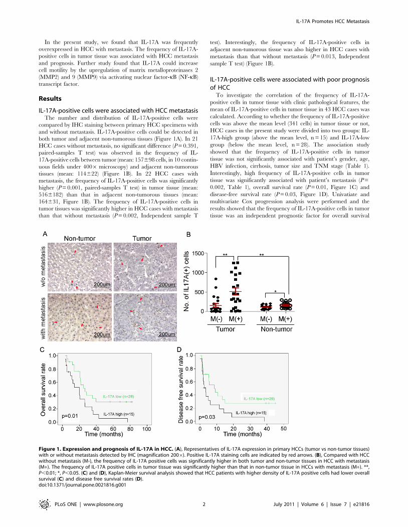
IL-17A-positive cells were associated with HCC metastasisThe number and distribution of IL-17A-positive cells were
compared by IHC staining between primary HCC specimens with
and without metastasis. IL-17A-positive cells could be detected in
both tumor and adjacent non-tumorous tissues (Figure 1A). In 21
HCC cases without metastasis, no significant difference (P = 0.391,
paired-samples T test) was observed in the frequency of IL-
17A-positive cells between tumor (mean: 157698 cells, in 10 contin-
uous fields under 4006 microscopy) and adjacent non-tumorous
tissues (mean: 114622) (Figure 1B). In 22 HCC cases with
metastasis, the frequency of IL-17A-positive cells was significantly
higher (P = 0.001, paired-samples T test) in tumor tissue (mean:
5166182) than that in adjacent non-tumorous tissues (mean:
164631, Figure 1B). The frequency of IL-17A-positive cells in
tumor tissues was significantly higher in HCC cases with metastasis
than that without metastasis (P = 0.002, Independent sample T
test). Interestingly, the frequency of IL-17A-positive cells in
adjacent non-tumorous tissue was also higher in HCC cases with
metastasis than that without metastasis (P = 0.013, Independent
sample T test) (Figure 1B).
IL-17A-positive cells were associated with poor prognosisof HCC
To investigate the correlation of the frequency of IL-17A-
positive cells in tumor tissue with clinic pathological features, the
mean of IL-17A-positive cells in tumor tissue in 43 HCC cases was
calculated. According to whether the frequency of IL-17A-positive
cells was above the mean level (341 cells) in tumor tissue or not,
HCC cases in the present study were divided into two groups: IL-
17A-high group (above the mean level, n = 15) and IL-17A-low
group (below the mean level, n = 28). The association study
showed that the frequency of IL-17A-positive cells in tumor
tissue was not significantly associated with patient’s gender, age,
HBV infection, cirrhosis, tumor size and TNM stage (Table 1).
Interestingly, high frequency of IL-17A-positive cells in tumor
tissue was significantly associated with patient’s metastasis (P =
0.002, Table 1), overall survival rate (P = 0.01, Figure 1C) and
disease-free survival rate (P = 0.03, Figure 1D). Univatiate and
multivariate Cox progression analysis were performed and the
results showed that the frequency of IL-17A-positive cells in tumor
tissue was an independent prognostic factor for overall survival
Figure 1. Expression and prognosis of IL-17A in HCC. (A), Representatives of IL-17A expression in primary HCCs (tumor vs non-tumor tissues)with or without metastasis detected by IHC (magnification 2006). Positive IL-17A staining cells are indicated by red arrows. (B), Compared with HCCwithout metastasis (M-), the frequency of IL-17A positive cells was significantly higher in both tumor and non-tumor tissues in HCC with metastasis(M+). The frequency of IL-17A positive cells in tumor tissue was significantly higher than that in non-tumor tissue in HCCs with metastasis (M+). **,P,0.01; *, P,0.05. (C) and (D), Kaplan-Meier survival analysis showed that HCC patients with higher density of IL-17A positive cells had lower overallsurvival (C) and disease free survival rates (D).doi:10.1371/journal.pone.0021816.g001
IL-17A Promotes HCC Metastasis
PLoS ONE | www.plosone.org 2 July 2011 | Volume 6 | Issue 7 | e21816
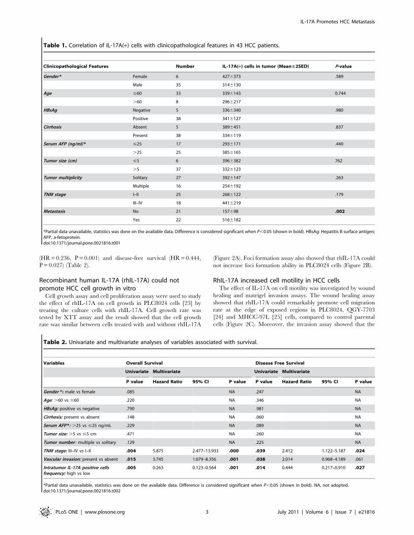
(HR = 0.236, P = 0.001) and disease-free survival (HR = 0.444,
P = 0.027) (Table 2).
Recombinant human IL-17A (rhIL-17A) could notpromote HCC cell growth in vitro
Cell growth assay and cell proliferation assay were used to study
the effect of rhIL-17A on cell growth in PLC8024 cells [23] by
treating the culture cells with rhIL-17A. Cell growth rate was
tested by XTT assay and the result showed that the cell growth
rate was similar between cells treated with and without rhIL-17A
(Figure 2A). Foci formation assay also showed that rhIL-17A could
not increase foci formation ability in PLC8024 cells (Figure 2B).
RhIL-17A increased cell motility in HCC cellsThe effect of IL-17A on cell motility was investigated by wound
healing and matrigel invasion assays. The wound healing assay
showed that rhIL-17A could remarkably promote cell migration
rate at the edge of exposed regions in PLC8024, QGY-7703
[24] and MHCC-97L [25] cells, compared to control parental
cells (Figure 2C). Moreover, the invasion assay showed that the
Table 1. Correlation of IL-17A(+) cells with clinicopathological features in 43 HCC patients.
Clinicopathological Features Number IL-17A(+) cells in tumor (Mean±2SED) P-value
Gender * Female 6 4276373 .589
Male 35 3146130
Age #60 33 3396143 0.744
.60 8 2966217
HBsAg Negative 5 3366340 .980
Positive 38 3416127
Cirrhosis Absent 5 3896451 .837
Present 38 3346119
Serum AFP (ng/ml) * #25 17 2936171 .440
.25 25 3856165
Tumor size (cm) #5 6 3966382 762
.5 37 3326123
Tumor multiplicity Solitary 27 3926147 .263
Multiple 16 2546192
TNM stage I–II 25 2686122 .179
III–IV 18 4416219
Metastasis No 21 157698 .002
Yes 22 5166182
*Partial data unavailable, statistics was done on the available data. Difference is considered significant when P,0.05 (shown in bold). HBsAg: Hepatitis B surface antigen;AFP, a-fetoprotein.doi:10.1371/journal.pone.0021816.t001
Table 2. Univariate and multivariate analyses of variables associated with survival.
Variables Overall Survival Disease Free Survival
Univariate Multivariate Univariate Multivariate
P value Hazard Ratio 95% CI P value P value Hazard Ratio 95% CI P value
Gender * : male vs female .085 NA .247 NA
Age: .60 vs #60 .220 NA .346 NA
HBsAg: positive vs negative .790 NA .981 NA
Cirrhosis: present vs absent .148 NA .060 NA
Serum AFP *: .25 vs #25 ng/mL .229 NA .089 NA
Tumor size: .5 vs #5 cm .471 NA .260 NA
Tumor number : multiple vs solitary .129 NA .225 NA
TNM stage: III–IV vs I–II .004 5.875 2.477–13.933 .000 .039 2.412 1.122–5.187 .024
Vascular invasion: present vs absent .015 3.745 1.679–8.356 .001 .038 2.014 0.968–4.189 .061
Intratumor IL-17A positive cellsfrequency: high vs low
.005 0.263 0.123–0.564 .001 .014 0.444 0.217–0.910 .027
*Partial data unavailable, statistics was done on the available data. Difference is considered significant when P,0.05 (shown in bold). NA, not adopted.doi:10.1371/journal.pone.0021816.t002
IL-17A Promotes HCC Metastasis
PLoS ONE | www.plosone.org 3 July 2011 | Volume 6 | Issue 7 | e21816
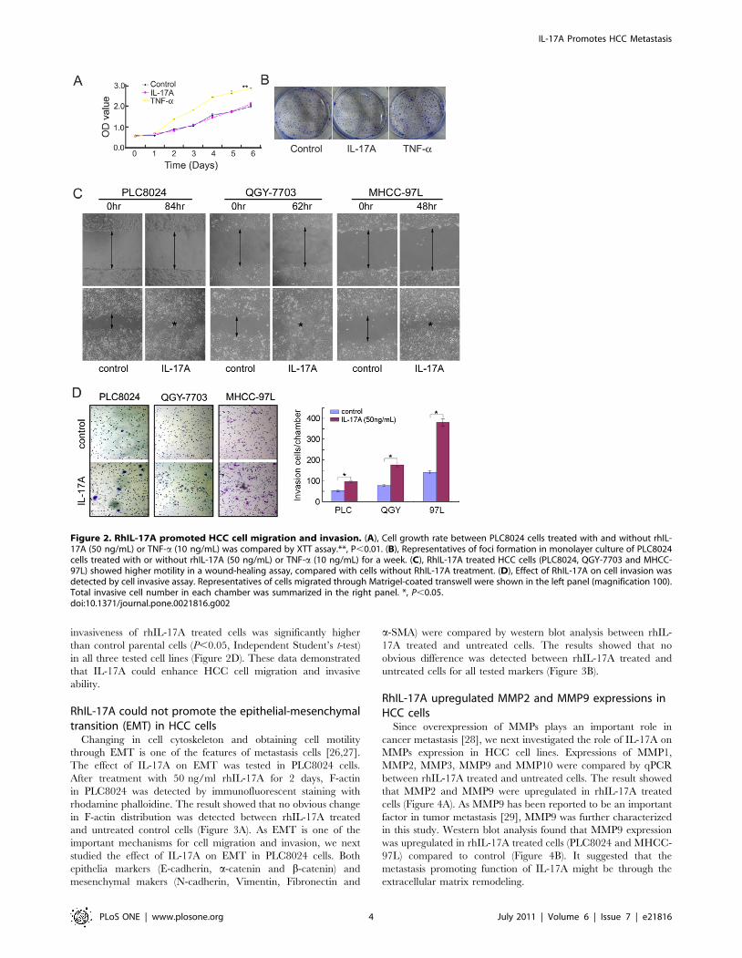
invasiveness of rhIL-17A treated cells was significantly higher
than control parental cells (P,0.05, Independent Student’s t-test)
in all three tested cell lines (Figure 2D). These data demonstrated
that IL-17A could enhance HCC cell migration and invasive
ability.
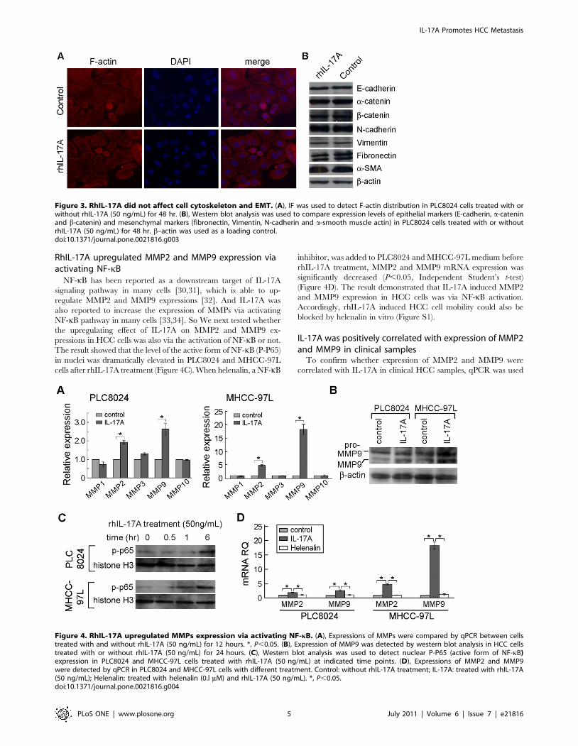
RhIL-17A could not promote the epithelial-mesenchymaltransition (EMT) in HCC cells
Changing in cell cytoskeleton and obtaining cell motility
through EMT is one of the features of metastasis cells [26,27].
The effect of IL-17A on EMT was tested in PLC8024 cells.
After treatment with 50 ng/ml rhIL-17A for 2 days, F-actin
in PLC8024 was detected by immunofluorescent staining with
rhodamine phalloidine. The result showed that no obvious change
in F-actin distribution was detected between rhIL-17A treated
and untreated control cells (Figure 3A). As EMT is one of the
important mechanisms for cell migration and invasion, we next
studied the effect of IL-17A on EMT in PLC8024 cells. Both
epithelia markers (E-cadherin, a-catenin and b-catenin) and
mesenchymal makers (N-cadherin, Vimentin, Fibronectin and
a-SMA) were compared by western blot analysis between rhIL-
17A treated and untreated cells. The results showed that no
obvious difference was detected between rhIL-17A treated and
untreated cells for all tested markers (Figure 3B).
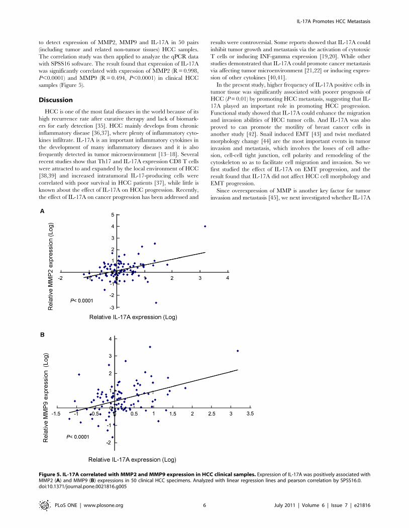
RhIL-17A upregulated MMP2 and MMP9 expressions inHCC cells
Since overexpression of MMPs plays an important role in
cancer metastasis [28], we next investigated the role of IL-17A on
MMPs expression in HCC cell lines. Expressions of MMP1,
MMP2, MMP3, MMP9 and MMP10 were compared by qPCR
between rhIL-17A treated and untreated cells. The result showed
that MMP2 and MMP9 were upregulated in rhIL-17A treated
cells (Figure 4A). As MMP9 has been reported to be an important
factor in tumor metastasis [29], MMP9 was further characterized
in this study. Western blot analysis found that MMP9 expression
was upregulated in rhIL-17A treated cells (PLC8024 and MHCC-
97L) compared to control (Figure 4B). It suggested that the
metastasis promoting function of IL-17A might be through the
extracellular matrix remodeling.
Figure 2. RhIL-17A promoted HCC cell migration and invasion. (A), Cell growth rate between PLC8024 cells treated with and without rhIL-17A (50 ng/mL) or TNF-a (10 ng/mL) was compared by XTT assay.**, P,0.01. (B), Representatives of foci formation in monolayer culture of PLC8024cells treated with or without rhIL-17A (50 ng/mL) or TNF-a (10 ng/mL) for a week. (C), RhIL-17A treated HCC cells (PLC8024, QGY-7703 and MHCC-97L) showed higher motility in a wound-healing assay, compared with cells without RhIL-17A treatment. (D), Effect of RhIL-17A on cell invasion wasdetected by cell invasive assay. Representatives of cells migrated through Matrigel-coated transwell were shown in the left panel (magnification 100).Total invasive cell number in each chamber was summarized in the right panel. *, P,0.05.doi:10.1371/journal.pone.0021816.g002
IL-17A Promotes HCC Metastasis
PLoS ONE | www.plosone.org 4 July 2011 | Volume 6 | Issue 7 | e21816
RhIL-17A upregulated MMP2 and MMP9 expression viaactivating NF-kB
NF-kB has been reported as a downstream target of IL-17A
signaling pathway in many cells [30,31], which is able to up-
regulate MMP2 and MMP9 expressions [32]. And IL-17A was
also reported to increase the expression of MMPs via activating
NF-kB pathway in many cells [33,34]. So We next tested whether
the upregulating effect of IL-17A on MMP2 and MMP9 ex-
pressions in HCC cells was also via the activation of NF-kB or not.
The result showed that the level of the active form of NF-kB (P-P65)
in nuclei was dramatically elevated in PLC8024 and MHCC-97L
cells after rhIL-17A treatment (Figure 4C). When helenalin, a NF-kB
inhibitor, was added to PLC8024 and MHCC-97L medium before
rhIL-17A treatment, MMP2 and MMP9 mRNA expression was
significantly decreased (P,0.05, Independent Student’s t-test)
(Figure 4D). The result demonstrated that IL-17A induced MMP2
and MMP9 expression in HCC cells was via NF-kB activation.
Accordingly, rhIL-17A induced HCC cell mobility could also be
blocked by helenalin in vitro (Figure S1).
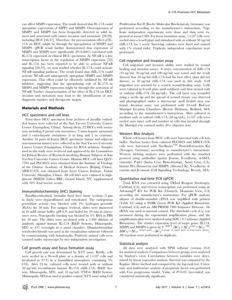
IL-17A was positively correlated with expression of MMP2and MMP9 in clinical samples
To confirm whether expression of MMP2 and MMP9 were
correlated with IL-17A in clinical HCC samples, qPCR was used
Figure 3. RhIL-17A did not affect cell cytoskeleton and EMT. (A), IF was used to detect F-actin distribution in PLC8024 cells treated with orwithout rhIL-17A (50 ng/mL) for 48 hr. (B), Western blot analysis was used to compare expression levels of epithelial markers (E-cadherin, a-cateninand b-catenin) and mesenchymal markers (fibronectin, Vimentin, N-cadherin and a-smooth muscle actin) in PLC8024 cells treated with or withoutrhIL-17A (50 ng/mL) for 48 hr. b–actin was used as a loading control.doi:10.1371/journal.pone.0021816.g003
Figure 4. RhIL-17A upregulated MMPs expression via activating NF-kB. (A), Expressions of MMPs were compared by qPCR between cellstreated with and without rhIL-17A (50 ng/mL) for 12 hours. *, P,0.05. (B), Expression of MMP9 was detected by western blot analysis in HCC cellstreated with or without rhIL-17A (50 ng/mL) for 24 hours. (C), Western blot analysis was used to detect nuclear P-P65 (active form of NF-kB)expression in PLC8024 and MHCC-97L cells treated with rhIL-17A (50 ng/mL) at indicated time points. (D), Expressions of MMP2 and MMP9were detected by qPCR in PLC8024 and MHCC-97L cells with different treatment. Control: without rhIL-17A treatment; IL-17A: treated with rhIL-17A(50 ng/mL); Helenalin: treated with helenalin (0.l mM) and rhIL-17A (50 ng/mL). *, P,0.05.doi:10.1371/journal.pone.0021816.g004
IL-17A Promotes HCC Metastasis
PLoS ONE | www.plosone.org 5 July 2011 | Volume 6 | Issue 7 | e21816
to detect expression of MMP2, MMP9 and IL-17A in 50 pairs
(including tumor and related non-tumor tissues) HCC samples.
The correlation study was then applied to analyze the qPCR data
with SPSS16 software. The result found that expression of IL-17A
was significantly correlated with expression of MMP2 (R = 0.998,
P,0.0001) and MMP9 (R = 0.494, P,0.0001) in clinical HCC
samples (Figure 5).
Discussion
HCC is one of the most fatal diseases in the world because of its
high recurrence rate after curative therapy and lack of biomark-
ers for early detection [35]. HCC mainly develops from chronic
inflammatory disease [36,37], where plenty of inflammatory cyto-
kines infiltrate. IL-17A is an important inflammatory cytokines in
the development of many inflammatory diseases and it is also
frequently detected in tumor microenvironment [13–18]. Several
recent studies show that Th17 and IL-17A expression CD8 T cells
were attracted to and expanded by the local environment of HCC
[38,39] and increased intratumoral IL-17-producing cells were
correlated with poor survival in HCC patients [37], while little is
known about the effect of IL-17A on HCC progression. Recently,
the effect of IL-17A on cancer progression has been addressed and
results were controversial. Some reports showed that IL-17A could
inhibit tumor growth and metastasis via the activation of cytotoxic
T cells or inducing INF-gamma expression [19,20]. While other
studies demonstrated that IL-17A could promote cancer metastasis
via affecting tumor microenvironment [21,22] or inducing expres-
sion of other cytokines [40,41].
In the present study, higher frequency of IL-17A positive cells in
tumor tissue was significantly associated with poorer prognosis of
HCC (P = 0.01) by promoting HCC metastasis, suggesting that IL-
17A played an important role in promoting HCC progression.
Functional study showed that IL-17A could enhance the migration
and invasion abilities of HCC tumor cells. And IL-17A was also
proved to can promote the motility of breast cancer cells in
another study [42]. Snail induced EMT [43] and twist mediated
morphology change [44] are the most important events in tumor
invasion and metastasis, which involves the losses of cell adhe-
sion, cell-cell tight junction, cell polarity and remodeling of the
cytoskeleton so as to facilitate cell migration and invasion. So we
first studied the effect of IL-17A on EMT progression, and the
result found that IL-17A did not affect HCC cell morphology and
EMT progression.
Since overexpression of MMP is another key factor for tumor
invasion and metastasis [45], we next investigated whether IL-17A
Figure 5. IL-17A correlated with MMP2 and MMP9 expression in HCC clinical samples. Expression of IL-17A was positively associated withMMP2 (A) and MMP9 (B) expressions in 50 clinical HCC specimens. Analyzed with linear regression lines and pearson correlation by SPSS16.0.doi:10.1371/journal.pone.0021816.g005
IL-17A Promotes HCC Metastasis
PLoS ONE | www.plosone.org 6 July 2011 | Volume 6 | Issue 7 | e21816
can affect MMPs expression. The result showed that IL-17A could
upregulate expressions of MPP2 and MMP9. Overexpression of
MMP2 and MMP9 has been frequently detected in solid tu-
mors and associated with tumor invasion and metastasis [28,29],
including HCC [46,47]. Therefore, the pro-metastasis effect of IL-
17A on HCC might be through the upregulation of MMP2 and
MMP9. qPCR result further demonstrated that expression of
MMP2 and MMP9 were significantly (P,0.0001) correlated with
IL-17A expression in clinical HCC specimens. As NF-kB is a key
transcription factor in the regulation of MMP9 expression [32]
and IL-17A has been reported to be able to activate NF-kB
signaling [30,31], we next studied whether IL-17A could activate
NF-kB signaling pathway. The result found that rhIL-17A could
activate NF-kB and subsequently upregulate MMP2 and MMP9
expression. This effect could be effectively inhibited by NF-kB
inhibitor, suggesting that the upregulating role of IL-17A in
MMP2 and MMP9 expression might be through the activation of
NF-kB. Further characterization of the effect of IL-17A on HCC
invasion and metastasis may lead to the identification of new
diagnostic markers and therapeutic targets.
Materials and Methods
HCC specimens and cell linesForty-three HCC specimens from archives of paraffin embed-
ded tissues were collected at the Sun Yat-sen University Cancer
Center (Guangzhou, China). Among them, 22 HCCs with metas-
tasis including 8 portal vein metastases, 9 intra-hepatic metastasis
and 5 extra-hepatic metastases (4 in lung and 1 in centrum).
Another 50 pairs of frozen HCC specimens (tumor and adjacent
non-tumorous tissues) were collected at the Sun Yat-sen University
Cancer Center (Guangzhou, China) for RNA isolation. Samples
used in this study were reviewed and approved by the Committees
for Ethical Review of Research involving Human Subjects at Sun
Yat-Sen University Cancer Center. Human HCC cell lines QGY-
7703 and PLC8024 were obtained from the Institute of Virology
of the Chinese Academy of Medical Sciences (Beijing, China).
MHCC-97L was obtained from Liver Cancer Institute, Fudan
University (Shanghai, China). All cell lines were cultured in high-
glucose DMEM (Gibco BRL, Grand Island, NY) supplemented
with 10% fetal bovine serum.
Immunohistochemistry (IHC) StainingParaffin-embedded, formalin fixed liver tissue sections (5 mm
in thick) were deparaffinized and rehydrated. The endogenous
peroxidase activity was blocked with 3% hydrogen peroxide
(H2O2) for 30 min. For antigen retrieval, slides were immersed
in 10 mM citrate buffer (pH 6.0) and boiled for 10 min in micro-
wave oven. Non-specific binding was blocked by 5% BSA in PBS
for 30 min. The slides were incubated with a 1:300 dilution of
antibody against human IL-17A (R&D Systems, Minneapolis,
MN) at 4uC overnight in a moist chamber. Diaminobenzidine
tetrahydrochloride was used as the visualization substrate followed
by counterstaining with hematoxylin. Positively stained cells were
counted under microscope by two independent investigators.
Cell growth assay and focus formation assayCell growth rate was determined by XTT assay. Briefly, cells
were seeded in a 96-well plate at a density of 16103 cells and
incubated at 37uC in a humidified atmosphere containing 5%
CO2. After 24 hr, cultured cells were treated with or without
50 ng/mL recombinant human IL-17A (rhIL-17A) (R&D Sys-
tem, Minneapolis, MN), and 10 ng/mL TNF-a (R&D System,
Minneapolis, MN)was used as positive control. XTT assay using Cell
Proliferation Kit II (Roche Molecular Biochemicals, Germany) was
performed according to the manufacturer’s instructions. Trip-
licate independent experiments were done and data were ex-
pressed as mean6SD. For focus formation assay, 16103 cells were
seeded onto a 6-well plate and stimulated with or without 50 ng/ml
rhIL-17A for 1 week. Surviving colonies were fixed and stained
with 1% crystal violet. Triplicate independent experiments were
performed.
Cell migration and invasion assayCell migration and invasion ability were studied by wound
healing and invasion assays. A series concentration of rhIL-17A
(10 ng/ml, 50 ng/ml and 100 ng/ml) was tested and the result
showed that 50 ng/ml rhIL-17A had the best effect (data did not
shown), so 50 ng/ml rhIL-17A was used in this study. Cell
migration was assessed by a scratch wound-healing assay. Cells
were cultured in 6-well plate until confluent and then treated with
or without rhIL-17A (50 ng/mL). The cell layer was wounded
using a sterile tip and the spread of wound closure was observed
and photographed under a microscope until healed area was
found. Invasion assay was performed with 24-well BioCoat
Matrigel Invasion Chambers (Becton Dicknson, Bedford, MA)
according to the manufacturer’s instructions. After cultured in
medium with or without rhIL-17A (50 ng/mL), 56104 cells were
seeded onto inner well and number of cells that invaded through
the Matrigel was counted under 206 objective lens.
Western Blot AnalysisWhole cell lysates from HCC cells were harvested with cell lysis
buffer. Nuclear lysates from cultured PLC8024 and MHCC-97L
cells were harvested with NucBusterTM ProteinExtraction Kit
(Novagen, Germany) according to manufacturer’s instructions.
Western blotting analyses were performed with the standard
protocol using antibodies against b-actin, E-cadherin, a-SMA,
vimentin, P-p65 (Santa Cruz Biotechnology, Santa Cruz, CA),
histone H3, fibronectin and MMP9, (Abcam,UK), N-cadherin, a-
catenin and b-catenin (Cell Signalling Technology, Beverly, MA).
Quantitative real-time PCR (qPCR)Total RNA was extracted using TRIzol Reagent (Invitrogen,
Carlsbad, CA), and reverse transcription was performed using an
AdvantageH RT for PCR Kit (Clontech, Mountain View, CA)
according the manufacturer’s instructions. For qPCR analysis,
aliquot of double-stranded cDNA was amplified with primers
(Table S1) using a SYBR Green PCR Kit (Applied Biosystems,
Carlsbad, CA) and an ABI PRISM 7900 Sequence Detector. 18s
rRNA was used as internal control. The threshold cycle (CT) was
measured during the exponential amplification phase, and the
amplification plots were analyzed using SDS 1.9.1 software (Applied
Biosystems). The relative expression level of target genes (IL17A,
MMP2 and MMP9) is given by 22DDCT (DCT =DCTtarget2DCT
18S,
DDCT =DCT(target gene)2DCT
(average of target gene in non-tumor tissue)).
All reactions were performed in duplicate.
Statistical analysisAll data were analyzed with SPSS software (version 16.0)
for statistical analysis. Comparisons between groups were analyzed
by Student’s t-test. Correlations between variables were deter-
mined by linear regression analysis. Survival was estimated by the
Kaplan–Meier method and compared by the log-rank test. Univa-
tiate and multivariate analysis of prognostic factor was performed
with Cox progression model. Value of P,0.05 (two-tailed) was
considered statistically significant.
IL-17A Promotes HCC Metastasis
PLoS ONE | www.plosone.org 7 July 2011 | Volume 6 | Issue 7 | e21816
Supporting Information
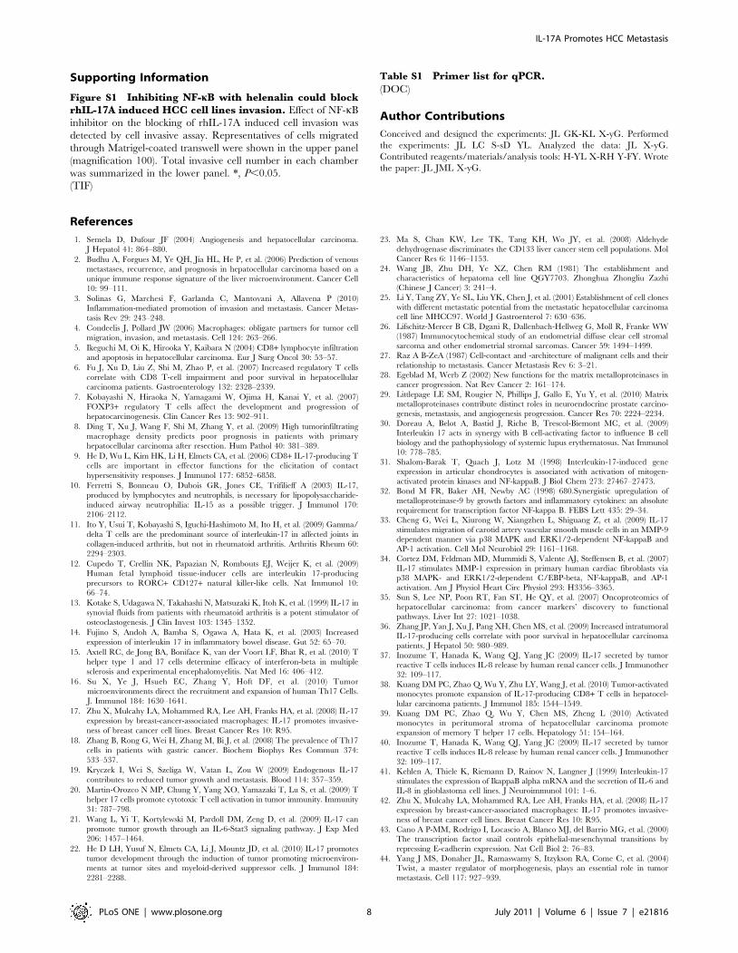
Figure S1 Inhibiting NF-kB with helenalin could blockrhIL-17A induced HCC cell lines invasion. Effect of NF-kB
inhibitor on the blocking of rhIL-17A induced cell invasion was
detected by cell invasive assay. Representatives of cells migrated
through Matrigel-coated transwell were shown in the upper panel
(magnification 100). Total invasive cell number in each chamber
was summarized in the lower panel. *, P,0.05.
(TIF)
Table S1 Primer list for qPCR.
(DOC)
Author Contributions
Conceived and designed the experiments: JL GK-KL X-yG. Performed
the experiments: JL LC S-sD YL. Analyzed the data: JL X-yG.
Contributed reagents/materials/analysis tools: H-YL X-RH Y-FY. Wrote
the paper: JL JML X-yG.
References
1. Semela D, Dufour JF (2004) Angiogenesis and hepatocellular carcinoma.
J Hepatol 41: 864–880.
2. Budhu A, Forgues M, Ye QH, Jia HL, He P, et al. (2006) Prediction of venous
metastases, recurrence, and prognosis in hepatocellular carcinoma based on a
unique immune response signature of the liver microenvironment. Cancer Cell
10: 99–111.
3. Solinas G, Marchesi F, Garlanda C, Mantovani A, Allavena P (2010)
Inflammation-mediated promotion of invasion and metastasis. Cancer Metas-
tasis Rev 29: 243–248.
4. Condeelis J, Pollard JW (2006) Macrophages: obligate partners for tumor cell
migration, invasion, and metastasis. Cell 124: 263–266.
5. Ikeguchi M, Oi K, Hirooka Y, Kaibara N (2004) CD8+ lymphocyte infiltration
and apoptosis in hepatocellular carcinoma. Eur J Surg Oncol 30: 53–57.
6. Fu J, Xu D, Liu Z, Shi M, Zhao P, et al. (2007) Increased regulatory T cells
correlate with CD8 T-cell impairment and poor survival in hepatocellular
carcinoma patients. Gastroenterology 132: 2328–2339.
7. Kobayashi N, Hiraoka N, Yamagami W, Ojima H, Kanai Y, et al. (2007)
FOXP3+ regulatory T cells affect the development and progression of
hepatocarcinogenesis. Clin Cancer Res 13: 902–911.
8. Ding T, Xu J, Wang F, Shi M, Zhang Y, et al. (2009) High tumorinfiltrating
macrophage density predicts poor prognosis in patients with primary
hepatocellular carcinoma after resection. Hum Pathol 40: 381–389.
9. He D, Wu L, Kim HK, Li H, Elmets CA, et al. (2006) CD8+ IL-17-producing T
cells are important in effector functions for the elicitation of contact
hypersensitivity responses. J Immunol 177: 6852–6858.
10. Ferretti S, Bonneau O, Dubois GR, Jones CE, Trifilieff A (2003) IL-17,
produced by lymphocytes and neutrophils, is necessary for lipopolysaccharide-
induced airway neutrophilia: IL-15 as a possible trigger. J Immunol 170:
2106–2112.
11. Ito Y, Usui T, Kobayashi S, Iguchi-Hashimoto M, Ito H, et al. (2009) Gamma/
delta T cells are the predominant source of interleukin-17 in affected joints in
collagen-induced arthritis, but not in rheumatoid arthritis. Arthritis Rheum 60:
2294–2303.
12. Cupedo T, Crellin NK, Papazian N, Rombouts EJ, Weijer K, et al. (2009)
Human fetal lymphoid tissue-inducer cells are interleukin 17-producing
precursors to RORC+ CD127+ natural killer-like cells. Nat Immunol 10:
66–74.
13. Kotake S, Udagawa N, Takahashi N, Matsuzaki K, Itoh K, et al. (1999) IL-17 in
synovial fluids from patients with rheumatoid arthritis is a potent stimulator of
osteoclastogenesis. J Clin Invest 103: 1345–1352.
14. Fujino S, Andoh A, Bamba S, Ogawa A, Hata K, et al. (2003) Increased
expression of interleukin 17 in inflammatory bowel disease. Gut 52: 65–70.
15. Axtell RC, de Jong BA, Boniface K, van der Voort LF, Bhat R, et al. (2010) T
helper type 1 and 17 cells determine efficacy of interferon-beta in multiple
sclerosis and experimental encephalomyelitis. Nat Med 16: 406–412.
16. Su X, Ye J, Hsueh EC, Zhang Y, Hoft DF, et al. (2010) Tumor
microenvironments direct the recruitment and expansion of human Th17 Cells.
J. Immunol 184: 1630–1641.
17. Zhu X, Mulcahy LA, Mohammed RA, Lee AH, Franks HA, et al. (2008) IL-17
expression by breast-cancer-associated macrophages: IL-17 promotes invasive-
ness of breast cancer cell lines. Breast Cancer Res 10: R95.
18. Zhang B, Rong G, Wei H, Zhang M, Bi J, et al. (2008) The prevalence of Th17
cells in patients with gastric cancer. Biochem Biophys Res Commun 374:
533–537.
19. Kryczek I, Wei S, Szeliga W, Vatan L, Zou W (2009) Endogenous IL-17
contributes to reduced tumor growth and metastasis. Blood 114: 357–359.
20. Martin-Orozco N MP, Chung Y, Yang XO, Yamazaki T, Lu S, et al. (2009) T
helper 17 cells promote cytotoxic T cell activation in tumor immunity. Immunity
31: 787–798.
21. Wang L, Yi T, Kortylewski M, Pardoll DM, Zeng D, et al. (2009) IL-17 can
promote tumor growth through an IL-6-Stat3 signaling pathway. J Exp Med
206: 1457–1464.
22. He D LH, Yusuf N, Elmets CA, Li J, Mountz JD, et al. (2010) IL-17 promotes
tumor development through the induction of tumor promoting microenviron-
ments at tumor sites and myeloid-derived suppressor cells. J Immunol 184:
2281–2288.
23. Ma S, Chan KW, Lee TK, Tang KH, Wo JY, et al. (2008) Aldehyde
dehydrogenase discriminates the CD133 liver cancer stem cell populations. Mol
Cancer Res 6: 1146–1153.
24. Wang JB, Zhu DH, Ye XZ, Chen RM (1981) The establishment andcharacteristics of hepatoma cell line QGY7703. Zhonghua Zhongliu Zazhi
(Chinese J Cancer) 3: 241–4.
25. Li Y, Tang ZY, Ye SL, Liu YK, Chen J, et al. (2001) Establishment of cell clones
with different metastatic potential from the metastatic hepatocellular carcinomacell line MHCC97. World J Gastroenterol 7: 630–636.
26. Lifschitz-Mercer B CB, Dgani R, Dallenbach-Hellweg G, Moll R, Franke WW(1987) Immunocytochemical study of an endometrial diffuse clear cell stromal
sarcoma and other endometrial stromal sarcomas. Cancer 59: 1494–1499.
27. Raz A B-ZeA (1987) Cell-contact and -architecture of malignant cells and their
relationship to metastasis. Cancer Metastasis Rev 6: 3–21.
28. Egeblad M, Werb Z (2002) New functions for the matrix metalloproteinases in
cancer progression. Nat Rev Cancer 2: 161–174.
29. Littlepage LE SM, Rougier N, Phillips J, Gallo E, Yu Y, et al. (2010) Matrixmetalloproteinases contribute distinct roles in neuroendocrine prostate carcino-
genesis, metastasis, and angiogenesis progression. Cancer Res 70: 2224–2234.
30. Doreau A, Belot A, Bastid J, Riche B, Trescol-Biemont MC, et al. (2009)
Interleukin 17 acts in synergy with B cell-activating factor to influence B cell
biology and the pathophysiology of systemic lupus erythematosus. Nat Immunol10: 778–785.
31. Shalom-Barak T, Quach J, Lotz M (1998) Interleukin-17-induced gene
expression in articular chondrocytes is associated with activation of mitogen-
activated protein kinases and NF-kappaB. J Biol Chem 273: 27467–27473.
32. Bond M FR, Baker AH, Newby AC (1998) 680.Synergistic upregulation ofmetalloproteinase-9 by growth factors and inflammatory cytokines: an absolute
requirement for transcription factor NF-kappa B. FEBS Lett 435: 29–34.
33. Cheng G, Wei L, Xiurong W, Xiangzhen L, Shiguang Z, et al. (2009) IL-17
stimulates migration of carotid artery vascular smooth muscle cells in an MMP-9dependent manner via p38 MAPK and ERK1/2-dependent NF-kappaB and
AP-1 activation. Cell Mol Neurobiol 29: 1161–1168.
34. Cortez DM, Feldman MD, Mummidi S, Valente AJ, Steffensen B, et al. (2007)
IL-17 stimulates MMP-1 expression in primary human cardiac fibroblasts viap38 MAPK- and ERK1/2-dependent C/EBP-beta, NF-kappaB, and AP-1
activation. Am J Physiol Heart Circ Physiol 293: H3356–3365.
35. Sun S, Lee NP, Poon RT, Fan ST, He QY, et al. (2007) Oncoproteomics of
hepatocellular carcinoma: from cancer markers’ discovery to functionalpathways. Liver Int 27: 1021–1038.
36. Zhang JP, Yan J, Xu J, Pang XH, Chen MS, et al. (2009) Increased intratumoralIL-17-producing cells correlate with poor survival in hepatocellular carcinoma
patients. J Hepatol 50: 980–989.
37. Inozume T, Hanada K, Wang QJ, Yang JC (2009) IL-17 secreted by tumor
reactive T cells induces IL-8 release by human renal cancer cells. J Immunother32: 109–117.
38. Kuang DM PC, Zhao Q, Wu Y, Zhu LY, Wang J, et al. (2010) Tumor-activatedmonocytes promote expansion of IL-17-producing CD8+ T cells in hepatocel-
lular carcinoma patients. J Immunol 185: 1544–1549.
39. Kuang DM PC, Zhao Q, Wu Y, Chen MS, Zheng L (2010) Activated
monocytes in peritumoral stroma of hepatocellular carcinoma promoteexpansion of memory T helper 17 cells. Hepatology 51: 154–164.
40. Inozume T, Hanada K, Wang QJ, Yang JC (2009) IL-17 secreted by tumorreactive T cells induces IL-8 release by human renal cancer cells. J Immunother
32: 109–117.
41. Kehlen A, Thiele K, Riemann D, Rainov N, Langner J (1999) Interleukin-17
stimulates the expression of IkappaB alpha mRNA and the secretion of IL-6 andIL-8 in glioblastoma cell lines. J Neuroimmunol 101: 1–6.
42. Zhu X, Mulcahy LA, Mohammed RA, Lee AH, Franks HA, et al. (2008) IL-17
expression by breast-cancer-associated macrophages: IL-17 promotes invasive-
ness of breast cancer cell lines. Breast Cancer Res 10: R95.
43. Cano A P-MM, Rodrigo I, Locascio A, Blanco MJ, del Barrio MG, et al. (2000)The transcription factor snail controls epithelial-mesenchymal transitions by
repressing E-cadherin expression. Nat Cell Biol 2: 76–83.
44. Yang J MS, Donaher JL, Ramaswamy S, Itzykson RA, Come C, et al. (2004)
Twist, a master regulator of morphogenesis, plays an essential role in tumormetastasis. Cell 117: 927–939.
IL-17A Promotes HCC Metastasis
PLoS ONE | www.plosone.org 8 July 2011 | Volume 6 | Issue 7 | e21816
45. Kessenbrock K, Plaks V, Werb Z (2010) Matrix metalloproteinases: regulators of
the tumor microenvironment. Cell 141: 52–67.46. Bu W HX, Tang Z (1997) The role of MMP-2 in the invasion and metastasis of
hepatocellular carcinoma (HCC). Zhonghua Yi Xue Za Zhi 77: 661–664.
47. Yang P YW, He J, Wang J, Yu L, Jin X, et al. (2009) Overexpression of EphA2,
MMP-9, and MVD-CD34 in hepatocellular carcinoma: Implications for tumor
progression and prognosis. Hepatol Res 39: 1169–1177.
IL-17A Promotes HCC Metastasis
PLoS ONE | www.plosone.org 9 July 2011 | Volume 6 | Issue 7 | e21816
Copyright © 2022 FDOKUMEN