Inter-Alpha Inhibitor Protein Administration Improves Survival From Neonatal Sepsis in Mice
15
Inter-Alpha Inhibitor Protein (IAIP) Administration Improves Survival From Neonatal Sepsis In Mice Kultar Singh, Ling Xiu Zhang, Kreso Bendelja, Ryan Heath, Shaun Murphy, Surendra Sharma, James F. Padbury, and Yow-Pin Lim Department of Pediatrics [K.S., S.S., J.F.P.], Women & Infants’ Hospital, Brown Medical School, Providence, Rhode Island, 02905; Division of Hematology/Oncology [L.X.Z., K.B., R.H., S.M., Y.- P.L.], Rhode Island Hospital, Brown Medical School, Providence, Rhode Island, 02903; ProThera Biologics [Y.-P.L.], East Providence, RI 02914 Abstract Inter-alpha Inhibitor proteins (IaIp) are serine proteases inhibitors which modulate endogenous protease activity and have been shown to improve survival in adult models of sepsis. We evaluated the effect of IaIp on survival and systemic responses to sepsis in neonatal mice. Sepsis was induced in 2-day-old mice with LPS, E. coli and Group B Streptococci. Sepsis was associated with 75% mortality. IaIp, given by intraperitoneal administration at doses between 15–45 mg/kg from 1–6 hours following the onset of sepsis improved survival to nearly 90% (p = 0.0159) in both LPS induced sepsis and with live bacterial infections. The greatest effect was on reversal of hemorrhagic pneumonitis. The effects were dose and time dependent. Systemic cytokine profile and tissue histology were examined. Survival was compared in interleukin 10 knock out animals. Systemic cytokine levels, including TNF-α and IL-10 were increased following induction of sepsis and modulated significantly following IaIp administration. Because the effect of IaIp was still demonstrable in IL-10 deficient mice, we conclude the beneficial effect(s) of IaIp is due to suppression of pro-inflammatory cytokines like TNF-α rather than augmentation of IL-10. IaIp may offer significant benefits as a therapeutic adjunct to treatment of sepsis in neonates and adults. INTRODUCTION Sepsis is a leading cause of morbidity and mortality in newborn and preterm infants (1–3). In a large, multicenter, prospective, cross-sectional study from the NICHD Neonatal Research Network, the incidence of late onset nosocomial infection was 16% of 2,416 infants enrolled at 12 sites (3). The high continuing morbidity and mortality due to sepsis, septic shock, and multiple organ failure may be attributable to the fact that the mediators/ factors responsible for the pathophysiologic alterations of sepsis are not fully understood (4). Antibiotic therapy may not be sufficient to reverse systemic inflammation and consequent organ injury accompanying sepsis. Mediators involved in the progression of sepsis can induce the activation of phagocytes that release neutrophil proteases (5). Experimental and clinical data have demonstrated that increased activity of neutrophil-derived serine proteases, such as leukocyte elastase and cathepsin G, play a prominent role in sepsis-related tissue damage (6). Administration of Corresponding Author: James F. Padbury, M.D., Department of Pediatrics, Women & Infants Hospital, 101 Dudley Street, Providence, RI 02905, Fax: 401-453-7571, Tel: 401-274-1122,x1205, [email protected]. DISCLOSURE:Y-P.L. has equity in the company ProThera Biologics where the IaIp protein preparations used in these experiments were produced and where the Western blot assays were performed. NIH Public Access Author Manuscript Pediatr Res. Author manuscript; available in PMC 2011 September 1. Published in final edited form as: Pediatr Res. 2010 September ; 68(3): 242–247. doi:10.1203/PDR.0b013e3181e9fdf0. NIH-PA Author Manuscript NIH-PA Author Manuscript NIH-PA Author Manuscript
-
Upload
independent -
Category
Documents
-
view
3 -
download
0
Transcript of Inter-Alpha Inhibitor Protein Administration Improves Survival From Neonatal Sepsis in Mice
Inter-Alpha Inhibitor Protein (IAIP) Administration ImprovesSurvival From Neonatal Sepsis In Mice
Kultar Singh, Ling Xiu Zhang, Kreso Bendelja, Ryan Heath, Shaun Murphy, SurendraSharma, James F. Padbury, and Yow-Pin LimDepartment of Pediatrics [K.S., S.S., J.F.P.], Women & Infants’ Hospital, Brown Medical School,Providence, Rhode Island, 02905; Division of Hematology/Oncology [L.X.Z., K.B., R.H., S.M., Y.-P.L.], Rhode Island Hospital, Brown Medical School, Providence, Rhode Island, 02903; ProTheraBiologics [Y.-P.L.], East Providence, RI 02914
AbstractInter-alpha Inhibitor proteins (IaIp) are serine proteases inhibitors which modulate endogenousprotease activity and have been shown to improve survival in adult models of sepsis. We evaluatedthe effect of IaIp on survival and systemic responses to sepsis in neonatal mice. Sepsis wasinduced in 2-day-old mice with LPS, E. coli and Group B Streptococci. Sepsis was associated with75% mortality. IaIp, given by intraperitoneal administration at doses between 15–45 mg/kg from1–6 hours following the onset of sepsis improved survival to nearly 90% (p = 0.0159) in both LPSinduced sepsis and with live bacterial infections. The greatest effect was on reversal ofhemorrhagic pneumonitis. The effects were dose and time dependent. Systemic cytokine profileand tissue histology were examined. Survival was compared in interleukin 10 knock out animals.Systemic cytokine levels, including TNF-α and IL-10 were increased following induction of sepsisand modulated significantly following IaIp administration. Because the effect of IaIp was stilldemonstrable in IL-10 deficient mice, we conclude the beneficial effect(s) of IaIp is due tosuppression of pro-inflammatory cytokines like TNF-α rather than augmentation of IL-10. IaIpmay offer significant benefits as a therapeutic adjunct to treatment of sepsis in neonates and adults.
INTRODUCTIONSepsis is a leading cause of morbidity and mortality in newborn and preterm infants (1–3).In a large, multicenter, prospective, cross-sectional study from the NICHD NeonatalResearch Network, the incidence of late onset nosocomial infection was 16% of 2,416infants enrolled at 12 sites (3). The high continuing morbidity and mortality due to sepsis,septic shock, and multiple organ failure may be attributable to the fact that the mediators/factors responsible for the pathophysiologic alterations of sepsis are not fully understood (4).Antibiotic therapy may not be sufficient to reverse systemic inflammation and consequentorgan injury accompanying sepsis.
Mediators involved in the progression of sepsis can induce the activation of phagocytes thatrelease neutrophil proteases (5). Experimental and clinical data have demonstrated thatincreased activity of neutrophil-derived serine proteases, such as leukocyte elastase andcathepsin G, play a prominent role in sepsis-related tissue damage (6). Administration of
Corresponding Author: James F. Padbury, M.D., Department of Pediatrics, Women & Infants Hospital, 101 Dudley Street,Providence, RI 02905, Fax: 401-453-7571, Tel: 401-274-1122,x1205, [email protected]:Y-P.L. has equity in the company ProThera Biologics where the IaIp protein preparations used in these experimentswere produced and where the Western blot assays were performed.
NIH Public AccessAuthor ManuscriptPediatr Res. Author manuscript; available in PMC 2011 September 1.
Published in final edited form as:Pediatr Res. 2010 September ; 68(3): 242–247. doi:10.1203/PDR.0b013e3181e9fdf0.
NIH
-PA Author Manuscript
NIH
-PA Author Manuscript
NIH
-PA Author Manuscript

protease inhibitors has been proposed as a therapeutic strategy to restore the balancebetween proteases and protease inhibitors in sepsis (7). One such inhibitor is the Inter-alphaInhibitor protein (IaIp) family (8–10). This is a group of structurally related serine proteaseinhibitors found at relatively high concentrations in human plasma. IaIp proteins arecomposed of heavy and light polypeptide subunits that are covalently linked by aglycosaminoglycan chain (10–12). The light chain, also called bikunin, contains the serineprotease inhibitory activity of the molecules (13). Bikunin is inactive when cross-linked inthese complexes until it is released by partial proteolytic degradation. After cleavage fromthe complex, bikunin is cleared rapidly from plasma by glomerular filtration and receptor-mediated uptake(14).
IaIp proteins are involved in many physiological and pathological activities. This includestumor invasion and metastasis (15), stabilization of the extracellular matrix (8),inflammation and wound healing (16). Recently, the involvement of IaIp in inflammatorydiseases has become an area of intensive investigation. The release of neutrophil proteases,especially human leukocyte elastase, has been implicated in the progression ofcomplications in both adult and neonatal patients with sepsis(9,17,18). Plasma IaIp isparticularly sensitive to cleavage by neutrophil elastase. The light chain released from theIaIp complex then exerts its inhibitory activity on serine proteases (19). In vitro, IaIp inhibitsseveral serine proteases that are involved in inflammation, including elastase, plasmin, andcathepsin G (20). Clinical studies of patients with severe sepsis have revealed significantreduction in circulating IaIp due to consumption and accompanied by an extended secretionof elastase (9,17,18).
We have demonstrated markedly decreased concentrations of IaIp in plasma of septic adults(9,21,22) and neonates (23,24). Further, the degree of reduction of IaIp in adults correlateswith increase mortality (9,21,22). In experimental models of sepsis, we have demonstratedimproved survival, improved hemodynamic and physiological stability, reduced hepaticinjury and reduced inflammatory cytokine production following IaIp (25,26). Jourdain et al.have shown administration of human IaIp markedly improved hemodynamics, tissueoxygenation and pulmonary hypertension in a porcine model of endotoxic shock (27). Thereare no experimental studies on the effects of IaIp administration in neonates. The goal of ourstudies was to examine the effects of IaIp administration on survival and inflammatorycytokine production in microbiologically relevant models of neonatal sepsis (28,29).
MATERIALS AND METHODSIaIp Preparation
Human IaIp was purified as previously described by a combination of cryo-precipitation,solid phase extraction and a series of chromatographic separations involving ion-exchange,heparin affinity and hydroxyl apatite columns (25,26). The purity is greater than 95%. Thishighly purified IaIp has shown no side effects including toxicity, thrombogenicity, orhypotension in studies in adult model systems (25,26,30).
Neonatal Sepsis ModelsAll animal experiments were conducted under a protocol approved by the InstitutionalAnimal Care and Use Committee, which conforms to the Guide for the Care and Use ofLaboratory Animals published by the US National Institutes of Health (NIH Publication No.85-23, revised 1996) and in accordance with the Declaration of Helsinki principles. All miceused in this study were housed in a specific pathogen-free facility under the care of theCentral Research Department of Rhode Island Hospital. All strains of mice, C57BL/6
Singh et al. Page 2
Pediatr Res. Author manuscript; available in PMC 2011 September 1.
NIH
-PA Author Manuscript
NIH
-PA Author Manuscript
NIH
-PA Author Manuscript
Interleukin 10 deficient animals (IL-10−/−), C57BL/6 wild type and Balb/C wild type, wereobtained from the Jackson Laboratory (Bar Harbor, ME).
The dose dependent effects of LPS were compared by injection of 5, 10 or 25 microgramssubcutaneously in 2-day-old, mixed-gender BALB/c wild type mice. The LPS preparationused was serotype O26:B6 E. coli LPS (Sigma Chemical, St. Louis, Missouri). Injection of25 micrograms of LPS subcutaneously resulted in survival of 25% after 80 hours. This dosewas used in subsequent experiments. Two day old mice (n=22) were injected with LPS andrandomized into two groups. The IaIp animals (n=12) received 30 mg/kg body weight ofhighly purified human IaIp intraperitoneally one hour after LPS. Controls (n=10) receivedequal amount of human serum albumin (HSA). Survival was observed every 2 hours for 80hours. Results were compared in the two groups. T the effect of delayed administration ofIaIp was tested by administration of IaIp up to 6 hours after the LPS dose. The dependenceof survival on the dose of IaIp was compared by administration of three different doses, 15mg/kg (n=5), 30 mg/kg (n=10) and 45 mg/kg (n=6) body weight 3 hours after LPS. Thisdosing range was based on prior studies in adult animal models (25,26)
Survival was also determined following injection of live E. coli or Type 1 GBS. The virulentstrain of K1 subtype of E coli we used was a clinical obtained from Dr. Stephen OpalMemorial Hospital of Rhode Island. The GBS strain was a clinical isolate obtained atWomen & Infants Hospital of Rhode Island. Bacteria from a fresh overnight culture wasinoculated into LB broth and grown to log phase in a shaking culture flask at 37°C andmonitored spectrophotometrically until achieving an OD560 of 0.38. Previous experimentsusing serial dilutions and direct plating of that innoculum were used to generate a standardcurve for colony forming units (CFU)/ml of culture. Fresh culture was serially diluted intosterile phosphate buffered saline. The final injection volume for all doses of bacteria was 25microliters. Survival was examined following subcutaneous administration of 104 to 107 E.coli CFU/pup. The effects of exogenous IaIp were examined following administration of 30mg/kg IaIp or HSA intraperitoneally one hour following the E. coli. Similarly, the dosedependent effects of GBS were determined by subcutaneous administration of 104 to 107
CFU/pup and the effects on survival were examined following a dose of 30 mg/kg IaIp onehour after the GBS. All experiments with animals were approved by the Institutional AnimalCare & Use Committee. The effect of IaIp was also compared in wild type animals and inanimals in which the anti-inflammatory cytokine IL-10 had been disrupted (IL-10−/−).Because of their heightened mortality in response to LPS, the dose response for lethality wasfirst established in IL-10−/− neonatal mice. The effect of IaIp on survival was then examinedafter LPS injection as in the experiments with wild type animals by intraperitoneal injectionof 30mg/kg IaIp or human albumin 1 hour after subcutaneous LPS.
Inflammatory Cytokine ResponsesIn order to examine the effects of IaIp on inflammatory responses, plasma cytokine levelswere measured in IaIp and albumin treated groups. LPS administered subcutaneously asabove was followed 1 hr later by 30 mg/kg body weight of intraperitoneal IaIp or Albumin.Animals were sacrificed at intervals and pooled trunk blood was collected from three pups at2, 6, 12, 24, 48 and 72 hours after the LPS injection, from both IaIp and albumin treatedgroups. Plasma was separated immediately by centrifugation and stored at −70°C formeasurement of MCP-1, TNF-a, IL-10, IL-12p70, IFN-g, and IL-6 by radioimmunoassay(9).
Singh et al. Page 3
Pediatr Res. Author manuscript; available in PMC 2011 September 1.
NIH
-PA Author Manuscript
NIH
-PA Author Manuscript
NIH
-PA Author Manuscript

StatisticsThe log rank test was used to calculate statistical differences in survival between the IaIpand the control group. Unpaired t-testing was used to compare group means wereappropriate. In both cases, p <0.05 was considered as statistically significant.
RESULTSWe administered graded doses of LPS subcutaneously to 2–3 day old BALB/c wild typemice (Figure 1, panel A). As can been seen, there was a dose dependent mortality whichreached 60–70% by 48 hours following the 25 microgram per pup dose. In Figure 1B, it canbe seen that administration of 30 mg/kg intraperitoneally of IaIp one hour after LPSchallenge reversed mortality from 60–70% by 40 hours to prolonged survival in nearly 90%of the pups. Increasing the dose to 45 mg/kg had no additional salutary effect. However,reducing the dose to 15 mg/kg decreased survival to 60% which was still statisticallysignificant, p<0.05. We examined the timing of IaIp administration. Administration of IaIpas long as up to 3 hours after the LPS was still highly protective. If IaIp was administeredone to three hours following LPS survival remained between 80 and 90%, Figure 1C.Delaying administration for six hours reduced survival, which was nonetheless stillsignificantly better than vehicle alone.
We examined the effects of IaIp on E. coli and Group B β hemolytic Streptococcus (GBS)sepsis, the two most relevant pathogens in neonatal sepsis in human infants, to assessveracity of protection (2,3,28,29,31). The virulent subtype of E coli was grown tologarithmic phase in culture and then administered subcutaneously at doses from 4×10⁵ upto 2×10⁷ CFU/animal, Figure 2A. There was a high degree of lethality associated with thetwo highest doses, whereas 4×10⁵ CFU per pup resulted in reproducible lethality between 60and 80% (Figure 2A, 2B). Administration of 30 mg/kg of IaIp one hour following the E coliimproved survival to nearly 90%, Figure 2B. Similar lethality and response to administrationof IAIP was seen with a clinical isolate of Type I GBS, Figure 2C. Subcutaneousadministration of GBS resulted in 60% overall mortality in the albumin-treated group.Survival was increased to 80% following administration of IaIp in the animals injected withGBS.
To examine the mechanism(s) of protection of IaIp, tissue samples were taken fromrandomly selected pups twenty four hours following administration of 25 micrograms ofLPS. Histological sections were reviewed by a pathologist not aware of the treatmentgroups. Sections of the lung parenchyma following LPS from an animal that received IaIpand a sections following LPS in a control animal that received albumin are shown in Figure3. The control lung showed marked thickening of the alveolar septae, a pronouncedpolymorphonuclear leukocyte infiltration and some pulmonary hemorrhage, Figure 3A. Incontrast, IaIp administration was associated with preservation of largely normal lungparenchymal architecture, Figure 3B. Changes in other organs (liver, kidney, intestine) weresimilar but less pronounced (not shown). Whole blood and plasma were collected andexamined by Western blot and ELISA to determine the time course for decrements inendogenous, mouse IaIp and the Pre-alpha Inhibitor. The plasma concentration of both IaIpand PaI decreased significantly the following LPS induced sepsis, Figure 4A. Thedifferences between each time point are all significant, 4B p<0.05.
In order to examine the effect of IaIp on systemic immune responses, LPS was administeredto animals as described above followed by timed sacrifice and collection of trunk blood forcytokine measurements. The results are shown in Figure 5. By 3 hours after LPS, the plasmaconcentration of the pro-inflammatory cytokine TNF-α was increased to almost 25,000 pg/ml and remained elevated for up to 12 hours. Similarly, the immunomodulatory cytokine
Singh et al. Page 4
Pediatr Res. Author manuscript; available in PMC 2011 September 1.
NIH
-PA Author Manuscript
NIH
-PA Author Manuscript
NIH
-PA Author Manuscript

IL-10 was increased following LPS injection, reaching a peak concentration of over 2,500pg/ml at 6 hours. Most remarkably, administration of IaIp (30 mg/kg) one hour followingLPS injection nearly completely attenuated the increase in the plasma concentration of TNF-α. TNF-α was reduced to almost baseline levels at 3 hours after IaIp (p<0.03). In contrast,the increase in IL-10 was augmented and prolonged by IaIp (p<0.04). IaIp similarlyaugmented the increase in plasma IL-12p70 (p< 0.002). The increase in IL-6 at 6 hours,followed the maximum increase in TNF-α and was modestly attenuated by IaIpadministration. There was much less of an effect of IaIp on the changes in MCP-1 followingLPS.
In order to determine the contribution(s) to the beneficial effect of IaIp on survival fromsuppression of pro-inflammatory cytokine production or augmentation of IL-10 secretion,we carried out similar experiments in animals in which the IL-10 gene had been disrupted(IL-10−/−). The IL-10−/− animals showed a markedly heightened susceptibility to mortalityfollowing LPS. We systematically compared 10, 2.5 and 0.25 micrograms in IL-10−/−
animals (Figure 5B). LPS was highly lethal but by reducing the dose to 0.25 micrograms/mouse, we observed a survival of up to 40% at 60 hours. Administration of IaIp to IL-10−/−
animals one hour following 0.25 micrograms LPS was associated with over 90% survival(Figure 6B). IaIp prolonged survival in the IL-10−/− animals receiving the higher LPS doses,albeit death was observed in all by 50 hours (p<0.05).
DISCUSSIONIn these studies, we show improved survival following the administration of IaIp in neonatalsepsis due to LPS and live E. coli and GBS, the most important organisms in theepidemiology of neonatal sepsis (3,20,31). Taken together with our other findings, theseresults suggest that administration of IaIp elicits potent immunomodulatory actions that leadto survival in neonatal sepsis even when administered at delayed time points. Theseobservations suggest that deficiency of these regulatory protease inhibitors may play apathophysiologic role in systemic inflammation and sepsis (32,33).
The host immune response leads to a proinflammatory cascade, which continues even afterthe responsible organisms are killed despite administration of antibiotics. Recently, studiesin adult humans with severe sepsis and organ dysfunction have demonstrated animprovement in survival following administration of antibiotics and recombinant activatedprotein C (34). The reduction in the risk of death was modest (6%) however there was anapparent increase in associated risk of bleeding. Activated protein C promotes fibrinolysisand suppresses immune system activation. The increase in bleeding with activated protein Cadministration is a relatively strong contraindication in newborn infants, especially preterminfants. Indeed, a randomized, controlled trial in children ages 38 weeks gestation to 17years of age failed to identify any benefits of activated protein C but noted an increase in therisk of bleeding in infants less than 60 days of age (35).
Clinical studies in patients with severe sepsis show significant reduction in the circulatingconcentration of IaIp which correlate with increased mortality in septic adult patients (9,21–24). We and others have shown improvement in survival of adult animals in experimentalmodels of sepsis associated with a decrease in organ dysfunction and a marked reduction inthe production/secretion of the pro-inflammatory cytokine TNF-α. (25–27). The experimentsreported here demonstrate that intraperitoneal administration of IaIp to 2-day-old micefollowing induction of sepsis syndrome was associated with a dose and time dependentimprovement in survival, suppression of secretion of pro-inflammatory cytokines like TNF-α and augmentation of IL-10 and IL-12p70 production. The improvement in survival wasseen in animals with genetic deficiency in IL-10, suggesting that the beneficial effect of IaIp
Singh et al. Page 5
Pediatr Res. Author manuscript; available in PMC 2011 September 1.
NIH
-PA Author Manuscript
NIH
-PA Author Manuscript
NIH
-PA Author Manuscript
on survival resulted from alteration in TNF-α levels and other cytokines rather thanaugmentation of IL-10 or IL-12p70 secretion.
Neutrophils, monocytes, macrophages and Kupfer cells all produce significant amounts ofTNF-α when exposed to high levels of endotoxin. Coeshutt and coworkers have furthershown that neutrophils contribute to augmented TNF-α and IL-1β secretion by release ofproteases such as proteas-3, elastase and cathepsin G in a TACE (TNF-α convertingenzyme) and ICE (IL-1β converting enzyme) independent manner (38). Il-6 is likewiseproduced in response to stimulation by endotoxin IL-1β and TNF-α (36). We observedmarked suppression in the levels of TNF-α in the animals treated with IaIp. Taken together,these findings stress different pathways of TNF-α biosynthesis/processing and provideinsight into mechanisms for the immunomodulatory effect of the protease inhibitor IaIp.Hepatic Kupfer cells are the main source of IL-10, an immunomodulatory cytokine thatprotects mice against endotoxic shock by preventing excessive production of pro-inflammatory cytokines (37). Because the effect of IaIp on survival was still demonstrable inIL-10 deficient mice, we conclude the beneficial effect(s) of IaIp is due more to suppressionof pro-inflammatory cytokines like TNF-α and others rather than augmentation of IL-10production.
Models of sepsis in experimental animals have been used widely to develop new andeffective therapies. Models that employ live organisms better recapitulate the characteristicsof disease progression and organ dysfunction that accompanies sepsis than models usingendotoxin alone (8,29). IaIp has been demonstrated to be effective now in a wide-range ofconditions (25,26,30). IaIp is not only therapeutic but may have both diagnostic andprognostic value. As already noted, low levels of IaIp are associated with sepsis in adulthumans and human neonates (9,21–24). Most recently, we have demonstrated that inlongitudinal studies of IaIp levels in severely septic patients that failure of IAIP levels torecover over the first five days of sepsis is associated with a poor outcome (22).Additionally, our most recent data looked cross-sectionally at large numbers of humannewborns with sepsis and gestational age and postnatally aged matched controls. We studiedthe receiver/operating characteristics needed to identify optimal predictive values of plasmaIaIp levels in neonatal sepsis (24). Using a computer derived cutoff value of 177 mcg/ml, wedemonstrated a sensitivity of 89.5%, a specificity of 99%, a positive predictive value of 95%and negative predictive value of 98%. The area under the ROC curve was 0.94, P < 0.001.
In the present studies, we observed comparable survival following induction of a systemicimmune response syndrome (SIRS) mimicking sepsis due to LPS in animals that receivedeither 30 or 45 mg/kg body weight IaIp, which suggests that larger doses are unlikely tooffer additional benefits. We also compared administration of IaIp after one hour withdelayed administration of IaIp until 3 or 6 hours. The beneficial effect(s) on survival wereseen even when IaIp administration was delayed 3 to 6 hours after induction of sepsis. Theresults further demonstrate the effects of IaIp administration on immune system activationand organ dysfunction. Our results suggest that administration of IaIp exerts potentimmunomodulatory actions that lead to a beneficial effect in septic newborn mice. IaIp maybe an effective adjunct to treatment of severe sepsis in neonates or adults and warrantsclinical investigation.
AcknowledgmentsSOURCE OF SUPPORT: This project was supported by Grants from the NIH NICHD R21 HD047600 andNCRR P20 RR018728.
Singh et al. Page 6
Pediatr Res. Author manuscript; available in PMC 2011 September 1.
NIH
-PA Author Manuscript
NIH
-PA Author Manuscript
NIH
-PA Author Manuscript
We appreciate intellectual contributions from Michael P. Sherman, MD, Professor, Southern Illinois UniversitySchool of Medicine and Rashmin C. Savani, Professor and Chief, Division of Neonatology, University of TexasSouthwestern Medical Center.
Abbreviations
CFU Colony forming units
GBS Group B β hemolytic Streptococcus
HSA Human serum albumin
IaIp Inter alpha Inhibitor Proteins
LPS Lipopolysaccharide
IL-10−/− 10 deficient animals
References1. Hyde TB, Hilger TM, Reingold A, Farley MM, O’Brien KL, Schuchat A. Active Bacterial Core
surveillance (ABCs) of the Emerging Infections Program Network. Trends in incidence andantimicrobial resistance of early-onset sepsis; Population-based surveillance in San Francisco andAtlanta. Pediatrics 2002;110:690–695. [PubMed: 12359781]
2. Puopolo KM, Madoff LC, Eichenwald EC. Early-onset group B streptococcal disease in the era ofmaternal screening. Pediatrics 2005;115:1240–1246. [PubMed: 15867030]
3. Stoll BJ, Hansen NI, Higgins RD, Fanaroff AA, Duara S, Goldberg R, Laptook AR, Walsh M, OhW, Hale E. Very low birth weight preterm infants with early onset neonatal sepsis: Thepredominance of gram-negative infections continues in the National Institute of Child Health andHuman Development Neonatal Research Network, 2002–2003. Pediatr Infect Dis J 2005;24:635–639. [PubMed: 15999007]
4. Cohen J. The immunopathogenesis of sepsis. Nature 2002;420:885–891. [PubMed: 12490963]5. Jochum M, Gippner-Steppert C, Machleidt W, Fritz H. The role of phagocyte proteases and
proteinase inhibitors in multiple organ failure. Am J Respir Crit Care Med 1994;150:S123–S130.[PubMed: 7952647]
6. Burg ND, Pillinger MH. The neutrophil: function and regulation in innate and humoral immunity.Clin Immunol 2001;99:7–17. [PubMed: 11286537]
7. Siebeck M, Fink E, Weipert J, Jochum M, Fritz H, Spannagl M, Kroworsch P, Shimamoto K,Schweiberer L. Inhibition of plasma kallikrein with aprotinin in porcine endotoxin shock. J Trauma1993;34:193–198. [PubMed: 7681484]
8. Bost F, Diarra-Mehrpour M, Martin JP. Inter-alpha-trypsin inhibitor proteoglycan family--a groupof proteins binding and stabilizing the extracellular matrix. Eur J Biochem 1998;252:339–346.[PubMed: 9546647]
9. Lim Y-P, Bendelja K, Opal SM, Siryaporn E, Hixson DC, Palardy JE. Correlation betweenmortality and the levels of inter-alpha inhibitors in the plasma of patients with severe sepsis. J InfectDis 2003;188:919–926. [PubMed: 12964125]
10. Zhuo L, Hascall VC, Kimata K. Inter-alpha-trypsin inhibitor, a covalent protein-glycosaminoglycan-protein complex. J Biol Chem 2004;279:38079–38082. [PubMed: 15151994]
11. Blom AM, Morgelin M, Oyen M, Jarvet J, Fries E. Structural characterization of inter-alpha-inhibitor. Evidence for an extended shape. J Biol Chem 1999;274:298–304. [PubMed: 9867844]
12. Fries E, Blom AM. Bikunin--not just a plasma proteinase inhibitor. Int J Biochem Cell Biol2000;32:125–137. [PubMed: 10687949]
13. Wachter E, Hochstrasser K. Kunitz-type proteinase inhibitors derived by limited proteolysis of theinter-alpha-trypsin inhibitor: IV. The amino acid sequence of the human urinary trypsin inhibitorisolated by affinity chromatography. Hoppe Seylers Z Physiol Chem 1981;362:1351–1355.[PubMed: 6171496]
Singh et al. Page 7
Pediatr Res. Author manuscript; available in PMC 2011 September 1.
NIH
-PA Author Manuscript
NIH
-PA Author Manuscript
NIH
-PA Author Manuscript
14. Sjoberg EM, Blom A, Larsson BS, Alston-Smith J, Sjöquist M, Fries E. Plasma clearance of ratbikunin: evidence for receptor mediated uptake. Biochem J 1995;308:881–887. [PubMed:8948446]
15. Kobayashi H, Suzuki M, Hirashima Y, Terao T. The protease inhibitor bikunin, a novel anti-metastatic agent. Biol Chem 2003;384:749–754. [PubMed: 12817471]
16. Fries E, Kaczmarczyk A. Inter-alpha-inhibitor, hyaluronan and inflammation. Acta Biochim Pol2003;50:735–742. [PubMed: 14515153]
17. Kingsmore SF, Kennedy N, Halliday HL, Van Velkinburgh JC, Zhong S, Gabriel V, Grant J,Beavis WD, Tchernev VT, Perlee L, Lejnine S, Grimwade B, Sorette M, Edgar JD. Identificationof diagnostic biomarkers for infection in premature neonates. Mol Cell Proteomics 2008;7:1863–1875. [PubMed: 18622029]
18. Mizon C, Piva F, Queyrel V, Balduyck M, Hachulla E, Mizon J. Urinary bikunin determinationprovides insight into proteinase/proteinase inhibitor imbalance in patients with inflammatorydiseases. Clin Chem Lab Med 2002;40:579–586. [PubMed: 12211652]
19. Hirose J, Ozawa T, Miura T, Isaji M, Nagao Y, Yamashiro K, Nii A, Kato K, Uemura A. Humanneutrophil elastase degrades inter-alpha-trypsin inhibitor to liberate urinary trypsin inhibitorrelated proteins. Biol Pharm Bull 1998;21:651–656. [PubMed: 9703243]
20. Potempa J, Kwon K, Chawla R, Travis J. Inter-alpha-trypsin inhibitor. Inhibition spectrum ofnative and derived forms. J Biol Chem 1989;64:15109–15114. [PubMed: 2475494]
21. Rucevic M, Fast LD, Jay GD, Trespalcios FM, Sucov A, Siryaporn E, Lim YP. Altered levels andmolecular forms of granzyme k in plasma from septic patients. Shock 2007;27:488–493. [PubMed:17438453]
22. Opal SM, Lim Y-P, Siryaporn E, Moldawer LL, Pribble JP, Palardy JE, Souza S. Longitudinalstudies of inter-alpha inhibitor proteins in severely septic patients: a potential clinical marker andmediator of severe sepsis. Crit Care Med 2007;35:387–392. [PubMed: 17205024]
23. Baek YW, Brokat S, Padbury JF, Pinar H, Hixson DC, Lim Y-P. Inter-alpha inhibitor proteins ininfants and decreased levels in neonatal sepsis. J Pediatr 2003;143:11–15. [PubMed: 12915817]
24. Chaaban H, Singh K, Lam J, Siryaporn E, Lim Y-P, Padbury JF. The Role of Inter-alpha InhibitorProtein in the Diagnosis of Neonatal Sepsis. J Pediatr 2009;154:620–622. [PubMed: 19324226]
25. Wu R, Cui X, Lim Y-P, Bendelja K, Zhou M, Simms HH, Wang P. Delayed administration ofhuman inter-alpha inhibitor proteins reduces mortality in sepsis. Crit Care Med 2004;32:1747–1752. [PubMed: 15286553]
26. Yang S, Lim YP, Zhou M, Salvemini P, Schwinn H, Josic D, Koo DJ, Chaudry IH, Wang P.Administration of human inter-alpha-inhibitors maintains hemodynamic stability and improvessurvival during sepsis. Crit Care Med 2002;30:617–622. [PubMed: 11990925]
27. Jourdain M, Carrette O, Tournoys A, Fourrier F, Mizon C, Mangalaboyi J, Goudemand J, Mizon J,Chopin C. Effects of inter-[alpha]-inhibitor in experimental endotoxic shock and disseminatedintravascular coagulation. Am J Respir Crit Care Med 1997;156:1825–1833. [PubMed: 9412562]
28. Buras JA, Holzmann B, Sitkovsky M. Animal models of sepsis: setting the stage. Nat Rev DrugDiscov 2005;4:854–865. [PubMed: 16224456]
29. Esmon CT. Why do animal models (sometimes) fail to mimic human sepsis? Crit Care Med2004;32:S219–S222. [PubMed: 15118521]
30. Opal SM, Artenstein AW, Cristofaro PA, Jhung JW, Palardy JE, Parejo NA, Lim Y-P. Inter-alpha-inhibitor proteins are endogenous furin inhibitors and provide protection against experimentalanthrax intoxication. Infect Immun 2005;73:5101–5105. [PubMed: 16041026]
31. Baker CJ. Group B streptococcal infections. Clin Perinatol 1997;24:59–70. [PubMed: 9099502]32. Garantziotis S, Hollingsworth JW, Ghanayem RB, Timberlake S, Zhuo L, Kimata K, Schwartz
DA. Inter-alpha-trypsin inhibitor attenuates complement activation and complement-induced lunginjury. J Immunol 2007;179:4187–4192. [PubMed: 17785858]
33. Wakahara K, Kobayashi H, Yagyu T, Matsuzaki H, Kondo T, Kurita N, Sekino H, Inagaki K,Suzuki M, Kanayama N, Terao T. Bikunin suppresses lipopolysaccharide-induced lethalitythrough down-regulation of tumor necrosis factor- alpha and interleukin-1 beta in macrophages. JInfect Dis 2005;191:930–938. [PubMed: 15717269]
Singh et al. Page 8
Pediatr Res. Author manuscript; available in PMC 2011 September 1.
NIH
-PA Author Manuscript
NIH
-PA Author Manuscript
NIH
-PA Author Manuscript
34. Bernard GR, Vincent JL, Laterre PF, LaRosa SP, Dhainaut JF, Lopez-Rodriguez A, Steingrub JS,Garber GE, Helterbrand JD, Ely EW, Fisher CJ Jr. Recombinant human protein C WorldwideEvaluation in Severe Sepsis (PROWESS) study group. Efficiency & safety of recombinant humanactivated protein C for severe sepsis. N Engl J Med 2001;344:699–709. [PubMed: 11236773]
35. Nadel S, Goldstein B, Williams MD, Dalton H, Peters M, Macias WL, Abd-Allah SA, Levy H,Angle R, Wang D, Sundin DP, Giroir B. Drotrecogin alfa (activated) in children with severesepsis: a multicentre phase III randomised controlled trial. Researching severe Sepsis and Organdysfunction in children: a gLobal perspective (RESOLVE) study group. Lancet 2007;369:836–843. [PubMed: 17350452]
36. Robache-Gallea S, Morand V, Bruneau JM, Schoot B, Tagat E, Realo E, Chouaib S, Roman-Roman S. In vitro processing of human tumor necrosis factor-alpha. J Biol Chem1995;270:23688–23692. [PubMed: 7559538]
37. van der Poll T, Marchant A, Buurman WA, Berman L, Keogh CV, Lazarus DD, Nguyen L,Goldman M, Moldawer LL, Lowry SF. Endogenous IL-10 protects mice from death during septicperitonitis. J Immunol 1995;155:5397–5401. [PubMed: 7594556]
38. Coeshott C, Ohnemus C, Pilyavskaya A, Ross S, Wieczorek M, Kroona H, Leimer AH, Cheronis J.Converting enzyme-independent release of tumor necrosis factor alpha and IL-1beta from astimulated human monocytic cell line in the presence of activated neutrophils or purifiedproteinase 3. Proc Natl Acad Sci USA 1999;96:6261–6266. [PubMed: 10339575]
Singh et al. Page 9
Pediatr Res. Author manuscript; available in PMC 2011 September 1.
NIH
-PA Author Manuscript
NIH
-PA Author Manuscript
NIH
-PA Author Manuscript
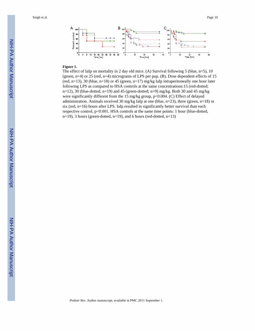
Figure 1.The effect of IaIp on mortality in 2 day old mice. (A) Survival following 5 (blue, n=5), 10(green, n=4) or 25 (red, n=4) micrograms of LPS per pup. (B). Dose dependent effects of 15(red, n=13), 30 (blue, n=18) or 45 (green, n=17) mg/kg IaIp intraperitoneally one hour laterfollowing LPS as compared to HSA controls at the same concentrations:15 (red-dotted;n=12), 30 (blue-dotted, n=19) and 45 (green-dotted; n=9) mg/kg. Both 30 and 45 mg/kgwere significantly different from the 15 mg/kg group, p<0.004. (C) Effect of delayedadministration. Animals received 30 mg/kg IaIp at one (blue, n=23), three (green, n=18) orsix (red, n=16) hours after LPS. IaIp resulted in significantly better survival than eachrespective control, p<0.001. HSA controls at the same time points: 1 hour (blue-dotted,n=19), 3 hours (green-dotted, n=19), and 6 hours (red-dotted, n=13)
Singh et al. Page 10
Pediatr Res. Author manuscript; available in PMC 2011 September 1.
NIH
-PA Author Manuscript
NIH
-PA Author Manuscript
NIH
-PA Author Manuscript

Figure 2.The effect of IaIp administration on survival following subcutaneous administration of livebacteria in 2-day-old BALB/c mice. (A) Survival following subcutaneous injection of 104
(blue), 105 (green),106 (red), 107 (black) CFU E. coli per pup. (B). Dose dependent effect ofIaIp on survival following E. coli administration. Control animals (red, n=28) receivedintraperitoneal HSA, and IaIp treated animals (blue, n=26) received 30 mg/kg IaIpintraperitoneally one hour later. IaIp treated animals had significantly better survival thaneither control group, p<0.05. (C) The effect of IaIp administration on survival followingsubcutaneous administration of group B streptococci. IaIp treated animals (blue, n=17)received 30 mg/kg IaIp intraperitoneally one hour later. IaIp treated animals hadsignificantly better survival than the HSA control group (red, n=16).
Singh et al. Page 11
Pediatr Res. Author manuscript; available in PMC 2011 September 1.
NIH
-PA Author Manuscript
NIH
-PA Author Manuscript
NIH
-PA Author Manuscript
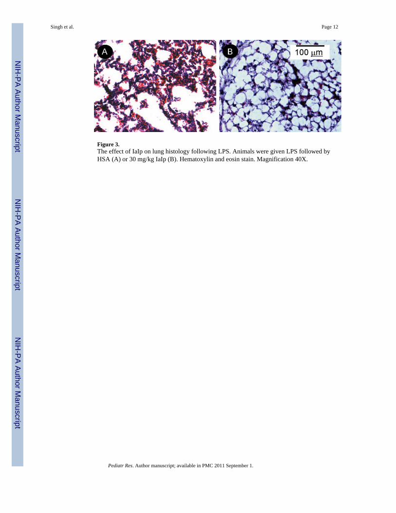
Figure 3.The effect of IaIp on lung histology following LPS. Animals were given LPS followed byHSA (A) or 30 mg/kg IaIp (B). Hematoxylin and eosin stain. Magnification 40X.
Singh et al. Page 12
Pediatr Res. Author manuscript; available in PMC 2011 September 1.
NIH
-PA Author Manuscript
NIH
-PA Author Manuscript
NIH
-PA Author Manuscript
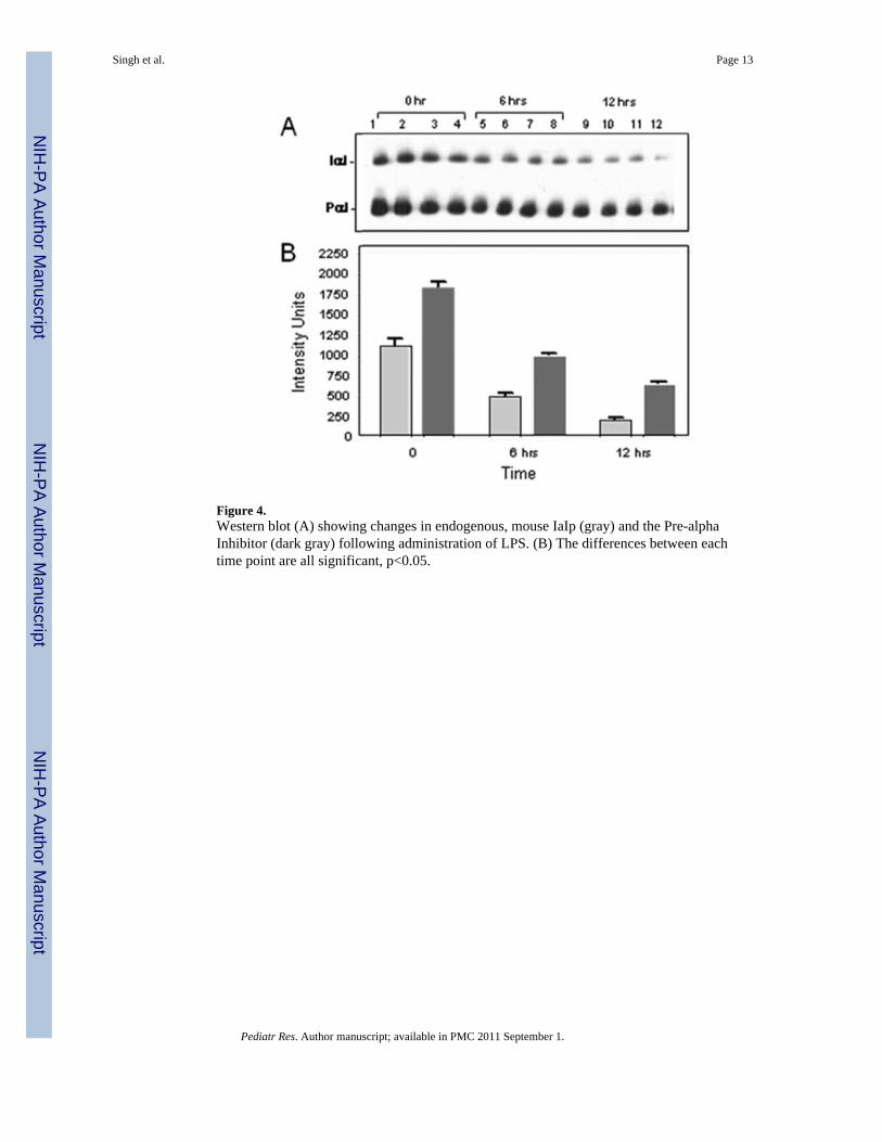
Figure 4.Western blot (A) showing changes in endogenous, mouse IaIp (gray) and the Pre-alphaInhibitor (dark gray) following administration of LPS. (B) The differences between eachtime point are all significant, p<0.05.
Singh et al. Page 13
Pediatr Res. Author manuscript; available in PMC 2011 September 1.
NIH
-PA Author Manuscript
NIH
-PA Author Manuscript
NIH
-PA Author Manuscript

Figure 5.The effect of LPS on systemic cytokine levels. Animals received LPS, HSA and IaIp asdescribed in Methods. Cytokines were measured by radioimmunoassay 3, 6, 12, 24, 48 and72 hours later. Results were compared by Two-way ANOVA. A) TNF-alpha, p=0.0313; B)IL-10, p=0.0377; C) IL-12p70, p=0.0024; D) IL-6, p=0.649; E) IFN-gamma, p=0.141; F)MCP-1, p=0.4948.
Singh et al. Page 14
Pediatr Res. Author manuscript; available in PMC 2011 September 1.
NIH
-PA Author Manuscript
NIH
-PA Author Manuscript
NIH
-PA Author Manuscript
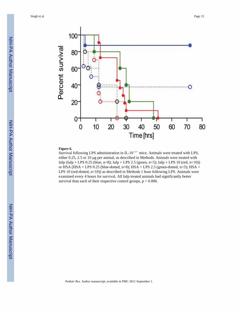
Figure 6.Survival following LPS administration in IL-10−/− mice. Animals were treated with LPS,either 0.25, 2.5 or 10 μg per animal, as described in Methods. Animals were treated withIaIp (IaIp + LPS 0.25 (blue, n=8); IaIp + LPS 2.5 (green, n=5); IaIp + LPS 10 (red, n=10))or HSA (HSA + LPS 0.25 (blue-dotted, n=8); HSA + LPS 2.5 (green-dotted, n=5); HSA +LPS 10 (red-dotted, n=10)) as described in Methods 1 hour following LPS. Animals wereexamined every 4 hours for survival. All IaIp treated animals had significantly bettersurvival than each of their respective control groups, p = 0.006.
Singh et al. Page 15
Pediatr Res. Author manuscript; available in PMC 2011 September 1.
NIH
-PA Author Manuscript
NIH
-PA Author Manuscript
NIH
-PA Author Manuscript