A Decade of the Moderate Resolution Imaging Spectroradiometer: Is a Solar–Cloud Link Detectable
Upload
independentCategory
view
2download
0
Biochemistry 1993, 32, 3557-3563 3557
Inhibitor Binding to the Phe53Trp Mutant of HIV-1 Protease Promotes Conformational Changes Detectable by Spectrofluorometry+
Evelyn J. Rodriguez,$ Christine Debouck,o Ingrid C. Deckman,§,ll Husam Abu-Soud,* Frank M. Raushe1,l and Thomas D. Meek'Jll
Departments of Medicinal Chemistry and Molecular Genetics, SmithKline Beecham Pharmaceuticals, 709 Swedeland Road, King of Prussia, Pennsylvania 19406, and Department of Chemistry, Texas A&M University,
College Station, Texas 77843-3255
Received October 27, 1992; Revised Manuscript Received December 21, 1992
ABSTRACT: HIV- 1 protease contains two identical, conformationally mobile loops, known as flaps, which form in part the binding pockets for substrates and inhibitors. We have constructed a site-specific mutant of the protease in which residues Phe-53 and Phe-153 at the end of the flaps have been mutated to Trp residues, in order to incorporate a specific fluorescent probe to monitor conformational changes upon the binding of an inhibitor. The Phe53Trp (F53W) mutant of HIV-1 protease was expressed in Escherichia coli and purified from bacterial lysates. Analysis of the purified mutant protease demonstrated that its kinetic properties were highly similar to those of the wild-type protease. While binding of a potent peptide- analogue inhibitor (Ki = 9 nM) to the wild-type enzyme led to no change in protein fluorescence, a 5-8% increase in fluorescence was observed with the F53W mutant, indicating an enhancement of the Trp fluorescence due to flap movement upon inhibitor binding. Investigation of the kinetics of the F53W proteaseinhibitor binding by stopped-flow spectrofluorometry revealed a rapid increase in protein fluorescence upon formation of the enzyme-inhibitor complex. These data were consistent with a one-step mechanistic model of inhibitor binding in which flap movement was concomitant with inhibitor binding, from which respective rate constants of association and dissociation of 2.5 X lo6 M-1 s-l and 0.023 s-1 were obtained.
Retroviruses encode a protease which plays an essential role in the maturation of viral polyproteins during virion assembly (Pearl, 1990). The protease from the human immunodeficiency virus-1 (HIV-1)' belongs to the family of the aspartic proteases. HIV-1 protease differs from these monomeric enzymes of >30 kDa in that its similar overall structure is achieved by the assembly of two 1 1-kDa polypep- tides into a homodimer (Meek et al., 1989) which contains the two highly conserved active site aspartyl residues (Asp-25 and Asp-125), each of which is contributed by a monomer (Wlodawer et al., 1989). Asdemonstrated in crystallographic structures of HIV- 1 protease-inhibitor complexes, the binding of a peptide analogue inhibitor, and presumably a peptide substrate, involves numerous hydrogen-bonding interactions with two highly mobile, glycine-rich loops (residues 47-53 and 147-153), knownasflaps (Miller et al., 1989). Movement of these flaps apparently accompanies the binding of the peptide analogue or substrate, which binds in an extended 0-sheet, such that hydrogen bonds are established between
This research was supported in part by National Institutes of Health Grants GM-33894 and GM-39526 (C.D.).
* Author to whom correspondence should be addressed. * Department of Medicinal Chemistry, SmithKline Beecham Phar-
8 Department of Molecular Genetics, SmithKline Beecham Pharma-
11 Present address: Bristol-Myers Squibb Pharmaceutical Research
1 Department of Chemistry, Texas A&M University. I Abbreviations: DTT, dithiothreitol; EDTA, ethylenediaminetet-
raacetic acid; F53W or PheS3Trp, mutation of both the Phe-53 and Phe- 153 residues in the homodimer; HIV- 1 , human immunodeficiency virus-1; kDa, kilodalton; MES, 2-(N-morpholino)ethanesulfonic acid; RP-HPLC, reversed-phase high-performance liquid chromatography; TFA, trifluoroacetic acid; Tris, tris( hydroxymethy1)aminomethane; WT, wild type. Residues on the monomers of HIV-1 protease are numbered 1-99 and 101-199.
maceuticals.
ceuticals.
Institute, Princeton, NJ 08543-4000.
0006-2960/93/0432-3557$04.00/0
complementary carbonyl oxygens and amide protons of the peptide and residues within the flaps. The presumed function of these flappeptide interactions is to entrap and align the scissile peptide sequence in the protease's active site.
One might expect that if the flap regions of HIV- 1 protease were the only mobile portions of the enzyme and contained a Trp or Tyr residue, then the conformational changes in the flap regions that occur upon peptide binding would give rise to changes in protein fluorescence that specifically reflect movement of the flaps. Such fluorescence changes could be investigated in detail by stopped-flow fluorometry to ascertain the kinetic significance of these conformational changes. We have chosen to prepare the Phe53Trp mutant of HIV- 1 protease to potentially provide a specific fluorescent probe of conformational changes in the flap regions of this enzyme. The characterization of this mutant form of HIV-1 protease and its potential use as a probe of this conformational change are reported herein.
EXPERIMENTAL PROCEDURES
Chemicals. The oligopeptide substrates Ac-Arg-Ala-Ser- Gln-Asn-Tyr-Pro-Val-Val-NH2, Ac-Ser-Gln-Asn-Tyr-Pro- Val-Val-NH2, and Ac-Arg-Lys-Ile-Leu-Phe-Leu-Asp-Gly- NH2 were prepared, purified, and characterized as described previously (Hyland et al., 1991a; Moore et al., 1989) or obtained commercially (Bachem Inc., Torrance, CA). All peptides were homogeneous by reverse-phase HPLC (>99%) and thin-layer chromatography, and their structures were confirmed by amino acid analysis and fast atom bombardment mass spectrometry. The competitive inhibitor Ala-Ala-PheW [CHOHCH2]Gly-Val-Val-OMe,inwhichPhe~[CHOHCH2]- Gly designates the incorporated hydroxyethylene dipeptide isostere of (4S,5S)-6-phenyl-5-aminc-4-hydroxyhexanoic acid, was synthesized and characterized as described (Dreyer et al., 1992). The following buffers were used: MEND buffer,
0 1993 American Chemical Society
3558 Biochemistry, Vol. 32, No. 14, 1993
50 mM 2-(N-morpholino)ethanesulfonic acid (MES), pH 6.0, 1 mM EDTA, 1 mM DTT, and 0.2 M NaCl; MENDT buffer, sameas MEND buffer with theadditionof O.l%TritonX-100, MENDG buffer, same as MEND buffer with the addition of 10% glycerol; FAMT-NED buffer, 50 mM each of sodium formate, sodium acetate, MES, and Tris-HC1, 0.2 M NaC1, 1 mM EDTA, and 1 mM DTT (pH 3-7). For all buffers, the ionic strengths were held constant, as measured by conduc- tance, by addition of small aliquots of 4 M NaCl to each titrated solution.
Construction of F53 WMutant HZV-I Protease. A Phe-53 (TTT) to Trp (TGG) substitution was introduced in the HIV- 1 protease coding region (IIIb-BH10 strain; Ratner et al., 1985) using the standard M13 site-specific mutagenesis procedure (Kunkel, 1987). The mutatedcoding region was then inserted into an Escherichia coli expression vector to create a construct identical to the PRO4 expression plasmid previously described for wild-type HIV-1 protease (Debouck et al., 1987). In this system, the protease is initially expressed in a precursor form that catalyzes its own processing into the mature 99 amino acid polypeptides. Both wild-type and mutant proteases in the PRO4 construct were induced for expression by a temperature shift from 32 to 42 OC in the AR58 strain of E. coli as described (Strickler et al., 1989).
Enzyme Purification. Wild-type and mutant HIV- 1 pro- teases were purified from bacterial lysates to apparent homogeneity as judged from NaDodS04-polyacrylamide gel electrophoresis using consecutive steps of ammonium sulfate, gel filtration on a Superose 12 column (Pharmacia), Q- Sepharose Fast-Flow (Pharmacia), and final concentration on an F-Sepharose Fast-Flow resin (Pharmacia) (Grant et al., 1991). Protease solutions were stored at -80 OC in 50 mM sodium acetate (pH 5.0), 1 mM dithiothreitol, 1 mM EDTA, 0.35 M NaC1, and 20% glycerol at concentrations of 15-400 pg/mL. Protein concentrations were determined by use of the Bio-Rad protein assay method with bovine serum albumen as a protein standard or by the chromatographic method described by Grant et al. (1991).
The molecular mass of purified F53W HIV-1 protease was determined by mass spectrometric analysis. Electrospray mass spectra were recorded on a Sciex API-I11 triple-quadrupole mass spectrometer fitted with a standard pneumatically assisted nebulization probe and an atmospheric pressure ionization source (Sciex, Ontario). Protein samples were desalted prior to mass spectrometric analysis by RP-HPLC on a Vydac C4 column (4.6 X 250 mm, 5-pm packing) with use of the linear gradient: t = 0-10 min, 90% A, 10% B; t = 10-20 min, 0% A, 100% B. A = 0.1% (v/v) TFA, and B = 0.1% (v/v) TFA and 90% (v/v) acetonitrile.
Enzyme Assays and Data Analysis. Peptidolysis of the oligopeptide substrates Ac-Ser-Gln-Asn-Tyr-Pro-Val-Val- NH2, Ac-Arg-Ala-Ser-Gln-Asn-Tyr-Pro-Val-Val-NH2, and Ac-Arg-Lys-Ile-Leu-Phe-Leu-Asp-Gly-NH2 as catalyzed by wild-type or F53W HIV-1 proteases was determined by the chromatographic method of Hyland et al. (1991a). Reaction mixtures (50 pL) containing buffer and 0.3-10 mM peptide substrate were equilibrated at 37 or 25 OC prior to the addition of enzyme (60 nM-1.2 pM; room temperature). The reactions were quenched by addition of an equal volume of 3% (v/v) trifluoroacetic acid. Initial rates were determined by single time points of reactions in which product formation had not exceeded 20% of the initial substrate concentration. Initial velocity data were obtained with at least five to six concen- trations of peptide substrate. The pH dependence of the kinetic parameters k,,, and kCat/Km for the cleavage of Ac-Arg-Ala-
Rodrfguez et al.
Ser-Gln-Asn-Tyr-h.o-Val-Val-NH2 catalyzed by F53W HIV- 1 protease was examined in FAMT-NED buffer using five concentrations of the substrate (1-10 mM) at each value of pH and 3 pg/mL enzyme at pH 3-4 and 6-7 and 1.6 pg/mL at pH 4-6. Kineticdata obtained in this manner were analyzed using the FORTRAN programs of Cleland (1979). Initial velocity data were fitted to eq 1, where A is the substrate concentration, Km is the Michaelis constant, u is the initial velocity, Et is the molar enzyme concentration, and kcat is the turnover number.
= kcatEtA/(Km + A ) (1)
Plots of pH-rate profiles in which the log of the kinetic parameter decreased at acidic and basic pH with respective slopes of 1 and -1 (“bell-shaped” curves) were fitted to eq 2, in which y is either k,t/Km or kcat, c is the pH-independent value of y , H i s the proton concentration, and Kl, K2, and K3 are apparent acid dissociation constants for groups on the substrate or enzyme. Profiles in which the log of the plotted parameter decreased to a lower “plateau” value at acidic pH (“wave-shaped” curve) were fitted to eq 3, in which YL and YH are values of y at the high and low pH values, respectively.
logy = log[c/(l + H/K, + K2/H)I (2)
log Y lOg[(yL + yH(K3/H))/(1 + K3/H)I (3)
Inhibition of the F53W HIV-1 protease by the peptide- analogue Ala-Ala-PheQ [CH(OH)CH2] Gly-Val-Val-OCH3 was investigated at variable concentrations of inhibitor (0-90 nM) and by changing fixed concentrations of enzyme (1.3-1 3 nM) at pH 6.0 in MENDT (25 and 37 “C) buffer using Ac- Arg-Ala-Ser-Gln-Asn-Tyr-Pro-Val-Val-NH2 ( 1 mM) as the substrate. Initial rates determined under these conditions were fitted to eq 4 (Williams 8c Morrison, 1979), in which u is the initial rate (nM/s), k,, is the turnover number (s-I), a is the fraction of active enzyme (0-1 .O), Et and Zt are total nanomolar concentrations of enzyme and inhibitor, respectively, and Ki‘ = Ki( 1 + A / K m ) in which Ki is the apparent inhibition constant, A is the fixed substrate concentration, and K m is the Michaelis constant.
u = kcatA{aEt - Zt - K{ + [ (K; + Zt + “E,)* - 4ZtaEt)] 1/2}/2(K + A ) (4)
Fluorescence Studies. Measurements of protein fluores- cence were conducted on a Perkin-Elmer MPF-66 fluorescence spectrophotometer equipped with a thermostated cell holder maintained at 25 OC by a water bath. Data accumulation and analysis were performed on a Perkin-Elmer 7500 pro- fessional computer interfaced to the instrument. HIV- 1 protease (8-16 pg/mL) or F53W HIV-1 protease (8.6-15.6 pg/mL) solutions were prepared in MEND buffer, which were then filtered through a 0.2-pm filter prior to fluorescence measurements. DTT was added to MEND buffer to a final concentration of 10 mM before addition of the enzyme. Ala- Ala-PheQ [CH(OH)CH2] Gly-Val-Val-OCH3 was added to the thermally equilibrated enzyme solutions to a concentration of 10 pM. The following spectral conditions were used: 280- nm excitation wavelength, 0.3-nm excitation slit width, 20- nm emission slit width, 290-nm emission filter, and 0 5 s response time. Emission spectra were recorded from 300 to 400 nm at 480 nm/min.
Rapid reaction kinetics were performed using a temperature- regulated Hi-Tech Model SF-5 1 stopped-flow apparatus
Phe53Trp Mutant Form of HIV-1 Protease Biochemistry, Vol. 32, No. 14, 1993 3559
Trp-53
Trp-42
c
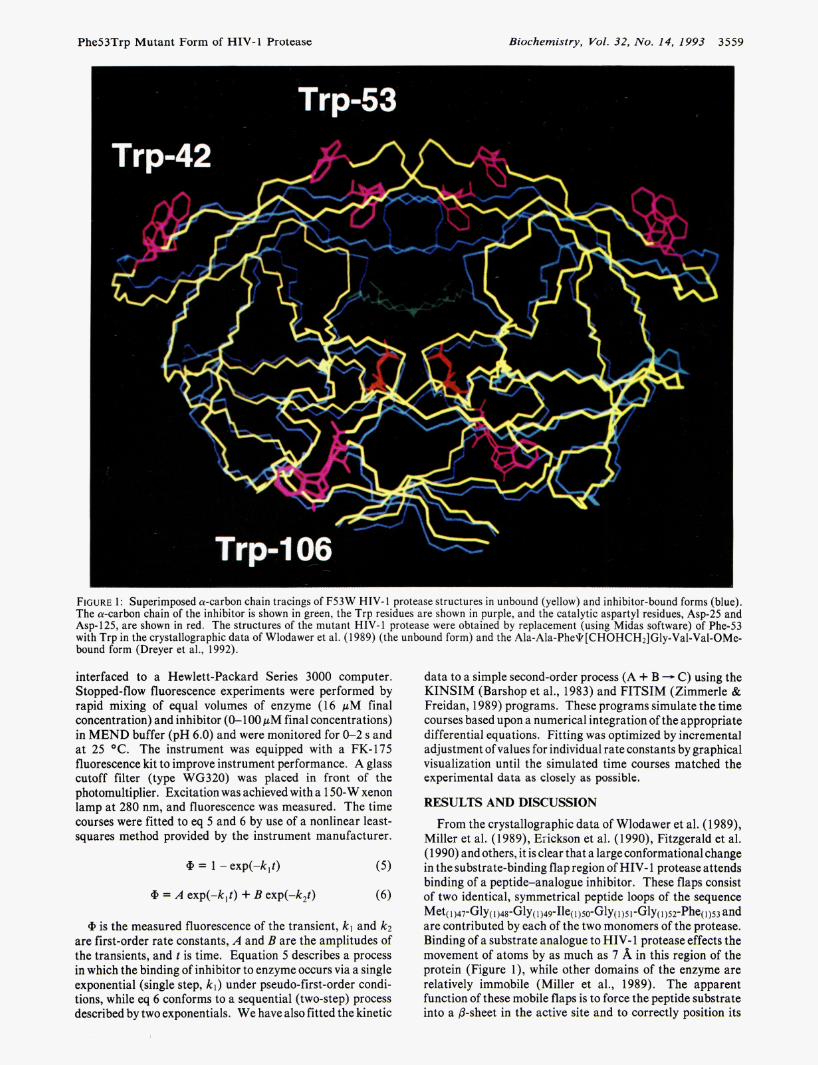
FIGURE 1: Superimposed a-carbon chain tracings of F53W HIV-1 protease structures in unbound (yellow) and inhibitor-bound forms (blue). The a-carbon chain of the inhibitor is shown in green, the Trp residues are shown in purple, and the catalytic aspartyl residues, Asp-25 and Asp-125, are shown in red. The structures of the mutant HIV-1 protease were obtained by replacement (using Midas software) of Phe-53 with Trp in the crystallographic data of Wlodawer et al. (1989) (the unbound form) and the Ala-Ala-Phe\k[CHOHCH2]Gly-Val-Val-OMe- bound form (Dreyer et al., 1992).
interfaced to a Hewlett-Packard Series 3000 computer. Stopped-flow fluorescence experiments were performed by rapid mixing of equal volumes of enzyme (16 pM final concentration) and inhibitor (0-100 pM final concentrations) in MEND buffer (pH 6.0) and were monitored for 0-2 s and at 25 O C . The instrument was equipped with a FK-175 fluorescence kit to improve instrument performance. A glass cutoff filter (type WG320) was placed in front of the photomultiplier. Excitation was achieved with a 150-W xenon lamp at 280 nm, and fluorescence was measured. The time courses were fitted to eq 5 and 6 by use of a nonlinear least- squares method provided by the instrument manufacturer.
data to a simple second-order process (A + B - C) using the KINSIM (Barshop et al., 1983) and FITSIM (Zimmerle & Freidan, 1989) programs. These programs simulate the time courses based upon a numerical integration of the appropriate differential equations. Fitting was optimized by incremental adjustment of values for individual rate constants by graphical visualization until the simulated time courses matched the experimental data as closely as p ~ ~ d l a l ~ .
RESULTS AND DISCUSSION
From the crystallographic data of Wlodawer et al. (1 989), Miller et al. (1989), Eiickson et al. (1990), Fitzgerald et al. (1 990) and others, it is clear that a large conformational change in the substrate-binding flap region of HIV- 1 protease attends binding of a peptide-analogue inhibitor. These flaps consist of two identical, symmetrical peptide loops of the sequence
are contributed by each of the two monomers of the protease. Binding of a substrate analogue to HIV- 1 protease effects the movement of atoms by as much as 7 %, in this region of the protein (Figure l ) , while other domains of the enzyme are relatively immabile (Miller et al., 1989). The apparent function of these mobile flaps is to force the peptide substrate into a &sheet in the active site and to correctly position its
Met(1)47'Gly( 1 ) 4 8 ~ ~ ~ ~ ( 1 ) 4 9 ~ ~ ~ ~ ( 1 ) 5 0 ~ ~ ~ ~ ( 1 ) 5 1 ' ~ ~ ~ ( 1)52'Phe(1)53 and
a = 1 -exp(-k,t)
a = A exp(-k,t) + B exp(-k,t)
@ is the measured fluorescence of the transient, kl and kz are first-order rate constants, A and B are the amplitudes of the transients, and t is time. Equation 5 describes a process in which the binding of inhibitor to enzyme occurs via a single exponential (single step, k l ) under pseudo-first-order condi- tions, while eq 6 conforms to a sequential (two-step) process described by two exponentials. We have also fitted the kinetic
3560 Biochemistry, Vol. 32, No. 14, I993 RodrIguez et al.
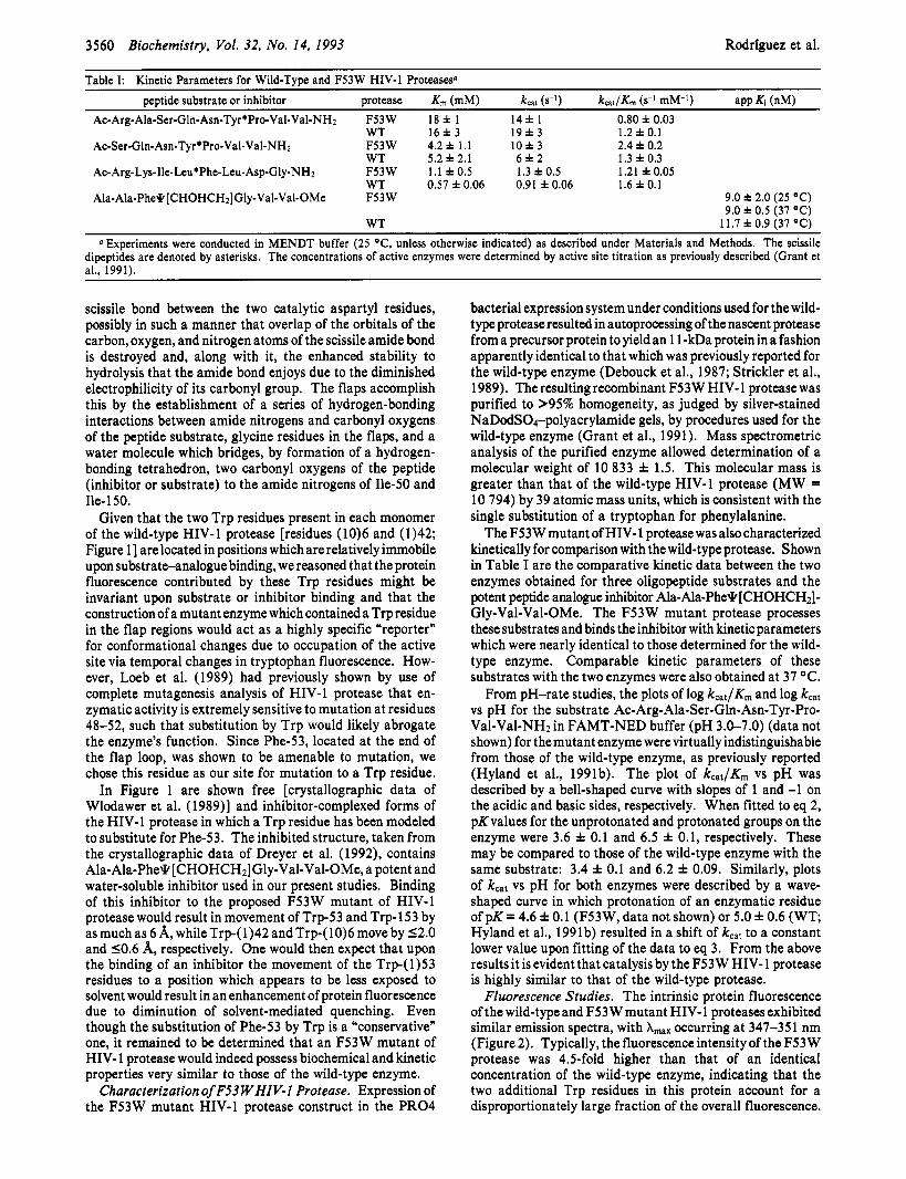
Table I: Kinetic Parameters for Wild-Type and F53W HIV-1 Proteaseso
peptide substrate or inhibitor protease Km (mM) k a t (s-') k a t / & (s-' mM-') app Ki (nM) Ac-Arg-Ala-Ser-Gln-Asn-Tyr*Pro-Val-Val-NHz F53W 18 f 1 1 4 f 1 0.80 f 0.03
WT 1 6 f 3 1 9 f 3 1.2 f 0.1 Ac-Ser-Gln-Asn-Tyr*Pro-Val-Val-NH2 F53W 4.2 f 1 . 1 1 0 f 3 2.4 f 0.2
WT 5.2 f 2.1 6 f 2 1.3 f 0.3 Ac- Arg-Lys-Ile-Leu'Phe-Leu-Asp-Gly-NHz F53W 1 . 1 f 0.5 1.3 f 0.5 1.21 & 0.05
WT 0.57 f 0.06 0.91 f 0.06 1.6 f 0.1 Ala-Ala-Phe9 [CHOHCH2]Gly-Val-Val-OMe F53W 9.0 f 2.0 (25 "C)
9.0 f 0.5 (37 "C) 11.7 f 0.9 (37 "C) WT
0 Experiments were conducted in MENDT buffer (25 "C, unless otherwise indicated) as described under Materials and Methods. The scissile dipeptides are denoted by asterisks. The concentrations of active enzymes were determined by active site titration as previously described (Grant et al., 1991).
scissile bond between the two catalytic aspartyl residues, possibly in such a manner that overlap of the orbitals of the carbon, oxygen, and nitrogen atoms of the scissile amide bond is destroyed and, along with it, the enhanced stability to hydrolysis that the amide bond enjoys due to the diminished electrophilicity of its carbonyl group. The flaps accomplish this by the establishment of a series of hydrogen-bonding interactions between amide nitrogens and carbonyl oxygens of the peptide substrate, glycine residues in the flaps, and a water molecule which bridges, by formation of a hydrogen- bonding tetrahedron, two carbonyl oxygens of the peptide (inhibitor or substrate) to the amide nitrogens of Ile-50 and Ile- 150.
Given that the two Trp residues present in each monomer of the wild-type HIV-1 protease [residues (10)6 and (1)42; Figure 11 are located in positions which are relatively immobile upon substrateanalogue binding, we reasoned that the protein fluorescence contributed by these Trp residues might be invariant upon substrate or inhibitor binding and that the construction of a mutant enzyme which contained a Trp residue in the flap regions would act as a highly specific "reporter" for conformational changes due to occupation of the active site via temporal changes in tryptophan fluorescence. How- ever, Loeb et al. (1989) had previously shown by use of complete mutagenesis analysis of HIV-1 protease that en- zymatic activity is extremely sensitive to mutation at residues 48-52, such that substitution by Trp would likely abrogate the enzyme's function. Since Phe-53, located at the end of the flap loop, was shown to be amenable to mutation, we chose this residue as our site for mutation to a Trp residue.
In Figure 1 are shown free [crystallographic data of Wlodawer et al. (1989)l and inhibitor-complexed forms of the HIV-1 protease in which a Trp residue has been modeled to substitute for Phe-53. The inhibited structure, taken from the crystallographic data of Dreyer et al. (1992), contains Ala-Ala-Pheq [CHOHCHI] Gly-Val-Val-OMe, a potent and water-soluble inhibitor used in our present studies. Binding of this inhibitor to the proposed F53W mutant of HIV-1 protease would result in movement of Trp-53 and Trp- 153 by as much as 6 A, while Trp-( 1)42 and Trp-( 10)6 move by 12.0 and 10.6 A, respectively. One would then expect that upon the binding of an inhibitor the movement of the Trp-( 1)53 residues to a position which appears to be less exposed to solvent would result in an enhancement of protein fluorescence due to diminution of solvent-mediated quenching. Even though the substitution of Phe-53 by Trp is a Kconservative" one, it remained to be determined that an F53W mutant of HIV- 1 protease would indeed possess biochemical and kinetic properties very similar to those of the wild-type enzyme.
Characterization of F53 WHIV-I Protease. Expression of the F53W mutant HIV-1 protease construct in the PRO4
bacterial expression system under conditions used for the wild- type protease resulted in autoprocessing of the nascent protease from a precursor protein to yield an 1 1 -kDa protein in a fashion apparently identical to that which was previously reported for the wild-type enzyme (Debouck et al., 1987; Strickler et al., 1989). The resulting recombinant F53W HIV- 1 protease was purified to >95% homogeneity, as judged by silver-stained NaDodSO4-polyacrylamide gels, by procedures used for the wild-type enzyme (Grant et al., 1991). Mass spectrometric analysis of the purified enzyme allowed determination of a molecular weight of 10 833 f 1.5. This molecular mass is greater than that of the wild-type HIV-1 protease (MW = 10 794) by 39 atomic mass units, which is consistent with the single substitution of a tryptophan for phenylalanine.
The F53W mutant of HIV- 1 protease was also characterized kinetically for comparison with the wild-type protease. Shown in Table I are the comparative kinetic data between the two enzymes obtained for three oligopeptide substrates and the potent peptide analogue inhibitor Ala-Ala-Pheq [CHOHCH2]- Gly-Val-Val-OMe. The F53 W mutant protease processes these substrates and binds the inhibitor with kinetic parameters which were nearly identical to those determined for the wild- type enzyme. Comparable kinetic parameters of these substrates with the two enzymes were also obtained at 37 OC.
From pH-rate studies, the plots of log kcat/Km and log kcat vs pH for the substrate Ac-Arg-Ala-Ser-Gln-Asn-Tyr-Pro- Val-Val-NH2 in FAMT-NED buffer (pH 3.0-7.0) (data not shown) for the mutant enzyme were virtually indistinguishable from those of the wild-type enzyme, as previously reported (Hyland et al., 1991b). The plot of kca,/K, vs pH was described by a bell-shaped curve with slopes of 1 and -1 on the acidic and basic sides, respectively. When fitted to eq 2, pK values for the unprotonated and protonated groups on the enzyme were 3.6 f 0.1 and 6.5 f 0.1, respectively. These may be compared to those of the wild-type enzyme with the same substrate: 3.4 f 0.1 and 6.2 f 0.09. Similarly, plots of kcar vs pH for both enzymes were described by a wave- shaped curve in which protonation of an enzymatic residue of pK = 4.6 f 0.1 (F53W, data not shown) or 5.0 f 0.6 (WT; Hyland et al., 1991 b) resulted in a shift of kcat to a constant lower value upon fitting of the data to eq 3. From the above results it is evident that catalysis by the F53W HIV- 1 protease is highly similar to that of the wild-type protease.
Fluorescence Studies. The intrinsic protein fluorescence of the wild-typeand F53W mutant HIV-1 proteases exhibited similar emission spectra, with A,,, occurring at 347-35 1 nm (Figure 2). Typically, the fluorescence intensity of the F53W protease was 4.5-fold higher than that of an identical concentration of the wild-type enzyme, indicating that the two additional Trp residues in this protein account for a disproportionately large fraction of the overall fluorescence.

Phe53Trp Mutant Form of HIV-1 Protease Biochemistry, Vol. 32, No. 14, 1993 3561
60 A
320 340 360 380 400 h, nm
280
224
168
112
56
I 300 320 340 360 380 400
Jan, nm
FIGURE 2: Conformational changes of F53W HIV-1 protease as detected by the increase in protein fluorescence upon addition of the inhibitor Ala-Ala-PheO[CHOHCH2]Gly-Val-Val-OMe. Protein fluorescence was obtained at 25 'C in MEND buffer (pH 6.0) using an excitation wavelength of 280 nm and an excitation slit width of 0.3 nm, and the fluorescence emission spectra maximum occurred as shown at a peak wavelength of 349 nm. Panels: (A) wild-type HIV-1 protease (0.75 pM) before (solid line) and after (dashed line) addition of Ala-Ala- PheO[CHOHCH2]Gly-Val-Val-OMe to a final concentration of 10 pM (emission slit width = 15 nm); (B) F53W HIV-1 protease (0.59 pM) before (solid line) and after (dashed line) addition of Ala-Ala-PheO[CHOHCH2]Gly-Val-Val-OMe to a final concentration of 10 pM (emission slit width = 20 nm).
Scheme I k l
k2 E + I E l
k l k3
k2 k4 E + I e El El'
Addition of a 100 pM quantity of the potent inhibitor Ala- Ala-Phe\k[CHOHCHz]Gly-Val-Val-OMe to a 0.75 pM quantity of the wild-type enzyme, at which concentration >99.9% of the enzyme is inhibitor-bound, resulted in no change in protein fluorescence (Figure 2A). Therefore, the movement of Trp(l0)6 and Trp(1)42, shown in the structures of the mutant protease (Figure 1) to be 12.0 A, was not sufficient to affect the intrinsic fluorescence of any of the four Trp residues in the wild-type enzyme. Accordingly, any observable change in fluorescence upon inhibitor binding to the F53W mutant protease must result from the approximate 6-A displacement of Trp-53 and Trp-153 as the flaps close down around the inhibitor.
As shown in Figure 2B, the addition of 10 pM Ala-Ala- Phe\k[CH(OH)CH2]Gly-Val-Val-OCH3 to 0.59 pM F53W HIV- 1 protease resulted in an increase in protein fluorescence intensity of 5 4 % with no apparent shift in the value of A,,,. Again, >99% of the F53W mutant protease was occupied by inhibitor in this study, such that the small increase in fluorescence observed represents the maximum change at- tainable by this conformational change. We found that this small overall change in fluorescence and the nanomolar inhibition constant prevented the reliable evaluation of the dissociation constant of Ala- Ala-Pheq [ CHOHCH21 Gly-Val- Val-OMe by fluorescence titration. That the fluorescence was increased rather than quenched suggested that Trp-53 and Trp-153 became less exposed to solvent in the protease- inhibitor complex, in agreement with the structural models. Therefore, the F53W mutant of HIV-1 protease constitutes a highly specific probe for investigating the dynamics of the substrate-binding flaps of the enzyme and the kinetic role of these conformational changes in the enzymatic reaction.
The binding of substrate-analogue to F53 W HIV- 1 protease could proceed via either a single-step or two-step mechanism as outlined in Scheme I. If the transient kinetics were characterized by a single-step mechanism, the stopped-flow
fluorescence data would be characterized by a single expo- nential if flap movement accompanied the formation of the E1 complex. Under these conditions, b b s = kl [I] + kz, and Xobs would display a linear dependence on [I] under pseudo- first-order conditions. Physically, in the single-step mecha- nistic model the conformational changes in the substrate- binding flaps of F53W HIV-1 protease would occur con- comitantly with enzyme-inhibitor association.
In the two-step mechanism, the timecourse of the formation of the proteaseinhibitor complex as exhibited by a biphasic transient "burst" of fluorescence would be described by two independent exponentials, a fast phase (X l ) and a slow phase (XZ). When [I] >> [E] (pseudo-first-order conditions), the transient rate constant, XI , for the fast phase equals kl [I] + k2 + k3 + kq, while for the slow phase (XZ), it has a hyperbolic dependence on inhibitor concentration (Johnson et al., 1992; Furfine et al., 1992). Therefore, plots of XI vs [I] would be linear, while at low inhibitor concentrations, a plot of X2 vs [I] would appear hyperbolic. However, in the case of the F53W protease, the collision of enzyme with inhibitor and the conformational change would occur in individual steps, kl and k3, respectively, such that one would expect to observe transient fluorescence changes only on the second step. In this case, b b s also would be best fitted by a single exponential but distinguishable from the single-step mechanism in that A,,k would not be linearly dependent on inhibitor concentration.
Stopped-flow spectrofluorometric studies were used to determine the transient kinetics of the binding of Ala-Ala- Phe\k[CHOHCH2JGly-Val-Val-OMe to F53W HIV-1 pro- tease. As shown in Figure 3, the change in protein fluorescence as measured by stopped-flow spectrofluorometry (excitation wavelength = 280 nm) of 16 pM F53 W protease rapidly mixed with a large excess of the inhibitor (100 pM; pseudo-first order conditions) was characterized by a rapid transient of increasing fluorescence. The time course was best fitted by a single exponential function (eq 5 ) , assuming pseudo-first- order conditions, and resulted in a transient rate, b b s , of 370 f 20 s-I at 25 "C in MEND buffer (pH 6.0). This transient rate is much greater than the k,,, values of peptide substrates found in Table I, indicating that the rates of the conformational changes which we ascribe to the binding of a substrate- analogue, and presumably substrates, are considerably faster than catalysis and are therefore not rate-limiting. The amplitude of the transient indicated a 5.6% increase in

3562 Biochemistry, Vol. 32, No. 14, 1993 Rodrlguez et al.
of the protein fluorescence. These authors attributed this effect to energy transfer from Trp residues within the enzyme to the proximal aromatic chromophores at the termini of the bound peptide-analogues. These investigators also showed that a tightly bound inhibitor (Ki = 40 nM) conformed to the single-step binding model of Scheme I (kl = 3.1 X lo6 M-I s-l, k2 = 0.12 s-l), while a less potent inhibitor (Ki = 120 nM) displayed transient kinetics consistent with the two-step binding mechanism. Our present results with the F53W mutant enzyme are very similar to those observed for binding of the more potent inhibitor to WT protease observed in this previous study, even though the changes in protein fluorescence arise from different phenomena. As Furfine et al. have suggested, the apparent single-step binding mechanism observed for potent inhibitors of the protease may in fact be a form of the two-step mechanism in which k3 >> k2 as to render the first step essentially irreversible. Thus, in the case of a potent inhibitor, collision of enzyme and the peptide-analogue promotes a rapid “closing” of the flaps so that the lifetime of the E1 complex of Scheme I is fleeting.
l o o t
0.000 0.005 0.010 0.015 0.020 Time (Seconds)
FIGURE 3: Time course of the binding of Ala-Ala-Phe\k- [CHOHCH2JGly-Val-Val-OMe (100pM) to F53W HIV-1 protease (16 pM) as determined by stopped-flow spectrofluorometry at 25 OC, pH 6.0. Protein fluorescence (arbitrary units) was obtained from an excitation wavelength of 280 nm, and the transient of protein fluorescence observed from 0 to 13 ms resulted from rapid mixing of equalvolumesof enzyme and inhibitor. Fitting of the experimental time course (solid line) to eq 5 resulted in a transient rate constant of 370 f 20 SKI, as indicated by the filled circles.
Table 11: Transient Rate Constants from Stopped-Flow Time Courses of Binding of Ala-Ala-Phe~[CHOHCH~]Gly-Val-Val-OMe to F53W HIV-1 Proteasea
app second-order fitted first-order rate contant second-order
inhibior rate constant (&,bs/[inhibitor], rate constant ( P M ) habs (s-’1 M-I s-I) (M-I s-’)
2.0 8.1 4.0 X lo6 0.76 f 0.04 X lo6 5.0 18 3.6 X lo6 2.1 0.2 x 10⁶
10 31 3.1 X lo6 2.8 f 0.1 X lo6 100 310 3.7 x 10⁶ 3.6 & 0.1 X lo6 a First-order rateconstants wereobtained using 16 rM HIV-1 protease
at 25 OC, pH 6.0, from fitting the transient time courses to eq 5, and the apparent second-order rate constant was obtained from data fitted to the first-order process (eq 5), as the quotient of &/[inhibitor]. The fitted second-order rate constants were obtained by fitting the experimental data to a simple second-order process using KINSIM and FITSIM, as described under Experimental Procedures.
fluorescence intensity upon binding of the inhibitor, similar to that observed in the static fluorescence studies. The most reasonable model for our present data is the single-step mechanism of Scheme I. Binding of the inhibitor is con- comitant with movement of the flaps. However, if the two exponentials of a two-step mechanism are not well separated kinetically, which would be true if k3 >> k2, then a two-step binding mechanism may appear to be defined by a single exponential (Furfine et al., 1992).
We further investigated the dependence of b o b s on inhibitor concentration to distinguish between mechanisms. As shown in Table 11, L b s increases with increasing inhibitor concen- trations in accord with a single-step binding mechanism, although pseudo-first-order conditions were not maintained throughout the concentration range used for these experiments. These data were also fitted to a second-order rate equation using the FITSIM program (Zimmerle & Freidan, 1989). The first-order and second-order rate constants are presented in Table 11. The second-order rate constant from a simul- taneous fit of all four time courses is 2.5 X lo6 M-l s-la From this rate constant and the apparent steady-state competitive inhibition constant, Ki = kz/kl = 9 X M, we calculate a value for k2 of 0.023 s-l.
Furfine et al. (1 992) have reported that binding of peptide- analogue inhibitors to WT HIV-1 protease led to quenching
ACKNOWLEDGMENT
We thank Michael Minnich and Dr. David Green for assistance in enzyme purification, Dr. Mark Hemling and Dr. Steven Carr for mass spectrometric analysis, Dr. Geoffrey Dreyer for a gift of HIV-1 protease inhibitor, and Dr. Eric Furfine for helpful comments. We also thank Professor Ronald Swanstrom for discussions, Wilson Francisco for performing data fitting, and Dr. Brian Metcalf and Dr. John Gleason for continued support.
REFERENCES
Barshop, B. A., Wrem, R., & Freisan, C. (1983) Anal. Biochem.
Cleland, W. W. (1979) Methods Enzymol. 63, 103-137. Debouck, C., Gorniak, J. G., Strickler, J. E., Meek, T. Do, Metcalf,
B. W., & Rosenberg, M. (1987) Proc. Natl. Acad. Sci. U.S.A.
Dreyer, G. B.,Lambert,D. M., Meek,T. D., Carr,T. J.,Tomaszek, T. A., Jr., Fernandez, A. V., Bartus, H., Cacciavillani, E., Hassell, A., Minnich, M., Petteway, S. R., Jr., Metcalf, B. W., & Lewis, M. (1992) Biochemistry 31, 6646-6659.
Erickson, J., Neidhart, D. J., VanDrie, J., Kempf, D. J., Wang, X. C., Norbeck, D. W., Plattner, J. J., Rittenhouse, J. W., Turon, M., Wideburg, N., Kohlbrenner, W. E., Simmer, R., Helfrich, R., Paul, D. A., & Knigge, M. (1990) Science 249,
Fitzgerald, P. M. D., McKeever, B. M., VanMiddlesworth, J. F., Springer, J. P., Heimbach, J. C., Leu, C.-T., Herber, W. K., Dixon, R. A. F., & Darke, P. L. (1990) J . Biol. Chem. 265,
Furfine, E. S., D’Souza, E., Ingold, K. J., Leban, J. J., Spector, T., & Porter, D. J. T. (1992) Biochemistry 31, 7886-7891.
Grant, S . K., Deckman, I. C., Minnich, M. D., Culp, J., Franklin, S., Dreyer, G. B.,Tomaszek, T. A., Jr., Debouck, C., & Meek, T. D. (1 99 1) Biochemistry 30, 8424-8434.
Hyland, L. J., Tomaszek, T. A., Jr., Roberts, G. D., Carr, S. A., Magaard, V. W., Bryan, H. L., Fakhoury, S. A,, Moore, M. L., Minnich, M. D., Culp, J. S., DesJarlais, R. L., & Meek, T. D. (1991a) Biochemistry 30, 8441-8453.
Hyland, L. J., Tomaszek, T. A., Jr., & Meek, T. D. (1991b) Biochemistry 30, 8454-8463.
Johnson, K. A. (1993) in The Enzymes (Boyer, P. D., Ed.) Vol. 20, 3rd ed., Academic Press, Orlando, FL (in press).
130, 3942-3947.
84, 8903-8906.
527-5 3 3.
14209-1 421 9.
Phe53Trp Mutant Form of HIV-1 Protease
Kunkel, T. A., Roberts, J. D., & Zakour, R. A. (1987) Methods Enzymol. 154, 367-385.
h b , D. D., Swanstrom, R., Everitt, L., Manchester, M., Stamper, S. E., & Hutchison, C. A., I11 (1989) Nature (London) 340, 397-400.
Meek, T. D., Dayton, B. D., Metcalf, B. W., Dreyer, G. B., Strickler, J. E., Gorniak, J. G., Rosenberg, M., Moore, M. L., Magaard, V. W., & Debouck, C. (1989) Proc. Natl. Acad.
Miller, M., Schneider, J., Sathyanarayana, B. K., Toth, M. V., Marshall, G. R., Clawson, L., Selk, L., Kent, S. B. H., & Wlodawer, A. (1989) Science 246, 1149-1152.
Moore, M. L., Bryan, W. M., Fakhoury, S. A., Magaard, V. W., Huffman, W. F., Dayton, B. D., Meek, T. D., Hyland, L., Dreyer, G. B., Metcalf, B. W., Strickler, J. E., Gorniak, J., & Debouck, C. (1989) Biochem. Biophys. Res. Commun. 159, 420-425.
Sci. U.S.A. 86, 1841-1845.
Pearl, L. H., Ed. (1990) Retrouiral Proteases: Control of
Biochemistry, Vol. 32, No. 14, 1993 3563
Maturation and Morphogenesis, Stockton Press, New York, NY.
Ratner, L., Haseltine, W., Pataraca, R., Livak, K. J., Starcich, B., Josephs, S. F., Doran, E. R., Rafalski, J. A., Whitehorn, E. A., Baumeister, K., Ivanoff, L., Petteway, S. R., Jr., Pearson, M. L., Lautenberger, L. A., Papas, T. S., Ghrayeb, J., Chang, N. T., Gallo, R. C., & Wong-Staal, F. (1985) Nature (London)
Strickler, J. E., Gorniak, J., Dayton, B. D., Meek, T., Moore, M., Magaard, V. W., Malinowski, J., & Debouck, C. (1989) Proteins 6, 139-154.
Williams, J. W., & Morrison, J. F. (1979) Merhods Enzymol. 63,437-466.
Wlcdawer, A., Miller, M., Jaskolski, M., Sathyanarayana, B. K., Baldwin, E., Weber, I. T., Selk, L. M., Clawson, L., Schneider, J., & Kent, S. B. H. (1989) Science 245,616-621.
Zimmerle, C. T., & Freidan, C. (1989) Biochem. J . 258, 381- 387.
31 3,277-284.
Copyright © 2022 FDOKUMEN