INHIBITION OF THEILER'S ENCEPHALOMYELITIS VIRUS (GDVII STRAIN) OF MICE BY AN INTESTINAL...
10
INHIBITION OF THEILER'S ENCEPHALOMYELITIS VIRUS (GDVII STRAIN) OF MICE BY AN INTESTINAL MUCOPOL¥SACCHARIDE II. PURIFICATION AND PROPERTIES OF I'~I~ MUCOPOLYSACCHAEIDE* BY BENJAMIN MANDEL,:~ I~.D., AND EFRAIM RACKER,§ M.D. (From the Department of Microbiology, New York University College of Medicine, New York) (Received for publication, June 25, 1953) In the preceding paper (1) it was shown that the intestinal tissue of adult mice contains a substance capable of inhibiting the GDVII strain of Theiler's encephalomyelitis virus of mice. The inhibitor was demonstrated to be active against the hemagglutinating, as well as the pathogenic activities of the virus. Evidence was presented which showed that the inhibitor is highly specific in its action, and that it functions by combining with the virus. In the present paper the method of purification of the inhibitor will be de- scribed. The results of studies of some of the physicochemical properties which indicate that the inhibitor is a mucopolysaccharide will be presented. Materials and Methods Acetone-Dried Pouder.--The preparation of an acetone-dried powder from tissue and the methods for measuring inhibitory activity are described in the preceding paper (i). Chromatographic Analyses.--The identification of amino adds was carried out following the method of Consden, Gordon, and Martin (2). Two dimensional analysis was carried out using Whatman 1 paper with water-saturated s-coUidineand water-saturated phenol as the solvents. With the latter solvent, the aqueous phase in the tank contained 3 per cent NHs and a trace of KCN was added as described (2). A tank was constructed following the directions of Toen- hies and Kolb (3). The spraying reagent was a 0.1 per cent solution of ninhydrin in butanol containing 1 per cent acetic acid (4). EXPE~NTAL Purification of the GDVII Virus Inh'~itor.--A large pool of acetone-dried powders of mouse intestine was extracted with 10 volumes of distilled water at 37°C. for 1 hour. After centrifuga- * This study was sponsored by the Army Medical Service Graduate School, and was sup- ported in part by the Medical Research and Development Board, Officeof the Surgeon General, United States Army, Washington, D. C., and was aided by a grant from The National Founda- tion for Infantile Paralysis, Inc. ~/Present address: The Division of Infectious Diseases, The Public Health Research Institute of the City of New York. § Present address: The Department of Physiological Chemistry, Yale University School of Medicine. 417 on October 10, 2016 Downloaded from Published November 1, 1953
Transcript of INHIBITION OF THEILER'S ENCEPHALOMYELITIS VIRUS (GDVII STRAIN) OF MICE BY AN INTESTINAL...

I N H I B I T I O N O F T H E I L E R ' S E N C E P H A L O M Y E L I T I S VIRUS ( G D V I I S T R A I N ) OF M I C E BY A N I N T E S T I N A L
M U C O P O L ¥ S A C C H A R I D E
II. PURIFICATION AND PROPERTIES OF I'~I~ MUCOPOLYSACCHAEIDE*
BY BENJAMIN MANDEL,:~ I~.D., AND EFRAIM RACKER,§ M.D.
(From the Department of Microbiology, New York University College of Medicine, New York)
(Received for publication, June 25, 1953)
In the preceding paper (1) i t was shown tha t the intest inal tissue of adu l t mice contains a substance capable of inhibi t ing the G D V I I s train of Theiler 's encephalomyeli t is virus of mice. The inhibitor was demonst ra ted to be act ive against the hemagglutinat ing, as well as the pathogenic activit ies of the virus. Evidence was presented which showed tha t the inhibitor is highly specific in i ts action, and tha t i t functions b y combining with the virus.
In the present paper the method of purification of the inhibi tor will be de- scribed. The results of studies of some of the physicochemical propert ies which indicate tha t the inhibi tor is a mucopolysaccharide will be presented.
Materials and Methods Acetone-Dried Pouder.--The preparation of an acetone-dried powder from tissue and the
methods for measuring inhibitory activity are described in the preceding paper (i). Chromatographic Analyses.--The identification of amino adds was carried out following the
method of Consden, Gordon, and Martin (2). Two dimensional analysis was carried out using Whatman 1 paper with water-saturated s-coUidine and water-saturated phenol as the solvents. With the latter solvent, the aqueous phase in the tank contained 3 per cent NHs and a trace of KCN was added as described (2). A tank was constructed following the directions of Toen- hies and Kolb (3). The spraying reagent was a 0.1 per cent solution of ninhydrin in butanol containing 1 per cent acetic acid (4).
E X P E ~ N T A L
Purification of the GDVII Virus Inh'~itor.--A large pool of acetone-dried powders of mouse intestine was extracted with 10 volumes of distilled water at 37°C. for 1 hour. After centrifuga-
* This study was sponsored by the Army Medical Service Graduate School, and was sup- ported in part by the Medical Research and Development Board, Office of the Surgeon General, United States Army, Washington, D. C., and was aided by a grant from The National Founda- tion for Infantile Paralysis, Inc.
~/Present address: The Division of Infectious Diseases, The Public Health Research Institute of the City of New York.
§ Present address: The Department of Physiological Chemistry, Yale University School of Medicine.
417
on October 10, 2016
Dow
nloaded from
Published November 1, 1953
418 INHIBITION OP THEILER'S VIRUS (GDVII). II
tion at 2500 ~.e.~. for 30 minutes, the supemate was decanted and saved. The sediment was washed once with a small volume of water, the washings combined with the supernate, and the sediment discarded. Advantage was taken of the fact that this crude extract contains phos- phatase. Magnesium chloride was added to a concentration of 0.005 M, the pH adjusted to 8, and the extract was incubated at 37°C. At the start of incubation the total phosphorus content was determined on a sample. After 2 to 3 hours' incubation nearly all the organic phosphorus was liberated as inorganic phosphorus.
To each liter of solution, 250 ml. of 25 per cent trichloracetic acid was added. After centrif- ugation at 2500 x.p.x~, for 30 minutes, the supernatant solution was decanted and saved. The sediment was washed with a small volume of 5 per cent trlchloracetic acid and the washings were added to the supernatant solution which was then dialyzed against distilled water in the cold for 24 hours with several changes of water. During dialysis a fine precipitate which de- veloped was removed by centrifugation. In order to remove any residual protein from this solution, 200 ml. of chloroform and 40 ml. of ~butanol were added to each liter and the mixture shaken vigorously for 30 minutes in a shaking machine. After centrifugation, the
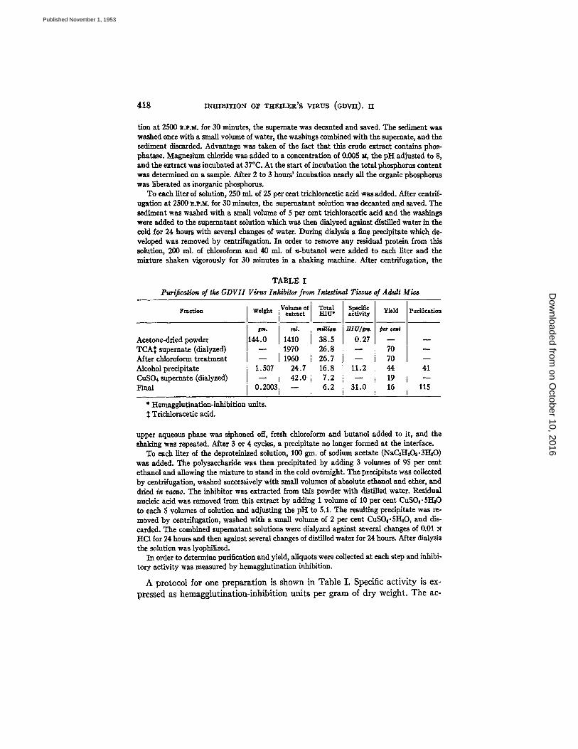
TABLE I
Puriflcaaon of the GDVII Virus Inhibilor from Intestinal Tissue of Adult Mica
Fraction
Acetone-dried powder rCAt superuate (dialyzed) ~dter chloroform treatment Mcohol precipitate CuSO~ supemate (dialyzed) Final
Weight
144.0
1.507
0.2OO3
Volume of extract
ml.
1410 1970 1960
24.7 42.0
Total HIU*
m//l/~n
38.5 25.8 26.7 15.8 7.2 6.2
;pecific mtivity
~ ~IU/rr, J.
0.27
11.2
31.0
giel,
70 70 44 19 16
Purificatim
41
115
* Hemagglutination-inhibition units. Trichloracotic acid.
upper aqueous phase was siphoned off, fresh chloroform and butanol added to it, and the shaldng was repeated. After 3 or 4 cycles, a precipitate no longer formed at the interface.
To each liter of the deproteinized solution, 100 gin. of sodium acetate (NaC2HsOv3HsO) was added. The polysaccharide was then precipitated by adding 3 volumes of 95 per cent ethanol and allowing the n~ ture to stand in the cold overnight. The precipitate was collected by centrifugation, washed successively with small volumes of absolute ethanol and ether, and dried Sn racuo. The inhibitor was extracted from this powder with distilled water. Residual nucleic acid was removed from this extract by adding 1 volume of 10 per cent CuSO4.5I-I20 to each 5 volumes of solution and adjusting the pH to 5.1. The resulting precipitate was re- moved by centrifugation, washed with a small volume of 2 per cent CuSO4.51~O, and dis- carded. The combined supematant solutions were dialyzed against several changes of 0.01 N HC1 for 24 hours and then against several changes of distilled water for 24 hours. After dialysis the solution was lyophlHzed.
In order to determine purification and yield, allquots were collected at each step and inhibi- tory activity was measured by hemagglutination inhibition.
A pro tocol for one p repara t ion is shown in Tab le I. Specific a c t i v i t y is ex-
pressed as hemagglu t ina t ion- inh ib i t ion uni ts per g ram of d ry weight . T h e ac-
on October 10, 2016
Dow
nloaded from
Published November 1, 1953
BENJAMIN MANDEL AND EFRAIM RACKER 419
tivity was determined on an extract of the dry powder in the case of the acetone powder and the alcohol precipitate. The final lyophilized material is com- pletely soluble. The data in Table I show that a lI5-fold purification was achieved with a yield of 16 per cent.
The purification method of Morgan and King (5), which has proved to be highly effective with the blood group substances, was tried for the purification of the GDVII virus inhibitor. Although a relatively pure product was obtained, the yield was only about I0 per cent of the amount obtained by the above de- scribed procedure.
0 . 7 5
,..,el" 0 . 5 0 - -
J 0.25
o 200 250 300 350 400
W A V E L E N G T H (m/J.)
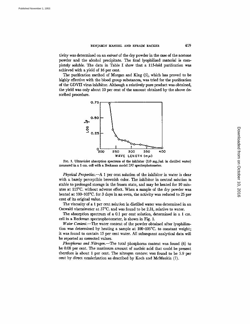
Fro. 1. Ultraviolet absorption spectrum of the inhibitor (1.0 mg./nd, in distilled water) measured in a 1 cm. cell with a Beckman model DU spectrophotometer.
Physical Properties.--A 1 per cent solution of the inhibitor in water is dear with a barely perceptible brownish color. The inhibitor in neutral solution is stable to prolonged storage in the frozen state, and may be heated for 10 min- utes at 115°C. without adverse effect. When a sample of the dry powder was heated at 100-105°C. for 3 days in an oven, the activity was reduced to 25 per cent of its original value.
The viscosity of a 1 per cent solution in distilled water was determined in an Ostwald viscosimeter at 37°C. and was found to be 2.31, relative to water.
The absorption spectrum of a 0.1 per cent solution, determined in a 1 era. cell in a Beckman spectrophotometer, is shown in Fig. 1.
Water Content.--The water content of the powder obtained after lyophiliza- tion was determined by heating a sample at 100--105°C. to constant weight; it was found to contain 15 per cent water. All subsequent analytical data will be reported as corrected values.
Phosphorus and Nitrogen.--The total phosphorus content was found (6) to be 0.08 per cent. The maximum amount of nucleic acid that could be present therefore is about 1 per cent. The nitrogen content was found to be 5.9 per cent by direct nesslerization as described by Koch and MeMeekin (7).
on October 10, 2016
Dow
nloaded from
Published November 1, 1953
420 I N H I B I T I O N O~ THEILER'S VIRUS (GDVII ) , II
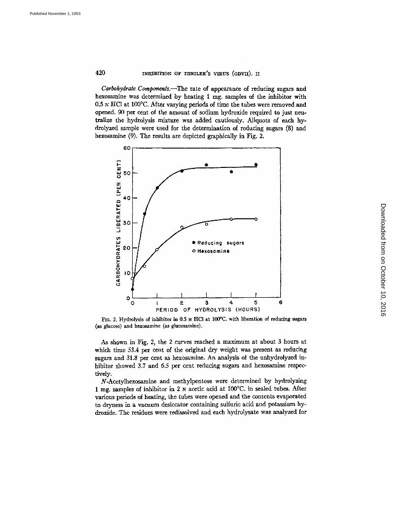
Carbohydrate Components.--The rate of appearance of reducing sugars and hexosamine was determined by heating 1 nag. samples of the inhibitor with 0.5 N HCt at 100°C. After varying periods of time the tubes were removed and opened. 90 per cent of the amount of sodium hydroxide required to just neu- tralize the hydrolysis mixture was added cautiously. Aliquots of each hy- drolyzed sample were used for the determination of reducing sugars (8) and hexosamine (9). The results are depicted graphically in Fig. 2.
6o~
"' 50 -- • 0
n~ LU O.
40 i Q hi I -
ra 5 0
' " • Reducing sugars 1-
2 0 o Hexosamine = i
0 m 10
o I I I I I I o t ~ 5 4 5
PERIOD OF HYDROLYSIS (HOURS) 6
FIG. 2. Hydrolysis of inhibitor in 0.5 z~ HC1 at 100°C. with liberation of reducing sugars (as glucose) and hexosamine (as glucosamine).
As shown in Fig. 2, the 2 curves reached a maximum at about 3 hours at which time 53.4 per cent of the original dry weight was present as reducing sugars and 31.8 per cent as hexosamine. An analysis of the unhydrolyzed in- hibitor showed 3.7 and 6.5 per cent reducing sugars and hexosamine respec- tively.
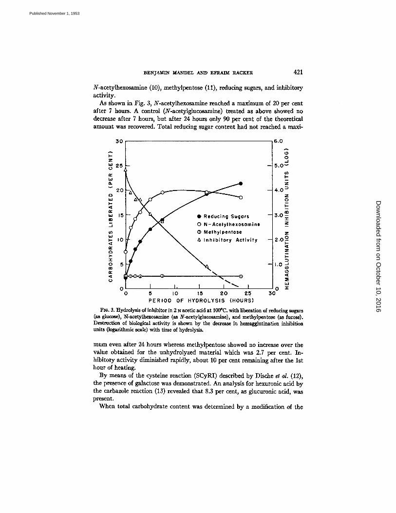
N-Acetylhexosamine and methylpentose were determined by hydrolyzing 1 mg. samples of inhibitor in 2 z~ acetic acid at 100°C. in sealed tubes. After various periods of heating, the tubes were opened and the contents evaporated to dryness in a vacuum desiccator containing sulfuric acid and potassium hy- droxide. The residues were redissolved and each hydrolysate was analyzed for
on October 10, 2016
Dow
nloaded from
Published November 1, 1953
BENJAMIN MANDEL AND EFRAIM RACKER 421
N-acetylhexosamine (10), methylpentose (11), reducing sugars, and inhibitory activity.
As shown in Fig. 3, N-acetylhexosamine reached a maximum of 20 per cent after 7 hours. A control (N-acetylglucosamine) treated as above showed no decrease after 7 hours, but after 24 hours only 90 per cent of the theoretical amount was recovered. Total reducing sugar content had not reached a maxi-
3 0 6 . 0 L z 2 5 - - 4 5 . O ~ (.)
B 20 4o i
~,5 ,~ 3.0; I d ~ o N-Ace,,,,.o.o.i.. l
..<,..,.,, o., ..,,.,,, 1..o I ' - . °
o 0 " ~ 0
O 5 IO 15 2 0 2 5 30 P E R I O D OF HYDROLYSIS (HOURS)
Fie. 3. Hydrolysis of inhibitor in 2 N acetic acid at 100°C. with liberation of reducing sugars (as gluco~), N-acetylhexommine (as N-acetylglucos~mlne), and methylpentose (as fucose). Destruction of biological activity is shown by the decrease in hemagglutination inhibition units (logarithmic scale) with time of hydrolysis.
mum even after 24 hours whereas methylpentose showed no increase over the value obtained for the unhydrolyzed material which was 2.7 per cent. In- hibitory activity diminished rapidly, about 10 per cent remaining after the 1st hour of heating.
By means of the cysteine reaction (SCyRI) described by Dische et al. (12), the presence of galactose was demonstrated. An analysis for hexuronic acid by the carbazole reaction (13) revealed that 8.3 per cent, as glucuronic acid, was present.
When total carbohydrate content was determined by a modification of the
on October 10, 2016
Dow
nloaded from
Published November 1, 1953
422 mmsi~mN or Tm~rL~X'S v i rus (oDv=). n
anthrone reaction (14), the value obtained was 21.9 per cent as glucose, which is considerably lower than the value of 53.4 per cent obtained by determining the reducing sugars on a hydrolyzed specimen. An investigation of the speci- ficity of the anthrone reaction showed that neither hexosamine nor hexuronic acid reacted with anthrone to produce the characteristic color. When the values of these 2 carbohydrates are added to that obtained with the anthrone reagent, there is an approximate agreement with the result obtained for reducing sugars after hydrolysis.
Amino Acid Components.--The "protein" content of the inhibitor was found to be 17.5 per cent by the method of Lowry et a/., (15) with a purified casein standard. A spectrophotometric assay (16) showed 11.2 per cent "protein." In view of the drastic deproteinizing measures employed in the purification procedure, it seems unlikely that any native protein as such is present. Further- more, since both methods are actually based upon the presence of certain amino acids, it seems preferable at present to view the above results as indicat- ing the presence of peptides or amino acids combined with the polysaccharide. On this basis the inhibitor has been considered to be a mucopolysaccharide.
The identification of the individual amino acids by partition chromatography was undertaken.
A sample of the inhibitor was hydrolyzed for 18 hours with 6 N HCI at ll0°C, in a sealed glass tube. The hydrolysate had become dark amber in color and contained a black precipitate. The precipitate was removed by centrifugation and washed. The washings and supernatant fluid were combined and evaporated to dryness in va~uo. The residue was dissolved in a small volume of water and analyzed for amino acids by two dimensional chromatography.
The hydrolysate of the inhibitor was resolved into 7 spots. One, with an R! of 0.08 in phenol, could not be identified. The remaining spots corresponded in their positions to those of aspartic acid, glutamic acid, serine, threonine, alanine, and isoleucine.
Periodate Oxidation.--It has been shown that the receptor substances for the influenza-mumps-Newcastle disease virus group are inactivated by perio- date (17). Similarly, the mucopolysaccharide blood group substances have been shown (18) to be oxidized by periodate. The stability of the GDVII virus inhibitor towards this reagent was investigated.
To 0.3 ml. of an inhibitor solution (1 mg./ml.), 0.15 ml. of 0.1 ~s lithium periodate was added and the mixture was held for 23 hours at room temperature. The reaction was stopped by adding 0.05 hal. of 50 per cent glucose. As a control, the periodate and glucose were mixed 3 hours prior to the addition of the inhibitor. The mixture was then held for 23 hours at room temperature. Each mixture was assayed for inhibitory activity in ~vo and/n dtro. As shown in Table II, destruction of the inhibitor by periodate was almost complete.
Homogeneily of the Purified Inhibitor.--From the preceding data it is clear that the most highly purified inhibitor preparation contains carbohydrate and
on October 10, 2016
Dow
nloaded from
Published November 1, 1953
BENJ'.4MTN MANDEL AND EFRAIM RACKER 423
amino acid components. Since da ta are not available regarding the homo- genei ty of the preparat ion, the question arises regarding the nature of the bio- logically act ive material . The possibilities to be considered are (a) ei ther the ca rbohydra te or the amino acid component is the act ive substance; (b) the biological ac t iv i ty is associated with bo th components; (c) the active substance is present as a th i rd component. This problem was approached b y a t t empt ing to separate the above components b y par t i t ion chromatography and deter- mining biological act ivi ty .
TABLE I I E~ec~ of Lithium Periodat~ on the Activity of the GDVII Virus Inhibitor
S~c~en
ntfibitor -{- periodate .'ontrol
Hema~qlutination inhibition
HIUIral.
100 3,200
Infectivity inhibition*
1
0 . 2 4 0.03
Virus control !
T
0.26
* Each specimen was diluted 1:4 before it was mixed with an equal volume of the virus suspension; consequently there were 12.5 and 400 HIU per ml. respectively in the inocula.
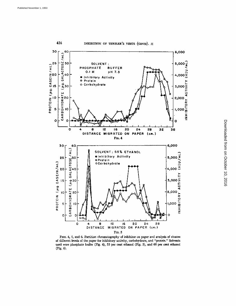
Six sheets of Whatman I paper, 40 by 4 cm. were washed with the solvent to be used. When dry, each sheet was marked off at the edges at 1 cm. intervals. On each of three of the sheets a solution containing 2 mg. of inhibitor was placed 4.5 cra. from the lower edge. The remaining 3 sheets served as controls. Each pair of sheets was suspended in a chromatography chamber with about 1 cm. of the lower edge immersed in the developing solvent. The following solvents were employed: (a) 0.1 M phosphate buffer, pH 7.3; (b) 55 per cent ethanol; (¢) 60 per cent ethanol. When the solvents had ascended approximately 30 cm., the sheets were removed, dried at room temperature, and cut into 1 cm. strips. Each strip w a s placed in a test tube containing 1.5 nil. water, the tubes were heated for 1 hour at 60°C. and the paper strips were removed. The eluates were centrifuged and the supernatant fluids were pipetted off, care being taken to avoid carrying over any paper fibers. Each eluate was analyzed for (a) carbo- hydrate by the SCyRI reaction of Dische (12) using galactose as a standard; (b) "protein" by the method of Lowry e~ a/. (15) with a purified casein standard; (¢) inhibitory activity by the hemaggintination-inhibition method. The values obtained for the control sheets were used as blank values and the experimental results were corrected accordingly.
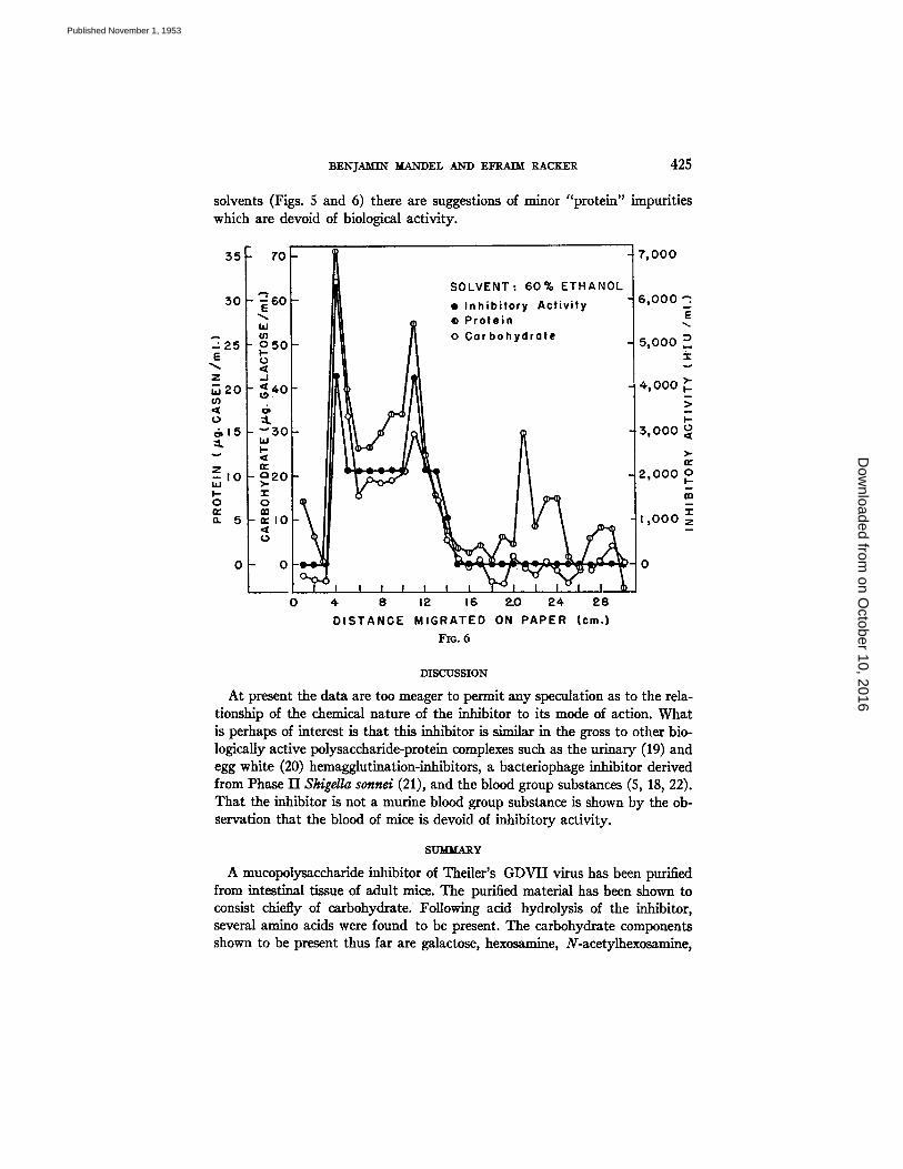
The results of these experiments are shown in Figs. 4 to 6. I t can be seen tha t the inhibi tor migra ted most rap id ly when the solvent was phosphate buffer (Fig. 4), and least rap id ly with the 60 per cent ethanol solvent (Fig. 6). Each a t t e m p t to separate the ca rbohydra te and "pro te in" components b y the use of a different solvent was unsuccessful, and in each case there is an approximate paral lel ism between the 2 components and biological act ivi ty . The inabi l i ty to separa te the 2 components from each other or from the biologically act ive substance s t rongly indicates tha t the carbohydra te and "pro te in" are combined and tha t this complex possesses the inhibi tory act ivi ty . Wi th the ethanol
on October 10, 2016
Dow
nloaded from
Published November 1, 1953
424 m m ~ r r i o N o F zm~z~R's v n ~ u s ( a D v n ) . r l
30 ~0
~ 2 5 - ~ 50 : 0
E ~
z 2 0 - ~ 4 0 ,'5, <
o 15 - ~ $ O
~k ,,,
z I0 - < 2 0 uJ ¢3 I - >-
o - ~ I0 n- 5 (1. rn
n.,
0 o 0
0
SOLVENT : PHOSPHATE BUFFER
0.1 M pH 7.:5
• Inh ib i tory Act iv i ty ¢ Protein o Carbohydrate
I I I I l l
8 12 16 2 0 24. 2 8 3 2 DISTANCE MIGRATED ON PAPER (cm.)
FI(;. 4
6 ,000
5 ,000
4 ,000 i
3 , 0 0 0
2,000
I , 0 0 0 7., - i - Z I
0
3 6
30 " 60
2 5 " ,,,50 03
20 - ~ 4 0 Z _ j
LU ' ~
15- ~ 3 0 o ::L
::L " ' I0- , ~ 2 0
z n- - Q
I - 0 5 " T I 0 n- 0 0 . n~
n-
O " o O
SOLVENT: 55% ETHANOL e l n h i b l t o r y Ac t i v i t y wProtein oCarbohydrate
4 B 12 16 2 0 2 4 2 8 DISTANCE MIGRATED ON PAPER (cm.)
FzG. 5
6 ,000
"3. 5 ,000 .~
' I " 4,000
I -
3 ,000 _~ I - 0
2 ,000 ~. n-
O 1.1.
1,000
Z
0
FIGs. 4, 5, and 6. Partition chromatography of inhibitor on paper and analysis of eluates of different levels of the paper for inhibitory activity, carbohydrate, and "protein." Solvents used were phosphate buffer (Fig. 4), 55 per cent ethanol (Fig. 5), and 60 per cent ethanol (Fig. 4).
on October 10, 2016
Dow
nloaded from
Published November 1, 1953
BEN]AMTN MANDEL AND EFRAIM RACKER 425
solvents (Figs. 5 and 6) there are suggestions of minor "protein" impurities which are devoid of biological activity.
55 70
.-.30 - ~ 6 0 {
425 - o 5 o I
920- ,o I
g 5 - n ' l O
SOLVENT: 60% ETHANOL • Inhibitory Activity ¢ Protein o Cor bohydrote
7,000
6 ,000 ;
5,000
4,000
,3, 0 O0 , >. I = 2,000 o
I ,000
0 - 0 i-e-e-~ ~ % W :,~';, --l-~ @~ ¢.'-* c ~-i 0
4 8 12 16 2.0 24 28 DISTANGE MIGRATED ON PAPER (cm.)
FIG. 6
DISCUSSION
At present the data are too meager to permit any speculation as to the rela- tionship of the chemical nature of the inhibitor to its mode of action. What is perhaps of interest is that this inhibitor is similar in the gross to other bio- logically active polysaccharide-protein complexes such as the urinary (19) and egg white (20) hemagglutination-inhibitors, a bacteriophage inhibitor derived from Phase II Shigella sonnel (21), and the blood group substances (5, 18, 22). That the inhibitor is not a murine blood group substance is shown by the ob- servation that the blood of mice is devoid of inhibitory activity.
SUMMARY
A mucopolysaccharide inhibitor of TheileFs GDVII virus has been purified from intestinal tissue of adult mice. The purified material has been shown to consist chiefly of carbohydrate. Following acid hydrolysis of the inhibitor, several amino acids were found to be present. The carbohydrate components shown to be present thus far are galactose, hexosamine, N-acetylhexosamine,
on October 10, 2016
Dow
nloaded from
Published November 1, 1953
426 INHIBITION OF TIt'EILER'S VIRUS (GDVII). II
methylpentose, hexuronic acid; an indication has been obtained that sedo- heptulose is also present. The available evidence strongly suggests that this mucopolysaccharide, and not some other substance present in minute amounts, is the inhibitor of the GDVII virus.
BIBLIOGRAPHY
1. Mandel, B., and Racker, E., J. Exp. Med., 1953, 98, 399. 2. Consden, R., Gordon, A. H., and Martin, A. J. P., Biochem. J., 1944, 38, 224. 3. Toennies, G., and Kolb, J. J., Anal. Chem., 1951, 23, 823. 4. Consden, R., and Gordon, A. H., Nature, 1948, 162, 180. 5. Morgan, W. T. J., and King, H. K., Biochem. J., 1943, 37, 640. 6. Lohmann, K., and Jendrassik, L., Biochem. Z., 1926, 178, 419. 7. Koch, F. C., and McMeekin, T. L., J. Am. Chem. Soc., 1924, 46, 2066. 8. Nelson, N., J. Biol. Chem., 1944, 153, 375. 9. Kabat, E. A., and Mayer, M. M., Experimental Immunochemistry, Spdngtield,
Illinois, Charles C. Thomas, 1948, 312. 10. Morgan, W. T. J., and Elson, L. A., Biochem. J., 1934, 28, 988. 11. Dische, Z., and Shetfles, L. B., Y. Biol. Chem., 1948, 175, 595. 12. Dische, Z., Shettles, L. B., and Osnos, M., Arch. Biochem., 1949, 22, 169. 13. Dische, Z., J. Biol. Chem., 1947, 167, 189. 14. Morris, D. L., Science, 1948, 107, 254. 15. Lowry, O. H., Rosebrough, N. J., Farr, A. L., and Randall, R. J., J. Biol. Chem.,
1951, 193, 265. 16. Warburg, 0., and Christian, W., Biochem. Z., 1941, 310, 384. 17. Hirst, G. K., J. Exp. Med., 1948, 87, 301. 18. Annison, E. F., and Morgan, W. T. J., Biod~m. Y., 1951, 50, 460; 1952, 52, 247. 19. Tamm, I., and Horsfall, F. L., Jr., J. Exp. Med., 1952, 95, 71. 20. I~nni, F., Sharp, D. G., Eckert, E. A., Dillon, E. S., Beard, D., and Beard,
J. W., J. Biol. Chem., 1949, 1"/9, 1275. 21. Jesaitis, M. A., and Goebel, W. F., J. Exp. M'ed., 1952, 96, 409. 22. Bendich, A., Kabat, E. A., and Bezer, A. E., J. Exp. Med., 1946, 83, 485.
on October 10, 2016
Dow
nloaded from
Published November 1, 1953