Inhibition of butyrate uptake by the primary bile salt chenodeoxycholic acid in intestinal...
11
Inhibition of Butyrate Uptake by the Primary Bile Salt Chenodeoxycholic Acid in Intestinal Epithelial Cells Pedro Gonc ¸alves, Telmo Catarino, Ine ˆs Grego ´rio, and Fa ´tima Martel * Faculty of Medicine, Department of Biochemistry (U38-FCT), University of Porto, Porto, Portugal ABSTRACT Colorectal cancer (CRC) is one of the most common cancers worldwide. Epidemiological and experimental studies suggest that bile acids may play a role in CRC etiology. Our aim was to characterize the effect of the primary bile acid chenodeoxycholic acid (CDCA) upon 14 C-BT uptake in tumoral (Caco-2) and non-tumoral (IEC-6) intestinal epithelial cell lines. A 2-day exposure to CDCA markedly and concentration- dependently inhibited 14 C-BT uptake by IEC-6 cells (IC 50 ¼ 120 mM), and, less potently, by Caco-2 cells (IC 50 ¼ 402 mM). The inhibitory effect of CDCA upon 14 C-BT uptake did not result from a decrease in cell proliferation or viability. In IEC-6 cells: (1) uptake of 14 C-BT involves both a high-affinity and a low-affinity transporter, and CDCA acted as a competitive inhibitor of the high-affinity transporter; (2) CDCA inhibited both Na þ -coupled monocarboxylate cotransporter 1 (SMCT1)- and H þ -coupled monocarboxylate transporter 1 (MCT1)-mediated uptake of 14 C-BT; (3) CDCA significantly increased the mRNA expression level of SMCT1; (4) inhibition of 14 C-BT uptake by CDCA was dependent on CaM, MAP kinase (ERK1/2 and p38 pathways), and PKC activation, and reduced by a reactive oxygen species scavenger. Finally, BT (5 mM) decreased IEC-6 cell viability and increased IEC-6 cell differentiation, and CDCA (100 mM) reduced this effect. In conclusion, CDCA is an effective inhibitor of 14 C-BT uptake in tumoral and non-tumoral intestinal epithelial cells, through inhibition of both H þ -coupled MCT1- and SMCT1-mediated transport. Given the role played by BT in the intestine, this mechanism may contribute to the procarcinogenic effect of CDCA at this level. J. Cell. Biochem. 113: 2937–2947, 2012. ß 2012 Wiley Periodicals, Inc. KEY WORDS: BUTYRATE UPTAKE; CHENODEOXYCHOLIC ACID; IEC-6 CELLS; Caco-2 CELLS; MCT1; SMCT1 C olorectal cancer (CRC) is a leading cause of cancer death in occidental countries [Jemal et al., 2011]. The causes of CRC are multifactorial, and a good correlation between a diet high in saturated fats and low in dietary fiber, fruit, and vegetables, and an increased risk of CRC has been well established by epidemiological studies [Martı ´nez et al., 2008]. Short-chain fatty acids [(SCFAs) acetate, propionate, and butyrate (BT)] are organic acids produced in the intestinal lumen by bacterial fermentation of mainly undigested dietary fiber. Among SCFA, BT plays a key role in colonic epithelium homeostasis, by having multiple regulatory roles at that level, including: (1) being the main energy source for colonocytes; (2) inhibition of colon carcinogenesis (by suppressing growth of cancer cells, inducing differentiation and apoptosis and inhibiting cell proliferation); (3) promotion of growth and proliferation of normal colonic epithelial cells; (4) stimulation of fluid and electrolyte absorption; (5) stimulation of mucus secretion and increase of vascular flow and motility; (6) reduction of visceral perception, intestinal discomfort, and pain; (7) inhibition of colon inflammation and oxidative stress; and (8) improvement of the colonic defense barrier function [Hamer et al., 2008]. As stated, one of the proposed beneficial effects of BT on human colonic health is the prevention/inhibition of colon carcinogenesis Journal of Cellular Biochemistry ARTICLE Journal of Cellular Biochemistry 113:2937–2947 (2012) 2937 Abbreviations used: ALP, alkaline phosphatase; BT, butyrate; Caco-2, human epithelial colon adenocarcinoma cell line; CaM, Ca 2þ /calmodulin; CaMK II, CaM-dependent protein kinase II; CDCA, chenodeoxycholic acid; CRC, colorectal cancer; IEC-6, rat non-tumoral small intestinal epithelial cell line; LDH, lactate dehydrogenase; MAP, mitogen-activated protein; MAPK, mitogen-activated protein kinase; MCT1, H þ -coupled monocarboxylate trans- porter 1; MTT, 3-{4,5-dimethyl-2-thiazolyl}-2,5-diphenyl-2H-tetrazolium bromide; PKA, protein kinase A; PKC, protein kinase C; PKG, protein kinase G; PTK, protein tyrosine kinases; qRT-PCR, quantitative real-time reverse transcription polymerase chain reaction; SCFA, short-chain fatty acid; SMCT1, Na þ -coupled monocarboxylate cotransporter 1; SRB, sulforhodamine B. No conflicts of interest are declared by the authors. Grant sponsor: Fundac ¸a ˜o para a Cie ˆncia e a Tecnologia (FCT), COMPETE, QREN and FEDER; Grant number: PTDC/ SAU-FCF/67805/2006. *Correspondence to: Prof. Fa ´tima Martel, Faculty of Medicine, Department of Biochemistry, University of Porto, Al. Prof. Herna ˆni Monteiro, 4200-319 Porto, Portugal. E-mail: [email protected] Manuscript Received: 15 September 2011; Manuscript Accepted: 19 April 2012 Accepted manuscript online in Wiley Online Library (wileyonlinelibrary.com): 2 May 2012 DOI 10.1002/jcb.24172 ß 2012 Wiley Periodicals, Inc.
-
Upload
independent -
Category
Documents
-
view
0 -
download
0
Transcript of Inhibition of butyrate uptake by the primary bile salt chenodeoxycholic acid in intestinal...

Inhibition of Butyrate Uptake by the Primary Bile SaltChenodeoxycholic Acid in Intestinal Epithelial Cells
Pedro Goncalves, Telmo Catarino, Ines Gregorio, and Fatima Martel*
Faculty of Medicine, Department of Biochemistry (U38-FCT), University of Porto, Porto, Portugal
ABSTRACTColorectal cancer (CRC) is one of the most common cancers worldwide. Epidemiological and experimental studies suggest that bile acids may
play a role in CRC etiology. Our aim was to characterize the effect of the primary bile acid chenodeoxycholic acid (CDCA) upon14C-BT uptake
in tumoral (Caco-2) and non-tumoral (IEC-6) intestinal epithelial cell lines. A 2-day exposure to CDCA markedly and concentration-
dependently inhibited 14C-BT uptake by IEC-6 cells (IC50¼ 120mM), and, less potently, by Caco-2 cells (IC50¼ 402mM). The inhibitory effect
of CDCA upon 14C-BT uptake did not result from a decrease in cell proliferation or viability. In IEC-6 cells: (1) uptake of 14C-BT involves both a
high-affinity and a low-affinity transporter, and CDCA acted as a competitive inhibitor of the high-affinity transporter; (2) CDCA inhibited
both Naþ-coupled monocarboxylate cotransporter 1 (SMCT1)- and Hþ-coupled monocarboxylate transporter 1 (MCT1)-mediated uptake of14C-BT; (3) CDCA significantly increased the mRNA expression level of SMCT1; (4) inhibition of 14C-BT uptake by CDCA was dependent on
CaM, MAP kinase (ERK1/2 and p38 pathways), and PKC activation, and reduced by a reactive oxygen species scavenger. Finally, BT (5mM)
decreased IEC-6 cell viability and increased IEC-6 cell differentiation, and CDCA (100mM) reduced this effect. In conclusion, CDCA is an
effective inhibitor of 14C-BT uptake in tumoral and non-tumoral intestinal epithelial cells, through inhibition of both Hþ-coupled MCT1- and
SMCT1-mediated transport. Given the role played by BT in the intestine, this mechanism may contribute to the procarcinogenic effect of
CDCA at this level. J. Cell. Biochem. 113: 2937–2947, 2012. � 2012 Wiley Periodicals, Inc.
KEY WORDS: BUTYRATE UPTAKE; CHENODEOXYCHOLIC ACID; IEC-6 CELLS; Caco-2 CELLS; MCT1; SMCT1
C olorectal cancer (CRC) is a leading cause of cancer death in
occidental countries [Jemal et al., 2011]. The causes of CRC
are multifactorial, and a good correlation between a diet high in
saturated fats and low in dietary fiber, fruit, and vegetables, and an
increased risk of CRC has been well established by epidemiological
studies [Martınez et al., 2008].
Short-chain fatty acids [(SCFAs) acetate, propionate, and butyrate
(BT)] are organic acids produced in the intestinal lumen by bacterial
fermentation of mainly undigested dietary fiber. Among SCFA, BT
plays a key role in colonic epithelium homeostasis, by having
multiple regulatory roles at that level, including: (1) being the main
energy source for colonocytes; (2) inhibition of colon carcinogenesis
(by suppressing growth of cancer cells, inducing differentiation and
apoptosis and inhibiting cell proliferation); (3) promotion of growth
and proliferation of normal colonic epithelial cells; (4) stimulation
of fluid and electrolyte absorption; (5) stimulation of mucus
secretion and increase of vascular flow andmotility; (6) reduction of
visceral perception, intestinal discomfort, and pain; (7) inhibition
of colon inflammation and oxidative stress; and (8) improvement of
the colonic defense barrier function [Hamer et al., 2008].
As stated, one of the proposed beneficial effects of BT on human
colonic health is the prevention/inhibition of colon carcinogenesis
Journal of CellularBiochemistry
ARTICLEJournal of Cellular Biochemistry 113:2937–2947 (2012)
2937
Abbreviations used: ALP, alkaline phosphatase; BT, butyrate; Caco-2, human epithelial colon adenocarcinoma cellline; CaM, Ca2þ/calmodulin; CaMK II, CaM-dependent protein kinase II; CDCA, chenodeoxycholic acid; CRC,colorectal cancer; IEC-6, rat non-tumoral small intestinal epithelial cell line; LDH, lactate dehydrogenase; MAP,mitogen-activated protein; MAPK, mitogen-activated protein kinase; MCT1, Hþ-coupled monocarboxylate trans-porter 1; MTT, 3-{4,5-dimethyl-2-thiazolyl}-2,5-diphenyl-2H-tetrazolium bromide; PKA, protein kinase A; PKC,protein kinase C; PKG, protein kinase G; PTK, protein tyrosine kinases; qRT-PCR, quantitative real-time reversetranscription polymerase chain reaction; SCFA, short-chain fatty acid; SMCT1, Naþ-coupled monocarboxylatecotransporter 1; SRB, sulforhodamine B.
No conflicts of interest are declared by the authors.
Grant sponsor: Fundacao para a Ciencia e a Tecnologia (FCT), COMPETE, QREN and FEDER; Grant number: PTDC/SAU-FCF/67805/2006.
*Correspondence to: Prof. Fatima Martel, Faculty of Medicine, Department of Biochemistry, University of Porto,Al. Prof. Hernani Monteiro, 4200-319 Porto, Portugal. E-mail: [email protected]
Manuscript Received: 15 September 2011; Manuscript Accepted: 19 April 2012
Accepted manuscript online in Wiley Online Library (wileyonlinelibrary.com): 2 May 2012
DOI 10.1002/jcb.24172 � � 2012 Wiley Periodicals, Inc.

[Hamer et al., 2008]. BT is transported into intestinal epithelial cells
by two specific carrier-mediated transport systems, the electro-
neutral Hþ-coupled monocarboxylate transporter 1 (MCT1) [Cuff
et al., 2005] and the Naþ-coupled monocarboxylate cotransporter 1
(SMCT1) [Gupta et al., 2006]. MCT1 [Cuff et al., 2005] and SMCT1
[Gupta et al., 2006] were recently proposed to function as tumor
suppressors, most probably due to their ability to mediate the entry
of BT into intestinal epithelial cells. Therefore, factors that interfere
with BT uptake into intestinal epithelial cells are potentially
detrimental to intestinal health and integrity by promoting CRC.
Primary bile acids, predominantly cholic acid and chenodeoxy-
cholic acid (CDCA), are produced in the liver by the metabolism of
cholesterol and are delivered into the intestinal tract as glycine or
taurine conjugates. Most bile acids are reabsorbed in the ileum as
conjugates. However, during each enterohepatic cycle, about 10% of
the bile acids escape into the colon where they are exhaustively
converted by the intestinal flora, and 1–3% of the bile acids will
eventually be excreted in the feces. Fecal bile acids are almost
completely deconjugated and have undergone several other
reactions, such as dehydroxylation, dehydrogenation, and epimer-
ization [Ridlon et al., 2006]. Intestinal epithelial cells are thus
exposed to various concentrations and compositions of bile acids.
Several epidemiological studies have found that fecal bile acid
concentrations are elevated in populations with a high incidence of
CRC [Reddy et al., 1977, 1978], and high-fat, high-cholesterol diets,
which increase the risk of CRC, also increase the total of bile acids in
the gut [Stadler et al., 1988]. In agreement with these observations,
secondary bile acids have been shown to be tumor promoters, both
in vitro and in animal studies [Reddy et al., 1977; Mahmoud et al.,
1999]. Their mechanism of action is still poorly understood,
although PKC, MAP kinases (MAPK), and nuclear factor-kappa B
(NF-kB) were identified as molecular targets for these compounds
[Qiao et al., 2000; McMillan et al., 2003].
In relation to primary bile acids, less information is available.
However, a significantly higher fecal concentration of the primary
bile salt CDCA was seen in patients with CRC/adenoma [Tong et al.,
2008] and CDCA was also shown to be tumor promoting in some
animal and cell culture studies [Mahmoud et al., 1999; McMillan
et al., 2003].
The majority of studies concerning the effects of BT and bile
acids on intestinal epithelial cells have considered these factors
individually. However, in vivo, both are present in the colon and
may directly or indirectly influence each other’s actions. Very
recently, we demonstrated a discrete interference of the bile salt
deoxycholic acid upon uptake of 14C-BT by intestinal epithelial cells
[Goncalves et al., 2011d]. So, the aim of the present study was to
investigate the hypothesis that interference with the apical uptake of
BT may be one of the mechanisms contributing to the effect of the
primary bile acid CDCA upon colon carcinogenesis.
MATERIALS AND METHODS
CACO-2 AND IEC-6 CELL CULTURE
The Caco-2 and IEC-6 cell lines were obtained from the Deutsche
Sammlung von Mikroorganismen und Zellkulturen (Braunschweig,
Germany) and were used between passage numbers 53–58 and
17–30, respectively. The cells were maintained in a humidified
atmosphere of 5% CO2–95% air. Caco-2 cells were cultured in
minimum essential medium (Sigma, St. Louis, MO) containing
5.55mM glucose and supplemented with 15% fetal calf serum,
25mM HEPES, 100U/ml penicillin, 100mg/ml streptomycin, and
0.25mg/ml amphotericin B (all from Sigma). IEC-6 cells were
cultured in Dulbecco’s modified Eagle’s medium:RPMI 1640
medium (1:1), supplemented with 10% fetal bovine serum, 0.1 U/
ml insulin, 5.96 g HEPES, 2.2 g NaHCO3, 100 U/ml penicillin,
100mg/ml streptomycin, and 0.25mg/ml amphotericin B (all from
Sigma). Culture medium was changed every 2–3 days and the
culture was split every 7 days. For sub-culturing, the cells were
removed enzymatically (0.25% trypsin–EDTA, 5min, 378C), split1:3, and sub-cultured in plastic culture dishes (21 cm2; Ø 60mm;
Corning Costar, Corning, NY). For use in experiments, cells were
seeded on 24-well plastic cell culture clusters (2 cm2 Ø 16mm;
TPP1), and experiments were performed 7–9 days after the initial
seeding (90–100% confluence).
TREATMENT OF THE CELLS
The effect of CDCA was tested by cultivating cells in culture medium
containing CDCA (or the respective solvent) for 1, 2, 3, or 7 days. The
effect of BT was tested by cultivating cells in culture medium
containing BT or the respective solvent for 2 days. The effect of
inhibitors of signaling pathways was tested by cultivating cells in
culture medium containing CDCA, these compounds or the
respective solvents for 1 day. The medium was renewed daily
and the end of the treatment period was always days 7–9 of cell
culture.
TRANSPORT STUDIES
Transport experiments were performed with Caco-2 and IEC-6 cells
incubated in glucose-free Krebs (GFK) buffer containing (mM): 125
NaCl, 4.8 KCl, 1.2 MgSO4, 1.2 CaCl2, 25 NaHCO3, 1.6 KH2PO4,
0.4 K2HPO4, and 20 MES, pH 6.5 [Goncalves et al., 2009]. Initially,
the culture medium was aspirated and the cells were washed with
0.3ml GFK buffer at 378C. Then, uptake was initiated by the additionof 0.3ml GFK buffer at 378C containing 14C-BT (10mM, except in
kinetic experiments). Incubation was stopped after 3min by
removing the incubation medium, placing the cells on ice and
rinsing the cells with 0.5ml ice-cold GFK buffer. The cells were then
solubilized with 0.3ml 0.1% (v/v) Triton X-100 (in 5mM, Tris–
HCl, pH 7.4) and placed at room temperature overnight.
Radioactivity in the cells was measured by liquid scintillation
counting. In the Naþ dependence experiments, NaCl was substituted
by an isotonic concentration of LiCl.
DETERMINATION OF CELLULAR VIABILITY
Two different methods were used to determine cell viability.
MTT (3-{4,5-dimethyl-2-thiazolyl}-2,5-diphenyl-2H-tetra-
zolium bromide) assay. After 45 h of treatment, 50ml MTT solution
(5mg/ml) were added to each well. The cells were then further
incubated for 3 h at 378C. The formazan crystals derived from
MTT cleavage were then measured as described by Mosmann
[1983].
2938 BUTYRATE UPTAKE AND CHENODEOXYCHOLIC ACID JOURNAL OF CELLULAR BIOCHEMISTRY

Lactate dehydrogenase (LDH) assay. After the treatment period
(48 h), cellular leakage of the cytosolic enzyme LDH into the
extracellular (culture) medium was measured spectrophotometrical-
ly, by quantification of the decrease in absorbance of NADH during
the reduction of pyruvate to lactate, as described by Bergmeyer and
Bernt [1974].
DETERMINATION OF CELLULAR PROLIFERATION
[SULFORHODAMINE B (SRB) ASSAY]
After the period of treatment (48 h), the whole cell protein was
determined, as described previously by our group [Goncalves et al.,
2011c].
DETERMINATION OF CELLULAR DIFFERENTIATION (ALKALINE
PHOSPHATASE ACTIVITY ASSAY)
After the period of treatment (48 h), cell differentiation was
measured by quantification of alkaline phosphatase (ALP) activity,
as previously described [Goncalves et al., 2011a]. ALP activity was
determined spectrophotometrically, by using p-nitrophenylpho-
sphate as substrate. The results are expressed as nmol p-nitrophenol/
min/mg protein.
QRT-PCR
Total RNA was extracted from IEC-6 cells using the Tripure1
isolation reagent, according to the manufacturer’s instructions
(Roche Diagnostics, Germany). cDNA synthesis and real-time PCR
was carried out as described by Goncalves et al. [2011abc].
Annealing temperatures (AT) and sequence of primers are indicated
in Table I. Data were analyzed using LightCycler1 4.05 analysis
software (Roche, Mannheim, Germany).
PROTEIN DETERMINATION
The protein content of cell monolayers was determined as described
by Bradford [1976], using human serum albumin as standard.
CALCULATION AND STATISTICS
For the analysis of the saturation curve of 14C-BT uptake, the
parameters of the Michaelis–Menten equation were fitted to the
experimental data by a nonlinear regression analysis using a
computer-assisted method [Muzyka et al., 2005].
Arithmetic means are given with SEM, and geometric means are
given with 95% confidence limits. Statistical analysis of the
difference between various groups was evaluated by the ANOVA
test, followed by the Student–Newman–Keuls test. Statistical
analysis of the difference between two groups was evaluated with
Student’s t-test. Differences were considered to be significant when
P< 0.05.
MATERIALS14C-BT ([1-14C]-n-butyric acid, sodium salt; specific activity 30–
60mCi/mmol; Biotrend Chemikalien GmbH, Koln, Germany);
40,5,7-trihydroxyisoflavone (genistein), 5,50-dimetil-BAPTA-AM,
antibiotic/antimycotic solution (100U/ml penicillin, 100mg/ml
streptomycin, and 0.25mg/ml amphotericin B), calmidazolium,
chelerythrine chloride, H7, H-89 dihydrochloride hydrate, KN-62,
MES (2-[N-morpholino]ethanesulfonic acid hydrate), nicotinamide
adenine dinucleotide (NADH), 5-nitro-2-(3-phenylpropylamino)-
benzoic acid (NPPB), 4-(hydroxymercuri)benzoic acid sodium
salt (pCMB), p-nitrophenylphosphate, serum albumin, sodium BT,
sodium chenodeoxycholate (CDCA), SRB, trichloroacetic acid
sodium salt, trypsin–EDTA solution (Sigma); fetal calf serum
(Invitrogen Corporation, Carlsbad, CA); PD 98058 and SB 203580
(Research Biochemicals International, Natick); dimethylsulfoxide
(DMSO), triton X-100 (Merck, Darmstadt, Germany).
Drugs to be tested were dissolved in water, DMSO, or ethanol; the
final concentration of these solvents in the culture medium and GFK
buffer was 1% and 0.1%, respectively. Controls for these drugs were
run in the presence of the respective solvent.
RESULTS
TIME- AND CONCENTRATION-DEPENDENCE OF THE EFFECT OF
CDCA UPON 14C-BT APICAL UPTAKE IN CACO-2 AND IEC-6 CELLS
Our group had previously shown that the apical uptake of 14C-BT in
both Caco-2 cells [Goncalves et al., 2009] and IEC-6 cells [Goncalves
et al., 2011b] was linear with time for up to 3min of incubation. So,
in the present work, cells were incubated with 14C-BT for 3min in
order to measure initial rates of uptake.
The effect of CDCA upon 14C-BT apical uptake by Caco-2 and IEC-
6 cells was investigated in a first series of experiments. For this,
CDCA (100mM) was tested over different time periods (1, 2, 3, and 7
days). As shown in Figure 1A, CDCA was devoid of a significant
effect upon the apical uptake of 14C-BT in Caco-2 cells. By contrast,
in IEC-6 cells, CDCA strongly decreased (�50%) 14C-BT apical
uptake, in a time-independent manner up to 7 days of exposure
(Fig. 1B).
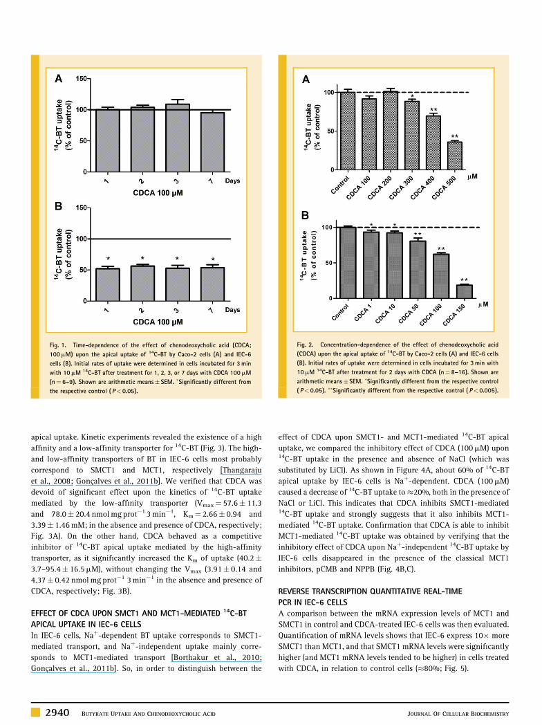
We further investigated the effect of a 2-day treatment with
CDCA upon the apical uptake of 14C-BT, by characterizing its
concentration-dependency. As shown in Figure 2, CDCA inhibited14C-BT uptake in both Caco-2 and IEC-6 cells in a concentration-
dependent manner, but with a higher potency in IEC-6 cells. Its IC50was found to be 402 (255–634)mM and 120 (88–164)mM in Caco-2
and IEC-6 cells, respectively.
EFFECT OF CDCA UPON THE KINETICS OF 14C-BT APICAL UPTAKE IN
IEC-6 CELLS
Next, we decided to investigate the effect of a 2-day exposure of IEC-
6 cells to CDCA (100mM) upon the kinetic parameters of 14C-BT
TABLE I. Primers Used in qRT-PCR
Gene name Primer sequence (50–30) AT (8C)
rGAPDH F: GGC ATC GTG GAA GGG CTC ATG AC 62R: ATG CCA GTG AGC TTC CCG TTC AGC
rMCT1 F: CAG TGC AAC GAC CAG TGA ATG TG 69R: ATC AAG CCA CAG CCA GAC AGG
rSMCT1 F: CGG GAT CAC CAG CAC CTA C 62R: GCA GGG GCA TAA ATC ACA ATC
rGAPDH, rat glyceraldehyde-3-phosphate dehydrogenase; rMCT1, rat monocar-boxylate transporter type 1; rSMCT1, rat Naþ-coupled monocarboxylate trans-porter type 1; F, forward; R, reverse.
JOURNAL OF CELLULAR BIOCHEMISTRY BUTYRATE UPTAKE AND CHENODEOXYCHOLIC ACID 2939
apical uptake. Kinetic experiments revealed the existence of a high
affinity and a low-affinity transporter for 14C-BT (Fig. 3). The high-
and low-affinity transporters of BT in IEC-6 cells most probably
correspond to SMCT1 and MCT1, respectively [Thangaraju
et al., 2008; Goncalves et al., 2011b]. We verified that CDCA was
devoid of significant effect upon the kinetics of 14C-BT uptake
mediated by the low-affinity transporter (Vmax¼ 57.6� 11.3
and 78.0� 20.4 nmolmg prot�1 3min�1, Km¼ 2.66� 0.94 and
3.39� 1.46mM; in the absence and presence of CDCA, respectively;
Fig. 3A). On the other hand, CDCA behaved as a competitive
inhibitor of 14C-BT apical uptake mediated by the high-affinity
transporter, as it significantly increased the Km of uptake (40.2�3.7–95.4� 16.5mM), without changing the Vmax (3.91� 0.14 and
4.37� 0.42 nmolmg prot�1 3min�1 in the absence and presence of
CDCA, respectively; Fig. 3B).
EFFECT OF CDCA UPON SMCT1 AND MCT1-MEDIATED 14C-BT
APICAL UPTAKE IN IEC-6 CELLS
In IEC-6 cells, Naþ-dependent BT uptake corresponds to SMCT1-
mediated transport, and Naþ-independent uptake mainly corre-
sponds to MCT1-mediated transport [Borthakur et al., 2010;
Goncalves et al., 2011b]. So, in order to distinguish between the
effect of CDCA upon SMCT1- and MCT1-mediated 14C-BT apical
uptake, we compared the inhibitory effect of CDCA (100mM) upon14C-BT uptake in the presence and absence of NaCl (which was
substituted by LiCl). As shown in Figure 4A, about 60% of 14C-BT
apical uptake by IEC-6 cells is Naþ-dependent. CDCA (100mM)
caused a decrease of 14C-BT uptake to�20%, both in the presence of
NaCl or LiCl. This indicates that CDCA inhibits SMCT1-mediated14C-BT uptake and strongly suggests that it also inhibits MCT1-
mediated 14C-BT uptake. Confirmation that CDCA is able to inhibit
MCT1-mediated 14C-BT uptake was obtained by verifying that the
inhibitory effect of CDCA upon Naþ-independent 14C-BT uptake by
IEC-6 cells disappeared in the presence of the classical MCT1
inhibitors, pCMB and NPPB (Fig. 4B,C).
REVERSE TRANSCRIPTION QUANTITATIVE REAL-TIME
PCR IN IEC-6 CELLS
A comparison between the mRNA expression levels of MCT1 and
SMCT1 in control and CDCA-treated IEC-6 cells was then evaluated.
Quantification of mRNA levels shows that IEC-6 express 10� more
SMCT1 than MCT1, and that SMCT1 mRNA levels were significantly
higher (and MCT1 mRNA levels tended to be higher) in cells treated
with CDCA, in relation to control cells (�80%; Fig. 5).
Fig. 1. Time-dependence of the effect of chenodeoxycholic acid (CDCA;
100mM) upon the apical uptake of 14C-BT by Caco-2 cells (A) and IEC-6
cells (B). Initial rates of uptake were determined in cells incubated for 3min
with 10mM 14C-BT after treatment for 1, 2, 3, or 7 days with CDCA 100mM
(n¼ 6–9). Shown are arithmetic means� SEM. �Significantly different from
the respective control ( P< 0.05).
Fig. 2. Concentration-dependence of the effect of chenodeoxycholic acid
(CDCA) upon the apical uptake of 14C-BT by Caco-2 cells (A) and IEC-6 cells
(B). Initial rates of uptake were determined in cells incubated for 3min with
10mM 14C-BT after treatment for 2 days with CDCA (n¼ 8–16). Shown are
arithmetic means� SEM. �Significantly different from the respective control
( P< 0.05). ��Significantly different from the respective control ( P< 0.005).
2940 BUTYRATE UPTAKE AND CHENODEOXYCHOLIC ACID JOURNAL OF CELLULAR BIOCHEMISTRY

EFFECT OF MODULATORS OF INTRACELLULAR SIGNALING
PATHWAYS ON CDCA-INDUCED INHIBITION OF 14C-BT UPTAKE IN
IEC-6 CELLS
We next investigated the intracellular signaling mechanisms
involved in the inhibition of 14C-BT uptake caused by CDCA. For
this, the effect of a 1-day exposure of IEC-6 cells to modulators of
intracellular signaling pathways, to CDCA or to a combination of
both was assessed.
First, the role of intracellular Ca2þ in CDCA-induced inhibition of14C-BT uptake was investigated, by testing the effect of the Ca2þ
chelator BAPTA-AM. No significant change in 14C-BT uptake was
found with BAPTA-AM, and the effect of CDCA was also not
changed in the presence of this agent (Fig. 6).
We then evaluated the role of the Ca2þ/calmodulin (CaM)
inhibitor calmidazolium, and of the CaM-dependent protein kinase
II (CaMK II) inhibitor KN-62 [Said et al., 1999]. Uptake of 14C-BT was
significantly reduced in the presence of calmidazolium, and slightly
increased in the presence of KN-62 (Fig. 6), suggesting that BT
uptake is dependent on intracellular CaM. The effect of CDCA upon
Fig. 3. Effect of chenodeoxycholic acid (CDCA; 100mM) upon the kinetic
parameters of the apical uptake of 14C-BT by IEC-6 cells. Initial rates of uptake
were determined in IEC-6 cells incubated at 378C with 0.01–0.1mM (high-
affinity transport, A) or 0.01–3mM (low-affinity transport, B) of 14C-BT for
3min, after treatment for 2 days with CDCA 100mM (closed squares) or the
respective solvent (control; closed circles; n¼ 4–8). Shown are arithmetic
means� SEM.
Fig. 4. Effect of chenodeoxycholic acid (CDCA; 100mM) upon SMCT1- and
MCT1-mediated apical uptake of 14C-BT by IEC-6 cells. A: Effect of CDCA
upon Naþ-dependent and Naþ-independent uptake. After treatment for 2 days
with CDCA or the respective solvent (control), cells were incubated at 378Cwith 14C-BT 10mM for 3min in GFK buffer containing NaCl (control) or an
isotonic concentration of LiCl (n¼ 10). B,C: Effect of CDCA and MCT1
inhibitors upon Naþ-independent uptake. After treatment for 2 days with
CDCA or the respective solvent (control), cells were preincubated for 20min
and then incubated at 378C with 14C-BT 10mM for 3min in GFK buffer
containing LiCl in the absence (control) or presence of p-chloromercuribenzo-
ate (pCMB) 0.5mM (n¼ 9) or 5-nitro-2-(3-phenylpropylamino)benzoic acid
(NPPB) 0.5mM (n¼ 9). �Significantly different from control ( P< 0.05).#Significantly different from CDCA (P< 0.05).
Fig. 5. Quantification of mRNA levels of MCT1 and SMCT1, by qRT-PCR, in
IEC-6 cells after treatment for 2 days with chenodeoxycholic acid (CDCA;
100mM) or its solvent (Control; n¼ 5). Results are shown as the expression of
MCT1 or SMCT1 relative to GAPDH (arithmetic means� SEM). �Significantlydifferent from control (P< 0.05).
JOURNAL OF CELLULAR BIOCHEMISTRY BUTYRATE UPTAKE AND CHENODEOXYCHOLIC ACID 2941

14C-BT uptake was not changed in the presence of KN-62, but was
reduced by calmidazolium (Fig. 6).
The involvement of MAPK was studied by testing the effect of
specific inhibitors of MAPK ERK1/2 (PD 98059) [Alessi et al., 1995]
and p38 MAPK (SB 203580) [Cuenda et al., 1995]. Interestingly
enough, both PD 98059 and SB 203580 were devoid of effect upon14C-BT uptake, but they were able to partially reduce the inhibitory
effect of CDCA upon 14C-BT uptake (Fig. 6).
Next, we tested the effect of H-7, a non-selective inhibitor of PKA,
PKC, and PKG [Hidaka et al., 1984]. Although H-7 was found to
cause a small (�10%) but significant decrease in 14C-BT uptake, it
was not able to change the effect of CDCA upon this parameter
(Fig. 6). We then studied the effect of specific inhibitors of
PKC (chelerythrine) and PKA (H-89) [Silva et al., 2010]. PKC
inhibition was found to cause a significant decrease (�17%)
and PKA inhibition caused a significant increase (�14%) in 14C-
BT uptake. This suggests that 14C-BT uptake by IEC-6 cells is
stimulated by PKC and inhibited by PKA activation, an observation
in perfect agreement with previous publications [Alrefai et al.,
2004; Narumi et al., 2010]. Interestingly enough, chelerythrine
(PKC inhibitor) was able to significantly reduce the inhibition of14C-BT uptake induced by CDCA, but H89 (PKA inhibitor) had no
effect (Fig. 6).
The involvement of protein tyrosine kinases (PTK) was studied
by testing genistein, a known PTK inhibitor [Said et al., 1999]. As
shown in Figure 7, neither 14C-BT uptake nor CDCA-induced
decrease of 14C-BT uptake were affected by this drug (Fig. 6).
We also evaluated the putative involvement of oxidative stress in
CDCA-mediated inhibition of 14C-BT uptake [Song et al., 2007;
Ignacio Barrasa et al., 2011], by testing the effect of the ROS
scavenger N-acetyl-cysteine (NAC) [Aruoma et al., 1989]. NAC was
devoid of effect upon 14C-BT uptake but it was able to reduce the
inhibitory effect of CDCA upon 14C-BT uptake (Fig. 6).
The involvement of NF-kB signal transduction pathway was
investigated by testing the effect of a specific inhibitor of IkBa
phosphorylation and degradation (BAY 11-7082) [Pierce et al.,
1997]. BAY 11-7082 (500 nM) was devoid of effect upon 14C-BT
uptake and did not change the inhibitory effect of CDCA upon this
parameter (results not show).
Finally, the involvement of cyclooxygenases (COX) in CDCA-
mediated inhibition of 14C-BT uptake was tested by using
quinacrine, a selective phospholipase A2 inhibitor [Winocour
et al., 1981]. Quinacrine (500mM) was devoid of effect upon 14C-
BT uptake and also did not affect the inhibitory effect of CDCA upon14C-BT uptake (results not shown).
EFFECT OF CDCA ON VIABILITY AND PROLIFERATION OF IEC-6
AND CACO-2 CELLS
The effect of CDCA on cell viability and proliferation was evaluated
in order to exclude these factors as responsible for its effect upon14C-BT uptake. For this, the effect of a 2-day exposure to increasing
concentrations of CDCA upon Caco-2 cellular viability and upon
IEC-6 cellular viability and proliferation was investigated. We
verified that CDCA up to 500mM did not affect Caco-2 cell viability
Fig. 6. Effect of inhibitors of intracellular signaling pathways upon the apical uptake of 14C-BT and upon CDCA-induced inhibition of 14C-BT uptake by IEC-6 cells. Initial
rates of 14C-BT uptake were determined in cells incubated for 3min with 14C-BT 10mM after treatment for 1 day with CDCA 100mM (CDCA), BAPTA AM 5mM (BAPTA),
CDCA 100mMþ BAPTA AM 5mM (CDCAþ BAPTA), genistein 10mM (GEN), CDCA 100mMþ genistein 10mM (CDCAþGEN), calmidazolium 0.5mM (CALM), CDCA
100mMþ calmidazolium 0.5mM (CDCAþ CALM), KN-62 1.5mM (KN), CDCA 100mMþKN-62 1.5mM (CDCAþ KN), PD 98058 2.5mM (PD), CDCA 100mMþ PD 98058
2.5mM (CDCAþ PD), SB 203580 2.5mM (SB), CDCA 100mMþ SB 203580 2.5mM (CDCAþ SB), H-7 10mM (H7), CDCA 100mMþH-7 10mM (CDCAþH7), chelerythrine
1mM (CHEL), CDCA 100mMþ chelerythrine 1mM (CDCAþ CHEL), H-89 10mM (H89), CDCA 100mMþH-89 10mM (CDCAþH89), N-acetyl-cysteine 1mM (NAC), CDCA
100mMþN-acetyl-cysteine 1mM (CDCAþNAC), or the respective solvents (control) (n¼ 6–9). Shown are arithmetic means� SEM. �Significantly different from the
respective control ( P< 0.05). #Significantly different from CDCA 100mM (P< 0.05).
2942 BUTYRATE UPTAKE AND CHENODEOXYCHOLIC ACID JOURNAL OF CELLULAR BIOCHEMISTRY

(Fig. 7A), and that CDCA up to 100mM did affect neither cell
viability (Fig. 7B,C) nor proliferation (Fig. 7D) of IEC-6 cells.
However, 200mM CDCA caused a significant decrease in IEC-6 cell
viability (Fig. 7B) and proliferation (Fig. 7D).
THE EFFECT OF THE INTERACTION BETWEEN BT AND CDCA UPON
IEC-6 CELLULAR VIABILITY, PROLIFERATION, AND
DIFFERENTIATION
In this final series of experiments, we investigated whether CDCA
was able to modify the effects of BT upon cell viability, proliferation,
and differentiation. The effect of BT upon IEC-6 cell viability,
proliferation, and differentiation was recently assessed by our group
[Goncalves et al., 2011c]. On the basis of these results, we selected BT
5mM for further experiments.
As shown in Figure 8, BT (5mM) caused a significant decrease in
IEC-6 cell proliferation (�20%) and viability (�13%) and a very
marked increase in cell differentiation (�1,000%). CDCA (100mM)
did not affect cellular viability or differentiation, and caused only a
small increase (�10%) in cell proliferation. Interestingly enough,
combination of CDCA with BT resulted in an attenuation of the
effects of BT upon cell proliferation and differentiation. On the
contrary, the effect of BT upon cell viability was potentiated by
CDCA (Fig. 8).
DISCUSSION
BT plays a key regulatory role in colonic epithelium homeostasis.
Therefore, factors that interfere with BT uptake into colonic
epithelial cells are potentially detrimental to intestinal health and
integrity. Very recently, we demonstrated a discrete interference of
some n-3 polyunsaturated fatty acids (PUFAs; docosahexaenoic
acid and eicosapentaenoic acid), n-6 PUFAs (linoleic acid,
g-linolenic acid, and arachidonic acid), conjugated linoleic acid,
and the bile salt deoxycholic acid upon uptake of 14C-BT by
intestinal epithelial cells [Goncalves et al., 2011d]. Secondary and
primary bile acids have tumor promoting effects, but the
Fig. 7. Effect of a 2-day exposure to increasing concentrations of chenodeoxycholic acid (CDCA) upon Caco-2 cellular viability (A) and IEC-6 cellular viability (B,C) and
proliferation (D). Cellular viability was determined by (A,B) quantification of extracellular lactate dehydrogenase activity (n¼ 12–17) and (C) the MTT assay (n¼ 10). Cell
proliferation was quantified by (D) the SRB assay (n¼ 6–12). A,B: Cells were seeded on 24-well plates and cellular viability was determined by quantification of extracellular
lactate dehydrogenase activity, as described in Materials and Methods Section. Results are shown as extracellular LDH activity (as % of total LDH activity). C: Cells were seeded
on 24-well plates and cellular viability was determined by the MTT assay, as described in Materials and Methods Section. Results are shown as % of control. D: Cells were seeded
on 24-well plates and cellular growth was determined by quantification of whole cellular protein with SRB, as described in Materials and Methods Section. Results are shown as
absorbance (% of control). Results are presented as arithmetic meansþ SEM. �Significantly different from control (P< 0.05).
JOURNAL OF CELLULAR BIOCHEMISTRY BUTYRATE UPTAKE AND CHENODEOXYCHOLIC ACID 2943

mechanisms involved remain to be fully elucidated. So, the aim of
the present study was to investigate the possibility that inhibition of
the apical uptake of BT may be one mechanism contributing to
the procarcinogenic effect of the primary bile acid CDCA at the
intestinal level. We found important to compare the effect of CDCA
in a colon adenocarcinoma cell line, the Caco-2 cells [Sambuy et al.,
2006] and a non-tumoral intestinal epithelial cell line, the IEC-6
cells [Wood et al., 2003], as comparison between a carcinogenic and
a non-carcinogenic cell line seemed interesting in the context of a
possible distinct effect of CDCA in these cells. Although not a
colonic cell line, the IEC-6 cells proved to be a good cellular model to
study colonic BT uptake [Borthakur et al., 2010; Goncalves et al.,
2011b], because they express the BT transporters (MCT1, SMCT1,
and BCRP) expressed in the human colon [Borthakur et al., 2010;
Goncalves et al., 2011b,c].
A 2-day exposure to CDCA markedly and concentration-
dependently inhibited 14C-BT uptake by IEC-6 cells (IC50¼ 120mM),
mM), and, less potently, by Caco-2 cells (IC50¼ 402mM). The effect
of CDCA in IEC-6 cells (up to 100mM) and in Caco-2 cells (up to
500mM) does not appear to be related to changes in cell viability or
proliferation. The inhibitory effect of CDCA (100mM) upon 14C-BT
uptake by IEC-6 cells was constant from 1 to 7 days of exposure, and
CDCA behaved as a competitive inhibitor of the high-affinity uptake
mechanism for 14C-BT.
Absorption of BT from the intestinal lumen involves both the Hþ-coupled MCT1 [Cuff et al., 2005] and the Naþ-coupled SMCT1
[Gupta et al., 2006], both of which are functionally present in IEC-6
cells [Borthakur et al., 2010; Goncalves et al., 2011b]. Interestingly
enough, the observation that CDCA reduced both the Naþ-dependent and the Naþ-independent uptake of 14C-BT, and that
inhibition of the Naþ-independent uptake of 14C-BT by CDCA
disappeared in the presence of MCT1 inhibitors (NPPB and pCMB)
clearly show that CDCA inhibits both MCT1- and SMCT1-mediated
BT uptake. It is important to note that IEC-6 cells, similarly to normal
human colonic epithelium, functionally express both MCT1- and
SMCT1-mediated transport, and CDCA was able to inhibit both
transport mechanisms. Moreover, the lower inhibitory potency of
CDCA in relation to 14C-BT uptake by Caco-2 cells (IC50¼ 402mM),
in which BT uptake is mainly MCT1-mediated [Goncalves et al.,
2009] and the fact that, in IEC-6 cells, CDCA (100mM) inhibits only14C-BT uptake mediated by the high-affinity transporter (SMCT1
andMCT1 being high- and low-affinity BT transporters, respectively
[Thangaraju et al., 2008]), suggests that the inhibitory effect of
CDCA is more pronounced for SMCT1 than for MCT1.
Next, we evaluated the effect of CDCA on the mRNA expression
levels of MCT1 and SMCT1 in IEC-6 cells. Contrary to the effect of
CDCA upon 14C-BT uptake, treatment of IEC-6 cells for 2 days with
CDCA caused a marked (�80%) increase in the steady-state mRNA
levels of SMCT1, and a tendency to an increase in the steady-state
mRNA levels of MCT1. Colchicine, an agent well known to affect the
membrane trafficking of cytoplasmic proteins, did not change the
effect of CDCA upon 14C-BT uptake (results not show), and so CDCA
appears to have no effect in the amount of transporters inserted in
the cell membrane. We thus conclude that CDCA is acting possibly
through decreased SMCT1 and/or MCT1 intrinsic activity, and we
speculate that the increase in SMCT1 mRNA level is a compensation
Fig. 8. Effect of a 2-day exposure to BT 5mM (BT), chenodeoxycholic acid
100mM (CDCA), or to a combination of both compounds (BTþ CDCA) upon
IEC-6 cellular proliferation (A), viability (B), and differentiation (C). A: Cellular
proliferation was determined by quantification of whole cellular protein with
SRB, as described in Materials and Methods Section. Results are shown as
absorbance (% of control; n¼ 12). B: Cellular viability was determined by
quantification of extracellular LDH activity, as described in Materials and
Methods Section. Results are shown as extracellular LDH activity (% of total
LDH activity; n¼ 17–18). C: Cell differentiation was determined by quantifi-
cation of alkaline phosphatase (ALP) activity, as described in Materials and
Methods Section. Results are shown as nmol p-nitrophenol/min/mg protein (%
of control; n¼ 18). Results are presented as arithmetic meansþ SEM.�Significantly different from control (P< 0.05). #Significantly different
from BT (P< 0.05).
2944 BUTYRATE UPTAKE AND CHENODEOXYCHOLIC ACID JOURNAL OF CELLULAR BIOCHEMISTRY

mechanism in response to the marked decrease in SMCT1 activity
caused by CDCA.
In relation to the mechanism(s) involved in the inhibitory effect of
CDCA upon 14C-BT uptake, several hypothesis can be advanced.
These include: (1) changes inmembrane structure, as bile acids cause
the release of cell membrane components (e.g., proteins and
phospholipids) prior to the occurrence of significant cell lysis
[Coleman and Holdsworth, 1976]; (2) changes in membrane fluidity,
as bile acids increase membrane fluidity [Zhao and Hirst, 1990], and
changes of the physical state of plasma membrane lipids can
influence the function of membrane carriers [Lundbaek et al., 2010];
(3) mitochondrial damage resulting in cellular ATP depletion, as this
mechanism was found to be associated with the strong inhibitory
effect of CDCA upon some ion transporters [Maleth et al., 2011]; (4)
changes in intracellular phosphorylation/dephosphorylation
mechanisms, as the amount/activity of many membrane transpor-
ters, including MCT1 [Alrefai et al., 2004; Narumi et al., 2010], is
modulated by these mechanisms, and intracellular signaling
mechanisms, such as PKC, MAPK, Ca2þ, and phosphoinositide-3
kinase have been identified as molecular targets for bile acids [Qiao
et al., 2000; McMillan et al., 2003]; and (5) increased production of
reactive oxygen species (ROS) and oxidative stress [Song et al.,
2007; Ignacio Barrasa et al., 2011].
We decided to clarify the role of intracellular regulatory pathways
in the regulation of 14C-BT uptake by CDCA in IEC-6 cells, by
examining the effect of inhibitors of various intracellular regulatory
pathways, including pathways mediated by CaM, PTK, PKA, PKC,
PKG, MAPK ERK1/2, and p38 MAPK. Our study revealed that the
inhibitory effect of CDCA upon 14C-BT uptake was partially reversed
by inhibition of CaM, PKC, and the MAPK pathways ERK1/2 and
p38. This observation suggests that CDCA-induced inhibition of 14C-
BT uptake is dependent on CaM, PKC, MAPK ERK1/2, and p38
activation. The stimulatory effect of CDCA upon MAPK is in
agreement with previous reports obtained with some other bile acids
[McMillan et al., 2003], and PKC is a known molecular target for bile
acids [Qiao et al., 2000; McMillan et al., 2003].
We also decided to investigate the role of oxidative stress and ROS
in the inhibitory effect of CDCA upon 14C-BT uptake, because we
recently demonstrated that oxidative stress decreases BT uptake in
IEC-6 cells [Goncalves et al., unpublished results], and proin-
flammatory cytokines, which inhibit SMCT1- and MCT1-mediated
BT uptake [Thibault et al., 2007; Borthakur et al., 2010], also lead to
the production of ROS [Seidelin and Nielsen, 2005; Babbar and
Casero, 2006]. Increased production of ROS appears to be involved
in the inhibitory effect of CDCA upon BT uptake, as NAC reduced the
inhibitory effect of CDCA upon 14C-BT uptake. This observation is of
great importance, because oxidative stress is associated with the
process of initiation and progression of colon carcinogenesis and
inflammatory bowel disease [Seril et al., 2003; Almenier et al.,
2012].
The ability of BT to modulate gene expression is often attributed
to histone hyperacetylation through inhibition of histone deace-
tylases (HDACi) [Hamer et al., 2008]. Because these effects are
dependent on the intracellular concentration of BT, it is expected
that inhibition of BT cellular uptake will change its cellular effects.
So, in the last part of this work, we evaluated the effect of BT in
conjunction with CDCA upon viability, proliferation, and differen-
tiation of IEC-6 cells.
BT (5mM; 2 days) caused a significant reduction in IEC-6 cell
proliferation and viability and a very marked increase on cell
differentiation. This observation is in perfect agreement with our
recent report obtained in this same cell line [Goncalves et al., 2011c],
and with the known effect of HDACi such as BT upon these
parameters [Hamer et al., 2008]. CDCA (100mM; 2 days) did not
affect cell viability and differentiation, and caused only a discrete
increase in IEC-6 proliferation. Interestingly enough, combination
of CDCA with BT caused a reduction in the inhibitory effect of BT
upon cell proliferation and differentiation. Unexpectedly, CDCA
potentiated the effect of BT upon cell viability, a result for which we
have at present no explanation.
In summary, we demonstrate that CDCA is an effective inhibitor
of 14C-BT uptake in both tumoral (Caco-2) and non-tumoral (IEC-6)
intestinal epithelial cells. In IEC-6 cells: (1) CDCA inhibits 14C-BT
uptake in a concentration-dependent manner, acting as a competi-
tive inhibitor of the high-affinity transporter of 14C-BT; (2) CDCA
decreases both MCT1- and SMCT1-mediated uptake of 14C-BT
(although the inhibitory effect upon SMCT1 appears more
pronounced); (3) CDCA induces an increase in SMCT1 mRNA
steady-state levels; (4) 14C-BT uptake is regulated by CaM, CaMKII,
PKC, and PKA; (5) inhibition of 14C-BT uptake by CDCA is dependent
on CaM, MAPK ERK1/2, and p38 and PKC activation; and (6) CDCA
significantly reduces the effects of BT upon cell proliferation and
differentiation.
Given the very important homeostatic role played by BT at the
intestinal epithelial level, knowledge on the interaction between an
endogenous compound present in high amounts at that level (in
cecum, 7–20% of total bile acids are CDCA [Hamilton et al., 2007]
and in colon high concentrations are also present) and BT uptake
into epithelial cells is very important. Indeed, the results of this study
are very interesting in the context of the well-known anti-
carcinogenic role played by BT in the intestinal epithelium, as they
suggest that inhibition of BT uptake into the intestinal epithelium
might contribute to the procarcinogenic effect of CDCA at this level.
REFERENCES
Alessi DR, Cuenda A, Cohen P, Dudley DT, Saltiel AR. 1995. PD 098059 is aspecific inhibitor of the activation of mitogen-activated protein kinase kinasein vitro and in vivo. J Biol Chem 270:27489–27494.
Almenier HA, Menshawy HH, Maher MM, Gamal SA. 2012. Oxidativestress and inflammatory bowel disease. Front Biosci (Elite Ed) 4:1335–1344.
Alrefai WA, Tyagi S, Gill R, Saksena S, Hadjiagapiou C, Mansour F,Ramaswamy K, Dudeja PK. 2004. Regulation of butyrate uptake in Caco-2cells by phorbol 12-myristate 13-acetate. Am J Physiol Gastrointest LiverPhysiol 286:G197–G203.
Aruoma OI, Halliwell B, Hoey BM, Butler J. 1989. The antioxidant action ofN-acetylcysteine: Its reaction with hydrogen peroxide, hydroxyl radical,superoxide, and hypochlorous acid. Free Rad Biol Med 6:593–597.
Babbar N, Casero RA, Jr. 2006. Tumor necrosis factor-A increases reactiveoxygen species by inducing spermine oxidase in human lung epithelial cells:A potential mechanism for inflammation-induced carcinogenesis. CancerRes 66:11125–11130.
JOURNAL OF CELLULAR BIOCHEMISTRY BUTYRATE UPTAKE AND CHENODEOXYCHOLIC ACID 2945

Bergmeyer HU, Bernt E. 1974. Lactate dehydrogenase. In: Bergmeyer HU,editor. Methods in enzymatic analysis. New York: Academic Press. pp 574–579.
Borthakur A, Anbazhagan AN, Kumar A, Raheja G, Singh V, Ramaswamy K,Dudeja PK. 2010. The probiotic Lactobacillus plantarum counteracts TNF-{alpha}-induced downregulation of SMCT1 expression and function. Am JPhysiol Gastrointest Liver Physiol 299:G928–G934.
Bradford MM. 1976. A rapid method for the quantitation of microgramquantities of protein utilizing the principle of protein-dye binding. AnalBiochem 72:248–254.
Coleman R, Holdsworth G. 1976. The release of membrane components priorto haemolysis during extraction of intact erythrocytes with bile salts.Biochim Biophys Acta 426:776–780.
Cuenda A, Rouse J, Doza YN, Meier R, Cohen P, Gallagher TF, Young PR,Lee JC. 1995. 203580 is a specific inhibitor of a MAP kinase homologuewhich is stimulated by cellular stresses and interleukin-1. FEBS Lett 364:229–233.
Cuff MA, Dyer J, Jones M, Shirazi-Beechey SP. 2005. The human colonicmonocarboxylate transporter isoform 1: Its potential importance to colonictissue homeostasis. Gastroenterology 128:676–686.
Goncalves P, Araujo JR, Pinho MJ, Martel F. 2009. Modulation of butyratetransport in Caco-2 cells. Naunyn Schmiedebergs Arch Pharmacol 379:325–336.
Goncalves P, Araujo JR, Pinho MJ, Martel F. 2011a. In vitro studies on theinhibition of colon cancer by butyrate and polyphenolic compounds. NutrCancer 63:282–294.
Goncalves P, Araujo JR, Martel F. 2011b. Characterization of butyrate uptakeby nontransformed intestinal epithelial cell lines. J Membr Biol 240:35–46.
Goncalves P, Gregorio I, Martel F. 2011c. The short-chain fatty acid butyrateis a substrate of breast cancer resistance protein. Am J Physiol Cell Physiol301:C984–C999.
Goncalves P, Araujo JR, Martel F. 2011d. Effect of polyunsaturated fattyacids and bile salts on butyrate uptake by intestinal epithelial cells. In: SchulzMA, editor. Caco-2 cells and their uses. New York: Nova Science PublishersInc. pp 89–99.
Gupta N, Martin PM, Prasad PD, Ganapathy V. 2006. SLC5A8 (SMCT1)-mediated transport of butyrate forms the basis for the tumor suppressivefunction of the transporter. Life Sci 78:2419–2425.
Hamer HM, Jonkers D, Venema K, Vanhoutvin S, Troost FJ, Brummer RJ.2008. Review article: The role of butyrate on colonic function. AlimentPharmacol Ther 27:104–119.
Hamilton JP, Xie G, Raufman JP, Hogan S, Griffin TL, Packard CA, ChatfieldDA, Hagey LR, Steinbach JH, Hofmann AF. 2007. Human cecal bile acids:Concentration and spectrum. Am J Physiol Gastrointest Liver Physiol 293:G256–G263.
Hidaka H, Inagaki M, Kawamoto S, Sasaki Y. 1984. Isoquinolinesulfona-mides, novel and potent inhibitors of cyclic nucleotide dependent proteinkinase and protein kinase C. Biochemistry 23:5036–5041.
Ignacio Barrasa J, Olmo N, Perez-Ramos P, Santiago-Gomez A, Lecona E,Turnay J, Antonia Lizarbe M. 2011. Deoxycholic and chenodeoxycholic bileacids induce apoptosis via oxidative stress in human colon adenocarcinomacells. Apoptosis 16:1054–1067.
Jemal A, Bray F, Center MM, Ferlay J, Ward E, Forman D. 2011. Global cancerstatistics. CA Cancer J Clin 61:69–90.
Lundbaek JA, Collingwood SA, Ingolfsson HI, Kapoor R, Andersen OS. 2010.Lipid bilayer regulation of membrane protein function: Gramicidin channelsas molecular force probes. R Soc Interface 7:373–395.
Mahmoud NN, Dannenberg AJ, Bilinski RT, Mestre JR, Chadburn A, ChurchillM, Martucci C, Bertagnolli MM. 1999. Administration of an unconjugatedbile acid increases duodenal tumors in a murine model of familial adeno-matous polyposis. Carcinogenesis 20:299–303.
Maleth J, Venglovecz V, Razga Z, Tiszlavicz L, Rakonczay Z, Jr., Hegyi P.2011. Non-conjugated chenodeoxycholate induces severe mitochondrialdamage and inhibits bicarbonate transport in pancreatic duct cells. Gut60:136–138.
Martınez ME, Marshall JR, Giovannucci E. 2008. Diet and cancer prevention:The roles of observation and experimentation. Nat Rev Cancer 8:694–703.
McMillan L, Butcher SK, Pongracz J, Lord JM. 2003. Opposing effects ofbutyrate and bile acids on apoptosis of human colon adenoma cells:Differential activation of PKC and MAP kinases. Br J Cancer 88:748–753.
Mosmann T. 1983. Rapid colorimetric assay for cellular growth and survival:Application to proliferation and cytotoxicity assays. J Immunol Methods65:55–63.
Muzyka A, Tarkany O, Yelizanof V. 2005. Non-linear regression (curve fit).GraphPad Prism for Windows. San Diego, CA: GraphPad Software.
Narumi K, Furugen A, Kobayashi M, Otake S, Itagaki S, Iseki K. 2010.Regulation of monocarboxylate transporter 1 in skeletal muscle cells byintracellular signaling pathways. Biol Pharm Bull 33:1568–1573.
Pierce JW, Schoenleber R, Jesmok G, Best J, Moore SA, Collins T,Gerritsen ME. 1997. Novel inhibitors of cytokine-induced I kappa B alphaphosphorylation and endothelial cell adhesion molecule expression showanti-inflammatory effects in vivo. J Biol Chem 272:21096–21103.
Qiao D, Chen W, Stratagoules ED, Martinez JD. 2000. Bile acid-inducedactivation of activator protein-1 requires both extracellular signal-regulatedkinase and protein kinase C signaling. J Biol Chem 275:15090–15098.
Reddy BS, Watanabe K, Weissburger JH, Wynder EL. 1977. Promoting effectof bile acids in colon carcinogenesis in germ-free and conventional F344rats. Cancer Res 37:3238–3242.
Reddy BS, Hedges AR, Laakso K, Wynder EL. 1978. Metabolic epidemiologyof large bowel cancer: Fecal bulk and constituents of high-risk NorthAmerican and low-risk Finnish population. Cancer 42:2832–2838.
Ridlon JM, Kang DJ, Hylemon PB. 2006. Bile salt biotransformations byhuman intestinal bacteria. J Lipid Res 47:241–259.
Said HM, Ortiz A, Kumar CK, Chatterjee N, Dudeja PK, Rubin S. 1999.Transport of thiamine in human intestine: Mechanism and regulation inintestinal epithelial cell model Caco-2. Am J Physiol 277:C645–C651.
Sambuy Y, De Angelis I, Ranaldi G, Scarino ML, Stammati A, Zucco F. 2006.The Caco-2 cell line as a model of the intestinal barrier: Influence of cell andculture-related factors on Caco-2 cell functional characteristics. Cell BiolToxicol 21:1–26.
Seidelin JB, Nielsen OH. 2005. Continuous cytokine exposure of colonicepithelial cells induces DNA damage. Eur J Gastroenterol Hepatol 17:363–369.
Seril DN, Liao J, Yang GY, Yang CS. 2003. Oxidative stress and ulcerativecolitis-associated carcinogenesis: Studies in humans and animal models.Carcinogenesis 24:353–362.
Silva E, Pinto V, Simao S, Serrao MP, Afonso J, Amaral J, PinhoMJ, Gomes P,Soares-da-Silva P. 2010. Renal aging in WKY rats: Changes in Naþ,Kþ-ATPase function and oxidative stress. Exp Gerontol 45:977–983.
Song S, Guha S, Liu K, Buttar NS, Bresalier RS. 2007. COX-2 inductionby unconjugated bile acids involves reactive oxygen species-mediatedsignalling pathways in Barrett’s oesophagus and oesophageal adenocarcino-ma. Gut 56:1512–1521.
Stadler J, Stern HS, Yeung KS, McGuire V, Furrer R, Marcon N, Bruce WR.1988. Effect of high fat consumption on cell proliferation activity ofcolorectal mucosa and on soluble faecal bile acids. Gut 29:1326–1331.
Thangaraju M, Cresci G, Itagaki S, Mellinger J, Browning DD, Berger FG,Prasad PD, Ganapathy V. 2008. Sodium-coupled transport of the short chainfatty acid butyrate by SLC5A8 and its relevance to colon cancer. J Gastro-intest Surg 12:1773–1781.
Thibault R, De Coppet P, Daly K, Bourreille A, Cuff M, Bonnet C, Mosnier JF,Galmiche JP, Shirazi-Beechey S, Segain JP. 2007. Down-regulation of the
2946 BUTYRATE UPTAKE AND CHENODEOXYCHOLIC ACID JOURNAL OF CELLULAR BIOCHEMISTRY

monocarboxylate transporter 1 is involved in butyrate deficiency duringintestinal inflammation. Gastroenterology 133:1916–1927.
Tong JL, Ran ZH, Shen J, Fan GQ, Xiao SD. 2008. Association between fecalbile acids and colorectal cancer: A meta-analysis of observational studies.Yonsei Med J 49:792–803.
Winocour PD, Kinlough-Rathbone RL, Mustard JF. 1981. The effect ofphospholipase inhibitor mepacrine on platelet aggregation, the platelet
release reaction and fibrinogen binding to the platelet surface. ThrombHaemost 45:257–262.
Wood SR, Zhao Q, Smith LH, Daniels CK. 2003. Altered morphology incultured rat intestinal epithelial IEC-6 cells is associated with alkalinephosphatase expression. Tissue Cell 35:47–58.
Zhao DL, Hirst BH. 1990. Comparison of bile salt perturbation of duodenaland jejunal isolated brush-border membranes. Digestion 47:200–207.
JOURNAL OF CELLULAR BIOCHEMISTRY BUTYRATE UPTAKE AND CHENODEOXYCHOLIC ACID 2947