INFORMATION TO USERS - CiteSeerX
232
INFORMATION TO USERS This manuscript has been reproduced from the microfilm master. UMI films the text diredly from the original or copy submitted. Thus, wme thesis and dissertation copies are in m e r face, Mile ottien may be ftom any type of cornputer printer. The qurlity of thb rrproducüon k dependent upon the qmlity d the copy submithd. Brokmorindistnct~nt,cakredorpoor~inu~sand photographs, print Meedttrrough, substandard margins, and impmper alignment can advecsely affect reproductiari. In the unlikely event that the authar dii not send UMI a compiete manuscript and there are missing pages, tnsso will be noted. Also, if unauthomed copyright material had to be remoued, a note wïll indikate the deletiorr. Oversize materials (e-g.. maps, drawirigs, charts) are reproduced by sedoning the original, beginning at the upper ~~d corner and cont i nu i ng frwn le to right in equal sections wilh smal overlaps. Photographs 'uicluded in the -inal manuscnscnpt have beem reproduoed xerographically in this copy. Highsr quality 6' x 9' bladc and nihite photographie prints are available for any photographs or illustrath$ appeanng in mis -y fw an additional charge. Corrtaa UMI direcüy to order. Bell 8 Houdl Infannation and Leaming 300 North Zeeb Road, Ann Arbor, MI 481-1346 USA
-
Upload
khangminh22 -
Category
Documents
-
view
0 -
download
0
Transcript of INFORMATION TO USERS - CiteSeerX

INFORMATION TO USERS
This manuscript has been reproduced from the microfilm master. UMI films the
text diredly from the original or copy submitted. Thus, wme thesis and
dissertation copies are in m e r face, Mile ottien may be ftom any type of
cornputer printer.
The qurlity of thb rrproducüon k dependent upon the qmlity d the copy
submithd. B r o k m o r i n d i s t n c t ~ n t , c a k r e d o r p o o r ~ i n u ~ s a n d
photographs, print Meedttrrough, substandard margins, and impmper alignment
can advecsely affect reproductiari.
In the unlikely event that the authar di i not send UMI a compiete manuscript and
there are missing pages, tnsso will be noted. Also, if unauthomed copyright
material had to be remoued, a note wïll indikate the deletiorr.
Oversize materials (e-g.. maps, drawirigs, charts) are reproduced by sedoning
the original, beginning at the upper ~~d corner and continuing frwn le to
right in equal sections wilh smal overlaps.
Photographs 'uicluded in the -inal manuscnscnpt have beem reproduoed
xerographically in this copy. Highsr quality 6' x 9' bladc and nihite photographie
prints are available for any photographs or illustrath$ appeanng in mis -y fw
an additional charge. Corrtaa UMI direcüy to order.
Bell 8 Houdl Infannation and Leaming 300 North Zeeb Road, Ann Arbor, MI 481-1346 USA


NOTE TO USERS
This reproduction is the best 'copy available
UMI


Li Long. ReguhImcm nf Twnor CeH Invmion unci Meranatir by rhe Tvpe / Insuiin-like Growrh Feror Receptor (IGF- f R)
rn Regulation of Tumor Cell Invasion and Metastasis
by the Type 1 Insulin-like Growth Factor
Receptor (IGF- IR)
Li Long
Department of Surgery, Division of Surgical Research
McGill University
Montreai Canada
Jdy, 1997
A Thesis submitted to the Faculty of Graduate Studies and Research
in partial fulfillment of the requirements of the degree of Doctor of
Philosophy

Nationai Library Bibliothèque nationale du Canada
Acquisitions and Acquisitions et Bibliographie Services services bibliographiques 395 Wellington Street 395, rue Wellington Otrawa ON K1A ON4 OrtawaON K l A W Cariada Canada
The author has granted a non- L'auteur a accordé une licence non exclusive Licence dowing the exclusive permettant à la National Libmy of Canada to Bibliothèque nationale du Canada de reproduce, loan, distribute or seil reproduire, prêter, distribuer ou copies of this thesis in microforni, vendre des copies de cette thèse sous paper or electronic formats. la fome de microfiche/nlm, de
reproduction sur papier ou sur format électronique.
The author retains ownership of the L'auteur conserve la propriété du copyright in this thesis. Neither the droit d'auteur qui protège cette thèse. thesis nor substantial extracts fiom it Ni la thèse ni des extraits substantiels may be printed or othexwise de celle-ci ne doivent être imprimés reproduced without the author's ou autrement reproduits sans son permission. autorisation.

The foiiowing excerpt is reprinted h m the "Guideiines for Thesis Reparatïon" of the
Faculty of Graduate Studies and Research of McGiIi University to infonn the reader of
Faculty reguiations:
Candidates have the option of including, as part of the tksis, the text of one or more
pupers submitted or to be submitted for publication, or the clearly-duplicated tex? of one
or more published papers. these texts must be b o d as an integral part of the thesis.
If this option is chosen, connecîhg tex& that provide logical bridges between the
diarent -ers cvc llLQndQfOry. The thesis must be writîen in such o way that it is more
rhan a mere collection of manuscript;r; in other worh, resuiis of a series of papers m m
be integrated
The thesis m u t still confonn to al1 other requirements of the "Guidelines for Thesis
Preparation". The thesis n w t include= A Table of Contents, an abstract in English and
French, an introduction which clearly States the rationale and objectives of the study, a
cornprehensive review of the literature, a final conclusion and S M M I I ~ ~ , and u thorough
bibliography or reference lis?.
Additioml merial maut be provided where appropriate (e.g. in appendices) and in
suflcient detail ro allow a clear anà precise judgment to be made of the importance md
originality of the research reported ùt the thesis.

In the case of manuscripts co-authored by the candidate and others, the candidote is
required to d e an exphkit sfatement Ur the daesis as &O who contdiuted to such work
and to what extent Supervisors must anest to the accuracy of such statements ar the
doctoral oral defense. Since the task of the examiners is made more dificuit in these
cases, it is in the candidate's interest tu make peflectly clear the responsibilities of al1 the
authors of the co-authured papers. Under no circumstances can a co-author of any
component of such a thesis as an examiner for that thesis.
In accordance with the above guidelines, 1 have decided to include the following published
papers as part of the body of the chesis:
Long, L., Nip, J., and Brodt, P. Paracrine growth stimulation by hepatocyte-
denved IGF-1: A regulatory mechanism for carcinoma cells metastatic to the
Iiver. Cancer Research. 54: 3732-3737, 1 994.
Long, L., Rubin, R., Baserga, R., and Brodt, P. Loss of The metastatic
phenotype in murine carcinoma cells expressing an antisense RNA to the
insulin-like growth factor receptor. Cancer Research. 55: 1006-1 009, 1995.
These two papers are reproduced h m Cancer Research by copyright permission of The
Amencan Association for Cancer Research. I am responsible for al1 of the experimental
work and anaiysis carrieci out in these two aforementioned papers with the exception of

a construction of plasmid vectors expressing IGF-IR cDNA in the sense or antisense
orientations. AU of the work was performed in the Iaboratory of Dr. Pnina Brodt
(Deparunent of surgery, Division of Surgical Research, McGïii University).

With the dream that one day I would be able to contribute to the fight against one of the
deadliest enemies of human health - cancer, 1 applied for Ph.D. snidies in the Department
of Surgery, Division of Surgical Research, McGill University. First and foremost, 1 would
iike to gratefuliy acknowledge the invaluable supervision, personai guidance, endless
encouragement and financial support given by Dr. Pnha Brodt. As a mentor, she has
generously given me the opportunity to develop my research skills, detailed suggestions
and guidance with great patience in ai l aspects of m y study. She has dways been available
and wiliing to assist me when 1 encountered any type of problem, research or otherwise,
despite her busy schedule. Dr. Brodt's role in m y professional development bas been
paramount and is highly appreciated.
A special thanks goes to my colleague, Dr. John Nip, who has generously aven me
enormous help and assistance in the fields of molecular biology, biochemistxy and
computer science when we studied togetber.
1 would Like to express my sincere appreciation to Drs. Raphael Rubin and Renato
Baserga for providing us the precious plasmid vectors required for portions of this study. -
for offenng their insight through discussions and for spending time to read and comment
on our rnanuscnpts.

1 would iïke to thank Mrs. Lucia Faüavoiiïta for her help in teaching me the numerous
tec hnical me thodologies and giving me sound practicd advice and generous assistance in
rnatters of research.
1 would Lice to acknowledge my colleagws and f'riends in the laboratory Dr. Hua Ling, for
her assistance in the field of molecular biology and her encouragementv and Dr. Margaret
Durko. Dr. Jian Wang, M s Roya Navab, Dr. Keguan Chen, Dr. Takayoki Assao and Mrs.
Grazieiia Vaknte for Liberal assistaoce and thoughtfùi discussions. As weU, the genemus
assistance, endless encouragement and invaluable fkiendship given by aU other members of
this Department, especiaüy Dr. Julius Gordon, Mrs. Nina Hassan , Mrs. Irene
Sidorenko, Mrs. Am Gordon and my fiends in Donner Building Mrs. Pat Guida . Ms. Linda Bazinet and Mr. Alberto Rodrigues, are appreciated. 1 have overcome mauy
difficulties through their help. Parties, dinners and cards are beautifid mernories that will
reside in my mind forever.
1 am grateful to Dr. B. R. Zetter (Department of surgery, Harvard Medical School,
Boston, MA), Dr. C. B. Srikant (Department of Medicine, McGii University, Montreal).
Dr. B. Massie (The Biotechnology Research Institute, National Research Council of
Canada, Montreai) and Dr. G. Karpati (Neuromuscular Research Group. Montreal
Neurological Institute, McGiU University, Montreai) for giving me the oppominity to
work in their labratories during some aspects of my study.

a 1 would like to thank Dr. Abdel M. Khatib for his kind assistance in the translation of my
abstract.
FmaUy, a speciai thanks to my wife Sining Wang, to my mother Mrs. Chunying Jiang, to
my parents in-law Mr. Kuiye Wang and Mrs. Huaming Liu, and to my son Randy Long for
their wholehearted support and encouragement.

Original Contributions to the Body of Knowledge
The major novel findings of the p m n t study are as foiiows:
1 . Liver derived IGF-1 is a major mitogmic and chernotactic factor for liver
metastatic cells;
2. Antisense mRNA to IGF-IR inhibits tumor ce11 gmwth and invasion
viîro and completely blocks metastasis in vn>o ; - 3. Overexpression of IGF-IR in a moderately metastatic tumot ce11 iine
increases the mitogenïc response to IGF-1 and celi invasion in vitro and
enables the cells to colonize the liver in vivo;
4. IGF-I is a major regulator of the expression of the Mr 72,000 type IV
collagenase (MMP-2, gelatinase A)

Abbreviations
DMSO
EGF
ECM
FN
HCM
IGF-1
IGF-IR
IGFBPs
1 s .
1.v.
MMP-2
MAb
PDGF
TIME's
TGF
Dimethyl sulfoxide
Epidermal growth factor
Extracellular matrix
Fibronectin
Hepatocyte conditioned medium
The type 1 insulin-like growth factor
Receptor for type 1 insulin-like jpwth factor
Insulin-like growth factor bindbg proteins
Intrasplenic
Intravenous
Matrix metalloproteinase 2
Monoclonal antibody
Platelet derived growth factor
Tissue inhibitor of metalloproteinases
Transforming growth factor

Table of Contents
.....................*......................... ....................*................*.. ..**.... A bstract .,., ..,... XVD
Chapter 1 . Molecular mediators of cancer cell invasion and metastasis . An
.......................................................... 1 . 1 . Introduction ............................................. -2
1.2. Growth factors and growth fa~tor receptors .............................................................. 3
......................................................... 1 .3 . The extracellular ma&-degradïng protehases 9
................................................................................ Cathepsins .................... .. 1 4
............................................................................. The metrix metaiioproteinases 15
....................................................................................................... Glycosidases 18
...................................................... ........... 1.4. The role of ceii adhesion molecules ... 1 9
........................................................................................ ............ In tegrins ... 20
.................................................................................................... The cadherins -22
............................ ...........*.... The immunoglobulin (Igs) supergene family ...... 24
..................................................................................... ................... Selectins .. 26
..................................................... .......................... 1.5. Angiogenesis and metastasis .... 28
..................................................................................................... 1.6. Motility factors -30
.............................................................................................................. 1.7. Summary 31
Chapter II . The insulin-like growtb factor 1 meptor: Structure, function and d e in
................................................................................................................ maügnancy 33
2.1. The type 1 insulin-like growth factor receptor (IGF-IR) ......................................... 34
................................................................................................. 2.2. The IGF-IR ligand -35

2.3. The IGF binding proteins ...... .. ............................................................................... 37
.................. ........................................................................... 2.4. IGF-IR signaling ,.. 39
2.5. Role of IGF-1 in cellular proliferation ...................................................................... 43
2.6. Role of IGF-I and IGF-IR in ceii death ................... ... ....................................... 45
........................................................................................ 2.7. IGF-IR and malignancy -45
............ Chapter III . Mahix met di op rote^ in cancer invasion and metastasis 48
.................................... The role of metalloproteinases in -or invasion and metastasis 52
Chapter IV . The d e of insuiin-lilre growth factor 1 in metastasis: Studies with the
................................................................................... Lewis lung carcinoma mode1 -56
4.1. Summary ....................................................................................... ....... 57 ...............
4.2. Sublines H-59 and M-27 of the Lewis lung carcinoma: Review of published data .... 58
Chapter V . The d e of insuün-iike growth factor 1 m p t o r system in cancer
me- (II): Enfimced invasion and liver-colonization in lung carcinoma c&
........................ .............. overexpressing the insulin-like growth factor 1 receptor .. 61
.............................................................................................................. 5.1. Overview -62
................................................................................................................. Introduction -64
.......................................................... ..................................... Materials and Methods .. -66
................................................................... .............................. Results .......... 7 1
.................................................................................................................... Discussion 73
Chapter VI . The role of iasulln-like growth factor 1 ~ e ~ e p t o r system in cancer
metastasis 0: Reguiation of Mr 72, 000 type N coïiageoase synthesis by the type 1
........................................................................ insulin-like growth factor receptor 82

................................................................................ ............................... Overview ...... 83
........................................................................................ ..................... Abstract ... 85
............................................ Materials and Methods .............................. .........., 88
.................................................................................................................... Results -92
................................................................................................................ Discussion 94
Chapter VIT . Tumors 8-59 and M-27 cek M e r in their rcsponses to IGF-I and
Merentially express other growth factors and receptors . Summ~ry of unpubbhed
................................................................................................................. results -102
....................................................................................................... 7.1. Introduction 103
....................................................................................... 7.2. Materials and Methods 103
............................................................................................................... 7.3. Results 107
........................................................................................................ 7.4. Discussion. 110
....................................................................................... C hapter VIII. Discussion 117
......... 8.1. Differential expression of growth factors and receptoa: tumor heterogeneity 118
.............. 8.2. The role of the IGF-IR/IGF-1 complex in cancer invasion and metastasis 120
..................................... ................................ 8.3. Suggestions for hiture research .... 127
.............................................................................................................. References 128

Li t5~n.v. Reg.uIuim r>/Tmar Ceff Invurü~n und Meranaris by ihc Tvpc 1 Inrufin-lik Gmwrh Fucmr Receprrw (IGF-1 RI
List of figures
Chapter IV
A ttached paper entitled " Paracrine growth stimulation by hepatocyte-derived
insulin-like gro wth factor4 A regulatory mechanism for carcinome cells
metastatic to the liver."
Finure 1. Bar graph showing the effects of conditioned media and growth factors on DNA
synthesis by the tumor ceh.
Figure 2. Line graph showing the dosedependent stimulation of H-59 ceiis by IGF-1.
Figure 3. Composite Line graph of inhibition and depletion of the mitogenic effect of
hepatocyte conditioned medium by a monocionai antibody to IGF-1.
Fiaure 4. Photograph showing the detection of IGF-1 in hepatocyte and lung tissue
conditioned media by Western blot analysis.
Figure 5. Scattered graph and autoradiograph composite demonstrate Scatchard analysis
of IGF-1 binding and Northem blot analysis for IGF-IR mRNA transcripts expresseci by
tumor cells.
Fipure 6. Autoradiograph and photograph composite showing the detections of KGF
binding proteins by Western ligand blot assay and Western blot assay.
Attached paper entitled "Loss of the metastatic phenolype in murine carcinoma
cells expressing an antisense RNA to the insulin-like growth factor receptor-'*
xiii

Figure 1. Autoradiograph and bar graph composite showing decreased IGF-IR mRNA
expression in tumor celis transfected by plasrnid vector canyîng an antisense sequence to
IGF-IR.
m u r e 2. Line and bar graph composite demonstrates the loss of response to IGF-1 and
hepatocyte couditioned medium in IGF-R antisense transfected ceiis.
m u r e 3. Line graph showing the growth of H-59 and IGF-IR antisense transfected cek
in vivo.
Chapter V
m u r e 1. M-27 c d s overexpressing IGF-IR proMerate in response to IGF-1 and HCM.
FiPure 2. Analysis of human IGF-IR expression in M-27 tnmsfectants by RT-PCR and
immunoprecipitation.
Figure 3. Increased invasiveness of M-27 cells overexpressing IGF-IR.
Chapter VI
Figure 2. Decreased invasiveness of H-59 ceils expressing IGF-IR antisense mRNA.
m u r e 2. The effect of modulation of IGF-IR expression on MW-2 mRNA production in
H-59 and M-27 celis - Analysis by RT-PCR
Figure 3. Western blot analysis of MMP-2 and TIMP-2 production in IGF-IR transfected
M-27 cells.
m u r e 4. Zymographic analysis of MMP-2 activity in M-27 ceils overexpressing IGF-IR.
xiv

Chapter VII.
Fiaure 7- 1 . Northern blot analysis of growth factors and receptors expressed in H-59 and
M-27 ceiis.
Figure 7-2. The chernotactic effect of IGF-1 on H-59 ceiis.
Figure 7-3. IGF-1 induced tyrosine phosphoryIation assessed by Western blot anaiysis.
Fiaure 7-4. IGF-I protects H-59 cells from apoptosis.

List of Tables
Chapter IV
Table 4-1. Summary of the phenotypic differences between H-59 and M-27 cells.
Chapter V.
Tabb 1. Increased anchorage-independent p w t & in tumor ceUs overexpressing IGF-IR.
Table 2. Enhanced liver-colonizing potentiai of M-27 ceik overexpressing IGF-IR.
xvi

Abstract
invasion and metastasis largely determine the clinical course of cancer. A better
understanding of the mechanisms required for metastases formation in secondary sites
will lead to the development of new and more successful tmument strategies. The present
work describes our resdts with a murine Lewis lmg carcinoma mode1 which consists of
two ce11 lines, Hg59 and M-27, with different patterns of metastasis in vivo. Using tbis
model, a positive coneiation was found b e e n the expression of Msulin-like growth
factor 1 receptor (IGF-Et) and the ability of the tumor celis to metastasize to the liver. The
highly invasive, highly metastatic, liver colonizing cell line H-59 expressed significantly
higher levels (5-fold) of IGF-IR than the poorly invasive M-27 cells which metastasize to
the lung only. When expression of IGF-IR in 8 5 9 cells was suppressed by transfection
with a plasmid vector expressing MiF-IR cDNA in an antisense orientation, the cells lost
m . their proliferative response to IGF-1 NI vitro , their ability to migrate in response to IGF-1
and their metastatic potential. To further study the role of IGF-IR in the process of liver
metastasis, IGF-IR was overexpressed in the Iung metastasizing M-27 cells by
transfection with a plasrnid vector expressing fil1 length human IGF-IR cDNA. The
stable transfectants had an cnhsnctd proliferative respome to IGF-1 and hepatocyte
conditioned medium ( K M ) and acquired an invasive potential as demonstrated in the
Matrigel invasion assay. When inoculated via the splenic/portal route in vivo, these celIs
but not the wild-type or mock-trmsfected cells gave rise to multiple liver nodules . TO M e r investigate the link between IGF-IR and invasion, metalloproteinase 2 (MMP-2)
expression in the tumor cells was investigated. It was found that the antisense h~nsfected

H-59 cells expressed significantly lower levels of MMP-2 as assessed by RT-PCR,
Western blot d y s i s and gelatin zymography. M-27 cells overexpmsing IGF-IR had a
marked increase in MMP-2 mRNA expression with a comsponding increase in the levels
and activity of the protein. Stimulation of these cells with IGF-1 resulted in increased
production of MMP-2 protein and increased gelatinolytic activity. In conclusion, our
results show that the IGF-IR/IGFII complex can modulate several cellular functions
which impact on the metastatic potential including growth, migration, invasion and fiver-
colonization. For tumors which an dependent on this system for proliferation, invasion
and metastasis, the IGF-IR could pmvide a specific target for effective anti-metastatic
tlierapy .

L'invasion et les métastases determinent cliniquement le développement et la
progression du cancer. La comprChension des mecanismes impliqués dans la
formation des métastases serait très utile a la mise en place de nouvelles
stratégies th6rapeutiques.
Ce travail décrit les résuitats obtenu in vivo avec le carcinome de poumons
de Lewis chez la souris, ceci en éhldiant les deux lignées cellulains modèles:
H-59 et M-27. Nos résultats montrent l'existence d' une corrélation positive
entre l'expression des récepteurs de Iïnsulin-like growth factor4 (IGF-IR)
et la capacité des œllules B envahir le foie par métastase.
Les œllules H-59, cellules invasives métastatiques, et envahissantes du foie,
expriment 5 fois plus & récepteurs IGF-IR que les cellules M27. qui
possèdent un faible pouvoir d'invasion et qui ne métastase que les poumons.
La transfection des cellules H59 avec un plasmide contenant de 1'ADNc des
rkcepteurs IGF-IR oriente dans un sens contraire (anti-sens), entraînanant la
suppression de l'expression des rCcepteurs IGF-IR, induit in vivo chez ces
cellules une perte de leurs capacitt de pmLif6rer et de migrer en présence de
lfIGF-1; et de leur pouvoir m6tastatique. Afin d'étudier le r61e du récepteur
de I'IGF-1 dans les processus des métastases du foie, ce dernier a et6 sur
exprimé dans les œllules M-27 B l'aide d'une transfection par un plasmi&
contenant I'ADNc complète du récepteur IGF-IR humain. Les cellules ;
trasfectées montrent une augmentation de leur capacitt de proliferer en
présence de lq1GF-I et du milieu conditionné des hépatocytes (HCM) et
acquièrent un pouvoir d'invasion. comme le &montre le test d'invasion du
Matrigel. L'inoculation in vivo de ces cellules au niveau de la veine porte
induit une augmentation des nodules du foie. M m de trouver la relation qui

existe entre les récepteurs IGF-IR et l'invasion , nous avons étudie
l'expression de la matria métallopmtéase 2 (MMP-2) dans les cellules
tumorales. Cette étude montre que les cellules H-59 transfectées n'expriment
que trés faiblement la MMP-2 comme le démontrent les technique de RT-
PCR, Western blot et la zymograpbie. Les œllules M27 qui sur expriment
les récepteurs IGF-IR présentent un taux très éleve des ARNm de la MMP-2
ainsi q'une activite augment6 de la protéine correspondante. La stimulation
de ces cellules avec de PIGF-1 se traduit par une augmentation de la
production de la MMP-2 et de son activité. En conclusion; nos résultats
montrent que le complexe IGF-IIIGF-IR peut moduler certaines fonctions
cellulaires qui ont un effet sur le pouvoir métastatique des cellules y compris
la croissance, la migration l'invasion et la colonisation du foie. Les Tumeurs
dont les cellules qui dependent de œ type de système pour leur prolif&ation,
invasion et métastase; le récepteur IGF-IR pourrait être une cible spécifique
m e pour la thérapie des métastases.

Molecular Mediators of Cancer Cell Invasion And Metastasis -
An Ovewiew

Li Long. Regdafion of Tumor Cell fnvusion a d l W e r ~ m i s by the Type I huuILn-lik Growih Factor Recepror (IGF-IU)
The clhical course of malignant disease is determined largely by the process of invasion
and metastasis. A better understanding of these processes is thetefore likely to lead to the
development of new and more successftl treatment strategies.
Over a century ago, Paget proposed that metastasis was due to the specific affhïty of
certain tumor cells (the "seed") for the milieu provided by certain organs (the "soil") (1)-
On the other han& the amtomid-mechanid theory of metastasis stresses the
importance of the vascular connections between the primary tumor and the secondary
site(s) of growth (2). Currentiy it is accepteci that both anatomid-mechanical and Seed-
soif factors play a role in the formation of metastases.
Metastasis is a cascade of Linked sequentiai steps involvïng multiple host-tumor
interactions (3-6). To successfuliy give nse to a metastatic colony, a ceil or group of
-or celis must detach h m the primay tumor, invade the local host tissue, enter the
cuculation, arrest at the distant vascular beâ, extravasate into the target organ interstitiun
and parenchyma, and proLiferate in the xcondary site. These processes are mediated by a
series of molecular interactions resulting h m disrupted positive and negative regdatory
mechanisms (7).
Invasion h m most primary epithelial tumors (carcinomas) requires disruption of the
basement membrane (8). This brings the tumor cells into contact with the underlying

Li Long. R4guIarion of T w o r CeII Inmaion and4CIe~ro1ü by rk Tipr f I~syIin-like Growtii Foccor Roccptor ffGF-IR)
a stroma and with parenchymal elements. The type of tissues encountered by the tumor
cells couId Vary dependîng on the tissue of origin of the malignant celis and theù pattern
of metastases, and rnay include connective tissue, muscle, bone, neuronal tissue and
epithelia (9). These tissues are impermeable to cells without prior alteration of their
composition and organhtion. Intravasation occurs at the level of lymph or blood
capillaries (9). Blood capillaries consist of an endothelium surromdeci by a continuous
basement membrane and pericytes; lymphatic capillaries M e r from blood capillaries
because they lack a basement membrane. When intravasaîion takes place at the level of
larger vessels, ~tumor ceils traverse additional layers of smooth muscle and connective
tissue. Tumor cells corne into contact with a multitude of serum factors and with cellular
elements cimilating in the blood or lymph. During extravasation, tumor celis transverse
the same structures encountered during intravasation. At each of the above-mentioned
steps, invasion may be accompanied by growth of the tumor cells, but growth and
invasion may also occur iadependentiy (10).
The following is a review of the factors and mechanisms involved in positive or negative
regdation of the process of metastasis.
1.2 Growth fwtom and gœwth factor receptora
The interaction of growth &tors, cytokines and hormones with their specific receptoa
triggers a cascade of intracellular biochemical signals, d t i n g in the activation and/or
0 repression of various subsets of genes (1 1). Genetic aberrations in growth factor signaling

Li Long. Regiriarion of Tumor Cd1 Invasion ami M e m w i t by the ïype I imfin-fi& Growth Factor Reeptor (IGF-IR)
a pathways are inexrricably Linked to developrnental abnormalities and to a variety of
chronic diseases, including cancer. Malignant cells mise as a resdt of a stepwise
progression of genetic events that include the dereguiated expression of growth factors,
theu receptors or components of their signai transduction pathways (1 1).
Growth factors enable ceils in the resting or Go phase to enter into and proceed through
the ceil cycle. The quiescent ceii must fkst advance into the G, phase of the ce11 cycle in
response to "~~mpetence~' ffactrs such as PDGF, procced through the GI phase, and then
become cornmitteci to DNA synthesis under the influence of "progression" f e r s such as
IGF-I and EGF (12-14). Growth factors and growth suppressers form a network of
regdatory signals in which either the overexpression of a positive signal or the decreased
expression of negative ones r e d t in a disturbance of ceil growth. These effects on
cellular proMeration are mediated via altered gene expression and protein synthesis (1 5).
Growth factors and their receptors have been implicated in tumor development (16-1 8)
including malignant transformation (19) and tumor progression (20). Expression of
PDGF and its receptor(s) has been documented in a high proportion of sarcomas as well
as giially denved neoplaîrns (21). in tissue culture, such tumor cells exhibit chronic
PDGF receptor activation, demonstrating a fimctional a u t o c ~ e loop induced by ligand
stimulation of receptoa produced by the same celi (22). Similarly, transforming growth
factor a (TGFa) is fkquentiy detected in carcinomas expressing high levels of the EGF
receptor (23). FGFs, such as basic fibroblast growth factor (bFGF) are upregulated in

a human melanoma cells which require bFGF to prolifetate (24). Among growth factor
receptors, the most fkequently implicated in human cancer have been memben of the
EGF receptor family. The EGF receptor gene is often overexpressed in squamous celi
carcinomas and glioblastomas (25). Similady, erbB-2, the oncogene which encodes a
receptor-like protein sharing high homology with EGF-R, is often overexpressed in
adenocarcinornas of the breast, stomach and ovary (26). Overexpression of either gene
under appropriate experimental conditions wnfm the t rdormed phenotype (27). The
erbB-3 gene is overexptessed in fertain breast carcinomas (28). Gene amplification or
overexpression of the mer gene encodiog the HGF receptor or of bek encoding a member
of the FGF family has been observed in human gastric carcinoma cell h e s (29).
Similarly, the ret gene encoding an EGF nceptor tyrosine kinase homolog is activated by
gene rearrangements in a large k t i o n of human thyroid carcinomas (30).
Many of these molecules have emerged h m oncogew cesearch as they were initially
found as products of oncogene-transformed cells. For example, the B-chain of platelet-
derived growth factor (PDGF) is encoded by the proto-oncogene C-sis (31). Several
oncogene pduc t s (e.g., hst or K-fg int-2, fjgf- and -6) share 4 0 4 % sequence
homology with basic fibroblast growth fector (bFGF) (32). The receptor for colony-
stllnulating factor 1 (CSF-1) is encoded by c-jmr (33). The v-erb-B-1 oncogene encodes a
tnincated receptor for e p i d e d growth fmor (EGF-R or p-c-erb-1). in which the
extracellular Ligand-binding domain is lacking, but the trammembme domain and an
intracelldar tyrosine-kinase domain is prrserved (34). Other oncogene products show at

Li Long. ReguIation of Tuaor Ce11 Illwuion and Merastasit by the Tjpe I InsnIin-lik G r o w l Factor Reccpror (IGF- IR)
a ieast some homology with various trammembrane receptors, such as the PDGF receptor
(kil, see ref 35) and the insulin receptor (ros and met, see ref. 36). Several other oncogene
products do not express a membrane spanning domain but becorne associated with the
inner side of the plasma membrane as they are myristylated (37). This group comprises
the products of the mc, 1161, andfis oncogene families. They exhibit a tyrosine-specific
kinase activity, similar to that of PDGF, EGF, or the insulin receptors (38).
Receptor activation through ligand binding altas the conformation of the "signai
particle" presenting an active kinase domain for phosphory1ation of specific substrates.
The phosphorylation of protein substrates located at the ceii membrane a d o r cytoplasm
on tyrosine triggers pst-receptor signal transduction d e s (39). Several proteins are
known to be phosphorylated through growth factor-associateci kinase activity and are
believed to be do- elements of receptor-associateci signal transduction pathways.
They include phospholipase Ç (PLC) (40). GTPase activating protein (GAP) (41),
phosphatidylinositol 3-kinase (PL3 kinase) (42). and the c-raf protooncogene product
(43). Phosphorylation of P L Ç results in the generation of inositol 1,4,5-triphosphate
(IP3) and lY2 diacylglycerol (DAO). In hun, DAG and ïP3-induced calcium release cm
stimulate protein lcinase C (PKC), a serine threonine kinase (39). PKC is one of the best
characterized in a series of protein-modifying enzymes which are thought to interact in
the integration and transmission of regulatoly stimuli which ultimatey influence ceIl
cornmitment to proliferaton or diffkrcntiation. The most immediately evident of receptor
signal transduction is the transcriptional activation of early response genes. Among these

Li Long. Regrchtion of Tm01 Cell I&on and Metasftwis by the Typc I IruuIui-liùe Growth Factor Reccpror (IGF-IR)
are at least hvo protooncogenes - c-fos, which itself encodes a transcription factor (44)- is
activated within 5 minutes of administering mitogens to quiescent 3T3 cells , while c-
myc, the product of which is r e q u i . for progression to DNA replication (45)- is
transcribed somewhat Iater (one to three hours, see tef. 46,47).
Growth factors may also influence celi growth by affecting the structure and composition
of the extracellular matrk and thus disturbing or altering the interaction of ceus with their
growth substratum. A numkr of growth mrs *ch can affect the proteolytic balance
of ceiis have ken identifid and they may play a role in pmmoting turnor invasion (48).
For example, EGF and TGFa stimulate the secretion of both iaokinase-type and tissue-
type plasminogen activators (u-PA and t-PA) (49) which can convert the inactive
plasminogen to plasmin - a widespectnrm proteinase. Basic FGF is also an effective
stimulator of u-PA synthesis and secretion (50).
On the other hanci, proteinases can contribute to the processing of growth factors thereby
regulating their bio-avdability. For example, plasmin was teported to convert the high
molecuiar weight precursor of TGFQ into the mature TGFB dimmer (51) while type IV
collagenases whose expression can be regulated by EGF were found to degrade the IGF
binding proteins produced by rat placenta and mouse osteoblast cells (52, 53) thereby
modulating IGF-1 hc t ion . Thus a reciprocal relatioaship exists between growth factor
production and proteinase synthesis and activation. The d e of the iasulin-like growth
factor I in malignancy which is the focus of this shdy is reviewed in greater detaii in

Li Long. Rc&uion of Timor Cell i m i o n and h&mtarü flic TF f fiuuIUI-Iikc Gmwth Factor Recrpior (IGF-IR)
a Chapter Ii. A summary of the polypeptide growth factors and thek properties is provided
in Table 1-1. In Table 1-2, the evidence for p w t h factor and growth factor receptor
involvement in malignant progression of various human tumors is summerized.
*References for Table 1-1: PDGF: ni. u. sq; EGF & TGFa: (5s. sq; TGFP: ( 5 3 ; IGF: (5s. m;
FGF: (60,611; MGSA: (62); IL-3: (63); M-CSF: (63); G-CSF: (63); GM-CSF: (63)

Table 1-1. Properties of Polypeptide Growth Factors implicated in malignancy
Known Sources Known Targets Receptors I Rcfenncfs Factors PDGFaa, ab Dimen of A (17 kDo) and B (16 k h ) 1 mdW chains. B chnin is produci of c-sis proie 1
-m
Two spcckti of glyqmceins. hah iyrosiiw Idnws. T y p a (170 kDa) binds dl PûGF dimus. Type B ( 180 kûn) binds PDGF bb
- . .
Plaielis, plnœnta, preiinplantation ernbryos, EDdMhclial cells
Mcscnchymal , glial end s d mwk cclls.
and ab V d d y Roccin tyrosine kinase (175 kDa). Roûuct of SS,56 Mnjor f m -6 kDa. Some larger spccics
TOP-a daecced. EGP and TGPa pcaeins am 4û% identicai. Boih r e W by pocadysis of membrane-bounâ
EGF: suômaxillary g l d , Brunncrs gland, a
mRNA (but no 6 kDa paiein) in vprieiy of newbom mouss b u s , TG-: Rcimpiantiiion m m cmbryos, her embryos, plaœnte Common in iruisfomiod œU1.
Epiihel id, mc~nchymal, and dirl œlls tb c c M prolo-omcogcne. Wc4xpcor for
EGP, TC3FJ-q and vwcinia vinir gmwth factor.
Typa 1 S@W kûa, type 2 1 15-140 kDa, type 3 280-330 &Da, Each typa binds TOPPI, -
snd -83, Type 1 nuy ôa main mdioior of ICIpoorcr. lGPl mmpor (130 kDa t 90 W8)1 proCCin tyrosim k b c , Mn& IGPl d 4. IGP-II rtcepoi (250 kDa) binâs IGP-Il, idcniicd IO
muuiofie.o.phos~ m p o r .
Wide vuiecy oîœll typer
I 1
lGP-l nd Il 1 7 kûa High homology tocach oihtr and 1 lGPl d n l y podwed in liver. IGPll mRNA in variety of celis, includhg sane tumor ah, kii patin r#naimer undcc6asbb. Bah pn#al in p h in associahm wiih rpscihc ôindinp pcoreins. Low mRNA hvtlr in wida mga of nomvl and msfdal , nadnr W l y dltnbured, associwcd wiib erinallular mslrlr.
I to proinsulin.
Melanom cclls, mRNA dcrecUMa in oWn.
140 kDi W e i n tyrosine kinase 63 150 kDa poiein tyrosine k i m . Roduct oî 63 ibs cfm pcoi0-011C~na monomus, Alremaiive pmducîs of
âiflcrcntirl splking. O-CSP 24 kDa glycopracin Macrophages, fibobh, endothclid cells,
OM-CSP 14 kDa glycopmtcin
Abbreviriions: PDGF, plnickt-denved gmwîh frclor; EGP, cpidrrmal growîh fWor; TGP, iransfomilng growth factw; KIF, fiboblrrr growih faclor; IGF, insulin-like growih firior; MGSA, ri~lrvioiiiu growth-riimiloiing nctivity; IL, iwrkukin; M., G-, and GM-CSP, --, granulocyte-, ud ~iocyidmecfOphPec-colcmy dmulsiing factor, This t g k incqoraes d i e s piblishtd by &r nuthon anâ dcmiùcd in the mcntioncd rcfcr#içef

Li Long. Replation of Twor Cd Ihasion and ACktcut4iu by rlir Typr Ilmiin-lik Growth Factor Roaptor (IGf-IR)
Table 1-2. Detection of Growth Factors and Growth Factor Rmptors in Various Stages of m.lignant progression
Tumor Type L u g Breast
S tomach
Liver Endometriu rn Kidney
Bladder Prostate
GF; Assay Normal Benign GFR
EGFR ICA IGF-II RNA + - * IGF-IR Protein - + TGF-a RIA + PDGF mRNA + FGFs mRNA + IGF-LI mRNA + p@ ICA + +
EGFR ICA + EGFR DNA + bFGF 1 mRNA 1 - 1 +
Ref.
Abbreviations: EGFR, epidennal growth factor receptor, E, estrogen; ER, estrogen receptor; IGF-II, insuüa-like growth factor II; TGFu, transforming growth factor a; PDGF, platelet-derived p w t h factor, FGFs, fibroblast growth Eictors; PgFt, progesterone receptor; TGF-P, transforming p w t h faftor P; bFGF, basic fibroblast growth factor, RIA, radioimmunochemicai assay; ICA, immimocytochemicai assay.
1.3 The Wace-ix - DagtadjllQ Protainaaaa
During the development of invasive tumors, tumor cek de@ the "social order" of organ
boundaries and cross into 'Toreign" tissues. The mammalim organism is divided into a

series of tissue compartments separated by the extracellular matrices consisting of
basement membranes and the interstitial stroma (6,72). During the transition kom in situ
to invasive carcinoma, tumor celis penetrate the epithelial basement membrane and enter
the underlying interstitial stroma to interact with the stromd cells. Thus, one definition of
the behavior of the metastatic tumor cell is the tendency to cross tissue cornpartment
boundaries and intermix with different celi types (6,73).
The contiuuous bascment membrane is a dense meshwork of collagen, glycoproteins, and
proteoglycans which does not n o r d y permit the passive passage of ceiis (72). It may
ais0 be a storage depot for latent proteinases and cytokines including angiogenesis
factors, which can be activated or released during processes such as wound heaLing and
aiso by mediators associated with invading celi pseudopodia (74). Once the tumor ceils
enter the stroma, they gain access to lymphatics and blood vessels for M e r
dissemination. The interaction of tumor cells with the ECM can be divided into three
steps namely, attachment, matruc dissolution, and migration. The nrst step in this process
is adhesion of the tumor ceii to basexnent membrane proteins. This adhesion is mediated
mainly by cell surface receptors of the integrin family (75, 76) and also by non-integrin
receptors such as the 61 kDa laminin receptor (77, 78). These receptors recognize
collagen as well as ECM glycoproteins such as laminin, vitronectin and fibmnectin.
Following adhesion, a locaiized zone of lysis is produced in the basement membrane at
the point of tumor cd-ECM contact (7). h viva -or cells can produce ECM degrading
enzymes (79) or îhey can induce host cells such as stroma1 cells and innltrating

Li Long. Regirlrrtion of Tmor Ce11 Inwuion and .Werawsis by the Typc I InsuIin-likc Growih Factor Recepror (IGF-I R)
a leukocytes to elaborate the proteinases (80). ECM Lysis generally occurs in a highly
localized fashion, in regions of celi-ECM contact (81), where the balance of active
proteinases and natural proteinase inhibitors has been dismpted resulting in excess
proteinase activity. Locomotion which propels the tumor cell across the basement
membrane and through stroma is the third step of invasion . It is now recognized that
random tumor ceIi motiiity can be regulated by tumor cell cytokines such as autocrine
motility factors (82) and scatter factors (83). In addition, the direction and site of tumor
ce11 locomotion may k inf luaid by host organderived chem-ts such as IGF-1
(84). Such chemoattractants could play a d e in organ-selective homing of metastatic
cells. This could wmplement 0 t h mechanisms of organ homing which include
preferential adhesion to organ-specific endothelium (see below) and preferential growth
in selected organs due to local growth factors (85,86).
The matrix degrading proteinases, which have been implicated in malignancy, can be
grouped into at least five classes on the basis of their active site, requirements for optimal
activity such as pH and cations and susceptibility to specifïc inhibitors (Table 1-3). The
evidence for their role in invasion and metastasis has been comprehensively reviewed
elsewhere (87)
Serine protehases
The serine proteinases most extensively investigated in the context of rnalignancy are the
plasminogen activators (PAS). These enzymes couvert plasminogen (Pg) to its

Li Long. Regdation of Tunuw Gd Invasion and Mcl~frasris by rhe Typc I Insulin-lik Gmwrh Factor Recepror (IGF-1 R)
Table 1-3. Major Claues of Matrix Degrading Enzymes and Inbibitors Implicated
Class of Members inhibitors Inhibitors References Proteinases (Natural) (Experirnental) Senne uP A PAI-1, PAI-2, PN-1 DFP, PMSF, TLCK, (88)
Zn". leupeptine, uanexamic acid, -dine
tPA PAL 1, a2-antiplasmia Idem (89) Pla~min PM-2, PN-1, a2- AproÉnin, SBTI, (90)
(91) antiplasmin EACA (92)
LEI TLNP Elas tinol Cathepsin G
Cysteine or Cathepsin B (-likc) Cys min, antipain, Iodoacetate, N-ethyl- (93) thiol MAF, ala2TPis rnaleimide, E64,
TLCK, 4- chIorornercuninzoa (94)
Cathepsin L Idem te, leupeptin
a Idem Carboxyl or Cathepsin D (-like) Pepstatin Diazoketones (95)
asparac Metallo- or MMP- 1 or MMP-8 TTMP EDTA, EGTA, Dm, (96)
Zinc 1.10-pheaantroiine (97) (98) MMP-2 Idem Idem, SC-44463 (99) MMP-9 Idem Idem (100) Collagenase V Idem Idem (101)
MMP-3 Idem Idem (1W MMP-IO Idem Idem (102)
MMP-7 Idem Idem m-EL Idem
Glycosidase P-N-Acetyl- (87) glucosaminidase Heparanase (87) Hvaluronidase (103)
Abbreviations: uPA: urinary type of plasrninogen activator, @A: tissue type of plasminogen activatot; 1-El: leukocyte-type elastase; TLNP: trypsin-like neutrat protease; MMP: matrix mctalloproteinase; MMP-1 or MMP-8: = interstitial collagenasc a vcrtcbnte collagenasc; MMP-2: 72 kDa type IV collagenasc = gelatinase-1; MMP-9: 92 kDa type IV collagenase = gelatinasc-2; MMP-3: strornclysin-1 = transin-1 = proteog 1 ycanase; M W - 10: sirornelysin-2 = transin-2. DFP: diisopropyl fluorophosphate; PMSF: phenyimethylsulfonyl fluoride; TLCK: Na-p-tosyl-L-lysine chloromethyl ketone; PM-1: endothelia1 type of pbminogen activator inhibitor or type 1; PAI-2: placental

interstitial colIagenase = vertebrate collagenase; MMP-2: 72 kDa type IV collagenase = gelathase-l; MMP-9: 92 kDa type IV collagenase = gelatllwe-2; MMP-3: stromelysin-1 = tramin-1 = pro teoglycanasc; MMP- 1 O: stromelysin-2 = transin-2. D FP: di isopropy 1 fluorophosphatc; PMSF: phen ylmethy lsulfony 1 fluoride; TLCK: Na-ptosy 1-L-lysine chlorornethyl ketonc; PAI-1: endothelial cypc of pIasminogcn activator inhibitor or type 1; PAI-2: placental type of plasminogen activaîor ïnhiiitor or type 2; PN-1: pro- nexin 1; SBTI- soybean üypsin inhibitor; EACA: E-aminocaproic acid; MAF: inhibitor human amniotic fluid; aldTPIs: al-, a2-thiol protease inhibitors; €64: L-trans-epoxysuccmyi-Icucylamido (4guanidino) butane; TIMP: tissue inhibitor of rnetalloproteinase; EPA: erythtoid potentiating -or; EDTA: ethylcnc-dîaminctetetraacetic acid; EGTA: ethylene glycol-bis (paminoethyl ether) N,N,NT,N'-tetraacctic acid; DIT: DL-dithiothrcitol; SC-44463: Searle cornpound 44463.
This table incorporates studies published by other authors and descriid in the relevant references-
and Val-561 peptide bond (88). Two different PAS have been identified in human cells
isolated fiom urine, hence its name (104). When fkst released h m by the celis, uPA is a
single-54 kDa-chain (105). First observed to be a zymogen in kldney ceil cultures and
termed prourokinacp!, this single chiiin uPA is converteci through proteolytic digestion by
plasmin or m i n at the Lys458 - Ile-159 peptide bond (106). This separates the A
and B chains producing a two-chained uPA held together by a disuIfide bridge (107). The
tPA is a 70 kDa glycoprotein produced under physiologic conditions primarily by
endothelial cells. It can be found in the plasma at a concentration of 5 ng/ml, mainly
bound to its inhibitor, PAI-1. It is rapidly cleared by liver d s , with a plauna ha-Me of
about 5 minutes (108). The plasminogen activaton are products of distinct genes and may
be produced by the same or distinct cells (88). The enzymatic activities of the PAS can be
inhibited by specinc cell-denved inhibitors of plasmiwgen activation (PAIS) including
PAI-1, PAI-2 and PN-1 (protease nexin 1) (109). Through the activity of PAIS, ECM
proteolysis is tightly regulated. Plasminogen is a ubiquitously distributed glycoprotein of
0 about 92 kDa produced in the liver and found in the semm at a concentration of 1-2pM

0 with a biologic half-life of 2.2 days. Plasmin has a broad degradative activity on ECM
substrates as well as an abiiity to activate zymogens particdarly procollagenases (1 10).
Another serine pmteinase implicated in malignancy is the Ieukocyte elastase (90). This
protease shows broad substrate specificity as it degrades, in addition to the highiy
insoluble elastin, severai other ECM molecules such as types III and N coliagen,
fibronectins and proteoglycans (1 1 1).
O
The Cathepsins
The cathepsins are a biochemically heterogeneous group of lysosorna1 proteases with
broad substrate specificity. They include thiol and carboxyl proteases, most of which
have optimal activity at acidic pH. Cathepsin G is a serine protease with optimal activity
at neutral pH and is found in the azurophi1 granules of neutrophils and monocytes (92). in
normal celis, cathepsin B, a cyrtine p r o t e k , is routed as an active 27 kDa form into the
lysosomes. However, in tumor cek cathepsin B activity was found in association with
plasma membrane fiactions and in shed membrane vesicles (93). Plasma membrane-
associated cathepsin B could not elute easily by various treatments, including maonose-6-
phosphate addition. A secreted high molecular weight cathepsin B form with Limited
proteolytic activity, i.e. the 43 kDa proenzyme, has been identified in culture media of
human breast carcinoma, mouse carcinoma and melanoma ceUs and rabbit carcinoma
cells (1 12). These secreted fonns can be M e r processed and activated extracellularly
either by limitecl pepsin digestion or by autoactivation at pH 3.0. Tumor-denved

Li Long. Rcgu farion of T.untof C d I . i m and LHC~CLSIQIU by rkr T m I Innrfin-Iik Growth Facror Recrpror ( IGf- IR)
cathepsin B have also been shown to have activity at neutral pH (1 13). In various tumors,
cathepsin B was detected at the invasive fiont and its expression at the ce11 surface or
secretion which are rarely observeci in normai cells were shown to correlate with the
invasive/metastatic phenotypes in several tumor celi lines (114). Another cysteine
protease which is normally targeted to the lysosomes where it is proteolytically activated
at acidic pH is catbepsin L (94). The precursor fomi of cathepsin L was identified as the
major excreted protein (MW) of ras-oncogene-transfomeci murine fibrobtasts (1 15).
This form shows limited activity at neutrai pH and becornes m e r activated
autocatalytically at a pH lower than 5 (1 16). Both the synthesis and secretion of MEP
were shown to be dramatidy i n d in tesponse to tumor promoters, growth factors
and viral transformation (1 17). In a study by Maciewicz et al, huxnan invasive colorectal
carcinoma celi lines were reporteci to secrete mature and active f o m of cathepsin L,
whereas adenorna-derived ce11 lines secmted inactive precursor forms of the enzyme
(1 18). Another cathepsin implicated in rnalipmcy particularly in breast cancer is the
carboxyl protease cathepsin D. Synthesis and extracellular secretion of cathepsin D are
inducible by estmgen and growth factors both in primary cultures and Wim (95).
Cathepsin D is semeteci as an inactive 52 kDa precursor form and autocatalytically
processed into a 5 1 kDa fom which is active at acidic pH (1 19). Cathepsin D, secreted by
breast cancer cell and autoactivated at pH 4.5, was reported to degrade p a the
subendothelid extracellular matrix (1 20).
The matri. metailoproteinma

The rnatrix metdoproteinases (MMPs) can be broadly divided into three major groups
on the basis of biochemical properties and structurai organization (121): They are the
interstitial vertebrate coilagauixs which degrade types 1, II and iIï collagens (MMP-1
and MMP-8); type IV collagenases or gelatinases (MMP-2 and MMP-9); and the
stromelysins ( a h known as proteogiycanases, transius and MMP-3, MMP- IO and MMP-
7). MetalIoproteinases in g e n d hction at n e d pH, require Zn2+ (and ca2> ions as
cofactors and can be inhibited by chelators such as EDTA, EGTA and by synthetic
inhibitors such as DIT or 1,lû-phenanthdine. Their activity is reguiated by naturai
inhibitors known collectively as tissue inhibitors of metalloproteinases (TïMPs) (122).
Al1 members of this family share several weli-co~lsecved domains, including an amino-
terminal domain in the mature active molecules, a fibmnectin-iike coilagen binding
a domain (in type N collage-), a central 2n2+ -binding domain carrying the active site
and a carboxy-terminal hemopexin-like domain (123). These enzymes are secreted in a
latent zymogen form following cleavage of a signal peptide. The naturai activators of
these enzymes Ur vivo are still a matter of active investigation. vitra , efficient
extracellular activation of the m e t a l l o p m t e ~ can be achieved by treatment with
organomercurïals such as m d y l , phenyimercuric acid and hophenyImercuric acetate
or APMA (124). This activation triggers autocataiytic cleavage resulting in the removal
of a propeptited N-terminaiiy to the conserved PRCGVPDV sequeme and a cysteine-zinc
bond is disrupted. This dissociation of the cysteine residue fkom the zinc atom and its
replacement by water is believed to expose the active site and convert the enzyme to a
catalytically active form, a process m e d the "cysteine switch" mechanism (125).

The net proteolytic activity mediated by the metalloproteinases can be regulated at
several Levels: at the tmscriptional level, at the level of translation, secretion, proenzyme
activation and proteolytic activity. TIMPs are ubiquitous and potent natural inhibitors of
the metalloproteinases (124). The best studied are TIMP-1 and TIMP-2. Both can form a
complex at 1: 1 stoichiometry with either activated metalloproteinases or the proenzyme
(124). Tt is of interest to note thaî the same microenvironmental factors which cm
modulate PA activity namely growth fhctors (e.g., EGF, TGF-a, PDGF, bFGF),
cytokines (e.g., IL- 1, TGF-p), stemid hormones (e.g., glucocortiwids, sex hormones),
ECM components (e.g., coilagen, fibmnectin, laminin), and naturai (e.g., retinoids,
lipopolysaccharides, eicosapentaenoic acid) or synthetic compounds (e.g., phorbol esters)
can also modulate net mdoprotease activity (126).
Studies using synthetic metalloproteinase inhibitors or genetic alteration in TIMP
production provided compelling evidence that metallopmteinase activity is indispensable
for the invasive/metastatic phenotypes. A m k e d reduction of TIMP production by
rnalignant vs. benign murine fibmblastic tumors and increased coiiagenolytic activity in
various mesenchymal and epithelial &ors have been reported by various groups (127,
228). The consistent inhibition of invasion &I v i t r ~ by 1,lO-phenanthroline or by natural
or recombinant TIMP provided striking evidence for their role (129). In addition the peri-
and intratumorai degradation of fibrillar collageos, which is regularly observeci in human
biopsies, impücate interstitial collagenase (MMP-1 or MMP-8) in tumor invasion and

Li Long. ReguL~ion of Tumor C d I'ion and ~Uefasfasu by r k 7ipc 1 ImIin-Iike Growth Factor Recepror (IGF- I R )
metastasis. The role of type IV coilagenases in invasion and metastasis will be discwed
in greater detaii in Chapter III.
Glycosidrrses
Glycosidases have also been implicated in malignancy. Abmant glycosylation has been
dernonstrated in malignant ceils (130) and altered glycosylation may influence both celi-
cell and celi-substrate interactions at various stages of the metastatic process. Tumor
lysosomal exocytosis has ken proposcd as one possible mechaniSm responsible for the
extracellular expression of P-N-acetyigiucosaminidase and endo-~glucuronidase (87).
An hcrease in the BON-acetylglucosaminidase levels was obsetved concomitantly with
the appearance of spontaneous (micro-)metastases of subcutaneously inoculated Lewis
a lung carcinoma ceiis (1 3 1). Similar fïndings were obtained with B 16 melanoma variants
and human carcinoma cells (132). ïa addition, glycosiks , in particular
endoglycosidases, rnay play an important d e in directional cell motility by as they may
contribute to local degradation of ECM. Sulfateci glycosaminoglycans and proteoglyuuis
can be considered ceii-immobWg molecules because they stabilize cell adhesion (1 33).
Indeed, proteoglycans such as heparin sulfates, were shown to be more susceptible to
degradation by d g n a n t cells (134) and a rat carcinoma was reportedly shown to secrete
a hyaluronidase (1 03). Endoglycosidases are synthesized by several normal ce11 types,
including fibroblasts, platelets and inilammatory macrophages (135). However, endo-p-
D-glucoronidase (hepamme) is preferentially expressed by highiy invasive and
metastatic tumor cells as compared to l e s dignant variants or normal cells. For

Li Long. RegirIation of T m w Cell Invaion anâ lUetasrasis by the Tjpe I Innfin-Iike Growth Factor Recepror (IGF-IR)
a example, increased expression ancilor activity of heparanase was found in a highly
invasive andor metastatic mouse B l6 melanoma variant (1 36), a mouse Eb T lymphoma
variant (137), rat rhabdomyosarcoma ceii variants (138) and a mouse fibrosarcoma (139).
The enzyme found in human and murine melanoma cell lines is a 96-kDa ceil-associated
protein with optimal activity at pH 5.6 but signifiant activity also at a physiological pH
(1 39). Specific inhibition of this hepiaanase by chemicaily modified heparias reduced
ECM-degradative and lung colonization potential of B 1 6-BL6 melanoma (1 40).
Treatment of rat mammgFl carcinoma ceUs with d a t e d polysaccharides likewise
resulted in the inhibition of a tumor ce11-derived heparanase, in decreased ECM
degradation and in decreased metastasis (141).
1.4 The role of cell adheaionmaleculeo
Cell-cell and ceii-matrix adhesion are of paramount importance in embryogenesis,
morphogenesis, idammatory responses, hemostasis, and maintenance of tissue integrity.
Quantitative andior qualitative changes in cell adhesion have b e n demonstrated in a wide
variety of pathological conditions including neuromuscular and neurological disorden,
chmnic inflammation, as well as in -or progression and metastasis (142-149). The
molecules mediating ceiiular adhesion have been grouped into several distinct families,
the most prominent of which are the integrins, rnembers of the imrnunoglobulin (Igs)
supergene family, cadherins and selectins. A large number of other adhesion molecules
which de@ categorization into any of these f d e s have been described and they include
cell surface proteoglycans such as CD44 (150, 151), glycopmteins (152).

a glycosphingoiipids (153) and several laminin receptors (154) some of these molecules
have also been implicated in metastasis.
-: The integrios are heterodimeric trammembrane giycoproteins consisting of an
a subunit noncovalently associated with a B subunit. To date, 11 B subunits and 15 a
subunits have been teported (155). These subunits can combine to form more than 20
distinct integrin heterodimers (155-157). Most integrins bind to extracellular matrix
proteins and promote ceU-substrafum adhcsion. However, some integrïns recognize
integral membrane proteins of the irnrnunogiobulin supcrfamiiy on other ceils and
mediate cell-ceii adhesion (158). Both the a and the $ subunits of integrins are membrane
glycoproteins with a large exîraceliular domah, a single membrane spanning segment
a and a short cytoplasmic portion. The ligand-binding pocket of the integrins consists of N-
terminal peptides of both subunits. Divalent cations, such as ca*, M~~ and MU* are
required for the association of the a and subunit and for ligand binding (159, 160).
While the extracellular portion of integrins binds to an extracellular ligand, the
cytoplasmic domains intetact with cytoskeletal elements (161). The integrins can transmit
signais fiom the extracellular matrix to the ceil interior (outside-in signaiing) (162). They
are also a target of regdatory signals originating h m the ce11 interior (inside-out
signaling) (163). The interaction of integrins with cytoskeletal elements leads to the
formation of specialized adhesive jimctions, such as focai adhesioas and
hemidesmosomes (164). In accordance with the e d y £Ming that focal adhesions contain
elevated levels- of phosphotyrosine (165), recent observations implicated tyrosine kinases

0 such as focai adhesion Linase (FAK) (166) and c n k (167), which localize in focal
adhesions, in mediating integrin signaling. Both FAK and c-crk also appear to be
involved in the signal transduction by growth factor ceceptors (168) indicating that they
may potentially integrate the signals originating h m integrias with those elicited by
growth factor receptors. In addition, adhesion of various ceU types to fibronectin and
other ligands also causes elevation of cytoplasrnic pH (169) and calcium influx (1 70),
suggesting that integrins may also activate the phospbatidyl-inositol polyphosphate and
protein kinase C pathways.
The role of integrin receptors in invasiodmetastasis cm Vary, depending on the receptor
subtypes and the histological type, grade, and stage of the cancerous lesion (155). In
0 general, transformed and malignant cells express reduced level of the integrin (171-
173) which may explain the general reduction noted in the adhesion of maügriant tumor
cells to fibronectin and extracellular matri.. In contrast, some carcinomas were shown to
express increased levels of the integrin laminin receptors a6P, or a6P4 (1 74-1 78) and this
rnay regulate their metastatidivasive ability. On the otbet hand, expression of many
other integrins such as a2P,, and a3P1 may e i t k remain unaltered or increase in some
tumor types but decrease in others (171,176-178). The vitroaectin receptor has been
irnplicated in the migration on vitronectin of severai diffèrent celi types including
endothelid cells (179), macrophages (180), neural crest cells (1 8 l), smooth muscle cells
(182) and malignant ceils such as lung and p a n M c carcinoma (183) and melanoma
cells (1 84). Vitronectin receptor-mediated migration on vitronectin is an important event

Li Long. Reguiacion of T h o r Celf invasion and lHutas~asis by tk Tjqu 1 Insufin-like Growrh Factor Receptor fIGF=I R)
a in physiological processes such as angiogenesis (185), wound healing (186), and
embryonic development ( 18 1). Vascular cells expressing the receptor can migrate to form
new blood vessels, a pmcess required for metastases formation, Tumor ceiis, including
malignant melanoma cells produce angiogenic factors such as bFGF which trïgger this
process (187). integrin a&-mediateci adhesion to vitronectin also appear to rescue
melanoma cells fiom apoptosis (187). A recent study in our labonitory revealed that the
expression of the uroliaase plasminogen activator receptor (uPAR) in metastatic
melanoma celis is linLcd to the expression 4 h c t i o n of the integrin vitronecth
receptor (188, 189). A positive correlation was also demonstrated between
expression of integrin a&, and the metastatic potential of melanoma cells (1 90, 19 1).
0 be C-: The cadherins are family of ca2+-dependent transmembrane
glycoproteins that mediate mainly homophilic but also heterophiiic cei.i«ii adhesion.
Three major subclasses, Le., E-cadherin (epithelial cadherin; uvomonilin; L-CAM), P-
cadherin (placental cadherin), and N-cadherin (neural cadherin; A-CAM) have initially
been identifieci and are well characterized at the molecdar level (146, 147). Ln recent
years, many new cadhcrins or cadherin-iiice molecules have been identifid including the
V-cadherias expressed on endothelid celis, R-cadherin in retina, B-cadherin in brain, M-
cadherin in muscle, T-cadherin that bas not been clearly defined, cadherins 4-1 1 found in
the nervous system, and desmosomal cadherins (146,147,155).

a The structure of a typical cadherin consists of an amino-terminal extemal domain having
five tandem repeats, a single trammembrane segment, and a cytoplasmic carboxy-
terminal domain of about 150 amino acids (192). The binding hinctiom of the cadherin
are localized in the amino-terminal tandem repeat, M e the other repeats contain
putative calcium binding sites (193). The cytoplasmic domain of caâherins interact
strongly with a group of intracellular proteins known as catenins and with plakoglobin
(194). The catenins are thought to mediate the interaction between the cadherins and the
cytoskeletal microfïlaments. Ranadrably, the cadherins cannot promote ceil adhesion
unless they are complexed with the catenins (195).
Similarly to integrins, cadherins are also Liaked intraceilularly either to microfilaments
a (for classic cadherins) via the a and f3 catenins, or to intermediate filaments (for
desmosomal cadherins) k u g h pldcogiobin and desnoplakllis. Cadherins utiiize the tri-
peptide His-Ala-Val present within the first extracellular domain as the recognition
sequence to initiate homophilic c d - c d interaction (146, 147). The regdated expression
of cadherins plays an important role in ceU-sorting, controlling ceil polarity, and
regulating morphogenesis.
An inverse correlation has ken noted between E -cadhe~ expression and the
invasivdmetastatic potential of animal and human tumors (155). Dom-regulated E-
cadherin expression is probably one mechaaisms responsible for the loss of cell-cell
contact, an initiating step in tumor progression and generation of metastatic variants. An

Li Long. Regulation of T m o r Cell Inwrion and ,Hetas~is by rlrr Tjpr I Insufin-likr Growth Factor Recepror ( K F - t R )
inverse correlation between E-cadherin expression and the degree of differentiation has
been reported in many carcinomas (196, 197). Transfection of E-cadherin cDNA into
epithelial tumor cells could restrict or reverse the invasive behavior (1 98, 1 99). Decreased
E-cadherin expression was recently correlated with increased grade of human prostatic
cancer and with poor prognosis in prostate cancer patients (200,201). The reduction of a-
and pcatenin expression, as weli as that of E-cadherin, was significantly associated with
tumor dediffitiation, Mtrative growth, and lymph no& metastasis. In squamous al1
cancer of the esophagus, the tumors expressing E-cadherin but with selectively reduced
expression of a-catenin did not show tight cell-cell adhesions and metastasized to lymph
nodes more fkquently than tumors expressing both E-caâherin and a-catenin molecules
(202). Moreover, the Spearman rauk correlation coefficient (rs) of a-catenin expression
. with differentiation or lyrnph nde metastasis was higher than that of E-cadherin in
human esophageal and breast cancers (202, 203) . These results indicated that the
reduction of a-catenin expression is more signincantly correlated with the invasive
phenotype and with lymph node metastasis wmpared with that of E-cadherin expression.
hterestingly, fells expressing mutant cadherhs that lack p-catenin binding sites were
shown to adhere to each other more tightly than those with normal cadherins (204).
0: Members of the Ig supergene family
(IgSF) are cell SUTface adhesion molecules which possess immunoglobulin-like folds in
the extracellular domain(s). Most members of this famiiy mediate ca2+-independent
adhesion, but some mediate ca2+-dependent adhesion processes (172, 205). They can be

Li Long. RegutOtton of Tmor CeU fnvarion a d Afetatrasu by the Typr I I w I i n - l i k e Growth Factor RPceptor (IGF-f R)
arbitrarily divided into several subgroups. These include immune ce11 receptors such as
the T ce11 receptor, Ig !I and L chah, CM, and CD8 (206), neuronal N-CAM, Ng-CAM
and Nr-CAM which ~ IE prototype of ce11 adhesion receptoa in this family playing role in
embryogenesis and morphogenesis (147, 207), growth factor receptor such as the PDGF
and CSFl receptors (172, 205), platelet and endotheîial ceil receptors such as PECAM-
lKD3 1, ICAM-1, and VCAM-1 (208, 209) which play a role in inflammation and
angiogenesis and members of the CEA (carcinoembryonic antigen) family (210, 21 1)
which are nonnally expressed during embryogenesis and are upregulated in some
malignant cells. Members of IgSF can be uivolved in homophilic adhesion (e.g. N-CAM
- N-CAM interactions) or in heterophilïc adhesion such as VCAM-1 binding to integrin
a43, and ICAM- 1 binding to integrins LFA- 1 and Mac- 1 . CEA and DCC (Deleted in
a Colon Carcinoma) appear to fiinction as dominant and recessive metastasis-relateci
oncogenes, respectively (212). CE& a widely useci human tumor marker, was shown to
mediate ca2+-independent, homotypic aggregation of human colon carcinoma cells and
colon carcinoma celi adhesion to collagen m M and was also locaiized to cell-ceii
contact sites in situ (2 10, 2 1 1). A direct positive correlation has also k e n observed
between serum CEA levels and the aggressiveness of human colorectal carcinoma cells in
nude mice (213). DCC is a tumor supprrsser gene located on human chromosome 18q
which encodes an NCAM-like adhesion molecule (214). It has k e n postulated that
deletion of this gene (similarly to loss of E-cadherin) resdts in the loss of celi-ce11 contact
(2 13). Another IgSF member, ICAM-1 is expressed on melanoma celis. Levels of ICAM-

Li Long- Rrguia~ion of T'or Ce[[ Imasion ami , W e m m u by the T j I Iitsuiin-like Gmwrk Factor Receptor (IGF-IR)
0 1 have been shown to positively correlate with metastasis and it has been proposed that it
is involved in homotypic ce11 aggregation of melanoma cells (21 1,215).
S e l e c m (du, t e d LEC-CAMs) are adhesion molecules which recognk cell surface
carbohydrate ligands. Stnicturally, all selectins contain an N-terminal lectin domain, an
epidermal growth factor-like module, a variable number of complement-binding repeats,
a transmembrane domain, and a short cytoplasmic tail (155). The selectins mediate
lymphocyte-homing and leukocytt migration and an exprrssad on Ieukocytes, platelets
and endotheliai cek (155). Three members have ken characterized. L-selectin (gp90m'4;
MEL14; LAM-1; LECAM-1) is expresscd on neutrophils, monocytes and lymphocytes.
This selectin, together with CD44 and integrin a& , mediates lymphocyte homing to
iymph nodes and neutrophil adhesion to the EC at sites of infhmmation (216). E-selectin
(also known as ELAM-1) is expressed on vascular EC stimulateci with IL-1, TNF-a or
endotoxin, and mediates targeted adhesion of neutropbiis and monocytes at sites of
inflammation (2 16,2 17). E-selectin mediates leukocyte rolling, the first step in a cascade
of leukocyte-EC interactions 1-g to tramendothelia1 migration (218). It also acts as a
specific homing receptor for a skin-associated memory T cell subset during chronic
inflammation (219). P-seIectin (GMP-140; CD62; PADGEM) is loçalized in the granules
of platelets and in Weibel-Palade bodies of EC. It is constitutively expressed in normal,
noninflamed tissues (2 16, 21 7, 220), but its translocation to the ce11 surface where it can
serve as an adhesion receptor for neutrophils and monocytes requires ce11 activation by
thrombin (platelets), histamine, phorbol esters, or oxygen radids (2 16, 220-222). The

Li Long. Rrguhrion of Tumor Cclf ?hasiun a d MefasIPIL by the T m I ImIin-fike Growth Factor Receptor (?CF-IR)
major h c t i o n of this adhesion molecule is thought to be the rnediation of neutrophils
adhesion to EC during the earIy phase of idammation (1 55). To date there has been no
evidence of selectin expression on tumor c e k However, various solid tumor cells
express abundant selectin Ligands, Le., siaiyl ~ewis ' and ~ewis* (sLea and sLeX) and the
expression of these ligands is positively correlateci with theîr metastatic potential (223,
224). Furthemore, expression of selectins on cytokine-activated vascular endothelial
cells was s h o w to increase tumor-EC adhesion as weli as experimental metastasis (225).
Taken together, the evidence suggests that this f d y of adliesion molecules may play an
important role in cancer metastasis.
Other adhesion molecules which have been implicated in the process of metastasis are
0 glycosyl-transfii, endogenous lectins, and glycosidases (226). In addition, CD44, a
multifiinctional, rnulti-isoform transmembrane hyaluronate receptor present in endothelial
cells, epithelial ceiis, chondrocytes, fibroblasts, and leukocytes has also been implicated
in the process. Many tumor ce11 types have elevated levels of CD44 protein or rnRNA, or
express new or aitered forms of the molecule (150). A variant CD44 gene was isolateci
fkom a metastatic rat carcinoma celi line which was not expressed by nonmetastatic
clones of the same ce11 line (227). Transfection of this variant gene into nonmetastatic
tumor cells code& on them a metastatic phenotype when injected into syngeneic rats.
A monoclonal antibody to this variant CD44 protein could retard metastasis formation by
the transfected ceil line (227). Subsequentiy human tumor cell lines were found to
express a receptor with sequence homology to the rat variant (228). This alternatively
Li Long. Rcgrrhtion of Timor Ce11 Irrvarion a d Mclp~tasstr by the Typr I fmlin-fïk Gmwth Facror Recepror flGF-IR)
a spliced CD44 may facilitate tumor ce11 movement thmugh the matrùc and adhesion to
endothelial cetls (228).
The formation of new blood vessels, or engiogenesis is essentiai for expansion of the
primary tumor IMSS. In addition, new blood vessels penetrating the tumor are fiequent
sites for tumor celi entry into the circulation (7, 229, 230). Vascuiar tumors may persist
as thin asymptomatic lesions, restricted by the levels of oxygen and by limiteci nutrient
diffusion. In contrast, vascularized tumors can expand l o d y and metastasize. n e first
inducers of angiogenesis to be identified were the basic and acidic fibroblast growth
factors @FGF and aFGF, reviewed in (23 1). Both proteins are members of a family of
0 growth factors that are chanictnipd by high afflnity bindiag to heparin; each lacking a
traditionai signal sequence for secretion. Both, however, can be released fiom celis under
certain circutnstances (23 1). A third, s e c d angiogenic factor was first identifïed by its
ability to elicit vascular permeability (232); Subsequently, this factor, terrned vascuiar
permeability factor or vascuiar endothelial gmwth factor (VPFNEGF), was shown to be
a potent inducer of angiogenesis (233). Recently, two relateâ endothelial growth factors,
VEGF-B and VEGF-C, have been identified (234). Al1 three VEGF genes as well as
acidic and basic FGF are widely expressed in normal addt organs of mice and humans,
suggestive of d e s in tissue homeostasis. Both albFGF aad VEGF bind to receptors on
endothelid cells that are transmembrane tyrosine kinases and are involved through a
signal transduction cascade in gene regulation. Expression of the three VEGF receptors,

Li Long. Regufarion of Tumor Ce11 /&asion ami ,4ktasmsu by rAr Typl f InsuiUr-like Gmwh Factor Receptor (IGF- I R )
tlk, Bt-1, and fl t4 (WGFR-1, 2 and 3), is restricted to the endothelial cells (233),
whereas the four FGF receptors, FGFRI-4, are more widely expressed (23 1). Tumors can
induce angiogenesis through the release of these soluble angiogenic factors or by
triggering their release by host cells such as s t r o d celis (229, 235, 236). bFGF and
VEGF are commonly e x p d in a wide variety of human and animal cancers.
Moreover, they can be detected at elevaîed levels in the urine and senim of cancer
patients (237, 238). However, angiogenesis is not a single event, but a cascade of
processes initiated by activation of m i c r o v d a r endothelial ceils. The cornmon feature
of many factors which induce angiogenesis, such as bFGF, is their ability to exert
multiple effects on capillary endothelial ceiis. Ther include induction of ceii motility,
proteolysis, and ceii prolifêration. Endothelid ceiis resting in the vesse1 are nimulated to
degrade the endothelial basement membrane, migrate into the perivascular space, and
form a capiilary sprout (239). Interestingiy, VEGF and bFGF have been shown to
synergize in angiogenesis assays in Y& (240), indicating that they c m serve
complementary fimctions.
In the pro- of angiogenesis, aeother group of factors also plays a important role - the
angiogenesis inhibitors. The £kst clues to the existence of endogenous angiogenesis
inhibitors came with the observations that a interferon (241) and platelet factor-4 (242)
could iahibit endothelial cell chernotaxis and proliferation, respectively. Several other
angiogenesis inhibitors have been recently identined including thrombospondin- 1 (243),
a 16 kDa proladin hgmemt (244), a 29 kDa fibronectin fragment (245) and the recently

Li Long. Regirfation of T'or Ceil favasion and . U e ~ i s by rhe f Imiin-likc Growch Facror Renptor fIGF-IR)
a identified angiostatin, a fragment of plasminogen (246). These inhibitors keep the activity
of angiogenic factors in check and serve to maintain the endothelid celis in alternative
States of quiescence or angiogenesis.
1.6 fa- -
Cellular rnotility is by no means exclusive to disseminating tumor celis, lymphocyte
trafic being an obvious example for migration of n o r d cells which involves active
motility at vesse1 walls and outside the circulation. Chernotactic molecules are the major
locomotiondriving signals. Cytohes (e.g., IL-1, TNFa, 1.N-y), bacterial products (e.g.,
N-formylated peptides) and other inûammatory products (e.g., complement-derived
peptide C5a; ECM degradation products) are well lcnown potent chemoattractants for
leukocytes (247-249). The gowth factors aFGF and bFGF are major chemoattractants for
endothelid celis and their production is antagonized by TGF-$ (250). On the other hanci,
TGF-P as weli as EGF can stimulate keratinocyte motiiity by inducing fibronectin and
thrombo&ndin synthesis which can in tum trigger motility ; iFN-y antagonizes
keratinocyte motility by inhibiting production of the ECM molecules and thereby motility
(251). A variety of molecules act as chemoattractants for fibroblasts including PDGF
(252), TGF-P (253), fibmnectin (254), and autocrine motility factor (82, 255). For most
of these factors specific cellular receptors have been identified which regulate the
migratory process. Some chemoattractants under restricted conditions can also elicit other
responses, including proliferation, secretion of hydrolases, release of arachidonic acid and
metabolites, incrcased pinocytosis, and increased cell-cell adhesion (256-258). Different

tumor cells can respond to the same chemoattractants (259). Primary tumon generaliy
trigger an inflammatory response resulting in an increased local concentration of
infiammatory chemoattractants which can then mobilue the tumor ceils. Injured vascular
walls are preferentiai sites for extravasaion by tumor cells and this is partialiy due to
local generation of infiammatory chemoattractants (260). Growth factors may also be
potent chemoattmctants for various ceils. Because many tumor celis elaborate gmwth
factors such as TGFs, PDGF and produts of the FGF-Like hst, uit2 and FGF-5
oncogenes, or ovetexpress growth factor rcceptors, it is conceivable that these fkctors aiso
stimuiate motility in an autocrine fashion (82). IGF-1 has been identifieci as a motility
factor for both normal and malignant ceiis (261). We also fouad that IGF-I is a
chemoattractant for the highly metastatic murine Lewis Lung carcinoma cells (please see
Chapter VII). - Tumor invasion and metastasis are not simply the outcome of mechanid pressure
exerted by the enlarging tumor mas, but rather the cumulative effect of multiple changes
in gene expression. Invasion d t s h m an imbalance and deregdation of positive and
negative signals which are tightly controlled under normal physiological conditions (7).
During nomial physiological pmcesses such as migration of smooth muscle cell fkom the
media to the intima (262), angiogenesis (230), embryogenesis and morphogenesis (263).
nerve growth cone extension and homing (264)- and tmphoblast implantation (265).

Li Long. Regdarion of Tumor Ceff 1-ion and Mktastasir by the Typr I Iiuulin-fi& Growth Factor Renptor (ZGF-IR)
Cellular motility and invasion are tightly regulated and the processes terminates when the
stimulus is removed. Thus for example, angiogenesis will cease when the source of the
angiogenic factor is removed or the concentration of angiogenic factors diminished (230).
Neurite migration and invasion are arrested when microenMronmentai signals "alert" the
neurites to destination arrivd (264). Diffetentiation of trophoblasts coincides with
termination of invasion (265). in contras& invading tumor celis appear to have lost the
control mechanisms which prevent normal cells fiam invading neighboring tissue at an
inappropriate time and space (7).
If one assumes that the malignant tumor ceii is inappropnately expressing cellular
mediatoa of invasion n o d y "silenced" in the non-malignant precursor ceiis, thni the
fuodamentai difference bctweni normal and maligaant ceii must be at the level of
regdation of these mediators. These implies that regulatory proteins which can activate
or downregulate the invasion are inappmpriately e x p d (266). A major goal,
therefore, is to understand which signais and signal transduction pathways are
maintaining mediators of ceii migration and invasion in a constitutively active fonn.

The Insulin-like Growth Factor I Receptor: Structure, Function and Role
In Malignancy

Li Long. Regu&tion of Timor Ccll fmtcuion anà ,cAc1~1141is by I/Y / J ~ ~ l ï n - i i k e Growth Factor Receptor /fGF-IR)
2.1. The type 1 insulin-lüre growth factor meptor (IGF-IR)
IGF-ZR was originally isolated fiom human placenta (267). Structuraily it was found to
have a strong similarity to the iasulin receptor but it binds iasulin with a relatively low
affinity (268). The human IGF-IR cDNA was cloned in 1986 by Axe1 Ullrich and
colleagues (269). It consists of an open reading fiame of 4101 nucleotides that encode a
protein of 1367 amino acids. The rat IGF-IR cDNA bas recently been cloned and
sequenced (270).
The IGF-IR is synthesizeâ as a single polypeptide chah which is then glycosylated and
proteolytically cleaved at a tetrabasic arg-lys-arg-arg sequence at position 707-710 into
a- and B- subunits (269). The receptor wmists of 2 a and 2 f3 chahs, with severai
alpha-alpha and alpha-beta disuifide bridges. The ligand binding domah is located on the
extracellular -alpha- subunit. Approximateiy a 1/3 of the (3 subunit is extracellular and is
comected to the intraceIlular portion by a single trammembrane domain. The tyrosine
kinase catalytic site and the ATP-binding site are locaîed on the cytoplasmic portion of
the B subunit (59).
The IGF-IR is expressed in nearly al l tissues and on cells in culture (59). Northem
analysis of IGF-IR mRNA reveals a major transcript of 11 kb with a minor transcript of
7 kb in some tissues (269). The liver has exceedingly low levels of IGF-IR mRNA.
However, receptor mRNA expression aud IGF-I binding sites increase during liver
regeneration and can also be found in fcîaî rat bers (271,272).

The molecular weight of the a and B subunits of the insulin receptor have been repted
to be between 130,000 and 135,000 and between 90,000 and 95,000, respectively (273)
(274). Similar molecular weights have been rcported for the IGF-1 receptor subunits (275)
(267), although there are dinérences between the molecular weights of the two
receptoa. For example, the subunit of the IGF-1 receptor h m IM-9 celis nuis on SDS
gels as a broader band with a siightiy higher molecular weight than that of the insuLin
receptor h m the same cells (276). Recent cloning of the iasuün receptor (36) and IGF-1
receptor (269) cDNA has, however, made it clear that although they share about 60%
homology at the amino acid level, they clearly are derived h m different genes, which,
perhaps surprisingiy, are located on different chromosomes (chromosome 19 for the
i d i n receptor and chromosome 15 for the IGF-1 receptor). The physiological properties
of the IGF-1R and insuLin receptors ciiffer substanttidy. 'Activation of the IGF-1R is
primarily mitogenic whereas the insulin receptor induces principally metabolic eEects
(59)-
2.2. The IGF-1R Iigands
The IGF-IR Ligands include IGF-1, IGF-II and insulin. The receptor binds IGF-1 with
high afWty (Kd 1nM) and IGF-II and insulin with considerably lower affinities (10 -fold
and 100-fold lower affTnities, respectively) (269). The gene and protein structures of IGF-
1 and IGF-II have been extensively studied. IGF-1 and IGF-II share about 70% sequence
homology and have close structural homology to insuiin. The mature 70 amiw acid IGF-

1 peptide, Like insulin, has an A and B domain. In addition, a C domain connects the A
and B domains, and a unique 8-amino acids D domain is present at the carboxy-terminal
end (277,278). The human IGF-I gene is located on chromosome 12 and is composed of
5 exons that are differently spliced to form two mRNA molecules, IGF-Ia and IGF-Ib
(279). Mature IGF-II also contains 4 domains (BCAD) and is 67 amho acids in Length.
IGF-1 plays a important role in the regdation of the dl cycle, in cellular transformation
and in maintenana of the dgnant phenotype (1 1). The induction of DNA synthesis
and mitosis generally require more than one growth factor. The effect of growth factors
on the ceU cycle has been most closely studied in the rnouse fibroblast line Balb/c 3T3
(280). In these celis, the induction of cornpetence, i.e., the ability to enter S phase, can be
mediated by several p w t h factors including platelet-derived growth factor (PDGF),
fibroblast p w t h factor (FGF), and epidermal growth &or (EGF). However,
progression through G1 into S phase will not occur without IGF-1. An intact IGF-IR
appears to be requind for at l e s t some of the biological effkcts of both the EGF and
PDGF receptors (281, 282). Thus when the human EGF receptor was overexpressed in
IGF-IR-deficient mouse fibroblasts, EGF treatment did not stimulate DNA synthesis,
cellular proliferation, or colony formation in sofi agar above background levels (28 1).
These defects were comted by expression in these cells of the IGF-IR, and EGF then
became effective (281). Similarly, overexpression of the PDGF b-receptor in cells lacking
the IGF-IR did not lead to ligand-activated proliferation or growth in sofi agar (282).
Again, expression of IGF-IR corrected these deficiencies (282).

2.3. The IGF Binding Proteins.
The physiological activities of IGF-1 are modulated by their association with the IGF
binding proteins (IGFBPs). To date, six IGFBPs have been identifïed comprising a
structurally related family of ~ecteted proteins that bind both IGF-1 and IGF-II with high
affinity (283-286). The IGFBPs Vary in length h m 216 to 289 amino acids and are
composed of s h d cysteine-rich amino- and carboxy1-terminal domains (286). In
contrast, the central portion of each IGFBP is unique (286). The IGFBP genes have a
simple and conserveci structure, and each gene is transcribed and processeci into one or at
most a few mRNAs (286).
The IGFBPs are modulators of IGF action. The IGFs present'in the blood and other
bioiogical fluids are bound to IGFBPs, and this interaction maintains reservoirs of these
growth factors in the circulation and elsewhere (284, 287). In conjunction with another
protein, termed the acid labile subunit (ALS), IGFBP-3 is primarily responsible for
maintainhg IGF levels in the blood (287,288). Other IGFBPs found in the bloodsfream,
including IGFBP-1, -2 and -4, can cross endothelid barriers and thus may transport IGFs
fiom the circulation to peripheral tissues (284, 286, 287). Severai IGFBPs are found in
the extracellular environments of many tissues and may regulate IGF accessibility to
receptors andor provide a I d storage depot The local hmctions of IGFBPs may be
modulated by interactions with the extracellular matrix and with the ceIl surface. In
addition, local levels of several IGFBPs are modified through specifk proteolysis (52,

Li Long. Regdation of T i o r Ceil inwuion a d .Uerusrarir by r k Typc I Innrlin-like Growrh Factor Rccepror (IGF-f R)
a 289). Extra-ceiidar matrix (ECM) -associated BP such as IGFBP- 5 have been described
(290). The association of BP-5 with the ECM, provides a mechanism for iGF-1 binding
and sequestration in the ECM where it can be exposed in processes such as wound
heding, tissue repair and tumor celi invasion.
The roles of individual IGFBPs in growth and development have not been examined in
detail, although a wealth of studies exists in tissue culture systems, as summarized
recently (284, 287). Transgenic mice overcxpressing IGFBP-1 showed mild growth
retardation and modest hypergiycermia (291), mice lacking IGFBP-2 by targeted gene
disruption were reported to be normal (292) and overexpresion of IGFBP-3 caused
selective increases in heart, liver, and spleen weights (293).
One fimction of IGFBPs is to buffer the effective concentrations of IGFsy which are
present at combined Ievels of about 750 ag/ml in serum, concentrations which are far
beyond their mitogenic thresholds. The h&life of semm IGF-1 depends upon its
association with the IGFBPs. The presentation of IGF-1 to ceiis by IGFBPs may account
for reported hdings that IGFBPs altematively enhruice or inhibit the mitogenic potency
of IGF-1 (294). For example, exogenous addition of IGFBP-1 blocked the mitogenic
effect of IGF-1 on human breast cancer MCF-7 ceus in one report (295) while another
report descnbed the reverse action for IGFBP-1 (296). The fÙnction(s) of the BPs are
not as yet fully understood. The experimental evidence suggests that they c m act to

Li Long. Regulo~mt of Tumor Cell l'ion and Metamsis ëy h e Typc I Inmfin-likr Gm wih Factor Receptor IIGF-IR)
a stabilize senun concentrations of IGF- 1, potentiate IGF- 1 function by facilitating the
ligand/receptor interaction or inhibit it by forming inactive complexes (297).
These actions can be summarized as foiiows:
1. transport IGFs h m the circulation to peripheral tissues
2. maintain a reservou of IGFs in the circulation
3. potentiate or inhibit IGF action
4. mediate IGF-independent biological effects
2.4. IGF-IR- Signaüng:
While signaling via the insuiin receptor has been a subject of active investigation for
several years, structure-fiundon analyses of the IGF-IR have ody rrcently begun to shed
light on the signaling pathway activateci upon ligand binding by this receptor (298).
Current analysis of the events thst follow gmwth f w r receptor activation comprises
both the identification of the cytosolic and nuclear mediators that lead to transcriptional
activation and the subsequent expression of mgdatory gene products. The evidence
currently avaiiable indicates that upon Ligand bînding the f3 subunit of the receptor is
autophosphorylated. This leads to phosphorylation of a cytoplasmic protein of - 165- 195
kDa (molecular mass varies in different reports) which was collectively termed the pp185
IRS-1 (innùin nccptor substrate-1)(299-301). This protein although it does not contain
SH2 or SH3 domains, has potentiai biading sites for SH2- containùlig proteins and
appears to act as a multi-site "docking" protein serving as a link between downstream

Li Long. Reguhtion of Tumor CeIf I . i m anâ ,Wtm&zsis by I/r Typr I Id in - f i& Growili Factor Receptor (fGF-IR)
substrates and the receptor. One of these substrates is the signaiing pathway intermediate
protein kinase C (PKC) (302). Recently the nuclear proto-oncogenes c-Jun and c-Fos
which had been identified as reguiaters of collagenase (MMP-1, MMP-3, MMP-9, and
MMP-10) were reported as nuclear substrates downstream of the IGF-IR signaiing
pathway suggesting that some of the receptor bctions are mediated via transcriptionai
activation of AP-1 binding sequences (303, 304). IRS- 1 dso associates with other SH2
dornain-containing proteins involved in growth f ~ t o r sipnaling pathways, inciuding Grb-
2, Nck (also an adaptor protein), and Syp (also known as PTPlD, PTPX, or SHPTP2, a
phosphotyrosine phosphatase) (305). Syp b m e tyrosyl-and threonine-phosphorylated
in response to PDOF and EGF stimulation (306). Syp is also lmown to associate with
IRS-i via its SEI2 domain which interact with specific phosphotyrosine-containing
sequences of IRS-l(307).
Another downstrearn substrate of insuiin and IGF-I receptor signaîing is the Shc (src-
homology/collagen) protein (308). The Shc family is comprised of three proteins of 46,
52, and 66 kDa thai contain a single SH2 domain (309). Like IRS-1, upon tyrosine
phosphorylation, Shc associates with Grb2, and subsequently activates Ras via a Grb2-
mSOS cornplex.. Since the Shc gene has transfomiing pmperties (309), it may play an
important role in the transfomiing ability of the IGF-1 receptor. The IGF-IR also directly
phosphorylates Crk (3 IO), a cellular homologue of v-crk. Members of the Crk family bear
SH.2 and SH3 domains, share homology with Grb2 and Nck, and interact with the ras-

binding protein mSOS (311). The relative importance of this pathway in signal
transduction by IGF-IR rernains to be elucidated.
Analysis of deletion and substitution mutations within the p-chain of the receptor has
yielded additional new information about IGF-IR function. As expected, the data
demonstrate that receptor tyrosine kinase activity is required for rnost signaihg bctions.
A mutation of lysine-1003, the ATP binding site, wmpletely disrupts receptor h c t i o n in
transfected NIH 3T3 fibmblasts (312). Minimal tyrosine phosphorylation occurred in
response to IGF-1, and Ligand binding did not trigger activation of PU-Linase, glucose
uptake, or DNA synthesis (312). Receptors containhg Dchain tnincated at amino acid
952, and thus Iaclring the kinase domain, did not transmit growth-promoting signals and
acted as dominant-negative inhibitors when transfected into ceUs expressing wild-type
receptors (313)A sidarly nonfimctionai receptor was created by substitution of
tyrosines- 1 13 1, -1 135, and - 1 136 with phenylalanine (3 14, 3 1 5). Alterations of a i l three
tyrosines, which are the major sites of receptor autophosphorylation in the IGF-IR,
resulted in no autophosphorylation, no phosphorylation of cellular substrates, including
iRS-1 and Shc, diminished ligand-activated receptor internalization, and no short-,
medium-, or long-term biological effêcts (314, 3 15). Mutation of individual residues in
this triple tyrosine cluster also caused a decrease in the extent of autophosphorylation and
diminished IRS- 1 and Shc phosphorylation (3 16-3 1 8). While substitution of
phenylalanine for tyrosine at residues 113 1 or 1135 did not inhibit induction of DNA
synthesis or cellular proliferaton in q n s e to IGF-1 in receptodeficient fibroblasts

MAP Kinase Kinases (MEKs)
MAP Kinases S6 kinase
(ERKs) \ /
Other Substrates -, l,#-,.,
YPKC signaling
- 0 . - e - Transcription Factors
Fig 2-1. Schemaîic representation of intncellular sigaaluig pathways of the IGF-1 receptor. Upon binding IGF-1, the IGF-1 receptor undergoes autophosphorylation at multiple tyrosine residues. The intrinsic kinase activity of the receptor also phosptiorylate IRS-1 at multiple tyrosine residues. Various SH-domaincontainhg proteins, including PI3-kinase, Grb2, Syp, and Nck, associate with specific phosphotyrosinetontaining motifs within IRS-1, as shown. Activation of IGF-1 receptors also results in tyrosine phosphorylation of Shc, which then cornpiexes with Grb2. Grb2 is tightly associated with the "an guanine nucleotide exchange factor SOS, which activates Ras. IGF-I c m apparently activate Ras via b t h the ES-l-Grb2-SOS or the Shc-Grb2-SOS pathways. This leads to the activation of a cascade of protein kinases including Raf-1 and one or more reiated kinases, MAP kinase kinases (or MEKs), the MAP kinases, and S6 kinase. These protein kinases, in tum, activate various other elements. including nuclear transcription factors. Alterations in expression of various IGF-I-responsive gens results in longer term effects of IGF-1, including growth and differentiation.

Li Long. Regdarion of Timor Ceff f l l ~a~ ion curd Meranasis by the Typc I I d i n - i i A r Growth Facror Recepror 1EF- IR )
a m f e c t e d with each mutant, modification of tyrosine-1 136 did reduce replication (3 17).
In contract, each substitution mutation blocked colony formation in soft aga. Taken
together, these results dernonstrate that each tyrosine in this cluster is not equivalent and
indicate that a M y tyrosine phosphorylated and presumably hilly functional receptor is
required for anchorage-independent growth but not for mitogenesis.
Tyrosine-950 is essentiat for binding and phosphorylation of IRS- 1 (3 19-32 1).
Substitution of phenylalanine for tyrosine at this site did not alter autophosphorylation or
ligand-activated teceptor intemakalion, but blunted phosphorylation of IRS-1 and
blocked other biological effects (320, 321). This tyrosine also has been found to bind to
another signahg intemediate, the rar GTPase-activating protein (GAP) (322). More
extensive mutations, deletkg residues 947-950 or 944-965 of the juxtamembrane domain,
additionally abrogated IGF-1-stimulated receptor internalization (3 19,32 1).
Mutation of tyrosine-1 3 16 near the carboxyl-terminal of the IGF-IR also interrupts
protein-protein intefactions between the activateci IGF-IR, the regdatory subunit of PI3-
kinase p85, and the tyrosine phosphatase SH-PTP2 (322, 323). The fbctional
consequences of these mutations have not been elucidated.
Two tyrosines at positions 1250 and 1251 of the IGF-IR are not found in the insulin
receptor (269). Substitution of either or both amino acids with phenylalanine had a
minimal effêct on autophosphorylation and substraîe phosphorylation in receptor-

Li Long. Regdation of Tùmor Celf Invasion and Merastaris by the T j f I d i n - l i k e Growrh Factor Rr~cpror (IGF-IR)
a deficient fibroblasts traasfwted with these mutants, and did not alter cellular proliferation
in response to IGF-1 (324). In contrast, the double mutation or substitution for tyrosine-
1251 alone caused a profound decrease in the efficiency of cellular tradormation, as
measured by diminished colony growth in soft agar (323,324). these results demonstrate
that the mitogenic and transforming properties of the IGF-IR cm be dissociated and
present an experimental mode1 to test the hypothesis that distinct signal-transduction
pathways mediate each biologicai effcct,
Additional determinants within the carboxyl-terminal tail of the IGF-IR may be involved
in mediating cellular bransformation. Mer trardection inîo receptordeficient fibmblasts,
receptoa lacking the last 108 amiw acids showed ligand-dependent autophosphorylation,
mediated phosphorylation of IRS-1, activated PI3-khase, and stimuiated cellular
proliferation, but did w t enhance colony formation in sofi agar (325). These studies also
indicate that signal transduction pathways responsible for the mitogenic and CransfoCIlling
properties of the IGF-IR may be separable.
2.5. Role of IGF-1 in Cellular Proliftration
The G1 phase of the ceU cycle is the interval when ceUs prepare for DNA synthesis (S
phase), which is marked by chromosoma1 replication. Studies with mouse fibroblasts
have demonsirateci roles for several growth factors in progression of the cell cycle nom
G1 to S (14). A puise of PDGF is required for fibroblasts to becorne competent to move
into G1, and either EGF or IGF-1 were found to be necessary for subsequent progression

Li Long. ReguIatron of Turnor Celf Invasion and r K e ~ ~ m L t by the rbpct I Illsulin-ii& Growrh Factor Reccpror (IGF-IR)
0 into S phase (14, 280). Pardee (14) also identifïed a point in Gl termed V, which was
opentionally defined as the halting stage observed in the absence of essential amino
acids. In addition to amino acids, which are necessary for protein synthesis, only IGF-1
was s h o w to be required for cells to move past V into S phase (14,280).
The range of ce11 types that require IGF-1 for survivl and growth is broad. It includes
fibroblasts, hematopoietic cells, smooth-muscle ceils, keratinocytes, osteoblasts and
chondrocytes, ~manrmary epithelid cells, thyroid ce&, and neutoglial celis (326). More
criticaily, recent obmations have demonstraied that a fundional IGF-IR is needed for
many aspects of normal cell cycle progression (327). In fibroblasts derived h m mouse
embryos with a nuU mutation in the IGF-IR, aii phases of the ceil cycle were slowed, and
0 doubling time after incubation in medium with 1W serum was proionged h m 44 h in
wild-type fibroblasts to 109 h in celis h m receptor-deficient mice (327). In addition, cell
cycle progression could not be stimulated to a normal rate in these fibroblasts by
expression of dominant oncogenes (327, 328). These vitro results potentidy provide
an explmation for the marked growth rebrdation seen in mouse embryos lacking the
IGF-IR (329), since delayed cell replication and consequently diminished ce11 number
codd be responsible for the dwarfism that results h m this mutation. These growth-
promoting effects of IGF-1 contribute to its role in tissue repair and regeneration (330).
IGF-I is also a survival factor rescuing responsive cells nom apoptosis. This effect is
thought to be independent of its mitogenic potential(33 1). In addition, IGF-1 was s h o w
to support the viability of non-proüferating cells, notably neaons, in culture (332).

Li Long. Rcgrcfut~on of Twnor Cell fhvation a d Lkt011cuu by the Typc I fiuufin-lïkc Growth Factor Recepror (IGF-IR)
2.6. Role of IGF-I and IGF-IR in ceii death
IGF-1 has been shown to hct ion as a Survivai fa ton for various ceIl types including
myoblasts, neurons, and oligodendrocytes. Both IGF-1 and PDGF have k e n found to
blunt apoptosis induced by c-Myc in seru~~l-deprived fibmblasts, and this effect was
independent of celi cycle progression or ongoing protein synthesis (333). In other studies,
IGF-1 and the IGF-IR were show to be requved for Survivl of cultured hematopoietic
ceils after trophic fiictor withdrawal (334). IGF-1 could dso prevent apoptosis in
fibroblasts exposed to the topoisornerase inhibitor etoposide (333, 3 3 5) and block the
death of a variety of turnor celis cultured for a short tenn in vivo (33 1,336).
The molecdar b i s of the anti-apoptotic effect of IGF-1 is poorly uaderstood (59,
3 35). 1GF-i could prevent apoptosis induced by overexpression of the c-myc oncogene in
rat4 fibroblasts (333). It could also inhibit apoptosis of IL-34ependent hematopoietic
cells following removal of IL-3 (334). The anti-apoptotic role of the IGF-IR in vivo bas
recently been examinecl (336). Several tumor lines expressing antisense IGF-IR RNA are
non-tumorigenic possibly because they die rapidly upon injection into animals (33 1, 335,
336).
2.7. IGF-IR and maügnancy.
Overexpression of the IGF-IR in fibroblasts tedted in the acquisition of tumorigenicity
when the transfected cells were injecteci into immunodeficient nude mice (337). This

0 transforming effect could be blocked by expression of a tnincated receptor lacking a
tyrosine kinase domain (313, 316, 317), possibly due to formation of heteromerk
complexes consisting of both wild-type and mutant ap-heterodimers. Fibroblasts lacking
the IGF-IR also do not form tumors in nude mice (327), nor do cells expressing receptors
with mutations in the tyrosine cluster at amino acids 1 13 1, 1 135, and 1 136 (3 14-3 16), or
the carboxyl-terminal tyrosine4251 (323, 324), thus indicating that intact receptors are
required to mediate tumorigenesis.
C6 rat glioblastoma ceUs form rapidly progressing malignant neoplasms when injected
into irnmunodeficient mice or syngeneic rats (331, 338). Cells pretreaîed with IGF-IR
antisense oligonucleotides fonned smaller tumors than controis (33 1). A decline in tumor
a ce11 survival in vivo aiso was observed in Cd giioblastomas stably expressing an IGF-IR
antisense cDNA (336).
The IGFs are potent mitogens for a wide range of hunor celi types in vitro. Activation of
IGF-IR can promote turnor celi growth by autocrine or paracrine mechanisms (339).
IGF-1 or IGF-IR expression have k e n documented in many hunor types including lung,
breast, thyroid, gastrointestinal tract, liver, pancreas, kidney and neuroendocrine tumors
(339). Several oncogenes have now been shown to affect IGF-1 and IGF-IR expression.
Oncogene c-niyb induces the expression of both IGF-1 and IGF-IR, and c-myc may also
increase the expression of IGF-IR (340). Overexpression of IGF-1R has in tum been
shown to induce cellular traiisformation f i vitra and pmmote colony foxmation in sofk

agar (337, 341), the &I v i t r ~ correlate of the tumorigenic phenotype. Reduction of IGF-
1R expression has been shown to inhibit cellular transformation and tumorigenicity as
mentioned above (342-344). Anti- IGF-1 (R) reagants suc6 as IGF-1 analog, antibodies
and antisense strategy have shown the potentid therapeutic application in the fûture (328,

Li Long. Regulorion of Timor Ccll l'-ion and Metosfaru by the Typr I Insuiin-il& Growih FPEIOI R r ~ p t o r (IGF-I R)
Chapter III
Matrix Metalloproteinases in
Cancer Invasion and Metastasis

Li Long. Rcguhion of Timor C d l'ion and ~Ue&asmis by the Type I Imiin-iike Growth Factor Receptor (IGF-IR)
Turner invasion and metastasis entail multiple specific interactions between the tumor
cells and the extracellular ma& (ECM, see r d 7). Ody metastatically comptent cells
can successfiilly navigate through these interactions. Initially, tumor ceU escape h m the
primary tumor is thought to require decreased adhesiveness to 0 t h tumor cells or ECM
proteins. However, the arrest of neoplastic cells in a target organ which leads to organ-
colonization is thought to be mediated by specific tumor-endothelid cell interactions and
adhesion to subendotheiid ECM proteins (145, 348). Attachment to ECM proteins may
be the trigger for ECM degrdation - a key step in -or invasion and metastasis.
The proteolytic enzymes or hydrolases which have b a n implicated in tumor invasion and
metastasis can be classified into at least five groups on the basis of their active site,
optimal pH, cation dependency and as a consequeme of the above, susceptibility to
specific inhibitors (349). These include the sezinc proteinases, cystein proteinases, the
aspartic proteinase cathepsin D, metallopmteinases and glycosidases. Of these groups, the
most extensively studied have been the serine proteinase uPA and plasmin and the matrù<
metalloproteinases (122).
The matrix metalloproteinases arc a family of ME - dependent hydcolases with well-
characterized structurai and catalytic properties (350). They share varying levels of
homology, and cm be inhibited by metd chelators or a family of endogenous inhibitors
known collectively as tissue inhibitors of metailoproteinases (TIMPs)(351). The
metalloproteinases are secreted as proenzymes that undergo proteolytic cleavage at an

e amino-ter- domain during activation. There are eight well-characterized members of
the family : interstitial collagenase, neutrophil collagenase, stromelysin- 1, stromelysin-2,
stromel ysin-3, matrilysin, gelatinase A and gelatinase B (1 22, 3 52). Additional members
have been identifid recently including collagenase 3 (353), metalloelastase (354) and
membrane-type mattix metalloproteinase (MT-MMP) (355).
The collagenases are zincdependent endopeptidases with a broad spectnim of proteolytic
activity against scveral components of the ECM (123). They include intersritid (type 1)
collagenase (MMP-1), coiiagenase-3 and type IV mllagenases (gelatiuases).
MMP-1: also known as Type 1 coiiagenase initiates the breakdown of type 1, II and III
0 fibnllar coilagens which together constitute the most abundant proteins in the body (356).
It therefore plays a critical role in tissue remodeling which occurs uuder both
physiological aud pathological conditions. Clesvage of all three coiiagen alpha c h a h
takes place at a single locus by hydrolysis of the peptide bond between residues 771 and
772 which is located immediately following a Gly residue. In addition, MMP-1 can also
promote the activation of MMP2 (357). The synthesis of this collagenase can be induced
by a variety of mediators including cytokines such as IL-1 and TNFa, growth factors
such as EGF, PDGF, and bFGF and chernical modulators such as phorbol esters,
cytochalasin B. and calcium phosphate crystais (96,358,359).

Li Long. RPgula~ion of Twor Cell Invusion ami Metastarir by the T m IInrrrlin-Me G r o h Factor Receptor flGF-I R)
MMP-2: MMP-2 is Also known as the 72 kIh type IV collagenase and gelatinase A
The major substrate for MMP-2 is type IV collagen - the major protein of basement
membranes. It has also been implicaîed in the degradation of type V, VI1 and X
collagens, gelatins, elastins and fibronectin (356, 360). Activation of the 72 kDa
proenyme by agents such as organommurials resuits in the autoçatalytic cleavage of an
amino-terminal domain giving rise to two activated forms of 66 and 62 kDa each (361).
The factors which regdate synthesis of this emyme have not been M y elucidated. TGFP
has k e n shown to induce expression of this eayme (81). However, it appears to be
rehctory to stimulation with photbol esters, IL-1 and EGF - factors which stimulate
synthesis of MMP-1. A recent study has shown that expression and secretion of this
enzyme could be positively regulated by ligation of the vitronecth receptor (362).
MMP-9: (also known as the 92 LDa type IV coüagenae and gehtiaase B) is similar to
MMP-2 in both structure and substrate specifkity (356) but is diaerently regulated (363).
h o n g the known substrates for this e q m e are gelatin, elastin, fibronectin,
proteoglycans, laminin and types IV, V, VII and X collagens (356,363). Transformeci or
maiïgnant celis and rheumatoid synovial fibroblasts Wuently express the enyme (364-
366). Ln these cells the expression of MMP-9 can be stimulated by inducers such as
phorbol esters, IL-IB, EGF and TNFa (363,367).
The activation and hct ion of the collagenases are tightiy regulated by a family of
m inhibitors known as the tissue inhibitors of metaHoproteinases (TIMPs). Of particular

Li Long. Regufation of T'or Ce11 i ï i i o u and Merastasu by the Typc I Iiuuiin-iikc Growtk Factor Rcceptor (IGF- I RI
relevance to the collagenases are TU.IP-1 and TIMP-2. While TIMP-I is generic and cm
inhibit the activities of several MMPs incIuding the collagenases and stromlysins (368).
TIMP-2 is more specific and inhibits mainly MMP-2 and MMP-1 (369).
The ro1e of metalloproteinua in tumor invasion and metastasis
The role of collagenases in tumor invasion and metastasis was orighally postulated on
the basis of observations, made as early as 1980, that metastatic tumor cells have
increased levels of type N collagenase as compared to the non-metast;itic counterparts
(370). This was nrst described by Liotta in the context of non-metastatic and metastatic
variants of BI6 melanoma ceils (370). The role of MMPs in tumor progression and
metastasis was also supporteci by the fïnding thai rat embryo fibroblasts (REF) which
were transformeci with both the rar and myc oncogenes were metastatic and expressed
high levels of ''type IV collagenase activity" (371). REF cells transformed with ras and
Ela, on the other han& were found to be equally turnorigenic but non-metastatic and
expressed low levels of ''type N coilagagenase activity". Subsequent studies identined this
"activity" as mediated by MMP-9 (372) andfor stromelysin-1 and -2 (373) . Studies
comparing the activity or production of MMPs in ceil Lines isolated h m lesions of
different stages of malignant disease have led to the conclusion that enhanced or de novo
MMP production is assaiiated with advanced stages of many cancers. For example,
MacDougall et al have found that cell lines denved nom advanced melanomas, that is,
ce11 Iines derived from lesions in which the patients succumbed to their disease. were
vimially 100% positive for expression of MMP-9 (374). On the other hand ceil Lines

a derived fiom early melanomas, i.e. those lesions that redted in a cure subseqwnt to
removal, were uniformly negative for MMP-9 expression Colon carcinama ce11 lines also
tend to produce increased amounts of MMP-2, although many tumor cells seem to be
uniformly positive for MMP-2 regardless of tumor stage or grade (375). hterestingly, it
has k e n found that prognosis in breast cancer correIated not with the expression of
MMP-2 but with its level of activation and that the presence of activateci MMP-2 was an
indicator of more aggressive lesions (376). Additional studies in this system revealed
that the activation of MMP-2 wuld be l o d i z d to the membrane k t i o n of breast
cancer cells (376). in addition, a marked reduction of TIMP production by malignant as
compared to benign cells was reportml in murine fibmblastic tumors (128). Plasma
membranes isolated h m human and mouse cancer cell lines were found to be highly
enriched in gelatinolytic and wllagenolytic açtivities (377). Increased collagenolytic
activity was also o b s e ~ e d in various mesenchymal and epithelid tumors (378).
Stimulation of the %h integrin receptor enhanced MMP-2 pduction and stimulated
melanoma ceU invasion (8 1) .
Much of the deikitive evidence for a d e of MMPs in the progression and metastasis of
tumors cornes fiom studies aimed at modulating the levels of MMP inhibitors. Work
fkom several groups has shown that transfection of cancer celis with the genes for either
TIMP- 1 or TIMP-2 had marked effects on their invasiveness in vitra or metastatic spread
in vivo in experirnental animals (379). Similar d t s were obtained following
administration of TIMP-1 or synthetic MMP inhibitors (380).

A growing number of reports, however, suggest that MMPs may also &ect the growth of
tumor cells at the primary site. This effect was initiaily described by Khokha et al for
Swiss 3T3 fibroblasts which were rendered tumorigenic as well as metastatic when
transfected with a TIMP-1 antisense expression vector (381). Using intravïtai
videomicroscopy, Koop et al. have found that overexpression of TIMP-1 in BI6
meianoma cells did not &èct the ability of ceus to extravasate but did teduce tumor celi
growth foilowing extravasation (382). Similady, DeClerck and ~oiieagues found that
transfection of melanoma cell with TIMP-2 cDNA reduced theu p w t h and metastasis
(383). On the other hand transfection of colon carcinoma celis with matrilysin cDNA
resuited in i n d tumongenicity (384) and the treatment of colon carcinoma or
ovarÏan carcinoma -karing mice with the synthetic MMPs inhibitor BB-94 r ed ted in a
ciramatic reduction of both tumor burden and metastasis (385). Taken together, these
studies strongly suggest that MMPs play a role not only in invasion but also in growth
regdation.
WhiIe the role of MMPs in facilitating invasion can be attributed to their ECM-
degrading function, the mechanisrn underlying their d e in p w t h enhancement at the
primary site is not clear. MMPs may facilitate the expansion of a growing tumor mass by
mediating degradation of the stroma1 ECM. They may activate latent growth factors or
inactivate growth inhibitory molecules indirectly Muencing the growth of tumor cells
(386). They may dso be involved in angiogenesis and thereby promote tumor

progression (386). In al1 of these processes, the MMPs may be produced by the tumor
cells or by adjacent stroma1 cells responding to tumordenved factors and contributhg to
the increased proteolytic activity in the tumor microenvironment.

Chapter IV
The Role of lnsulin-like Growth Factor I in Metastasis:
Studies with the Lewis Lung Carcinoma Model

Li Long. Reguhrion of Tumur Celf Invos~on and .Wewtaris by I/w TF I Idin-Iikc Growrh Factor âeceptor ffGF- IR)
0 4.1
Results of my studies on the role of the insulin-iike growth factor receptor system in
cancer metastasis are presented in part in this chapter in the form of two published papers.
I was responsible for al1 of the experimental work described in this chapter. The plasmid
vector expressing antisense IGF-IR cDNA was a gift h m Dr. Renato Baserga and
colieagws at the Jeffmon Cancer Institute, Thomas Jeffetson University in Philadelphia.
Al1 of the studies were carrieci out with the murine Lewis lung carcinoma model which
consists of two subiines with divergent metastatic pmperties namely, carcinoma H-59
cells which are highly and preferentiaiiy metastatic to the liver and M-27 ceils which are
moderatel y and specifidy metastatic and to the lung (Please see table 4- 1).
in the nrst paper Cong, L., Np, J., and Brodt, P. Paracrine growth stimulation by
hepatocyte-derived insulin-lüce growth factor-1: A regdatory mechanism for carcinoma
cells metastatic to the liver. Cancer Res. 54: 3732-3737, 1994), H-59 and M-27 cells were
used to study the mie of paracrine factors in regdating the ability of these tumor ceiis to
form liver metastases. H-59 celis express significantly higher levels (5-fold) of IGF-1
receptor than M-27 cells (Fig Sc in paper). Hepatocyte conditioned medium (HCM) was
found to be mitogenic for H-59 but not M-27 cells whereas lung tissue conditioned
medium (lung CM) was mitogenic for both celi lines (Fig 1 in the nrst paper), raising the
possibility that target organderived growth fmoi(s) can regulate organ specific
a dissemination in this model. Subsequently it was found that IGF-1 was mitogenic for H-

a 59 but not M-27 cells whereas other growth factors tested such as EGF, PDGF, HGF,
and TGFa did not have differential effects on the two ce11 lines. Furthemore, the
rnitogenic effécts of HCM on H-59 cells could be depleted by a monoclonal antibody to
IGF-1, ident-g IGF-1 as the HCM growth factor. Western blot analysis connmied the
presence of IGF-1 in HCM.
In the second paper (Long, L., Rubin, R, Baserga, R, and Brodt, P. Loss of the
metastatic phenotype in murine carcinoma cek expressing an an- RNA to the
Win- l ike growth factor receptor. Cancer Res. 55: 10061009, 1995), the d e of the
IGF-I receptor in metastasis in this mode1 was M e r hvestigated using H-59 ceiis
tramfected with a plasmid vector expressing IGF-IR cDNA in the antisense orientation.
The antisense transfectants lost their responses to IGF-1 and HCM in vitro and their
metastatic potentiai in vivo.
9 eview O- *
Sublines H-59 and M-27 were originally derived h m metastatic colonies to lung (M-
27) and Liver (H-59) which developed in animais bearing a ss. Lewis lung carcinoma
(3 LL) tumors (3 87). The spontaneous hepatic and pulmonary metastases were removed
and serially implanted S.C. (seven or more consecutive implantations) in the axillary
region of new recipient mice until a stable pattern of organ-selective @ver and lung,
respectively) metastasis was established.

Li Long. ReguIation of Timor Cell Imasion and M e t a s ~ i s by üte T ~ p c I Idin-Iïke Growrh Factor Recepror (IGF- I R)
Common and unique properties of sublines H-59 and M-27 cells:
Summary of previously pubüsheà data
Both tumor sublines rrtained the characteristic morphology of the parent line originaliy
described as a "poorly differentiated epidermoid carcinoma" (3 87, 3 88). No significant
differences were found between the incidence or rate of growth of these two tumor celis
in vivo foliowing S.C. inoculation. However M-27 ceUs are moderately metastatic
specifically to the lung, while H-59 ceiis are highly metastatic predominantly to lymph
nodes and liver but also to lung, adrenal gland, kidneys and even the heart. In this model,
tumor cell adhesion to cryostat sections or cultwed cells denved h m the preferred target
organ of metastasis in vitro was found to correlate with their patterns of organ selective
a metastasis in vivo. Thus tumor H-59 cell adhered significantly better to cryostat sections
of lymph nodes and isolated hepatocyte monolayers than M-27 celis suggesting that celi
adhesion may play an important role in organ-selective meuistasis in this model (388,
389). Using a monoclonal antibody specifk to H-59 cells @Wb C-11), a Mr 64,000
plasma membrane glywptein was identifieci on H-59 cells and which is involved in the
adhesion of the tumor celis to hepatocytes (390). ~rea&ent of H-59 celis with this
monoclonal antibody markediy and specincally reduced the ability of the cells to fomi
hepatic metastases (391). In addition, Merences were noted in the repertoires of
basement membrane degradhg enzymes produceci by the two subünes. H-59 cells were
found to secrete higher levels of the 72 kDa collagenase (MMPZ, gelatiaase A) and
cathepsin L whereas M-27 cells were found to express higher levels of the urokiaax type

plasminogen activator @PA) and cathepsin B, suggesting that invasion by these ceiis is
differentially regdated (392).
These finduigs are swnmarized in table 4-1.
Table 4-1.
3LL Meîastasis Adhesion Expression of Matrix ûegrading Proteinases
Subline Liwr Lung LN' LN ~ e p ? MMP-2 m p C 6 -p. L "PA*
a LN, lymph node b- Hep., he~atoc~te c. Cathep., cathepsin d. uPA, urokinase plasmmogen activator

(C'.L.CER R E S l A R C l I -54. 172-377 . JJb 15. 1-1
Paracrine Growth Stimulation by Hepatocyte-derived Insulin-like Growth Factor-1: A Regulatory Mechanism for Carcinoma Cells Metastatic to the ~ iver '
f Li Long, John Nip, and Pnina ~rodt' Lh-parrmcn:.~ of Sur.rcn /L. L.. J 'V.. P. 8 / und Qnccdq~~. lP B. f. .W&N L:ruwrsiiv and he Rmd Vkmria Hmptml /P. B.1. \fon!reuI. Quebec H3.4 1.44. C m h
Tumor H-59 is a sublinc o t tbc Lewis lung arcimoma which is highly and pmfercntially metastatic to the liver. We uxd tbb atdwmm m o d to investigate the rok of paracriac growth regulation by liverderived factors in this organ-selective pattern of metastasis, We observed that scnrm-free medium conditioatd by primary cultures of mouse bepato- c y t a was highly and specifialty mitogcaic lor H-59 crlls but bad littk etYect oa the proliferation of a second sublinc. Le.. carcinoma M-27. which is metastatic only to the lung. This mitogenic activity was bepatmtc- specific and could bc blocked o r depkttd by a monoclod antibody to insulin-like growth factor 1 tIGF-I). IGF-t cwld in turn be detected in hepatqtc conditioned medium by Ihe Western blot assay. and whea added to xrumdeprivcd cells, IGF-1 could stimuhte the proliferatioa of H-19 but not M-27 cells, Furthermote. when expressioa of the IGF-I receptor was analyzed by the Sorthcra Mot assay. we l i ~ ~ n d hat H-59 cells expressed signifhxatly higber kv& of mRWA manscripts eacodiag IGF-1 receptor. A ligand binding assay revealed that the number o f IGF-I binding sites on H-19 ctlls was 3.4-fold higher than that on 51-27 cells. The d t s identify ICF-L as the growth factor mediating tbe p d f e m t i v t effect of hepatocyte conditioned medium and suggest that porrcrinc growth stimulation by hcpotocytc-derived IGF-1 is a potential mechanism of selection in the process of liver colonization by these carcinoma cells.
The mechanisms u hich determine the hilit! of cancer celIs to t o m metastases in specific organ sites are diverse. Evidence derived from clinical and experimental studies suggests rhx. in addition to mcchan- i d factors such as blood flow and lymph dminag. specific intenc- rions between the cancer cells and the ce1Iuhr and cxtracellular elements of a target o r p site 3re involved (1-5). These host-tumor interactions include cellceIl and celltxtnccllular matrix adhesion (6-10) and growth regulation by orzan sire-derived or -activated growth factor(s) ( 1 1-13].
To study the host-rumor interactions regulating liver metastases formation. we developed an animal model based on two sublines of the murine Lewis lung carcinoma which are metastatic. one prefer- entially to the liver (carcinoma H-59) and rhe other specifically to the lung (carcinoma M-27; Refs. 1 1 and 15). We investigated the role of pancrinc faaors in regulating the ability of these tumor cells to form liver mctastues. HCM3 was found to be mitogenic for H-59 but not 31-17 cells. raising the possibility chat a hepsroqte-derived growth f a c t ~ ~ r (or factors) can regulate hepatic dissemination in this model.
To identiiy the hepatocytederived factor(s) and the mechanisms underlying the divergent responses of the two sublines. a combination
ZCCCIVCJ 3.3 OJ. ;~~xepfcu 5. 10194. Thc costs of publication of this anicle wcrc dcfnycd In pan bv the payment of pagc
charges. This rrrticlc mmt rhercfnrc be hercby marked udwrrzmcnr in xcord3ncc wth IS L'S.C. Section 1754 wlcly to indiafc t h a fact. . ' This study was supprtcd bv 3 grant from rhc M d i d R e d Council of Cam&. 'To whom requests for reprints a u l d be addrrucd at Deprrmcnt of Surgery.
Diklsion of Surgial Research. %lcGill University. Donncr Btdg. 740 Dr. Prniicld Awe., Munxc~l . Quebec HjA i.44. Canada.
' The abbreviations used arc: HC.U heptocytc conditioned mcdiumr IGF. insulin-likc g r w t h factor: IGFBP. insalin-likc growlh factor binding pmrcin: C!bf. conditioned mcdk PDGF. piatclet-derivcd growth factor. TGF. rransfoming p w h hcror: W. mono- dons1 antibody: cDKA complementary DNAt SF. serum I=: BSA. buvine =rum dlrurn~n. SDS. M i u r n dodcql culfatc: SDSPAGE SDS-palyacryhide gel clcam- phore\i\: EGF. epidermal growth haor: HGF, heproqte growth fsaor.
of immunochzrnistry. [isand binding assays. and Nonhcrn blot a n d - yses were w d . We show here that IGF-L is the major mitogenic factor for H-59 celIs in HCM and that differences in the IGF-1 receptor density on the variant cell lines and possibly in the synthesis of IGFBPs a n account for thcir J ivcrgnt responses to this growth factor-
Cell Lines. Tumors H-59 and M-17 were established from hepatic and lung mecrtskxs of the p m n t line 3LL rapcctively f la t Thc tumors were main- tained m vivo by s . ~ . implanntton o r lung ( ,M-17) or liver r H-59) rnera<txxs derived from tumor-bearing mice into new recipient animals. In vitro mono- layer cultures of the tumors were maintained 3s detailed elsewhere I ~ I . To prepare tumor cell CM. subconflucnl culturcs were washed rwia with SF- RPSfZ IdM and then incubated with the SF medium for J8 h- The media werc tollccrerf. aliquoted and stared at -2D°C until used.
Reagents. Human recombinant [GF-1. IGF-2. PDGFs rU and BB. EGF. TGF-a and TGF-8. and the antibodies to these f;rctors. LL. J m o w M.4b to human [GF-I. a rabbit antrxrum to bovine IGFBPZ 3 nbbit .inriserum to EGF. and 3 goat antiserum to human PGDF-AB. were purchased f n m C'BI t L l i e Placid. SY). The bovine HGF was a kind gift from Dr. M. Park (Departmat of Oncology. Royal Victoria Hospi~aI. Sfontreal. Quebec. Canada), The moitx MAb to human TGF-83 was from Oncogene Science ~Uniondalc. SY,. Al- kalinc phosphatrrse-conjuga~ed anti-mom. rat- and nbbit antibodirs ucre from B[O-ChV Sc-isntific i51Lssis.s1ug~ Ontario. Canada). cDS.4 1t.r rbs human [GF-1 receptor was tibt~ined from the .4merian Tpc Culture Cdlec- tion ~Rockvilk. %ID). and an otigonucleotide probe for IYS rRV.4 was a kind eift from Dr. 0. Blaschuk (Division of Urology. Royal Victoria Hospiul. .Montreal. Quebec). Protein .A Ssphamsc C L l B was from Pharmacia Biotech. Inc. (Bait d'Urfe. Quekc. Canada). immobilized P r o n w from Pierce :Rock- ford. IL). m o w albumin from Sigma Chemiul Co. (St. Louis. 510). and bovine x rum albumin from Boshringer Slannheirn Canada ( L i b s l . Oucbcs. Canada t. [$'P)ATP t 3 0 0 Ci mmol). [a"PldCTP (30(lI) C i mmol). ~ n d ['H]thymidine (2-0 fummol) wcrc from Du Pont Canada (Sf'ksisiup. On- r;lrio. Canada). "I-labeled IGF-1 (2IX)c) C i mmol) was obtained fmm .-er- sham Canada (Oakvillc, Ontario. Canada).
Preparation o f Mouse Hepotqtcs and Lung T i c CM. Ci-BLo fcmale mice. 7-12 weeks old (Charles River Canada. St. Consrant. Quebec. Canada). werc u x d as the wurce of organs. Pnman cultures of hcpatoc~tcs wcre prepared by the method we described previously (15). The hepatocyte monolayers were washed wic t with SF-RPMI 1MO 48-72 h after plating and then werr cultured with SF-RPVI IW) for an additionai 4.S h. HC51 s3s
colleaed. aIiquotrd. and stored at -20°C for future use. The lung Clcf uas prepared using the method described by U'halrn and Sharif ( 16). Bnefl>. rfic l u n g wcrc minced to yield 1- to 3-mm' fralynenls. and the fragments ucre washed several times with phosphate-buffered saline. placed in culture dishes containing SF-RPMI 1640 ( 1 mL mg tissue). and incubated at 37°C for 14 -ib h. The medium was colkaed and centrifuged to remove tissue debris. Lrber- organ culture CM was prepared in a similar manner. To concentrate the CXf. they were lyophilizcd in 3 Hf3OfRAP CT L 10 Iyophilizcr (Heto Hi@ Technology. Wndinavia. Denmark). Prior to use. the lyophilhtc was rccon- stitured in 0.1 st acetic acid and dialyzed for 12 b at J'C against 0.0 1 M acetic acid.
Tumor Cell Proliferation Assay. Semiconfluent rvitures ot H-59 and M-27 celk gmwn in RPMI 1640 with 10% fetal calf serum were washcd twice with SF-RPMI 1640 and cultured in SF-medium for 24 IL h'lhc cells were dispersed with phosphate-buffed d i n e - E D T h and 2 x I@ cclWwel1 wcre seeded onto 96well polystyrene plates (Falcon) together with the growth factors or CSl and with or without antibodies s specified. Following a 5-l-h

incubation. the cclls were pulsed with 0.1 pCiml of ['Hlthymidinc for 18 b Thc cclls uen: lyscd by rcpeatcd i r c a i n g and rhawing. and the cc11 lymtcs wcre hamesrcd onto p p c r filtcr -inp the Micromitic 146 h a m a t e r ( P x k a r d Insinimenr Cwnpany. Inc.. Xleriden. CT). ILHJThymidine incwrpimtion w;r\
monitorcd usinp the Beckman LS 8000 liquid scintilhtion counter (Beckrnan Imtruments. lnc.. Fullrnon. CA).
~ M S C Digestioa of HCM. HCM was incubaicd wirh irnrnobilitcd Pro- nase (Pierce. Rockford. IL) for 12 h at 3PC. The final con~rn tmt ion of the enzyme was c~lculated to pive an enrymcisubstntc-protcin n t i o of 1: LOO. The Prcinase sas rcmoved by ccntriiugation at IIWWI .r. S. Conrrul H m 1 sÿmplcï u c r r i n ~ u t u t e d iit j7'C for 12 h without Pronac.
Rcmoval of IGF-I and PDGF from HCM. Niquors of HCM wcrc incvbarcd with a rnouse M A b to IGF-1 or a goar polyclond antibody 10 PDGF-AB ( 1-20 pg ml) for 12- l h h at J'C with agitarion. Tc) m c h aliquot. rabhit .mi-mouse lgG or rrihbit ami-goat IgG u t u n t d Proiein A-Scphamsc CL4B beads wert added for an additional incubation of 12-16 h at 4-c (- 100 p l k a d s ml HCV). Prntrin .a k a d s wcrc rcmovcd by centrifugation. ControI HCM w p l c s wcrc trtatrd in an identical rnanncr but withour thc addition of the t in t anribody -
Western Bloc A n a l y i s of Crowth Factors and tGFBP 2 ia rhc CM- wcsicm blot anal!& w s crrried out usine cs~btished p m c e d u m (17. 12%). Conccntrrirrd a m p l e s of mcdia conditioncd by hcpaiocyta. H-59 and 51-27 ccils. o r lung o r g m frrigmcnrs wcrc loadcd onto 15% p o l y a ~ l a m i d c gels; at 150 r- prorrin per Ianr: and wprinred by elearuphorcsk undcr nonreducïng condiuons using an LKB minigel systcm (7050 rnidgcgct Pb;innacia LKB Biotcch Inc.. Baie d'lfrfc. Que). Folluwing electrophoresis, the proteins wcrc rrrinsftrred onto nitrocellulose using a T n w B I o t apparatus (Bio-Rad Labo- rritarics. Richmond CA). The blols wcrr p m k d with mtibociics CO p w r h factors and io IGfSPt (LW). Following a 12-h incubation with the prima- mtihoriics. blors u e r e \*shed and incubaied with fiie appropriate alkaline phosphar3scconju~~lcd second mtibodics (6IO.C.'; Scieririfici. The! were dcvcloped with the substme nitro b h e tttr;uolium5-brorno-k-hlo~>in- dolyl-phospharc as we dcscribcd prcvioUly { 19).
Watcrn Ligand Blotting .Asa? for 1GF Binding Proteins, The asmu was performed asentially JS d m - b e d bu Holly r.r UL (3)). Brisfly. samplcs of CM ucrc: Irudrd onto LUC; p l j a q l a m i d c gels nt J srinccntmtion of 150 p g protein lane and ricpar;itcd eltarophoretic.il~ undcr non-rcducing conditionx The separaicd proteins uerc t n m k r r c d onto the n i t r ~ e l l u l o s c tilters ;L..
describeci above. The filters werr blocked with Tris bufirred saline containing CCr skirn rnilk. 3 5 BSA. 11.15 Tuecn-20 and incubard obemight al 4'C with ["IIlGF-1 adjusted ro a soncrnrnrion of 4 7 10' cpm ml of blocking buffer with or m-ithout 3W ng colci IGF-1. Bands cormponding to the [GF-1 binding proteins uerc derecled using auiomdiography. h x r dcmitomecry with 3 LKB Bromma Cltro-imn SL Enhancd h r ïknsitomercr sas u x c i to quantitate the rçlaii\e intensitj o r rhr. bands.
N o n h e r n Biot -4nalysis. Cdlular RVA &ris cxtrriacd irom W-59 and 51-'7 c c l b b> the phcnol-chloroform rnsrhori of Chomcz'nski and Sacchi (21). The R S X ,pccies wcrc r~ .wl \cd bu slcctruphlires~r in 1'; g a r u x $1'. iuni;iininp - 3 . 3 fomri tdeh~de 1221. Approximattly 50 pg of total RSA wsrc loaded onto sach trine. The tmaionateci RNA species ncre tnnsfenrd onto charecd n r l m m r r n b r ~ n r s (GencStxen Plus: Ncw Engiand Nuclear Rcxarch Products. Bosron. MA) b' the L-apillriry blocring rnethd. .A S - k b EÇURI resiriaion fr;igrncni ui the human IGF-i rrsrptor CDS.\ uzcd a, a hjbndiz;itii>n probe attcr if UL+ rdi tkibelrd by ihe rrindorn primer methocl of Fsinbrrg and \ argrl?;rrin ( 3 1 . The blocs wcrc incubrited uith the r~diolahclcd prolx for ' 4 h ai 37'C. uÿshed. and subjectcd to a u t o r ~ d i o p p h y . The relative amountj of the mRSA tramcripis uerc analyzed by laser dcnsitometry usine an LM3 Bromrna Cltrowan !U cnhanced laser densitometer and normalizcd rcbrivc 10
rhr 18s rRSX. Binding of '31-CGF-l to Xloadaycr Cultum olTumor Cdls. T o quan-
rirais rhe binding of IGF-1 to the turnor cells. the rncthod dcscribed by Phillips c.r ul. (24) w;is u x d . Bricfly. H-59 and M-27 e l l s wrre culnrrcd with RPMI IMObfecal n l f wrurn in 2 l w c l l plates for 1-3 daus. The culture medium w u rrmoved and rcplaced with SF-RPMI 1630 conwining 1 msfml 8SA. Binding ;LS.W)\ nere carricd uut 12 h bter. To each ..vcll. SF-RPW 1oJO containing S-1500 PM of l"I-labekd IGF-1. 1 m y m l BSA. and 1 pgiml leupcptin wcre addcd. with or without p d c d concenuadons of unlabelcd IGF-1 for a 1-h incubation at 37°C. Thc cells wcre cinxd twicr with icecold binding medium and wlubilized in (1.01 \ SaOH mnain ing 1). 1 5 Triton S-l(Wi and (1.15 SOS.
Cc11 numùcs/wclI at the cime of ihc sisay wcrc &temincd h m triplicatc conirol wells which wcre rnanipuhrcd in rhe samc manner. An diquot from c x h wcll mi rcrnovcd, and the mdioactivity urar monitoml in an LKB gamma ~xwntsr. The d l \ . i a t i o n cumiant ~ n d the nurnkr of IGF-1 binding sites f a a c h d l lins w m c a l c u l ~ e d using the Li@ p m p u n (3. 3)-
To test whether l iver parcnchymal cells produccd factors which ~r,ulJ mcdulatc carcinoma ceIl grawth. wc culrurrd ucrum-\tan.cd H-59 and M-17 cells in SF medium conditioned bu HCM. Hrpatic endothdial celis and fibroblasts were used as conrrok. Rrsults of a repmentative expenment are shown in Fig. Ia. HCM was highly mitogcnic for H-59 but not 31-27 cclls. incrcz~ing ['Hlthumidinc uptdce by 1 1-fold and resulting in an S-fold incrrasc: in ce11 number. In a ioal o f five additional expenrnents. the increase in ['Hlthymidine uptrikc in rssponx to HCM nnged €rom 6.2- to 27-1-fold for H-59 ~rlls but was negligible for M-17 cells. Media conditioned by fibro- btasts md rndothslial c e k had no effçct on proiifrntion of citbrr turnor Iine. whereas Iung or-- c u f t u r c CM stimuhtd both cclf types [Fi& 1dL
Scsvenl Iiverderîved polypeptide growth factors have previousiy brrn idrntifird. They include IGF- 1 (27-30). IGF-2 (3 1 ). TGF-a(S2). PDGF (33). and HGF (34)- We tested the cffects of these factors on the proliftmtion of H-59 crlls and compared them ro the sffect of

HCM. Results of 3 representative experiment are wmmarized in Fig. Ib. IGF-1 stirnuiated DKX synthesis in H-59 but not 34-27 cells. This effect was dose dependent (Fig. 3, reaching a 4-5-fold increase rclativr to control cell3 at a dose of 0.h n\l ( 5 n s mi) IGF-I. In tive adifitir~nal evpenments. the incrcas in [ '~lthymidine uptake in rc- sponse to 0.6 nsc IGF-l ranged from 2-1- to 4.6-fold for H-59 cells and u a s ne_gligible for 51-27 cells. Other growth factors were tested at concentrations r~ng ing from 0.001-13 nat. IGF-I and PDGF-aa had a minor stirnulaton effect on H-59 cells. whereas all other faaors testrd h d either no effect ur 3 minor prolifer~tive effect on both H-54 and 51-2' cells. Representative results obtained with J concentration of 0.h nst of these factors are shown in Fig. 1b.
Initial chamcterization of the HCM factor mediatin2 the mitogenic cffect was provided hy subjecting the HCM to Pronasr digestion or to a temperature of IoCIcC. These treatments reduced the prolifentivc effect of HCM by 70 and 9 Y 5 . respectively. indimtins that 3 protein factor u a s intolved (data not shown).
To identify the growth factorts) mediatins the stirnulatop cffea of HC\1. nrutr~lizing M.47 were subsequently used. The results shown in Fig. 3a dsrnon.;tr:itt: :i significant and specific inhibition nf !he mitoernic effect with MAb to IGF-1. The mito_penic activip could be depleted from the medium when antibodies to IGF-1 were u x d in combination u-ith immobilized Protein A This effect was dose de- pendent and w= not seen when a polyclonal antibody to PDGF-AB ubrd as a control or only protein A beads u e r s added (Fig. 3b). Depletion t ) t IGF-1 from lung CS1 also resulted in a decrease in the \timulator? effect o i the medium o n H-59 cells. reducing it bv a factor of 35. but it had no effect on stimulation of Xf-27 cells (not shown).
When the Western blot assay was subsequently w d to detect [GF- I in HCM w i!h an anti-IGF- 1 MAb. two specific bands of :\I, 4S.OW and 2S.(HX) were seen (Fiy. 4). suggesting that IGF-1 in the CM was present in a complex with other. h i e e r molecular weight proteins. IGF-L was also detected in lung C M .but no IGF-1 could be detected in media conditioned by either of the nvo tumors.
The objective of subsequent experiments was to elucidate the factofls) C respunsihle for ihc di f fc~nce in the response of the two sublines to [GF-1. Two possible rncchmkrns were investigated: (a) differences in IGF-I receptor ( IGF-I R) expression: and (6) differences in the produc- [ion of IGF-I binding proteins. A lipnd-bindins assag wa5 w d to
measure receptor &nsity and the bimling affinity of IGF-I to the tumor cells. RcsuIts in Fig. 5 show that the number of [GF-I receptols on M-27 cells (b. 937 binding sitedcell) was significantly lower than that on H-59 eIIs (ct. 3200 binding sitescsll). In addition. the binding affinity of ICF- I to M-27 cells (K, = 950 pw) was found to be si_enificintly lower than that to H-59 cells (K, = 193 pv).
Funher confirmation for the difference in IGF-IR expression in these variant cell lines was obtained from Nonhem blot assays. An I I-kb mRNA transcript corresponding to the IGF-I R could he de- tected in total RNX cxtmctc of h ~ t h turnon. Htw i.t er- ~ n a l ~ s i 3 o l thr bands by laser densitorneuy revealed that H-59 cclls expressed 5-fold more IGF- IR mRNA than M-27 cells (Fig. 3c).
IGFBPs are a g o u p of polyeptidcs which a n bind IGF-I and augment or inhibit ifs function. We investigated the pussihiIity that the difference in the potentids of H-59 and M-27 cells to respond to IGF-I may also be related to differences in the ability of the.% c ~ l l s to produce IGFBPs and bind IGF-I from HCM- Production of IGFBPs was andi~_vted by the Western li~and-binding assay using CM derived from the tumor cells o r from liver and lung or_ean cultures and
0 1 2 3 4 5 7 8 9 1 0 t 1 1 2 1 3 Prorain concmntration of HCM (pglmL)
Fig. 3. Inhchlt~on and Jeplctim of rhc rniropn~s c? t s t ur HCM h? a m~~nm-lmal mibody to IGF-1. 4 earn-starved H-59 cells were inrruhtcd iar 7 h with dilfehlnt cuncenuarions o f H a in (hc abscncc (3 or presence of m u M i a to IGF-I ( C ) or EGF ( 4 ). b. diquoa of HCM were incubated with lCWb m tGF-I md nbbif anti-mouse I& antihdv-utuntcrl pmrein A-Scphrrox heads i - I lU) pL ml; t Thc k.rr3& acre rcrn~ircrl by centrifugation. A pol~clorwl antibody TO PDGF w s uxd ;LI ;I cunuul and Jcplctd uith nbbit mti%wat IgG anribody~runtcd protein A-Sephmxe kds (m. Scrurn-scmed H-59 cclls sere culturtd u-ith COOMI or antibody treated H-I for 72 h The raults ;rrc
m a n of niplic=rc wmpla: Iwrx SD. They rrr c n p m ~ the pcmnnge ~rimulation rchtcvc to H - 5 s cdts incubated wth f;CM pretrcarcd u ~ r h Pnmxn .A onl?.

'"1-labclcd IGF-1. Two binding proteins of .c1, 55.000 and 24.000. 6 7 corrnponding to binding pmtcinr iGFBP3 (M, S 5 . m ) and IGFBPS
or I (M, 21.0003. wcre detected in CM h m the tumor's and organ culturcs {Fig. 6a). A third binding protan w u specificdly recoynized by sn antibody to IGFBP2 (Fio. hhl. H-59 cclls producrd si_pnifiantly mort IGFBP-2 than M-27 cells (a 4-kild differenco). Intercstingly. the
C concentration of IGFBE in luns CM was signifirxntly higher than in cither liver CM or in HCM.
Fig 4 Dcimion of IGF-l in H a and ui Iung CM b) W ~ m i bIot mdysis. C-M pmicim 11-9 pglanc) uert w e d by SDS-PAGE tr;insfmcd onro nitnxrllulox rnemhrsnfi. m d incub~lcd wirh mubody IO IGF-1 t l petni). &ne 1. 5 0 op human rltiF-l Ldncr 2. -;. J. md 5. mcdta conditioncd by hcpaoqia . lung iiuuc. H-Cu. ~ f l d
)cf-:' :CIL. rcyxai%cl% Lunes o and 7. HCSt and lune CM p M w i h a ~cunrml munnc SfAb ri, ln r n c b a n : proretn. Lzfr pic ions cri molcculu uci@r
DISCUSSION
Tbc cellular and molccular interactions which regulate the potential of tumor crlIs to rnctrist=izc IO different orgin bites are diverse and both tumor and organ specific. Evidrncr: derived from several exper- imentai models suggests chat paracrine stimulation of tumor cells by or_eandrrived growth factors is one mechanism which determines the target organ prefcrence of disseminating cancer cetis (2-5. 10.1 1. 16).
Usine two carcinoma sublines with distinct patterns of dissemina- tion. we dcmonstratc here that the ability of tumor H-59 celis to colonize to the Iivrr was ascciatcd with a signifrcmt increase in their prolifenrive response to hepatoqtederived IGF-1. We iound thar thcsc tumor celIs cnpressed increased Ievels of IGF- 1 R (as compared to M-27 cclls) and produccd at l em nvo IGF-binding proteins. yct they did not secrete detectable lsvcls of IGF-1. The lack of IGF-1 prociuctioa was alm confirmed by a Nonhem bloi m r i y which t i k d to detect the bands corresponding to IGF-1 mnscrîpts in RNA ex- tram of H-59 (not shown). Thus. dthough these rurnor crlls have the cripabiIity to respond IO the growth hctor. they appear to be dependent on an exogenous source of [GF-1 ro triggrr a prolifentivz responsc. This dependence on a pancxint source rendes the liver a favorable environment for their growth.
51-27 cells aIso express the recrptor for IGF- 1 and produce dercct- cible lsvels of [GF-1 hinding protriins. Thrir failure to rrspund ro IGF-1 may be related to the Iow lrvel of IGF- 1 R expression and'or the significantly deaeasrd binding affinity of IGF-1. While rhc mscha-
Fig. 5. Scatchard Yialysis of IGF-f b i n d h and Nonhcm blot -y for IGF-IR rnR'iA tmncnp exprcsscd by tumor cells. Differcni coacarr;ilions of '31-Gf-1 acre addcd 10
26). Toul RNA (50 w l a n c ) dcrivcd from thc w o c c U liaes was maiyzed using z 2-7-kb IGF-IR cDNA as bybnd~rion p r o k (c). The 28s and 18s nbmonul RN& wcrc uxd as H-59 (a) and M-27 ( b ) monolayers i t 37.C for 1 b The d i ï i i o n CO-i and the n u m k of IGF-1 biading s i i a for cJch cc11 linc wcrc d c u h i d uYng the Ligsnd p m (5.
sizc msrkca. Thc r a u i t s of hybridiition of the u m c filier with the 18s oligoaudeotidc pmbt ut sbom at Ihe m m . Bands were m n c d using the LKB Bmmm U ~ ~ ~ S C M XL cnhanccd laser dcnsilomcicr. and the diffcrtmx in the qwntq of m&SA i w n p t s w s dcuhtcd darive to the 1s smbrds. The inrtnsiry of i k I I - k b band dcrivcd from H-59 celb =as 5-fold higher t b n i h i denved fmm M-27 alis.
3735

nism responsible for the low-ered affinity is not yet cfear. it could concriv~bly be due to posttranslational modifitxtions in the recrptor. Wr also crinnot exclude the possibility that defects in signaling rivents downstrcam of the IGF- 1 AGF-1R interaction contributs ru the lack of rnito_eenic response by M-27 cells.
Taken to_eether. t h s e results sugxst t h a ~ in the prexnt model. the divergent ptentials or' the urmoma ce& to memusire to the liver is replated by theu ability to respond hvorably to hepatoqtederivcd IGF- 1. Thus. our resuits provide rhc: first line oicvidence that IGF- t ma); rcplare wncer cell dksemïnation to the Iiver. In th& respec~ it is relevmt to note recent reports t h t colorertaI carcinoma tells. for which the Liver is p w n l l y a prim- site of metasusis ui vko. were similady stimulated tn \in-O in responx to 0.WS63 nw IGF-1 (3437).
The role of IGFE3Ps in genenl (38-11) and in our mode1 in particular is nor !et full? understood. In preliminary esperimrnrs, we found rhat antibodies to IGFBPZ enhancrd the prolifer~tive rtsponsr of H-59 crlls to HCM (cirita not shown). This is consisrent with results rrpunrd by other investigstors (12) and may indicrite that IGFBPl could play a nezative re_eulrtton. r ok in IGF-1 function. rUternatively. antibody binding ma'; result in conformationaI changes which in- c reax the affinity of the IGF-1:IGFBP complex for the receptor. or it ma! incrcrtsc the stabilit- of the complex. .As rintibodies rractive with murine IGFBPs 1. 3. and 5 rire not prexntl); rivailable. their function in our modrl mnnot br casil? drtrrmined. Howrver. the hi* Ievel of H-59 prolifention in responx to HCM suggests that the net contri- bution of the binding proteim prewnt in this medium is to suppon the mitognic rfftxt of IGF- 1.
We found char lung CM. HChf. and IGF-1 wrre 311 xnitogenic for H-5'1 cells. but only lung CM stimu1arrd the p w t h of hi-27 crlls r Fig. 1). This suggtsts thar. in lung ClIf. a factor (or factor';) other than IGF-1 ma' hc prcsenr which crin prornore the prolifention of hl-27 cells. In r-iw, this factor ma! contributr ro the seleaivr jrowth of metastatic hl-27 cells in the h g . On the other hmd. the preferentia1 metaïtasis of H-59 cells to the liver. despite the prrscncr of high Ievels of IGF-1 in the lune (Ref. 23: Fig. 3) could be resulated by addirional fricton such as specific cei l te i i intcnctions (19). The findinz of high levels of IGF-1 in lune CM may also rxplain the abilit? of H-59 cells to grow in this organ following the inoculation of high doses of tumor crlls directly into the pulmonary vasculature. as we previously repmed (14). It is a h possible that undcr these conditions autocrine mechanisrns ma) promotri tumor ceil protifen- tion in the lung independentl'; of paracrine growth factors.
Our resulrs raise the possibiliry that the IGF-I receptor couid provide a rrirgrt for specific thenpeutic intervention aimed at inhib-
iting the purfr of hepatic metastriscs. Rectnt studies have shown thst a monoclonal antibody to the human IGF-I receptor could inhibit the growth of the human breast carcinoma ceil line .MCF-7 in rirro (43). This mtibody also inhibitrd human melanoma cri1 _murth in culture and in athymic micc (44). uthibition of nirnor ccll growth could also k schieved with IGF-f a n a I a p s and following tnnsfection of mnlignant ceils with plasmici vectors expressing [GF-1 or IGF-1R cDNAs in the amisense orientation (45-49). For carcinomas which are respnsive to IGF-I and metastatic to the liver. thex and similar approaches coouid lead to the devdopment of new stntegics for the prevention and ueatment of liver metastases.
REFERENCES
anrinoms v-JnJnt H-Sir idcntiiin s plwm mcmbnnc prurcin uith Jppucnr rclc- iwcc ir> I>mpn n d c dheicn and mcusc*i> C~nccr RCL 50- I%i;-l+S. : UUO. Bror l~ P. Adhaion rnciunisms tn lympiuiis mcasfs i s . C w c r %fcms[;i,is Rm.. f«: LM' 1991. Pauli. B. C.. Johnson. R C.. Widom. J.. md Chan+ C-F. Endi~thclial ~ ~ 1 1 rdhc- \ion molsolles snd rhcir rolc in o r y n prcfcrcncc of rncr;ÿmr\. TrcnLi GLyma. GIymc~hnol, 4: 405-414. LW- Pauli. B. C.. Au-auin-Vocs K G.. U-Sabbui. M. IL. Johnson. R C.. and Hmmcr. D. A Orgin-preic~cncr: or mcwstum. Cancer Slemt;lsis Rcv.. 9: I Ï t l S U . LW. Rusamo. O., LL>rcnzoni, P, and Burgcr. Y. M. The wle o f horh spccific cellular adhoion and grnulfi pmmorion in livcr ~vlonintion by fs cmbryon~l &nom;\ alk h[nt S. Cancer. J8: 45û-456. 1991. Rdimky. R. Gmuth factors and rhcir rctcptors in rncu4iJsis. Semin. Cancer B I I L 2: lbq-[y. [ W I . Saiio. S.. GWvJni. R. snd Fïdlcr. 1. J . Corrchiion kwccn the ur vizro intenaion o f -or cclls with an o r g m cnvironmcnt and mcr;ismtic bcbvior m vnv. Invasion .U~W~SU 2 I b - 3 . 1 9 s . Honk. L. Darling. D. L. and Tmn. D . b l y % i s of or- \pcofic cffccri un

mcu~otic ntmor formation by sudia III rim. J. NaiL Currr lm. 76: 913si-t. l w s .
14. B m d ~ P. ChurcicMiion of nro highly merasntic vaciau of the Lewis lung c~nmwrna wih diffcrcnt orpn cpecikitics. Cancxf Re.. 46: 342-:LUI. 19%.
15. Undi. P Sclccrion nf J h@ly mctsatic l i ux~ .bnu inp wbpopulaimn of Lrwr5 lung amnoma variani H-59 using murine bcpoioc)l<e m o c i d a y m Clin. Erp. \ f c - ias~~ ' 525-5.W. IWQ.
lh. Uhalcn. G.. ~ n d Shini. S. b l l y incrcrsai mcasoiic cffecncy a r fusan for prtfcrcniial rnctasasis of wlid tumm io lymph nodu Ann. Su+ 21% 166-171. I Q Q I
1, Twbin. H.. and Codon. J. lmmunoblotting and dot i rnmunobind inmnt e t u s and ourlook. J. Immunol Mcthods. 71.- SL-L-;U). 1981.
IS Touhin. H.. Siachciin. T. ~ n d Gordon. 1. Ek7~ph0rCt i~ innsfer of prnlein.. frum

Loss of the Metastatic Phenotype in Murine Carcinoma Cells Expressing an Antisense RNA to the Insulin-like Growth Factor ~eceptor'
Li Long, Raphael Rubin. Renato Baserga. and Pnina ~rod?
ï h e abitity of malignant cells to form mc(rrtnra in ueoiiQn r i ta remains a major obstacle to the curative trcatment d cancer. P r e v i d y . w e ideatified type I insulin-like growth faciof t [CF-1 8s 8 purrine mitogen fat highly mecastatic munnc arcinomn, H-59 c c k Htre the rok of IGF-1 and its rcceptor t IGF-LR) in w ~ b t u d e r iavatiltptd using H-19 relis transfected 4th a p&dd vcctor upm KGF-IR cDSA in the anthense orientation. The t d e c ï a n t s bod a markedly reâuced expression o f [CF-IR and lost the ability to respoad ro IGF-t in vitro. When injected in vivo. cicber d i d y inîo tbe m i c r o v a s c u l a ~ of tbe Lvtr or lung (experimcnlnl mdashdsl or s.c to rllow tbt go* of primary local tumors t spontanmus melpnnsist. tbcic ctlls did mt give rW CO any mefastases under conditions which allowcd wild-type or convol transfectants to form multipk heprtic and pubmmy +bc results demonstrate that the [CF-IR can piay a criticai role in the reffu- htion of carcinoma metastasis.
Introduction
Despite major advances in our understanding of the molecular mechanisms underlying rnalignant transformation, progess in the clinical manasement of cancer remains lirnited. This is due in large pan ru the lirnitec! success of conventional thrnp? in the rrearment of second- metastatic lesions (1). Cancer metastasis is a complex rnuhistcp process involving detachmen; of mmor ceils from the pri- ma- site. invasion of and migration through blood or lymph vessels. c'ctrrivasation into distant organs. and ceil proliferau'on in the target orgrin in response to autocrine andior louil:; relcrasctd growth factors ( 2 . 3). Sumcrous studies have shown chat the acquisition of a malig- nant phrnotype is associated with altered responses to various growth factors due to changes in the expression of growh factor receptors andor the altered regularion of grouzh factor-induced sipal tnns- ducrion mechanisms (4).
Evidence is npidly accumularing that I G F - I ~ and IGF-IR play a crucial role in malignant transformation (5). Recent studies have shoun chat antiscnse expression piasmids to IGF-l(6) or the receptor ('1 and anrihodies to the [GF-IR ( Y ) could inhibit turnorigenicit). In sorne rnodrls. this was artrïbuted, at Iras paniall~. to rnoduIation of curnor immunogenicity (6). The role of IGF-1R and i ls ligand in the regulation and maintenance of the metastatic phenotype has not. however. been investigated.
Tumor H-59 is s highly metastatic variant of the iewis lung carcinoma (9. 10). Previously. we have shown that the potencial for
Rcaived 1: 20i9-k amprcd 1; 19.95. Thc cosu of publicauon of this Ytictc wcrc dcfnyed in p ~ r by k pymcnt of page
charges. ni6 article mus chenfort bc hcrcby mutcd uhenircmrnr in accordancc 6th l b U.S.C. Seaion 1% wlclv IO indicilc this f 3a
Thb work was supponcd by a gnnc €rom the Medicd Rts tmh Couml of Chuda (ro P. B.) and a i s by Zm( G m u A.A-0123 and IU-07309 (to R R) and CA-53484 (rn F. B.) - To wham rcquats for reprints sbauld bc JddrtsKd
' Thc ~ b b r c u i ~ t i o ~ u u d JE: IGF-1. type 1 insulin-like grouth haoc IGF-IR [CF-1 rrccptor. DSlSO. dimethyi iulfoxidc; HCM. heptocyrc conditionni medium. inm- \plcnic; STM. w u m frcc medium
hepatic metastasis in this mode1 correiated wilh increased responsive- ness to hepatic IGF-1. which w s in turn due to increased expression of the IGF-1 R (1 1). T o study the role of IGF-IR in H-59 growth and merastasis in vivo. tixe mmor cclis were transfcctcd with a vector in which IGF-1R cDNA is constitutively exprcssed in the antisense or (as control) sense orientations relative to the SVJO promoter. We show here that the antisense uansfecrants lost the abiiity to respond to IGF-1 and hepatocyteconditioned medium in virro and failed to give r ix to memtases in vivo.
Ceîi Lints. Tumor H-59 was cstablishcd €rom a hepatic metastases of the parent Iinc 3 U (9). The m o r *-as maintahcd in L-~L-O by s.c. ïmplanmion of liser metastasci dcrived frorn tumor-k3nng mice inro neu recipicnt mimals. In \.irro monolayer culnirrs of the turnor were mainnined in RPMI cunuining 5% FCS aç dctailtd e k w h e r e (3-
Constn~ctioa of IGF-IR Plnzmids. .4n .WI-.YhoI frqtnent correspond- iog ro 1-339 base pairs of the IGF-1 recepror cDNA ( 131 was lïgatcd into the .Ybat-BmHI site of the CVN veffor (14) in thc xnsc or sntiscnse oricnmions relative to the SV40 cari! pmrnoter o n e . The vector 3lw fiintains :hr dihydrofolate rcduaase (DHFRi and neomycin resistancr f.%uR J ~mding x- quençrtlr. both under the m n m l of an SVUI promoter.
Transfcctions. The mils were t m s f m e d by clccuopontioo (15) and then culnirrd in RPMI lbu3 containing 104 FCS. which was supplementcd €rom day 1 onward with LOO p s m l GA18 (GIBCO-BRL 8urlington. Ontario. Crinach). Rsisfant c lona wcrc isoiared 12-14 days lacer- Tumor C d Rditemtioa .-y. H-59 cells and transfecrruits wert cul-
ruced in SF-RPMI for 24-h and ihcn disperscd and xeded inio 9b-well plates (Falcon, Lincoln Park 'iJI î t 3 dcnsity of T x ld cclkwell and incubated for 51 h wtrh medium containing 5 5 FCS. IGF-1. or heparqte-conditioncd medium prepmd ;S u.e d k k d pr-iousiy ( LI). The cells wcrr p u k d with 0. I mCi ml of ['Hlthymidine (Du Pont C u a k Mkiiciaugri. Ontririu, C~iada) for 18 h. and thymidine incorpontion was monirored as deeiled elxwhcre (If).
Sortbern Blot A d p i s . Cellular RWA was exmatd from H-59 and tnnsfectcd crlk by the phenol~hloroform method of Chomaymki and Sacchi (161 as we descriid in r f c ~ i l eixwncrr t II). Blots werr prob~d uirh 4
ndioLklcd 0.7-bIobse ELYURI rcszriaion fragment of the human IGF-1 reccptor cDS.4 (.berican Type Culture Colleaion Rockvillc MD). mal>red by laser densitometry. and normdized relativc to the 18s rRVA Ligand Binding Assay. TO quanuntc the binding of IGF-1 to the turnor
celIs. uie method of Phillips cr al. ( 1 9 ws i s d as we d c - m h d in demil previously [ I l ) . execpt that cells wcre grown in RPMI containine 5 5 FCS prior to assay.
MetastPsis h y s , Turnor celis werc inoculatcd by the intmplenic route to gineratc expcrimental Iiver metastases (18) or by tail vein injedons to gencratc lung memwws. Liver rnctastaxs wcrr: enurneratcd immcdiaicly aftcr the rcmoval of or-. Lune werc furcd in Bouin's fimative. and nodules were cnumented as rcponcd previously ( 19). For spontsneous rncmsiyis assays. the tumor cells were injectcd 5.c. into the IatcroaWorninal region. The mean turnor diamctcr for individual turnoa was calculateci h m mtjsurtments in two plana at right mgla. Animals were sacrifice& and mewsrjxs were enumcr- ated when the mean mmor diamctcr for the group rlrached 1.1 cm. In some animais. mmor immunogtnici:y u3s 3ssesscd Sy simul~ntous :njec:ion oi l&

ROLE OF IGF-IR IN CARCWOMA m A S f A S l S
wild-type and trarufeacd cclls at symmetrid conttaiatd sites in ibe la- abdominal region.
To study the rote of IGF-1 R in H-59 growth and metastasis in vivo. the tumor cells were transfected with a vector in which LGF-IR cDNA is constitutively expresscd in the antiscnse or (as control) sense orientations relative to the SV40 promoicr. Several neomycin-mis- tant clones were obtained, and two wcrc srlected for funhcr study. WC found thrit the expression of IGF-IR m W A in thc antisense-trans- fecred clone SA-9 was reduccd by 75% relative to parental H-59 ails or the sense-transfeaed donc SS-2 (Fig. 1). This was rcflccted in rcductions of 65 and 754. respeaively. in the number of IGF-1 binding sites on anfisense-transfectcd SA-9 cells (22137 sites/ccll) relative to sense transfected (64.490 sitcs/cell) or parental H-59 cclb (87.297 sitrs,cell). as assessed by a ndioIabeled ligand binding =y. In vitro. ceIl growth in senim-supplcmcntcd (5%) medium was not affected. 3s shown in Fig. 2. This was also confirmcd in a second m a y where 7-(1,5-dimcthylthi~I-2-yt)-tS-diphenyt-tetrau,Iium hromide (10) was used ro monitor ceil -mowth daily for 5 days (results not shown). However. the amisense-transfected cells lost their ability to respond to IGF-1 or hepatocyteconditioned medium, which are highly mitogenic for parental H-59 and sense-transfectcd cells (fig- 2; Ref. 1 1 ).
To test the effect of IGF-IR suppression un the ability of the tumor cells to colonize secondary organs, they were inoculaicd into synge- neic mice by threr different routes. Following inoculation by the intrrisplrnic route. as means of delivering the ccl!~ into the hepatic circulation via the portal vein (18). ai1 the animals which werr injected
Paptidm Concentration (nu)
8
fi5 2. Lw of rcsporiu ro IGF-1 mi hcparocytccoadirioncd rnedzum t HCMi in
IGF-XR snUxnw asnsfmcd celis. .i. rrum-uuved H FY (3. SS-2 (8). a d %-Y t )
ccUs uerc xtdcd in % w l I plates es u akxntntion OC' i a 10' a l l s u c l l mui~ incubateri wiih difftrcnt con~coaatiom ot' IGF-1 ar 5-C tor 72 h. f'HjThyuudinc u z added drnng the lm IS h of incubaiion B. nimor H-59 m d the tr~nsfcacd cclls wcrc in~ubaicd in the absena (m or pmcucc iC) of 5% FCS or wilh HCW (m. and Ihymidinc uptakc m e s u d T2 h Lter. Rciults arc of 3 qmcntativc cxpcrirncnt dune ia niplkatm. FOL ~rlls culturcd in ururnconnining m d u m thc relative inrxc;iws m ('Hith>midhc incorpontron ucn: C O - . :L6-. md !W-iold +ad for dis r~inired with HCM. iks: .%Zr=
2.1-. 4 . 5 . ~ n d 1.2-iuld for H-9. 55-2 md 5.4-b ~ r l l s . rcsp«xi\cI' Bur. SD.
with 3 X id H-59 or sense-tnnsfectcd ceils developed multiple hepatic nodules by day 14. ranging from 81-30 and from 31-26 nodules F r liver. respectively. In conuast. none of the animdc inoc- ulated with IGF-IR antisew-naasfccted cells dcveloped any noduIes for up to 30 days foIlowiog inoculation (Table 1).
in a second eirpcnmental metastasis =y, animals werc inoculated i,v. via the tail vein with 3 X 1@ turnor cells. Prcviously, we have , shown chat H-59 a l l s preferentially colonki: the l u n e when inocu- lated by this route (9). In the prcsent cxpcriment. anirnals injected with parental or sense-uansfccted celis developed multiple lung col- onies withii 21 days of the injection. rangirig from LW-317 and 8 1-357 colonies per lung. respeaively. However. none of the anirnrils

inoculated with antisenw-transfected cells had detectable lung nod- ules (TabIe 1). To test rhe effect of IGF-IR suppression on spontaneous metastasis
h m a prima? site. animals were injetted s.c. with IO0 tumor cells- Anim3ls in al1 groups developed local tumors. although in animals injeaed with IGF-I R antisense-transfected cells. tumor appeannce was delayed (Fig. 3). The animais were sacrificrd when the mean diameter of their local tumors reached approxirnately 1.1 c m (11.1 days for control mice and JO days for mice injected with cintisense crans- fecrantsh Mile SO? i S of IO) of mimais bexinz tumors of parental or xnx-tnnsfected H-59 celis had liver metastases. none of the animals bearing tumors of ancisense-uansfected cells had detectable livzr metastas& (Table 1). The absence of micrometastases was contimçd by histology using iiver paraffin sections (data not shown). Nonhern biot anaIysis of cells derived from th- tumors when the anirnals were sacrificed confumed thar IGF-IR mRNX ievels in the tumors remained suppressed (Fig. Lb).
In a second experimcnt, snimds were injected s.c. with 2 :K i(I tumor crlls. In rhis sxperirnent, al1 anirnals injected with tumor cells deceioped tumors which grcw rapidly and metastsized to the tivcr in 1005 of the mice. However. the animals injected with anusense- tmnsfrctsd cells did not dcveIop nimors for up to 70 days follow.ing inoculation (Table 1). suggesting tbat 1GF-IR wcis also involved in regularing H-59 growth in the subcutis.
Similar findings were recently reported by Resnicoff er al. (7) using the C6 rat glioblastoma. In these and other srudies with the glioblas- roma model. the reduced tumorigenicity of cells uansfecred with IGF-1 or IGF-1R amisense cDNA was amibuted to increased immu-
jgeniciry of the tnnsfectrd ctlls (6). To trsr xhether the host immune response also played a rolc in the failure of SA-9 cells to develop local tumors when injectcd with the lower dose of 2 X 10' crlls. Ge rechallenged the rumir-free mice with a second inmlurn of I~ wild-type H-59 crlls 75 days after the first injection. Additionally.
WC injectecl H-59 and antiscnsc-transfcaed œlls simultuiwusly at -ai. contraiaicd sites of ihc Iateroabdomiiul region- In both expcrimcnts, parental H-59 cells formcd local nimors. which werc indistinguishable in t h e of q p u a n œ and rate of growth h m tumors in the concsponding control gioups. suggcsting that the mti- sense-uansfcctcd çclls did not indue an effective immune rcsponse in these animais (Table 1).
Our results show tbat H-59 ceils expressing IGF-1R a n k n s e RNA lost iheir ability to metastasüc spontaneously to ~ h c liver or lung nom p r i r n q s.c. tumors and could not colonüe these organs. cven when inmlarcd ditectly into their rnicrovasculature. These results are in agrctmcnt with several reccnt snidies which implicated the IGF-lR in the conml of tumor growth (5.7) and funher extend thtse finding by showing that KiF-IR can play a crucial role in the regdation of mmor ceII potcntial to disseminate and form metasrases in secondary organs.
!Several rnechanisms may be invoked in interpreting our findings. Previously we reportrd that H-59 celis do not p d u c t detrctable tevels of IGF-1 but express high levels of IGF-1R (Ref. 11: Fig. I L The cells require. therefore. an çxogenous source of [GF-1 for acti- vation of the IGF-1R signal transduction pathway. This dependence on pafacrine mcchanisms rendes the livcr and lune favorable target o r g a ~ for rnetastasizing H-59 celfs because hi& levels of IGF-1 are producrd in both organs ( 2 1 )- In celis expressing IGF- L R antisense RNA, the growth advantage in thest sites is apparently lost This may lead to tumor cell death dur: to apoptosis. a proces of ce11 death which c-n nonnally be inhibited by [GF-Li? (22). or it rnay increase nimor cell vulacrability to natural host rcsistance rnechanisms. including the nunoricida1 effecu of resident Kupffer ceils in the Iiver and alveolar macrophages in the lung (19). with a rcsultinp rapid clearance of the cells from the local circulation. tt remains to bç determined. howevsr. whether the failure 01 IGF-LR-suppressed celIs :O rnetastasize is >et another manifecation of the critical role this recrptor and its ligand play in _erow~h conuol or whether other tumor ce11 propenies essential for m e t ~ ~ s formation. such as invasiveness. are also affected. The
O 10 20 30 40 50 Days
fi& 3. Grovrth of H-59 and pinsfmed cclls in wtvo. One million H-59 (C). SS-2 (.), and SA-9 (CI alk wcrc injrctcd +c inm CSïûL6 mier The m u n turnor Climcrcr for indmdiul nimors iwas calaahltd from mriwûcmcnts in NO p h n a st right angler To dctcminc m a n NmOr dkrnctcr (kn. 5 0 ) for die -mup. the sum of fhc individuai mcasurcrnenrs ws diwidcd by the number of iumorhemng ( 11) of 10) m i e

following mu l t s argue in support of tbc latrcr: (a) the p w t h of antisense tnnsfected cells in vitro in serurn-supplemented medium was unchanged relative to conuols; and (b) the amisense tnnsfectants which grew s.c. and gave rise to prïmary tumors failed to rnetastasize to the Iiver or lung (Table 1; Fig, 3).
The failure of IGF-IR antknse-transfected cells to givc rise to tocal tumors folIowing the S.C. injection of 2 X KIS but not 10b celis s u s e s t s that IGF-1 R is atso a Iimiting factor for mmor cell survival and g r ~ w t h in the subcutis and that. in rhis microenvironment, the
- requirement for IGF-1R can bt overcorne undcr conditions of high cell density. as has also k e n suggested prcviously (2). ft is possible th& at high cell densicies, ceII growth in the subcutis could pmcred through autocrine growth mechanisms or other paracrine factors in- dependent of IGF-I (3 3). Alternatively. since the antisense-tram- fectrd cells expressed residual low lrvels of IGF- 1 R (Fig. 1 ). it is alsu possibie that. under conditions of high cell densip. this was sufficient tu r c x u r a srnall subpopulation of the ceIIs expressing the required ibresho1d levef of IGF- 1 R: these nlis cvcntually e i n g rise to the s-c- tumors. Tfiis is consistent with Our observarion thar. in animals in- jected wirh IO" antisense-tnnsfected cells. the latent pend preceding the spperirmce of tumors was considenbly prolonged as compared to conuols. It is also in accordance witb tbe rcported role of IGF-1R in the control of cell survival through inhibition of apoptosis (22)-
ln r-icu of these t'ïnding. the possibilily a ~ o t be rukd out chat the cornplete failure of IGF- i R antisense-UaaSfecfed ceils IO colooize the Iiver or lung is elated to the conditions inhewnt in the rnrrasntic procrss, i.c. the rirrest in the microvascular k d of s d numkrs of single ce& andor s m d m o r ceil emboti (23). This impties that niicrometas~â~s- are Iikcly to be highly susceptible to targeted disrupaon of _mwh procrsxs during these early stages of diimination.
S r w approaches for m c e r therapy are k i n g dcveloped that aim to intenene rit various ';teps in growth factor rrceptor-signaIing prith- ways. One of the most prornisinp targets of more specific thenpy is the r o w t h t'ircror receptor itself. the activit) of which a n br sup- pressed by monoclonal antibodies inducing recrptor down rcgulation (Y) . b' rrceptor kinase inhibitors (14). or by the use of an snt isenx stntesz_v (6 . 7). Our data suggest that. for tumors which are dependent on IGF- 1 for proliferation. the IGF-I R could providr a specific t a r p tor rffscrivs antirnetastatic thcrapy.
References

Li Long. Replotion O/ TUIIKK CeU Ibasion and .WlorlruU by tk T j I Idin-iiAr G r o d Fator RLcepror (IGF-f R)
Chapter V
The Role of Insulin-like Growth Factor I Receptor System in Cancer Metastasis
(11)
Enhanced Invasion And Liver-colonùstion In
Lung Carcinoma Cells Overexpressing the
Insuiin-like Growth Factor 1 Receptor

5.1 m i e w
The role of the IGF-IR in cancer metastasis was M e r studied by transfecting M-27
ceiis with a plasmid vector expressing full length human IGF-IR cDNA. The
invasive/metastatic properties of the transfectants were then analyzed. The d t s of these
anaiyses are presented in this chapter in the form of a submitted manuscript. 1 was
responsible for ai l of the experimentai work described in this chapter with the exception
of the construction of the plasmid vector expressing HGF-IR cDNA, which was
performed by Dr. Raphael Rubin and coiieagues at the Jefferson Medical Coiiege,
Thomas Jefferson University in PhiIadelphia
We found that the transfêcbnts had an enhanced proliferative response to IGF-1 and
hepatocyte-conditioned medium and an i n d clonogenic potential in semisolid
medium. Moreover, they acquired an invasive potential as demonstrated in a reconstituted
basement membrane (Matrigel) invasion assay. When inoculated via the splenic/portai
route in vivo, these celis gave rise to multiple tumor nodules which were not seen
following injection of cells transfected with the vector alone.

Li Long. Reguiation of Tmor Cell f m i o n attd.Crasrasis by r / w Typt I InmIin-iike Growfh Factor Renpror (IGF-IR)
The receptor for the type 1 insulin-like growth factor (IGF-IR) and its ligands IGF-1 and
IGF-iI play important roles in the maintenance of the malignant phenotype. In previous
studies with two sublines of the Lewis lung carcinoma (H-59 and M-27, expressing high
and low levels of IGF-IR, respectively) we have shown that receptor expression in these
tumors correlates with the metastasis to the liver. When IGF-IR expression in highiy
metastatic H-59 celis was suppressed by antisense RNA, the metasbtic potentid was
abolished. In the present study we asked whether the metastatic pmperties can be
modulated by overexpression of the receptor. M-27 carcinoma ceils were transfected
with a plasmid vector expressing fidi length cDNA for human IGF-IR High expression
9 of human IGF-IR in the stable transfectants was confumed by RT-PCR,
immunoprecipitation analysis and a ligand binding -y. These cells had an enhanced
proiiferative response to IGF-1 and hepatocyteanditioned medium and an i n c r e d
clonogenic potential in semisolid medium. Moreover, they aquired an invasive potential
as demonstrated in a teconstituted basement membrane (Matrigel) invasion assay.
When inoculatecl via the splenidporîal route in vivo, these cells gave nse to multiple
tumor nodules which were not seen foilowing injection of cells transfected with the
vector alone. The results suggest that IGF-IR c m modulate several cellular fiuictions
which impact on the metastatic potential including invasion and liver-colonization.

Li bng. Regularion of Twnor Cell Invasion anâ Metarash by the TLgc I I~uIin-likc Grouth Factor Recepror (IGF-IR)
Introduction
Invasion and metastasis are the most life-threatening aspects of malignant disease. The
process of metastasis involves multiple sequential interactions between the disseminating
tumor cells and host tissue. These include cell-ce11 and cell-extracellular matrix (ECM)
interactions mediated through specific adhesion reccptors such as integrins (393),
degradation of ECM by enzymes such as metallo- and serine proteinases (386) and the
regulation of tumor ce11 proliferation by host cell-denved growth factors (1 1).
The receptor for type 1 insulin-like growth factor (IGF-1R) and its ligand IGF- 1 play a
crucial role in the regulation of cellular proliferation, apoptosis and transformation (59).
Recent studies using antibodies to the receptor (347), IGF-1 analogues (345) as well as
antisense expression vecton to IGF-I (394) or IGF-IR (346) have provided evidence
that the IGF-I receptor is involved in the induction and maintenance of the transfonned
phenotype. Using an IGF-IR antisense expression vector, we have previously shown that
suppression of IGF-lR expression in the highly metastatic carcinoma H-59 cells
abrogated their metastatic ability (395). Simila. results have recently b e n reported by
Burfeind et al using a rat prostate carcinoma mode1 (473).
*--
M-27 carcinoma cells are moderately and selectively metastatic to the Iung (387), express
low endogenous IGF-IR and do not proliferate in response to TGF-I(396). We used them
in the present study to invcstigate whether the expression of IGF-IR cauld modulate
metastatic potency, particularly the ability of the tumor cells ta colonize the liver. We

found that ovetexpression of the receptor augmentai both invasion and liver colonization
by îhese ceiis.

Li Long. ReguIution of Tumor Ceil lkasionand ,WemwU ëy the Typc l f i I in- l ike Growth Factor Rccepror (IGF-IR)
Materials and Methods
Cell lines. The origin, metastatic phenotype and methods of transplantation for M-27
cells were describeci in detail previously (387). Briefly, the tumor line was maintained &
vivo by S.C. implantation of lung metastases dexived h m tumor-bearing mice into new
recipient animais. Monolayer cultures were prepared from dispersed tumors and
maintained v i t r ~ for up to 4 weeks (388). Human breast carcinoma MCF-7 celis (397)
were a kind gift h m Dr. M. Pollak (Lady Davis Research Institute, Montreal, Canada).
Ail cultures were maintaineci in RPMI medium containhg 10% FCS (RPMLFCS).
IGF-1R plasmid. The pCVN piasmid vector expresshg a fidl length human IGF-IR
cDNA under the control of the SV-40 promoter was a kind gift h m Dr. EL Baserga
(Jefferson cancer Institute, Philadelphia , PA). The neomycin-resistance gene is also
expressed in this plasmid under the control of the SV40 promoter (269).
Transfection. Transfdon was carried out using Lipofectin, as desmibed elsewhere
(398). Ceils were cultured in RPMI-FCS supplemented h m day 2 onward with
200pg/ml G418 (GIBCO-BR. Canada, Burlington, Ontario, Canada). Nwmycin-
resistant clones were isolated 12-14 days later. Control clones were obtained following
transfection of M-27 cells with the CVN vector alone (mock-transfectants).
Tumor ceU prolireration assay. M-27 celis aad transfectants were senam-starved for 24
hr, dispersed and seeded into 96-weii plates (Falcon@, Becton D e c b n , Lincoln Park,

Li Long. Ruguhation o/Tumor Ceil I l l ~ ~ i o n and lHctastzasu by f i te Type i ImIin-like Growrh Factor Receptor (IGF--IR)
NJ) at a density of 2 x 10' cells/well and incubated for 68 h with medium containing IGF-
1 (Upstate Biotechnology Inc., Lake Placid, NY) or hepatocyte-condiùoned medium
(HCM), prepared as we prewiously describeci (396). Ce11 proliferation was measured by
the MîT [3- (4,s-dimethylthiazol 2-yl)-2,5aiphenyltetrau,lium bromide] assay as was
RT-PCR The reverse transcription &on was performed with 2pg of total cellular
RNA extractecl as we prevïously describeci (396). For amplification, primers
correspondhg to genomic sequences specific for the human IGF-1R (with no homology
to rat IGF-1 R) (269) were used; a sense primer spanning nucieotides 3334-3359 and an
antisense primer comsponding to nucleotides 41 39-4160. Amplification was carried out
in a PTC-100 Programmable Thermal Controiler (Atomic Energy of Canada Ltd.
Montreal, Que., Canada) using estabfished procedures (400). Thirty five cycles of
amplification were pecfonned consisting of denaturation at 94OC for 1 min, reamealing at
55°C for 1 min and extension at 72°C for 1 min, followed by incubation at 72OC for an
additional 5 min. The amplifieci DNA firagments were anaiyzed on 1.2% agarose gels.
The PCR-amplifleci 826-bp product was purifieci and cloned into the pMOSBlue vector
(Arnersham Canada Ltd., Oakville, Ont., Canada) for sequencing. Sequence analysis
was performed by the McGill Sheldon Biotechnology Center (Montreal, Que., Canada)
using the Thenno Sequenase fluorescent labeled primer cycle sequencing kit (Amersham
Canada Ltd., Oakville, Ont., C d ) . Primers corresponding to sequences in the muMe

p-actin gene were used as intemal controls (401). Total RNA fiom the human breast
carcinoma ce11 line MCF-7 was used as a positive control for MGF-IR expression (397).
Ligand binding issay. To quantitate IGF-1 binding to the amior cells, the method of
Phillips et al (402) was used as we previously describeci (395,396) except that celis were
cultured in RPMI medium containhg 5% FCS prior to the assay. Data analysis was
performed using the Ligand program (43) .
125 Immunoprecipitation of IGF-IR Tumor cc11 d a c e proteins were 1-labeled uskg
the lacto-peroxidase method (404). Equal numbers of cells (10') h m each ceil line were
lysed for 15 min on ice with 0.5% NP40 containing ImM PMSF, lmg/ml leupeptin, and
lmg/rnl pepstatin A (ail h m Sigma). The lysaies were cleared by centrifugation at
10,000 g for 15 min. Immmoprecipitation was d e d out using the murine anti-human
IGF-1 receptor MAb aiR3 (Oncogene Science inc., Manhasset, NY) and a rabbit anti-
mouse IgG-saturateci protein A coupled to CL4B Sepharose beads (Phamuiçia Biotech).
The beads were washed repeaîediy with 0.1% NP 40 in PBS, resuspended in SDS sample
b a e r containhg 100 mM dethiothreitol (ICN Biochemical Inc., Cleveland, OH), and
boiled for 15 min. The eluted proteins were separated on an 8% SDS-polyacrylamide gel
and the gels dned and exposed to X-ray film (X-Omat AR, Eastman Kodak Co.,
Rochester, NY) for 7-14 days.

Li Long. Reguhtion of Tmor Ce11 fmasion and Metasrasu by the Type / fmfin-like Growh Factor Receptor (IGF- f R)
a Anchorage-independent growth assay. To mesure anchorage-independent growth, a
modification of the standard soft agar cloning assay was used (405). Briefly, tumor cells
were mixed with a solution of 0.8% agar (Difco Labotatories Inc., Detroit, MI) added to
an equal volume of a 2x concentrateci RPMI-FCS medium, and plated in six-well plates
(Fisher Scientifïc Canada, Montreai, Que., Canada) precoated with 2% agar at a
concentration of 103 ceiidweil. The overlay was aliowed to solidify and then
supplemented with lml RPMI-FCS. The medium was replenished on aiternate days for
12 days. Colonies which exceeded 250 pm in diameter were enumerated using a
microscope equipped with an ocular grid (Diaphot-TMD Inverteci, Nikon Canada).
CeN invasion assay. Turnor celi invasion was assessed in v i t r ~ by the recortstituted
basement membrane (Matrigel) invasion assay as we previously described (392). Bnefly,
Matrigel (Collaborative Researchh, Bedford, MA) diluted to a concentration of 0.23 mglml
was applied to 8 p filters. To each filter 5x10~ celis in semm fke (SF) -RPMI
containing 0.2% BSA were added, Rat fibronectin (5 pg/ml, Gibco BRL) was used as a
chemoattraçtant in the lower chamber. Incubation was for 48 h at 37OC. After removal of
the cells fkom the upper d a c e of the nIter with a cotton swab, the cells on the lower
surface of the filter were stained with 0.2% crystal violet and counted with the aid of a
Nikon inverted microscope. Duplicate filters were used for each ce11 type and 20 random
fields were counted per filter. To control for changes in ceil migration, some fiiters were
coated with 7.5 pg/fïlter of human placenta] type IV collagen (Sigma) which does not
constitute an invasion barrier (392).

Liver colonization way. Mice were anesthetized by an i.p. injection of 28.6 mgkg
sodium pentobarbital. Tumor ceiis (103 w m inocdated by the intrasplenic@ortai mute
to generate experimental tiver metastases (406) and the animals splenectomized 1 min
Iater. Mice were sacrificed and the livers exadneci 16 days later. The metastases were
enumerated immediaîely, without prior fixation.
Statistics. The Stuâent t-test was used to anaiyze ciiffierences in cloning efnciency and the
ManuWhitney test to analyze metastasis data.

M-27 cells were transfected with a plasmid vector expressing a full length cDNA for
human IGF- 1 R under the control of the SV 40 promoter. Control cells were transfected
with the vector alone (mock-transfectants). Neomycin-tesistant clones were obtained and
several were selected for £ûrther study. Expression of human IGF-1R mRNA in these
clones was confirmeci by RT- PCR using primers specifk for the human IGF-1R
sequence (269) for ampiificaîion. An 826-bp PCR product couId be amplified h m
RNA obtained b m clones R3, R4, R9 and R19 and h m the human carcinoma MCF-7
cells (a positive contml) but not h m wiid-type and vector transfected M-27 cells, or
fiom 8 5 9 celis, although the latter express high levels of the endogenous moue IGF-IR
mRNA (Fig la) (395,396). DNA sequence anaiysis confhmed that the PCR product was
amplined h m human IGF-IR RNA. In addition, MA\, aIR3 to human IGF-IR
immunoprecipitated two bands of Mk 112,000 and 94,000 corresponding to the
molecular masses of the a and B subunits of human IGF-iR, h m ceii lysates of R3 and
R4 but not h m mock-transfected and wild-type M-27 cells or h m H-59 cells (Fig lb).
Finally, a ligand-binding assay using '%IGF-1 demonstratecl that the transfection
increased the IGF-1 binding sites h m 21,214 sites/ceii in M-27 cells to 67,487 sitedcell
in R3 cells.
The effect of increased IGF-IR expression on several cellular fllnctions which could
impact on the metastatic potentiai of the cells aamely, cell pmlûeraton, clonogenicity
and invasion was then assessed. We found that the IGF-IR aransfectants acquired a

Li Long. Regdation of Tiuniw CeII I-ion a d ,Uèm&uu by rlY T j p I ImI in- i ik Growth Fpcror Recepror (IGF-IR)
0 mitogenic response to IGF-1 and to hepatocyte-conditioaed medium (Fig 2), two
properties also characteristic of the liver - metastasking H-59 cells (395, 396). In
addition these ceii had an increased cloning efficiency in semisolid as compared to M-27
(3-4 fold increase) or mock transfected (7-8 fold) cells (Table 1).
When the invasive potential of the cells was assessed using nucleopore filters coated
with recomtituted basement membrane (Matngel), we found that the HGF-IR-
transfectants were 8-10 fold more invasive than e i t k Ad-fype or mock-transfected M-
27 cells (Fig 3).
Finaliy, the effect of IGF-1R overexpression on the ability of M-27 cells to colonize the
liver was evaluated 16 days foliowing tumor ceil injection via the intrasplenidportal
route (406). None of the animais (0/5) inoculateci with mock-transfected CVNl and
CVN3 celis developed any liver nodules while al l the mice injected with clones R3, R4
and R9 celis developed multiple hepatic tumor noduies (median numbers ranging h m
7 1-1 10 nodules per liver, see Table 2). Only 10/13 animals injected with wild-type M-27
cells developed hepatic metastases and the median numbers of metastases in these mice
was 5 (jH0.01).

Li Long. Regufation of Tumor C d Imtasion d ,Wtastasis by the Tjpe /frrnrf~-likr Growrh Factor Receptor ([GF-IR)
Discussion
The IGF-IR has been strongly implicated in the induction and maintenance of the
transformed phenotype (see review in ref. 59). Ovetexpression of this receptor in
different ce11 types lead to induction of a t d o r m e d phenotype (327, 328), while the
loss of its expression was shown to renders cells resistant to transformation and could
reverse the transformed phenotype (325). However the d e of this receptor plays in
regdation of the invasivefmetastatic phenotype is still poorly understood (407).
Previously, wc reported that suppression of IGF-IR expression in highly metastatic H-59
ceiis c a w d an abrogation of the metastatic potentiai (395). We demonstrate here that
overexpression of KGF-IR in turnorigenic cells can enhance their invasiveness and
increase their iiver-colonizing potentiai. Thus the d t s extend our previous fïndings and
implicate the IGF-1 receptor in several ceiiular ftnctions which can impact on the
metastatic potential including cellular prolifition, anchorage-independent growth,
invasion and the ability to grow in the hepatic microenvin,nment
Severai mechanisms may be invoked in inîerpreting our fïndings. The increased liver-
colonking potential of the transfectants may be directly relateci to the increased
proliferative response to IGF-1. This may provide the cells with a growth-advantage in the
hepatic microenviroment and therefore with the ability to more effectively evade
resident host defense mechanisms (e.g., tumonciclal Kupffer celis). On the other hand the
increased invasiveness of the cells may facilitate theù extravasation into the parenchyma
positioning them in closer proximity to the IGF-1-producing hepatocytes (20). IGF-1 has

Li Long. UeguIatlion of Tumor CelI Invarion and ,Wemmasis by rliv Typr I wuf fin-like Growth F ' o r &cepror (IGF-I R)
O also been identifïed as ce11 swivai factor (408) and the metastatic phenotype has been
linked to increased resistance to apoptosis (336). It is conceivable therefore that IGF-1-
mediated suppression of apoptosis, which in turn may be induced by local cytokines (e.g.,
Kupffer ce11 derived TNFa) provides tumor cells overexpressing IGF-IR with a suMval
advantage in the liver.
Unlike other models used to investigate the role of IGF-IR in tumongenesis (e.g.,
fibroblasts), M-27 cells are tumorigenic and moderately metastatic to the lung.
Interestingly, in these ceUs overexpression of IGF-IR did not result in a signifïcant
change in either tumongenicity, m e d as the incidence and growth rate of
subcutaneous tumors, or the potential to colonize the lung following i.v. inoculation (&ta
not shown). In fact the major effect which IGF-IR overexpression had on these cells
vivo was a marked increase in their liver-colonizing potential to levels normaUy observed
with carcinoma H-59 (395, 396). Recently we found that M-27 cells produce EGF,
PDGFa and PDGFP and express the respective receptors, whereas H-59 cells do not
express detectable levels of EGF or EGFR and have signincantly lower levels of PDGF
and PDGFR (please see chapter IV, Fig 4-1). growth of M-27 cells v i v ~ may therefore
be regulated in an autocrine fashion by PDGF and EGF. IGF-1 is a progression factor
which acts in concert with cornpetence factors such as PDGF to drive the ce11 into S
phase (1 1). Since M-27 celis do not produce detectable levels of IGF-1 (chapter IV, Fig
4-la), the celis which overexpress IGF-IR depend on an exogenous source of IGF-1 to
activate the IGF-IR signaling pathway. It is conceivable therefore that the growth-

Li Long. Regcbtion of T w r Celt Invusion and iU~14stas~ by the T m I /miin-lik Grorvth Factor Receptor (iGF- I R )
a promoting effect of IGF- 1 R on these celis could best be manifest in a microenvironment
with high levels of IGF-1, such as the liver (20). Thus, regionai (Le., organ-specific)
differences in the relative abundance of various growth factors, as weil as the repertoire
of growth factors and receptors expressed on the malignant cells may ultimately
determine the potential of disseminating cancer ceiis to co1on.k different organ sites.
Mutationai analyses have recently suggested that the mitogenic and transforming
activities of the IGF-IR are nguiated through activation of distinct signahg pathways
(409). In our system, the overexpresion of IGF-IR redted not o d y in enhanced
mitogenic response to IGF-l a d increased clonogenicity (a measure of the transfomieci
phenotype) but also in incrrased invasiveness. Further studies are required to determine
whether increased c d invasion in this system is the b c t i o d outcome of a distinct,
IGF-1-induced signaling d e .
Tumor invasion and metastasis represent the outcome of a complex series of sequential,
interlinked steps. Recent &ta have indicated that through its binding proteins, IGF-1 can
also modulate ce11 adhesion and consequentiy motility (410). Changes in any one of
these cellular parameters, independentiy of growth, is likely to impact significantiy on
the metastatic potential of the turnor ceiis. The present tumor system provides therefore a
model for M e r dissection of the d e of IGF-IR in metastasis in general, and in
particular its contribution to organ-selective pattems of turnor dissemination.

a Acknowldgmcnts
We are grateful to Dr. Renato Baserga (Thomas Jefferson University, PPhiladelphia, PA)
for helpful discussions and to Mrs. Irene S i d o d o for help in the preparation of the

O ! m w a w m w 1 w m m w 4 O 2 4 6 8 10 12 14
1GF-1 Concentration [nM]
Fig 1. IGF- IR' M-27 ceUs proLiferate in respanse to IGF-1 and HCU a: Sem-starved M-27 (*), CVN3 O, R3 (A) and R4 (O) celis were seeded in 96-weli plues a a concentration of 2 x lo3 celidweii and incubateci with different concentrations of IGF-1 at 37°C for 72 h. MTT was ddd during the last 4 h of incubation- b: CeUs were cultureci in HCM . R e d t s are of a representative experiment curied out in tripkatests Light micrographs d e p i h g colonies characteristidy developed by the twnor celis are shown in c (Next page): 1-4: M-27, CW3, R3 and R4 cuitured in SF-RPMI supplamiaed with SOngIml IGF-1, 5-8: the same ceUs cultureci in HCM.


Fig 2. RT-PCR based analysis of hIGF-IR expression in M-27 transféctants. a: Total
RNA (2pg) isolateci h m M-27 ceiis, (mock-transfected) CVN3 and (receptor
transfected) R3, R4 and R9 ceiis were reverse transcribed and then amplified using hIGF-
IR or B-actin specific primers as detailed in Materials and Methods. RNAs fkom MCF-7
and H-59 ceiis were used as positive and negative controls, respectively. b: Protein
expression was anaiyzed by imrnuuoprecipitatiioe '25~-radiolabeled proteins were
precipitated h m NP40 extracts of M-27, H-59, CVN3, R3, R4 and MCF-7 celis using
mAb aIR3. The irnrnunoprecipitated proteins were separated by electmphoresis on 8%
polyacrylamide gels under ducing conditions and the protein bands visualized by
autoradiography. The estimated molecular weights are indicated on the Ieft.


Fig 3. Increased invasiveness in tumor d i s overexpressing IGF-IR Ceils (SX lo4 in 100
pi medium) were dded to Matrigel-coated filters in scnim-hec d u m and iaaibated for
48 h at 37OC. Resuhs are presented as percent of invasion reiative to control ceils. Bars
denote SD.

e Table 1. Increased anchorage-independent growth potential in tumor ceils overexpressing
IGF- IR Cells were cloned in semisoiid agar as descnied in Materials and Methods.
Shown are the means and SD of triplicate plates. The cloning efficiency of R3 and R4 cells
were signifjcantly higher than that of M-27 celis (P<0.0025 and P<0-01 respectively). The
cloning efficiency of CVN3 cells was lower than parental M-27 cens (P<0.025).
Cell Line No. of ColoniesiPlate MeanBD

Table 2. Enhanced tiver-colonizing potential of M-27 ceUs overexpresshg IGF- 1 R
Animds- were inoculated with 10' -or c e b by the intrasplenidportal route and
splenectomized 1 min later. Livers were analyzed 16 days d e r the injection. The results
are expressed as medians and (range). The number of liver metastases in alI animals
injected with hIGF-IR' M-27 celis was signincantly higher than that in rnice injected with
Cell Tumor Cell Metastases NoduleslLiver Line lnoculum (Incidence)
(cellslmouse)

Chapter VI
The Role of Insulin-like Growth Factor I Receptor System in Cancer Metastasis
(W
Regulation of Mr 72,000 Type IV Collagenase Synthesis by the Type 1 Insulin-like Growth
Factor Receptor.

Li Long. Regufarron ~J-Tumor Cell Invasion ami .Wettumsu by r h e Typr I I ' i n - I t k e Grovth Factor Rccepror (IGF-IR)
The findings descnbed in this chapter demonstrate that IGF-1 regulates the expression of
the Mr 72,000 type IV coiiagenase (MMP-2, gelatinase A). They are presented in the
form of a submitted m m d p t . 1 was responsible for dl of the experimental work
descnbed in this mmuScTipt
In this study, the expression of MMP-2 was investigated in tumor H-59 and M-27 celis
and the respective IGF-IR (antisense or sense) transfectants (see chapters 4 and 5). H-
59 celis in whkh IGF-IR expression was suppressed by transfection with an IGF-1R
antisense expressing vector were signincantly less invasive than wild-type or sense
controls in a reconstituted basement membrane (Matrigel) model. These ceiis expresed
lower levels of MMP-2 mRNA and protein as assesseci by RT-PCR, Western blot
analysis and gelatin zymography. Conversely, overexpression of IGF-IR in M-27 ceiis
with low endogenous levels of the receptor caused a marked increase in MMP-2 mRNA
expression with a comspoeding increase in the levels and activity of the protein. In both
models the addition of IGF-1 to the culture medium of receptor-expressing ceiis resulted
in the production of MMP-2 M A .

Li Long. RcgirIation of Timor Ceil Invasion and ,Uerm~pris by th 7' I hfin- i ike Grouch F4ctor Rcccpror (NTF- I R )
CIassz~cafion: CeU Biology
Reguiation of Mr 72,000 Type IV Collagenase Synthesis by the Type 1 Insuiin-like Growth Factor Receptor.
(matrix metaiioproteinases / type N collagenase / type 1 insulin like growth factor / invasion / metastasis)
LI LONG, HUA LING AND PMNA BRODT'
Department of Surgery, Division of SUtgicai Rcsearch, McGiIl University, Royal Victoria Hospital, Montreai, Quebec, Canada H3A 1Al
1. To whom the quest for reprint should be addressed.
Abbreviations: MMP, mat& metallopmteinase-, IGF-1, type 1 insulin-lïke p w t h factor, IGF-1R receptor for type 1 insulin like growth factor, EGF, epidermai growth fàctor; TIMP, tissue inhibitor of metalloproteinase; SFM, serum fk medium.
Address for correspondence:
Dr. P. Brodt, Surgical Research, Royal Victoria Hospitd, 687 Pine Ave. W., Room H6.25, Montreal, Quebec, Canada H3A 1Al

Abstract
We învestigated the role of the type 1 insulin-Wre growth factor in regulating tumor ce11
invasion and the synthesis of the 72 kDa type IV collagenase @Mi?-2). Highly invasive
Lewis lung carcinoma subline H-59 cells in which expression of the receptor for type 1
insulin like gmwth factor was inhibited by antisense RNA had a signincantly reduced
invasive potentiai in a reconstituted basement membrane Matrigel) invasion mode1 as
compared to controls. These cells expresseâ lower Levels of MMP-2 mRNA and protein
as assessed by RT-PCR, Wesfern blot d y s i s and gelaîin zymogmphy. Conversely,
overexpression of IGF-IR in a second, p r l y invasive carcinoma subline, M-27 with low
endogenous levels of the receptor caused a d e d i n c m in MMP-2 mRNA expression
with a correspondhg increase in protein Levels and invasion. Finally, ligand-mediated
activation of IGF-IR signifïcatltiy increased MMP-2 synthesis in both celi types. The
results idenifL IGF-1 as a regulator of MMP-2 expression and cellular invasion.

0 The receptor for type 1 insulin-like growth factor (IGF-1 R) and its ligand play a critical
role in the regdation of cellular proliferation, apoptosis and transformation (59. 328). We
are investigating the role of the receptor in the regdation and maintenance of the
metastatic phenotype using a murine carcinoma mode1 of two sublines of the Lewis lung
carcinoma, H-59 and M-27, with divergent potentials to metastas* to the Liver which
correlate with IGF-IR levels. Previously we reported that H-59 cells expressed higher
levels of the Mr 72,000 type N collagenase (MMP-2) tban M-27 cells and this correlateci
with invasiveness as measured in the reconstituted basement membrane (Mairigel) assay
(392). Suppression of IGF-IR expression in H-59 cells by stable transfection with a
plasmid vector expressing IGF-IR antisense cDNA abrogated their metastatic potential
(395) while overexpression of IGF-IR in M-27 cells d t e d in enhanced invasiveness
and an increaseû potentiai to colonke the liver (Long, L. et al, m. submitted).
The dissolution of extracellular matrices by proteinases is an essentid step in the process
of metastasis. The matrix metailoproteinases (MMPs) are a f d y of zinc-binding
proteinases which play a role in processes such as idammation and wound healing.
Severai memkrs of this farnily, including type 1 collagenase (MMP-l), the gelatinases
(MMP-2 and MMP-9) and stromelysins (MMP-3 and MMP-IO) have been implicated in
cancer ce11 invasion (122, 411). The evidence is particularly compelhg for the
involvement of MMP-2 in the dissolution of basement membrane barriers, a process
thought to be required for tumor ceii invasion into blood vessels and for tumor
extravasation (8 1, 122,412). MMP-2 and otha mdoproteinases may also facilitate the

0 expansion of a growing tumoi m a s in the primary site either directly by proteolytic
cleavage of ECM proteins or indirectiy by activation of other proenzymes (122). These
enzymes can also activate latent, ECM bound growth factors &or inactivate growth
inhibitory molecules (122,370,412) thereby indirectly affecting cellular proliferation.
The fkding that in our mirrine carcinoma model, the invasive and metastatic potentials of
the cells correlateci with IGF-IR expression and MMP-2 b e l s prompted us to investigate
whether the IGF-IR/IGF-1 axk plays a role in the regdation MMP-2 synthesis. To this
end, we used clonal populations of the tumors in which IGF-IR levels were either
suppresseà @-59) or enhanad (M-27) by gene transfer. Our evidence suggests that IGF-
IR can regdate the expression of MMP-2. This implies that in addition to its p w t h
moduiating effocts, IGF-1 can also impact on the metastatic potential by increasing the
collagenolytic activity of the ceiis.
Li Long. Regrrhion of Tmor Ccll f m i m and Muttasu by the T m I Insufin-like Crowth Factor Reccrpror (iGF-I R)
Materiais and Methods
CeII lines. The origin, metastatic phenotypes and methods of transplantation for tumors
H-59 and M-27 were d e d b e d in detail previously (387). Transfection was perfonned
using Lipofectin (398). Culture medium was supplemented with 200pg/ml G418 two
days after the ttansfêction and thereafter. Al1 tissue culture reagents were obtained h m
Gibco BRL (Burlington, Ont, Canada), SA-1, SA-8, SA-9 and SA40 were clonal lines
derived h m H-59 ceils transfected with the CVN plasmid expressing IGF-1R antisense
cDNA; Clone SS-2 was derived h m H-59 ceiis transfited with the same plasmid
expressing the IGF-IR cDNA in the sense orientation. The origin and phenotypes of
these clones were described in detail previously (395). Clones R3, R4 and R9 were
0 denved h m M-27 cells transfected witb îhe CVN vector expressing fbll length human
IGF-lR cDNA; CVNl and CVN3 are control clones derived h m M-27 ceiis transfected
with the vector only. The NM 3T3 mouse fibmblast ce11 line was obtained h m Dr.
Clmord Stauners (McGill Cancer Center, McGill University, Montteal, Canada). It was
maintained in RPMI supplemented with 10% fetal calf~mnn.
Ceil invasion assay. Turnor ce11 invasion was assesseci in v- using the reconstïtuted
basement membrane (Matrigel) invasion assay as we described in detail elsewhere (392).
Matrigel (Collaborative Research, Beaord, MA) was used at a concentration of 0.23
m g h l and rat fibronectin (5 pg/ml, Gibco BRL) was used as a chemoattrstctant Cells on
the lower surface of the fiiter were enumerated using a Nikon inverted microscope and

Li Long. Reguiation of Twnor C d Iiriwion and .%ferastasis by the T7yprr I Insuth-fi& Growth Factor Uece~tor (KF-IR)
duplicate samples were analyzed for each assay condition. To control for changes in ceII
migration some fiIters were coated with 7.5 pg/nlter of human placentai type IV coiiagen
(Sigma-Aldrich Canada, Ltd., Mississauga, Ont., Canada), a concentration which does
not constitute a migration barriet (3).
RT-PCR Total cellular RNA was e-ed using the procedure we previously described
(396). Two pg of totat RNA were reverse transcribed using a cocktail of 50 mM Tris-HCI
(pH 8.3), 30 mM KCl, 8 mM MgC12, 10 m M Dm, 2 mM each dNTP, 50 ng/ml of
random hexadeoynucleotide primer, 8 U / d RNase inhibitor, and 8U of aMan
myeloblastosis virus reverse traasaiptase (aii h m Phamuicia Biotech, Baie D'Urfe,
Que., Canada). The mixture was incubated for 10 min at 23OC, then for 45 min at 42°C
and finaily for 5 min at 9S°C. One-tenth of the cDNA product was used in the PCR
reaction Primers speciflc to mouse MMP-2 were designed h m the lcnown cDNA
sequence (413). The sequence for the upstream primer comsponded to nucleotides 1381-
1401 and the sequence for the dowmtream antisense primer comsponded to nucleotides
2099-2 1 19. Using these primers a 738-bp product was amplifïed which hybridized to a y-
S2p]~TP end-labeled MW-2 oligonucleotide (bp 1742-1769) in a Southern blot assay.
Two primers for the murine actin gene (correspondhg to bp222-240 and 1052-1070)
were used as intemal contmls (401). Thirty-five cycles consisting of 1 min incubation
each at 94OC, 5S°C and 7Z°C were useà and followed by a 5 min incubation at 72OC.
The amplined DNA hgments were analyzed without fiutha purification by

Li Long. Regufatron of Tmor Cell I d o n and Metauasis by r /w T m I Ihuiin--lik Growrh Factor Receptor (rGF-f&
electrophoresis on 1.2% agarose gels. Total RNA h m mouse fibroblast NIH 3T3 cells
was used as a positive control for MMP-2 (41 4).
The optimal number of PCR cycles for which exponentiai production of the PCR
products of interest (MMP-2 and p-actin) can still be observeci was detennined in
preliminary experiments using NIH 3T3 RNA (Fig Sa). Reverse transcribed tumor ce11
RNPL was then amplifieci using 35 cycles to ensure a linear range. Because the quantity of
amplined p-actin hgments is assumed to be proportional to the initial amount of mRNA,
the relative level of expression of MMP-2 wuld bc detennined by normalizing to the p-
actin level, using densitometry.
Western blot assay. To preparr tumot conditioned media, confiuent monolayers (8x 1o6
cells) were washed extensively to remove the senim, the cells culhaal for 72 h at 37OC in
serum fiee (SF) medium with or without 6.67 n M recombinant human IGF-1 (UBI, Lake
Placid, NY) and the supernatants lyophilized and reconstituted (50-fold concentration)
until used. Western blot analysis was c e e d out as we previously described (41 5). The
blots were probed with polyclonal antibodies to MMP-2 and TIMP-2 @th kind gi&
£iom Dr. Stetler-Stevenson, Laboraîoxy of Pathology, NCI, NiH, MD) (17) and the
relative amounts of MMP-2 and TIMP-2 were assessed using an LKB Bromma üitroscan
XL Enhanced Laser Densitometer.
Gelatin zymography. The gelatinolytic activity of MMP-2 was analyzed by ymography
as described elsewhere (3). nie concentrateci conditioned media nom H-59, M-27 and

m. the transfectants were electrophocesed on an SDS poiyacrylamide gel containhg lmg/mi
gelatin. The gels were stained with Coomassie blue and destallied with 10% acetic acid-
50% methano1 until the desired color intensity was obtained. The gelatinolytic activity
seen as a clear zone on the blue background was quantitateci by densitometry-using
photographie negatives of the gels.

Raults
To study the effect of aitered IGF-IR expression on tumor ce11 invasion, the Matrigel
assay was used. The results shown in Fig 1, demonstrate that in H-59 cells the
suppression of IGF-1R expression caused a reduction of up to 50% in tumor ceil
invasion. This was in accord with earlier îïndings that overexpression of IGF-1R in the
poorly invasive M-27 ceils sisnificantly augmented (8-10 fold) their invasive capability
(Long, L. et al, ms. submitted). The major basement membrane degradhg proteinase
implicated in Matrigel invasion is the 72 kDa type IV collagenase (MMP-2) (370,416),
we therefore investigated whether its expression in these cells was altered. Quantitative
RT-PCR analysis was used to analyze changes in MMP-2 mRNA levels. Results shown
in Fig 2(b) indicate that in M-27 celis overexpressing IGF-IR, the expression of MMP-2
mRNA increased 2.5-7.7 fold as wmpand to wild-type or mock-transfected controls.
Conversely in H-59 cells expressing IGF-IR antisense mRNA, MMP-2 mRNA levels
were reduced 2.5-6 fold relative to wild-type or control transfectants. In these ceils the
expression of TIMP-2 mRNA as measured by the Northem blot assay was unaltered (data
not shown).
To assess MMP-2 and TIMP-2 expression at the protein level, tumor-conditioned media
were analyzed by the Western blot assay using antibodies to MMP-2 and TIMP-2.
Results shown in Fig 3(a) confîrmd that in M-27 cells overexpressing IGF-IR, the
increase in MMP-2 mRNA was parailelecl by a comsponding increase (2-4 fold) in
MMP-2 protein levels. Furthexmore. when the cells were serum staived and then cultured

in medium supplemented with IGF-1 (SOngM), MMP-2 levels in al1 cells increased by
2-4 fold relative to unstimulated cells. In the same cells TMP-2 leveIs remained
unchanged (Fig 3b).
In cells overexpressing IGF-IR, the increased production of MMP-2 was refiected in
increased gelatiwlytic activity as assessed by ymography. Results shown in Fig 4
demonstrate that in tumorconditioned media, two zones of lysis corresponding to the
latent (72 kDa) and the activated (66-68 kDa) forms of MMP-2 were present. These
zones of lysis couid not be seai in the presence of EDTA (not shown) indicating that they
were produced by a metallopmteinase. These gelatinolytic activities were more
prominent in medium conditioned by M-27 cells overexpressing IGF-IR than in media
conditioned by wiId-type or mock-transfected cells when the d s were cultiired in the
absence of IGF-1 (10-1 1 fold) and were f.urther increased in dl ceils in the presence of
IGF-1. This stimulatory effwt could not be seen when epidermal p w t h factor was used
(results not shown). Conversely, a reduction (2.5-5 fold) in gelatinolytic activity was
seen in H-59 ceils expressing IGF-IR antisense mRNA as compared to wild-type or
sense-transfected cells. Momvet, while in the latter cells, this activity was enhanced up
to 2-2.4 fold following addition of IGF-1 to the culture medium, no hcrease in
gelatinolytic activity was seen when antisense expressing cells were treated in a similar
manner.

Li Long. Regdarion of Tumor Ce11 l l m i o n and Mctas~pru by the T m CIll~uIin-iike Growrh Factor Recepror IIGF-/RI
Discussion
Proteolytic degradation of the extracellular ma& is an essential and recurring process in
the course of cancer metastasis. Cleavage of type N collagen, the major constituent of
basement membranes permits local invasion at the primary site, is required during tumor
ce11 intravasation and extravasation and is a major trigger for angiogenesis (7). Prominent
among the proteinases involved in bascment membrane degradation are the type N
collagenases or gelahases also known as MMP (matrix metallopmteW )-2 and MMP-
9 (370,386). MMP-2, is secreted as a 72 kDa ymogen and is acdivated extracellularly to
produce partiaüy and M y activated gelatinases of 6268kDa (361). It has been
implicated in invasion and metastasis of diverse tumors (1 22,3 52). Suppression of MMP-
2 activity by synthetic or natural iabibitors has been shown to block -or invasion in
and meîastasis &z vivo (1 22,380,4 17-4 1 9).
The regulation of MMP-2 synthesis in manudian cells is stiii not weU understood.
Unlike other metalloproteinases such as MMP- 1, MMP-3 and MMP-9, MMP-2
synthesis is not inducible by cytokines such as TNFa and I L 1 (367, 420) and is
refiactory to the protein Kinase C (PKC) activator Phorbol-12-myristate-13 acetate
(PMA) (81, 363). Among the factors reported to activate MMP-2 synthesis are the
cytokine TGF-P (81, 363), i n c d concentrations of intracellular calcium (421) and
the ECM proteins laminin -which may fùnction through a phospholipase D activating
pathway (422, 423) and vitronectin which activates signal transduction through the

Li Long. RrguIation o/Tmor CeIf Invasion and ~ l o ~ r a s i r & the T~pr 1 Inntlin-like Growrh Factor ûecepror (IGF-IR)
integrin vitronetin receptor a$3 (362). The role of oncogenes such as Ha-ras and c-erb
in the regulation of MMP-2 synthesis, although it was suggested by some reports is still a
matter of controversy (424,425). One reason for the conflicting reports on the regulation
of MMP-2 may be the involvement of tissue-specific enhancer - promoter elements in its
transcriptional regulation, has been suggested by a ment report (426).
Our r e d t s provide the f h t LUie of evidence implicating IGF-1 in the regdation of MMP-
2
synthesis. They, therefore, add new insight into the d e which the IGF-IR plays in tumor
progression implicating it in the regdation of late events in this process, namely the
acquisition by transfomed cells of an invasive/metastatic phenotype.
IGF-1 has been implicated in transcriptional regulation of various genes including the
early response genes fos (304, 427, 428) and jwi (429). ceil cycle intermediates such as
cyclin-Dl and cdc2 (430) and recently, the urokinase plasminogen activator inhibitor
PAI-1 (43 1). IGF-1 has also been implicated in the regulation of the synthesis of ECM
proteins such as collagen (432) and proteogiycans (433). The findiag that it also regulates
MMP-2 synthesis suggests that IGF-1 could play a dual role in the coordination of ECM
tumover and maintenance of homeostasis. As IGF-1 has been shown to enhance steady
state mRNA Ievels through a direct effect on gene transcription or by pst-transcriptional
modification of mRNA, which stabilize the mesage (431), the precise molecuiar
mechanism involved in regulation of MMP-2 synthesis remains to be elucidated.

The rnolecular events which are triggereci foilowing activation of the IGF-I receptor are
currently the subject of active investigation and are not yet fÙUy understood. The
evidence suggests that multiple signal transduction pathways can be turned on in
response to ligand binding by IGF-IR. They include a tyrosine phosphoryIation cascade
involving IRS- 1 (insului receptor substrate-l), Shc, Grb-2 and mSOS which are linked to
the ras signahg pathway (reviewed in tef. 59), activation of phospholipase C and the
accumulation of IP3. While the link between these signaling intermediates and MMP-2
synthesis remains to be elucidated, it is of interest to note that IRS-1 was recently found
to be associated with the integrin vitronecth receptor a& (434), a modulator of MMP-2
synthesis which was also recently identifIed as a ceii sucface receptor for MMP-2 (435).
The signal transduction pathways activated by IGF-IR and appear therefore to
converge with the likely result of signal amplification and an enhancement of the
proteolybç and invasive capabilities of the c d .
la a recent report, it was shown that the inhibition of TGF-1 P receptor-mediated calcium
influx blocked the activation of MMP-2 transcripption in three different human tumor lines
(421). This suggests that calcium mobWtion is a criticai component of TGF-1P-
mediated activation of MMP-2 synthesis. As ligand binding by IGF-IR is bown to
induce to a rapid accumulation of IP3, resuiting in increased cytoplasmic levels of fke
calcium (436), it may be one mechanisms for IGF-1-mediated regulation of MMP-2.

a The link between IGF-IR and MMP-2 may be reciprocal. Thus, recent studies have
implicated MMP-2 in proteolytic processing of IGF-1 binding proteins 3,.- and 5 resulting
in increased bioavailability of IGF-1 (52, 53, 289) which in may lead to M e r
amplification of MMP-2 synthesis. As IGFBP-S is reportdy incoprated into the
extracellular matrix, it may also provide the celis with an ECM-bound resewoir of IGF-1
(290) which can become accessible during ECM degradation. Enhanced production of
MMP-2 may thereby d t not ody in increased invasion but also in augmented cellular
proliferation.
Taken together with ou. findings, the data suggest that the activities of IGF-1 and MMP-2
are coordinated at the levels of both transcription and fiinction and that together they play
a centrai role in regulat& the metastatic phenotype. This provides a wmpelling
rationale for targeting the IGF-IR and IGF-IR-activated signaling pathways in the design
of anti-metastatic therapy.

Fig 1. The invasiveness of H-59 ceiîs correlates with IGF-IR expression- H-59 ceUs
(5 x 1 O' in 100 pi medium) were added to the Matrigel-coated filters and incubated for 48 h
at 37°C. Resuits are expressed as percent of invasion relative to control cells. Bars denote
SD.

Li Long. ReguIation of Tumor Cell I n v ~ ~ i o n Md Ickmcaru by ht Tjipc I I d i n - l i k e Growrh Facror Reecpror (ICF-IR)
Fig 2. RT-PCR based d y s i s of MMP-2 expression in IGF-IR tdectants . a:
Optimal conditions for quantitative analysis of PCR-based amplification of MM.-2
cDXA were determineci using cDNA derived h m NM 3T3 fibroblast Two sets of
primers for MMP-2 and B-aain and 20-41 amplification cycles w m used. PCR products
were separated by electrophoresis on 1% agarose gel. The yields were analyzed by
densitometry, and the relative yield of MMP-2 proportionally to p-actin was calculated
for each PCR cycle. b: Total mRNA was reverse transcribed and the cDNA amplified
using the same MMP-2 and p-actin primers for 35 PCR cycles. NIH 3T3 fibroblaots were
used as controls to quantitate relative levels of MMP-2. The positions of the size markers
are shown on the 1eR Results of the densitometry are shown were nomializcd relative to
p-actin and are shown in the bottom graph.

Fig 3. Western blot analysis of MMP-2 expression. Media wnditioned by M-27, mock-
transfectant CVN3 and receptor transfected R3 and R4 cells were separated by
electrophoresis on 10% SDS-polyacrylamide gels and the protein transferred onto
nitrocellulose nIteis and pmbed with rabbit anti-mouse MMP-2 (a) and TiMP-2 (b)
antisenr Laser densitometry was performed on transparencies The results of the
densitometric analysis were normalized relative to untreated wild type M-27 ceUs which
were assigned a value 1 and arc shown in the botiom panel. The estimateci molecular
weights are shown on the left.

Li Long. Rrguhtion of Timor CeIl Iùwrion d M e ~ p s m i r by rlic T' 1 I . d & Growrh Factor Receptor (IGF-f R)
Fig 4. Zymographic anaîysis of gelatinssc activity. Conditionad media werr harvested
fkom cells incubated for 72 h with or without IGF-1 and concentrated by 50 fold. Results
of densitometry are shown in the bottom panel. The values were normaiized relative to
the respective unstimulateci, wild type cells. The estimated molecuiar weights are
indicated on the le&

Chapter VI1
Tumors H-59 and M-27 Cells Differ in their
responses to IGF-1 and differentiaiïy Express
other Growth Factors and Receptors-
Summary of Unpublished Results

Li Long. Replatton of Timor Ceif hvasion und .Clor~t&as& by the Type I Insuiin-li& Growth Factor Receptor {IGF-IR)
In Chapter IV, a summary of the phenotypic diffierences between H-59 and M-27 cells
was provided based on published data. Several unpubüshed observations are relevant to
interpretation of the datû described in Chapters IV, V, and VI and are therefore described
in this chapter. 1 was responsible for all of the experimental work described in the
present chapter.
Northern blot analysis was used to investigate growth f&ctor and receptor expression in
the two tumor celî lines. The resuits showed that both ceil lines do not express detectable
levels of IGF-1 mRNA. Similar levels of mRNA for TGFa are detected in both ce11 lines.
M-27 ceiis expresse simiificantly higher levels of mRNA for EGF, EGF receptor, PDGFa
and PDGF receptor bha in than tumor H-59 cells. In addition, ceil migration in response
to IGF-1 was measured and the d t s indicated that IGF-1 is chernotactic for tumor H-
59 but not M-27 celis. A western blot analysis with antibodies to phosphotyrosine
showed that foliowing IGF-I bindiag several proteins are phosphorylated in H-59 but
not in M-27 cells. Finally, a DNA hgmentaîion assay showed that 8 5 9 ceils expressing
IGF-IR antisense RNA but not wild-type ceils undergo apoptosis when incubated in
serum -fkee medium supplemented with IGF-1 only.
Reagents and eDNA probes Recombinant human [GF-1 and the monoclonal antibody
0 to phosphotyrosine wem purchascd h m UBI (Lake Placid, NY). Rat fibronectin was

Li Long. Reguhtion of Tumof Ceil IIIV(U~OR anà . k t a r a s u by rtic Typz I f~uu i in - f i& Growth Factor Recrptor (IGF-I R)
0 from Sigma (St. Louis, MO). The [GF-I cDNA probe was a kind gift fiom Dr. Henry G-
Friesen (Department of Physiology, University of Manitoba, Canada), cDNA probes for
IGF-I (437), EGF (438), EGFR (439), TGFa (440), PDGFa (Ml), and PDGF-RB (442)
were purchased fiom the ATCC (Rockville, MD). The alkaline phosphatase-conjugated
goat anti-mouse antibody was fkom BIOKAN Scientinc (Mississauga, Ont ).
Northern blot anaiysis: Cellular RNA was extracted by the phenol-chloroform method
of Chomczynski and Sacchi (443). The RNA species were rcsolved by electrophoresis on
1.2 % agarose gels containhg 3.7% formaidehyde (444). Approximately 30pg of total
RNA were loaded ont0 each lane. The htionated RNA species were transferred ont0
charged nylon membranes (GeneScreen Plus, New England Nuclear Research Products,
Boston, MA) by the capiliary blotting method me cDNA probes were radiolabeled by
0 the random primer method of Feinberg and Vogelstein (445). The blots were incubated
with the radiolabekd probes for 24 h at 37OC, washed and subjected to autoraàiography.
The relative amounts of the mRNA &pts were analyzd by laser densitometry ushg
an LKB Bromma Ultroscan XI, Enhanceà Laser Densitometer and normalized relative to
the 18s rRNA.
Boyden chamber migration usay: Eight p polyvioyl-pyrrolidine-ke filtecs
(Nucleopore Corp., Pleawinton, CA) were coated for 15 min with rat fibronectin diluted
in phosphate-buffered saline (without calcium or magnesiimi) to a final concentration of
13.3 pg/mi. The wated filters were placed on a 48 weii migration chamber (Neuroprobe,
Cabin John, MD) containhg dinerent dilutions of IGF-I in serum fhe RPMI and the

0 migration chamber sealed. To the upper wells 15,000 tumor cells in 50 pl of RPMI were
added and the chamber incubated at 37OC for 4 h in a 5% CO2 atrnosphere. The side of
the filter onto which the cells were loaded was then scraped fiee of cells, the migrating
cells fixed in f o d i n for 45 min_ the filters washed in phosphate-bufEered saline and
stained overnight in GU'S triple strrngth hematoxyh (Polyscienca, Warrington, PA)
and then washed again in phosphate-buffered caline, and mounted in glycerol. AU the
ceils within the perimeter of each weli were counted using a Light microscope.
Detection of tyrosincphospho ylihd proteins by Watern blotting. Subconfluent
cultures of tuxnor cells were washed twice with serum-ike RPMI and incubated in serum-
fkee RPMI for 18-24 h. The medium was removed, and the ceiIs supplemented with h s h
serum-6ree medium containing 50 nghl IGF-1 and incubateci at 37OC for different
intervals indicated in the te- The medium was removed, the ceil monolayers quickly
lysed using the RIPA lysis b e e r (50 mM Tris, pH7.5, 1 50 m . NaCl, 1 % Triton X- 100,
1% sodium deoxycholate, 0.1% SDS, 1 mM EDTA, 1 mM dithiothreitol, 100 p M sodium
orthovanadate) containhg 1% Trasylol and 20 p M leupeptin. The lysed celis were
tramferred to wld mictofhge tubes, vortexed, incubated on ice for 10 min and then
centrifuged for 10 min to remove nuclei and ceil debris. The ce11 lysates were aliquoted
and stored at -20°C until used. For analysis the sa&les were separated by electrophoresis
using 7.5% polyacrylarnide gels under reducing conditions. The separated proteins were
transblotted onto a nitrocellulose membrane, non specific protein binding blocked with
blocking buffer (3% BSA and 5% skim m2.k powder in 1M Tris pH 8.0) and the

a membranes Uicubated first with an a n t i i y to phosphotyrosïne ( 1 500) ovemight at 4OC
and then with a 1 :200 dilution o f an alkaline phosphatase conjugated second antibody for
mouse IgG. Protein bands were developed using the substrate NBT/BCIP (nitro blue
tetrazolium/5-brorno-4i:hlom-3 -indolylqhqhk) as we described previously (390).
DNA bgmentation rwsay. Tumor H-59, SS-2 and SA-9 cells (see chapter 4 and 6 )
were first cultureci in RPMI containhg 10% FCS. Confluent ceiis (8x10⁹ were washed
three times with serum the medium and re-cdttired with serum Eree medium and IGF-1
(1Ong/ml, UBI Inc., Laite Placid, NY) for 72 h. Ceils were then washed with ice-cold
PBS, centfigeci at 1200 rpm for 10 min, and resuspended in Lysis bUner consisting of 10
mM Tris-HC1 (pH 8.0), 10 mM EDTA, 0.5% SDS, 100 pdmi Rnase A, and 100 pghl
proteinase K (Sigma). After incubation at 37OC for 16 h, samples were extracted twice
with pheno1:chlorofonn (v/v) and then precipitated ovemight at -20°C by adding 1 :10
(v/v) sodium acetate (3 M, pH 5.2) and 2 volumes of absolute ethawl. DNA was then
peileted by centrîfiigation at 13,000 x g for 10 min, washed with 1 volume of 70%
ethanol, and air dried. DNA was fe~u~pe~lded in 10 mM Tris-HCl - 1 m M EDTA b a e r
(pH 8.0) containing the loading buffer (50% glycetol-0.05% bromophenol blue-0.05 %
xylane cyan01 FF). The samples were electmphoresed on 1.2 % agarose slab gels
containing ethidium bromide and the DNA bands visualized with a UV transilluminator.

Dinerential expression of growth factors and receptors in 8-59 and M-27 ceUs.
The results of Northern blot analyses of growth factors and receptors expressed by these
celis can be sumrnarized as follows:
IGF-1: Anaiysis of mRNA extracted h m mMae hepaîocytes (the positive control)
revealed two traascripts of 7.0 and 1.5 kb (Fig 701% lane Hep.). Neither of these
transcripts codd be daodod in Northern blots of H-59 and M-27 (Fig 7-la).
EGF and EGFR : Two EGF transcripts of 6.4 and 4.0 kb were detected in mRNA
extracted h m M-27 but not h m H-59 ceUs (Fig 7-lb). Similarly, in M-27 but not in
8 5 9 blots probed with an EGFR cDNA, three transcripts of 10.2, 5.6 and 2.5 kb were
detected @ig 7- 1 c).
TGFa: A major 5 kb and two additional (1.5 and 0.8 kb respectively) transcripts were
detected in extracts of both celi lines using a buman TGFa cDNA probe (Fig 7- Id).
PDGFa and PDGF receptor bhain:: M-27 express significantly higher levels of mRNA
transcnpts for PDGFa and PDGF receptor bchain than H-59 cells (Fig 7-le and f ). The
results are also summarized in table 7-1.
Table 7-1. Dinerential expression of growth factors rnd meptorr in 8-59 and M-
27 ceus.
EGF EGF-R TGFa PDGFa PDGF-R$

Li Long. Reguhtion of Timor Cell Inwion a d Metas~asis éy the Type I Ikuiin-like Growrh Factor Recvpror (IGF-IR)
a IGF-1 is chernotactic for Li-59 but not M-27 ceîis. Tumor cell ability to migrate toward
an IGF-1 gradient was assayed in a modined Boyden multiwell chernotaxis chamber. As
shown in Fig 7-2, M-27 cells migrated poorly in response to [GF-1, while H-59 celis
migrated in a dose-dependent manner.
IGF-1- induced protein tyrosine phosphorylation in tumor H-59 ce&. IGF-1 induced
tyrosine phosphorylation was d y z e d by the Western blot assay using a monoclonal
anti-phosphotyrosine antibody. Three phosphotyrosine bands of Mi 150,000- I60,OOo
@p 1 50- 16O), A4k l90,OOO @p 190) and 76,Oûû-8 1,000 (pp76-8 1) codd be detected in
H-59 extracts foliowing addition of IGF-1. Phosphorylation of the pp190 protein was
apparent as eady as 10 sec foiiowing IGF-1 addition, reached maximal intensity at 30
seconds and began to niminish after 5 min. In the absence of IGF-1, only weak
phosphotymsine bands were seen at the 150-160 and 7681 kDa range. foilowing the
addition of IGF-1, the intensiîy of these bands increased reaching a plateau after 60 sec.
H-59 ceb expraming IGF-IR antheme RNA unnot be rescueà h m apoptosis by
IGF-1. During apoptosis, 10s of membrane integrity is typicaliy preceded by chromatin
condensation and internucleosomal cleavage of genomic DNA (446). When cultured in
senun fke medium supplemented with XGF-X for three days, the XGF-IR antisense cDNA
transfected H-59 clone SA09 ceiis contained large amounts of low molecular weight
DNA, which produced a characteristic "ladder" on agarose gels (Fig 7-4, lane 1). The
degraded DNA was present in oligomen that were multiples of approximately 180-200
base pairs suggesting internucleosomal cleavage. In contrast, this DNA fÎagmentation
pattern was not obxrved in B59 or sense transfected SS-2 ceus cultured under the same
conditions (Fig 74, lane 3 and 2 respectively) indicatiug that in these ceiis the activated
IGF-IR could protect the ceUs h m apoptosis.


Li Long. Replation of Timor CeAI Inwuon and .Ue~cutasÛ by r k Tvpe I Insufin-Iikz Growth Factor Receptor (IGF-IR)
The data on the differentiai expression of growth factors/receptors in the tumor Lines add
another level of complexity to the anaiysis of cellular and molecular factors involved in
target organ specificity in our turnor model. Growth factors cause celis in the Go phase
to enter and pmgress through the ce11 cycle. The quiescent ce11 m u t first advance into the
G1 phase of the celi cycle by '%ompetence" factors such as PDGF. This factor and its
receptor are e x p d in both H-59 and M-27 cells although the level of expression is 5-
2-fold higher for PDGFa and PDGF-RB respedvely, in M-27 as compared to H-59 celis
(Fig 7-le and f). Once celis enter G1 they can becorne committed to DNA synthesis
under the infiuence of 'cprogression" factors such as IGF-1 and EGF provided they
express the respective receptors. Expression of i'progressi~n'' fators and their receptoa
in H-59 and M-27 cells are sigdicantly different. Namely, tumor H-59 celis express
significantly higher levels of the IGF-1 receptor than M-27 ceils (Please see Chapter IV)
whereas the latter express sigaificantly higher levels of EGF and EGF receptor, indicating
that the two d s may utilize different progression mechanisms. Interestingly, neither
tumor expresses detectable levels of IGF-1, suggcstiag that exogenous IGF-1 is required
to induce IGF-LR dependent "progression", particularly for H-59 d i s . These results raise
the possibility that the differences in the patterns of metastasis of these sublines iq vÏvo
are affecteci by their distinct repertoks of growth factorts) and receptor(s) although the
mechanisrn is not entirely clear.
IGF-I is also a ce11 migration factor (407, 410). As shown in Fig 7-2, H-59 migrated
significantiy better than M-27 cells towards an IGF-1 gradiant probably as a result of the
increased number of binding sites on these cells. As the liver is the major site of IGF-1
production (Fig 7-la), it is conceivable that IGF-1 plays a role in directing H-59 to the
liver as well as supporting H-59 growth in the liver once the cells are arrested in this

a organ. Tumor M-27 cells do not migrate in responce to IGF-1 and therefore may have a
preference for the tung where high levels of PDGF have been reported (20).
Many growth factors stimulate cellular mitogenesis by interaction with a family of cell-
surface receptors that possess an intrinsic, Ligand-sensitive, protein tyrosine kinase
activity in the receptor cytoplasmic domain (447). The critical events foilowing the
binding of IGF-1 to the IGF-1 receptor are the activation of the intrinsic tyrosine kinase
within the intracelluiar domain leading to tyrosine autophosphorylation and subsequent
phosphorylation of tyrosines on other cytoplasmic substrates (448). A major substrate of
the insulin receptor is W-1, a cytoplasmic protein which migrates as a 160-185 kDa
band in SDS-PAGE (299). Following insulin/IGF-1 stimulation, IRS-1 is rapidly
phosphorylated on sultiple tyrosines (299). This r d t s in docking of several SH2 domain
proteins, including: the p85 subunit of PI Ikinase (449), an upstream element in insulin-
stimdated glucose transport and activation of p70 S6 kinase (450); Grb2, an adapter
molecule linking IRS-1 to activation of Ras and mitogm-activateci protein (MAP) kinase
(45 1); and the tyrosine phophatase SHPTP2 (307). IGF-1 receptor can also phosphorylate
other cytoplasmic proteins. These include Shc, a cytoplasmic protein with reported MW
of 46, 52, and 66 kDa which biads to Grb-2 (308, 452), a p62 protein which associates
with Ras-GAP (453). and a 55-60-kDa protein which associates with PI 3-kinase (454).
Recentiy, a Mr 180,000 protein desiguated IRS-2, has been rrported which is tyrosine-
phosphorylated within 1 min of insuiin stimulation, binds to PI 3-kinase and is not
recognized by anti-IRS-1 antibodies (455). The substrates of IGF-IR in H-59 cells
following activation by IGF-1 need to be firrther investigated but the protein bands
identified by Western blotting with anti-phosphotyrosine antibodies comspond to IRS-1
(pp- 1 50- 1 6O), IRS-2 (pp 1 90) and subunit of PI3 kinase @p76-8 1) in their estimated MW.

Numerous reports have recently identifieci IGF-1 as an auti-apoptosis factor. IGF-I was
show to maintain the viability of non-proliferating celis in culture, notably neurones
(332,408,456458)- IGF-1 can also rescue neuronal celis in culture h m ischemia (459)
and cerebeiia granule cells h m potassium shock (460,461). Evan and coworkers have
demonstrated that IGF-I can prevent c-myc-induai apoptosis in rat- 1 fibmblasts (333).
Activation of the IGF-1 meptor couid dso protects BALB/c 3T3 fibroblasts from
apoptosis induced by serum deprivation or exposure to the topoisornerase inhibitor
etoposide (335, 462, 463). In other studies, IGF-1 and the IGF-IR were shown to be
required for swival of cuitured hmmtopoietic ceiis aAer tmphic fmor withdrawal(334)
and to block the death of a variety of tumot ceii Lines cultured for short term in vivo (33 1,
336). One general conclusion that emerges h m these varius observations in that IGF
action can prevent the premature death of many ceii types, a conclusion consistent with
the rnarked cellular hypoplasia in tissues of mice lacking a fimctioning XGF-Et (329).

b. EGF h
d. TGFa h f ;
EGF-R
10.2 kb
Fig 7-1. Cornparison of expression of gmwth factors and p w t h factor receptor mRNA
transcripts in 8 5 9 and M-27 ceiis. a IGF-1; b. EGF; c. EGF-R; d. TGFa; e. PDGFa; f.
PDGFRQ. The sizes of major mRNA bands are shown on the left. The fïiter was also
hybridized with a cyclophilin cDNA probe (panel b - f )

Fig 7-2
Fig 7-2. Migration of tumor ceiis in response to IGF-1. Ho59 (e) and M-27 (m) cells were anaiyzed as mean (f SD) of triplicaîe weiis. The ôars qresent standard deviation of the mean.

Li Long. Regdation of T i o r C d Inwrioi, and ~ ~ ~ P I U by the Typr I fkmIin-likc Growtli Factor Rcceptw (IGF-IR)
Fig 7-3. Westem blot Analysis of IOF-1 -inducecl tyrosine phosphorylation. Smim-
starved H-59 celis wm incubated with 50 n g h i of IGF-1 at 37OC for the indicated
intervals and immediately lyseci. Anti-phosphotymsine Mab was used at a dilution of
1:500. The positions of M W standards are marked on the le& The estimated molecular
weights of phosphorylated protein are marked on the ri@.
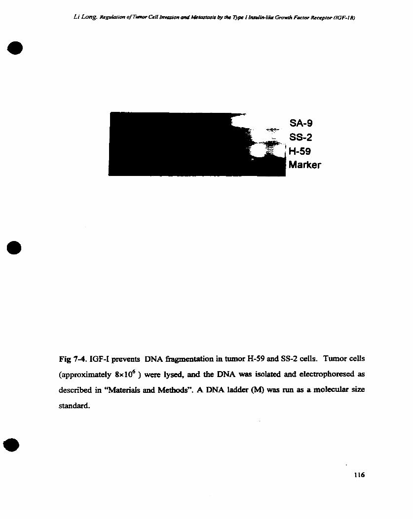
Fig 74. IGF-1 prevents DNA fragmentation in tumor H-59 and SS-2 ceiis. Tumor cells
(approximately 8x10~ ) were lysed, and the DNA was isolated and electrophoresed as
described in "Materiais and Methods''. A DNA ladder (M) was run as a molecular size
standard.

Chapter Vlll
Discussion

Li Long. Regufarian of Tumor Cefl f ~ i m and M'tastp~ir by t/ru Tjpe I f'fin-lilr Growth Factor Rrccptor (fGF-f R)
The evidence described demonstrates that the insulin-like growth factor I receptor is
involved in carcinoma growth, invasion and metastasis to the liver. These results provide
the fi& indication that the IGF-MGF4 complex play a role in preferential homing of
cancer cells to selected target organs and in regulating cellular invasion through its role in
regulating expression of the Mi 72,000 type IV collagenase - a major mediator of ECM
proteolysis.
8.1 Dinetentid erprruion of p w t h factorr and recepton: tumor heterogeneity.
Metastasis, the spread of cells b m the primary neoplasm to distant sites and their growth
there, contributes to the death of most cancer patients. The two major reasons for the
failure to treat metastases are the anatomid location of the metastatic lesions and the
biologic heterogeneity of ceils in primary and secondary neoplasms. This heterogeneity is
manifest in a wide range of genetic, biochemical, immunological, and biological
characteristics including ceII sdace receptors, enzymes, karyotypes, c d morphology,
growth properties, sensitivity to various therapeutic agents, and the ability to invade and
metastasize (464). A century ago. Paget questioned whether the distribution of metastases
was random and therefore analyzed a large number of autopsy records of women with
breast cancer. The noorandom pattern of visceral breast cancer metastases suggested to
Paget that the process was not due to chance, but rather th certain favored tumor cells
(the "seed") had a specinc e t y for growth in the milieu provided by certain organs

Li Long. Regulafiom of Tmor Cell fnvasion ami d c k ~ a s ~ i s by the Typr I fnsutin-like Growth factor âeceptor (IGF-IR)
(the "soil"). Metastases resulted only when the "seed and the soir were compatible (1).
This concept has gained wide acceptance and currently is based on three principles:
1. Neoplasms are heterogeneous in respect to biologic and metastatic properties;
2. The process of metastasis is not random, but consists of a series of iinked, sequential
steps that must be completed by tumor cells if a metastasis is to develop;
3. The successfùl establishment of metastasis depends on the outcome of interaction
between the multiple metastatic celis and the cliffixent target organ microenvironment.
Previous studies in our laboratory revealed that the Lewis lung carcinoma sublines H-59
and M-27 différ in a range of phenotypes including ceii adhesion (388-391.46s) and the
expression of ECM degrading proteinases (392) ali of which may contribute to their
organ selective pattems of metastasis. These p h e n ~ ~ i c difference may be related to the
divergent expression of several growth factors and their receptors in these ceiis as
described in the present study. The molecular mechanisms which underlie the
development of tumor heterogeneity are divene. They rnay reflect genetic events such as
mutations chromasomal deletions or transtocations which alter gene expression.
Aitematively altered gene expression rnay be due to epigenetic mechanisms such as DNA
methylation and demethylation, processes which can be induced environmentally and
developmentally (466,467). The mechanism which caused the development of the Lewis
lung carcinoma sublines with distinct invasive/metastatic properties used in this study
are not presently clear.

Li Long. Rrgulation of Tiunor Ceii Inwsion ami .Uemrusis by the T ~ p e I lnnriui-like Growrh Facror Recrpcor fIGF-I R)
The overexpression of IGF-IR in tumor H-59 cells (seed) may be a major mechanism
regulating their preferential metastasis to the liver (soil) since the latter is a major sources
of IGF-1 production (Chapter W. Fig la, and see ref. (20,468). This is supported by our
fmding that M-27 cells transfected with full length IGF-IR cDNA acquired an incfea~ed
liver-colonizing potential (Chapter V). Neither H-59 nor M-27 cells express detectable
IGF-1 levels (Chapter W, Fig la). The activation of the IGF-iR in these cells therefore
relies on an exogenous source of IGF-1 rendering the liver a suitable target organ. IGF-1
has also been identifieci as a chernotactic andior motility factor for many normal or
malignant cells including endotheliai celis (469), keratinocytes (470). osteoblasts (471),
smooth muscle ceii (261), melanoma (84), breast carcinoma (472), bladder and ovarian
carcinomas (472). In our studies it was found that IGF-1 is a chemoattractant for H-59 but
not for M-27 ceiis (Chapter W, Fig 2). It is conceivable that the chernotactic effect of
IGF-1 also plays a role in selcctively directing tumor migration towards organs with high
local concentrations of this factor. In addition, the mitogenic (Chapter IV and V), anti-
apoptotic (33 1, 335, 336, 409) and invasion-inducing effect of IGF-1 (Chapter VI)
probably contribute to its role in promoting target organ colonization. A similar
interpretation can also be applied to lymph node metastasis of 8 5 9 cells since IGF-1 is
aiso produced in the lymph nodes (20). The influence of the differential expression of
other growth factors and receptors on invasion and metastasis in our tumor mode1
requises M e r investigation.
8.2 The rote of the IGF-IR/IGF-1 compkx in cancer invasion and metastrsis

Li Long. Re&ation of Tmor Cell fn\wlion and .Hokastaris by I/w Tjpc 1 f~~~IUi--liitc Growrli Facroc Reccpror (lGF-C R)
0 The process of metastasis is a cascade of Linked sequential steps involving multiple host-
tumor interactions. This complex process requires the cells to eriter into the circulation,
arrest at a distant vascular bed, exüavasate into the orgau interstitiun and patenchyma,
and proliferate as a secondary colony. increased expression of IGF-IR andor IGF-1 has
k e n noted in a wide range of tumor types including carciinornas of the lung, breast,
thyroid, gastrointestinal tract, iîver, pancreas and kidney, and neuroendocrine tumors
(339). In vitro stuàîes have irnplicated this receptor in cellular transformation (3 13, 327,
328), proiifèration (326, 330), ceU protection h m apoptosis (331, 335, 336) and
migration (84,261,407,410). Ilan and CO-workers have found that dsense RNA to the
IGF-IR could inhibit tumor growth and prevent invasion rat prostate cancer cells vivo
(473). Ioterestingiy, a signiscant reduction in expression of both tPA and uPA which play
a important rolc in matrix degredatîon o c c d in IGF-IR antisense transfected celis
(473).
As we have discussed in Chapter 6, IGF-I has been implicated in transcriptional
regulation of various genes including the early response genes fus (304,427,428) andjun
(429), cell cycle intermediates such as cyclin-Dl and cdc2 (430) and recently, the
urokinase plasminogen activator Uihibitor PM4 (43 1). IGF-1 has also been implicated in
the regulation of the synthesis of ECM pmteins such as collagen (432) and proteoglycans .
(433). As IGF-I has been shown to enhance steady state mRNA levels through a direct
effect on gene transcription or by pst-transcriptional modification of m.RNA, which

e stabilize the message (43 l), the precke molecular mechanism involved in regulation of
MMP-2 synthesis rem- to be elucidated.
Multiple signal transduction pathways can be hnned on in response to ligand binding by
IGF-IR. They include a tyrosine phosphorylation cascade involving IRS-1 (insulin
receptor substrate-l), Shc, Grb-2 and mSOS which are Linked to the ras signaling
pathway (reviewed in ref. (S9), activation of phospholipase C and the accumulation of
IP3. While the link between these signahg intermediates and MMP-2 synthesis remains
to be elucidated, it is of interest to note that IRS-1 was recently found to be associated
with the integrin vitronecth receptor a& (434), a modulator of MMP-2 synthesis which
was also recently identifieci as a cell surface receptor for MMP-2 (435). The signal
transduction pathways activated by IGF-IR and a& appear therefore to converge with
the iikely result of signal amplincation and an enhancement of the proteolytic and
invasive capabilities of the ceii. In a recent report, it was shown that the inhibition of
TGF-1 B receptor-mediateci calcium influx blocked the activation of MMP-2 transcription
in three different human tumor hes (421). This suggests that calcium mobiiization is a
critical component of TGF-lp-mediated activation of MMP-2 synthesis. A s Ligand
binding by IGF-IR is known to induce to a rapid accumulation of IP3, resulting in
increased cytoplasmic leveb of fÏee calcium (436). it may be one mechanisms for IGF-I-
mediated regulation of MMP-2.

Li Long. &guIation of T-of Ce4 i . . i o n und .Uecprtosir & r i te T m I In~Iin-Iike Growrh Factor Receptor (IGF-IR)
While selective H-59 metastasis to the liver may be regulated by IGF-1, the mechanisms
which regulate M-27 metastasis to the lung are not presently entirely ciear. M-27 cells
express significantly higher levels of EGF, PDGF and the respectiire recptors than H-59
cells (Chapter W, Fig 7-1). Overexpression of IGF-IR in these ceiis did not result in a
significant change in either tumongenicity, meanad as the incidence and growth rate of
subcutaneous tumors, or the potential to colonize the lung following i.v. inoculation
suggesthg that the growth of these ce& in vivo in these sites may be regulated
predominantly by autocrine mechanisrno mediateci by growth factor(s) such as PDGF and
EGF. In addition, M-27 ceUs produce significantly higher leveis of urokhase-type
plasminogen activator @PA) and cathepsin B than tumor H-59 celis and it is wnceivable
that the differences in the types of basement membrane d e m g proteinases released by
these tumor cells are also a factor in their distinct patterns of dissemination. Other
factor@) such as transfh have been identifiai as regdators of lung metastases
formation and they may also play a d e in our system (474). The hding that the major
effea of increased IGF-IR expression on M-27 c d growth ip v i v ~ was a rnarked increase
in their liver-coloniring potentid to levels normally observed with carcinoma H-59
supports the conclusion that IGF-I plays an important role in the pmcess of liver
metastasis.
The involvement of IGF-IR in the mgdation of type IV collagenase synthesis may e t
not only the invasive potential of the ceiis but also cellular proliferation. Recent studies
by Dr. Fowlkes and colleagues have demo~l~trated that IGF binding proteins 3 and 5

(IGFBP3 and 5) are processed by MMPs (MMP-2 and MMP-9) both in vitro and
suggesting a role for MMPs in regulating semm and tissue IGF bioavailability (52, 53,
289). Taken together wîth our data, this is suggestive of a possible IGF-UIGF-IR:MMP
:IGFBPs loop regulating IGF-1 function, namely, IGF-1 can upregdate the expression of
MMP-2 (Chapter VI), active MMP-2 in tum is involved in the processing of IGF
binding proteins (52, 53,-289) which can modulate the interaction of the IGFs with theu
receptors (284). A schematic illustration of the proposed IGF-MGF-IR:MMPs:IGFBPs'
loop is shown in Fig. 8-1.
modulation of IGFaGF-CR interaction
upctguiation ? transcription ? mRNA stability

Li Long. Regukz~ion of Tumor Ce fi I l l ~ ~ ~ i o n and Mkku&tsïs by clir Typc I IlLnJWik Gmwh Facror Rrcepror (fGF-/ R)
Fig. 8-1. The schematic Uustration of proposeci IGF-VIGF-1R:MMPs:IGFBPs' Loop

Li Long. Replation of Tùmor Cell Invasion Md ,UerartasU d u f farulin-lik Growih Factor Reccpror (IGF-IR)
Cellular invasion depends on adhesion as well as proteolytic modification of the
extracellular matrix and an association between the expression and fùnction of adhesion
receptor; and ECM-degrading proteinases has been noted in ciiffernt systems (122, 143,
475). For example, Brooks et ai have recently reported on the colocalization of MMP-2
and the vitronectin receptor integrin a,,& on angiogenic blood vessels and melanoma
cells Jn vivo. Expression of q P 3 on cultureci melawma cells was necessary for binding of
MMP-2 in a proteolyticaIiy active form, facilitating cell-mediateci collagen degradation
(435). In our laboratory, a fùnctional and rcguiatory link between a& and the urokinase
-type plasminogen activator receptor @PAR) has been identifiecl in metastatic melauoma
cells (189). Interestingiy. Jones et al have recently reported that ligand-dependent
. activation of a$3 is necessary for IGF-1-mediated stimulation of ce11 migration (410),
providing a mechanism for coordination of ceil adhesion, migration, invasion and growth
through molecular cross-talk. The expression and the d e of the vitronecth receptor
in our tumor mode1 have not been studied. A M 64,000 plasma membrane glycoprotein
has k e n icientifïed as an adhesion molecule mediating the speciiic interaction between
tumor H-59 cells and hepatocytes, an important step in liver colonipltion by tuxnor H-59
cells (390.391). The relationship between tbis d e s i o n molecule, the IGF-IR and matrix
degrading proteinases, if any, remains to be determineci.
Recent insight gained into the molecular mechanisrns underlyiag the pmcess of ceIl
migration and invasion has led to the identification of two major categories of cellular
targets for therapeutic intervention. The k t category includes celi M a c e and secmted

Li Long. Rrgufation of Tiunor CeII fmasiion anâ lUetas~asis tk Tjlpc I Insuiin-like Growth Factor Rcceprar ffGF-f R)
a proteins such as adhesion receptors, degradative eazymes and their inhibitors, growth
factordreceptors and motility stimuiating cytokines (1 1, 157, 199, 3 70, 476-478). The
second category is defined as intracelîular regulatory proteins which mediate signal
transduction mechanisms such as G-pmteins, and tyrosine kinases (342,479-482). Many
studies indicate that the IGF-VIGF-IR complex and metalloproteinases may provide
effective targets for cancer therapy (331,342,343,345-347,394,417,473,483486). The
present hdings confirm and extend previous findings on the critical role of the IGF-
VIGF-IR system in cancer celi growth, invasion and me-. Together they suggest
that the IGF-VIGF-IR system codd provide a target for anti-metastatic therapy.
8 3 Suggestions for Fuhuc Rcscarch
The following aspects requin M e r investigation:
1. Elucidating the signalhg pathways linking MMP-2 transcription to IGF-1 receptor.
2. Analyze the relaîionsbip between IGFBP production and MMP-2 synthesis and
function in H-59 and M-27 cells.
3. Gene therapy of metastatic tumors by transduction with a viral vector expresshg IGF-
1 R antisense DNA.
4. Mutational analysis of the IGF-1 receptor to identiSr receptor domains critical for its
diverse f'unctions.

Li Long. Regubtion of Twnor Ceff Invasion and Me~cutasu by the Tjpc C I~ufin- l ik Growth Factor Receptor UGF-1 R)
Refcnnccs
Paget, S. Distribution of secondary growths in cancer of the breast. Lancet. I:
57 1-573, 1889.
Ewing, 3. A treatise on tumors., 3rd edition. Philadelphia: W. B. Saunders, 1928.
Fidier, 1. and Balch, C. The biology of cancer metastasis and implications for
therapy. Curr. Pmb. Surg. 24: 137,1987.
Fidler, 1.' and Hart, 1. biologic diversity in metastatic neoplastus - origins and
implications. Science (Washington DC). 21 7: 998- 100 1, 1982.
Schimnancher, V. Cancer Maastasis: Experimental approaches, theoretical
concepts, and impacts for treatment strategies. Adv. Cancer Res. 43: 1-32, 1985.
Liotta, L., Rao, C., and Barsky, S. Tumor invasion and the extraceliular matrix.
Lab. Invest. 49: 363, 1983.
Liotta, L., Steeg, P., and Stetler-Stevenson, W. Cancer metastasis and
angiogenesis: An imbalance of positive and negative regdation. Cell. 64: 327-
336,1991.
Bernstein, L. R. and Liotta, L. A. Molecular mediators of interactions with
extraceiluiar ma* cornponents in metastasis and angiogenesis. meview]. Curr.
0pin.Oncol. 6: 106-13, 1994.
Stracke, M. L. and Liotta, L. A. Multi-step cascade of -or celi metastasis.
Ipeview]. In Vivo. 6: 309-16, 1992.
Mareel, M. M., Van Roy, F. M., and Brade, M. E. How and when do tumor celis
metastasize? Crit. Reviews Oncagen. 4: 5 59-94, 1993.

Li Long ReguIario~ of TÙmor Ceif I'arion ond .WetpsrasLr by he T j p I fmfin-fi.& Growrh Foctor Recrpror (ZGF-f R)
Aaronson, S. Growth factors and cancer. Science (Washington DC). 254: 1 146-
L 153, 1991.
Pledger, W., Stiles, C., Antonides, H., and Scher, C. Proc. Natl. Acad. Sci. USA.
73: 448 1, 1977.
Pledger, W., Stiles, C., Antonides, H., and Scher, C. Proc. Natl. Acad. Sci. USA.
75: 2839, 1978.
Pardee, A. G1 events and regdation of celi proliferation. Science (Washington
DC). 246: 603408,1989.
Cross, M. and dexter, T. Growth factors in development, trandormation, and
tumorigenesis. Ceii. 64: 271 -280, 199 1.
LeRiche, V. K, Asa, S. L., and E m t , S. E p i d e d p w t h factor and its receptor
(EGF-R) in human pituitary adenornas: EGF-R correlates with tumor
aggressiveness. J. Ch. Endocrinol. Metab. 81: 65662,1996-
Kimura, G., Kasuya, I., Giannini, S., Honda, Y., Mohan, S., Kawachi, M.,
Akimoto, M., and Fujita-Yamaguchi, Y. Insulin-like growth factor (IGF) system
components in human prostatic cancer ceii-lines: LNCaP, DU145, and PC-3 cells.
ht. J. Urol. 3: 3946,1996.
Potapova, O., Fakhrai, H., and Mercola, D. Growth factor PDGF-B/v-sis coafea a
hunorigenic phenotype to human tumor c e k bearing PDGF receptors but not to
ceUs devoid of receptors: evidence for an autocrine, but not a paracrine,
mechanism. ht. J. Cancer. 66: 669-77, 1996.

Robbins, K., Antoniades, H., Devare, S., Hunkapiller, M., and Aaronson, S.
Structural and immunological similan'ties between simian sarcoma virus gene
products and human platelet derived growth factor. Nature. 305: 605608, 1983.
D'Ercole, A. I., Stiles, A. D., and UnderwOOd, L. E. Tissue concentrations of
somatomedin C: Further evidence for multiple sites of synthesis and paracine or
autocrine mechanisms of action. Proc. Natl. Acad. Sci. USA. 81: 93 5-939, 1984.
HeIdin, C. and Westermark, B. Platelet-derived growth factors: a farnily of
isoforms that bind to two distinct receptors. Br. Med. BU. 4 2 453-464, 1989.
Fleming, T., Matsui, T., Moiloy, C., Robbias, K., and Aaronson, S. Autocrine
mechanism for v-sis transformation re;luires ceii d a c e localization of internally
activated growth factor receptors. Proc. Natl. A d . Sci. USA. 86: 8063-8067,
1989.
Di Marco, E., Pierce, J., Flemming, T., Kraus, M., Molloy, C., Aaronson, S., and
Di Fiore, P. Autocrine interaction between TGF dpha and the EGF-receptor:
quantitative requirements for induction of the malignant phenotype. Oncogene. 4:
83 1-838, 1989.
Halaban, R, Kwon, B., Ghosh, S., DeUi Bovi, P., and Baird, A. bFGF as an
autocrine growth fsctor for human melanomas. Oncogene Res. 3: 177-1 86, 1988.
Liberniano, T., Nusbaum, H., h n , N., Kris, R, Lax, I., Soreq, Hm, Whittle, N.,
Waterfield, M., Ulirich, A., and Schlessinger, J. Amplification, enhanced
expression and possible reaxrangement of EGF receptor gene in primary human
brain tumours of glial on&. Nature. 31 3: 144-147, 1985.

Li Long. ReguIa~ion of Tumor CeII I l l ~ ~ ~ i o n anci Metastasu by tk Typc î Insufin-fiAP Growth Factor Receptor (IGF-1 R)
Slamon, D., Clark, G., Wong, S., Levin, W., Ullrich, A., and McGuire, W. Human
breast cancer: correlation of relapse and survival with amplincation of the HER-
2neu oncogene. Science. 235: 177- 182, 1985.
Di Fiore, P., Pierce, J., Kraus, M., Segatto, O., King, C., and Aaronson, S- erbB-2
is a potent oncogene when overexpressed in W 3 T 3 cells. Science. 237: 178-
182,1987.
Kraus, M., Issing, W., Mili, T., Popescu, N., and Aaronson, S. Isolation and
charactrization of ERBB3, a third member of the ERBB/epidermal growth factor
receptor family: evidence for ovetexpression in a subset of human mammary
tumors. Proc. Natl. A d . Sci. USA. 86: 9 193-9 197, 1989.
Soman, N., Coma, P., Ruiz, B., and Wogan, G. The TPR-MET oncogenic
rearrangement is present and exprrsseâ in human gastric carcinoma and precürsor
lesions. Proc. Nad. Acad. Sci. USA. 88: 48924896, 1991.
Grieco, M., Santom, M., Berlingieri, M., Melillo, R., Donghi, R, Bongarzone, I.,
Pierotti, M., Delia Porta, G., Fusco, A., and Vecchio, G. PTC is a novel
rearranged fonn of the ret proto-oncogene and is kquently detected in vivo in
human thyroid papülary carcinomas. Celi. 60: 557-563,1990.
Heldin, C. H. and Westermack, B. Platelet-derived growth factor and its relation
to oncogenes. ISI Atlas. Sci. Immunol. 441,1988.
Nusse, R The int genes in mammary tumorigenesis and in nomai development.
Trends Genet. 4: 291, 1988.
Sherr, C. J. Thefmr onwgene. Biophys. Acta. 948: 225,1988.

Hayman, M. J. erb-B: growth factor receptor turned oncogene. Trends Genet. 2:
260,1986,
Besmer, P., Murphy, J. E., George, P. C., Qiu, F., Bergold, P. J., Lederman, L.,
Snyder, H. W., Brodeur, D., Zuckerman, E. E., and Hardy, W. D. A new acute
transformiDg feline retrovirus and relationship of its oncogene v kit with the
protein kinase gene family. Nature. 320: 415,1986.
Ullrich, A., BeU, J. R, Chen, E. Y., Herrera, R, Pertuzzelli, L. M., Duii, T. J.,
Gray, A., Coussens, L., Liao, Y. C., Tsubokawa, M., Mason, A., Seeburg, P.,
G d e l d , C., Rosen, O. M., and Ramachandran, J. Human insulin receptor and its
relatiomhip to the tyrosine kinase family of oncogenes. Nature. 313: 756, 1985.
Sefton, B. M. and Buss, J. E. The covatent modification of eukaryotic proteins
with lipid. J. Ceil Biol. 104: 1449, 1987.
Sefion, B. M. Owogenes encoding protein kinases. Trends Genet. 1: 306, 1985.
Hollywood, D. Signal transduction. Br. Med. Bulletin. 47: 99-1 15, 199 1.
Miesenhelder, J., Suh, P., Rhee, S., and Hmter, T. Phospholipase
overexpression on PDGF induced second messengers and mitogenesis. Cell. 57:
1109-1122, 1989.
Molloy, C., Bottaro, D., Fleming, T., Marshall, M., Gibbs, J., and Aaronson, S.
PDGF induction of tyrosine phosphorylation of GTPase activating protein.
Nature. 342: 71 1-714,1989.
Kaplan, D., Whitman, D., Schaffhausen, B., Pallas, D., White, M., Cantley, L.,
and Roberts, T. Common elements in growth futor stimulation and oncogenic

Li Long. Regufation of Tumor Cell lmasion a d Memwasis by ri# 7Jpe I Insufin-liku Growth Factor Recepror (IGF- IR)
transformation: 85 kD phosphoprotein and phosphatidylinositol kinase activity.
Cell. 50: 1021-1029, 1987.
Momson, D., Kaplan, D., Escobedo, J., Rapp, U., Roberts, T., and Wiams, L.
Direct activation of the serine-threonine kinase activity of Ebf-1 through tyrosine
phosphorylation by the PDGF 8-receptor. Ceil. 58: 649-657, 1989.
Curran, T. and Franza, B. J. fos and Jun: the AP-1 connection. Cell. 55: 395-397,
1988.
Heikkla, R, Schwab, G., W i c b m , E., Lobe, S., Pl- D., Watt, R, and
Neckers, L. A c-mye antiseme oligodeoxynucleotide inhibits entry into S phase
but not progress h m GO to G1. Nature. 328: 445449,1987.
Kelly, K., Cochran, B., Stiles, C., and Leder, P. Cell-specific regulation of the c-
myc gene by lymphocyte mitogeils and plateletderived growth fiador. Celi. 35:
603-610,1983.
Greenberg, M. and Zin, E. Stimulation of 3T3 ceils induces transcription of the c-
fos prot-oncogene. Naîure. 311: 433438,1984.
Keski-Oja, J., Koli, K., Lohi, J., and Laiho, M. Growth factors in the regulation of
plasminogen-plasmin system in tumor ceus. Semia Thromb. Hemost 1 7: 23 1 -
239,1991.
Lee, L. and Weinstein, 1. E p i d e d growth factor, iike phorbol esters, induces
plasminogen activator in HeLa cells. Nature. 274: 696-697, 1978.

Montesano, R., Vassalli, J., Baird, A., Guillemin, R, and Orci, L. Basic fibroblast
growth factor induces angiogenesis in vitro. Proc. Natl. Acad. Sci. USA. 83:
7297-730 1, 1986.
Lyons, R., Keski-Oja, J., and Moses, H. Proteolytic activation of latent
transfoming growth factor-f3 fiom fibroblast conditioned medium. I. Ceil Biol.
106: 1659-1665, 1988.
Fowlkes, J., Suzuki, K., Gagase, H., and ïhiküi, K. Proteolysis of insulin-like
growth factor binding protein-3 during rat pregnancy: A role for matrix
metalloproteinases. Endocrinol. 135: 28 10-28 1 3,1994.
Thrailkili, L, Quarles, L., Nagase, H., Suzuki, K., Sema, D., and Fowlkes, J.
Chanicterization of insulin-like growth factor-binding protein 5-degrading
proteases ptoduced throughout miaine osteoblast differentiation. Endocrinol. 236:
3 527-3 533, 1995.
Ross, R., Bowden-Pope. D., and Raines, E. Platelet-derived gmwth factor and its
role in heaith and disease. Phil. Trans. Ry. Soc. (Lond.). B 327: 155-1 69, 1990.
Rappoiee, D., Brenner. C., Schultz, R, Mark, D., and Werb. 2. Developmend
expression of PDGF, TGF-a, and TGF-f3 genes in prcimplantation mouse
embryos. Science. 241: 1823- 1825,1988.
Gill, G., Bertics, P., and Santon, J. E p i d e d growth factor and its receptor. Mol.
Cek Endocrinol. 51: 169-1 86,1987.
Barnard, J., Lyons, R, and Moses, H. The ceii biology of traosforming growth
factor p. Biochim. biophys. Acta 1032: 79-87, 1990.

Li Long. Rrgulafion of Tumor Cell Invasion and .Clemtasir by liw Typr I fl~~1IU1-lik Growth Frrcior Rewpfor (ICF-I R)
Haselbacher, G., Irminger, I., Zapf, J., Ziegler, W., and Humbel, R Insulin-like
growth factor II in human adrenal pheochromocytomas and Wilms m o r s :
expression at the mRNA and protein levei. Proc. Natl. Acad Sci. USA. 84: 1 104-
1 106,1987.
Rubin, R and Baserga, R Insulin-like growth factor4 receptor. Its role in ce11
proliferation, apoptosis, and tumongenicity. E.eview]. Lab. Invest. 73: 3 1 1-3 1,
1995.
Burgess, W. and Maciag, T. The heparin-binding (fibroblast) f e l y of proteins.
Ann. Rev. Biochem. 58: 575-606,1990.
Acland, P., Dixon, M., Peters, G., and Dickson, C. Subcellular fate of the int-2
oncoprotein is determineci by choice of initiation codon. Nature. 343: 662465,
1990.
Richmond, A-, Balantien, E., Thomas, H., Flaggs, Ge, Bargton, D., Spiess, J.,
Bordoni, R, Franke, U., and Derynck, R Molecular characterization and
chromosomal mapping of melauorna growth stimulating activity, a p w t h factor
structuraUy related to B-thromboglobulin. EMBO J. 7: 2025-2033, 1 988.
Whetton, A. and Dexter, T. Myeloid haemopoietic growth fztors. Biochim.
Biophys. Acta. 989: 1 1 1-132,1989.
Hwang, D., Tay, Y., Lin, S., and Lev-Ran, A. Expression of epidennal gmwth
factor receptors in human lung tumors. Cancer. 58: 2260, 1986.
Thijssen, J. and Blankenstein, M. Oestrogens in breast tumours and fat. Proc. R.
Soc. Edinburnh Sect. B. 95: 161.1989.

Cartlidge. S. and Elder, I. Transfomiing gmwth faftor a and epidermal p w t h
factor levels in normal human gastrointestinal mucosa. Br. J. Cancer. 60: 657,
1989.
Cariani, E., Lasserre, C., Seurin, D-, Hameiin, B., Kemeny, F., Franco, D., Czech,
M., Ulirich, A., and Brechot, C. Differential expression of insulin-like p w t h
- factor II mRNA in human primary liver cancers, benign tumors, and Liver
cirrhosis. Cancer Res. 48: 6844,1988.
Bergeron, C., Ferenczy, A., TOR D., and Shyamala, g. Immunocytochemical
study of progesten,ne recqtors in hypeqlastic and neoplastic endometrial tissues.
Cancer Res. 48: 6132,1988.
Gomeila, L., Sargent, E., Wade, T., Anglard, P., Linehan, W., and Kasid, A.
Expression of transforming growth factor a in nomal human adult kidney and
enhanced expression of transforming growth m o r s a and 01 in rend ceU
carcinoma. Cancer Res. 49: 6972,1989.
Smith, K., Fenueiiy, J., Neai, D., Hali, R, and Harris, A. Characterizattion and
quantitation of the epidennal growth factor receptor in invasive and superficial
bladder tutnom. Cancer Res. 49: 58 IO, 1989.
Davies, P., Eaton, C., France, T., and Phillips, E. Growth factor receptoa and
oncogene expression in prostate ceils. Am. J. Clin. Oncol. 1 I : sl , 1988.
Y urchenco, P. and Schittny, J. Molecdar architecture of basement membranes.
FASEB J. 4: 1577-1 590, 1990.

Barsky, S., siegal, G., Jannotta, F., and al, e. Loss of basement membrane
components by invasive tumors but not by their genign counterparts. Laboratory
Investigation. 49: 140, 1983.
Kohn, E. and Liotta, L. Molecular insights into cancer invasion: Strategies for
prevention and intemention. Cancer Res. 55: 1856-1 862,1995.
Hynes, R Integrins: a family of celi s u f f i receptors. Cell. 48: 549, 1987.
Hymphnes, M., OIden, IC, and Yamada, IC A synthetic peptide fiom fïbronectin
inhibits expcrimental metastasis of murine melanoma celis. Science (Washington
DC). 233: 467,1986.
Rao, C., Castronovo, V., Scbmitt, M., Wewer, U., and Claysmith, A. Evidence for
a precursor of the high-aff?nity metastasis associated murine laminin receptor.
Biochemistry. 28: 7476-7486,1989.
Aznavoorian, S., Stracke, M., K W h , H., and al, e. signal transduction for
chernotaxis and haptotaxis by matrix molecules in tumor celis. J. Cell Biol. 110:
1427,1990.
Gottesman, M. The mle of proteases in cancer. Semin. Cancer Biol. 1: 97-160,
1990.
Nakajima, M., Morikawa, K., Fabm A., Bucana, C., and Fidler, 1. Influence of
organ environment on elcaa~eiiular matrix degradative activity and metastasis of
human colon carcinoma ceiis. J. Nat. Cancer Mt. 82: 1890- 1898,199 1.
Brown, P., Levy, A., Marguîies, 1.. Liotta, L., and Stetler-Stevenson, W.
Independent expression and cellular processhg of the 72-kDa type IV collagenase

Li Long. Reguiarion of Timor Crll f l l ~ ~ ~ i o n a d Metasasis by r k T m I Insuiin-Iïke Growrh Forrw Recepror (IGF-/ R)
and intersth1 coilagenase in human tumorîgenic ce11 Lines. Cancer Res. 50:
6184-6191, 1990.
Liotta, L., mander, R, Murano, G., Katz, D., Gordon, R., Chiang, P., and
Schifmiann, E. Tumor celi autocrine motility factor. Proc Natl Acad Sci USA. 83:
330293306, 1986.
Gherardi, E., Gray, J., Stoker, M., Perryman, M., and Furlong Purification of
scatter factor, a fibroblast derived basic protein that modulates epitheiial
interactions and movement. Roc Natl Acad Sci USA. 86:, 1989.
Stracke, M., Engel, J., Whn, L., Rechler, M., Liotta, L., and Schiffinann, E. The
type I insulia-like growâh factor receptor is a motility receptor in human
melanoma celis. J. Bi01 Chem. 264: 21544-2 1549,1989.
Nicolson, G. L. Paracrine and autocrine growth mechanisms in tumor metastasis
to specinc sites with particular emphasis on brai. and lung metastasis. Cancer
Metast, Rev. 12: 32543,1993.
Nicolson, G. L. Tumor microenvironment: paracrine and autocrine growth
mechanisms and metastasis to specific sites. Front. Radiat. Ther. Oncol. 28: 1 1-
24, 1994.
Bemacki, R., Niedbala, M., and Korytnyk, W. Glycosidases in cancer and
invasion. Cancer Metast. Rev. 4: 8 1, 1985.
Danf, K., Addreasen, P., Grfûdahl-Hansen, J., Kristemen, P., Nielsen, L., and
Skriver, L. Plasmigogen activators, tissue degradation and cancer. Adv. Cancer
Res. 44: 139, 1985.

Li Long. Reguiat~on of Tunor Cclf Invarion and M i i t a r r ~ ~ i r by he ripe I Insulin-Iirtr Growth Fator Recepror (IGF-IR)
D e , M. and O'Grady, P. Plasminogen activator and cancer. Eur. J. Cancer Clin.
Oncol. 20: 557, 1984.
Mainardi, C., Hasty, D., Seyer, J., and Kang, A. Specific cleavage of human type
III coliagen by human polymorphonuclear leukocyte elastase. J. Biol. Chem. 25.5:
12006,1980.
Zucker, S., Lysik, R, Meman, J., Willcie, D., and Lane, B. Diversity of human
pancreatic cancer cell proteinases: role of cell membrane metaiioproteinases in
collagenolysis and cytolysk. Cancer Res. 45: 6 186,1985.
Senior, R and Campbell, E. Cathepsin G in human mononuclear phagocytes:
cornparisons bmveen monocytes and U937 monocyte-lü<e celis. J. Immunal. 132:
2547,1984.
Sloane, B. and Honn, K. Cysteine proteinases and metastasis. Cancer Metast. Rev.
3: 249,1984.
Portnoy, D., Erickson, A., Kochan, J., Ravetch, J., and Unkeless, J. Cloning and
characterization of a mouse cysteine proteinase. J. Biol. Chem. 261: 14697, 1986.
Rochefort, H., Augereau, P., Briopo, P., Capony, F., Cavailles, V., Freiss, G.,
Garcia, M., Maudelonde, T., Morisset, M., and Vignon, F. Structure, function,
regdation and clinical sigdicance of the 52K pro-cathepsin D secreted by breast
cancer ceus. Biochimie. 70: 943,1988.
Wilhelm, S., Eisen, A., and Teter, M. Human fibroblast collagenase:
Glycosylation and tissue-specinc levels of enzyme synthesis. P m . Natl. Acad.
Sci. USA. 83: 3756-3760,1986.

Li Long. Regdation of Twnw Ceil Invasion anâ Me~rutasu éy the Type I Idin-liAe Growrh Factor Receptor (IGF-I R)
Collier, I., Wilhelm, S., Eisen, A., Marmer, B., Grant, G., Seltzer, J., Kronberger,
A., He, C., Bauer, E., and Goldberg, G. H-ras oncogene-transfomed human
branchial epithelial cells (TBE-1) sectete a single metalloprotease capable of
degrading basement membrane collagen. J. Bio 1. Chem. 263: 6579, 1988.
Murphy, G., Hembry, R, McGarrity, A., Reynolds, J., and Henderson, B.
Gelatinase (type N coliagenase) irnmunolocalization in ceils and tissues: use of
an antiserum to rabbit bone gelatinase that identifies high and low Mr forms. I.
CeU Sci. 92: 487,1989.
Liotta, L., Lanzer, W., and Garbisa, S. Identiiication of a type V cbilagenolytic
enzyme. Biochem. Biophys. Res. Cornmum. 98: 184,1981.
Mder , D., Quantin, B., Gesnel, M., Miilon-Collard, R, Abecassis, J., and
Breathnach, R The collagenase gene family in humans consists of at least four
rnembers. Biochem. J. 253.- 187,1988.
Nicholson, R, Murphy, G., and Breaîhnach, R Humna and rat malignant-tumor-
associateci mRNAs encode stromelysin-like metailoproteinases. Biochemistcy. 28:
5195,1989.
Banda, M. and Werb, 2. Mouse macrophage elastase. Purification and
chafacterization as a metalloprotease. Biochem. J. 193: 589, 198 1.
Fiszer-Szafarz, B. and Gulho, P. Hyaluronidase activity of normal and neoplastic
interstitial fluid. Roc. Soc. Exp. Biol. Med. 133: 805, 1970.

Sobel, G., Mohler, S., Jones, N., Miller, K., and Peluso, R. Urokinase: An
activator of plasma profibrinolysin extracted nom urine. Am. J. Physiol. 171:
768-769, 1952.
Gunzler, W., Steffens, G., Otting, F., Kim, S., Frankus, E., and FIohe, L. The
primary structure of high molecular mass urokinase fkom human urine. The
complete amino acid sequence of the A cbain. Hoppe Seylers Z Physiol. Chem.
363: 1155-1 165, 1982.
Be& M. and Ohler, E. Plasminogen activator and proactivator- (utokinase
precursor) in lung cultures. J. Am. M d Wom. Assoc. 31: 465472,1976.
Ichinose, A., Fujikawa, K., and Suyama, T. The activation of pro-urokinase by
plasma kallikrein and its inactivation by thrombin. J. Biol. Chem. 261: 3486-
3489,1983.
Verstraete, M., Su, C., Tansweii, P., Feuerer, W., and CoUen, D.
Pharmacokinetics and effects on fibrinolytic and coagulation panmeters of two
doses of recombinant tissue-type plasminogen activator in heaithy volunteers.
Thromb. Haemost. 56: 14,1986.
Sprengers, E. and Kluft, C. Plasminogen activator inhibitors. B l d 69: 381,
1987.
Trygg~son, K., Hoyhtya, M., and Saio, T. Proteolytic degradation of
extracellular ma& in tumor invasion. Biochim. Biophys. Acta. 907: 19 1, 1987.

Li Long. Rrgufu~ion of T i o r Ccll fnvasion a d Merastasis by the Tjrpc I fmlin-liùe Growh Focror Recepror fIGF-i R)
Zeydel, M., Nakagawa, S., biempica, L., and TakahaShi, S. Collagenase and
elastase production by mouse mammary adenocarcinorna primary cultures and
cloned ceus. Cancer Res. 46: 6438, 1986.
Mort, J., Recklies, A., and Poole, A. Charactemion of a thiol pmteinase secreted
by malignant human breast tumors. Biochim. Biophys. Acta. 614: 134, 1980.
Pietras, R and Roberts, J. Cathepsin B-iike enzymes: subce11ular distribution and
properties in neoplastic and conttoI cells h m human ectocervix. J. Biol. Chem.
256: 8536,1981.
Keren, 2. and LeGnie, S. Idenification of cell d a c e cathepsin B-like d v i t y on
murine melanomas and fibrosarcomas: modulation by butanol extraction. Cancer
Res. 48: 1416,1988.
Denhardt, D., Hamilton, R, Parfiett, C., Edwards, De, Pierre, R, Waterhouse, P.,
and Nilsen-Hamilton, M. Close relationship of the major excreted protein of
transformed murine fibroblasts to thiol-dependent cathepsins. Cancer Res. 46:
4590,1986.
Masun, R, Gd, S., and Go~tesman, M. The identification of the major excreted
protein (MEP) h m a ttansformed mouse fibroblast celi Line as a catalytically
active precutsor form of cathepsin L. Biochem. 1.248: 449, 1987.
Gottesman, M. Transfodon-dependent secretion of a tow molecular weight
protein by miaine fibroblasts. Proc. Natl. A d Sci. USA. 75: 2767,1978.

Maciewicz, R., Wardale, R, Etherington, D., and Paraskeve, C. Immunodetection
of cathepsin B and cathepsin L present in and secreted nom human pro-maiignar?:
and malignant colorectal tumour ce11 Lines. Int. J. Cancer. 43: 478, 1989.
Capony, F., Morisset, M., Barrett, A., Capony, I., broquet, P., Vigono, F.,
Chambon, M., Louisot, P., and rochefort, H. Phosphorylation, glycosylation, and
proteolytic activity of the 52-kD estrogen-induced protein secreted by MCF-7
cells. J. Ce11 Biol. 104: 253, 1987.
B n o p ~ , P., Morisset, M., Chpony, F., Rougeot, C., and Rochefort, H. In vitro
degradation of extracelidar matrix with mr 52,000 cathepsin D secreted by breast
cancer celis. Cancer Res. 48: 3688,1988.
Murphy, G., Reynolds, J., and Hembry, R Metailoproteinases and cancer
invasion and metastasis. Int. J. Cancer. 44: 757, 1989.
Stetler-Stevenson, W. G., Liotta, L. A., and Kieher, D. E., Jr. Extracellula. matrix
6: role of matrix metalIoproteinases in tumor invasion and meîastasis. peview].
FASEB J. 7: 1434-41,1993.
Matrisian, L. Metalloproteinases and their inhibiton in matrix ~modeling. TIG.
6: 121-125, 1990.
Stricklin, G., Jefney, J., Roswitt, W., and Eisen, A. Human skin fibroblast
procollagenase: mechanism of activation by organomercurials and trypsin.
Biochemistry. 22: 61, 1983.

a 125. Sanchez-Lopez, R., Nicholson, R., Gesnel, M., Matrisian, L., and Bceathnach, R
Structure-fimction relationships in the collagenase faniily member transin. J. Biol.
chem. 263: 1 1892,1988.
126. Mareel, M., De Baetselier, P., and Van Roy, F. Mechanisms of Invasion and
Metastasis, p. 565. Boston: CRC Press, 1 99 1.
127. Pauli, B., Schwartz, Dmy Thonar, E., and Kuetnner, K. Tumor invasion and host
extracellular maîrix. Cancer Metast. Res. 2: 129, 1983.
128. Hicks, N., Ward, R, and Reynolds, J. A fibrosucorna mode1 derived h m moue
embryo cells: growth properties and secretion of collagenase and
metalloproteinase inhibitor w) by tumour ceU lines. Int J. Cancer. 33: 835,
1984.
129. Mignatti, P., Robbins, E., and Ri&in, D. Tumor invasion through the human
amniotic membrane: requirement for a proteinase cascade. Ceii. 47: 487, 1986.
130. Warren, L., Fuhrer, J., and Buck, C. Sinface glycoproteins of normal and
transformeci cells: a diffimmce determineci by sialic acid and a growth-dependent
sialyl transferese. Proc. Natl. Acad. Sci. USA. 69: 1838,1972.
13 1. Dobrossy, L., Pavelic, 2.. Vaughan, M., Porter, N., and Bernaclci, R Elevation of
lysosomal eiryme in prirnary Lewis lung tumor comlated with the initiation of
metastasis. Cancer Res. 40: 3281, 1980.
132. Bosmann, H., Bieber, G., Brown, A., Case, K., Gersten, D., Kimmerer, T., and
Liow, A. Chernical parameters correlateci with tumor ceil implantation. Nature.
246: 487,1973.

Li Long. Repùuion of Tuaor Ceff fnvasion and Merasmu by Ikc Type I InfuIin-fÏke Growth Factor &cepior (IGF- f R)
Gallagher, J-, Lyon, M., aud Steward, W. Structure and h c t i o n of heparan
sulfate proteoglycans. Biochem. J. 236: 3 13, 1986.
Kramer, R., Vogel, K., and Nicolson, G. Solubilizattion and degradation of
subendotheliai rnatrix glycoproteins and proteoglycans by metastatic tumor cells.
J. Biol. Chem. 257: 2678, 1982.
Nakajima, M., Irimura, Tey and Nicolson, G. Heparanases and tumor metastasis. J.
Cell, Biochem. 36: 157, 1988.
Nakajima, M., Irimura, T., Di Ferrente, N., Di Ferrente, N., and Nicolson, G.
Heparan sulfate degradation: relation to tumor invasive and metastatic properties
of mouse B 16 melanoma sublines. Science. 220: 61 1,1983.
Vlodavsky, I., Fuks, Z., Bar-Ner, M., Ariav, Y., and Schirrmacher, V. Lymphoma
cell-mediateci degradation of sulfated proteoglycans in the subendotheliai
extracellular ma*: relationship to tumor metastasis. Cancer Res. 43: 2704,
1983.
Becker, M., Moczar, M., Poupon, M., and Moczar, E. Solubilintion and
degradation of extraceliuiar matrix by various metastatic ce11 lines derived fiom a
rat rhabdomyosarcoma. J. Natl. Cancer Inst, 77: 41 7,1986.
Ricoveri, W. and Cappelletti, R. Heparan sulfate endoglycosidase and metastatic
potentiai in murine fibrosarcoma and melanoma. Cancer Res. 46: 3855, 1986.
Irimura, T., Nakajima, M., and Nicolson, G. Chemically modified heparins as
inhibitors of hep- sulfate specific endo-P-glucuronidase @eparanase) of
metastatic melanoma cens. Biochemistry. 25: 5322,1986.

Coombe, D., Parish, C., Ramshaw, I., and Snowden, J. Analysis of the inhibition
of tumour metastasis by sulphated polysaccharides. Int- J. Cancer. 39: 82, 1987.
Liotta, L. Tumor invasion and metastasis: role of the extracellular matrix. Cancer
Research. 46: 1-7, 1986.
Nicolson, G. L. Tumor and host molecules important in the organ preference of
metastasis. Semin Cancer Biol. 2: 143-54, 1991.
Belloni, P. and Tressler, R Microvascular endothelid ceii heterogeneity:
interactions with 1eukocytes and tumor cells. Cancer Metast. Rev. 8: 353-389,
1990.
Pauii, B., Augustin-Voss, R, EL-Sabban, M., Hohnson, R., and H a m , D.
Organ prefetence of metastasis: the role of endotheliai celi adhesion molecules.
Cancer Maast Rev. 9: 175-189,1990.
Takeichi, M. Cadherins: a molecular family important in selective cell-ce11
adhesion. Ann. Rev. Biochem. 59: 237-252, 1990.
Edeiman, G. a d Crossin, K. Ceii adhesion molecules: implications for a
molecular hitology. Annu. Rev. Biochem. 60: 1 55-1 90, 199 1.
Smith, C. and Anderson, D. PMN adhesion and extravasation as a paradigm for
tumor ce11 dissemination. Cancer Metast. Rev. IO: 6 1-78, I 99 1.
Butcher, E. Leukocytecndothelial cell recognition: three (or more) steps to
specificity and diversity. Ceil. 61: 1033-1036,1991.
Haynes, B., Liao, H., and Patton, K. The tnuismembtane hyaluconate receptor
(CD44): Multiple hction, multiple fomis. Cancer CeUs 3: 347-350, 1991.

Haynes, B., Telen, M., Hale, L., and Deaaing, S. CD44 a moiecule involved in
leukocyte adherence and T-ce11 activation. Immunol. Today. 10: 423428,1989.
Irimura, T., Ota, D., and Cleary, K. Ulwx uropeus agglutinin 1-reactive high
molecular weight giycoproteins of adenocarcinorna of distal colon and rectum and
their possible relationship with metastatic potential, Cancer Res. 47: 88 1, 1987.
Hakomori, S. GIycosphingolipids in cellular interaction, differentiation, and
oncogenesis. Annu Res. Biochem. 50: 733, 1 98 1 . Castronovo, V., Taraboletti, G., Liotta, L., and Sobel, M. Modulation of laminin
receptor expression by estmgen and progestins in human breast cancer ceii lines.
J. Natl. Cancer Inst. 8 1: 78 1, 1989.
HOM, K. and Tang, D. Adhesion molecules and tumor ce11 interaction with
endothelium and subendothelia1 ma&. Cancer Metast. Rev. II: 353-375,1992.
ALbelda, S. and Buck, C. Integrins and other ceii adhesion molecules. FASEB
Journal. 4: 2868-2880,1990.
Hynes, R and Lander, A. Contact and adhesive specificities in the associations,
migrations, and targeting of ceiîs and axons. Ceil. 68: 303-322, 1992.
Springer, T. Adhesion receptors of the Mmune system. Nature. 346: 425434,
1990.
Kirchhofer, D., Grzesiak, J., and Pierschbacher, M. Calcium as a potential
physiological regulator of integrin-mediated ce11 adhesion. J. Biol. Chem. 266:
44714477,1991.

Gailit, J. and Ruoslahti, E. Regdation of the fibronectin receptor affinity by
divalent cations. J. Biol. Chem. 263: 1292% 12933, 1988.
Burridge, K., Fath, K, Kelly, T., Nuckoils, G., and Turner, C. Focal adhesions:
transmembrane junctions between extracellular matrix and the cytoskeleton. Am.
Rev. Cell BioI. 4: 487-525, 1988.
Jdiano, R and Haskiil, S. Signal transduction h m the extracellular matrix. J.
Ce11 Biol. 120: 577-585, 1993.
Ginsberg, M., Du, X., and Plow, E. Insideout integrin signalling. Cm. Op. Cell.
Biol. 4: 766-771, 1992.
Giancotti, F. and Mainiero, F. Integrin-mediated adhesion and signaling in
tumorigenesis. Biochim. Biophys. Acta. 1198: 4744,1994.
Comoglio, P., Di R e m , M., Tarone, G., Giancotti, F., Naldini, L., and Marcbisio,
P. Detection of phosphotyrosine-contahing proteins in the detergent-insoluble
fiaction of RSV-transformed fibroblasts by azobenzene phosphonate antiboâies.
EMBO J. 3: 483-489,1984.
Schaller, M., Borgmaa, C., Cobb, B., Wmes, R, Reynolds, A., and Parsons, J.
ppl2SFAK a structurally distinctive protein-tyrosine kinase associated with focal
adhesions. Proc. Natl. Acad. Sci. USA. 89: 5 192-5 196, 1992.
Birge, R, Fajardo, J., Reichman, C., Shoelson, S., Songyan, Z., Cantley, L., and
Hanafusa, H. Identification and characterhtion of a high-affinity interaction
between v-Crk and tyrosine-phosphorylatcd paxillin in CT10-transfonned
fibroblasts. Mol- Cell. Biol. 13: 46484456, 1993.

Li Long. Rrgufarion of Tumor Cefl Invasion and .Uerasmis by the T~pc 1 I d i n Jike Gro wih Facror Reclpror (GF-/ R)
Hampstead, B., Birge, R, Fajardo, J., Mahadeo, D., Glassman, R., Kraemer, R.,
and Hanafka, H. Expression of the v-crk oncogene product in PC12 cells results
in rapid differentiation by bot& nerve growth factor- and epidermal growth factor-
dependent pathways. Mol. Cell Biol. 14: l964-l97l,l994.
Schwartz, M., Lechene, C., and Ingber, D. Insoluble fibronectin activates the
NaîH antiporter by clustering and immobilizing integrin alpha 5 beta 1,
independent of ce11 shape. Proc. Natl. Acad. Sci. USA. 88: 7849-7853, 1991.
Jaconi, M., Theler, J., Schlegel, W., Appel, R, Wright, S., and Lew, P. Multiple
elevations of cytosolic-fhx Ca2+ in human neutrophils: initiaiion by adherence
receptors of the integrin family. J. Ceii. Biol. 112: 1249-1 257, 1991.
Zutter, M., Mamujian, G., and Santom, S. Decreased expression of integrin
adhesive protein receptors in adenocafcinoma of the breast. Am. J. Pathol. 137:
863-870, 1990.
Feldman, L., Shin, K, Natale, R., and Tod, R. 1. betal integrin expression on
human srnall ceIl lung cancers. Cancer Res. 51: 10654070,199 1.
Dedhar, S. and Sautnier, r. Alterations in integrin receptor expression on
chemifally transformed human cells: Specifk enhsuicement of laminin and
collagen receptor complexes. J Cell Biol. 110: 48 l-48gYl99O.
Perroti, D., Cimino, L., Falecioni, r., Tibursi, G., Gentileschi, M., and Sacchi, A.
Metastatic phenotype: Growth factor dependence and integrin expression.
Anticamer Res. 10: 587-598, 1990.

Lin, C., Shang, K., and Kramer, R alpha 6 integrin is up-regulated in step
increments accompanying neoplastic transformation and tumorigenic conversion
of humau fibmblasts. Cancer Res, 53: 295092953, 1993.
Viak, J., Thomas, L., Etoh t, Bniijn, J., Mihm, M., Gattoni-Celli, S., and Byers,
H. RoIe of beta-1 inte- in organ specific adhesion of melanoma ceus in vitro.
Lab Invest. 68: 192-203, 1993.
Mechtersheimer, G., Barth, t, Quenmeier, A., and Moller, P. Differential
expression of betal, beta3, and beta4 integrin subunits in nomeoplastiv neural
cells of the peripheral and autonomie nervous system and in tumors derived h m
these cells. Lab Invest. 70: 740-752, 1994.
Mechtersheimer, G., Barth, t, Quentmeier, A., and Moiier, P. deffetential
expression of betal integrins in nonneoplastic smooth and striateci muscle cells
and in tumors devived h m these cells. Am. J. Pathol. 144: 1 172-1 182, 1994.
Leavesley, D., Schwarg M., Rosenfeld, M., and Cheresh, D. Integrin pl - and P3-
mediated endothelid ceil migration is triggered through distinct signahg
mechanisms. J. Ceii Biol. 121: 163-170, 1993.
De Nichilo, M. and Burns, G. Granulocyte-macrophage and macrophage colony-
stimulating factors diffintially regulate av integrin expression on cultured
human macrophages. Proc. Natl. A d . Sci. USA. 90: 25 1 7-252 1, 1 993.
Delannet, M., Martin, F., Bossy, B., Cheresh, D., Reichardt, L., and Duband, J.
Specific role of the aval, avp3 and ave5 intepins in avian neural crest ce11
adhesion and migration on vitronectin. Development. 120: 2687-2702,1994.

Li Long. &&arion of Tmor CelI Illyyuion and .Wt(utasb by the T m I Imfin-f ike Gmwth Factor Receptor (IGF-IR)
Brown, S., Lundgren, C., Nordt, T., and Fujii, S. Stimuiation of migration of
human aortic smooth muscle cells by vitronectin: implications for atherosclerosis.
Cardiovas. Res. 28: 1 8 1 5- 1 820, 1994.
Leavesley, D., Ferguson, G., Wayner, E., and Cheresh, D. Requirement of the
integrin 83 subunit for carcinoma ce11 spreading or migration on vitronectin and
fibrïnogen. J. Ceil Biol. I I 7: 1 10 1-1 107, 1992.
Sanders, L., Felding-Habermann, B., Mueiier, B., and Cheresh, D. Role of av
integrk and vitronectin in human melanoma celI growth. In: Cold Spring Harbor
Symposia on Quantitative Biology 1992, pp. 233-240.
Brooks, P., Clark, R, and C h e D. Requirement of vascular integrin ave3 for
angiogenesis. Science. 264: 569-571.1994
Gailit, J., Welch, M., and Clark, R TGF-BI stimulates expression of keratinocyte
integrins during re-epitheliaiimtion of cutaneous wounds. J. Invest. Dennatol.
103: 221-227, 1994.
Brooks, P., Montgomery, A., Rosedelci, M., Reisfeld, R, Hu, T., Kier, G., and
Chemh, D. Integrin avp3 antagonists promote tumor tegression by induchg
apoptosis of angiogenic blood vessels. Cell. 79: 1 157-1 164, 1994.
Nip, J. and Br& P. The role of the integrin vitronectin receptor, alpha v beta 3 in
melanoma metastasis. peview]. Cancer Metast Rev. 14: 241 -52, 1995.
Nip, J., Rabbani, S. A., Shibata, H. R, and Brodt, P. Coordinated expression of
the vitronecth receptor and the urokinase-type plasminogen activator receptor in
metastatic melanoma cells. J. Clin. Invest. 95: 2096-1 03,1995.

Li Long. &&arion of Tumor Cell Inwion and M c f a s ~ ~ r u & t k T m 1 I d i n - l i k e Growrh Factor Rcccpmr fIGF-IRJ
Hart, I., Buch, M., and Marshell, J. Celi adhesion receptor expression during
melanoma progrssion and metastasis. Cancer Me- Rev. 1 O: 1 15- 128, 199 1.
Gehlsen, K., Davis, G., and Srirarnarao, P. Integrin expression in human
melanoma cells with differing invasive and metastatic properties. Clin. Exp.
Metast. 10: 11 1-120, 1992.
Takeichi, M. The cadherins: cell-cell adhesion molecules controliing animal
morphogenesis. Develop. 102: 639,1988.
Nose, A., Nagafbcbi, A., and Takeichi, M. Isolation of placental cadherin cDNA:
identification of a novel gene f d y of celî-cell adhesion molecules. EMBO J. 6:
3655,1987.
Hinck, L., Nathke, L, Papkoff, J., and al, e. Dynamics of cadherin/catenin
complex formation: novel protein interactions and pathways of complex
assambly. J. Cell Biol. 125: 1327-1340, 1994.
NagdÛchi, A. and Takeichî, M. Ceil bindhg thction of E-cadherin is regulated
by the cytoplasmic do& EMBO J. 7: 3679-3684,1988.
Sommers, C., Thompson, E., Tom, J., Kemler, R, &imam, E., and Byers, S.
Cell adhesion molecule womonilin expression in human breast cancer cell liues:
Relationship to morphology and invasive capacities. Cell Growth Diner. 2: 365-
372,1991.
Schipper, J., Frixen, U., Behrens, J., Unger, A., Jahnke, K., and Bitchmeiet, W. E-
c a d h e ~ expression in squamous cell carcinoma of head and neck: Inverse

correlation with tumor de-differentiation and lymph node metastasis. Cancer Res.
51: 6328-6337, 199 1.
Vleminckx, K., Vakaek, L., Mareel, M., Fiers, W., and Van Roy, F. Genetic
manipulation of Etadherin expression by epithelial tumor ceiis reveals an
invasion suppressor d e . CeU. 66: 107-1 19, 199 1.
Frixen, U., Behrens, J., Sachs, M., Eberle, G., Voss, b., Warda, A., Lochner, D.,
and Birchmeier, W. E-cadherin-mediateci cekeii adhesion prevents invasiveness
of human carcinoma cells. J CeU Bio. 113: l73-785,199 1.
Umbas, R, Issacs, W., Bringuier, P., Schaafsm8, H., Karthaus, H., Oosterhof', G.,
Debruyne, F., and Schalken, J. Decreased E-cadherh expression is associated
with poor progwsis in patients with prostate cancer. Cancer Res. 54: 3927-3933,
1994.
Umbas, R, Schalken, J., Aalders, T., Carter, B., Kartbaw, H., Schaafsma, H.,
Debniyne, F., and Issacs, W. Decreased expression of the cellular adhesion
molecule E d e r i n is reduced or absent in high-grade prostatic cancer. Cancer
Res. 52: 5 104-5 lO9,l992.
Kadowaki, T., Shiozaki, H., houe, M., Tamura, S., Oka, H., Doki, Y., and et, a
E-cadherin and a-catenin expression in human esophageal cancer. Cancer Res.
54: 196-291, 1994.
Takayama, T., Shiozaki, H., b u e , M., Tamura, S., Oka, H., Kadowaki, T-, and
et, a Expression of E-cadheri. and a-catenin molecules in human breast cancer

Li Long. Rrcgulation of Tmo? Ce11 l'ion and rWe~<uuuis by rhr Type I /d in-I ike Growth Factor Recepror (IGF-IR)
tissues and association with clinicopathological features. Int. J. Oncol. 5: 775-780,
1994.
Shiozaki, H., Oka, H., houe, M., Tamura, S., and Monden, M. E-cadherin
mediated adherion system in cancer celis. Cancer. 77: 1605- 16 13, 1996.
WiIIiams, A. and Barclay, A. the immunoglobulin superfamily - domains for ce11
surface recognition. AM. Rev. Immunol. 6: 381-405, 1988.
Robinson, M. and Kindt, T. Major histocompatibility complex antigens and
genes., p. 489. New York: Raven Rws, 1989.
Edelman, G. Morphoregdatory molecules. Biochemistry. 27: 3533,1988.
Albelda, S., Mueller, W., Buck, CA,, and Newman, P. Molecuiar and cellular
properties of PECAM-1 (endoCAM/CDî 1): a novel vascular cell-ceii adhesion
molecule. J Cell Biol. 11 4: 1 05- 1 68,199 1.
Tang, D., Gao, X., Chen, Y., and Honn, K. Tumor celis express PECAM-l-lïke
molecules involved in mediating tumor ceil adhesion to endothelium. Proc. Am.
Assoc. Cancer Res. 33: 33,1992,
Benchmol, S., Fuks, A., Jothy, S., Beauchemin, N., Shirota, K., and Stanners, C.
Carciwembryonic antigen, a human hunor marker, hctions as an intercellular
adhesion molecule. Cell. 57: 327-3 34, 1989.
Johnson, J. Cell adhesion molecules of the immmoglobdin supergene family and
their role in malignant transformation and progression to metastatic disease.q.
Cancer Metast. Rev. 10: 1 1-22, 199 1.

Birchmeier, W., Beherens, I., Weidner, K., Frixen, U., and Schipper, J. Dominant
and recessive genes incolved in tumor ceIl invasion. Cm. Opin. Ce11 Biol. 3: 832-
840,1991.
Tang, D. and HOM, K. Adhesion molecules and tutuor metastasis: An update.
invasion Metast. 14: 109- 122, 1994-95.
Fearson, E., Cho, K., Nigro, J., Kem, S., Simons, J., Ruppert, J-, Hamilton, S.,
Preisinger, A., Thomas, G., Kinzler, K., and Vogelstein, B. Identification of
chromosome 18q gene that is a l t d in colorectal cancer. Science (Washington
DC). 247: 49-56, 1990.
Johnson, J., Stade, B., Holnnana, B., Schwabte, W., and Riethmder, G. De novo
expression of intercellular adhesion motecuiar-1 in melanoma correlates with
i n d risk of metastasis. Roc Nat1 Acad Sci USA. 86: 641-644, 1989.
McEver, R Selectk: novel teceptors that mediate leukocyte adhesion during
inflammation. b m b . Haemost. 65: 223-228, 199 1.
Rosen, S. The LEC-CAMs: an emrrging f d y of cell-cell adhesion receptors
based upon carbohydrate recognition. Am. J. Respir. Ceil. Mol. Biol. 3: 397402,
1990.
Pober, J. Cytokine-mediated activation of vascular endothelium. Am. J. Pathol.
133: 426,1988.
Picker, L., Kishimoto, T., Smith, C., Wsv~lock, R, and Butcher, E. ELAM-1 is an
adhesion molecule for skin-homing T celis. Nature (London). 349: 796-799,
1991.

Li Long. Rc?guIation of Tmor C d I . o n and ~Ckcastasu by r k TLpr I Imlin-Iiùe Growih Factor Recepror (IGF-IR)
Celi, A., Furie, B., and Furie, B. PADGEM: an adhesion receptor for leukocytes
on stimulated platelets and endothelial ceus. Proc. Soc. Exp. Biol. Med. 198: 703-
709,1991.
Geng, J-, Bevilacqua, M., Moore, K., McIntyre, T., Prescott, S., Kim, J., Bliss, G.,
Zimrnerman, G., and McEver, R Rapid neutrophd adhesion to activated
endotheiium mediated by GMP-140. Nature. 343: 757-760,1990.
Patel, K., Zimrnerrnan, G., Prescott, S., McEver, R, and McIntyre, T. Wxygen
radicals induce human endotheiiai cek to express GMP-140 and bind neutrophils.
J. Ceii Biol. 112: 749-759,1991.
Hoff, S., Matsushita, Y., Ota, D., Cleary, K, Yamori, T., Hakomori, S., and
Irimura, T. Increased expression of sialydimeric LeX antigen in liver metastases
of human colorectai carcinoma. Cancer Res. 49: 6883-6888, 1989.
Matsuchita, Y., Hoff, S., Nudeiman, E., Otaka, M., Hakomori, S., Ota, D., Cleary,
K., and Irimura, T. Metastatic behavior and celi d a c e properties of HT-29
human colon carcinoma variant celis se1ected for their ciiffirent expression of
sialyldimeric LeX antigen. Clin. Exp. Metast. 9: 283-299, 199 1.
Bevilaqua, M. and Nelson, R Endothelial-leukucyte adhesion molecules in
inflammation and metastasis. Thromb. Hemost. 70: 1 52- 1 54, 1 993.
Hakomori, S. Tumor-associateci carbohydrate antigens. Ann. Rev. Immunol. 2:
103-126, 1990.
Gunthert, U., Hoffmann, M., Rudy, W., Reber, S., Zoller, M., Haussmann, I.,
Matzkus, A., Weaztl, A., Ponta, H., and Herrlich, A. A new variant of

glycoprotein CD44 confers metastatic potential to rat carcinoma cells. Cell. 65:
13-24, 1991.
Hohann, M., Rudy, W., Zoller, M., Tolg, C., Ponta, H., Herrlich, P., and
Gunthert, U. CD44 splice variants confer metastatic behavior in rats: Homologous
sequences are expressed in human tumor celi lines. Cancer Res. 51: 5292-5297,
1991.
Folkman, J. and Klagsbnm, M. Angiogenic factors. Science (Washington DC).
235: 442447,1987.
Folkman, J. and Shing, Y. Angiogenesis. J Bi01 Chem. 267: 1093 1-4, 1992.
Christofori, G. The rote of fibroblast gmwth factors in tumor progression and
angiogenesis. Oxford: Oxford University Press, 1996.
Senger, D., Gaiii, S., Dvorak, A., Pernizzi, C., Harvey, V., and Dvorak, H. Tumor
cells secrete a vascuiar permeability factor that promotes accumulation of ascites
fluid. Science. 219: 983-985, 1983.
Brown, L., Detmar, M., ClafEey, K., Nagy, J., Feng, D., Dvorak, A., and Dvorak,
H. Vascuiar permeability factor/vascular endothelial growth factor: a
multifhctional aagiogenic cytokine. Berlin: BirLhauser Verlag, 1996.
Olofsson, B., Pajusola, K., Kaipainen, A., von Euler, G., Joukov, V.. Saksela, O.,
Orpana, A., Pettersson, R, Alitalo, K., and Eriksson, U. Vascular endothelial
growth factor B, a novel growth factor for endotheiial cells. Proc. Natl. Acad. Sci.
USA. 93: 2576-2581,1996.

Li Long. Re@'urion of T~iior CeII Iiwarion and M C I O I ~ ~ by the T m f IiuuIin-like Growch Facror Recepror ffGF-I R)
235. Pepper, M., Belin, D., Montesano, EL, Orci, L.. and Vassalli, J. Transforming
growth factor-beta 1 modulates basic fibroblast growth factor-induced proteolytic
and angiogenic properties of endothelial cells in vitro. J. Cell Biol. 1 11: 743-755,
1990.
236. Follmian, J., Watson, K., Ingber, D., and Hanahan, D. induction of angiogenesis
during the transition h m hyperplasia to neoplasia. Nature. 339: 58-6 1, 1989.
237. Folkman, J. Tumot angiogenesis., p. 206232. Phifadelphia: W.B. Saunders Co.,
1995.
238. Folkman, J. Clinid applications of research on angiogenesis. N. En@. J. Med.
333: 175701763, 1995.
239. Auspnmk, D. and Folkman, J. Migration and proliferation of endotheLia1 celis in
prefomed and newly fomed blood vessels during angiogenesis, Microvasc. Res.
14: 5345,1977.
240. Pepper, M., Ferrara, N., Orci, L., and Montessano, R Potent synergism between
vascular endothelial growth factor and basic fibroblast growth factor in the
induction of angiogenesis in vitro. Biochern. Biophys. Res. Commun. 189: 824-
831,1992.
241. Brouty-Boye, D. and Zetter, B. Inhibition of ce11 motility by interferon. Science.
208: 5 16-518, 1980.
242. Taylor, S. and Foikman, J. Protamine is an inhibitor of angiogenesis. Nature. 297:
307-3 12, 1982.

Li Long. Regufation o/Twnor Ceil I m i o n and Mertmasis by the T m I Insufin-iïke Growch Factor Recepior (IGF-IR)
243. Good, D., Polverini, P., Rasthejad, F., Le Beau, M., Lemons, R, Frazier, W., and
Bouck, N. A tumor suppressor-dependent inhibitor of angiogenesis is
irnmunologicaily and hctionaily indïstinguishable h m a fiagrnent of
thrombospondin. Proc. W. Acad. Sci. USA. 87: 6624-6628, 1990.
244. Clapp, C., Martial, J., Gupnan, R, Rentier-Delnie, F., and Weiner, R The 16 kDa
N-terminai fhgment of human prolactin is a potent inhibitor of angiogenesis.
Endoc~ol . 133: 1292-1 299,1993.
245. Hornandberg, G., W'iams, J., Grant, D., Schumacher, B., and Eisenstein, R
Heparin-binding hgments of fibmnectin are potent inbibitors of endotheiial ce11
growîh. Am. J. Pathol. 120: 327-332, 1985.
246. OReiliy, M., Holmgren, L., Shing, Y., Chen, C., Rosenthal, R, Moses, M., Lane,
W., Cao, Y., Sage, E., and Foikman, J. Angiostatin: a novel angiogenesis inhibitor
that mediates the suppression of metastases by la Lewis lung carcinoma Ceil. 79:
3 15-328, 1994.
247. Devreotes, P. and Zigmonci, S. Chernotasis in euhryotic cells: a focus on
leukocytes and Dictyostelium. Ann. Rev. Cell. Biol. 4: 649, 1988.
248. Snydennan, R and Pike, M. Chemoattractant receptors on phagocytic cells. Ann.
Rev. Immunol. 2: 257, 1984.
249. Berman, J., Cruikshank, W., Beer, D., Kodeld, H., Bernardo, J., Theodore, A.,
and Center, D. Lymphocyte motility and lymphocyte chemoattractant factors.
Immunol invest. 17: 625, 1988.

L i Long. Rrgufution of Tumor Celf Invasion und Metastaris by the Type f fandiin-likc G~owrk Factor Recepror flGF-f R)
250. Muller, G., Behrens, J., Nussbaumer, U., Bohlen, P-, and Birchmeier, W.
Inhibitory action of transforming growth factor B on endothelid cells. Proc. Natl.
Acad. Sci. USA. 84: 5600, 1987.
251. Nickoloff, B., Mitra, R, Riser, B., Dixit, V., and Varani, J. Modulation of
keratinocyte motility: correlation with production of extracellular matrix
molecules in response to p w t h promothg and antiproliferative factors. Am. J.
Pathol. 132: 543, 1988.
252. Grotendorst, G. Alteration of the chemotactic tesponse of NIW3T3 cells to PDGF
by growth factors, traiisforrnation, and tumor promoters. Celi. 36: 279, 1984.
253. Postlethwaite, A., Keski-Oja, J., Moses, H., and h g , A. Stimulation of
chemotactic migration of human fibroblasts by transfonning growth fxtor beta. J.
Exp. Med. 16.5: 251,1987.
254. Mbini, A., Aüavena, G., Melchiori, A., Giancotti, F., Richter, H., Comoglio, P.,
Parodi, S., Mattin, G., and Tarone, G. Chernotaxis of 3T3 and SV3T3 ce11 to
fibronectin is mediated tbrough the celi-attachment site in fibronectin and
fibronectin ce11 surface receptor. J. Cell Biol. I OS: 1 867.1 987.
255. Rosen, E. and Godlberg, 1. Protein factors which regulate dl motility. in Vitro
Cell Dev. Biol. 25: 1079, 1989.
256. Kishimoto, T., Jutila, M., Berg, E., and Butcher, E. Neutrophil Mac4 and MEL-
14 adhesion proteins inversely regulated by chemotactic factors. Science
(Washington DC). 245: 1238,1989.

Nickoloff, B., Mitra, R., Riser, B., Dixit, V., and Varani, i. Modulation of
keratinocyte motility: correlation with production of extracellular ma&
molecules in response to growth promoting and antiproliferative factors. h. J.
Pathol. 132: 543,1988.
Keski-Oja, J., Postlethwaite? A., and Moses, H. Transforming growth factors in
the regdation of malignant ceii growth and invasion. Cancer Invest 6: 705, 1988.
Varani, J. Chernotaxis of metastatic tumor cells. Cancer Meta* Rev. 1: 17,1982.
Nicolson, G. L. Tumor cell interactions with the vascular endothelium and their
role in cancer metastasis. Exs. 74: 123-56, 1995.
Bodeldt, K. E., Raines, E. W., Nakano, T., Graves, L. M., Krebs, E. G., and
Ross, R. Insulin-like p w t h factor4 and platelet-derived growth factor-BB induce
directed migration of human iuterial srnooth muscle cells via signaling pathways
that are distinct h m those of prolifération. J. Clin. Invest 93: 126674, 1994.
Bendeck, M., Zempo, N., Clowes, A., and Reidy, M. Smooth muscle c d
migration and matrix metalloproteinase expression after arterial injury in the nit.
Circ. Res. 75: 539-545,1994.
Talhouk, R, Bisell, M., and Werb, 2. Cootdiaated expression of extracellular
matrix degrading proteinases and their inhibitors regulates mammary epithelial
fiinction during involution. J. Cell Biol. 118: 1271 -1 282, 1992.
Wu, D. and Goldberg, D. Regulated tyrosine phosphorylation at the tips of p w t h
cone filopodia J. Cell Biol. 123: 653-664.1993.

Li Long. RegulatÏon of Tumor Ceff fnvasion and .C(efast~~ü by the T m I Z~yiin-Iik Growch Factor Recepror (IGf -1 R)
265. Lola, P. and Graham, C. Mechanisrns of trophoblast invasiveness and their
control: the role of proteases and protease inhibitors. Cancer Matast, Rev. 9: 369-
380,1990.
266. Nicolson, G. L. Gene expression, cellular diversification and tumor progression to
the metastatic phenotype. Bioessays. 13: 337-342, 1991.
267. Bhaufnick, B., Bala, R, and Hiienberg, M. sometomedin receptor of human
placenta: solubilization, photolabeling, partial purification, and cornparison with
insulin receptor. Roc. Natl. A d Sci .USA. 78: 42794283,198 1.
268. Chernausek, S., Jacobs, S., and Van Wyk, J. Structural similarities betweni
human receptors for sornatomedin C and iosuün: analysis by afflinity labeiing.
Biochemistry. 20: 734507350, 198 1.
269. ULlrich, A., Gray, A., Tam, A., Tang-Feng, T., Tsubodawa, M., Collins, C.,
Hemel, W., LeBon, T., Kaîchurea, S., Cehn, E., Jacobs, S., Franke, D.,
Ramachadram, J., and Fujuta-Yamaguchi, Y. Lnsulin-like growth factor 1 receptor
primary structure. Cornparison with insuiin receptor suggests structurai
determinants that define fllnctional specincity. EMBO J. Sr 2503-2512, 1986.
270. Pedrini, M., Giorgino, F., and Smith, R cDNA cloning of the rat IGF 1 receptor:
structurai analysis of rat and human IGF 1 and insulin receptors reveals
dflerences in alternative splicing and receptor-specific domah conservation.
Biochem. Biophys. Res. Commun. 202: 103 8- 1046,1994.

Caro, I., Poulos, J., Ittoop, O., Pories, W., Flicking, E., and Sinha, M. insuli-like
growth factor 1 binding in hepatocytes fkom human liver hepatoma, and normal,
regenerating, and fetal rat liver. J. Clin. Invest. 81: 976-98 1, 1988.
Santos, A., Yusta, B., Femandez, -. M., MD., and Blazquez, E. Expression of
insulin-like growth factor4 (IGF-1) receptor gene in rat brain and liver during
development and in regenerating adult rat liver. Mol. Cell Endocrinol. 101.- 85-93,
1994.
Jacobs, S., Shechter, Y., Bissell, K., and Cutmasu, P. q. Purification and
properties of iasulin cecepfors h m rat liver membranes. Biochem. Biophys. Res.
Commun. 77: 981-988, 1977.
Yip, C., Yeung, C., and Moule, M. Photoaffinity labelhg of insuiin receptor
proteins of liver plasma membrane preparations. Biochemistry. 19: 70-76,1980.
Massague, J. and Czech, M. The subunit structures of two distinct receptors for
insulin-like growth factors 1 and II and their relationship to the insulin receptor. J.
Biol. Chem. 257: 5038-5045, 1982.
Dmonio, V., Jacobs, S., and Cuatrecasas, P. Complete glycosylation of the insulin
and insulin-like growth f-r 1 receptors is not necessary for their biosynthesis
and hction. Use of swainsonine as an inhibitor in IM-9 cells. J. Biol. Chem.
261: 970-975, 1986.
Sara, V. and Hali, K. Insuiin-like growth factors and theù binding proteins.
Physiol. Rev. 70: 591-614, 1990.

Humbel, R uisulin-like growth factors 1 and II. Eur. J. Biochem. 190: 445-462,
1990.
Rotwein, P., Pollock, K, Didier, D., and Krivi, G. Organization and sequence of
the human insulin-like growth factor 1 gene. J. Biol. Chem. 261: 48284832,
1986.
Baserga, R and Rubin, R CeU cycle and growth control. Crit. Rev. Euk. Gene
Exp. 3: 47-6 1, 1993.
Coppola, D., Ferber, A., Miura, M., SeU, C., D'Ambrosio, C., Rubin, R, and
Baserga, R A fùnctional insulin-lile growth f m r 1 receptor is rquired for the
mitogenic and transfonning activities of the epidermal growth factor receptor.
Mol. Celi. Biol. 14: 4588495,1994.
Deangelis, T., Ferber, A, and Baserga, R Insulin-like growth fator 1 receptor is
required for the mitogenic aud transformhg activities of the platelet-derived
growth factor receptor. J. Cell. Physiol. 164: 2 14-22 1, 1995,
Clemmons, D. Insulin-like p w t h factor binding proteins., p. 15 1-179. Boca
Raton: CRC Press Boca Raton, 1 99 1.
Jones, J. and Clemmons, D. Insulin-like p w t h factors and their binding proteins:
biological actions. Endocr. Rev. 16: 3-34, 1995.
Baxter, R and Martin, J. Structure of the Mr 140,000 growth hormonedependent
insuiin-like growth factor binding protein cornplex: determination by
reconstitution and affinity-labeling. Proc. Nati. Acad. Sci. USA. 86: 6898-6902,
1989.

286. Bach, L. and Rechler, M. Insulin-like growth factor binding proteins. Diabetes
Res. 3: 38-61, 1995.
Baxter, R Circulating binding proteins for the insulin-like g~owth factors. Trends
E n d h o l . Metab. 4: 91-96, 1993.
Leong, S., Baxter, R, Camerato. T., Dai, J., and Wood, W. Structure and
functionai expression of the acid-labile subunit of the insulin-like growth factor-
binding protein cornplex. Mol. Endocrinol. 6: 870-876, 1992.
Fowlkes, J., Enghiid, I., Suzuki, IL, and Nagase, H. Matrix metaUoproteinascs
degrade insulin-iike growth factor binding protein-3 in dermai fibroblast cultures.
J. Biol. Chem, 269: 25742.25746, 1994.
Jones, J., Gockennan, A,, Busby, W. J., Camacho-Hubner, C., and Clemmons, D.
Extracellular matrix contains insulin-ke p w t h factor binding protein-5:
potentiation of the effects of IGF-1. J. Cell Biol. 121: 679-687, 1993.
Rajkumar, K., Barron, D., Lewitt, M., and Murphy, L. Growth retardation and
hyperglycemia in iasulin-like growth factor binding protein-1 transgenic mice.
Endocrinology. 136: 4029-4034, 1 995.
Wood, T., Rogler, L., Streck, R, Cerro, J., Green, B., Grewal, A., and Pintar, J.
Targeted disruption of IGFBP-2 gene. Growth Regul. 3: 5-8, 1993.
Murphy, L., Molnar, P., Lu, X., and Huang, H. Expression of human iasulin-like
growth factor-binding protein-3 in transgenic mice. J. Mol. Endocrinol. 15: 293-
303,1995.

Li Long. ReguIation of Twnor Cefi invasion and Metastasis by the Tycn IInsufin-Cike Growth Factor Recepror (IGF-I R)
294. Clemmons, D., Jones, L, Busby, W., and Wright, G. Role of insulin-like growth
factor binding proteins in m o m g IGF actions. NY Acad. Sci. 692: 10-2 1,
1993.
295. McGuire, W. J., Jackson, J., Figueroa, J., Shhasaki, S., Powell, D., and Yee, D,
Regulation of insulia-like growth factor-binding protein (IGFBP) expression by
breast cancer cells: Use of IGFBP-1 as an inhi'bitor of insulin-like growth factor
action. J. Nd. Cancer Inst, 84: 1336- 1341,1992.
296. Busby, W. J., Klapper, D., and Clemmon, D. Purincation of a 3 1,000dalton
insulin-iike p w t h factor binding protein h m human amniotic fiuid. Isolation of
two form~ with different biologic actions. J. Biol. Chem. 263: 14203-14210,
1988.
297. Cohick, W. and Clemmons, D. The insulin-like growth factors. Annu. Rev.
Physiol. 55: 13 1-153, 1993.
298. Werner, H. e. a , p. 17-47. Boca Raton: CRC press, 1991.
299. Sun, X., Miralpeix, M., Myers, M., Glasheen, E., Backer, J., Kahn, C., and White,
M. The expression and hc t ion of IRS-1 in insulin signal transmission. J. BioI.
Chem. 267: 22662-22672,1992.
300. Giorgetti, S., Baiiotti, R, Kowalski-Chauvel, A., Tartare, S., and Van Obberghen,
E. The insulin and insulin-like growth factor -1 receptor substrate IRS-1
associates with and activates phosphatidylinositol3-àinase in vitro. J. Biol. Chem.
268: 7358-7364,1993.

Li Long. Replation o/Tumor Ceii Invarion a d M e i c u t ~ u by the Type I IitsuIUi-likr Gro wth Factor ficepior fGF- I R)
Myers, M., Sun, X., Cheatham, B., Bozena, R., Glasheen, E., Backer, J., and
White, M. IRS-1 is a commn element in insulin and insulin-like growth factor-i
signaling to the phophatidylinositol3'-kinase. EndocrinoIl. 132: 142 1 - 143 0, 1 993.
Penberthy, W. and Dahmer, M. Insulin-like growth factorl-enhanced secretion is
abolished in protein kinase Cdeficient chmrnaflh ceiis. J. Neurochem. 62: 1707-
1715,1994,
Rosenzweig, S., Oemar, B., Law, N., Shankavaram, U., and Miller, B. Insulin Like
growth factor 1 receptor signai transduction to the nucleus. Adv. Exp. Med.
Biology. 343: lS9-68,1993.
Heidenreich, K. A. Insulin and IGF-1 receptor signahg in culturrd neutons. Am.
NY Acad. Sci. 692: 72-88, 1993.
Sun, X., Crimmins, D., Myers, J. M., Miraipeix, M., and White, M. Pleiotropic
insulin signals are engaged by muitisite phosphorylation of IRS-1. Mol. Ce11 Biol.
13: 74 l8-747428,l993.
Lechleider, R, Suginoto, S., Bemett, A., KashiShian, A., Cooper, A., Shoelson,
S., Walsh, C., and Neil, B. Activation of the SH2-containhg phosphotyrosine
phosphatase SH-PTP2 by its binding site, phosphotyrosine 1009, on the human
platelet-denved growth factor receptor P. J. Biol. Chem. 268: 2 1478-2 148 1, 1993.
Sugimoto, S., Wandless, T., Shoelmn, S., and Neii, B. W., CT. Activation of the
SH2-containing protein tyrosine phosphatase, SH-PTP2, by phosphotyrosine-
containing peptides derived h m insuiin receptor substrate-1. I. Biol. Chem. 269:
1361413622,1994.

Li Long. ReplPIion of Tumor Ceff Imrarion and . W ~ ~ ~ S ~ C U U & the T m i Imlin-like Growth Factor Recepor (IGF-IR)
Giorgetti, S., Pelicci, P., Pelicci, G., and Van Obberghen, E. Involvement of Src-
homology/coiiagen (SHC) proteins in signaling through the insulin receptor and
the insulin-me ph-factor-1-receptor. Eur. J. Biochem. 223: 195-202, 1994.
Pelicci, G., Lanfbcone, L., Grignani, F., McGlade, J., Cavalio, F., Forni, G.,
Nicoletîi, I., Grignani, T., Pawson, T., and Pelicci, P. A novel transforming
protein (SHC) with an SH2 domain is implicated in mitogenic signal transduction.
Ceil. 70: 93-104, 1992.
Beitner-Johnson, D. and LeRoith, D. Insulin-like growth mt-1 stimulates
tyrosine phosphorylation of endogenous c-Ch J. Biol. Chem. 2 70: 5 1 87-5 190,
1995.
Matsuda, M., Hashimoto, Y., Mmya, K., Hasegawa, H., Kurata, T., Tanaka, S.,
Nakamura, S., and Hattori, S. CRK protein binds to two guanine nucleotide-
releasing proteins for the Ras family and modulates neme p w t h fixtor-induced
activation of Ras in PC12 ceiis. Mol. CeU Biol. 14: 5495-5500, 1994.
Kato, H., Faria, T., Stannard, B., Roberts, C. J., and Leroith, D. Role of tyrosine
kinase activity in signal transduction by the insulin-like growth factor4 (IGF-1)
receptor. J. Biol. Chem. 268: 2655-266 1, 1 993.
Prager, D., Li, H., Asa, S., and Meimed, S. Dominant gegative inhibition of
himongenesis in vivo by human insulin-like growth factor 1 receptor mutant.
Proc. Natl. A d . Sci. USA. 91: 218 1-2185, 1994.

Li Long. Rrgulution of T'or Cell Imasion and ~Cfetcuiasu by r k T m I InsuIùr-like Growih Factor &cep for (IGF- 1 R)
3 14. Gronborg, M., WuE, B., Rasmussen, J., Kjeldsen, T., and Gammeltoft, S.
Structure-fiinction relationship of the insuiin-iike growth factor4 receptor tyrosine
kinase. J. Biol. Chem. 268: 2343523440, 1993.
3 15. Kato, H., Faria, T., Stannard, B., Roberts, C. J., and LeRoith, Dg Essential role of
tyrosine residues 1 1 3 1, 1 13 5, and 1 1 3 6 of the insutin-like growth factor -1 (IGF-1)
receptor in IGF-I action. Mol. Endocrinol. 8: 40-50, 1994.
3 1 Fe~lii~ldez-Sanchez, C., Blakesley, V., Kalebic, T., H e h a q Le, and Leroith, D.
The role of the tyrosine kinase domain of the insulin-like growth factor 1 receptor
in intraceiiular signaLing, cellular proliferation, aiid tumorigenesis. J. Biol. Chem.
270: 29176-29181,1995.
317. Li, S., Ferber, A., Miura, M., and Baserga, R Mitogenicity and transforming
activity of the insulin-like growth façt0r-I receptor with mutations in the tyrosine
kinase domain. J. Biol. Chem. 269: 32558-32564, 1994.
318. Stannard, B., Blakesley, V., Kato, H., Roberts, C. J., and LeRoith, D. Single
tyrosine substitution in the insulin-like growth factor I receptor inhibits ligand-
induced receptor autophosphorylation and intenialization, but not mitogenesis.
Endocrinol. 136: 49 1 84924,1995.
3 19. Hsu, D., Knudson, P., ZapE A., Rolbanà, G., and M., O. NPXY motif in the
insuiin-like growth factor4 receptor is required for efficient ligand-mediated
receptor intenialization and biological sigaaling. Endocrinol. 134: 744-750, 1 994.

320. Miura, M., Li, S., and Baserga, R Effect of a mutation at tyrosine 950 of the
insulin-like growth factor 1 receptor on the growth and transformation of cells.
Cancer Res- 55: 663-7, 1995.
321. Yamasaki, H., Prager, D., Gebremedhin, S., and S., M- Human insulin-like giowth
factor 1 receptor 95%yrosïne is required for somatotroph p w t h factor signal
transduction, J. Biol. Chem, 267: 20953-20958, 1992.
322. Seely, B., Reichart, D., Staubs, P., Jhun, B., Hsu, D., Maegawa, H., Milarski, K.,
Saltiel, A-, and OlefsSr, J. Loîalizaton of the iiisulin-lile growth k t o r 1 receptor
binding sites for the SIE domah proteins p85, Syp, and GTPase activahg
protein. J. Biol. Chem. 270.- 1915149157, 1995.
323. Blakesley, V., Kalebic, T., Hel- L., Stannard, B., Faria, T., Roberts, C. J., and
LeRoith, D. Tumorigenic and mitogenic capacities are reduced in transfêcted
fibroblasts expressing mutant hdin-like growth factor (IGF)-1 receptors. The
role of tyrosine residues 1250, 1251 and 13 16 in the carboxy-terminuus of the IGF-
I receptor. Endocrinol. 13 7: 4 10-41 7, 1996.
324. Miura, M., Surmacz, E., Burgaud, J., and Saserga, R. Different effectts on
mitogenesis and transformation of a mutation at tyrosine 1251 of the iosulin-like
growth factor 1 receptor. J Biol. Chem. 270: 22639022644, 1995.
325. Surmacc E., Sell, C., Swmtek, J., Kato, H., Roberts, C. T., Jr., LeRoith, D., and
Baserga, R Dissociation of mitogenesis and traosfomiing activity by C-terminal
truncation of the insulin-like growth factor4 receptor. Exp. Ce11 Res. 218: 370-80,
1995.

Goldring, M. and Goldring, S. Cytokines and ce11 growth control. Eukar. Gene
Expression. 1: 30 I -326, 1 99 1.
Sell, C., Dumenil, G., Deveaud, C., Miura, M., Coppola, D., DeAngelis, T.,
Rubin, R, Efstratiadis, A., and Baserga, R Effect of a null mutation of the
insulin-tike growth factor 1 receptor gene on growth and transformation of mouse
embryo fibroblasts. Mol. Celi. Biol. 14: 3604-12, 1994.
Seli, C., Rubini, M., Rubin, R, Liu, J. P., Efstratiadis, A., and Baserga, R Simian
virus 40 large tumor antigen is unable to transfomi mouse embryonic fibroblasts
lacking type 1 insulin-Iike growth factor receptor. Roc. Nat A d Sci USA. 90:
11217-21, 1993.
Liu, J., Baker, J., Perkins, A., Robertson, E., and Efktratiadis, A. Mice carxying
nul1 mutations of the genes encoding insuiin-like growth fstor 1 (IGF-1) and type
1 IGF receptor (Igflr). Cell. 75.- 59-72, 1993.
Lowe, W. Biological actions of the insulin-like growth factors. In: D. LeRoith
(ed.) Insulin-like growth factors: Molecuiar and cellular aspects., pp. 1 1 1 - 1 50.
Boca Raton: CRC Press, 199 1.
Resnicoff, M., Burgaud, J. L., Rotman. H. L., Abraham, D., and Baserga, R
Correlation between apoptosis, tumorigenesis, and levels of insulin-like growth
factor 1 meptors. Cancer Res. 55: 3739-41, 1995.
Sara, V. and Carlsson-Skwinit, C . Insulin-like growth factors in the central
nervous system: biosysthesis and biological d e . In: V. Sara, K. Hdi, and H. Low

(eds.), Growth Factors: From genes to clinical applications., pp. 1 79- 19 1. New
York: Raven Press, 1990.
Harrington, E., Bennett, M., Fanidi, A., and Evan, G. c-myc-induced apoptosis in
fibroblasts is inhibited by specinc cytokines. EMBO J. 13: 3286-3295, 1994.
Rodrïguez-Tarduchy, G., Collins, M., Garcia, L, and Lopez-Rivas, A. Insulin-LiLe
growth factor4 inhibits apoptosis in L34ependent hemopoietic ceUs. J.
Tmmiuiol. 149: 535-540,1992.
Sell, C., Basaga, R, and Rubin, R Iiwlin-ke growth factor 1 (IGF-1) and the
IGF-1 receptor prevent etoposide-induced apoptosis. Cancer Res. 55: 303-6, 1995.
Resnicoff, M., Abraham, D., Yutanawiboonchai, W., Rotman, H. L., Kajstuta, J.,
Rubin, R, Zoltick, P., and Baserga, R The insulin-(ike growth factor 1 receptor
protects tumot c d s h m apoptosis in vivo. Cancer Res. 55: 2463-9,1995.
Kaleko, M., Rutter, W., aad MiUer, D. Overexpression of the human insulin-lile
growth factor 1 receptor, promotes Liganddependent neoplastic transformation.
Mol. Ce11 Biol. 10: 464-473, 1990.
ResnicoE, M., Seii, C., Rubino, M., Coppola, D., Axnbrose, D., Baserga, R., and
Rubin, R. Rat glioblastoma cells expressing an antisense RNA to the insulin-like
growth factor4 (IGF-1) receptor are non-turnorigenic and induce regression of
wild-type tumors. Cancer Res. 54: 22 18-2222, 1994.
Macaulay, V. Insulin-Like growth factors and cancer. Br. J. Cancer. 65: 3 1 1-320,
1992.

Li Long. Regulution of Tumor Ceff Invasion and A U e ~ u by !he Typc I Imfin-like Growrh Fucror Rrcepror (IGF-1 R)
Travali, S., Reiss, K., Ferber, A., Petralia, S., Mercer, W., Calabretta, B., and
Basserga, R Constitutively expressed c-myb abrogates the requirement for
insulin-like growth factor 1 in 3T3 fibroblasts. Mol. Ce11 biol- 11: 73 1-736, 199 1.
Liu, D., Rutter, W., and Wang, L. Enhancement of transfotming potential of
human insulin-like grow?h factor 1 receptor by N-terminal tnincation and fiision
to avian sarcoma virus UR2 gag sequence. J. Vuil. 66: 374-385, 1992.
Resnicoff, M., Rubini, M., Baserga, R, and Rubin, R Ethanol inhibits insulin-
Lüre p w t h factor-1-mediated signalLing and proliferaîion of C6 rat glioblastoma
ceils. Lab, Invest. 71: 657-62, 1994.
Remicoff, M., Coppola, D., Sell, C., Rubin, R, Ferrone, S., aud Baserga, R
Growth inhibition of human melanoma cells in nude mice by antisense strategies
to the type 1 iiwlin-like growth fktor receptor. Cancer Res. 54: 4848-50, 1994.
Shapiro, D., Jones, B., Shapiro, L., Dias, P., and Houghton, P. Antisense-
mediated reduction in insuiin-like p w t h factor 1 receptor expression suppresses
the malignant phenotype of a human alveolar rhabdo myosarcoma. I. Ch. Invest.
94: 1235- 1242,1994,
Pietnkowski, Z., Mubolland, G., Gomella, L,, Jarneson, B. A., Wernicke, D., and
Baserga, R Inhibition of growth of prostatic cancer ce11 lines by peptide
analogues of insulin-like gmwth factor 1. Cancer Res. 53: 1 102-6, 1993.
Resnicoff, M., Li, W., Basak, S., Herlyn, D., Baserga, R, and Rubin, R. Lnhibition
of rat C6 glioblastoma tumor growth by expression of insulin-like p w t h factor I
receptor antisaise mRNA. Cancer Immunol. Immunother. 42: 64-8, 1996.

Li Long. R e m r i o n of Timor Ce1 f i"im and Mefiurastr by h e ïjp I I d n - I i k e Growth Factor &-for (IGF-IR)
Arteaga, C. L. interference of the IGF system as a strategy to inhibit breast cancer
growth, Bteast Cancer Res. Treat. 22: 10 1-6, 1992.
Zhu, D., Cheng, C., and Pauli, B. Mediation of lung metastasis of murine
melanomas by a lung specifk endothelîal adhesion molecule. P m . Natl. Acad.
Sci. USA, 88: 9568-9572, 199 1.
Pauli, B. and Knudson, W. Tumor invasion : a consequence of destructive and
compositional matrix alteratiom. Hum. Pathol. 19: 628, 1988.
Birkedal-Hansen, H., Moore, W., Bodden, M., Wmdsor,.L., Bitkedal-Hansen, B.,
DeCarlo, A., and Engier, J. Matrix metauoproteinases: a review. Crit, Rev. Orai
Biol. Med. 4: 197-250, 1993.
Nagase, H., Barrett, A., and Woesmer, J. Nomenclature and glossary of the matrix
metallopmteinases. New York: Gustav fischer Verlag, 1992.
Stetler-Stevenson, W. Type IV collagenases in tumor invasion and metastasis.
Cancer Metast. Rev. 9: 289-303,1990.
Freije, J., Diez-Itza, I., Balbin, M., and et, a Molecular cloning and expression of
coiiagenase-3, a novel human matrix metalloproteinase produced by breast
carcinoma. J. Biol. Chem. 269: 16766- 16773, 1994.
Shapiro, S., Kobayashi, D., and Ley, T. Cloning and characterization of a unique
elastolytic metalloproteinase produced by hiiman alveolar macrophages. J. Biol.
Chem. 268: 23824-23 829,1993.
Sato, H., Takino, T., Okada, Y., and et, a A matrut metalloproteinase expressed
on the SUrfâce of invasive tumour cclls. Nature. 370: 61-65, 1994.

Li Long. Rrgulation of Tumr Cell fnwian a d MtasrasU by the Tjpe Ifmiin-lik Growch Facror Recepror fIGI=-IR)
Woessner, .J. j. Mat& metalloproteinases and their inhibitors în connective tissue
remodeling. FASEB J. 5: 2 145-2 154, 199 1.
Crabbe, T., O'Comell, J., and Smith, B. Reciprocated matrix metaüoproteinase
activation: a process performed by interstitial collagenase and progelatinase A.
Biochem. 33: 14419-14425,1994-
Conca, W., Kaplan, P., and Krane, S. Increases in levels of procollagenase
messenger RNA in cuitured fibroblasts induced by human recombinant
interieukin 1j3 or sennn foliow c-jun expression and are dependent on new protein
synthesis. J. Clin. hvesî. 83: 1753-1 757, 1989.
Richards, C., R-erty , J., and Reynolds, J. e. a Procine collagenase h m
synovial fibroblasts: cDNA sequence and modulation of expression of RNA in
vitro by various cytokines. Matrix. I I : 16 1 - 167,199 1.
Murphy, G., Cockett, M., and al., W. R e. Matrix metalloproteinases degradation
of elastin, type N coUagen and proeoglycan. Biochem. J. 2 77: 277-279, 199 1.
Stetler-Stevenson, W., Knitzsch, H., and Wacher, M. e. a The activation of
human type N collagenase proenzyme. J. Biol. Chem, 264: 1353-1356,1989.
Seftor, R E., Seftor, E. A., Stetler-Stevenson, W. G., and Hendrix, M. J. The 72
kDa type IV collagenase is moduiated via differential expression of alpha v beta 3
and alpha 5 beta 1 integrins during human melanoma ce1 invasion. Cancer Res.
53: 341 1-5,1993.
Huhtala, P., Tuuttila, A., and Chow, L. e. a Complete structure of human gene for
92 KDa type IV collagenase. Divergent ngulation of expression for the 92 and

Li Long. Rcgufation of T m r Ceff l.nvieriorr UrdrHrlostasU by the T ~ p c f fmfin-li& Growth Factor Recepror (IGF-IR)
72-kilodolton enzyme genes in HT-1080 cells* J. Biol. Chem. 266: 16485-16490,
1991.
364. Unemori, E., Hibbs, M., and Amento, E. Constitutive expression of a 92-kD
gelatinase (type IV collagenase) by rheumatoid synovial nbroblasts and its
induction in normal human fibmblasts by innammatory cytokines. J. Clin. Invest.
88: 1 6%- 1662, 1 99 1.
365. Goldberg, G., Strongin, A,, Collier, I., and et, a Interaction of 92-kDa type-IV
coiiagenase with the tissue inhibitor of metalloproteinases prevents dimerzation,
complex formation with interstitiai coiiagenase, and activation of the proenzyme
with stromelysin. J. Biol. Chem. 267: 45834591, 1992.
366. J u e I., Clayman, G., Nakajima, M., and et, a Role and reguhtion of
expression of 92-kDa type- N collagenase (MMP-9) in 2 invasive squamous-ceU-
carcinoma ceii lines of the oral cavity. int. J. Cancer. 55: 10- 18, L 993.
367. Sato, H. and Seiki, M. Regdation mechanism of 92 Kd type IV collagenase gene
expression which is associated with invasiveness of tumor cells. Oncogene. 8:
395-405, 1993.
368. Murphy, G., Gawston, T., and Reynolds, J. An inhibitor of collagenase from
human amniotic fluid, Purification, cbaracterization and action on
metalloproteinases. Biochem. J. 195: 167-1 70, 198 1.
369. Stetler-Stevenson, W., Knitzsch, H., and Liotta, L. Tissue inhibition of
metalloproteinase (TMP-2). I. Biol. Chem. 264: 17374-1 7378,1989.

Liotta, L., Tryggvason, K., Garbisa, S., Hart, I., Foie C., and Sha£ie, S.
Metastatic potential correlates with enzymatic degradation of basement membrane
coilagen. Nature. 284: 67-68, 1980.
Garbisa, S., Popatti, R, Muschel, R., Safnotti, U., Ballin, M., Goldfarb, R.,
Khoury, G., and Lioîîa, L. Secretioc of type IV collagenolytic protease and
metastatic phenotype: induction by transfdon with c - H a m but not c-Ha-ras
plus Ad2-Ela Cancer Res. 47: 15234528,1987.
Bernhard, E., Muschel, R., and Hughes, E. Mr 92,000 gelatinase release forrelates
with the metastatic phenoîype in transformed rat embryo celis. Cancer Res. 50:
3 872-3877, 1990.
Sreenath, T., Matrisian, L., Stetler-Stevenson, W., Gattoni-Celii, S., and Popatti,
R. Expression of ma& metallopmteinase genes in transfonned rat ceil lines of
high and low metastritic potential. Cancer Res. 52: 49424947, 1992.
MacDougall, J., Bani, M., Lin, Y., Rak, I., and Kerbel, R. The 92 kDa gelatinase
B is expressed by advanced stage melanoma ceiis: suppression by somatic ceii
hybridization with early stage melanoma cells. e c e r Res. 55: 4 1 74-4 1 8 1, 1 995.
Levy, A., Cioce, V., Sobei, M., Garbisa, S., Grigioni, W., Liotta, L., and Stetler-
Stevenson, W. Increased expression of the Mt 72,000 type N collagenase in
human colonic adenocarcinorna, Cancer Res. 51: 439-444, 199 1.
Azzam, H., Arand, G., Lippman, M., and Thompson, E. MMP-2 activation
potentiai associates with metastatic progression in humao breast cancer ceIl lines,

Li Long. Regulmon of Tmor Cell Imiasion a d Memtasis by the Type I InsuIin-likr Growth Facror Recepror {[CF-IR)
and is independent of MMP-2 production. J. Nad Cancer Inst. 85: 1758-1764,
1993.
Zucker, S., Wieman, J., Lysik, R-, Wilkie, D., Rarnamurthy, N., Golub, L., and
Lane, B. Enrichment of coiiagen and gelatin degrading activities in the plasma
membranes of human cancer celis. Cancer Res. 47: 1608,1987.
Wooley, D- Coliagenolytic mechanisms in tumor ce11 invasion. Cancer Metast.
Rev. 3: 361,1984.
Khokha, R, Zimmer, M., Wilson, S.. and Chambers, A. Upreguiation of TlMP-1
expression in B 16-F 10 melanoma celis suppreses their metastatic ability in chick
embryo. Ch. Exp. M e t e 10: 365-370, 1992.
Chinvi, R, Garofalo, A., Crimmjn, M., Bawden, L., Stoppacciaro, A., Brown, P.,
and Giavazzi, R. Inhibition of the metastatic spread and growth of B16-BL6
murine melanoma by a synthetic matrix metaUoproteinase inhibitor. Int J.
Cancer. 58: 460-464,1995.
Khokha, R, Waterhouse, P., Yagel, S., Lala, P., Overall, C., Norton, G., and
Denhardt, D. Antisense RNA induced rtduction in mutine TIMP levels confers
oncogenicity on Swiss 3T3 ceils. Science (Washington DC). 243: 947,1989.
Koop, S., Khokha, R, Schimdt, E., MacDonald, I., Moms, V., Chambers, A., and
Groom, A. Overexpnssion of metallopmteinase inhibitor in B 16F 10 cells does
not affect extravadon but reduces tumor p w t h . Cancer Res. 54: 47914797,
1994.

Li Long. Regrthtion of Timor Ce/[ Itmasion and .Uefartasu by !Ac Type I fnrirlin-hk Growrh &etor Rrcepror //CF-I R)
Montgomery, A., Mueller, B., Reisfeld, R, Taylor, S., and DeClerck, Y, Effect of
tissue inhibitor of the matrix metalloproteioases-2 expression on the gowth and
spontaneous metastasis of a human melanoma ceU line. Cancer Res. 54: 5467-
5473,1994.
Witty, J., M c D o ~ e & S., NeweU, K., Cannon, P., Navre, M., Tressler, R., and
Matrisian, L. Modulation of matrilysin levels in colon carcinoma ceii lines affects
tumorigenecity in vivo. Cancer Res. 54: 48054812,1994.
Wang, X, Fu, X, Brown, P., Crimmin, M., and Hofbm, R Mat&
metalloproteinase inhibitor BB-94 (Bahastat) inhibits human colon -or
growth and spread in a patient-l&e orthotopic mode1 in nude mice. Cancer Res.
54: 4726-4728,1995.
Ray, J. M. and Stetler-Stevenson, W. G. The role of matrk metailoproteases and
their inhibitors in tumour invasion, metastasis and angiogenesis. Ipeview]. Eu..
Resp. J. 7: 2062-72,1994.
Brodt, P. C m t i o n of two highly metastatic variants of Lewis lung
carcinoma with different orga. specincities. Cancer Res. 46: 2442-8, 1986.
Brodf P. Sdection of a bighly metastatic liver-colonking subpopuiation of Lewis
lung carcinoma variant H-59 using mu- hepatocyte monolayers. Clin. Exp.
Metast. 7: 525-39, 1989.
Brodt, P. Tumor ce11 adhesion to h z e n lymph node sections-an in vitro correlate
of lymphatic metastasis. Clin. Exp. Me- 7: 343052,1989.

Li Long. RrguIatton of Tumor Cdf I-im and Mc-U by the T~pc I Inrulin-like Grouth Factor h p t o r (KF-IR)
390. Wang, I., Fallavohta, L., and Brodt, P. Identification of an Mr 64,000 plasma
membrane glycoprotein mediating adhesion of tumor H-59 cells to hepatocytes.
Cancer Res. 51: 3578-84, 1991.
391. Wang, J., Fallavollita, L., and Brodt, P. Inhibition of experimental hepatic
metastasis by a monoclonal mtibody that blocks tumor-hepatocyte interaction. J.
Immunother. Emph. Tumor Imrnunol. 16: 294-302, 1994.
392. Brodt, P., Reich, R, Moroz, L. A., and Chambers, A. F- DBerences in the
repertok of ôasement membrane degrading enzymes in two carcinoma subiines
wîth distinct patterns of site-seleaive metastasis. Biochim. Biophys. Acta. 1139:
77-83, 1992.
393. Zetter, B. R Adhesion molecules in tumor metastasis [see comments]. Semin.
Cancer Biol. 4: 2 l9-29,1993.
394. Trojan, J., Johnson, T. R, Rudin, S. D., ilan, J., Tykocinski, M. L., and Ilan, J.
Treatmmt and prevention of rat glioblastoma by immunogenic C6 ceiis
expressing antisense insuiin-like growth factor 1 RNA. Science. 259: 94-97,
1993.
395. Long, L., Rubin, R, Baserga, R, and Brodt, P. Loos of The Metastatic Phenotype
In Murine Carcinoma Cells Expressing an Antisense RNA to the Insuiin-like
Growth Factor Receptor. Cancer Res. 55: 1006- 1009, 1995.
396. Long, L., Nip, J., and B d t , P. Paracrine growth stimulation by hepatocyte-
derived IGF-1: A regdatory mechanism for carcinoma cells metastatic to the
liver. Cancer Res. 54: 3732-3737, 1994.

Li Long. Regalarion of Taunor CeIf Invarion and Mefasrasir by the 7ypl I Inmiin-like Gmwrh Factor Roccpror (IGF-IR)
397. Yang, X., Beamer, W., Huynh, H., and Poilak, M. Reduced growth of human
breast cancer xenografts in hosts homozygous for the lit mutation. Cancer Res.
56: 1509-1511,1996.
398. Felgner, P. L., Gadek, T. R., Holm, M., Roman, R, Chan, H. W., Weaz, M.,
Northrop, J. P., Ringold, G. M., and Danielsen, M. Lipofeçtion: A highly efficient
Iipid-mediated DNA-transfection procedure. Proc. Nd. Acad. Sci. USA. 84:
74 13-74 17, 1987.
399. Carmichael, J., De-, W. G., Gazda, A. F., Mïnna, J. D., and Mitchell, J. B.
Evaiuation of a tetrazolim- based semiautomatecl colorimetrjc assay: Assessrnent
of chemosensititvity testhg. Cancer Res. 47: 936-942,1987.
400. Steller, M., Delgado, C., Bartels, C., Wdworth, C., and Zou, 2. Overexpression
of the insulin-like growth factor4 receptor and autocrine stimulation in human
cervicai cancer cells. Cancer Res. 56: 17614765, 1996.
401. Alonso, S., Minty, A., Boulet, Y., and Buckingham, M. E. Cornparison of t h
actin-coding sequences in the mouse: Evolutionary relatiomhips between the actia
genes of warm-blooded vertebrates. i. Mol. Evol. 23: 1 1-22, 1986.
402. Phillips, P. D., Pignolo, R J., and Cristofalo, V. J. Insulin-like growth factor-1:
Specinc binding to high and low affinity sites and mitogenic action throughout
the life span of WI-38 cells. J. Celi. Physiol. 133: 135-143, 1987.
403. McPherson, G. A practical cornputer based approach to the analysis of radioligand
binding experiments. Cornputer Programs in Biomedicine. 1 7: 107- 1 14, 1 983.

Li Long- Regufation of Twnor Cell f m i o n and Merps~U by rhe Type l ImfinJike Growrh Focior Recepior (IGF-I R)
lohnstone, A. and Thorpe, R Immunochemistry in practice., 2 edition, p. 122-
124. London: Blackweh Scientifîc Publications Ltd., 1987.
Saiga, T., Ohbayashi, T., Tabuchi, Kio and Midorikawa, O. A model for
tumorigenicity and metastatic potentiai: growth in 1.0Y0 agar cultures. in Vitro.
23: 850-854, 1987.
Lhniere, R and Rosenberg, S. A. A novel appmach to the generation and
identification of experimental hepatic metastases in a murine model. J. Nat.
Cancer M t . 76: 309-3 15,1986.
Doerr, M. and Jones, J. The d e s of integrins and extmceliular maîrix proteins in
the insulin-like growth factor 1-stimulateci chernotaxis of human breast cancer
cells. J. Biol. Chem. 271: 244302447,1996.
Bozyczko-Coyne, D., Glicksnan, M., prantaer, J., McKenna, B., Connors, T.,
Friedman, C., Dasgupta, M., and Neff, N. IGF-1 supports the SUTVival and/or
differentiation of multiple types of central nervous system neurons. Am. NY
A c d Sci. 692: 31 1-313, 1993.
Li, S., Resnicoff, M., and Baserga, R Effect of mutations at serines 1280-1283 on
the mitogenic and transforming activities of the insulin-like growth factor 1
receptor. J. Biol. Chem. 2 71: 1225460, 1996.
Jones, J., Prewette, T., Gockemian, A., and Clemmons, D. Ligand occupancy of
the alpha-V-Ma3 integrin is necessary for smooth muscle cells to migrate in
response to insuiin-like gmwth factor. P m . Nat. Acad. Sci. USA. 93: 2482-
2487,1996.

Kleiner, D. and Stetler-Stevenson, W. Structural biochemism and activation of
rnatrix metalloproteinases. Curr. Opin. Cell Biol. 5: 89 1-897, 19%.
Stetier-Stevenson, W., Aznavoonan, S., and Liotta, L. Tumor ce11 interaction with
the extraceilular ma& during invasion and metastasis. Ann. Rev. Ce11 Biol. 9:
541-573, 1993.
Reponen, P., Sahlberg, C., Huhtala, P., Hwkainen, T., Thesleff, I., and
Tryggvason, K. M o l d a r cloning of miaine 72-kDa type IV collagenase and its
expression during mouse devdopment. J. Biol. Chem. 267: 78567862,1992-
Xuan, J., Wilson, S., Chin, J., and Chambers, A. Metastatic NI).I 3T3 x LTA ceil
hybrids express 72 kDa type IV coliagenase. Anticancer Res. 15: 1227-1233,
1995.
Towbin, H., Staehelin, T., and Gordon, J. Electmphoretic transfer of proteins fiom
polyacrylamide gels b nitroceiiulose sheets: procedure and some applications.
Proc. Nat. A d , Sci. U.S.A. 76: 43504354, 1979.
Crawf'ord, H. and Matrisian, L. Tumor and stroma1 expression of matrix
metalloproteinases and their role in tumor progression. Invasion metast. 14: 234-
245,19941995,
DeClerck, Y., Perez, N., Shimada, H., and al, e. Inhibition of invasion and
metastasis in cells transfcted with an inhibitor of metalloproteinases. Cancer Res.
52: 1992,1992.
Schultz, R, Silbemian, S., Persicy, B., Bajkowski, A., and Carxnichael. D.
Inhibition by human recombinant tissue inhibitor of metalloproteinases of human

Li Long. Regukàrion of Timor Cell Imasion and hWet~rasis by rhc Qpe I Innrln-iik Growrh Factor Recepror (IGF- I R)
anmion invasion and lung colonization by murine B 16-F 10 melanoma cells.
Cancer Res. 48: 5539-5545, 1988.
Alvarez, O., Carmichael, D., and DeClerck, Y. Inhibition of collagenolytic
activity and metastatic of tumor ceUs by a recombinant human tissue inhibitor of
metailoproteinases. J. Natl. Cancer ïnst. 82: 589-595, 1990.
Kawashima A, Nakanishi 1, Tsuchiya H, Roessner A, Obata K, and Oka& Y
Expression of ma& metailoproteinase 9 (92-kDa gelahadtype N coliagenase)
induced by tumour necrosis nidor alpha correlates with metastatic ability in a
human osteosarcorna cell line. Virchows Archiv. 424: 54742,1994.
Kohn, E., Jacobs, W., Kim, Y., Alessandm, R, Stetler-Stevenson, W., and Liotta,
L. Calcium influx modulates expression of met& metalloproteinase-2 (72kDa
type N coiiagenase, gelatinase A). J. Biol. Chem. 269: 2 1505-2 1 5 1 1, 1994.
Reich, R, Blumenthal, M., and Liscovitch, M. Role of phosphotipase D in
laminin-induced production of gelatinase A (MMP-2) in metastatic cells. Clin.
Exp. Metasî. 13: 13440, 1995.
Williger, B., Reich, R, Neeman, M., Bercovici, T., and Liscovitch, M. Release of
gelatinase A (matrix metalloproteinase 2) induced by photolysis of caged
phosphatidic acid in HT 1080 metastatic fibrosarcoma cells. J. Biol. Chem. 270:
29656-9, 1995.
Giunciuglio, D., Culty, M., Fassina, G., Masiello, L., Melchiori, A., Paglialunga,
G., Arand, G., Ciardiello, F., Fasolo, F., and Thompson, E. e. a Invasive

Li Long. Regulation of T w o r Ccll Invarion and .WewtPrir the T m 1 Insufin-fike Groivrh Factor Recepfor (IGF4ü.l
p henotype of MCF 1 OA cells ovetexpressing c-Ha-ras and c-erbB-2 oncogenes.
Int. J. Cancer. 63: 815-822, 1995.
Yokota, J., Yamamoto, T., Toyoshiam, K.? Terada, M., Sugimura, T., Battifora,
H., and Cline, M. Amplincation of c-erbB-2 oncogene in human adenocarcinornas
in vivo. Lancet. i: 765-767, 1986.
Harendza, S., Pollock, A., Mertens, P., and Lovett, D. Tissue-specifïc enhancer-
promoter interactions regdate high level constitutive expression of matrix
metalloproteinase 2 by giomerular mesangid cells. J. Biol. Chem. 270: 18786-
18796,1995.
Wieland, M., Bahr, F., Hohne, M., Schurmann, A., Ziehm, D., and Joost, H. The
s i m g potentiai of the receptors for insulin and insulin-like growth factor I
(IGF-1) in 3T3-Li adipocytes: cornparison of glucose transport activity, induction
of oncogene c-fos, glucose transporter mRNA and DNA-synthesis. J. Cell.
Physiol. 149: 428-435, 1991.
Heidenreich, K., Zeppelin, T., and Robinson, L. Insuiin and insulin-like growth
factor 1 induce c-fos expression in postmitotic neurons by a protein kinase C-
dependent pathway. J. Biol. Chem. 268: 14663- 14670,1993.
Chiow, S. and Chang, W. Insulin-like growth factor 1 stimulates transcription of
the c-jun protosncogenes in BALB/c 313 cells. Biochem. Biophys. Res.
Commun. 183: 524-53 1,1992.

Li Long. Regdation of Twnor C d litvusion Pnd M k ~ t a u by thr Type 1 Inmiin-Iik Grouth Factor Ruccpior (fGF-IR)
Furlanetto, R, Harweii, S., and Frick, K. insuiin-Like growth factor4 induces
cyclin-Dl expression in MG63 human osteosarcorna cells in vitro. Mol.
Endocrinol. 8: 5 10-5 17, 1994.
Fattd, P., Schneider, D., Sobel, B., and Büladclio, J. Post-transcriptional
regdation of expression of placminogen activator inhibitor type 1 mRNA by
insulin and inmilin-like growth fàctor 1. J. Biol. Ch- 2676: 124 12- 124 1 5, 1 992.
Thiebaud, D., Guenther, H., Porret, A., burckhardt, P., Fleisch, H., and Hofstetter,
W. Regulaîion of coiiagen type 1 and bigiycan mRNA levels by homones and
growth factors in normal and immoaalized osteoblastic celi lines. J. Bone Mine.
Res. 9: 1347-1 354, 1994.
Van der Kraan, P., Vitters, E., Postma, N., Verbunt, J., and van den Berg, W.
Maintenance of the synthesis of large proteogiycaus in anatomîcally intact murine
articuler cartilage by steroids and insuiin-like growth factor 1. AM. Rheumet Dis.
52: 734741,1993.
Vuon, K. and Ruoslahti, E. Association of ihsulin receptor substrate-l with
integriris. Science. 266: 1576- 1578, 1994.
Brooks, P., Stromblad, S., Sanders, L., von Scbalscha, T., Aimes, R, Stetler-
Stevenson, W., Quigley, I., and Cheresh, D. Locaihtion of ma&
metalloproteinase MMP-2 to the Junhce of invasive cells by interaction with
integrin avB3. Cell. 85: 683-693, 1996.

Kojuma, I., Matsunaga, H., Kurokawa, K., Ogata, E., and Nishimoto, 1. Calcium
idlux: an ïntcacellular message of the mitogenic action of insulin-iike growth
factor-1. J. Bioi. Chem. 263: 16561-16567, 1988.
Murphy, L., Bell, G., DuckWorth, M., and Friesen, H. Identification,
characterization, and regulation of a rat complementary deoxyribonucleic acid
which encodes insulin-like growth factor4 Endocriaol. 121: 684-69 1, 1987.
Scott, J., Urdea, M., Quiroga, M., Sanchez-Pescador, R, Fong, N., Selby, M.,
Rutter, W., and Beil, G. Stmcture of a mouse submarcillary messenger RNA
encoding epidemial p w t h fa*ar and seven related proteins. Science
(Washington DC). 221: 236-240, 1983.
Ullrich, A., Coussens, L., Hayfiick, J., Dull, T., Gray, A., Tarn, A., Lee, J.,
Yarden, Y., Libermann, T., Schlessinger, J-, Downward, J., Mayes, E., Whittle,
N., Waterfield, M., and Seeburg, P. Human epidennal p w t h factor receptor
cDNA sequence and aberrant expression of the arnpMed gene in A431
epidermoid carcinoma cells. Nature. 309: 4 18-425, 1984.
Tricoli, J., Nakai, H., Byers, M., Rail, L., Bell, G., and Shows, T. ïhe gene for
human transfomhg growth factor alpha is on the short arm of chromosome 2.
Cytogenet Celi Genet. 42: 94-98, 1986.
Khan, A., Wilcox, A., Polymeropoulos, M., Hopkins, J., Stevens, T., Robinson,
M., Orpana, A., and Sikela, J. Single pass sequencing and physical and genetic
mapping of human brain cDNAs. Nat Genet. 2: 180-1 85,1992.

Gronwald, R, Grant, F., Haldeman, B.. Hart, C., OHara, P., Hagen, F., Ross, R.,
Bowen-Pope, D., and Murray, M. Cloning and expression of a cDNA coding for
the human plateletderivesi giowth fmor receptor: evidence for more than one
receptor class. P m . Natl. Acad. Sci. USA. 85: 3435-3439, 1988.
Chomczynski, P. and Sacchi, N. Single-step method of RNA isolation by acid
guanidinium thiocyanate-phenolchlorofom extraction. Anal. Biochem. 162:
156- 159,1987.
Lehrach, H., Diamonci, D., Womcy, J. M., and Boedtker, H. RNA molecular
weight determinations by gel electrophoresis under denahning conditions, a
critical reexaminatioa Biochemistry. 16: 4743-475 1,1977.
Feinberg, A. P. and Vogelstein, B. A technique for radiolabeiing DNA restriction
endonuclease fragments to high specific activity. Anal. Biochem. 132: 613,1983.
Wyllie, A, Kerr, J., and C d e , A. CeU death: the significance of apoptosis. Int.
Rev. Cytol. 68: 25 1-306, 1980.
Yarden, Y. and UIrich, A. Growtb factor receptor tyrosine kinases. Ann. Rev.
Biochem. 57: 443478,1988-
LeRoith, D. and Roberts, C. T., Jr. Insulin-lie growth factors. Ann, NY Acad.
Sci. 692: 1-9, 1993.
Backer, J., Myers, M. J., Sun, X., Chin. D., Shoelson, S., Miralpeur, M., and
Wbite, M. J. Biol. Chem. 268: 82048212,1993.
Okada, T., Kawauo, Y., Saicakibam, T., Hazeki, O., and Ui, M. 3. Biol. Chem.
269: 3568-3573,1994.

Li Long. Resuuzlion of Timor Cefl fnvasion a d ~Uetasmsir by the Type I Insufin-fi& Growth Factor R e w t o r fIGF-1 R)
Skolaik, E., Batzer, A., Li, N., Lee, C., Lowenstein, E., Moharnmadi, M.,
Margolis, B., and Schlessinger, J. Science. 260: 1953- 1955, 1993.
Pronk, G., McGlade, J., PeIicci, G., Pawson, T., and Bos, I. J. Biol. Chem. 268:
5748-5753,1993.
Sung, C., Sanchez-Margalet, V., and Goldfine, 1. I. Biol. Chem. 269: 12503-
12507,1994.
Lavan, B. and Lienhaid, G. J. Biol. Chem. 268: 5921-5928,1993.
Patti, M., Sun, X., Bruening, J., Araki, E., Lipes, M., White, M., and Kahn, C.
4PSAnsulin tecepfor substrate m)-2 is the alternative substrate of the iasuün
receptor in IRS-ldeficient mice. J. Biol. Chem. 270: 24670-24673. 1995.
Beck, K. Functions of brain-denved neurotrophic factor, insulin-like growth
factor-1 and basic fibmblast growth factor in the development and maintenance of
dopaminergic neurons. h g . Neurobiol. 44: 497-5 1 6, 1 994.
LeRoith, D., Roberts, C. J., Werner, H., Bondy, C., Raizada, M., and Adamo, M.
Insulin-like growth factors in the brain. Neurotrophic Factors: Academic Press,
1993.
Svizic, D. and schubert, D. bulin-ke gmwth factor 1 supports embryonic nerve
ce11 survival. Biochem. Biophys. Res. Comm. 172: 54-60, 1990.
Giucbnan, P., Klempt, N., Guan, J., Mallard, C., Sirimanne, E., Dragunow, M.,
Klempt, M., Shgh, K., Williams, C., and Nikolics, K. A role for IGF-1 in the
rescue of CNS neurons foliowing hypoxic-ischemic injury. Biochem. Biophys.
Res. Comm. 182: 593499,1992.

Li Long. ReguIution of Tumor Cefl luvarion and Merus~cuir by tk Tjpe I f d i n - f i k Growth Fac~or Recepror (IGF-I R)
D'Mello, S., Galii, C., Ciott, 2, and Calissano, P. induction of apoptosis in
cerebellar grande neurons by low potassium: inhibition of death by insulin-like
growth factor 1 and CAMP. Ref. Nat. Acad. Sci. USA. 90: 10989- 10993, 1993.
G a i , C., Meucci, O., Scorzieilo, A., Werge, T., Calissano, P., and Schenini, G.
Apoptosis in cerebeliar granyule ceiis is blacked by high KCl, forskolin, and IGF-
1 through distinct mechanisrns of action: the involvement of intraceUular calcium
and RNA synthesis. J. Neurosci. 15: 1 172-1 179,1995.
Tamm, 1. aad Küaichi, T. bulh-W<e growth fmor-1 (IGF-0, b s u h , ami
epidennal p w t h factor (EGF) are survivai fators for demity-inhibited,
quiescent BALB/c-3T3 m h e fibrobiasts. J Cell Physiol. 143: 494-500, 1990.
Zumstein, P. and Stiles, C. Molecular clonhg of gene sequences that are regulated
by insulin-like growth factor 1. J Bi01 Chem. 262: 11252-1 1260, 1987.
Liotta, L. A. Cancer ce11 invasion and metastasis. Scientific American. 266: 54-9,
62-3, 1992.
Shestowsky, W., Fallavoilita, L., and Brodt, P. A monodonai antibody to Lewis
lung carcinoma variant H-59 identaes a plasma membrane protein with apparent
relevance to lymph node adhesion and metastasis. Cancer Res. 50: 1948-1953,
1990.
Holliday, R The inheritance of epigenetic defects. Science. 238: 163, 1987.
Jones, P. DNA methylation and cancer. Cancer Res. 46: 461, 1986.

Li Long. Rcgulclrion of Tumor Crlf Iitvaszon d .Hctas tasu by the T p II'lin-like Growth Factor Recepror IIGF-IR)
Bell, G., S tempien, M., Fong, N., and Rd, L. Sequence of liver cDNA encoding
huo different mouse iasulin-like growth factor precurson. Nucleic Acids Res. 14:
7873-7882, 1986.
Fiorelli, G., Orlando, C., Benvenuti, S., and et, a. Characterization, regdation, and
hc t ion of specific ceii membrane receptors for insulin-me growth factor 1 on
bone endothelia1 ceUs. J. Bone Miner Res. 9: 329-337,1994.
Ando, Y. and Jensen, P. E p i d d growth factor and insulin-Iike growth factor 1
enhance keratinocyte imgration. J. &est. Dermstol. 100: 633-639, 1993.
PanagaLos, F. Insulin-like growth factors4 and -II stimulate chernotaxis of
osteoblasts isolateci h m fetal rat calvaria, Biochimie. 75: 99 1 -994, 1 993.
Kohn, E., Francis, E., Liotta, L., and Schiffman, E. Heterogeneity of the motility
response in malignant tumor celis: a biologicai b i s for the divmity and homing
of metastatic celis. Int. J. Cancer. 46: 287-292,1990.
Burfeind, P., Chemicky, C., Rininsland, F., Ilan, J., and Ilan, J. Antisense RNA to
the type 1 insulin-like growth factor receptor suppresses tumor growth and
prevents invasion by rat prostate cancer cells in vovo. P m . Natl. A d . Sci. USA.
93: 726307268,1996.
Cavanaugh, P. and Nicolson, G. Lung-denved growth factor that stimulates the
growth of lung-metastesizing turnor cells: Identification as transfertin. J. Cell.
Biochem. 47: 26 1-27 1,199 1.

Durko, M. and Brodt, P. The metalloproteinases and their inhibitors. In: P. Brodt
(ed.) Ce11 adhesion and invasion in cancer metastasis., pp. 113-150. Berlin:
S p ~ g e r Verlag, 1996.
Seftor, R, Sefior, E., Gehlsen, K-, Stetler-Stevenson, W., Rouslahti, E., and
Hendrix, M. Role of the ava3 integrin in human melanoma ceIl invasion. Proc.
Nati. Acad. Sci. USA. 89: 1557-1561, 1992.
Rong, S., Segal, S., Anver, M., Resau, J., and Vande Woude, G. hvasiveness and
metastasis of NIH 3T3 ceUs induced by Met-hepatocyte gmwth f8ctodscatter
factor autocrine stimulation Proc. Nad. A d . Sci. USA, 91: 473 1-4735, 1994,
Stracke, M., Krutzsch, H., Umorth, E., Arestad, A, Cioce, V., Schiffmann_ E.,
and Liotta, L. Identification, purification, and partial sequence analysis of
autotaxin, a novel motility-stimulating protein. J. Biol. Chem. 267: 2524-2529,
1992.
Powis, G. Signaiing targets for anticancet h g development Trends Phannacol.
Sci. 12: 188-194, 1991.
IMne, R uiositol phosphates and ~ a 2 + entry: toward a proMeration or a
simplification. FASEB J. 6: 3O85-3O9lYl992.
Cole, K. and Kohn, E. Calcium-mediated signal transduction: biology,
biochemistry, and therapy. Cancer Metast. Rev. 13: 33-4 1, L 994.
Gibbs, J., OlB, A., and Kohl, N. Fernesyltraasferase inhibitors: ras research
yields a potential cancer therapeutic. Celi. 77: 1 75- 1 78, 1 994.

m 483. Thorgeinson, LI., Liotta, L., and Kaiebic, T. Effect of the natural protease
inhibitors and a chemoattractaut on tumor invasion in vitro. J. Natl. Cancer Inst.
69: 1049-1054, 1982.
484. Albini, A., Melchiori, A., Santi, L., and ai, e. Tumor ceii invasion inhibited by
TIMP-2. J. Natl. Cancer k t . 83: 775-779, 1991.
485. Wang, X., Fu, X., Brown, P., Crimmin, M., and HofEnan, R. Matrix
metaiioproteinase inhibitor BB-94 (Batimasbt) inhibits human colon tumor
growth and spread in a patient-iiice orthotopic mode1 in nude mice. Cancer Res.
54: 4726-4728,1994.
486. Davies, b., bmwn, P., East, N., Crimrnin, M., and Balkwill, F. A synthetic matrix
metalloproteinase inhibitor decreases tumor burden and prolongs survival of mice
be-g human ovarien carcinoma xenogdk. Cancer Res. 53: 2087-2091,1993.