![Synthesis of novel pyrano[3,2-f]quinoline, phenanthroline derivatives and studies of their interactions with proteins: An application in mammalian cell imaging](https://static.fdokumen.com/doc/165x107/63316fde576b626f850ceff3/synthesis-of-novel-pyrano32-fquinoline-phenanthroline-derivatives-and-studies.jpg)
Induction of apoptosis in yeast and mammalian cells by exposure to 1,10-phenanthroline metal...
8
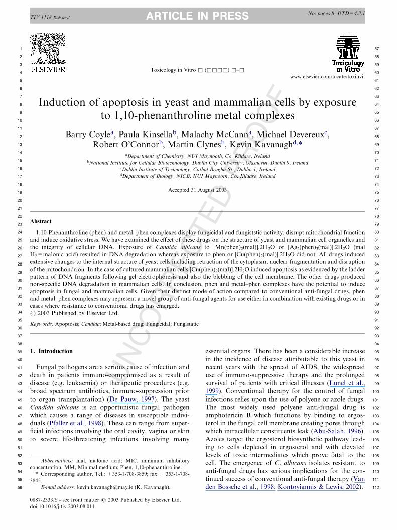
TIV 1118 Disk used No. pages 8, DTD=4.3.1 ARTICLE IN PRESS UNCORRECTED PROOF Induction of apoptosis in yeast and mammalian cells by exposure to 1,10-phenanthroline metal complexes Barry Coyle a , Paula Kinsella b , Malachy McCann a , Michael Devereux c , Robert O’Connor b , Martin Clynes b , Kevin Kavanagh d, * a Department of Chemistry, NUI Maynooth, Co. Kildare, Ireland b National Institute for Cellular Biotechnology, Dublin City University, Glasnevin, Dublin 9, Ireland c Dublin Institute of Technology, Cathal Brugha St., Dublin 1, Ireland d Department of Biology, NICB, NUI Maynooth, Co. Kildare, Ireland Accepted 31 August 2003 Abstract 1,10-Phenanthroline (phen) and metal–phen complexes display fungicidal and fungiststic activity, disrupt mitochondrial function and induce oxidative stress. We have examined the effect of these drugs on the structure of yeast and mammalian cell organelles and the integrity of cellular DNA. Exposure of Candida albicans to [Mn(phen) 2 (mal)].2H 2 O or [Ag 2 (phen) 3 (mal)].2H 2 O (mal H 2 =malonic acid) resulted in DNA degradation whereas exposure to phen or [Cu(phen) 2 (mal)].2H 2 O did not. All drugs induced extensive changes to the internal structure of yeast cells including retraction of the cytoplasm, nuclear fragmentation and disruption of the mitochondrion. In the case of cultured mammalian cells [Cu(phen) 2 (mal)].2H 2 O induced apoptosis as evidenced by the ladder pattern of DNA fragments following gel electrophoresis and also the blebbing of the cell membrane. The other drugs produced non-specific DNA degradation in mammalian cells. In conclusion, phen and metal–phen complexes have the potential to induce apoptosis in fungal and mammalian cells. Given their distinct mode of action compared to conventional anti-fungal drugs, phen and metal–phen complexes may represent a novel group of anti-fungal agents for use either in combination with existing drugs or in cases where resistance to conventional drugs has emerged. # 2003 Published by Elsevier Ltd. Keywords: Apoptosis; Candida; Metal-based drug; Fungicidal; Fungistatic 1. Introduction Fungal pathogens are a serious cause of infection and death in patients immuno-compromised as a result of disease (e.g. leukaemia) or therapeutic procedures (e.g. broad spectrum antibiotics, immuno-suppression prior to organ transplantation) (De Pauw, 1997). The yeast Candida albicans is an opportunistic fungal pathogen which causes a range of diseases in susceptible indivi- duals (Pfaller et al., 1998). These can range from super- ficial infections involving the oral cavity, vagina or skin to severe life-threatening infections involving many essential organs. There has been a considerable increase in the incidence of disease attributable to this yeast in recent years with the spread of AIDS, the widespread use of immuno-suppressive therapy and the prolonged survival of patients with critical illnesses (Lunel et al., 1999). Conventional therapy for the control of fungal infections relies upon the use of polyene or azole drugs. The most widely used polyene anti-fungal drug is amphotericin B which functions by binding to ergos- terol in the fungal cell membrane creating pores through which intracellular constituents leak (Abu-Salah, 1996). Azoles target the ergosterol biosynthetic pathway lead- ing to cells depleted in ergosterol and with elevated levels of toxic intermediates which prove fatal to the cell. The emergence of C. albicans isolates resistant to anti-fungal drugs has serious implications for the con- tinued success of conventional anti-fungal therapy (Van den Bossche et al., 1998; Kontoyiannis & Lewis, 2002). 0887-2333/$ - see front matter # 2003 Published by Elsevier Ltd. doi:10.1016/j.tiv.2003.08.011 Toxicology in Vitro & (&&&&) &–& www.elsevier.com/locate/toxinvit 1 2 3 4 5 6 7 8 9 10 11 12 13 14 15 16 17 18 19 20 21 22 23 24 25 26 27 28 29 30 31 32 33 34 35 36 37 38 39 40 41 42 43 44 45 46 47 48 49 50 51 52 53 54 55 56 57 58 59 60 61 62 63 64 65 66 67 68 69 70 71 72 73 74 75 76 77 78 79 80 81 82 83 84 85 86 87 88 89 90 91 92 93 94 95 96 97 98 99 100 101 102 103 104 105 106 107 108 109 110 111 112 Abbreviations: mal, malonic acid; MIC, minimum inhibitory concentration; MM, Minimal medium; Phen, 1,10-phenanthroline. * Corresponding author. Tel.: +353-1-708-3859; fax: +353-1-708- 3845. E-mail address: [email protected] (K. Kavanagh).
-
Upload
independent -
Category
Documents
-
view
4 -
download
0
Transcript of Induction of apoptosis in yeast and mammalian cells by exposure to 1,10-phenanthroline metal...
No. pages 8, DTD=4.3.1
TIV 1118 Disk used ARTICLE IN PRESSPROOFInduction of apoptosis in yeast and mammalian cells by exposure
to 1,10-phenanthroline metal complexes
Barry Coylea, Paula Kinsellab, Malachy McCanna, Michael Devereuxc,Robert O’Connorb, Martin Clynesb, Kevin Kavanaghd,*
aDepartment of Chemistry, NUI Maynooth, Co. Kildare, IrelandbNational Institute for Cellular Biotechnology, Dublin City University, Glasnevin, Dublin 9, Ireland
cDublin Institute of Technology, Cathal Brugha St., Dublin 1, IrelanddDepartment of Biology, NICB, NUI Maynooth, Co. Kildare, Ireland
Accepted 31 August 2003
RREC
TED
Abstract
1,10-Phenanthroline (phen) and metal–phen complexes display fungicidal and fungiststic activity, disrupt mitochondrial functionand induce oxidative stress. We have examined the effect of these drugs on the structure of yeast and mammalian cell organelles and
the integrity of cellular DNA. Exposure of Candida albicans to [Mn(phen)2(mal)].2H2O or [Ag2(phen)3(mal)].2H2O (malH2=malonic acid) resulted in DNA degradation whereas exposure to phen or [Cu(phen)2(mal)].2H2O did not. All drugs inducedextensive changes to the internal structure of yeast cells including retraction of the cytoplasm, nuclear fragmentation and disruption
of the mitochondrion. In the case of cultured mammalian cells [Cu(phen)2(mal)].2H2O induced apoptosis as evidenced by the ladderpattern of DNA fragments following gel electrophoresis and also the blebbing of the cell membrane. The other drugs producednon-specific DNA degradation in mammalian cells. In conclusion, phen and metal–phen complexes have the potential to induceapoptosis in fungal and mammalian cells. Given their distinct mode of action compared to conventional anti-fungal drugs, phen
and metal–phen complexes may represent a novel group of anti-fungal agents for use either in combination with existing drugs or incases where resistance to conventional drugs has emerged.# 2003 Published by Elsevier Ltd.
OKeywords: Apoptosis; Candida; Metal-based drug; Fungicidal; FungistaticUNC
1. Introduction
Fungal pathogens are a serious cause of infection anddeath in patients immuno-compromised as a result ofdisease (e.g. leukaemia) or therapeutic procedures (e.g.broad spectrum antibiotics, immuno-suppression priorto organ transplantation) (De Pauw, 1997). The yeastCandida albicans is an opportunistic fungal pathogenwhich causes a range of diseases in susceptible indivi-duals (Pfaller et al., 1998). These can range from super-ficial infections involving the oral cavity, vagina or skinto severe life-threatening infections involving many
essential organs. There has been a considerable increasein the incidence of disease attributable to this yeast inrecent years with the spread of AIDS, the widespreaduse of immuno-suppressive therapy and the prolongedsurvival of patients with critical illnesses (Lunel et al.,1999). Conventional therapy for the control of fungalinfections relies upon the use of polyene or azole drugs.The most widely used polyene anti-fungal drug isamphotericin B which functions by binding to ergos-terol in the fungal cell membrane creating pores throughwhich intracellular constituents leak (Abu-Salah, 1996).Azoles target the ergosterol biosynthetic pathway lead-ing to cells depleted in ergosterol and with elevatedlevels of toxic intermediates which prove fatal to thecell. The emergence of C. albicans isolates resistant toanti-fungal drugs has serious implications for the con-tinued success of conventional anti-fungal therapy (Vanden Bossche et al., 1998; Kontoyiannis & Lewis, 2002).
0887-2333/$ - see front matter # 2003 Published by Elsevier Ltd.
doi:10.1016/j.tiv.2003.08.011
Toxicology in Vitro & (&&&&) &–&www.elsevier.com/locate/toxinvit
1
2
3
4
5
6
7
8
9
10
11
12
13
14
15
16
17
18
19
20
21
22
23
24
25
26
27
28
29
30
31
32
33
34
35
36
37
38
39
40
41
42
43
44
45
46
47
48
49
50
51
52
53
54
55
56
57
58
59
60
61
62
63
64
65
66
67
68
69
70
71
72
73
74
75
76
77
78
79
80
81
82
83
84
85
86
87
88
89
90
91
92
93
94
95
96
97
98
99
100
101
102
103
104
105
106
107
108
109
110
111
112
Abbreviations: mal, malonic acid; MIC, minimum inhibitory
concentration; MM, Minimal medium; Phen, 1,10-phenanthroline.
* Corresponding author. Tel.: +353-1-708-3859; fax: +353-1-708-
3845.
E-mail address: [email protected] (K. Kavanagh).

No. pages 8, DTD=4.3.1
TIV 1118 Disk used ARTICLE IN PRESSC
UNCORRE
Metal-based drugs represent a novel group of anti-fungal agents with potential applications for the controlof fungal infections. 1,10-Phenanthroline (phen) andsubstituted derivatives, both in the metal-free state andas ligands co-ordinated to transition metals, disturb thefunctioning of a wide variety of biological systems(Butler et al., 1969). Furthermore, when the metal-freeN,N-chelating bases are found to be bioactive it isusually assumed that the sequestering of trace metals isinvolved, and that the resulting metal complexes are theactive species (MacLeod, 1952; Dwyer et al., 1969) Pre-vious work has demonstrated that in RPMI medium at37 �C the metal-based drugs [Cu(phen)2(mal)].2H2O,[Mn(phen)2(mal)].2H2O and [Ag2(phen)3(mal)].2H2O(phen=1,10-phenanthroline; malH2=malonic acid)inhibit the growth of C. albicans by around 95% at aconcentration of 5 mg/ml (Coyle et al., 2003). It wasestablished that both metal-free phen and the metal–phen complexes affect mitochondrial function, retardthe synthesis of cytochromes b and c and uncouplerespiration. Treatment of fungal cells with the Cu(II)and Ag(I) complexes resulted in a reduced amount ofergosterol in the cell membrane and subsequent increasein its permeability. Cells exposed to metal-free phen andthe Cu(II) and Mn(II) complexes [but not the Ag(I)complex] demonstrated an elevation in oxygen uptake.Indeed, part of the mode of action of this group ofdrugs seems to lie in their ability to induce oxidativestress within the cell as evidenced by the decreasedreduced:oxidized glutathione ratios (GSH:GSSG) andincreased levels of lipid peroxides in Candida cells trea-ted with [Cu(phen)2(mal)].2H2O (McCann et al., 2000).The aim of the work presented here was to further
characterise the mode of action of these drugs in termsof their effects on the morphology of fungal and mam-malian cells. Due to their different mode of actioncompared to the polyene and azole anti-fungal drugs(Coyle et al., 2003), metal based drugs may represent anovel group of anti-fungal agents with potential appli-cations either alone or in combination with conven-tional anti-fungals. In addition, they may be applicablein situations where resistance to conventional anti-fungaldrugs has emerged.
2. Materials and methods
2.1. Fungal strain and culture conditions
C. albicans ATCC 10231 was obtained form theAmerican Type Culture Collection, (VA, USA). Cul-tures were grown on Sabouraud dextrose agar (SDA)plates at 37 �C and maintained at 4 �C for short-termstorage. Cultures were routinely sub-cultured every 4–6weeks. Cultures were grown to the stationary phase(approximately 1�108/ml) overnight at 30 �C and 200
TEDPR
OOF
rpm in minimal medium (MM) [2% w/v glucose, 0.5%w/v yeast nitrogen base (without amino acids orammonium sulphate), 0.5% w/v ammonium sulphate].
2.2. Human cell culture
The HEp-2 cell line (ATCC CCL23) was obtainedfrom the American Type Culture Collection (VA, USA)and cells were grown in MEM (Sigma Aldrich ChemicalCo., Dorset, UK) supplemented with 5% v/v foetal calfserum (Gibco, Paisley, UK), 4 mM l-glutamine and 1%v/v Penn-Strep (Sigma Adrich). The DLKP cell line wasobtained from the National Cell and Tissue CultureCentre (Dublin, Ireland) and is derived from a poorlydifferentiated cell carcinoma from a lymph node meta-stasis of a primary lung tumour. DLKP cells were cul-tured under the same conditions as HEp-2 cells.Adherent cells were grown in 80 cm2 culture flasks at37 �C and 5% CO2 in a humidified atmosphere and sub-cultured by trypsinisation every 3–4 days.
2.3. Drugs
Chemicals were obtained from commercial sourcesand used without further purification. [Cu(phen)2(mal)].2H2O, [Mn(phen)2(mal)].2H2O and [Ag2(phen)3(mal)].2H2O were prepared as previously described(McCann et al., 2000).
2.4. In vitro toxicity testing
Sub-confluent DLKP cells were harvested by trypsi-nisation washed and resuspended in PBS. Cells wereenumerated microscopically and diluted with MEM togive a final cell density of 2�104/ml. Ninety-six-wellplates (NUNC) were seeded with 100 ml of this suspen-sion per well and incubated at 37 �C and 5% CO2 in ahumidified atmosphere for 24 h to allow cell attach-ment. Subsequently, a range of concentrations of metalbased drug was added to the rows of wells and theplates were re-incubated until controls reached 80–90%confluency (typically 5–6 days). Cell growth in toxicityassays was quantified as described previously (Martin &Clynes, 1993).
2.5. Extraction of DNA from C. albicans
Yeast cells were grown in the presence of drug (10 mg/ml) to the late exponential phase (18–24 h) in MM at30 �C in an orbital incubator. Cells were harvested bycentrifugation and washed with 1 mm EDTA. Cells wereresuspended in spheroplasting buffer (1 M sorbitol, 0.1M EDTA, 6 mg/ml lyticase and 0.05 M dithiotheritol,pH 7.5) and incubated at 37 �C for 2 h. Spheroplastswere harvested by centrifugation, resuspended in lysingbuffer (50 mm EDTA, 50 mm Tris (pH 8), 1% w/v SDS
2 B. Coyle et al. / Toxicology in Vitro& (&&&&)&–&
1
2
3
4
5
6
7
8
9
10
11
12
13
14
15
16
17
18
19
20
21
22
23
24
25
26
27
28
29
30
31
32
33
34
35
36
37
38
39
40
41
42
43
44
45
46
47
48
49
50
51
52
53
54
55
56
57
58
59
60
61
62
63
64
65
66
67
68
69
70
71
72
73
74
75
76
77
78
79
80
81
82
83
84
85
86
87
88
89
90
91
92
93
94
95
96
97
98
99
100
101
102
103
104
105
106
107
108
109
110
111
112

No. pages 8, DTD=4.3.1
TIV 1118 Disk used ARTICLE IN PRESSC
UNCORRE
and 8 mg/ml proteinase K) and incubated at 65 �C for0.5 h. DNA was precipitated with two volumes of chil-led ethanol (95% v/v) and incubated at �20 �C over-night. DNA was harvested by centrifugation, washedwith ethanol (20% v/v), harvested by centrifugation at4000�g for 25 min and allowed air dry. The DNA wasresuspended in TE buffer, 1 mg/ml RNase and incu-bated at 37 �C for 0.5 h. Ethanol (95% v/v) and 3 m
sodium acetate solution (pH 5.2) was added and thesample stored at �20 �C overnight.
2.6. Extraction of DNA from cultured human cells
Sub-confluent HEp-2 cells that had been cultured inthe presence of 3 mm of metal-based drug for 24 h wereharvested by trypsinisation, washed and resuspended inPBS. Cells (1�106) were resuspended in lysis buffer [20mm EDTA, 0.8% (w/v) sodium lauryl sarcosiate, 100mm Tris (pH 8.0)] and 10 mg/ml RNase (BoehringerManneheim, Sussex, UK) and incubated at 37 �C for 18h. Proteinase K (1 mg/ml) was subsequently added andthe samples were incubated for a further 2 h at 50 �C.
2.7. DNA gel electrophoresis
Purity and concentration of DNA was determined byUV spectroscopy (260–280 nm). DNA from yeast andmammalian cells was run at a concentration of 40 mg/mlon a 0.8% (w/v) agarose gel at 40 V for 18 h. Followingstaining with ethidium bromide DNA was visualisedusing a UV transilluminator.
2.8. Electron microscopy
Primary fixation of stationary phase yeast cells was ina 3% solution of gluteraldehyde in 0.1 m phosphatebuffer for 2 h. Secondary fixation was in a 2% solutionof osmium tetroxide in 0.1 m phosphate buffer for 1 h.Dehydration of samples was in an alcohol series of 10,30, 50, 75, 95 and 100%, each for 15 min. Sampleswere embedded in Agar 100 resin (Agar Scientific Ltd.,UK) and viewed using a Hitachi H-7000 TransmissionElectron Microscope operating at 100 kv acceleratingvoltage.
3. Results
3.1. Determination of effect of metal-based drugs onyeast cell DNA
The aim of the work presented here was to establishwhether drug-induced oxidative stress altered the struc-ture of the cellular organelles and affected the integrityof yeast DNA. The minimum inhibitory concentrationof each drug was used as determined previously (Coyle
TEDPR
OOF
et al., 2003). Cells of C. albicans were grown to the sta-tionary phase in MM containing 10 mg/ml of each drugfor 24 h. Cells were harvested by centrifugation, theDNA was extracted as described and visualised by ethi-dium bromide staining following agarose gel electro-phoresis. The DNA extracted from yeast cells exposedto [Ag2(phen)3(mal)].2H2O shows extensive degradation(Fig. 1). Smaller amounts of degradation, as demon-strated by smearing, are also visible in cells treated with[Cu(phen)2(mal)].2H2O and [Mn(phen)2(mal)].2H2O,whereas incubation of cells with metal-free phen causeslittle or no DNA breakdown under the conditionsemployed here.
3.2. Electron microscopic examination of C. albicans fol-lowing growth in the presence of metal-based drugs
Cultures of C. albicans were grown to the stationaryphase overnight in MM medium at 30 �C and 200 rpmin the presence of each drug at a final concentration of10 mg/ml. Cells were harvested by centrifugation,washed with PBS (pH 7.2) and placed on ice prior topreparation for TEM examination (as described). Cellsgrown in the absence of drug showed normal cellularmorphology with a distinct cell wall, an intact nucleusand numerous membranous organelles (Fig. 2a). Incontrast, cells grown in the presence of metal-free phendemonstrated a distended cell wall, ruptured internalorganelles and the withdrawal of the cytoplasmic mem-brane from within the cell wall (Fig. 2b). Cells treatedwith [Ag2(phen)3(mal)].2H2O possessed a distended cellwall, ruptured organelles and, in some cases, a frag-mented nucleus (Fig. 2c). The most obvious feature of[Cu(phen)2(mal)].2H2O-treated cells was the occurrence
Fig. 1. DNA banding pattern of C. albicans cells treated with phen
and metal-phen complexes for 24 h. Lane 1: DNA from control cells,
Lane 2: phen, Lane 3: [Cu(phen)2(mal)].2H2O, lane 4: [Mn(phen)2(-
mal)].2H2O, Lane 5: [Ag2(phen)3(mal)].2H2O.
B. Coyle et al. / Toxicology in Vitro& (&&&&)&–& 3
1
2
3
4
5
6
7
8
9
10
11
12
13
14
15
16
17
18
19
20
21
22
23
24
25
26
27
28
29
30
31
32
33
34
35
36
37
38
39
40
41
42
43
44
45
46
47
48
49
50
51
52
53
54
55
56
57
58
59
60
61
62
63
64
65
66
67
68
69
70
71
72
73
74
75
76
77
78
79
80
81
82
83
84
85
86
87
88
89
90
91
92
93
94
95
96
97
98
99
100
101
102
103
104
105
106
107
108
109
110
111
112

No. pages 8, DTD=4.3.1
TIV 1118 Disk used ARTICLE IN PRESSof an enlarged nucleus which, in some cells, was crescentshaped (Fig. 2d). With this drug the internal organellesappeared intact but some shrinkage of the cytoplasmwithin the cells was apparent. Cells exposed to[Mn(phen)2(mal)].2H2O had, in most cases, completelydisrupted organelles. Some of these cells possessedenlarged nuclei while others appeared to contain distinctnuclear fragments (Fig. 2e).
F3.3. Effect of metal-based drugs on integrity of mamma-lian DNA
Previous studies into the effect of metal-phen com-plexes on fungal cell viability indicated that the drugsdisrupt mitochondrial function (Coyle et al., 2003). Invitro toxicity assays were performed to establish theconcentration of 1,10-phen and [Cu(phen)2(mal)].2H2O
O
UNCORREC
TEDPR
O
Fig. 2. Electron-micrographs of C. albicans cells exposed to phen and metal-phen complexes for 24 h. (a) Control, (b) Phen, (c) [Ag2(phen)3(mal)].2H2O, (d) [Cu(phen)2(mal)].2H2O, (e) [Mn(phen)2(mal)].2H2O. (Bar=1 mM).
4 B. Coyle et al. / Toxicology in Vitro& (&&&&)&–&
1
2
3
4
5
6
7
8
9
10
11
12
13
14
15
16
17
18
19
20
21
22
23
24
25
26
27
28
29
30
31
32
33
34
35
36
37
38
39
40
41
42
43
44
45
46
47
48
49
50
51
52
53
54
55
56
57
58
59
60
61
62
63
64
65
66
67
68
69
70
71
72
73
74
75
76
77
78
79
80
81
82
83
84
85
86
87
88
89
90
91
92
93
94
95
96
97
98
99
100
101
102
103
104
105
106
107
108
109
110
111
112

No. pages 8, DTD=4.3.1
TIV 1118 Disk used ARTICLE IN PRESSC
O
ORRE
capable of killing cultured human cells. The resultsindicated IC50 values of 0.002 mg/ml and 0.001 mg/ml foreach drug, respectively against DLKP cells.HEp-2 cells were chosen to determine whether the
metal–phen complexes induced apoptosis since theappearance of the DNA fragmentation pattern asso-ciated with this mode of cell death (Verhaegen, 1998) iseasier to visualise in this cell line than in the DLKP line.HEp-2 cells were cultured for 24 h in the presence of 3mm of metal-free phen or the Cu(II), Mn(II) and Ag(I)phen complexes, and the DNA was subsequentlyextracted and separated by agarose gel electrophoresis.Degradation of high molecular weight DNA was evi-dent in those cultures treated with Cu(II), Mn(II) andAg(I) phen. Cells treated with metal-free phen alsoshowed extensive degradation of DNA. Treatment ofcells with the copper–phen complex produced a DNApattern which was divided into distinct fragments. Theappearance of this DNA fragmentation ‘ladder’ is indi-cative of apoptosis (or programmed cell death) occur-ring in the cells in response to the metal based drug.Cultures treated with Mn or Ag complexes demon-strated DNA degradation but did not produce a specific‘ladder’ fragmentation pattern at this concentration.
3.4. Microscopic examination of cultured human cellsgrown in the presence of [Cu(phen)2(mal)].2H2O
Previous work has established that the metal-phencomplexes affect the oxygen uptake rate of C. albicansand induced oxidative stress within the fungal cell(McCann et al. 2000; Coyle et al., 2003). We sought todetermine whether these complexes affected the growthand cellular morphology of cultured mammalian cells inorder to assess their toxicity in vitro towards cellsderived from human tissue.In order to examine the morphological changes taking
place in cultured cells following exposure to the most
COF
toxic of the metal–phen complexes sub-confluent DLKPcells were exposed to [Cu(phen)2(mal)].2H2O (3mM) andincubated at 37 �C for 48 h. Untreated cells show thetypical morphology of adherent epithelial cells (Fig. 4A).Exposure to [Cu(phen)2(mal)].2H2O for 24 h producedcells showing evidence of membrane ‘blebbing’(Fig. 4b)— a feature of cell death by apoptosis (Ver-haegen, 1998). After 48 h incubation a number of cellshad fragmented to give clusters of small sub-cellularpackets (Fig. 4c)— tentatively identified as apoptoticbodies, a feature of the latter stages of apoptosis (Ver-haegen, 1998).
TEDPR
4. Discussion
The in vitro antibacterial action of phen has beendemonstrated on several species of bacteria (Dwyer etal., 1969; Feeney et al., 1957). Whereas metal–phencomplexes can be bacteristatic (Dwyer et al., 1969) andbactericidal (Butler et al., 1969) towards many Gram-positive bacteria they are relatively ineffective againstGram-negative organisms. In addition, dilute aqueoussolutions of phen and its Cu(II) and Mn(II) complexeswere highly toxic to clinical isolates of Candida species(Geraghty et al., 2000; Geraghty et al., 1998).Earlier in vitro experiments in our laboratories have
shown that phen and a number of transition metalcomplexes incorporating this chelating ligand are extre-mely active anti-fungal drugs (Geraghty et al.,1999a,b,c; Devereux et al., 2000a,b; McCann et al.,2000; Geraghty et al., 2000). The compounds haveminimum inhibitory concentrations in the range 1.25–5.0 mg/ml and, at a concentration of 10 mg/ml, displaysome fungicidal activity. Treating exponential and sta-tionary phase yeast cells with phen and the Cu(II) andMn(II) complexes induces a dramatic increase in oxygenconsumption. All of the drugs cause reductions in thelevels of cytochromes b and c in the cells, while theAg(I) complex also lowers the amount of cytochromeaa3. Cells treated with phen and the Cu(II) and Ag(I)species show reduced levels of ergosterol while theMn(II) complex induces an increase in the sterol con-tent. Extensive studies with [Cu(phen)2(mal)].2H2Oindicated that this drug induces significant cellular oxi-dative stress (decreased reduced:oxidized glutathioneratios (GSH:GSSG) and increased levels of lipid per-oxides). Furthermore, as the drugs were not uniformlyactive this suggested that their bioactivity has a degreeof metal-ion dependency. The drugs disrupt mitochon-drial function, uncouple respiration and promote oxi-dative stress in the organism (Coyle et al., 2003). Assuch, phen and the metal–phen complexes may repre-sent a novel set of highly active anti-fungal agentswhose mode of action is significantly different to that ofthe polyene and azole prescription drugs.
UN
Fig. 3. DNA banding pattern from HEp-2 cells exposed to phen and
metal–phen complexes for 24 h. Lane 1: molecular weight standards,
Lane 2: control, Lane 3: phen, Lane 4: [Cu(phen)2(mal)].2H2O, lane 5:
[Mn(phen)2(mal)].2H2O, Lane 6: [Ag2(phen)3(mal)].2H2O.
B. Coyle et al. / Toxicology in Vitro& (&&&&)&–& 5
1
2
3
4
5
6
7
8
9
10
11
12
13
14
15
16
17
18
19
20
21
22
23
24
25
26
27
28
29
30
31
32
33
34
35
36
37
38
39
40
41
42
43
44
45
46
47
48
49
50
51
52
53
54
55
56
57
58
59
60
61
62
63
64
65
66
67
68
69
70
71
72
73
74
75
76
77
78
79
80
81
82
83
84
85
86
87
88
89
90
91
92
93
94
95
96
97
98
99
100
101
102
103
104
105
106
107
108
109
110
111
112

No. pages 8, DTD=4.3.1
TIV 1118 Disk used ARTICLE IN PRESSThe work presented here is a progression of the abovemechanistic studies and examines the effects of themetal–phen complexes on the integrity of DNA andcellular morphology of C. albicans and cultured mam-malian cells. Exposing C. albicans to [Ag2(phen)3(mal)].2H2O or [Mn(phen)2(mal)].2H2O leads to non-specific DNA cleavage. In the case of the Mn(II) com-plex this may be as a result of oxidative damage to thecells caused by elevated levels of oxygen uptake (Coyleet al., 2003). The Cu(II) complex and metal-free phenseem to have little effect on fungal DNA. This latterfinding is surprising since phen and copper(I)–phencomplexes are known to cleave DNA with the samepreferences as micrococcal nuclease (Jessee et al., 1982;Chen & Sigman, 1986). It is possible that either theconcentration used was not optimal to induce fungalcell DNA degradation or that the cells were capable ofrepairing the damage over the timeframe of the experi-ment. In addition, it is possible that the oxidative stressinduced by these compounds can inhibit Caspase activ-ity which is sensitive to the redox balance within the cell(Green & Kroemer, 1998).Electron-micrographic examination of fungal cells
exposed to phen and the metal–phen complexes revealssevere disruption of internal cellular structures (Fig. 2a–e).
C
TEDPR
OOF
In particular, nuclear disruption is evident followingexposure to [Ag2(phen)3(mal)].2H2O [Mn(phen)2(mal)].2H2O and [Cu(phen)2(mal)].2H2O. This is con-sistent with the cleavage of fungal DNA evident inFig. 1. Nuclear fragmentation is characteristics ofapoptosis as is nuclear ‘crescent’ formation evident inmany cells (Cohen, 1993). Apoptosis in fungal cells fol-lows many of the same steps evident in animal cellsincluding fragmentation of the nucleus, degradation ofDNA and disruption of internal organelles (Roze &Linz, 1998). Discrete apoptotic bodies are not formed.Exposure of mammalian cells to [Cu(phen)2
(mal)].2H2O at a concentration of 0.003 mg/ml results ina DNA fragmentation pattern which is characteristic ofcells dying by apoptosis (Cohen, 1993; Verhaegen, 1998).In this mode of cell death, the injured cell plays an activerole in its own demise and one part of the process is thecleavage of nuclear DNA into specific sized fragments byan endonuclease giving rise to a ‘ladder’ pattern of frag-ments upon gel electrophoresis (Cohen, 1993; Cotter andAl-Rubeai, 1995). Copper–phen complexes have pre-viously been shown to induce apoptosis in a range of celllines (Zhou et al., 2002 a,b; De Vizcaya-Ruiz et al., 2002).Exposure of cultured mammalian cells to metal-free phen,[Ag2(phen)3(mal)].2H2O or [Mn(phen)2(mal)].2H2O did
UNCORRE
Fig. 4. Micrographs of DLKP cells cultured in the presence of [Cu(phen)2(mal)].2H2O. (a) Cells showing membrane ‘blebbing’ after 24 h exposure,
(b) Apoptotic bodies after 48 h exposure, (c) Dead and detached cells after 96h exposure. (MB: membrane blebbing; AB: Apoptotic bodies). (Ori-
ginal magnification: (a) and (b) �400, (c) �100).
6 B. Coyle et al. / Toxicology in Vitro& (&&&&)&–&
1
2
3
4
5
6
7
8
9
10
11
12
13
14
15
16
17
18
19
20
21
22
23
24
25
26
27
28
29
30
31
32
33
34
35
36
37
38
39
40
41
42
43
44
45
46
47
48
49
50
51
52
53
54
55
56
57
58
59
60
61
62
63
64
65
66
67
68
69
70
71
72
73
74
75
76
77
78
79
80
81
82
83
84
85
86
87
88
89
90
91
92
93
94
95
96
97
98
99
100
101
102
103
104
105
106
107
108
109
110
111
112

No. pages 8, DTD=4.3.1
TIV 1118 Disk used ARTICLE IN PRESSC
P
UNCORRE
not give rise to a specific DNA fragmentation ladderpattern but extensive non-specific DNA fragmentationis visible. From this it is possible to conclude that[Cu(phen)2(mal)].2H2O activates mammalian cell deathby apoptosis and that the other drugs induce non-spe-cific cleavage of DNA possibly due to the nuclease-likeactivity of the phen ligand (Jessee et al., 1982; Chen andSigman, 1986).Examination of cultured mammalian cells exposed to
[Cu(phen)2(mal)].2H2O for 24 h show cells undergoingmembrane ‘blebbing’. After 48 h a number of cells wereobserved to have undergone fragmentation to yieldstructures tentatively identified as ‘apoptotic bodies’(Fig. 4). These findings and the DNA degradation pat-tern observed previously with this drug (Fig. 3) areconsistent with the induction of cell death by apoptosis(Verhaegen, 1998). A similar finding was made by Viz-caya-Ruiz et al. (2000) who demonstrated the ability ofcopper based anti-cancer drugs to induce apoptosis inhuman ovarian carcinoma cells. In that case the induc-tion of apoptosis was monitored by changes in cellmorphology, activation of caspases and the degradationof DNA to give a ladder pattern of fragments. Copper–1,10-phenanthroline complexes have been shown toinduce G1-phase specific apoptosis in a liver carcinomacell line (Zhou et al., 2002a,b), further supporting theview that the [Cu(phen)2(mal)].2H2O employed in thiswork is capable of activating cell death by apoptosis.Copper chelators have been shown to accumulate cop-per within thymocytes which can trigger oxidative-stressinduced apoptosis (Nobel et al., 1995).In conclusion, the metal-phen complexes examined
here have a potent anti-fungal effect being capable ofinhibiting growth of C. albicans by 95% at a concen-tration of 5 mg/ml (Coyle et al., 2003). Yeast and mam-malian cells exposed to these complexes at aconcentration of 10 mg/ml show DNA cleavage. In thecase of mammalian cells [Cu(phen)2(mal)].2H2O inducesa DNA fragmentation pattern indicative of apoptosis.TEM examination of yeast cells reveals gross distortionof cellular structures and nuclear fragmentation. Thiswork indicates that, in addition to its effect on mito-chondrial function and oxygen uptake (Coyle et al.,2003), [Cu(phen)2(mal)].2H2O also plays a role in indu-cing cell death by apoptosis in yeast and mammaliancells. The mitochondrion plays a central role in govern-ing the induction of apoptosis (Green & Kroemer, 1998)and the drugs examined here, particularly [Cu(phen)2(mal)].2H2O, interfere with mitochondrial function(Coyle et al., 2003) and integrity which may be sufficientto push the cell towards apoptotic cell death. Whetheror not apoptosis is a direct effect of exposure to themetal based drugs or is related to an effect on the mito-chondrion induced by them (increased oxygen uptake,disruption of cytochrome synthesis (Coyle et al., 2003)is currently being investigated.
OF
The conventional polyene and azole anti-fungal drugstarget ergosterol in the fungal cell membrane or inhibitergosterol biosynthesis, respectively. We have demon-strated that the metal–phen complexes examined herehave a distinct mode of action and may represent anovel group of anti-fungal agents to be used alone or incombination with existing anti-fungal drugs. In addi-tion, metal-based drugs may offer the possibility ofover-coming the emerging problem of resistance toconventional anti-fungal drugs (Van den Bossche et al.,1998; Kontoyiannis & Lewis, 2002).
RO
Uncited references
Brandt, 1954; McNaught and Owen, 1949 and Tur-ian, 1951 are not cited in the text.
TEDReferences
Abu-Salah, K.M., 1996. Amphotericin B: an update. British Journal
of Biomedical Science 53, 122–133.
Butler, H.M., Hurse, A., Thursky, E., Shulman, A., 1969. Bactericidal
action of selected phenanthroline chelates and related compounds.
Australian Journal of Experimental Biological and Medical Science
47, 541–552.
Brandt, W.W., Dwyer, F.P., Gyarfas, E.C., 1954. Chelate complexes
of 1,10-phenanthroline and related compounds. Chemical Reviews
54, 959–1017.
Chen, C.H., Sigman, D.S., 1986. Nuclease activity of 1,10 phenan-
throline-copper: sequence specific targeting. Proceedings of the
National Academy of Science 83, 7147–7151.
Cohen, J.J., 1993. Overview: mechanisms of apoptosis. Immunology
Today 14, 126–130.
Cotter, T.G., Al-Rubeai, M., 1995. Cell death (apoptosis) in cell cul-
ture systems. Trends in Biotechnology 13, 150–155.
Coyle, B., Kavanagh, K., McCann, M., Devereux, M., Geraghty, M.,
2003. Mode of anti-fungal activity of 1,10-phenanthroline and its
Cu(II), Mn(II) and Ag(I) complexes. BioMetals. 16, 321–329.
De Pauw, B.E., 1997. Practical modalities for prevention of fungal
infections in cancer patients. European Journal of Clinical Micro-
biology and Infectious Disease 16, 32–41.
De Vizcaya-Ruiz, A., Riverro-Muller, A., Ruiz-Ramirez, L., Kass,
G.N., Kelland, L.R., Orr, R.M., Dobrota, M., 2000. Induction of
apoptosis by a novel copper-based anticancer compound, Casiope-
nia II, in L1210 murine leukaemia and CH1 human ovarian carci-
noma cell. Toxicology in vitro 14, 1–5.
Devereux, M., McCann, M., Leon, V., Geraghty, M., McKee, V.,
Wikaira, J., 2000a. Synthesis and fungitoxic activity of mangane-
se(II) complexes of fumaric acid: X-ray crystal structures of
[Mn(fum)(bipy)(H2O)] and [Mn(phen)2(H2O)2](fum).4H2O
(fumH2=fumaric acid; bipy=2,20-bipyridine; phen=1,10-phenan-
throline). Polyhedron 19, 1205–1211.
Devereux, M., McCann, M., Leon, V., Geraghty, M., McKee, V.,
Wikaira, J., 2000b. Synthesis and biological activity of mangane-
se(II) complexes of phthalic and isophthalic acid: X-ray crystal
structures of [Mn(ph)(Phen)2(H2O)].4H2O, [Mn(Phen)2(H2O)2]2(I-
soph)2(Phen).12H2O and {[Mn(Isoph)(bipy)]4.2.75bipy}n (phH2=
phthalic acid; Isoph=isophthalic acid; Phen=1,10-phenanthroline;
bipy=2,2-bipyridine). Metal-Based Drugs 7, 275–288.
Dwyer, F.P., Reid, I.K., Shulman, A., Laycock, G.M., Dixon, S.,
1969. The biological actions of 1,10-phenanthroline and 2,20-bipyr-
B. Coyle et al. / Toxicology in Vitro& (&&&&)&–& 7
1
2
3
4
5
6
7
8
9
10
11
12
13
14
15
16
17
18
19
20
21
22
23
24
25
26
27
28
29
30
31
32
33
34
35
36
37
38
39
40
41
42
43
44
45
46
47
48
49
50
51
52
53
54
55
56
57
58
59
60
61
62
63
64
65
66
67
68
69
70
71
72
73
74
75
76
77
78
79
80
81
82
83
84
85
86
87
88
89
90
91
92
93
94
95
96
97
98
99
100
101
102
103
104
105
106
107
108
109
110
111
112

No. pages 8, DTD=4.3.1
TIV 1118 Disk used ARTICLE IN PRESSC
ORREidine hydrochlorides, quaternary salts and metal chelates and rela-
ted compounds. 1. Bacteriostatic action on selected gram-positive,
gram-negative and acid-fast bacteria. Australian Journal of Experi-
mental and Biological Medical Science 47, 203–218.
Feeney, R.E., Petersen, I.M., Sahinkaya, H., 1957. ‘‘Liesegang-like’’
rings of growth and inhibition of bacteria in agar caused by metal
ions and chelating agents. Journal of Bacteriology 73, 284–290.
Geraghty, M., McCann, M., Devereux, M., Cronin, J.F., Curran, M.,
McKee, V., 1999a. Synthesis and anti-Candida activity of cobalt(II)
complexes of benzene-1,2-dioxyacetic acid (bdoaH2). X-ray crystal
structures of [Co(bdoa)(H2O)3]3.5H2O and {[Co(phen)3]
(bdoa)2}2.24H2O (phen=1,10-phenanthroline). Metal-Based Drugs
6, 41–48.
Geraghty, M., Sheridan, V., McCann, M., Devereux, M., 1999b.
Synthesis and anti-Candida activity of copper(II) and manganese(II)
carboxylate complexes: X-ray crystal structures of [Cu(sal)(bi-
py)].C2H5OH.H2O and [Cu(norb)(phen)2]6.5H2O.(salH2=salicylic
acid; norbH2=cis-5-norbornene-endo-2,3-dicarboxylic acid; bipy
=2,20-bipyridine; phen=1,10-phenanthroline). Polyhedron 18,
2931–2939.
Geraghty, M., McCann, M., Devereux, M., McKee, V., 1999c.
Synthesis and anti-Candida activity of cobalt(II) complexes of octa-
nedioic acid (odaH2) and nonanedioic acid (ndaH2): X-ray crystal
structures of [Co(phen)3]oda.14H2O and [Co(phen)3]nda.11.5H2O
(phen=1,10-phenanthroline). Inorganic Chimica Acta 293, 160–
166.
Geraghty, M., Cronin, J.F., Devereux, M., McCann, M., 2000.
Synthesis and anti-microbial activity of copper(II) and manga-
nese(II) a,o-dicarboxylate complexes. BioMetals 13, 1–8.Geraghty, M., Cronin, J.F., Devereux, M., McCann, M., 1998.
Activity of Copper(II) and Manganese(II) Carboxylate Complexes:
X-Ray Crystal Structures of [Cu(sal)(bipy)].C2H5OH.H2O and
[Cu(norb)(phen)2].6.5H2O (salH2=salicylic acid; norbH2=cis-5-
norbornene-endo-2,3-dicarboxylic acid; bipy=2,20-bipyridine;
phen=1,10-phenanthroline). Polyhedron 18, 2931–2939.
Green, D., Kroemer, G., 1998. The central executioners of apoptosis:
caspases or mitochondria? Trends in Cell Biology 8, 267–271.
Jessee, B., Gargiulo, G., Razvi, F., Worcel, A., 1982. Analogous clea-
vage of DNA by micrococcal nuclease and a 1,10-phenanthroline-
cuprous complex. Nucleic Acid Research 10, 5823–5833.
Lunel, F.M., Meis, F.G., Voss, A., 1999. Nosocomial fungal infec-
tions: Candidemia. Diagnostic Microbiology and Infectious Dis-
eases 34, 213–220.
UNC
TEDPR
OOF
Kontoyiannis, D.P., Lewis, R.E., 2002. Antifungal drug resistance in
pathogenic fungi. Lancet 359, 1135–1143.
Martin, A., Clynes, M., 1993. Comparison of 5 microtitre colormetric
assays for in vitro cytotoxicity testing and cell proliferation assays.
Cytotechnology 11, 49–58.
McCann, M., Geraghty, M., Devereux, M., O’Shea, D., Mason, J.,
O’Sullivan, L., 2000. Insights into the mode of action of the anti-
Candida activity of 1,10-phenanthroline and its metal chelates.
Metal-Based Drugs 7, 185–193.
MacLeod, R.A., 1952. The toxicity of o-phenanthroline for lactic acid
bacteria. Journal of Biological Chemistry 197, 751–761.
McNaught, M.L. and Owen, E.C. (1949). Metals and rumen bacteria.
In: Int. Congr. Biochem., Abstr. of Communs., 1st Congr., Cam-
bridge, UK, pp. 340–341.
Nobel, C., Kimland, M., Lind, B., Orrenius, S., Slater, A.F., 1995.
Dithiocarbamates induce apoptosis in thymocytes by raising the
intracellular level of redox-active copper. Journal of Biological
Chemistry 270, 26202–26208.
Pfaller, M.A., Jones, R.N., Messer, S.A., Edmond, M.B., Wenzel,
R.P., 1998. National surveillance of nosocomial blood stream
infection due to Candida albicans: frequency of occurrence and
anti-fungal susceptibility in the SCOPE programme. Diagnostic
Microbial Infectious Disease 31, 327–332.
Roze, L.V., Linz, J.E., 1998. Lovastatin triggers an apoptosis-like
death process in the fungus Mucor recemosus. Fungal Genetics
Biology 25, 119–133.
Turian, G., 1951. Tuberculostatic action of o-phenanthroline.
Schweiz. Z. allgem. Pathol. U. Bakteriol 14, 338–344.
Van den Bossche, H., Dromer, F., Improvissi, I., Lozane-Chiu, M.,
Rex, J.H., Sanglard, D., 1998. Anti-fungal drug resistance in
pathogenic fungi. Medical Mycology 36 (Supp. 1), 119–128.
Verhaegen, S., 1998. Microscopical study of cell death via apoptosis.
Microscopy Analysis 1, 5–7.
Zhou, H., Zheng, C., Zou, G., Tao, D., Gong, J., 2002a. G1-phase
specific apoptosis in liver carcinoma cell line induced by copper-
1,10-phenanthroline. International Journal of Biochemistry and Cell
Biology 34, 678–684.
Zhou, H., Liu, Y., Zheng, C., Gong, J., Liang, Y., Wang, C., Zou, G.,
2002b. Microcalorimetric studies of the synergistic effects of copper-
1,10-phenanthroline combined with hyperthermia on a liver hepa-
toma cell line Bel-7402. Thermochimica Acta 7149, 1–9.
8 B. Coyle et al. / Toxicology in Vitro& (&&&&)&–&
1
2
3
4
5
6
7
8
9
10
11
12
13
14
15
16
17
18
19
20
21
22
23
24
25
26
27
28
29
30
31
32
33
34
35
36
37
38
39
40
41
42
43
44
45
46
47
48
49
50
51
52
53
54
55
56
57
58
59
60
61
62
63
64
65
66
67
68
69
70
71
72
73
74
75
76
77
78
79
80
81
82
83
84
85
86
87
88
89
90
91
92
93
94
95
96
97
98
99
100
101
102
103
104
105
106
107
108
109
110
111
112