Indomethacin can downregulate the levels of inflammatory mediators in the hippocampus of rats...
6
Indomethacin can downregulate the levels of inflam- matory mediators in the hippocampus of rats sub- mitted to pilocarpine-induced status epilepticus Michele Juliane Vieira, I,II Sandra Regina Perosa, I Gustavo Adolfo Argan ˜ araz, III Jose ´ Anto ˆ nio Silva Jr, IV Esper Abra ˜ o Cavalheiro, I Maria da Grac ¸a Naffah-Mazzacoratti I,V I Universidade Federal de Sa ˜ o Paulo, Departamento de Neurologia/Neurocirurgia, Sa ˜ o Paulo/SP, Brazil. II Sociedade Beneficente Israelita Brasileira, Hospital Albert Einstein, Neurofisiologia Clı´nica, Sa ˜ o Paulo/SP, Brazil. III Universidade de Brası ´lia, Faculdade de Cie ˆ ncias da Sau ´ de, Laborato ´ rio de Patologia Molecular, Brasilia/DF, Brazil. IV Universidade Nove de Julho, Departamento de Educac ¸a ˜o Fı´sica, Programa de Po ´ s-Graduac ¸a ˜ o em Cie ˆ ncias da Reabilitac ¸a ˜o, Sa ˜ o Paulo/SP, Brazil. V Universidade Federal de Sa ˜ o Paulo, Departamento de Bioquı ´mica, Sa ˜ o Paulo/SP, Brazil. OBJECTIVE: Refractory status epilepticus is one of the most life-threatening neurological emergencies and is characterized by high morbidity and mortality. Additionally, the use of anti-inflammatory drugs during this period is very controversial. Thus, this study has been designed to analyze the effect of a low dose of indomethacin (a COX inhibitor) on the expression of inflammatory molecules. METHOD: The hippocampus of rats submitted to pilocarpine-induced long-lasting status epilepticus was analyzed to determine the expression of inflammatory molecules with RT-PCR and immunohistochemistry. RESULTS: Compared with controls, reduced levels of the kinin B2 receptors IL1b and TNFa were found in the hippocampus of rats submitted to long-lasting status epilepticus and treated with indomethacin. CONCLUSIONS: These data show that low doses of indomethacin could be employed to minimize inflammation during long-lasting status epilepticus. KEYWORDS: Indomethacin; Inflammatory Mediators; Epilepsy. Vieira MJ, Perosa SR, Argan ˜ araz GA, Silva Jr JA, Cavalheiro EA, Naffah-Mazzacoratti MG. Indomethacin can downregulate the levels of inflammatory mediators in the hippocampus of rats submitted to pilocarpine-induced status epilepticus. Clinics. 2014;69(9):621-626. Received for publication on November 26, 2013; First review completed on January 13, 2014; Accepted for publication on April 10, 2014 E-mail: [email protected] Tel.: 55 11 5576-4846 & INTRODUCTION Epilepsy has traditionally been considered to be a neuronal disease and several recent studies have suggested that astrocytes, microglia, blood-derived leukocytes and blood-brain barrier (BBB) breakdown are involved in the pathogenesis of this disease (1,2). Experimental and clinical evidence has demonstrated the increased synthesis of specific inflammatory mediators and the upregulation of their receptors in the epileptic brain, indicating that some proinflammatory pathways are activated in seizure foci. Patients with refractory temporal lobe epilepsy (TLE) frequently present with hippocampal sclerosis (HS), which has been related to high levels of IL1b and nitric oxide (NO) in the hippocampal formation (3). Lymphocyte infiltrates were also present in the hippocampus of these patients, indicating that the immune system participates in this disease. Several molecules are involved in these proinflammatory pathways, including molecules involved in disturbance to the BBB, molecules in the cyclooxygenase (COX2) signaling pathway, related prostaglandins, classical cytokines and their downstream targets and toll-like receptors (1,4). Refractory status epilepticus (SE) is one of the most life- threatening neurological emergencies and it is characterized by high morbidity and mortality (5). This severe condition sometimes worsens the prognosis and, at times, long-lasting seizures lead to refractory TLE in adulthood (6). Therefore, several approaches have been used to diminish brain sequelae due to SE. Treatment involves intravenous anesthetics and antiepileptic drugs, including topiramate, which has been associated with a decrease in tissue excitability, blocking glutamate a-amino-3-hydroxy-5- methyl-4-isoxazolepropionic acid (AMPA) receptors (7). Previous studies from our group have shown increased levels of prostaglandin F2a (PGF2a) in the hippocampus of rats submitted to pilocarpine-induced long-lasting SE (8). We have also demonstrated that the administration of high doses of indomethacin (a COX inhibitor) increases rat death due to tonic seizures, blocking changes in the PG concentration. Copyright ß 2014 CLINICS – This is an Open Access article distributed under the terms of the Creative Commons Attribution Non-Commercial License (http:// creativecommons.org/licenses/by-nc/3.0/) which permits unrestricted non- commercial use, distribution, and reproduction in any medium, provided the original work is properly cited. No potential conflict of interest was reported. DOI: 10.6061/clinics/2014(09)08 BASIC RESEARCH 621
-
Upload
independent -
Category
Documents
-
view
1 -
download
0
Transcript of Indomethacin can downregulate the levels of inflammatory mediators in the hippocampus of rats...
Indomethacin can downregulate the levels of inflam-matory mediators in the hippocampus of rats sub-mitted to pilocarpine-induced status epilepticusMichele Juliane Vieira,I,II Sandra Regina Perosa,I Gustavo Adolfo Arganaraz,III Jose Antonio Silva Jr,IV
Esper Abrao Cavalheiro,I Maria da Graca Naffah-MazzacorattiI,V
I Universidade Federal de Sao Paulo, Departamento de Neurologia/Neurocirurgia, Sao Paulo/SP, Brazil. II Sociedade Beneficente Israelita Brasileira, Hospital
Albert Einstein, Neurofisiologia Clınica, Sao Paulo/SP, Brazil. III Universidade de Brasılia, Faculdade de Ciencias da Saude, Laboratorio de Patologia
Molecular, Brasilia/DF, Brazil. IV Universidade Nove de Julho, Departamento de Educacao Fısica, Programa de Pos-Graduacao em Ciencias da Reabilitacao,
Sao Paulo/SP, Brazil. V Universidade Federal de Sao Paulo, Departamento de Bioquımica, Sao Paulo/SP, Brazil.
OBJECTIVE: Refractory status epilepticus is one of the most life-threatening neurological emergencies and ischaracterized by high morbidity and mortality. Additionally, the use of anti-inflammatory drugs during thisperiod is very controversial. Thus, this study has been designed to analyze the effect of a low dose ofindomethacin (a COX inhibitor) on the expression of inflammatory molecules.
METHOD: The hippocampus of rats submitted to pilocarpine-induced long-lasting status epilepticus wasanalyzed to determine the expression of inflammatory molecules with RT-PCR and immunohistochemistry.
RESULTS: Compared with controls, reduced levels of the kinin B2 receptors IL1b and TNFa were found in thehippocampus of rats submitted to long-lasting status epilepticus and treated with indomethacin.
CONCLUSIONS: These data show that low doses of indomethacin could be employed to minimize inflammationduring long-lasting status epilepticus.
KEYWORDS: Indomethacin; Inflammatory Mediators; Epilepsy.
Vieira MJ, Perosa SR, Arganaraz GA, Silva Jr JA, Cavalheiro EA, Naffah-Mazzacoratti MG. Indomethacin can downregulate the levels ofinflammatory mediators in the hippocampus of rats submitted to pilocarpine-induced status epilepticus. Clinics. 2014;69(9):621-626.
Received for publication on November 26, 2013; First review completed on January 13, 2014; Accepted for publication on April 10, 2014
E-mail: [email protected]
Tel.: 55 11 5576-4846
& INTRODUCTION
Epilepsy has traditionally been considered to be aneuronal disease and several recent studies have suggestedthat astrocytes, microglia, blood-derived leukocytes andblood-brain barrier (BBB) breakdown are involved in thepathogenesis of this disease (1,2). Experimental and clinicalevidence has demonstrated the increased synthesis ofspecific inflammatory mediators and the upregulation oftheir receptors in the epileptic brain, indicating that someproinflammatory pathways are activated in seizure foci.Patients with refractory temporal lobe epilepsy (TLE)frequently present with hippocampal sclerosis (HS), whichhas been related to high levels of IL1b and nitric oxide (NO)in the hippocampal formation (3). Lymphocyte infiltrateswere also present in the hippocampus of these patients,
indicating that the immune system participates in thisdisease.
Several molecules are involved in these proinflammatorypathways, including molecules involved in disturbance tothe BBB, molecules in the cyclooxygenase (COX2) signalingpathway, related prostaglandins, classical cytokines andtheir downstream targets and toll-like receptors (1,4).
Refractory status epilepticus (SE) is one of the most life-threatening neurological emergencies and it is characterizedby high morbidity and mortality (5). This severe conditionsometimes worsens the prognosis and, at times, long-lastingseizures lead to refractory TLE in adulthood (6). Therefore,several approaches have been used to diminish brainsequelae due to SE. Treatment involves intravenousanesthetics and antiepileptic drugs, including topiramate,which has been associated with a decrease in tissueexcitability, blocking glutamate a-amino-3-hydroxy-5-methyl-4-isoxazolepropionic acid (AMPA) receptors (7).
Previous studies from our group have shown increasedlevels of prostaglandin F2a (PGF2a) in the hippocampus ofrats submitted to pilocarpine-induced long-lasting SE (8). Wehave also demonstrated that the administration of high dosesof indomethacin (a COX inhibitor) increases rat death due totonic seizures, blocking changes in the PG concentration.
Copyright � 2014 CLINICS – This is an Open Access article distributed underthe terms of the Creative Commons Attribution Non-Commercial License (http://creativecommons.org/licenses/by-nc/3.0/) which permits unrestricted non-commercial use, distribution, and reproduction in any medium, provided theoriginal work is properly cited.
No potential conflict of interest was reported.
DOI: 10.6061/clinics/2014(09)08
BASIC RESEARCH
621

These data reveal the dual effect of anti-inflammatory drugsin the treatment of epilepsy. In accordance with our previousresults, Jeong et al. (9) reported that acetylsalicylic acidtreatment also leads to increased death among epilepticanimals. In contrast, several authors have shown that COX2inhibition can control P-glycoprotein (PgP) expression,improving the penetration of antiepileptic drugs into thebrain and restoring the pharmacosensitivity to these drugs(10,11). Additionally, COX2 deficiency reduces excitotoxicdamage to the epileptic brain (12). According to Levin et al.(4), COX2 knockout mice presented with earlier mortalityafter pilocarpine-induced SE. Dore et al. (13) also reportedthat antagonists of the PGE2 receptor provide a neuropro-tective effect, decreasing cell damage and reducing strokeseverity. These findings make this issue very controversial.Thus, this study has been designed to analyze the effect of alow dose of indomethacin on inflammatory moleculeexpression in the hippocampus of rats submitted topilocarpine-induced, long-lasting SE.
& MATERIALS AND METHODS
Wistar rat treatmentThe animal experiments were performed with institu-
tional ethics approval and all efforts were made to minimizeanimal suffering. Moreover, the animals were givenassistance with eating and hydration during the initialrecovery period after SE to improve their condition andendurance. Wistar adult male rats, weighing 250 g, werehoused in groups of three to four per cage and kept at acontrolled room temperature, humidity and light–dark cycle(12512 h). Chow pellets and tap water were available adlibitum. The rats received a single dose of pilocarpine(350 mg/kg, intraperitoneal [i.p.]). To prevent peripheralcholinergic effects, scopolamine methyl nitrate was sub-cutaneously injected at a dose of 1 mg/kg 30 min beforepilocarpine administration. Animals that progressed to SEwere divided into two groups: pilo (group B) andpilo+indomethacin (group C) (0.5 mg/kg i.p.). The indo-methacin was injected at different time intervals after SE(30 min, 1 h, 2 h and 4 h). The animals were sacrificed 5 hafter SE. Saline-treated animals (group A) were sacrificed5 h after saline and scopolamine methyl nitrate injectionsand were used as control groups.
These groups were used to quantify the mRNA levels ofkinin B1 and B2 receptors, TNFa and IL-1b with a real-timePCR assay (n = 4 per group). The spatial and temporallocalization of both kinin receptors and cytokines wereanalyzed using immunohistochemistry (n = 3 per group).
Quantitative real-time TaqmanTM PCRFor the biochemical evaluation of kinin B1 and B2
receptors, TNFa and IL-1b, brains were collected from thefollowing groups: saline-treated rats (control group; groupA); animals that received scopolamine methyl nitrate pluspilocarpine injection (350 mg/kg i.p.) and were sacrificed5 h after SE onset (pilocarpine-treated group; group B); andanimals that received scopolamine methyl nitrate pluspilocarpine injection (350 mg/kg i.p.) plus indomethacin(0.5 mg/kg i.p.) (indomethacin-treated group; group C).Rats from the aforementioned groups (n = 4 for each group)were sacrificed and their brains were isolated and dissected.Rat hippocampi were then frozen in liquid nitrogen andstored at -80 C. Thawed tissue was homogenized in 1 ml of
TRIzol reagent (Gibco BRL, Gaithersburg, MD) and totalRNA was isolated according to the manufacturer’s instruc-tions. Samples were amplified in 20 ml reactions using theTaqMan
TM
Amplification System with an ABI PRISMH 7000Sequence Detection System (Applied Biosystems, FosterCity, CA, USA). Real-time PCR was performed with 900 ngof cDNA for kinin B1 and B2 receptors, IL1b and TNFa,whereas 100 ng of cDNA was used for glyceraldehyde 3-phosphate dehydrogenase (GAPDH), which served as aninternal standard. Oligonucleotide primer and fluorogenic
probe sets for TaqmanTM Real-Time PCR were designed forkinin receptors and GAPDH using the Assays-by-Design
Service (Applied Biosystems) to meet all TaqmanTM designguidelines. Probes were synthesized with the reporter dye6-carboxyfluorescein (6-FAM) covalently linked to the 59
end and the quencher dye 6-carboxy-tetramethyl-rhoda-mine (TAMRA) linked to the 39 end of the probe. Eachreaction was carried out with 10 ml of Master Mix (AppliedBiosystems), 1 ml of a mix containing two primers (18 mleach) and a probe 5 ml specific to mRNA of Kinin B1receptor (forward primer: 59-CCAGGGTTCGTCATCACT-ATCTG-39, reverse primer: 59-GCAAAAGGAAGAAGGAC-AAGACTAA-39), the kinin B2 receptor (forward primer: 59-CCCTTCCTCTGGGTCCTCTT-39, reverse primer: 59-CAG-AACACGCTGAGGACAAAGA-3), IL-1b (forward primer:59-CACCTCTCAAGCAGAGCACAG-39, reverse primer: 59-GGGTTCCATG GTGAAGTCAAC-39), TNFa (forwardprimer: 59- AAATGGGCTCCCTCTATCAGTTC-39, reverseprimer: 59-TCTGCTTGGTGGTTTGCTACGAC- 39), orGAPDH (forward primer: 59-GGGCAGCCCAGAACATC-AT-39, reverse primer: 59-CCGTTCAGCTCTGGGATGAC-39). The cycling conditions were as follows: 50 C for 2 minand 95 C for 10 min, followed by 50 cycles of 95 C for 15 s(melting step) and 60 C for 1 min (annealing/extension step).
Data analysisIncreases in the level of reporter dye fluorescence during
the 50 cycles of amplification were monitored usingSequence Detector Software (SDS version 1.6; AppliedBiosystems). The PCR amplification cycle in which a givenfluorescence threshold is reached is the first parameter usedfor analyzing mRNA expression and is referred to as Ct. Thehigher the initial copy level, the lower the Ct value. Anormalized value is obtained by subtracting the Ct ofGAPDH from the Ct of kinin B1, B2, IL-1b or TNFa,resulting in the DCt. It is uncommon to use DCt as ameasure of relative expression due to its logarithmic nature;thus, the 2-DCt parameter is used to express the relativeexpression level (14). The mRNA expression levels of thekinin B1 and B2 receptors and cytokines are presented as n-fold differences relative to the levels expressed duringdifferent experimental model phases and were comparedusing one-way ANOVA followed by Tukey’s post-test. Thedata are presented as the means ¡ S.E.M.; p,0.05 wasconsidered statistically significant.
ImmunohistochemistryImmunohistochemistry, as a non-quantitative method,
was employed to analyze alterations in the spatial andtemporal distribution of the kinin B1 and B2 receptors,TNFa and IL-1b in all studied groups. The rats wereanesthetized and transcardially perfused with PBS (phos-phate-buffered saline, 15 ml) followed by 4% paraformal-dehyde (40 ml). The brains were removed immediately after
Indomethacin, inflammation, temporal lobe epilepsyVieira MJ et al.
CLINICS 2014;69(9):621-626
622

perfusion and post-fixed in paraformaldehyde (4%). Thebrains were sectioned at 40 mm, collected and washed withPBS (pH 7.4). All sections were treated with 3% H2O2 in PBSto quench the endogenous peroxidase and were thenincubated for 2 h in PBS containing 0.3% Triton X-100 andbovine serum albumin. Subsequently, the sections wereincubated overnight at 4 C with the following primaryantibodies: anti-B2 receptor (BD Transduction) diluted15100, anti-IL1b (IBL) diluted 15100, anti-TNFa (IBL)diluted 15100 and anti-B1 (Santa Cruz) diluted 1550 in 2%albumin. After three washes in PBS, the sections wereincubated with the secondary biotinylated antibody for 2 h.The complex was then incubated with the ABC Kit (Vector)and visualized using 3,39-diaminobenzidine (1 mg/ml in1% H2O2). The sections were subsequently dehydrated,coverslipped and analyzed by light microscopy usingbright-field illumination.
& RESULTS
Immunoreactivity of the kinin B1 and B2 receptors,IL-1b and TNFa in the hippocampus
Immunohistochemical analysis revealed the distributionof the kinin B1 receptor in all hippocampal formations of allstudied groups (Figure 1, panel A–C). Regions such as CA1,CA2, CA3 and the hilus as well as the granular cells fromthe dentate gyrus were stained. However, the stainingintensity of kinin B1 was very different among the groupsstudied. Increased immunoreactivity was observed inthe CA1 region of rats in groups B and C (Figure 2,panels A-C). On the other hand, the CA3 region showed
less immunoreactivity for the kinin B1 receptor whengroups B and C were compared with group A (Figure 3,panels A-C).
Immunoreactivity for the kinin B2 receptor was alsovisualized in all hippocampal formations of all studiedgroups (Figure 1, panels A’-C’). Increased staining wasnoted in the CA1 and CA3 regions of rats from group Bcompared with rats from group A (Figures 2 and 3, panelsA’-B’).
Immunohistochemical analysis of IL-1b showed increasedstaining in the CA1 region of rats from group B comparedwith those of group A (Figures 2 and 3, A’’-C’’).
TNFa staining was decreased in the CA3 of rats fromgroups B and C compared with rats from group A (Figure 3,panels A’’’-C’’’). Interestingly, the CA1 and CA2 regionswere not labeled by TNFa antibody. Note that allhippocampal regions of rats treated with indomethacinhad decreased levels of cytokines.
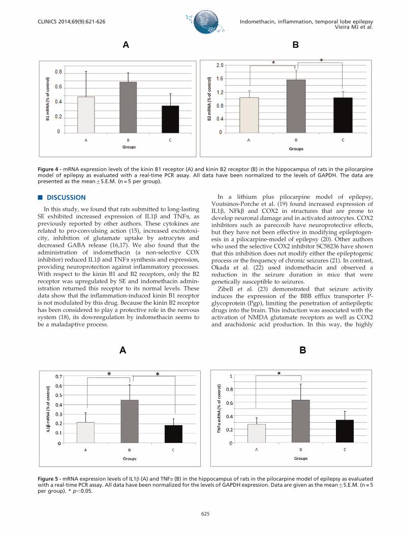
Real-time PCR analysis of the kinin B1 and B2receptors, IL-1b and TNFa
The mRNA levels of the kinin receptors, IL1b and TNFain Wistar rats were analyzed using a semi-quantitative RNAassay. There were no alterations in kinin B1 mRNA in any ofthe groups that were studied (Figure 4 A). The kinin B2receptor mRNA level was increased in group B(1.565¡0.2841) compared with group A (1.042¡ 0.2057)(p,0.05) (Figure 4 B). However, when comparing theanimals in group B (1.565¡0.2841) with those in group C(1.038¡0.1857), we observed a decrease in the expression ofthis receptor (p,0.05) (Figure 4 B). There was an increase in
Figure 1 - Photomicrographs of hippocampi processed for immunohistochemistry showing expression of the kinin B1 receptor (A, B andC), the kinin B2 receptor (A’, B’ and C’), IL1b (A’’, B’’ and C’’) and TNFa (A’’’, B’’’ and C’’’). Groups: A – saline-treated group; B –pilocarpine-treated group; and C – indomethacin-treated group. Scale bars: 0.3 mm.
CLINICS 2014;69(9):621-626 Indomethacin, inflammation, temporal lobe epilepsyVieira MJ et al.
623

the IL-1b mRNA in group B (0.4450¡0.1622) compared withgroup A (0.2150¡0.09950) (p,0.05) (Figure 5 A). On theother hand, the level of IL-1b mRNA was decreased ingroup C (0.1850¡0.06608) compared with group B(0.4450¡0.1622) (p,0.05) (Figure 5 A). Quantitative real-time PCR also revealed a significant increase in the mRNA
expression of TNFa in the animals in group B(0.6275¡0.2364) compared with those in group A(0.2700¡0.09661) (p,0.05) (Figure 5 B). When the animalsin group B (0.6275¡0.2364) were compared with theanimals in group C (0.3325¡0.1284), no differences inTNFa expression levels were observed.
Figure 2 - Photomicrographs of the CA1 region of hippocampi processed for immunohistochemistry showing expression of the kinin B1receptor (A, B and C), the kinin B2 receptor (A’, B’ and C’) and IL1b (A’’, B’’ and C’’). Groups: A – saline-treated group; B – pilocarpine-treated group; and C – indomethacin-treated group. Scale bars: 0.02 mm.
Figure 3 - Photomicrographs of the CA3 region of hippocampi processed for immunohistochemistry showing expression of the kinin B1receptor (A, B and C), the kinin B2 receptor (A’, B’ and C’), IL1b (A’’, B’’ and C’’) and TNFa (A’’’, B’’’ and C’’’). Groups: A – saline-treatedgroup; B – pilocarpine-treated group; and C – indomethacin-treated group. Scale bars: 0.02 mm.
Indomethacin, inflammation, temporal lobe epilepsyVieira MJ et al.
CLINICS 2014;69(9):621-626
624
& DISCUSSION
In this study, we found that rats submitted to long-lastingSE exhibited increased expression of IL1b and TNFa, aspreviously reported by other authors. These cytokines arerelated to pro-convulsing action (15), increased excitotoxi-city, inhibition of glutamate uptake by astrocytes anddecreased GABA release (16,17). We also found that theadministration of indomethacin (a non-selective COXinhibitor) reduced IL1b and TNFa synthesis and expression,providing neuroprotection against inflammatory processes.With respect to the kinin B1 and B2 receptors, only the B2receptor was upregulated by SE and indomethacin admin-istration returned this receptor to its normal levels. Thesedata show that the inflammation-induced kinin B1 receptoris not modulated by this drug. Because the kinin B2 receptorhas been considered to play a protective role in the nervoussystem (18), its downregulation by indomethacin seems tobe a maladaptive process.
In a lithium plus pilocarpine model of epilepsy,Voutsinos-Porche et al. (19) found increased expression ofIL1b, NFkb and COX2 in structures that are prone todevelop neuronal damage and in activated astrocytes. COX2inhibitors such as parecoxib have neuroprotective effects,but they have not been effective in modifying epileptogen-esis in a pilocarpine-model of epilepsy (20). Other authorswho used the selective COX2 inhibitor SC58236 have shownthat this inhibition does not modify either the epileptogenicprocess or the frequency of chronic seizures (21). In contrast,Okada et al. (22) used indomethacin and observed areduction in the seizure duration in mice that weregenetically susceptible to seizures.
Zibell et al. (23) demonstrated that seizure activityinduces the expression of the BBB efflux transporter P-glycoprotein (Pgp), limiting the penetration of antiepilepticdrugs into the brain. This induction was associated with theactivation of NMDA glutamate receptors as well as COX2and arachidonic acid production. In this way, the highly
Figure 4 - mRNA expression levels of the kinin B1 receptor (A) and kinin B2 receptor (B) in the hippocampus of rats in the pilocarpinemodel of epilepsy as evaluated with a real-time PCR assay. All data have been normalized to the levels of GAPDH. The data arepresented as the mean¡S.E.M. (n = 5 per group).
Figure 5 - mRNA expression levels of IL1b (A) and TNFa (B) in the hippocampus of rats in the pilocarpine model of epilepsy as evaluatedwith a real-time PCR assay. All data have been normalized for the levels of GAPDH expression. Data are given as the mean¡S.E.M. (n = 5per group). * p,0.05.
CLINICS 2014;69(9):621-626 Indomethacin, inflammation, temporal lobe epilepsyVieira MJ et al.
625

selective COX2 inhibitor NS-398, an indomethacin-relatedmolecule, blocked the overexpression of Pgp in pilocarpine-induced seizures.
Taken together, these data show that the inhibition ofCOX during the development of neurodegenerative pro-cesses can be dose- and time-dependent, which wasreported previously by other authors (23,24). As reportedby our group (8), the administration of high doses ofindomethacin prior to pilocarpine injection increases themortality rate due to tonic seizures in rats, indicating a dualeffect involving COX inhibition in this epileptic model.Additionally, numerous specific cell types, includingneurons of various subcategories and different glial cells,express and respond to inflammatory mediators in dis-tinctly different ways. Therefore, a given molecule might bebeneficial for a particular population of cells but harmful forother populations (25). Several authors have debated theefficacy of anti-inflammatory drugs in epilepsy treatment,highlighting the antiepileptic action of some types of PGs,such as PGD2 and PGE2, but noting that PGs maycontribute to the feedback inhibition of neurotransmitterrelease, possibly reducing excitation (26). In this sense, theblockage of or decrease in COX activity in the early stages ofSE, which reduces the levels of inflammatory mediators,seems to be insufficient for blocking epileptogenesis, but itcould be effective in neuronal protection or for the control ofotherwise refractory seizures.
Therefore, our data show that the administration ofindomethacin decreases the levels of inflammatory media-tors when used in the early phases of SE onset in an epilepsymodel induced by pilocarpine. However, caution should beexercised when using anti-inflammatory drugs in epilepsybecause several pathways are activated during this process.
& ACKNOWLEDGMENTS
This work was supported by Fundacao de Amparo a Pesquisa do Estado de
Sao Paulo (FAPESP), Conselho Nacional de Desenvolvimento Cientıfico e
Tecnologico (CNPQ), Institutos Nacionais de Ciencia e Tecnologia
(INCT) and Programa de Apoio a Nucleos de Excelencia (PRONEX).
& AUTHOR CONTRIBUTIONS
Vieira MJ and Perosa SR developed the experimental model of epilepsy
and the immunohistochemistry protocol; additionally, they assisted with
manuscript writing. Silva Jr JA performed real-time PCR. Cavalheiro EA,
Arganaraz GA and Naffah-Mazzacoratti MG outlined the main ideas and
wrote the manuscript.
& REFERENCES
1. Friedman A, Dingledine R. Molecular cascades that mediate theinfluence of inflammation on epilepsy. Epilepsia. 2011;52 Suppl 3:33-9,http://dx.doi.org/10.1111/j.1528-1167.2011.03034.x.
2. Devinsky O, Schein A, Najjar S. Epilepsy associated with systemicautoimmune disorders. Epilepsy Curr. 2013;13(2):62-8, http://dx.doi.org/10.5698/1535-7597-13.2.62.
3. Varella PP, Santiago JF, Carrete H Jr., Yacubian EM, Centeno RS, CabocloLO, et al. Relationship between fluid-attenuated inversion-recovery(FLAIR) signal intensity and inflammatory mediator’s levels in thehippocampus of patients with temporal lobe epilepsy and mesialtemporal sclerosis. Arq Neuropsiquiatr. 2011;69(1):91-9, http://dx.doi.org/10.1590/S0004-282X2011000100018.
4. Levin JR, Serrano G, Dingledine R. Reduction in delayed mortality andsubtle improvement in retrograde memory performance in pilocarpine-treated mice with conditional neuronal deletion of cyclooxygenase-2gene. Epilepsia. 2012;53(8):1411-20, http://dx.doi.org/10.1111/j.1528-1167.2012.03584.x.
5. Sutter R1, Marsch S, Fuhr P, Ruegg S. Mortality and recovery fromrefractory status epilepticus in the intensive care unit: a 7-yearobservational study. Epilepsia. 2013;54(3):502-11.
6. Cendes F, Ragazzo PC, da Costa V, Martins LF. Corpus callosotomy intreatment of medically resistant epilepsy: preliminary results in apediatric population. Epilepsia. 1993;34(5):910-7, http://dx.doi.org/10.1111/j.1528-1157.1993.tb02111.x.
7. Hottinger A, Sutter R, Marsch S, Ruegg S. Topiramate as an adjunctivetreatment in patients with refractory status epilepticus: an observationalcohort study. CNS Drugs. 2012;26(9):761-72, http://dx.doi.org/10.2165/11633090-000000000-00000.
8. Naffah-Mazzacoratti MG, Bellıssimo MI, Cavalheiro EA. Profile ofprostaglandin levels in the rat hippocampus in pilocarpine model ofepilepsy. Neurochem Int. 1995;27(6):461-6, http://dx.doi.org/10.1016/0197-0186(95)80003-4.
9. Jeong KH, Kim JY, Choi YS, Lee MY, Kim SY. Influence of aspirin onpilocarpine-induced epilepsy in mice. Korean J Physiol Pharmacol.2013;17(1):15-21.
10. Zibell G, Unkruer B, Pekcec A, Hartz AM, Bauer B, Miller DS, et al.Prevention of seizure-induced up-regulation of endothelial P-glycoproteinby COX-2 inhibition. Neuropharmacology. 2009;56(5):849-55, http://dx.doi.org/10.1016/j.neuropharm.2009.01.009.
11. Schlichtiger J, Pekcec A, Bartmann H, Winter P, Fuest C, Soerensen J,Potschka H. et al. Celecoxib treatment restores pharmacosensitivityin a rat model of pharmacoresistant epilepsy. Br J Pharmacol.2011;60(5):1062-71.
12. Takemiya T, Maehara M, Matsumura K, Yasuda S, Sugiura H, YamagataK. Prostaglandin E2 produced by late induced COX-2 stimulateshippocampal neuron loss after seizure in the CA3 region. NeurosciRes. 2006;56(1):103-10, http://dx.doi.org/10.1016/j.neures.2006.06.003.
13. Dore S. GPCR antagonists as an alternative to COX-2 inhibitors: a casefor the PGE2 EP1 receptor. Trends Pharmacol Sci. 2006;27(9):458-60,http://dx.doi.org/10.1016/j.tips.2006.07.001.
14. Livak KJ, Schmittgen TD. Analysis of relative gene expression data usingreal-time quantitative PCR and the 2(-Delta Delta C(T)) Method.Methods. 2001;25(4):402-8, http://dx.doi.org/10.1006/meth.2001.1262.
15. Vezzani A, Conti M, De Luigi A, Ravizza T, Moneta D, Marchesi F, et al.Interleukin-1beta immunoreactivity and microglia are enhanced in therat hippocampus by focal kainate application: functional evidence forenhancement of electrographic seizures. J Neurosci. 1999;19(12):5054-65.
16. Ye ZC, Sontheimer H. Cytokine modulation of glial glutamate uptake: apossible involvement of nitric oxide. Neuroreport. 1996;7(13):2181-5,http://dx.doi.org/10.1097/00001756-199609020-00025.
17. Zeise ML, Espinoza J, Morales P, Nalli A. Interleukin-1beta does notincrease synaptic inhibition in hippocampal CA3 pyramidal and dentategyrus granule cells of the rat in vitro. Brain Res. 1997;768(1-2):341-4.
18. Arganaraz GA, Regina Perosa S, Cristina Lencioni E, Bader M, AbraoCavalheiro E, da Graca Naffah-Mazzacoratti M, et al. Role of kinin B1and B2 receptors in the development of pilocarpine model of epilepsy.Brain Res. 2004;1013(1):30-9.
19. Voutsinos-Porche B, Koning E, Kaplan H, Ferrandon A, Guenounou M,Nehlig A, et al. Temporal patterns of the cerebral inflammatory responsein the rat lithium-pilocarpine model of temporal lobe epilepsy.Neurobiol Dis. 2004;17(3):385-402, http://dx.doi.org/10.1016/j.nbd.2004.07.023.
20. Polascheck N, Bankstahl M, Loscher W. The COX-2 inhibitor parecoxib isneuroprotective but not antiepileptogenic in the pilocarpine model oftemporal lobe epilepsy. Exp Neurol. 2010;224(1):219-33, http://dx.doi.org/10.1016/j.expneurol.2010.03.014.
21. Holtman L, van Vliet EA, van Schaik R, Queiroz CM, Aronica E, GorterJA. Effects of SC58236, a selective COX-2 inhibitor, on epileptogenesisand spontaneous seizures in a rat model for temporal lobe epilepsy.Epilepsy Res. 2009;84(1):56-66, http://dx.doi.org/10.1016/j.eplepsyres.2008.12.006.
22. Okada K, Yamashita U, Tsuji S. Cyclooxygenase system contributes tothe maintenance of post convulsive period of epileptic phenomena in thegenetically epileptic El mice. J UOEH. 2006;28(3):265-75.
23. Baran H, Vass K, Lassmann H, Hornykiewicz O. The cyclooxygenaseand lipoxygenase inhibitor BW755C protects rats against kainic acid-induced seizures and neurotoxicity. Brain Res. 1994;646(2):201-6.
24. Serrano GE, Lelutiu N, Rojas A, Cochi S, Shaw R, Makinson CD, et al.Ablation of cyclooxygenase-2 in forebrain neurons is neuroprotectiveand dampens brain inflammation after status epilepticus. J Neurosci.2011;31(42):14850-60, http://dx.doi.org/10.1523/JNEUROSCI.3922-11.2011.
25. Dedeurwaerdere S, Callaghan PD, Pham T, Rahardjo GL, Amhaoul H,Berghofer P, et al. PET imaging of brain inflammation during earlyepileptogenesis in a rat model of temporal lobe epilepsy. EJNMMI Res.2012;2(1):60, http://dx.doi.org/10.1186/2191-219X-2-60.
26. Herting G, Seregi A. Formation of eicosanoids in central nervous system.In: Arachidonic acid Metabolism in Nervous System (Barkai A.I andBazan N. eds) New York Academy of Science, New York. 1989;84-99.
Indomethacin, inflammation, temporal lobe epilepsyVieira MJ et al.
CLINICS 2014;69(9):621-626
626