Indirect CD4 + TH1 Response, Antidonor Antibodies and Diffuse C4d Graft Deposits in Long-Term...
12
American Journal of Transplantation 2009; 9: 697–708 Wiley Periodicals Inc. C 2009 The Authors Journal compilation C 2009 The American Society of Transplantation and the American Society of Transplant Surgeons doi: 10.1111/j.1600-6143.2009.02556.x Indirect CD4 + TH1 Response, Antidonor Antibodies and Diffuse C4d Graft Deposits in Long-Term Recipients Conditioned by Donor Antigens Priming C. Ballet a , K. Renaudin b , N. Degauque a, † , H. L. Mai a , F. Bo¨ effard a , D. Lair a , L. Berthelot a , C. Feng a , H. Smit a , C. Usal a , M. Heslan a , R. Josien a , S. Brouard a and J.-P. Soulillou a, ∗ a Institut National de la Sant ´ e et de la Recherche M´ edicale (I.N.S.E.R.M), "Immunointervention dans les allo et x´ enotransplantations" et Institut de Transplantation et de Recherche en Transplantation (I.T.E.R.T), Chu Hˆ otel Dieu, Nantes, Cedex 01, France b Departement d’anatomopathologie, Chu-Hotel Dieu, Nantes, Cedex 01, France †Recipient of a Postdoctorate fellowship from the transplantation society. This work was supported in part by RTRS ‘Centaure’ and the Progreffe foundation. ∗ Corresponding author: Jean-Paul Soulillou, [email protected] Priming of recipients by DST induces long-term sur- vival of mismatched allografts in adult rats. Despite these recipients developing inducible T regulatory cells able to transfer long-term graft survival to a sec- ondary host, a state of chronic rejection is also ob- served. We revisited the molecular donor MHC targets of the cellular response in acute rejection and analyzed the cellular and humoral responses in recipients with long-term graft survival following transplantation. We found three immunodominant peptides, all derived from LEW.1W RT1.D u molecules to be involved in acute rejection of grafts from unmodified LEW.1A recipients. Although the direct pathway of allorecognition was reduced in DST-treated recipients, the early CD4 + in- direct pathway response to dominant peptides was almost unimpaired. We also detected early and sus- tained antidonor class I and II antibody subtypes with diffuse C4d deposits on graft vessels. Finally, long-term accepted grafts displayed leukocyte infiltration, endar- teritis and fibrosis, which evolved toward vascular nar- rowing at day 100. Altogether, these data suggest that the chronic graft lesions developed in long-term graft recipients are the result of progressive humoral injury associated with a persisting indirect T helper response. These features may represent a useful model for un- derstanding and manipulating chronic active antibody- mediated rejection in human. Key words: Alloantibodies, allotransplantation, antibody-mediated rejection, chronic allograft rejec- tion, complement C4d, heart allograft, transplantation Received 09 July 2008, revised 21 November 2008 and accepted for publication 15 December 2008 Introduction Long-term graft survival through manipulation of the host ‘peripheral’ immune response has been achieved in a num- ber of experimental models (1–4). However, closer atten- tion to the graft itself has recently shown that some of the recipients with long-term graft survival (>100 days), de- velop histological lesions of chronic rejection (5–7). Such lesions can coexist with the presence of regulatory T cells that are able to transfer the capacity of immediate graft ‘acceptance’ to na¨ ıve hosts, but unable to prevent the reappearance of chronic lesions (5,7,8). This reinforces the need for careful pathological analyses, particularly in the usual non-life-sustaining, heterotopic heart graft model, to assess the mechanisms leading to chronic rejection. More- over, these models may contribute to the understanding of chronic rejection in humans. Using molecularly defined donor-derived MHC peptides, we revisited the direct and indirect pathway responses of CD4 + and CD8 + T cells as well as the humoral response following the protocol of DST-induced long-term graft sur- vival. We show that, according to previous data empha- sizing transcriptional downmodulation of a number of Th1 cytokines (9), there was strong inhibition of the IFNc re- sponse triggered by the direct pathway of allorecognition in this model. CD8 + T cells, despite undergoing a strong clonal selection, lost their capacity to respond to dominant donor peptides in the indirect pathway. However, we also noted an unambiguous CD4 + -mediated IFNc response by the indirect pathway of allorecognition of dominant donor MHC peptides in the week following transplantation. Fur- thermore, we show that long-term surviving animals de- veloped an unbiased and sustained antidonor humoral re- sponse with diffuse intragraft C4d deposition and severe cellular inflammation associated with vascular occlusion suggestive of an ‘active humoral’ type of chronic rejection recently defined in humans (10). Altogether, these data 697
Transcript of Indirect CD4 + TH1 Response, Antidonor Antibodies and Diffuse C4d Graft Deposits in Long-Term...

American Journal of Transplantation 2009; 9: 697–708Wiley Periodicals Inc.
C© 2009 The AuthorsJournal compilation C© 2009 The American Society of
Transplantation and the American Society of Transplant Surgeons
doi: 10.1111/j.1600-6143.2009.02556.x
Indirect CD4+ TH1 Response, Antidonor Antibodiesand Diffuse C4d Graft Deposits in Long-TermRecipients Conditioned by Donor Antigens Priming
C. Balleta, K. Renaudinb, N. Degauquea,†,
H. L. Maia, F. Boeffarda, D. Laira, L. Berthelota,
C. Fenga, H. Smita, C. Usala, M. Heslana,
R. Josiena, S. Brouarda and J.-P. Soulilloua,∗aInstitut National de la Sante et de la Recherche Medicale(I.N.S.E.R.M), "Immunointervention dans les allo etxenotransplantations" et Institut de Transplantation et deRecherche en Transplantation (I.T.E.R.T), Chu Hotel Dieu,Nantes, Cedex 01, FrancebDepartement d’anatomopathologie, Chu-Hotel Dieu,Nantes, Cedex 01, France†Recipient of a Postdoctorate fellowship from thetransplantation society.This work was supported in part by RTRS ‘Centaure’ andthe Progreffe foundation.∗Corresponding author: Jean-Paul Soulillou,[email protected]
Priming of recipients by DST induces long-term sur-vival of mismatched allografts in adult rats. Despitethese recipients developing inducible T regulatory cellsable to transfer long-term graft survival to a sec-ondary host, a state of chronic rejection is also ob-served. We revisited the molecular donor MHC targetsof the cellular response in acute rejection and analyzedthe cellular and humoral responses in recipients withlong-term graft survival following transplantation. Wefound three immunodominant peptides, all derivedfrom LEW.1W RT1.Du molecules to be involved in acuterejection of grafts from unmodified LEW.1A recipients.Although the direct pathway of allorecognition wasreduced in DST-treated recipients, the early CD4+ in-direct pathway response to dominant peptides wasalmost unimpaired. We also detected early and sus-tained antidonor class I and II antibody subtypes withdiffuse C4d deposits on graft vessels. Finally, long-termaccepted grafts displayed leukocyte infiltration, endar-teritis and fibrosis, which evolved toward vascular nar-rowing at day 100. Altogether, these data suggest thatthe chronic graft lesions developed in long-term graftrecipients are the result of progressive humoral injuryassociated with a persisting indirect T helper response.These features may represent a useful model for un-derstanding and manipulating chronic active antibody-mediated rejection in human.
Key words: Alloantibodies, allotransplantation,antibody-mediated rejection, chronic allograft rejec-tion, complement C4d, heart allograft, transplantation
Received 09 July 2008, revised 21 November 2008 andaccepted for publication 15 December 2008
Introduction
Long-term graft survival through manipulation of the host‘peripheral’ immune response has been achieved in a num-ber of experimental models (1–4). However, closer atten-tion to the graft itself has recently shown that some of therecipients with long-term graft survival (>100 days), de-velop histological lesions of chronic rejection (5–7). Suchlesions can coexist with the presence of regulatory T cellsthat are able to transfer the capacity of immediate graft‘acceptance’ to naıve hosts, but unable to prevent thereappearance of chronic lesions (5,7,8). This reinforces theneed for careful pathological analyses, particularly in theusual non-life-sustaining, heterotopic heart graft model, toassess the mechanisms leading to chronic rejection. More-over, these models may contribute to the understanding ofchronic rejection in humans.
Using molecularly defined donor-derived MHC peptides,we revisited the direct and indirect pathway responses ofCD4+ and CD8+ T cells as well as the humoral responsefollowing the protocol of DST-induced long-term graft sur-vival. We show that, according to previous data empha-sizing transcriptional downmodulation of a number of Th1cytokines (9), there was strong inhibition of the IFNc re-sponse triggered by the direct pathway of allorecognitionin this model. CD8+ T cells, despite undergoing a strongclonal selection, lost their capacity to respond to dominantdonor peptides in the indirect pathway. However, we alsonoted an unambiguous CD4+-mediated IFNc response bythe indirect pathway of allorecognition of dominant donorMHC peptides in the week following transplantation. Fur-thermore, we show that long-term surviving animals de-veloped an unbiased and sustained antidonor humoral re-sponse with diffuse intragraft C4d deposition and severecellular inflammation associated with vascular occlusionsuggestive of an ‘active humoral’ type of chronic rejectionrecently defined in humans (10). Altogether, these data
697
Ballet et al.
shed new light on the classical model of tolerance induc-tion in the rat and provide evidence that this model carriesseveral features observed in chronic humoral rejection inhuman transplantation.
Materials and Methods
Animals and transplantation
Naıve adult MHC-mismatched congeneic LEW.1A (RT1a), LEW.1W (RT1u)and BN (RT1n) rats were purchased from Janvier (France). LEW.1A, LEW.1Wor BN heart grafts were implanted heterotopically onto LEW.1A recipientsusing the Ono and Lindsey technique (11) and function was monitored dailyby palpation through the abdominal wall. Rejection was defined as completecessation of heartbeat.
Donor-specific transfusions
Blood (1 mL) collected from a LEW.1W donor by cardiac puncture wasinjected intravenously into LEW.1A recipients on pretransplant day −14and −7 (12). The regimen induces donor MHC-specific long-term allograftsurvival (>100 days), but with grafts showing signs of chronic rejection atday 100 (6).
Peptide synthesis
Synthetic peptides 16 amino-acids in length and spanning the polymor-phic regions of RT1.Au (a1, a2 and a3 domains), RT1.Bu (b1 domain) andRT1.Du (b1 domain) or the invariable regions of RT1.A (a2 and a3 domains)molecules (13) (Figure 1) were prepared by Genepep (France). Lyophilizedpeptides were dissolved in deionized water/0.4% DMSO and used in cul-tures at a final concentration of 10 lg/mL.
Cells and cell sorting
Cells from mesenteric lymph nodes and spleen cells isolated by passingthrough a stainless steel mesh. Graft infiltrating cells were extracted fromhearts after cutting the graft into small pieces, incubated with collagenaseD (2 mg/mL Boerhringer Mannheim) for 20 min at 37◦C and isolated bycentrifugation over Ficoll. Blood samples were collected in heparin tubesand PBMC were isolated after erythrocytes lysis by osmotic shock. SpleenT cells were prepared using nylon wool adhesion columns followed bydepletion of NK cells, monocytes and B cells with specific mAbs (clone3.2.3, clone OX42 and clone HIS24 (BD Pharmingen) respectively), followedby anti-mouse IgG-coated dynabeads (Invitrogen). CD4+ or CD8+ T cellswere negatively sorted using anti-mouse IgG dynabeads after T-cell stainingwith anti-CD8 (OX8) or anti-CD4 (W3/25) mAbs (European collection of cell,Salisbury, UK). CD4+CD25high T cells and their negative counterparts weresorted using an ARIA flow cytometer (BD) after staining with anti-CD4 FITCand anti-CD25 Alexa 647 mAbs (W3/25 and OX39).
IFNc ELISPOT assays
ELISPOT plates (BD Biosciences) were coated with purified anti-rat IFNc
(5 lg/mL), incubated at 4◦C overnight and blocked 2 h with RPMI 1640medium containing 10% Fetal Calf Serum (FCS) Glutamine (Sigma) and1% Penicillin-Streptomycin-L-Glutamine (Sigma). For assessment of IFNc -producing cell frequency, 4 × 10⁵ total fresh cells from spleen, lymph nodes,blood or grafts from untreated or DST-treated recipients, were seeded in trip-licate in the presence of irradiated donor splenocytes (direct pathway), 16amino-acid peptides from LEW.1W MHC molecules (10 lg/mL) (Genepep)(Indirect pathway) or medium alone. To test the T-cell responses in the differ-ent subsets, 2 × 10⁵ CD4+, CD8+, CD4+CD25high-depleted T cells, non-Tcells, total recipient spleen T cells or 2.5 × 10⁴ CD4+CD25high T cells werestimulated in the presence of 2 × 10⁵ irradiated recipient splenocytes. Cells
were incubated for 24 h at 37◦C 5% CO2. For detection of spots, 2 lg/mL ofbiotinylated antirat IFNc were added and plates incubated for 2 h, followedby a 1-h incubation with streptavidin-horseradish peroxidase. The plateswere developed using 20 lL of 3-Amino-9-ethylcarbazole (AEC chromogen)added per 1 mL of AEC Substrate. The number of spots was countedon a computer-assisted-enzyme-linked immunospot image analyzer(AID).
Serum antibody detection
For the assessment of posttransplant antidonor IgG subtypes, decomple-mented sera (30 min at 56◦C) from naıve, rejecting (day 7 and 10), orDST-treated animals and syngeneic recipients (day 7, 30 and 100) wereincubated with donor splenocytes. Donor splenocytes were subsequentlystained with purified mouse anti-rat IgG1, IgG2a, IgG2b or IgG2c mAbs(University of Louvain, kindly provided by F. Nisol) and revealed with PE-conjugated donkey anti-mouse mAbs (Jackson ImmunoResearch). At thesame time, donor splenocytes were stained with Alexa 647-conjugatedanti-rat TCR alpha/beat (R7–3) mAbs obtained from the European collectionof animal cell culture (Salisbury, UK). Cells were collected on a FACScanand analyzed using CellQuest software (BD Biosciences). The analysis wasperformed on TCR positive (for anticlass I antibody detection) or negativecells (for anticlass I and class II antibody detection). Data were expressedas mean PE fluorescence intensity.
In vitro production and characterization of alloantibodies
Organocultures were performed as previously described (14). Microdis-sected heart allografts were recovered from 4 DST-treated rats 100 daysafter transplantation or 3 naıve hearts and placed in cold sterile RPMI 1640medium containing Glutamine (Sigma) and 1% Penicillin-Streptomycin-L-Glutamine (Sigma) and 25 lg/mL Fungizone (GIBCO). Tissues were washedthree times in fresh medium. Each sample was fragmented with a sterilerazor blade, placed into 2 mL of fresh medium, and cultured in 12-well platesat 37◦C. Culture supernatants were recovered after 5 days of culture. Theanalysis of the specificity (anticlass I and II or anticlass I) of the antibod-ies present in the organoculture-derived supernatants was performed byflow cytometry by using LEW.1W (donor) splenocytes. One hundred mi-croliters of each organoculture-derived supernatant were incubated with2 × 105 LEW.1W cells for 30 min at 4◦C. The binding of antibodies on thecell surface was then determined with purified mouse anti-rat IgG1, IgG2a(University of Louvain, kindly provided by F. Nisol) and revealed with PE-conjugated donkey anti-mouse mAbs (Jackson ImmunoResearch). At thesame time, cells were stained with FITC-conjugated anti-rat HIS24 mAbs(BD Pharmingen). Cells were collected on a FACScan and analyzed usingCellQuest software (BD Biosciences, Le Pont de Claix, France). The analysiswas performed on HIS24 negative (class I) or positive (class I and II) cells.Data are expressed as mean fluorescence intensity.
Histology and immunostaining for C4d
Heart allografts were removed and fixed in 4%-diluted fomalin. Tissueblocks were routinely processed and embedded in paraffin. Serial sectionsof 6 lm were obtained for hematoxylin-eosin-saffron staining. Leukocyteinfiltration, fibrosis, vascular damage and luminal occlusion of arteries wereevaluated.
C4d deposition was localized by immunoperoxidase staining with an affinity-purified polyclonal rabbit antibody to C4d kindly provided by K. Murata (15)(department of pathology, John Hopkins Medical Institutions, BaltimoreMD) used at a 1:1000 dilution. Deparaffinized tissue sections were sub-mitted to heat-induced antigen retrieval and processed using the Menarinirevelation kit (A. Menarini diagnostics) with 3,3’-diaminobenzidine and ahematoxylin counterstain.
698 American Journal of Transplantation 2009; 9: 697–708

An Animal Model for Antibody-Mediated Chronic Rejection
Figure 1: Sequence alignment and peptides. Sixteen amino acid-long overlapping peptides (61 peptides) were chosen along thepolymorphic regions of the RT1Au (a1, a2 and a3 domains), RT1.Bu (b1 domain) and RT1.Du (b1 domain) molecules. Peptides 1, 2, 3, 66and 67 were chosen in the nonpolymorphic sequences of the RT1Au molecule.
American Journal of Transplantation 2009; 9: 697–708 699
Ballet et al.
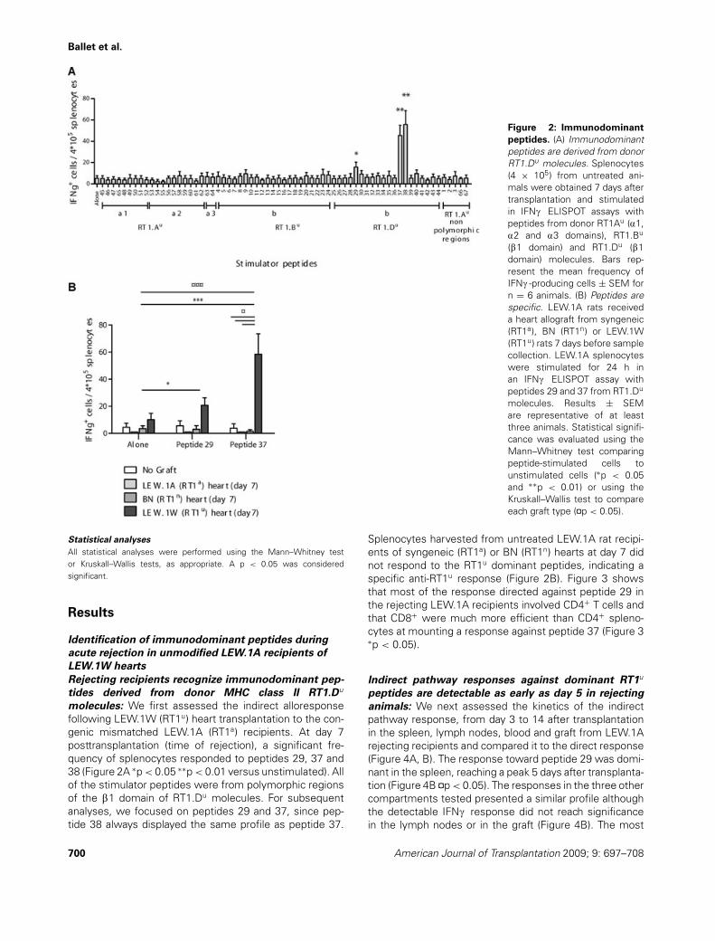
Figure 2: Immunodominant
peptides. (A) Immunodominantpeptides are derived from donorRT1.Du molecules. Splenocytes(4 × 10⁵) from untreated ani-mals were obtained 7 days aftertransplantation and stimulatedin IFNc ELISPOT assays withpeptides from donor RT1Au (a1,a2 and a3 domains), RT1.Bu
(b1 domain) and RT1.Du (b1domain) molecules. Bars rep-resent the mean frequency ofIFNc -producing cells ± SEM forn = 6 animals. (B) Peptides arespecific. LEW.1A rats receiveda heart allograft from syngeneic(RT1a), BN (RT1n) or LEW.1W(RT1u) rats 7 days before samplecollection. LEW.1A splenocyteswere stimulated for 24 h inan IFNc ELISPOT assay withpeptides 29 and 37 from RT1.Du
molecules. Results ± SEMare representative of at leastthree animals. Statistical signifi-cance was evaluated using theMann–Whitney test comparingpeptide-stimulated cells tounstimulated cells (∗p < 0.05and ∗∗p < 0.01) or using theKruskall–Wallis test to compareeach graft type (¤p < 0.05).
Statistical analyses
All statistical analyses were performed using the Mann–Whitney testor Kruskall–Wallis tests, as appropriate. A p < 0.05 was consideredsignificant.
Results
Identification of immunodominant peptides during
acute rejection in unmodified LEW.1A recipients of
LEW.1W hearts
Rejecting recipients recognize immunodominant pep-
tides derived from donor MHC class II RT1.Du
molecules: We first assessed the indirect alloresponsefollowing LEW.1W (RT1u) heart transplantation to the con-genic mismatched LEW.1A (RT1a) recipients. At day 7posttransplantation (time of rejection), a significant fre-quency of splenocytes responded to peptides 29, 37 and38 (Figure 2A ∗p < 0.05 ∗∗p < 0.01 versus unstimulated). Allof the stimulator peptides were from polymorphic regionsof the b1 domain of RT1.Du molecules. For subsequentanalyses, we focused on peptides 29 and 37, since pep-tide 38 always displayed the same profile as peptide 37.
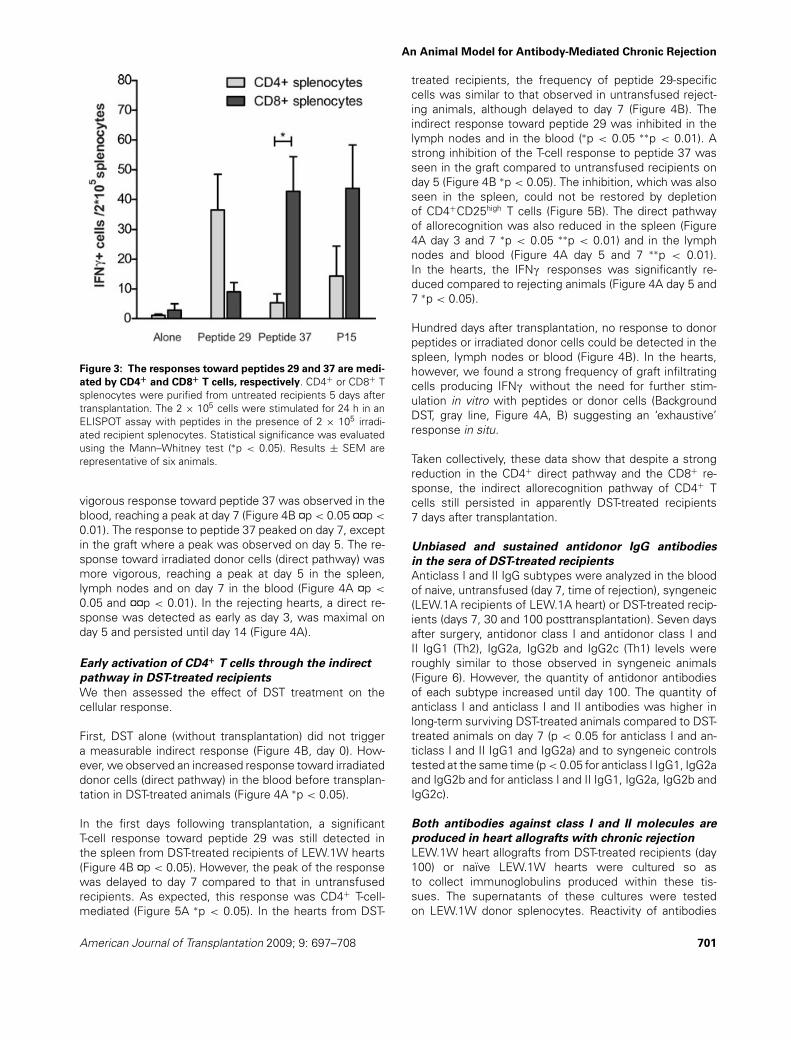
Splenocytes harvested from untreated LEW.1A rat recipi-ents of syngeneic (RT1a) or BN (RT1n) hearts at day 7 didnot respond to the RT1u dominant peptides, indicating aspecific anti-RT1u response (Figure 2B). Figure 3 showsthat most of the response directed against peptide 29 inthe rejecting LEW.1A recipients involved CD4+ T cells andthat CD8+ were much more efficient than CD4+ spleno-cytes at mounting a response against peptide 37 (Figure 3∗p < 0.05).
Indirect pathway responses against dominant RT1u
peptides are detectable as early as day 5 in rejecting
animals: We next assessed the kinetics of the indirectpathway response, from day 3 to 14 after transplantationin the spleen, lymph nodes, blood and graft from LEW.1Arejecting recipients and compared it to the direct response(Figure 4A, B). The response toward peptide 29 was domi-nant in the spleen, reaching a peak 5 days after transplanta-tion (Figure 4B ¤p < 0.05). The responses in the three othercompartments tested presented a similar profile althoughthe detectable IFNc response did not reach significancein the lymph nodes or in the graft (Figure 4B). The most
700 American Journal of Transplantation 2009; 9: 697–708
An Animal Model for Antibody-Mediated Chronic Rejection
Figure 3: The responses toward peptides 29 and 37 are medi-
ated by CD4+ and CD8+ T cells, respectively. CD4+ or CD8+ Tsplenocytes were purified from untreated recipients 5 days aftertransplantation. The 2 × 10⁵ cells were stimulated for 24 h in anELISPOT assay with peptides in the presence of 2 × 10⁵ irradi-ated recipient splenocytes. Statistical significance was evaluatedusing the Mann–Whitney test (∗p < 0.05). Results ± SEM arerepresentative of six animals.
vigorous response toward peptide 37 was observed in theblood, reaching a peak at day 7 (Figure 4B ¤p < 0.05 ¤¤p <
0.01). The response to peptide 37 peaked on day 7, exceptin the graft where a peak was observed on day 5. The re-sponse toward irradiated donor cells (direct pathway) wasmore vigorous, reaching a peak at day 5 in the spleen,lymph nodes and on day 7 in the blood (Figure 4A ¤p <
0.05 and ¤¤p < 0.01). In the rejecting hearts, a direct re-sponse was detected as early as day 3, was maximal onday 5 and persisted until day 14 (Figure 4A).
Early activation of CD4+ T cells through the indirect
pathway in DST-treated recipients
We then assessed the effect of DST treatment on thecellular response.
First, DST alone (without transplantation) did not triggera measurable indirect response (Figure 4B, day 0). How-ever, we observed an increased response toward irradiateddonor cells (direct pathway) in the blood before transplan-tation in DST-treated animals (Figure 4A ∗p < 0.05).
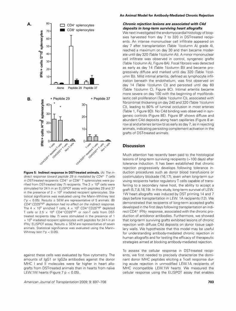
In the first days following transplantation, a significantT-cell response toward peptide 29 was still detected inthe spleen from DST-treated recipients of LEW.1W hearts(Figure 4B ¤p < 0.05). However, the peak of the responsewas delayed to day 7 compared to that in untransfusedrecipients. As expected, this response was CD4+ T-cell-mediated (Figure 5A ∗p < 0.05). In the hearts from DST-
treated recipients, the frequency of peptide 29-specificcells was similar to that observed in untransfused reject-ing animals, although delayed to day 7 (Figure 4B). Theindirect response toward peptide 29 was inhibited in thelymph nodes and in the blood (∗p < 0.05 ∗∗p < 0.01). Astrong inhibition of the T-cell response to peptide 37 wasseen in the graft compared to untransfused recipients onday 5 (Figure 4B ∗p < 0.05). The inhibition, which was alsoseen in the spleen, could not be restored by depletionof CD4+CD25high T cells (Figure 5B). The direct pathwayof allorecognition was also reduced in the spleen (Figure4A day 3 and 7 ∗p < 0.05 ∗∗p < 0.01) and in the lymphnodes and blood (Figure 4A day 5 and 7 ∗∗p < 0.01).In the hearts, the IFNc responses was significantly re-duced compared to rejecting animals (Figure 4A day 5 and7 ∗p < 0.05).
Hundred days after transplantation, no response to donorpeptides or irradiated donor cells could be detected in thespleen, lymph nodes or blood (Figure 4B). In the hearts,however, we found a strong frequency of graft infiltratingcells producing IFNc without the need for further stim-ulation in vitro with peptides or donor cells (BackgroundDST, gray line, Figure 4A, B) suggesting an ‘exhaustive’response in situ.
Taken collectively, these data show that despite a strongreduction in the CD4+ direct pathway and the CD8+ re-sponse, the indirect allorecognition pathway of CD4+ Tcells still persisted in apparently DST-treated recipients7 days after transplantation.
Unbiased and sustained antidonor IgG antibodies
in the sera of DST-treated recipients
Anticlass I and II IgG subtypes were analyzed in the bloodof naive, untransfused (day 7, time of rejection), syngeneic(LEW.1A recipients of LEW.1A heart) or DST-treated recip-ients (days 7, 30 and 100 posttransplantation). Seven daysafter surgery, antidonor class I and antidonor class I andII IgG1 (Th2), IgG2a, IgG2b and IgG2c (Th1) levels wereroughly similar to those observed in syngeneic animals(Figure 6). However, the quantity of antidonor antibodiesof each subtype increased until day 100. The quantity ofanticlass I and anticlass I and II antibodies was higher inlong-term surviving DST-treated animals compared to DST-treated animals on day 7 (p < 0.05 for anticlass I and an-ticlass I and II IgG1 and IgG2a) and to syngeneic controlstested at the same time (p < 0.05 for anticlass I IgG1, IgG2aand IgG2b and for anticlass I and II IgG1, IgG2a, IgG2b andIgG2c).
Both antibodies against class I and II molecules are
produced in heart allografts with chronic rejection
LEW.1W heart allografts from DST-treated recipients (day100) or naıve LEW.1W hearts were cultured so asto collect immunoglobulins produced within these tis-sues. The supernatants of these cultures were testedon LEW.1W donor splenocytes. Reactivity of antibodies
American Journal of Transplantation 2009; 9: 697–708 701

Ballet et al.
Figure 4: Direct and indirect pathways of allorecognition in rejecting or DST-treated animals. The 4 × 10⁵ cells harvested fromspleen, lymph nodes, blood and grafts before transplantation (day −14, −7 and day 0) and from day 3 to 100 after transplantation ofLEW.1W heart allografts, were stimulated 24 h in IFNc ELISPOT assay. (A) Direct pathway. LEW.1A cells were stimulated with donorirradiated splenocytes (1 × 10⁵). (B) Indirect pathway. Cells were tested for their reactivity against peptides 29 and 37 derived from donorMHC class II (RT1.Du) molecules. (A) and (B) DST-treated recipients (circles, dotted lines), untreated recipients (squares, full lines). Thegray line in the graphs showing the response in the graft, represent the frequency of IFNc -producing cells in the absence of stimulationin vitro (Background DST). Note that because frequencies of activated cells were higher in the direct than in the indirect pathway, scalesare different. Significant differences were calculated by the Kruskall–Wallis test (¤) and by the Mann–Whitney U test (¤ and ∗p < 0.05;¤¤ and ∗∗p < 0.01). Results at each day ± SEM are representative of at least three animals.
702 American Journal of Transplantation 2009; 9: 697–708
An Animal Model for Antibody-Mediated Chronic Rejection
Figure 5: Indirect response in DST-treated animals. (A) The in-direct response toward peptide 29 is mediated by CD4+ T cellsin DST-treated recipients. CD4+ or CD8+ T splenocytes were pu-rified from DST-treated (day 7) recipients. The 2 × 10⁵ cells werestimulated for 24 h in an ELISPOT assay with peptides 29 and 37in the presence of 2 × 10⁵ irradiated recipient splenocytes. Sta-tistical significance was evaluated using the Mann–Whitney test(∗p < 0.05). Results ± SEM are representative of 5 animals. (B)CD4+CD25high depletion had no effect on the indirect response.The 4 × 10⁵ enriched T cells, 4 × 10⁵ CD4+CD25high depletedT cells or 2.5 × 10⁴ CD4+CD25high or non-T cells from DST-treated recipients (day 7) were stimulated in the presence of 1× 10⁵ irradiated recipient splenocytes with peptides for 24 h in anIFNc ELISPOT assay. Results ± SEM are representative of sevenanimals. Statistical significance was evaluated using the Mann–Whitney test (∗p < 0.05).
against these cells was evaluated by flow cytometry. Theamounts of IgG1 or IgG2a antibodies against the donorMHC I and II molecules were far higher in heart allo-grafts from DST-treated animals than in hearts from naıveLEW.1W hearts (Figure 7 p < 0.05).
Chronic rejection lesions are associated with C4d
deposits in long-term surviving heart allografts
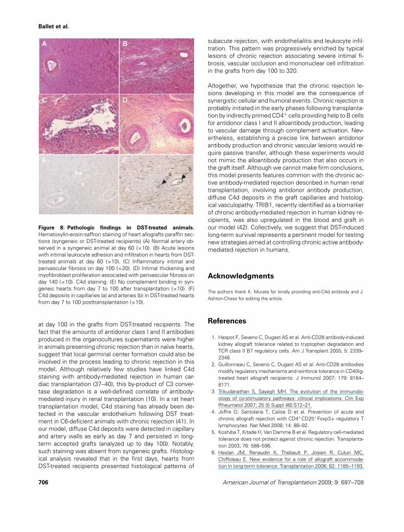
We next investigated the endomyocardial histology of biop-sies harvested from day 7 to 320 in DST-treated recipi-ents. An intense mononuclear cell infiltrate appeared onday 7 after transplantation (Table 1(column A) grade 4),reached a maximum on day 30 and then became moder-ate until day 320 (Table 1(column A)). A minor mononuclearcell infiltrate was observed in control, syngeneic grafts(Table 1(column A), Figure 8A). Focal fibrosis was detectedas early as day 14 (Table 1(column B)) and became pro-gressively diffuse and marked until day 320 (Table 1(col-umn B)). Mild intimal arteritis, defined as lymphocyte infil-tration beneath the endothelium, was first observed onday 14 (Table 1(column C)) and persisted until day 60(Table 1(column C), Figure 8C). Intimal arteritis becamemore severe on day 100 with the beginning of myofibrob-lastic cell proliferation (Table 1(column C)), associated withfibrointimal thickening on day 240 and 320 (Table 1(columnC)), leading to 80% of luminal occlusion in most arteries(Table 1, Figure 8D). No C4d binding was observed in syn-geneic controls (Figure 8E). Figure 8F shows diffuse andabundant C4d deposits along heart capillaries (Figure 8 ar-row a) and arteries (arrow b) as early as day 7, as in rejectinganimals, indicating persisting complement activation in thegrafts of DST-treated animals.
Discussion
Much attention has recently been paid to the histologicallesions of long-term surviving recipients (>100 days) aftertolerance induction. It has been established that chronicrejection progressively develops following tolerance in-duction procedures such as donor blood transfusions orcostimulatory blockade (16,17), even when long-term sur-viving recipients harbor regulatory T cells capable of trans-ferring to a secondary naive host, the ability to accept agraft (5,7,8,18,19). In this study, long-term survival of LEW.1W heart allografts was induced by DST priming 14 and 7days before transplantation in LEW. 1A recipients (12). Wedemonstrated that recipients of long-term accepted graftsdeveloped in the first days following transplantation an indi-rect CD4+ IFNc response, associated with the chronic pro-duction of antidonor antibodies. Furthermore, we showedthat long-term surviving grafts exhibited lesions of chronicrejection with diffuse C4d deposits on donor tissue capil-lary walls. We hypothesize that this model may be usefulfor understanding antibody-mediated chronic rejection inhuman allografts and for testing the efficacy of therapeuticstrategies aimed at blocking antibody-mediated rejection.
To assess the cellular response in DST-treated recipi-ents, we first needed to precisely characterize the domi-nant donor MHC peptides eliciting a T-cell response dur-ing acute rejection in unmodified LEW.1A recipients ofMHC incompatible LEW.1W hearts. We measured thecellular response using the ELISPOT assay that enables
American Journal of Transplantation 2009; 9: 697–708 703

Ballet et al.
Figure 6: Unbiased and sustai-
ned circulating antidonor MHC
class I and class II antibodies in
DST-treated animals. Assessmentof posttransplant antidonor class Iand class I and II IgG subtypes(IgG1, IgG2a, IgG2b, IgG2c) in thesera of 2 naıve, 3 rejecting (day 7),5 rejecting (day 10) or 3 DST-treatedanimals and 3 syngeneic recipients(day 7, 30 and 100). For anticlassI and II antibody detection, decom-plemented sera (1/20e) were incu-bated with donor splenocytes. Cellswere then stained with purifiedmouse anti-rat IgG1, IgG2a IgG2bor IgG2c mAbs and revealed withPE-conjugated donkey anti-mousemAbs. At the same time, spleno-cytes were stained with Alexa647-conjugated anti-rat TCR (R73)mAbs (T cells). Cells were analyzedusing a FACScan with CellQuestsoftware (BD Biosciences). Theanalysis was performed on TCRpositive cells (for antidonor class Iantibody detection) or TCR negativecells (for antidonor class I and classII antibody detection). Data are ex-pressed as mean PE fluorescencechannel. Statistical significance wasevaluated using the Mann–Whitneytest (∗p < 0.05).
Figure 7: IgG1 and IgG2a antibod-
ies against class I and II MHC
molecules are locally produced in
hearts presenting chronic rejec-
tion. The presence of antidonor classI and II IgG1 and IgG2a antibodiesin the supernatant of organoculturesof hearts from DST-treated animalswas assessed by flow cytometry us-ing LEW.1W donor splenocytes andcompared to that observed in naıveLEW.1W animals. Statistical signifi-cance was evaluated using the Mann–Whitney test (∗p < 0.05).
704 American Journal of Transplantation 2009; 9: 697–708

An Animal Model for Antibody-Mediated Chronic Rejection
Table 1: Histological analyses of heart allografts
Vascular Luminal % Number of C4dLeukocytes Fibrosis damage occlusion occluded arteries staining DST/
Groups (A) (B) (C) (D) arteries examined syngeneic
Syngeneic D140 1;1;1 1;0;0 0;0;0 0;0;0 0;0;0 12;12;10DST D7 4;4;4 0;0;0 0;0;0 0;0;0 0;0;0; 21;26;22 ± (×3)DST D14 4;4 0;1 1;0 2;0 30;0 40;42 ± (×2)DST D30 4;4;3 1;1;3 1;0;1 1;0;1 3;0;4 36;57;56 ± (×3)DST D60 3;2–3;2–3 2;2;4 1;0;1 2;0;1 5;0;8 18;19;13 ± (×3)DST D100 2;2;2–3 3;2;4 2;0;2 2;0;4 4;0;81 28;20;16 ± (×3)DST D140 2;2;3 2;1;3 2;2;2 4;2;3 44;37;76 9;8;17DST D240 1;1;2 2;2;2 2;3;3 3;2;2 73;7;50 8;14;8DST D320 3;2;3 3;3;4 2;2;3 3;4;4 50;33;70 10;6;10
Hematoxylin-eosin-saffron staining of heart allografts from DST-treated (day 7, 14, 30, 60 and 100) or syngeneic (day 140) animals. (A)Leukocytes: none = 0; minor = 1; moderate = 2; dense = 3; intense = 4. (B) Fibrosis: none = 0; focal = 1; diffuse minor = 2; diffusemoderate = 3; diffuse marked = 4. (C) Vascular damage: normal = 0; leukocyte adhesion to endothelial cells = 1; inflammatory intimalproliferation = 2; fibrointimal thickening = 3; atheroma = 4. (D) Luminal occlusion: none = 0; <20% = 1; 20 to 50% = 2; 50 to 80% =3; >80% = 4.
quantification of the frequency of T cells already commit-ted to antigen. IFNc was chosen since it has been shownto offer the best correlation to rejection in various studies(20–22). Using this method, we first identified three 16amino-acid long immunodominant peptides from the RT1u
MHC of LEW.1W donors (referred to as peptides 29, 37and 38), which triggered a potent IFNc response duringLEW. 1W heart rejection as early as day 5 after transplan-tation for peptide 29 and day seven for peptides 37 and 38.Testing of purified CD4+ or CD8+ subsets revealed thatthe responses toward peptides 29 and 37 were CD4+ andCD8+-mediated, respectively. Several studies have previ-ously shown that indirectly primed T cells are involved in al-lograft rejection in many experimental models (23–26) andin humans (27,28). We have also shown that in the samestrain combination, LEW. 1W hearts depleted of residentDC are acutely rejected following the same tempo, indi-cating that the indirect pathway can rapidly develop in un-modified LEW. 1A hearts (29,30). The dominant peptidesconcerned the donor class II RT1.Du molecule sequence,in agreement with data obtained in different strain com-binations (31–33). None of these peptides activated cellsharvested from naive LEW. 1A, syngeneic graft recipients(LEW. 1A to LEW. 1A) or RT1n BN heart recipients, indicat-ing a specific response. A higher frequency of alloreactivecells was found to result from the direct pathway of acti-vation, notably on day five and seven after transplantation.This was in agreement with previous data from Benichouet al. who showed that acute rejection was principally gov-erned by the direct pathway.
Hyporesponsiveness of directly activated alloreactive Tcells (9,29) associated with an overall downmodulation ofTh1 cytokines (34) and inflammatory monokines (35) wasobserved in the first days following transplantation in thesame strain combination. We confirmed a strong reductionin the direct allorecognition pathway in DST-treated recipi-ents in the first days following transplantation in all testedcompartments. Considerable inhibition of the indirect path-
way of allorecognition mediated by CD8+ IFNc - producingcells was also observed in all compartments tested fromDST-treated recipients in the first days after transplanta-tion. Because depletion of CD4+CD25high regulatory cellscould not restore the CD8+ IFNc response, it is unlikelythat naturally occurring T regulatory cells are involved inthis inhibition. Our data also show an early activation ofCD4+ T cells specific to peptide 29 in the spleen and graftof DST-treated recipients in the first days following trans-plantation, and this response was as robust as in rejectinganimals.
Because several studies have shown a close relationshipbetween chronic rejection and the indirect pathway of al-lorecognition (28,36), we also analyzed T-cell responses100 days after transplantation, when chronic lesions arewell established. We found the persistence of IFNc -producing graft infiltrating cells without the need for fur-ther stimulation in vitro with donor cells or peptides sug-gesting that graft infiltrating cells themselves are stronglyactivated.
We then asked whether IgG alloantibodies could be de-tected in this model and could be implied in the chronicrejection process. In the RA (RT1p) to PVG (RT1c) model of‘tolerance’ induction by DST priming, Th2 polarized IgG1production associated with Il4 production was reported tooccur 30 days after transplantation (5). In our model, noincrease in Il-4 or Il-13 protein was detected in any of theserum samples from DST-treated recipients tested follow-ing transplantation (data not shown), suggesting an ab-sence of Th2 polarization. This corroborated the sustainedproduction of both Th1 and Th2 antidonor alloantibodyIgG subtypes against class I and II MHC antigens, whichreached a maximum 100 days after transplantation. Be-cause previous data emphasized the role of the lymphoidneogenesis in the graft itself during chronic rejection (14),we also investigated whether locally produced antibodiesdirected against donor MHC molecules could be detected
American Journal of Transplantation 2009; 9: 697–708 705
Ballet et al.
Figure 8: Pathologic findings in DST-treated animals.
Hematoxylin-eosin-saffron staining of heart allografts paraffin sec-tions (syngeneic or DST-treated recipients) (A) Normal artery ob-served in a syngeneic animal at day 60 (×10). (B) Acute lesionswith intimal leukocyte adhesion and infiltration in hearts from DST-treated animals at day 60 (×10). (C) Inflammatory intimal andperivascular fibrosis on day 100 (×20). (D) Intimal thickening andmyofibroblast proliferation associated with perivascular fibrosis onday 140 (×10). C4d staining. (E) No complement binding in syn-geneic hearts from day 7 to 100 after transplantation (×10). (F)C4d deposits in capillaries (a) and arteries (b) in DST-treated heartsfrom day 7 to 100 posttransplantation (×10).
at day 100 in the grafts from DST-treated recipients. Thefact that the amounts of antidonor class I and II antibodiesproduced in the organocultures supernatants were higherin animals presenting chronic rejection than in naıve hearts,suggest that local germinal center formation could also beinvolved in the process leading to chronic rejection in thismodel. Although relatively few studies have linked C4dstaining with antibody-mediated rejection in human car-diac transplantation (37–40), this by-product of C3 conver-tase degradation is a well-defined correlate of antibody-mediated injury in renal transplantation (10). In a rat hearttransplantation model, C4d staining has already been de-tected in the vascular endothelium following DST treat-ment in C6-deficient animals with chronic rejection (41). Inour model, diffuse C4d deposits were detected in capillaryand artery walls as early as day 7 and persisted in long-term accepted grafts (analyzed up to day 100). Notably,such staining was absent from syngeneic grafts. Histolog-ical analysis revealed that in the first days, hearts fromDST-treated recipients presented histological patterns of
subacute rejection, with endothelialitis and leukocyte infil-tration. This pattern was progressively enriched by typicallesions of chronic rejection associating severe intimal fi-brosis, vascular occlusion and mononuclear cell infiltrationin the grafts from day 100 to 320.
Altogether, we hypothesize that the chronic rejection le-sions developing in this model are the consequence ofsynergistic cellular and humoral events. Chronic rejection isprobably initiated in the early phases following transplanta-tion by indirectly primed CD4+ cells providing help to B cellsfor antidonor class I and II alloantibody production, leadingto vascular damage through complement activation. Nev-ertheless, establishing a precise link between antidonorantibody production and chronic vascular lesions would re-quire passive transfer, although these experiments wouldnot mimic the alloantibody production that also occurs inthe graft itself. Although we cannot make firm conclusions,this model presents features common with the chronic ac-tive antibody-mediated rejection described in human renaltransplantation, involving antidonor antibody production,diffuse C4d deposits in the graft capillaries and histolog-ical vasculopathy. TRIB1, recently identified as a biomarkerof chronic antibody-mediated rejection in human kidney re-cipients, was also upregulated in the blood and graft inour model (42). Collectively, we suggest that DST-inducedlong-term survival represents a pertinent model for testingnew strategies aimed at controlling chronic active antibody-mediated rejection in humans.
Acknowledgments
The authors thank K. Murata for kindly providing anti-C4d antibody and J.Ashton-Chess for editing the article.
References
1. Haspot F, Seveno C, Dugast AS et al. Anti-CD28 antibody-inducedkidney allograft tolerance related to tryptophan degradation andTCR class II B7 regulatory cells. Am J Transplant 2005; 5: 2339–2348.
2. Guillonneau C, Seveno C, Dugast AS et al. Anti-CD28 antibodiesmodify regulatory mechanisms and reinforce tolerance in CD40Ig-treated heart allograft recipients. J Immunol 2007; 179: 8164–8171.
3. Trikudanathan S, Sayegh MH. The evolution of the immunobi-ology of co-stimulatory pathways: clinical implications. Clin ExpRheumatol 2007; 25 (5 Suppl 46):S12–21.
4. Joffre O, Santolaria T, Calise D et al. Prevention of acute andchronic allograft rejection with CD4+CD25+Foxp3+ regulatory Tlymphocytes. Nat Med 2008; 14: 88–92.
5. Koshiba T, Kitade H, Van Damme B et al. Regulatory cell-mediatedtolerance does not protect against chronic rejection. Transplanta-tion 2003; 76: 588–596.
6. Heslan JM, Renaudin K, Thebault P, Josien R, Cuturi MC,Chiffoleau E. New evidence for a role of allograft accommoda-tion in long-term tolerance. Transplantation 2006; 82: 1185–1193.
706 American Journal of Transplantation 2009; 9: 697–708

An Animal Model for Antibody-Mediated Chronic Rejection
7. Guillonneau C, Hill M, Hubert FX et al. CD40Ig treatment resultsin allograft acceptance mediated by CD8CD45RC T cells, IFN-gamma, and indoleamine 2,3-dioxygenase. J Clin Invest 2007;117: 1096–1106.
8. Lair D, Degauque N, Miqueu P et al. Functional compartmental-ization following induction of long-term graft survival with pregraftdonor-specific transfusion. Am J Transplant 2007; 7: 538–549.
9. Josien R, Pannetier C, Douillard P et al. Graft-infiltrating T helpercells, CD45RC phenotype, and Th1/Th2-related cytokines in donor-specific transfusion-induced tolerance in adult rats. Transplanta-tion 1995; 60: 1131–1139.
10. Solez K, Colvin RB, Racusen LC et al. Banff 07 classification ofrenal allograft pathology: Updates and future directions. Am JTransplant 2008; 8: 753–760.
11. Ono K, Lindsey ES. Improved technique of heart transplantationin rats. J Thorac Cardiovasc Surg 1969; 57: 225–229.
12. Soulillou JP, Blandin F, Gunther E, Lemoine V. Genetics of theblood transfusion effect on heart allografts in rats. Transplantation1984; 38: 63–67.
13. van Denderen B, Peche H, Gagne K, Usal C, Cuturi MC, SoulillouJP. Identification of immunodominant donor MHC peptides fol-lowing rejection and donor strain transfusion-induced toleranceof heart allografts in adult rats. Eur J Immunol 2001; 31: 1333–1339.
14. Thaunat O, Field AC, Dai J et al. Lymphoid neogenesis in chronicrejection: Evidence for a local humoral alloimmune response. Pro-ceedings of the National Academy of Sciences of the UnitedStates of America 2005; 102: 14723–14728.
15. Minami K, Murata K, Lee CY et al. C4d deposition and clearancein cardiac transplants correlates with alloantibody levels and re-jection in rats. Am J Transplant 2006; 6 (5 Pt 1):923–932.
16. Pirenne J, Kitade H, Kawai M et al. Regulatory cells, TH1/TH2unbalance, and antibody-induced chronic rejection in operationaltolerance induced by donor-specific blood transfusion. Transplan-tation 2005; 79 (3 Suppl):S25–27.
17. Ashton-Chess J, Brouard S, Soulillou JP. Is clinical tolerance real-istic in the next decade? Transpl Int 2006; 19: 539–548.
18. Degauque N, Lair D, Braudeau C et al. Development of CD25-regulatory T cells following heart transplantation: Evidence fortransfer of long-term survival. Eur J Immunol 2007; 37: 147–156.
19. Jovanovic V, Lair D, Soulillou JP, Brouard S. Transfer of toleranceto heart and kidney allografts in the rat model. Transpl Int 2008;21: 199–206.
20. Cunningham DA, Dunn MJ, Yacoub MH, Rose ML. Local pro-duction of cytokines in the human cardiac allograft. A sequentialstudy. Transplantation 1994; 57: 1333–1337.
21. Nast CC, Zuo XJ, Prehn J, Danovitch GM, Wilkinson A, JordanSC. Gamma-interferon gene expression in human renal allograftfine-needle aspirates. Transplantation 1994; 57: 498–502.
22. D’Elios MM, Josien R, Manghetti M et al. Predominant Th1 cellinfiltration in acute rejection episodes of human kidney grafts.Kidney Int 1997; 51: 1876–1884.
23. Benichou G, Takizawa PA, Olson CA, McMillan M, Sercarz EE.Donor major histocompatibility complex (MHC) peptides are pre-sented by recipient MHC molecules during graft rejection. J ExpMed 1992; 175: 305–308.
24. Watschinger B, Gallon L, Carpenter CB, Sayegh MH. Mechanismsof allo-recognition. Recognition by in vivo-primed T cells of specificmajor histocompatibility complex polymorphisms presented aspeptides by responder antigen-presenting cells. Transplantation1994; 57: 572–576.
25. Gallon L, Watschinger B, Murphy B, Akalin E, Sayegh MH, Carpen-ter CB. The indirect pathway of allorecognition. The occurrence
of self-restricted T cell recognition of allo-MHC peptides early inacute renal allograft rejection and its inhibition by conventionalimmunosuppression. Transplantation 1995; 59: 612–616.
26. Shirwan H, Leamer M, Wang HK, Makowka L, Cramer DV. Pep-tides derived from alpha-helices of allogeneic class I major histo-compatibility complex antigens are potent inducers of CD4+ andCD8+ T cell and B cell responses after cardiac allograft rejection.Transplantation 1995; 59: 401–410.
27. Liu Z, Colovai AI, Tugulea S et al. Indirect recognition of donorHLA-DR peptides in organ allograft rejection. J Clin Invest 1996;98: 1150–1157.
28. Najafian N, Salama AD, Fedoseyeva EV, Benichou G, Sayegh MH.Enzyme-linked immunosorbent spot assay analysis of peripheralblood lymphocyte reactivity to donor HLA-DR peptides: Potentialnovel assay for prediction of outcomes for renal transplant recipi-ents. J Am Soc Nephrol 2002; 13: 252–259.
29. Josien R, Heslan M, Brouard S, Soulillou JP, Cuturi MC. Criti-cal requirement for graft passenger leukocytes in allograft toler-ance induced by donor blood transfusion. Blood 1998; 92: 4539–4544.
30. Roussey-Kesler G, Brouard S, Ballet C et al. Exhaustive deple-tion of graft resident dendritic cells: Marginally delayed rejectionbut strong alteration of graft infiltration. Transplantation 2005; 80:506–513.
31. Sayegh MH, Khoury SJ, Hancock WW, Weiner HL, CarpenterCB. Induction of immunity and oral tolerance with polymorphicclass II major histocompatibility complex allopeptides in the rat.Proceedings of the National Academy of Sciences of the UnitedStates of America 1992; 89: 7762–7766.
32. Sayegh MH, Perico N, Imberti O, Hancock WW, Carpenter CB,Remuzzi G. Thymic recognition of class II major histocompatibilitycomplex allopeptides induces donor-specific unresponsiveness torenal allografts. Transplantation 1993; 56: 461–465.
33. Vella JP, Magee C, Vos L et al. Cellular and humoral mecha-nisms of vascularized allograft rejection induced by indirect recog-nition of donor MHC allopeptides. Transplantation 1999; 67: 1523–1532.
34. Bugeon L, Cuturi MC, Hallet MM, Paineau J, Chabannes D,Soulillou JP. Peripheral tolerance of an allograft in adult rats–characterization by low interleukin-2 and interferon-gamma mRNAlevels and by strong accumulation of major histocompatibilitycomplex transcripts in the graft. Transplantation 1992; 54: 219–225.
35. Gagne K, Brouard S, Guillet M, Cuturi MC, Soulillou JP. TGF-beta1 and donor dendritic cells are common key componentsin donor-specific blood transfusion and anti-class II heart graft en-hancement, whereas tolerance induction also required inflamma-tory cytokines down-regulation. Eur J Immunol 2001; 31: 3111–3120.
36. Vella JP, Spadafora-Ferreira M, Murphy B et al. Indirect allorecog-nition of major histocompatibility complex allopeptides in humanrenal transplant recipients with chronic graft dysfunction. Trans-plantation 1997; 64: 795–800.
37. Crespo-Leiro MG, Veiga-Barreiro A, Domenech N et al. Humoralheart rejection (severe allograft dysfunction with no signs of cellu-lar rejection or ischemia): Incidence, management, and the valueof C4d for diagnosis. Am J Transplant 2005; 5: 2560–2564.
38. Smith RN, Brousaides N, Grazette L et al. C4d deposition in cardiacallografts correlates with alloantibody. J Heart Lung Transplant2005; 24: 1202–1210.
39. Chantranuwat C, Qiao JH, Kobashigawa J, Hong L, Shintaku P,Fishbein MC. Immunoperoxidase staining for C4d on paraffin-embedded tissue in cardiac allograft endomyocardial biopsies:
American Journal of Transplantation 2009; 9: 697–708 707
Ballet et al.
Comparison to frozen tissue immunofluorescence. Appl Immuno-histochem Mol Morphol 2004; 12: 166–171.
40. Rodriguez ER, Skojec DV, Tan CD et al. Antibody-mediated rejec-tion in human cardiac allografts: Evaluation of immunoglobulinsand complement activation products C4d and C3d as markers.Am J Transplant 2005; 5: 2778–2785.
41. Qian Z, Lee CY, Murata K et al. Antibody and complement me-diated injury in transplants following sensitization by allogeneicblood transfusion. Transplantation 2006; 82: 857–864.
42. Ashton-Chess J, Giral M, Mengel M et al. Tribbles-1 as a novelbiomarker of chronic antibody-mediated rejection. J Am SocNephrol 2008; 19: 1116–1127.
708 American Journal of Transplantation 2009; 9: 697–708