Independent Regulation of Na and K+ Balance by the Kidney
14
Fax +41 61 306 12 34 E-Mail [email protected] www.karger.com Review Med Princ Pract DOI: 10.1159/000332580 Independent Regulation of Na + and K + Balance by the Kidney María Castañeda-Bueno Juan Pablo Arroyo Gerardo Gamba Molecular Physiology Unit, Instituto Nacional de Ciencias Médicas y Nutrición Salvador Zubirán, Instituto Nacional de Cardiología Ignacio Chávez, and Instituto de Investigaciones Biomédicas, Universidad Nacional Autónoma de México, Mexico City, Mexico a normal plasma K + concentration. The triggers of this acute response (particularly in hyperkalemia) include stimulation of the -adrenergic response, which will ac- tivate the Na + /K + ATPase pump and, thus, drive K + into the cells. This is a relatively effective temporary mecha- nism. However, the total amount of K + in the body re- mains unchanged. Thus, this is a transient response that buys time until a corrective (K + -excreting) mechanism, i.e. the kidney, restores normal K + levels. In addition to adrenergic stimulation, insulin can also promote activa- tion of the Na + /K + ATPase, thus favoring K + uptake. This mechanism is presumably important after eating. Acute changes in Na + can be divided in two ways: vol- ume versus osmolarity. Volume deficit or overload im- plies a change in the amount of fluid in the extracellular compartment that generally does not change the salt-to- water ratio. Na + -related osmolarity derangements on the other hand refer to changes in either Na + or water, thus leading to changes in the salt-to-water ratio. There is, of course, considerable overlap of both these two entities in clinical practice, e.g. hypotonic diarrhea, which causes both volume and osmolarity derangements. However, in the interest of clarity, we will explain these concepts sep- arately so as to understand the body’s response to each one. Volume deficit elicits a much more robust acute re- sponse than volume overload. During a volume-depleted state, blood pressure and thus tissue perfusion can be- come compromised. Thus, the acute mechanisms in- volved in regulation of volume are oriented towards Key Words Angiotensin II Aldosterone Sodium balance Potassium balance Na + /Cl – cotransporter Renal outer medullary K + channel Apical Na + channel WNK kinases Abstract The understanding of the independent regulation of sodium and potassium by the kidney has remained elusive. Recent evidence now points to dissimilar regulatory mechanisms in ion handling, dependent on the presence of either aldoste- rone alone or angiotensin II with aldosterone among other factors. This review summarizes past and present informa- tion in an attempt to reconcile the current concepts of differential regulation of sodium and potassium balance through the with-no-lysine (K) kinase (WNK) system and the previous knowledge regarding ion transport mechanisms in the distal nephron. Copyright © 2011 S. Karger AG, Basel Introduction Acute and chronic regulation of Na + and K + plasma concentrations is essential in maintaining homeostasis. Acutely, changes in plasma K + can alter membrane volt- age with the potential of causing life-threatening ar- rhythmias. Thus, the body’s rapid response involves shifting K + in or out of K + -rich cells in order to maintain Received: February 23, 2011 Accepted: May 15, 2011 Published online: October 25, 2011 Gerardo Gamba Molecular Physiology Unit Vasco de Quiroga No. 15 14000 Mexico City (Mexico) Tel. +52 55 5513 3868, E-Mail gamba @ biomedicas.unam.mx © 2011 S. Karger AG, Basel 1011–7571/11/0000–0000$38.00/0 Accessible online at: www.karger.com/mpp
-
Upload
vanderbilt -
Category
Documents
-
view
0 -
download
0
Transcript of Independent Regulation of Na and K+ Balance by the Kidney
Fax +41 61 306 12 34E-Mail [email protected]
Review
Med Princ Pract DOI: 10.1159/000332580
Independent Regulation of Na + and K + Balance by the Kidney
María Castañeda-Bueno Juan Pablo Arroyo Gerardo Gamba
Molecular Physiology Unit, Instituto Nacional de Ciencias Médicas y Nutrición Salvador Zubirán, Instituto Nacional de Cardiología Ignacio Chávez, and Instituto de Investigaciones Biomédicas, Universidad Nacional Autónoma de México, Mexico City , Mexico
a normal plasma K + concentration. The triggers of this acute response (particularly in hyperkalemia) include stimulation of the � -adrenergic response, which will ac-tivate the Na + /K + ATPase pump and, thus, drive K + into the cells. This is a relatively effective temporary mecha-nism. However, the total amount of K + in the body re-mains unchanged. Thus, this is a transient response that buys time until a corrective (K + -excreting) mechanism, i.e. the kidney, restores normal K + levels. In addition to adrenergic stimulation, insulin can also promote activa-tion of the Na + /K + ATPase, thus favoring K + uptake. This mechanism is presumably important after eating.
Acute changes in Na + can be divided in two ways: vol-ume versus osmolarity. Volume deficit or overload im-plies a change in the amount of fluid in the extracellular compartment that generally does not change the salt-to-water ratio. Na + -related osmolarity derangements on the other hand refer to changes in either Na + or water, thus leading to changes in the salt-to-water ratio. There is, of course, considerable overlap of both these two entities in clinical practice, e.g. hypotonic diarrhea, which causes both volume and osmolarity derangements. However, in the interest of clarity, we will explain these concepts sep-arately so as to understand the body’s response to each one. Volume deficit elicits a much more robust acute re-sponse than volume overload. During a volume-depleted state, blood pressure and thus tissue perfusion can be-come compromised. Thus, the acute mechanisms in-volved in regulation of volume are oriented towards
Key Words
Angiotensin II � Aldosterone � Sodium balance � Potassium balance � Na + /Cl – cotransporter � Renal outer medullary K + channel � Apical Na + channel � WNK kinases
Abstract
The understanding of the independent regulation of sodium and potassium by the kidney has remained elusive. Recent evidence now points to dissimilar regulatory mechanisms in ion handling, dependent on the presence of either aldoste-rone alone or angiotensin II with aldosterone among other factors. This review summarizes past and present informa-tion in an attempt to reconcile the current concepts ofdifferential regulation of sodium and potassium balance through the with-no-lysine (K) kinase (WNK) system and the previous knowledge regarding ion transport mechanisms in the distal nephron. Copyright © 2011 S. Karger AG, Basel
Introduction
Acute and chronic regulation of Na + and K + plasma concentrations is essential in maintaining homeostasis. Acutely, changes in plasma K + can alter membrane volt-age with the potential of causing life-threatening ar-rhythmias. Thus, the body’s rapid response involves shifting K + in or out of K + -rich cells in order to maintain
Received: February 23, 2011 Accepted: May 15, 2011 Published online: October 25, 2011
Gerardo Gamba Molecular Physiology Unit Vasco de Quiroga No. 15 14000 Mexico City (Mexico) Tel. +52 55 5513 3868, E-Mail gamba @ biomedicas.unam.mx
© 2011 S. Karger AG, Basel1011–7571/11/0000–0000$38.00/0
Accessible online at:www.karger.com/mpp
Castañeda-Bueno/Arroyo/Gamba Med Princ Pract 2
maintaining an adequate blood pressure in the presence of diminished extracellular fluid (ECF). These responses include the activation of the sympathetic nervous system and the pressor response of angiotensin II (Ang II), which will increase blood pressure without changing blood vol-ume. Volume overload leads to a decreased sympathetic output, which prevents dangerous increases in blood pressure. Alterations in osmolarity can be caused by many factors, including changes in Na + or water content. When ECF Na + increases with no net change in water, or when ECF water decreases with not net change in Na + , the increased osmolarity will trigger the release of an-tidiuretic hormone (ADH) by the hypothalamus, thus leading to water retention by the kidney. Conversely, in-creased water content with no net change in total body Na + leads to a decreased Na + concentration and osmolar-ity, which will decrease ADH secretion and, thus, water diuresis.
With the exception of osmolarity derangements, the other acute responses to changes in Na + and K + concen-tration require adaptive mechanisms that are only tem-porary as the initial problem, i.e. increased/decreased K + or increased/decreased volume, is not corrected. Chronic responses that do correct the initial problem are carried out by the kidney. Thus, the kidney is the major long-term regulator of Na + and K + homeostasis. The renal han-dling of both these ions involves aldosterone, hinting to common regulatory pathways. Nevertheless, the kidney is able to distinguish between these two contrasting situ-ations. The way in which the kidney independently han-dles Na + and K + homeostasis is the focus of this review.
Renal Mechanisms for K + and Na + Handling
Potassium The kidney normally excretes between 90 and 95% of
our daily K + intake. The rest is excreted through the co-lon. Potassium is freely filtered in the glomerulus and it is both, reabsorbed and secreted across the nephron in a regulated manner in order to achieve the correct level of urinary excretion according to the level of ingestion. Po-tassium handling in the proximal nephron (before the macula densa) usually does not vary with K + intake. The bulk of filtered K + is reabsorbed in the proximal tubule (about 80%) and another 10% is reabsorbed in the thick ascending limb of Henle’s loop. Thus, only about 10% of the filtered K + remains in the lumen fluid by the time it gets to the distal tubule [1–4] . Beyond the macula densa, K + handling is modulated by plasma K + level. When it is
low (as in low K + intake), distal K + reabsorption is stimu-lated and the final urinary K + excretion may represent only 1–3% of the filtered load [4, 5] . On the other hand, when K + intake is normal to high, renal retention is not warranted, hence it is secreted along the distal nephron, in which case urinary output can represent from 10 to 150% of the filtered load [4] . Normal K + intake in a mod-ern diet is approximately 80–120 mmol [6] . With a nor-mal glomerular filtration rate (GFR) of approximately 180 liters/day and a plasma K + concentration of 4 m M , around 720 mmol of K + are filtered per day. Thus, under low K + intake and maximal distal K + reabsorption, the2% of the filtered load that is excreted, represents about 14 mmol. However, with high plasma K + , distal nephron secretion of K + is stimulated, and excretion can go from 72 to 1,080 mmol (10–150% of the filtered load, respec-tively). Thus, the system allows important variations in dietary intake, i.e. the kidney possesses the capacity to adjust K + excretion and maintain a balance in the face of intakes 10 times lower or 10 times higher than normal modern dietary intake values. Intakes higher than these values are very rare so that hyperkalemia more common-ly occurs due to impaired output that may result from renal disease.
The reabsorption and secretion of K + involve trans-cellular ion transport mechanisms. Reabsorption takes place within � intercalated cells (IC) of the distal convo-luted tubules (DCT), connecting tubules (CNT) and the initial and cortical collecting ducts (ICD and CCD). ICs express an apical H + /K + pump, whose activity is driven by ATP hydrolysis, which is necessary for transporting K + against its electrochemical gradient. K + then exits the cell through K + channels present in the basolateral mem-brane [5, 7] .
Regarding K + secretion, initial studies with micro-puncture and microcatheterization, in which secretion and reabsorption were estimated by comparing K + amounts found in fluid that was collected from different nephron segments and urine, demonstrated that secre-tion takes place mainly within the late portion of the clas-sical distal tubule, i.e. in the CNT and ICD. It was also shown that K + excretion also takes place in the CCD but in less important levels [4, 8] . Further studies focused on demonstrating that principal cells of the collecting ducts (CDs) are capable of secreting K + and that this activity can be modulated by factors which are known to modu-late K + excretion in vivo, e.g. aldosterone [1, 9–11] . Thus, principal cells were long considered to be the major site for regulation of K + excretion and their activity was thought to be crucial for maintenance of K + balance.
Sodium and Potassium Balance Med Princ Pract 3
More recently, the importance for K + balance of more proximal nephron segments (late DCT and CNT) has been highlighted. Reilly and Ellison [3] showed that a large percentage of K + secretion occurs between 20 and 80% of superficial distal tubule length which comprises mainly DCT2 and CNT. Different cell types in charge of K + secretion are found in each of these nephron segments, DCT cells in DCT2, CNT cells in CNT and principal cells in ICD and CCD. These diverse cell types share mecha-nisms for K + secretion. The basolateral Na + /K + pump me-diates K + entry and Na + exit. This promotes the increase in intracellular K + and the generation of an electrochem-ical gradient necessary for K + exit through the apical membrane which occurs mainly through the small-con-ductance renal outer medullary K + (ROMK) channel [1, 12] . Other exit routes through the apical membrane are the large Ca 2+ -activated K + channel (MaxiK or BK chan-nel) [1, 15–17] , which normally does not contribute to se-cretion, but that becomes activated by enhanced flow through the tubule [18] and a K + -Cl – cotransport pathway which has also been proposed to participate in the DCT [13, 14]. In addition to the K + -secreting channels, the presence of an epithelial Na + channel (ENaC) is necessary for an adequate adaptation to K + loading. ENaC mediates Na + movement from the tubule lumen to the cell down a favorable concentration gradient generated by the Na + /K + ATPase [19, 20] . This flux of positive charges generates a lumen negative potential which drives K + secretion. Both, Na + /K + pump activity and Na + reabsorption through ENaC are regulated during changes in plasma K + and, consequently, K + secretion through apical channels is in-directly affected [1] .
In humans, impaired ENaC activity presents as pseu-dohypoaldosteronism type I (PHAI), a syndrome charac-terized by hypotension due to urinary Na + loss and hy-perkalemia due to impaired renal K + secretion [21] . Ge-netic mouse models for this syndrome have been generated by disruption of ENaC activity in specific nephron segments. Interestingly, mice with specific dis-ruption of ENaC in the CDs do not display a PHAI-like phenotype [22] , while mice lacking ENaC activity in the CNT as well as the CDs present a mild PHAI phenotype, with hyperkalemia and renal salt loss, as compared to the severe phenotype observed in humans [23] . These results clearly demonstrate an important role for CNT K + secre-tion in the maintenance of K + balance. That the pheno-type is not as severe as in humans leaves the possibility of an essential role for DCT2 in K + balance regulation. Al-ternatively, the difference could be due to variations be-tween species.
Sodium The average intake of Na + in a Western diet is about
120 mmol/day. In order to maintain an adequate ECF vol-ume, the kidneys must excrete the same amount. This means that from the about 25,500 mmol of Na + that are filtered daily in the glomerulus (Na + plasma concentra-tion of 140 mmol/l ! 180 liters/day) only a small fraction must be excreted in the urine (approximately 0.4% of the filtered load, i.e. 100 mmol). Thus, the majority of filtered sodium is reabsorbed. As with K + , the bulk reabsorption takes place in the proximal nephron (before the macula densa): 60–70% in the proximal tubule and 25% in the thick ascending limb of Henle’s loop. Of the remaining 8%, about 5% is reabsorbed in the DCT, CNT and ICD and about 3% in the medullary collecting duct. The cur-rent knowledge of several genetic diseases that result from altered renal Na + handling has contributed to a bet-ter understanding of the role of each transport protein and nephron segment in tubular Na + reabsorption. Sev-eral reviews on this subject have been written by others [24–26] and they present an alternative way of describing renal Na + handling.
The macula densa, a group of specialized epithelial cells located precisely in the site where the ascending loop of Henle passes between the afferent and efferent arteri-oles, is an important center for regulation of kidney func-tion. For instance, it orchestrates a regulatory mechanism called tubuloglomerular feedback (TGF) which func-tions in maintaining relatively constant levels of GFR. Changes in GFR are sensed as changes in NaCl delivery to the macula densa through the Na + /K + /2Cl – cotrans-porter (NKCC2), expressed in these cells [27, 28] . An in-crease in delivery produces a response that affects renal arteriolar tone, causing a decrease in GFR. The opposite occurs with decreased distal NaCl delivery [29] . This mechanism has implications for renal Na + handling be-cause it limits the possibility of altering urinary Na + ex-cretion by affecting GFR or proximal tubular reabsorp-tion since these alterations would be sensed as changes in NaCl delivery to the macula densa and, therefore, would exert a compensatory modulation of GFR. A very clear demonstration of this is the fact that inhibitors of Na + re-absorption mechanisms in proximal tubule, e.g. acetazol-amide, do not have an important diuretic effect in con-trast to that produced by inhibitors of distal Na + reab-sorption, e.g. thiazides and amiloride. Loop diuretics are an exception to the rule because, even when they block reabsorption in the thick ascending limb of Henle’s loop, its target is precisely NKCC2, the macula densa sensor, so that, although salt delivery to the macula densa is in-
Castañeda-Bueno/Arroyo/Gamba Med Princ Pract 4
creased, it is not detected because the sensor is blocked by the loop diuretic, impairing TGF [30] . This implies that physiologic responses which regulate renal Na + excretion would not be able to produce a response by altering filtra-tion or proximal reabsorption unless they alter the TGF mechanism.
Distal reabsorption, however, is not subjected to a feedback mechanism of this type. Thus, as demonstrated by the use of thiazide diuretics, modulation of distal re-absorption can be readily reflected in urinary Na + excre-tion. This idea is reinforced by the fact that all the mono-genic genetic disorders characterized to date with altered blood pressure are caused by mutations in genes coding for proteins affecting renal distal salt handling [25, 31] . In spite of the low level of Na + reabsorption that takes place in this segment as compared to that occurring in the proximal nephron, if altered, important changes in uri-nary excretion can be produced, suggesting that this site is perhaps more adequate for exerting the fine tuning of renal Na + excretion.
Na + reabsorption in the distal nephron is exclusively an active transcellular process driven by the activity of the basolateral Na + /K + ATPase, which extrudes Na + to the extracellular space generating a concentration gradient which favors Na + entry across the apical membrane. Two different pathways are involved. In the DCT (early and late), the thiazide-sensitive, electroneutral Na + /Cl – co-transporter (NCC) mediates apical Na + and Cl – entry thanks to the electrochemical Na + gradient [32, 33] . Re-absorption of Na + in the late DCT, the CNT, and CD is mediated by the amiloride-sensitive ENaC [3, 34] . In both situations, maintenance of the Na + /K + ATPase activity re-quires potassium extrusion to the extracellular space. The electroneutral Na + uptake through NCC is coupled to both, apical and basolateral K + exit. The importance of this K + recycling for NCC activity has been recently high-lighted by the demonstration that inactivating mutations in the DCT basolateral K + channel KCNJ10 are the cause of SeSAME/EAST syndrome. This syndrome is charac-terized by a pattern of alterations similar to those devel-oped in Gitelman’s syndrome, which is caused by inacti-vating mutations in NCC (see below) [35, 36] . However, no net reabsorption or secretion of K + takes place in this segment. In contrast, ENaC activity is tightly coupled to K + secretion since the apical exit of the K + is greatly fa-vored by the lumen-negative transepithelial potential es-tablished by the electrogenic Na + uptake through ENaC [21] .
Integration of Sodium and Potassium Handling by the Kidney As we have seen, the distal nephron is the site where
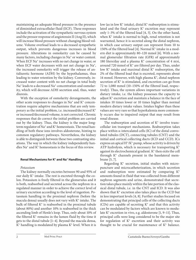
the fine tuning of Na + and K + balance takes place. As such, ion transport proteins along this segment have a very specific distribution ( fig. 1 ). This in turn will gener-ate three different distal nephron segments: the early DCT or DCT1, in which NCC and ROMK are expressed, the late DCT or DCT2 where NCC, ENaC and ROMK are expressed, and the CNT/CD where ENaC, ROMK and BK channels are expressed [3, 37–40] . This differential protein expression will render these nephron segments specific for different functions. These are: (1) DCT1 – so-dium reabsorption, (2) DCT2 – sodium reabsorption 1 potassium secretion, and (3) CNT/CD – potassium secre-tion 1 sodium reabsorption.
Sodium reabsorption can be either electroneutral (via NCC) or coupled to K + secretion (via ENaC) (see above). Electroneutral reabsorption of Na + via NCC inhibits K + secretion since it competes with ENaC-mediated reab-sorption. The more Na + that is reabsorbed by NCC, the less to be transported by ENaC and thus no potassium secretion takes place. Similarly, increased availability of Na + favors reabsorption via ENaC and thus promotes K + secretion. Additionally, increased flow to the distal neph-ron will stimulate the BK channels, favoring K + secretion [18, 40] . Analyzed in this light, the clinical consequences of the different classes of diuretics become readily appar-ent. In addition to the Na + wasting they provoke, diuret-ics can be classified into two families depending on their effect on potassium, i.e. K + -wasting or K + -sparing diuret-ics. Loop diuretics (which inhibit NKCC2) and thiazide diuretics (which inhibit NCC) increase the distal delivery of Na + and, through the Na + /K + exchange and flow-de-pendent mechanisms (see above), favor K + secretion [41] . The site of action of potassium-sparing diuretics is lim-ited to the region of the distal nephron specialized in K + secretion, and through the inhibition of ENaC, amiloride and triamterene block the ENaC/ROMK-mediated Na + /K + exchange, which leads to increased plasma levels of K + [41] .
Regulation of Renal K + Excretion: High-K + versus
Low-K + Diet
Variations in plasma K + must elicit adequate compen-satory mechanisms in order to maintain adequate neuro-muscular function, which is dependent on ECF K + levels. These in turn are a direct result of IC K + levels, where 95%
Sodium and Potassium Balance Med Princ Pract 5
of the total body K + is stored. With variations in daily K + intake, the kidney must adjust accordingly. An in-depth discussion of hyper- and hypokalemia is beyond the scope of this review; however, the renal adaptation to high- or low-K + diet elicits the same molecular response as hyper/hypokalemia, respectively, albeit dietary varia-tions elicit a more discrete response in contrast to life-threatening variations in K + .
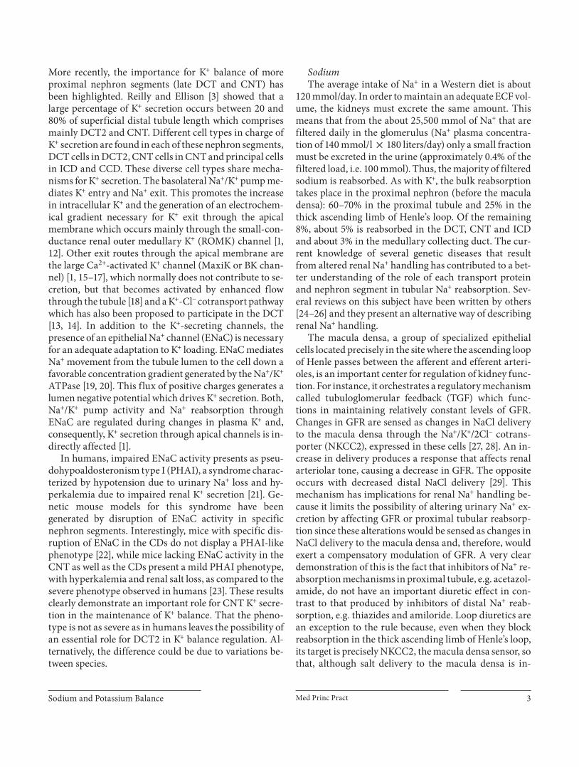
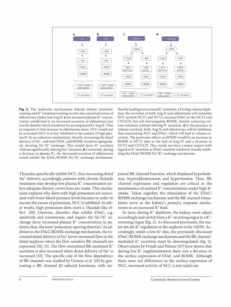
A high-K + diet will favor increased K + secretion, with minimal impact on Na + balance. When faced with an in-creased K + intake, the kidney must adjust K + excretion rapidly in order to prevent life-threatening increases of plasma K + . This response is carried out through both the mineralocorticoid hormone aldosterone and aldoste-rone-independent mechanisms [42] . Expression of the inward-rectifying pore channels in the glomerulosa cells of adrenal glands, TWIK and, more specifically, TASK, maintains the membrane potential close to the equilib-rium potential for K + , thus making the glomerulosa cells sensitive to changes in plasma K + [43–45] . Therefore, changes in plasma K + will depolarize the glomerulosa cells, favoring Ca 2+ entry which will stimulate aldoste-rone synthesis [43, 44] . The increased levels of aldoste-rone will in turn stimulate the mineralocorticoid recep-
tor in the aldosterone-sensitive distal nephron (ASDN) favoring K + secretion. The clinical relevance of this mech-anism is emphasized in patients treated with spironolac-tone (mineralocorticoid receptor antagonist) in which plasma K + concentration increases, sometimes danger-ously. Increased aldosterone stimulation in the ASDN will lead to an increase in both ENaC and ROMK activ-ity [46] . The increase in the Na + /K + exchange mechanism, however, is dependent on an increased distal delivery of Na + . This would require inhibition of Na + reabsorption upstream of the ASDN. Nonetheless, any variations in Na + handling by the proximal tubule and the loop of Henle would be offset by a TGF feedback mechanism ren-dering variations in these nephron segments relatively in-effective to regulate adequate Na + delivery. However, downregulation of NCC would result in an adequate in-creased distal delivery of Na + that could be coupled to K + secretion ( fig. 2 ). Indeed, Frindt and Palmer [47] have re-cently shown that in rats under a high-K + diet, the surface expression of NCC is decreased while Vallon et al. [48] have shown that a high-K + diet in mice has a negative ef-fect upon NCC N-terminal phosphorylation (which is re-lated to its level of activity). A parallel can be drawn here to the mechanism of action of thiazide-type diuretics.
DCT1 DCT2 CNT/CD
Maculadensa
NCC
ROMK
ENaC
BK
Na+/K+
ATPase
Tubular lumen
Basolateral space
Na+ Cl– Na+ Cl–
K+
Na+
K+
K+
Na+ Na+
K+ K+
Na+
K+
Na+
K+
Fig. 1. The distal nephron consists of cells distal to the macula densa which are sub-divided into cell types based on their pro-tein expression pattern. The presence of the NCC defines the DCT, which is further subdivided into two sections: DCT1, which is relatively insensitive to aldoste-rone, and DCT2, which is sensitive to al-dosterone and expresses the ENaC. The expression of flow-sensitive large Ca 2+ -ac-tivated K + channel (BK) is limited to the CNT/CDs, while ROMK spans the entire distal nephron. The Na + /K + ATPase pro-vides the electrochemical gradient neces-sary for reabsorption and secretion of Na + and K + to occur.
Castañeda-Bueno/Arroyo/Gamba Med Princ Pract 6
Thiazides specifically inhibit NCC, thus increasing distal Na + delivery; accordingly, patients with chronic thiazide treatment may develop low plasma K + concentration un-less adequate dietary corrections are made. This mecha-nism explains why diets with high potassium are associ-ated with lower blood pressure levels because in order to excrete the excess of potassium, NCC is inhibited. In oth-er words, high potassium diets exert a ‘thiazide-like ef-fect’ [49] . Likewise, diuretics that inhibit ENaC, e.g. amiloride and triamterene, and impair the Na + /K + ex-change favor increased plasma K + concentration in pa-tients, thus, the term ‘potassium-sparing diuretics’. In ad-dition to the ENaC/ROMK exchange mechanism, the in-creased distal delivery of Na + favors increased flow in the distal nephron where the flow-sensitive BK channels are expressed [50, 51] . The flow-stimulated BK-mediated K + secretion is also increased when distal delivery of Na + is increased [52] . The specific role of the flow dependence of BK channels was studied by Grimm et al. [18] by gen-erating a BK channel � 1-subunit knockout, with im-
paired BK channel function, which displayed hyperkale-mia, hyperaldosteronism and hypertension. Thus, BK channel expression and regulation are critical in the maintenance of normal K + concentrations under high K + intake. Taken together, the stimulation of the ENaC/ROMK exchange mechanism and the BK channel stimu-lation serve as the kidney’s primary response mecha-nisms to an increased K + load.
In turn, during K + depletion, the kidney must adjust accordingly and switch from a K + -secreting organ to a K + -retaining organ ( fig. 2 ). As discussed previously, the ma-jor site for K + regulation in the nephron is the ASDN. Ac-cordingly, under a low-K + diet, the previously discussed ENaC/ROMK exchange mechanism and the BK channel-mediated K + secretion must be downregulated ( fig. 2 ). Observations by Frindt and Palmer [47] have shown that during low-K + supplementation there was a decrease in the surface expression of ENaC and ROMK. Although there were not differences in the surface expression of NCC, increased activity of NCC is not ruled out.
F K+ f K+
F AldoF RAAS
F Ang II
f RAASf Aldo
F ENaC
F ROMK f ROMK
f ENaC
f NCC ? NCC
F K+ secretion f K+ secretion , K+ secretion , K+ secretion
, Na+ + waterreabsorption
F Na+ + waterreabsorption
f Na+ + waterreabsorption
, Na+ + waterreabsorption
f Volume
F Aldo f Aldo
F NCC(DCT1/2)
F ENaC
F ROMKf ROMK
F Volume
f Ang II
f ENaC
F ROMK f ROMK
F ENaC f ENaC
F NCC(DCT2)
f NCC(DCT1/2)
f NCC(DCT2)
a b c d
Fig. 2. The molecular mechanisms behind volume retention/wasting and K + retention/wasting involve the concerted action of aldosterone (Aldo) and Ang II. a An increased plasma K + concen-tration would lead to an increased secretion of aldosterone (see text for details) which would not be accompanied by Ang II. Thus in response to this increase in aldosterone alone, NCC would not be activated (NCC is in fact inhibited in the context of high plas-ma K + by an unknown mechanism), thereby increasing the distal delivery of Na + , and both ENaC and ROMK would be upregulat-ed, favoring Na + /K + exchange. This would favor K + secretion without significantly altering Na + retention. b Conversely, during a decrease in plasma K + , the decreased secretion of aldosterone would inhibit the ENaC/ROMK Na + /K + exchange mechanism,
thereby leading to increased K + retention. c During volume deple-tion, the secretion of both Ang II and aldosterone will stimulate NCC in both DCT1 and DCT2, increase ENaC in the DCT2 and CNT/CD, but will downregulate ROMK, thereby achieving vol-ume retention without altering K + secretion. d In the presence of volume overload, both Ang II and aldosterone will be inhibited, thus inactivating NCC and ENaC, which will lead to volume ex-cretion. The particular effects on ROMK would be an increase in ROMK in DCT1 (due to the lack of Ang II) and a decrease in DCT2 and CNT/CD. This would not have a major impact with regard to K + secretion as ENaC would be inhibited, thereby inhib-iting the ENaC/ROMK Na + /K + exchange mechanism.
Sodium and Potassium Balance Med Princ Pract 7
Regulation of Renal Na + Excretion: High-Na + versus
Low-Na + Diet
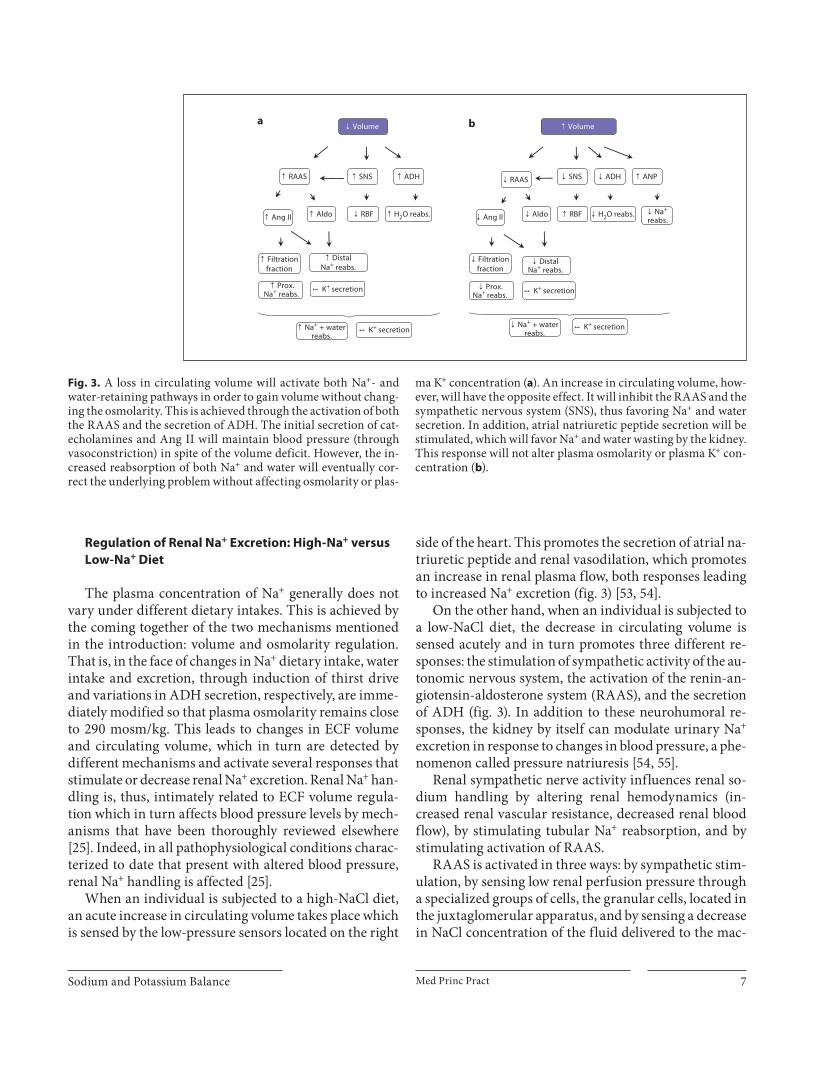
The plasma concentration of Na + generally does not vary under different dietary intakes. This is achieved by the coming together of the two mechanisms mentioned in the introduction: volume and osmolarity regulation. That is, in the face of changes in Na + dietary intake, water intake and excretion, through induction of thirst drive and variations in ADH secretion, respectively, are imme-diately modified so that plasma osmolarity remains close to 290 mosm/kg. This leads to changes in ECF volume and circulating volume, which in turn are detected by different mechanisms and activate several responses that stimulate or decrease renal Na + excretion. Renal Na + han-dling is, thus, intimately related to ECF volume regula-tion which in turn affects blood pressure levels by mech-anisms that have been thoroughly reviewed elsewhere [25] . Indeed, in all pathophysiological conditions charac-terized to date that present with altered blood pressure, renal Na + handling is affected [25] .
When an individual is subjected to a high-NaCl diet, an acute increase in circulating volume takes place which is sensed by the low-pressure sensors located on the right
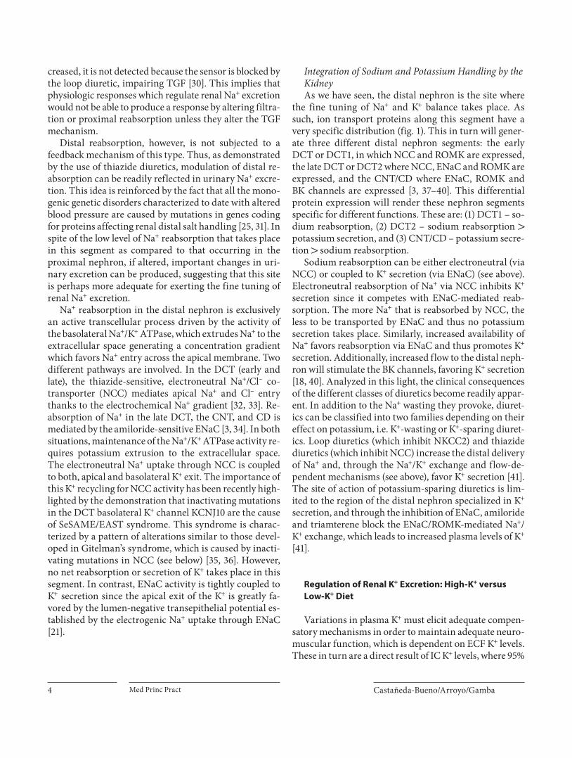
side of the heart. This promotes the secretion of atrial na-triuretic peptide and renal vasodilation, which promotes an increase in renal plasma flow, both responses leading to increased Na + excretion ( fig. 3 ) [53, 54] .
On the other hand, when an individual is subjected to a low-NaCl diet, the decrease in circulating volume is sensed acutely and in turn promotes three different re-sponses: the stimulation of sympathetic activity of the au-tonomic nervous system, the activation of the renin-an-giotensin-aldosterone system (RAAS), and the secretion of ADH ( fig. 3 ). In addition to these neurohumoral re-sponses, the kidney by itself can modulate urinary Na + excretion in response to changes in blood pressure, a phe-nomenon called pressure natriuresis [54, 55] .
Renal sympathetic nerve activity influences renal so-dium handling by altering renal hemodynamics (in-creased renal vascular resistance, decreased renal blood flow), by stimulating tubular Na + reabsorption, and by stimulating activation of RAAS.
RAAS is activated in three ways: by sympathetic stim-ulation, by sensing low renal perfusion pressure through a specialized groups of cells, the granular cells, located in the juxtaglomerular apparatus, and by sensing a decrease in NaCl concentration of the fluid delivered to the mac-
f Volume F Volume
F RAAS F SNS F ADH
F Filtrationfraction
f RBF
F Prox.Na+ reabs.
F H2O reabs.
, K+ secretion , K+ secretion
F AldoF Ang II
F DistalNa+ reabs.
F Na+ + waterreabs.
f Na+ + waterreabs.
, K+ secretion
f RAAS f SNS f ADH
f Filtrationfraction
F RBF
f Prox.Na+ reabs.
f H2O reabs.f Aldof Ang II
f DistalNa+ reabs.
, K+ secretion
F ANP
f Na+
reabs.
a b
Fig. 3. A loss in circulating volume will activate both Na + - and water-retaining pathways in order to gain volume without chang-ing the osmolarity. This is achieved through the activation of both the RAAS and the secretion of ADH. The initial secretion of cat-echolamines and Ang II will maintain blood pressure (through vasoconstriction) in spite of the volume deficit. However, the in-creased reabsorption of both Na + and water will eventually cor-rect the underlying problem without affecting osmolarity or plas-
ma K + concentration ( a ). An increase in circulating volume, how-ever, will have the opposite effect. It will inhibit the RAAS and the sympathetic nervous system (SNS), thus favoring Na + and water secretion. In addition, atrial natriuretic peptide secretion will be stimulated, which will favor Na + and water wasting by the kidney. This response will not alter plasma osmolarity or plasma K + con-centration ( b ).
Castañeda-Bueno/Arroyo/Gamba Med Princ Pract 8
ula densa. RAAS activation involves stimulation of renin secretion by the granular cells [56] . The enzyme renin promotes the conversion of angiotensinogen to angioten-sin I, which in turn is converted to Ang II by the angio-tensin-converting enzyme (ACE). Ang II exerts various effects on the kidney and also stimulates aldosterone se-cretion by the adrenal cortex which will in turn increase volume retention by the kidney [56] . Ang II modifies re-nal hemodynamics by decreasing GFR and increasing the filtration fraction [57] , stimulates proximal Na + reab-sorption by promoting activity of the apical Na + /H + ex-changer and the basolateral Na + bicarbonate cotrans-porter [58–60] , and also stimulates distal Na + reabsorp-tion through the actions of Ang II and aldosterone on membrane transport proteins (see below). All these ac-tions promote Na + retention. As mentioned before, the kidney possesses a mechanism called TGF through which NaCl delivery to the macula densa is sensed and GFR is modulated accordingly. As we have seen, this mechanism normally prevents changes in GFR and can also prevent changes in distal NaCl delivery caused by changes in proximal reabsorption, i.e. decreased proximal reabsorp-tion would be compensated with a decrease in GFR, which will in turn correct distal delivery. Thus, given the existence of this mechanism, can the RAAS really exert an effect upon urinary sodium excretion through its ef-fects on renal hemodynamics and proximal reabsorp-tion? The explanation that has been given to support the idea that indeed it can, is that Ang II can increase the sen-sitivity of the TGF mechanism by stimulating macula densa NaCl transport. That is, in the presence of high Ang II, a decrease in NaCl delivery to this site can occur without compensatory changes in GFR [55, 61] . In any case, modulation of distal Na + transport by Ang II and aldosterone could be a more effective way of altering uri-nary Na + excretion, given that changes in reabsorption at this point are not subjected to the scrutiny of the TGF mechanisms. Additionally, at this nephron site, a much smaller proportion of Na + reabsorption takes place, which, nevertheless, if affected, can have a high impact on final urinary excretion. This is the reason why distal nephron has been seen as the site at which the fine tuning of urinary Na + excretion takes place [3] .
An interesting observation concerning Na + and K + balance can be made regarding aldosterone action. As mentioned above, aldosterone stimulates the Na + /K + ATPase, the K + channel ROMK and the Na+ channel ENaC expressed in the ASDN [46] . When K + levels vary, the modulation of these proteins affects urinary K + excre-tion, leading to correction of plasma K + levels. In contrast,
when aldosterone is secreted in response to a low-salt diet, the purpose of stimulating these proteins is to in-crease distal Na + reabsorption while K + excretion should remain unchanged. That is, even when the system for K + secretion is predicted to be activated, only Na + reabsorp-tion must occur. This phenomenon has been described under the name of ‘aldosterone paradox’ [62, 63] . The clue must be that the system for K + excretion is not really ac-tivated. The reason must lie in a condition that differs between both situations in which aldosterone is stimu-lated, high plasma K + or hypovolemia.
It is well known that distal Na + delivery can affect K + secretion by the ROMK/ENaC system (see above) be-cause when less Na + is available for reabsorption through ENaC, the electrical driving force for K + secretion is not generated [4, 64] . In addition, Na + delivery is related to flow delivery to the distal nephron and, under low Na + /flow delivery, flow-sensitive K + channels (BK channels) are inhibited [18] . Thus, reduced distal delivery during low Na + intake may contribute to inhibition of K + excre-tion under high aldosterone levels. This reduced Na + de-livery may be due to the actions of sympathetic activity and Ang II on renal hemodynamics and proximal reab-sorption. Moreover, recent evidence suggests that Ang II can induce activation of the DCT-specific NCC [65–67] ( fig. 2 ). Indeed, NCC surface expression is stimulated in rats on a low-salt diet [68] . Activation of this cotrans-porter in the first two segments of the distal nephron (DCT1 and DCT) promotes Na + reabsorption and has a negative effect upon K + secretion: the opposite picture of what happens with the administration of thiazide-type diuretics which block NCC (see above). That activation of NCC can lead to a reduced K + secretion and inactiva-tion has the opposite effect is also demonstrated by ge-netic disorders in which NCC activity is affected. Pa-tients with pseudohypoaldosteronism type II (PHAII) develop high blood pressure with hyperkalemia and these alterations are thought to be mainly caused by in-creased NCC activity [69–72] . On the other hand, pa-tients with Gitelman’s syndrome, caused by inactivating mutations in the gene SLC12A3 that encodes for NCC, present with hypotension and hypokalemia [73, 74] and one of the known secondary effects of antihypertensive therapy with thiazide-type diuretics is precisely hypoka-lemia [74] .
Nevertheless, decreased distal Na + delivery for ENaC-mediated reabsorption and decreased distal flow prob-ably do not completely explain the different effects of aldosterone under hypovolemia and hyperkalemia or, in other words, the independence of regulation of Na + and
Sodium and Potassium Balance Med Princ Pract 9
K + balance. The reason is that prevention of K + secretion by diminished distal Na + delivery has to involve a nega-tive effect upon ENaC-mediated reabsorption, ques-tioning the idea that ENaC activation during hypovole-mia is important for achieving Na + retention. Therefore, ENaC activity during hypovolemia must be at some lev-el enhanced but uncoupled to K + secretion. The rela-tively recent description of a group of kinases that dif-ferentially regulate the activity of several renal epithe-lial transport proteins has shed light in this direction (see next section).
The WNK System in the Independent Regulation of
Na + and K + Balance
So far, we have seen that regulation of Na + and K + bal-ance is orchestrated by the differential action of various epithelial transport proteins under distinct physiological conditions. However, a more detailed description of the specific regulatory mechanisms is warranted. A family of serine/threonine kinases which lack a highly con-served catalytic lysine residue and are thus named the with-no-lysine (K) or WNK kinases have been shown to be key players in the regulation of NCC, ENaC and ROMK. The WNK family in mammals is conformed of 4 different proteins named WNK 1–4, which consist of an N-terminal domain, a highly conserved catalytic do-main, and a C-terminal domain [75, 76] . Interestingly, a splice variant of WNK1 produces a kidney-specific vari-ant of WNK1 (KS-WNK1). This KS-WNK1 lacks the N-terminus and most of the kinase domain of WNK1. Three of the four members of the WNK family (WNK1, 3 and 4) are expressed in the distal nephron and are ca-pable of modulating the function of transport proteins [62, 70, 76–78] . The role of the WNKs as regulators of ion transport in the distal nephron became evident when Lifton’s group [70] discovered that mutations in the genes that encode either WNK1 or WNK4 are the cause of PHAII also known as familial hyperkalemic hyperten-sion (FHHt). As mentioned above, patients with PHAII present with hyperkalemia, hypertension and metabolic acidosis, which are corrected with low doses of thiazides [71] . This evidence highlighted the importance of the WNK kinases as regulators of Na + and K + homeostasis. A large amount of knowledge has been generated regard-ing their function and interactions. However, for the purpose of this work, we will focus on their role as inter-mediaries in the hormonal effects upon distal nephron ion transport proteins.
Regulation of NCC by the WNK Kinase System The discovery of the WNK system has begun to shed
light on how the regulation of NCC takes place. WNK4 is coexpressed with NCC [70] and it has been shown in vitro that it functions as a negative regulator of NCC activity [79] , apparently, at least in part, through lysosomal-me-diated endocytosis [80] . This inhibitory effect is lost in the WNK4 harboring a PHAII-type mutation, leading to activation of NCC [79] . Additionally, WNK4 mouse mod-els generated by Lalioti et al. [72] corroborated these find-ings as the mice which carried two extra alleles of wild-type WNK4 were hypotensive and the mice that carried two extra alleles of WNK4 with a PHAII mutation were hypertensive.
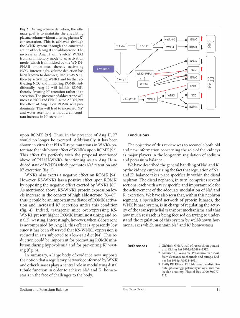
WNK1, in which mutations causing PHAII have also been found, can also modify NCC function. WNK1 serves as an indirect upregulator of NCC by inhibiting WNK4 [81] . Evidence shows that WNK1 is able to inhib-it the WNK4-mediated inhibition of NCC, thus activat-ing NCC [81] . All PHAII-type mutations in WNK1 gene consist of deletions in intron 1 which lead to overexpres-sion of the kinase, thus providing a plausible molecular mechanism behind the WNK1 mutations that generate PHAII [70] . Interestingly, another indirect regulator of NCC function is KS-WNK1. KS-WNK1 inhibits WNK1 and therefore abrogates the WNK1-mediated inhibition of WNK4. Thus, KS-WNK1 functions as an indirect in-hibitor of NCC [82] .
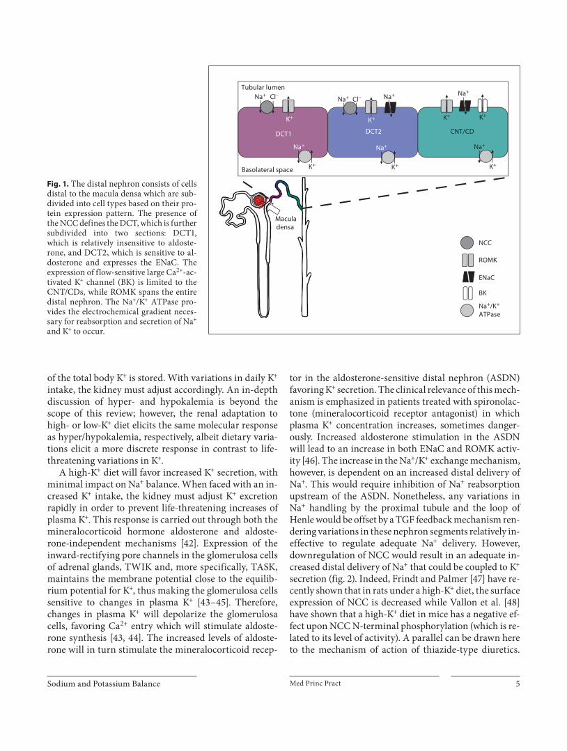
As discussed previously, regulation of NCC is crucial for both Na + and K + handling by the kidney. The WNK system is one of the mechanisms that allow the differen-tial regulation of NCC and, thus, Na + and K + ions. The presence of aldosterone without Ang II, i.e. hyperkale-mia, leads to an upregulation of KS-WNK1 [83–85] . KS-WNK1 is presumed to inhibit NCC, thereby increasing the distal delivery of Na + and favoring the ENaC-ROMK Na + and K + exchange mechanism ( fig. 4 ). Indeed, NCC N-terminal phosphorylation, which has been clearly pos-itively correlated with its level of activation, is inhibited in mice that overexpress KS-WNK1 [86] . Conversely, KS-WNK1 expression is reduced in rats placed on a low salt diet. Accordingly, this may lead to an increase in NCC activity, as further supported by the observation that mice which have a blunted expression of KS-WNK1 have an increased level of NCC phosphorylation ( fig. 5 ) [86, 87] . Additionally, it has been shown that Ang II stimu-lates NCC membrane translocation [66] and it has been shown in vitro that Ang II activates NCC through a WNK4-dependent pathway [65] ( fig. 5 ). This effect has been paralleled with the activation mediated by the

Castañeda-Bueno/Arroyo/Gamba Med Princ Pract 10
WNK4-PHAII mutations, in that mutations in WNK4 which activate NCC mimic Ang II-mediated stimulation of the WNK4-NCC pathway [65] . Phosphorylation and thus activation of NCC by Ang II in vivo has been cor-roborated in adrenalectomized rats, eliminating the pos-sibility that Ang II effects are transduced by aldosterone [67] . This upregulation of NCC in the scenario of hypo-volemia may be important for promoting Na + retention and also for competing with the ENaC-ROMK Na + and K + exchange mechanism, thereby preventing K + wasting in the context of high aldosterone.
Regulation of ENaC by the WNK Kinase System In addition to the well-known aldosterone-mediated
regulation of ENaC through the serum-glucocorticoid-induced kinase 1 (SGK1)-neural precursor cell expressed, developmentally down-regulated 4-2) (Nedd4-2) (SGK1-Nedd4-2) pathway [88] , ENaC is also regulated by the WNK kinase system. In vitro studies suggest that WNK4, which functions as a negative regulator of ENaC, can be inhibited by aldosterone-induced SGK1 through phos-phorylation of serine 1169 in the C-terminal region ( fig. 4 ) [46, 89] . Thus, the WNK4-mediated inhibition of ENaC would be abrogated during high-aldosterone states, i.e. hypovolemia and hyperkalemia ( fig. 4 , 5 ). This, however, is different from the regulation of NCC in that KS-WNK1, which functions as an inhibitor of NCC [81] , activates ENaC ( fig. 4 ). Indeed, transfection of KS-WNK1 increas-es amiloride-sensitive currents in mineralocorticoid re-
ceptor-transfected, M1 cells [83] . The increased expres-sion of KS-WNK1 and activation of ENaC during high-aldosterone states would be warranted, albeit different to the regulation of NCC. KS-WNK1 induced by high K + or aldosterone would inhibit NCC but would disinhibit ENaC, thus favoring K + secretion aided with distal deliv-ery of Na + . The role of WNK1 as a regulator of ENaC is apparently through the SGK1 pathway, as WNK1 phos-phorylates SGK1 and thus activates ENaC [90] , although there is still no evidence demonstrating the modulation of this kinase in high-aldosterone states. The exact role of the interaction between WNK1-KS, WNK1 and WNK4 with regard to the regulation of ENaC still remains to be elucidated.
Regulation of ROMK by the WNK Kinase System In addition to NCC, ROMK is another protein that is
‘switch’ regulated by the WNK kinase system. WNK4 functions as an inhibitor of ROMK under steady-state conditions. However, the phosphorylation of WNK4 on serine 1169 by SGK1 inhibits WNK4 and thus releases the WNK4-mediated inhibition of ROMK [46] . This effect would be important when the induction of SGK1 by al-dosterone is necessary to promote K + secretion ( fig. 4 ). In the presence of hypovolemia, this would prove counter-intuitive. However, Ang II is capable of restoring inhibi-tion in the presence of SGK1 [91] ( fig. 5 ). Although this pathway has been elucidated in an in vitro system, there is also in vivo evidence of the negative effect of Ang II
F Plasma K+ F Aldo
F KS-WNK1
F SGK1
ENaC
WNK1
WNK4 NCC
ROMK
Nedd4-2 ENaC
WNK4
*
DCT2/CNT/CD
*
Fig. 4. The effects of aldosterone on the WNK kinase system dur-ing an increase in plasma K + are directed towards increasing the distal delivery of Na + , while favoring the ENaC/ROMK-mediated Na + and K + exchange mechanism. This is achieved through the upregulation of KS-WNK1 (predominantly in the DCT) which will inhibit WNK1, thus releasing the WNK1-mediated inhibi-tion of WNK4, which will lead to an increased inhibitory effect of WNK4 on NCC in DCT1. This increased distal delivery of Na +
will be coupled with an increase in ENaC and ROMK mediated by SGK1 which will phosphorylate both WNK4 and Nedd4-2, thereby inhibiting them and releasing ENaC and ROMK, facilitat-ing the exchange of Na + for K + . In addition, KS-WNK1 upregula-tion may also be important for promoting the increase in ENaC- and ROMK-mediated Na + /K + exchange as suggested by in vivo and in vitro experimental evidence (see text). * These effects are lost due to the action of SGK1 on WNK4 in the ASDN.
Sodium and Potassium Balance Med Princ Pract 11
upon ROMK [92] . Thus, in the presence of Ang II, K + would no longer be excreted. Additionally, it has been shown in vitro that PHAII-type mutations in WNK4 po-tentiate the inhibitory effect of WNK4 upon ROMK [93] . This effect fits perfectly with the proposal mentioned above of PHAII-WNK4 functioning as an Ang II-in-duced state of WNK4 which promotes Na + retention and K + excretion ( fig. 5 ).
WNK1 also exerts a negative effect on ROMK [94] . However, KS-WNK1 has a positive effect upon ROMK, by opposing the negative effect exerted by WNK1 [85] . As mentioned above, KS-WNK1 protein expression lev-els increase in the context of high aldosterone [83–85] , thus it could be an important mediator of ROMK activa-tion and increased K + secretion under this condition ( fig. 4 ). Indeed, transgenic mice overexpressing KS-WNK1 pre sent higher ROMK immunostaining and re-nal K + wasting. Interestingly, however, when aldosterone is accompanied by Ang II, this effect is apparently lost since it has been observed that KS-WNK1 expression is reduced in rats subjected to a low-salt diet [84] . This re-duction could be important for promoting ROMK inhi-bition during hypovolemia and for preventing K + wast-ing ( fig. 5 ).
In summary, a large body of evidence now supports the notion that a regulatory network conformed by WNK and other kinases plays a central role in modulating distal tubule function in order to achieve Na + and K + homeo-stasis in the face of challenges to the body.
Conclusions
The objective of this review was to reconcile both old and new information concerning the role of the kidneys as major players in the long-term regulation of sodium and potassium balance.
We have described the general handling of Na + and K + by the kidney, emphasizing the fact that regulation of Na + and K + balance takes place specifically within the distal nephron. The distal nephron, in turn, comprises several sections, each with a very specific and important role for the achievement of the adequate modulation of Na + and K + excretion. We have also seen that, within this nephron segment, a specialized network of protein kinases, the WNK kinase system, is in charge of regulating the activ-ity of the transepithelial transport mechanisms and that now much research is being focused on trying to under-stand the regulation of this system by well-known hor-monal axes which maintain Na + and K + homeostasis.
f Volume
F Aldo
f KS-WNK1
F SGK1
ENaC
WNK1WNK4 NCC
ROMK
Nedd4-2 ENaC
WNK4
*
F Ang II
ROMK
WNK4
WNK4-PHAIINCC
ENaC
DCT2 CNT CD
ROMK
DCT1DCT2 CNT CD
Fig. 5. During volume depletion, the ulti-mate goal is to maintain the circulating plasma volume without altering plasma K + concentration. This is achieved through the WNK system through the concerted action of both Ang II and aldosterone. The increase in Ang II will ‘switch’ WNK4 from an inhibitory mode to an activation mode (which is mimicked by the WNK4-PHAII mutations), thereby activating NCC. Interestingly, volume depletion has been known to downregulate KS-WNK1, thereby activating WNK1 and further ac-tivating NCC and inhibiting ROMK. Ad-ditionally, Ang II will inhibit ROMK, thereby favoring K + retention rather than secretion. The presence of aldosterone will increase NCC and ENaC in the ASDN, but the effect of Ang II on ROMK will pre-dominate. This will lead to increased Na + and water retention, without a concomi-tant increase in K + secretion.
References 1 Giebisch GH: A trail of research on potassi-um. Kidney Int 2002; 62: 1498–1512.
2 Giebisch G, Wang W: Potassium transport: from clearance to channels and pumps. Kid-ney Int 1996; 49: 1624–1631.
3 Reilly RF, Ellison DH: Mammalian distal tu-bule: physiology, pathophysiology, and mo-lecular anatomy. Physiol Rev 2000; 80: 277–313.

Castañeda-Bueno/Arroyo/Gamba Med Princ Pract 12
4 Malnic G, Klose RM, Giebisch G, Seldin DW: Micropuncture study of renal potassium ex-cretion in the rat. 1964. J Am Soc Nephrol 2000; 11: 1354–1369.
5 Okusa MD, Unwin RJ, Velazquez H, Giebisch G, Wright FS: Active potassium absorption by the renal distal tubule. Am J Physiol 1992; 262:F488–F493.
6 Adrogue HJ, Madias NE: Sodium and potas-sium in the pathogenesis of hypertension. N Engl J Med 2007; 356: 1966–1978.
7 O’Neil RG, Hayhurst RA: Functional differ-entiation of cell types of cortical collecting duct. Am J Physiol 1985; 248:F449–F453.
8 Wright FS: Sites and mechanisms of potas-sium transport along the renal tubule. Kid-ney Int 1977; 11: 415–432.
9 Grantham JJ, Kurg MB, Obloff J: The nature of transtubular Na and K transport in iso-lated rabbit renal collecting tubules. J Clin Invest 1970; 49: 1815–1826.
10 Sansom SC, Agulian S, Muto S, Illig V, Giebisch G: K activity of CCD principal cells from normal and DOCA-treated rabbits. Am J Physiol 1989; 256:F136–F142.
11 Muto S: Potassium transport in the mamma-lian collecting duct. Physiol Rev 2001; 81: 85–116.
12 Wang WH, Giebisch G: Regulation of potas-sium (K) handling in the renal collecting duct. Pflügers Arch 2009; 458: 157–168.
13 Ellison DH, Velazquez H, Wright FS: Stimu-lation of distal potassium secretion by low lu-men chloride in the presence of barium. Am J Physiol 1985; 248:F638–F649.
14 Velazquez H, Ellison DH, Wright FS: Chlo-ride-dependent potassium secretion in early and late renal distal tubules. Am J Physiol 1987; 253:F555–F562.
15 Woda CB, Bragin A, Kleyman TR, Satlin LM: Flow-dependent K + secretion in the cor-tical collecting duct is mediated by a maxi-K channel. Am J Physiol Renal Physiol 2001; 280:F786–F793.
16 Taniguchi J, Imai M: Flow-dependent activa-tion of maxi K + channels in apical membrane of rabbit connecting tubule. J Membr Biol 1998; 164: 35–45.
17 Grantham JJ, Kurg MB, Obloff J: The nature of transtubular Na and K transport in iso-lated rabbit renal collecting tubules. J Clin Invest 1970; 49: 1815–1826.
18 Grimm PR, Irsik DL, Settles DC, Holtzclaw JD, Sansom SC: Hypertension of Kcnmb1–/– is linked to deficient K secretion and aldoste-ronism. Proc Natl Acad Sci USA 2009; 106: 11800–11805.
19 McDonald FJ, Price MP, Snyder PM, Welsh MJ: Cloning and expression of the � - and � -subunits of the human epithelial sodium channel. Am J Physiol 1995; 268:C1157–C1163.
20 Ahn YJ, Brooker DR, Kosari F, Harte BJ, LiJ, Mackler SA, Kleyman TR: Cloning and functional expression of the mouse epithe-lial sodium channel. Am J Physiol 1999; 277:F121–F129.
21 Chang SS, Grunder S, Hanukoglu A, Rosler A, Mathew P M, Hanukoglu I, Schild L, Lu Y, Shimkets RA, Nelson-Williams C, Rossier BC, Lifton RP: Mutations in subunit of the epithelial sodium channel cause salt wasting with hyperkalaemic acidosis, pseudohypoal-dosteronism type 1. Nat Genet 1996; 12: 248–253.
22 Rubera I, Loffing J, Palmer LG, Frindt G, Fowler-Jaeger N, Sauter D, Carroll T, McMa-hon A, Hummler E, Rossier BC: Collecting duct-specific gene inactivation of � ENaC in the mouse kidney does not impair sodium and potassium balance. J Clin Invest 2003; 112: 554–565.
23 Christensen BM, Perrier R, Wang Q, Zuber AM, Maillard M, Mordasini D, Malsure S, Ronzaud C, Stehle JC, Rossier BC, Hummler E: Sodium and potassium balance depends on � ENaC expression in connecting tubule. J Am Soc Nephrol 2010; 21: 1942–1951.
24 Kleta R, Bockenhauer D: Bartter syndromes and other salt-losing tubulopathies. Neph-ron Physiol 2006; 104: 73–80.
25 Lifton RP, Gharavi AG, Geller DS: Molecular mechanisms of human hypertension. Cell 2001; 104: 545–556.
26 Arroyo JP, Ronzaud C, Lagnaz D, Staub O, Gamba G: Aldosterone paradox: differential regulation of ion transport in distal nephron. Physiology (Bethesda) 2011; 26: 115–123.
27 Oppermann M, Mizel D, Huang G, Li C, Deng C, Theilig F, Bachmann S, Briggs J, Schnermann J, Castrop H: Macula densa control of renin secretion and preglomerular resistance in mice with selective deletion of the B isoform of the Na,K,2Cl co-transport-er. J Am Soc Nephrol 2006; 17: 2143–2152.
28 Oppermann M, Mizel D, Kim SM, Chen L, Faulhaber-Walter R, Huang Y, Li C, Deng C, Briggs J, Schnermann J, Castrop H: Renal function in mice with targeted disruption of the A isoform of the Na-K-2Cl co-transport-er. J Am Soc Nephrol 2007; 18: 440–448.
29 Vallon V: Tubuloglomerular feedback in the kidney: insights from gene-targeted mice. Pflügers Arch 2003; 445: 470–476.
30 Plata C, Meade P, Vazquez N, Hebert SC, Gamba G: Functional properties of the api-cal Na + -K + -2Cl– cotransporter isoforms. J Biol Chem 2002; 277: 11004–11012.
31 Lifton RP: Molecular genetics of human blood pressure variation. Science 1996; 272: 676–680.
32 Ellison DH, Velazquez H, Wright FS: Thia-zide-sensitive sodium chloride cotransport in early distal tubule. Am J Physiol 1987; 253(3 pt 2):F546–F554.
33 Gamba G: Molecular physiology and patho-physiology of the electroneutral cation-chlo-ride cotransporters. Physiol Rev 2005; 85: 423–493.
34 Fuller CM, Awayda MS, Arrate MP, Bradford AL, Morris RG, Canessa CM, Rossier BC, Benos DJ: Cloning of a bovine renal epithe-lial Na + channel subunit. Am J Physiol 1995; 269:C641–C654.
35 Scholl UI, Choi M, Liu T, Ramaekers VT, Hausler MG, Grimmer J, Tobe SW, Farhi A, Nelson-Williams C, Lifton RP: Seizures, sensorineural deafness, ataxia, mental retar-dation, and electrolyte imbalance (SeSAME syndrome) caused by mutations in KCNJ10. Proc Natl Acad Sci USA 2009; 106: 5842–5847.
36 Bockenhauer D, Feather S, Stanescu HC, Bandulik S, Zdebik AA, Reichold M, Tobin J, Lieberer E, Sterner C, Landoure G, Arora R, Sirimanna T, Thompson D, Cross JH, van’t Hoff W, Al Masri O, Tullus K, Yeung S,Anikster Y, Klootwijk E, Hubank M, Dillon MJ, Heitzmann D, Arcos-Burgos M, Knep-per MA, Dobbie A, Gahl WA, Warth R, Sheridan E, Kleta R: Epilepsy, ataxia, senso-rineural deafness, tubulopathy, and KCNJ10 mutations. N Engl J Med 2009; 360: 1960–1970.
37 Loffing J, Kaissling B: Sodium and calcium transport pathways along the mammalian distal nephron: from rabbit to human. Am J Physiol Renal Physiol 2003; 284:F628–F643.
38 Loffing J, Loffing-Cueni D, Valderrabano V, Klausli L, Hebert SC, Rossier BC, Hoen-derop JG, Bindels RJ, Kaissling B: Distribu-tion of transcellular calcium and sodium transport pathways along mouse distal nephron. Am J Physiol Renal Physiol 2001; 281:F1021–F1027.
39 Obermuller N, Bernstein P, Velázquez H, Reilly R, Moser D, Ellison D H, Bachman S: Expression of the thiazide-sensitive Na-Cl cotransporter in rat and human kidney. Am J Physiol 1995; 269:F900–F910.
40 Pluznick JL, Wei P, Grimm PR, Sansom SC: BK- � 1 subunit: immunolocalization in the mammalian connecting tubule and its role in the kaliuretic response to volume expan-sion. Am J Physiol Renal Physiol 2005; 288:F846–F854.
41 Ernst ME, Moser M: Use of diuretics in pa-tients with hypertension. N Engl J Med 2009; 361: 2153–2164.
42 Matsubara M: Renal sodium handling for body fluid maintenance and blood pressure regulation. Yakugaku Zasshi 2004; 124: 301–309.
43 Brenner T, O’Shaughnessy KM: Both TASK-3 and TREK-1 two-pore loop K channels are expressed in H295R cells and modulate their membrane potential and aldosterone secre-tion. Am J Physiol Endocrinol Metab 2008; 295:E1480–E1486.
44 Davies LA, Hu C, Guagliardo NA, Sen N, Chen X, Talley EM, Carey RM, Bayliss DA, Barrett PQ: TASK channel deletion in mice causes primary hyperaldosteronism. Proc Natl Acad Sci USA 2008; 105: 2203–2208.
45 Czirjak G, Fischer T, Spat A, Lesage F, Enye-di P: TASK (TWIK-related acid-sensitive K + channel) is expressed in glomerulosa cells of rat adrenal cortex and inhibited by angioten-sin II. Mol Endocrinol 2000; 14: 863–874.

Sodium and Potassium Balance Med Princ Pract 13
46 Ring AM, Leng Q, Rinehart J, Wilson FH, Kahle KT, Hebert SC, Lifton RP: An SGK1 site in WNK4 regulates Na + channel and K + channel activity and has implications for al-dosterone signaling and K + homeostasis. Proc Natl Acad Sci USA 2007; 104: 4025–4029.
47 Frindt G, Palmer LG: Effects of dietary K on cell-surface expression of renal ion channels and transporters. Am J Physiol Renal Physiol 2010; 299:F890–F897.
48 Vallon V, Schroth J, Lang F, Kuhl D, Uchida S: Expression and phosphorylation of the Na-Cl-cotransporter NCC in vivo is regulat-ed by dietary salt, potassium and SGK1. Am J Physiol Renal Physiol 2009; 297:F704–F712.
49 Huang CL, Kuo E: Mechanisms of disease: WNK-ing at the mechanism of salt-sensitive hypertension. Nat Clin Pract Nephrol 2007; 3: 623–630.
50 Pluznick JL, Sansom SC: BK channels in the kidney: role in K + secretion and localization of molecular components. Am J Physiol Re-nal Physiol 2006; 291:F517–F529.
51 Estilo G, Liu W, Pastor-Soler N, Mitchell P, Carattino MD, Kleyman TR, Satlin LM: Ef-fect of aldosterone on BK channel expression in mammalian cortical collecting duct. Am J Physiol Renal Physiol 2008; 295:F780–F788.
52 Liu W, Morimoto T, Woda C, Kleyman TR, Satlin LM: Ca 2+ dependence of f low-stimu-lated K secretion in the mammalian cortical collecting duct. Am J Physiol Renal Physiol 2007; 293:F227–F235.
53 West DB, Wehberg KE, Kieswetter K, Gran-ger JP: Blunted natriuretic response to an acute sodium load in obese hypertensive dogs. Hypertension 1992; 19(suppl I):I-96–I-100.
54 Knox FG, Granger JP: Control of Sodium Ex-cretion: An Integrative Approach. Oxford, Oxford University Press, 1992, pp 927–967.
55 Granger JP, Hall JE: Role of the kidney in hy-pertension; in Lip GYP, Hall JE (eds): Com-prehensive Hypertension. Amsterdam, Else-vier, 2007, pp 241–263.
56 Hall JE, Brands MW: The renin-angioten-sin-aldosterone systems – renal mechanisms and circulatory homeostasis; in Seldin DW, Giebisch GH (eds): The Kidney: Physiology and Pathophysiology. New York, Raven Press, 1992, pp 1010–1045.
57 Olsen ME, Hall JE, Montani JP, Guyton AC, Langford HG, Cornell JE: Mechanisms of angiotensin II natriuresis and antinatriure-sis. Am J Physiol 1985; 249:F299–F307.
58 Schuster VL, Kokko JP, Jacobson HR: Angio-tensin II directly stimulates sodium trans-port in rabbit proximal convoluted tubules. J Clin Invest 1984; 73: 507–515.
59 Houillier P, Chambrey R, Achard JM, Frois-sart M, Poggioli J, Paillard M: Signaling pathways in the biphasic effect of angioten-sin II on apical Na/H antiport activity in proximal tubule. Kidney Int 1996; 50: 1496–1505.
60 He P, Klein J, Yun CC: Activation of Na + /H + exchanger NHE3 by angiotensin II is medi-ated by inositol 1,4,5-triphosphate (IP 3 ) re-ceptor-binding protein released with IP 3 (IRBIT) and Ca 2+ /calmodulin-dependent protein kinase II. J Biol Chem 2010; 285: 27869–27878.
61 Schnermann J, Levine DZ: Paracrine factors in tubuloglomerular feedback: adenosine, ATP, and nitric oxide. Annu Rev Physiol 2003; 65: 501–529.
62 McCormick JA, Yang CL, Ellison DH: WNK kinases and renal sodium transport in health and disease. An integrated view. Hyperten-sion 2008; 51: 588–596.
63 Arroyo JP, Ronzaud C, Lagnaz D, Staub O, Gamba G: Aldosterone paradox: differential regulation of ion transport in distal nephron. Physiology (Bethesda) 2011; 26: 115–123.
64 Good DW, Wright FS: Luminal influences on potassium secretion: sodium concentra-tion and fluid flow rate. Am J Physiol 1979; 236:F192–F205.
65 San Cristobal P, Pacheco-Alvarez D, Rich-ardson C, Ring AM, Vazquez N, Rafiqi FH, Chari D, Kahle KT, Leng Q, Bobadilla NA, Hebert SC, Alessi DR, Lifton RP, Gamba G: Angiotensin II signaling increases activityof the renal Na-Cl cotransporter througha WNK4-SPAK-dependent pathway. Proc Natl Acad Sci USA 2009; 106: 4384–4389.
66 Sandberg MB, Riquier AD, Pihakaski-Maunsbach K, McDonough AA, Maunsbach AB: Angiotensin II provokes acute traffick-ing of distal tubule NaCl cotransporter (NCC) to apical membrane. Am J Physiol Re-nal Physiol 2007; 293:F662–F669.
67 van der LN, Lim CH, Fenton RA, Meima ME, Jan Danser AH, Zietse R, Hoorn EJ: Angio-tensin II induces phosphorylation of thethiazide-sensitive sodium chloride cotrans-porter independent of aldosterone. Kidney Int 2010; 79: 66–76.
68 Frindt G, Palmer LG: Surface expression of sodium channels and transporters in rat kid-ney: Effects of dietary Na. Am J Physiol Re-nal Physiol 2009; 297:F1249–F1255.
69 Gordon RD, Hodsman GP: The syndrome of hypertension and hyperkalaemia without re-nal failure: long-term correction by thiazide diuretic. Scott Med J 1986; 31: 43–44.
70 Wilson FH, Disse-Nicodeme S, Choate KA, Ishikawa K, Nelson-Williams C, Desitter I, Gunel M, Milford DV, Lipkin GW, Achard JM, Feely MP, Dussol B, Berland Y, Unwin RJ, Mayan H, Simon DB, Farfel Z, Jeunemai-tre X, Lifton RP: Human hypertension caused by mutations in WNK kinases. Sci-ence 2001; 293: 1107–1112.
71 Mayan H, Vered I, Mouallem M, Tzadok-Witkon M, Pauzner R, Farfel Z: Pseudohy-poaldosteronism type II: marked sensitivity to thiazides, hypercalciuria, normomagne-semia, and low bone mineral density. J Clin Endocrinol Metab 2002; 87: 3248–3254.
72 Lalioti MD, Zhang J, Volkman HM, Kahle KT, Hoffmann KE, Toka HR, Nelson-Wil-liams C, Ellison DH, Flavell R, Booth CJ, Lu Y, Geller DS, Lifton RP: WNK4 controls blood pressure and potassium homeostasis via regulation of mass and activity of the dis-tal convoluted tubule. Nat Genet 2006; 38: 1124–1132.
73 Gitelman HJ, Graham JB, Welt LG: A new family disorder characterized by hypokale-mia and hypomagnesemia. Trans Assoc Am Physicians 1966; 79: 221–235.
74 Simon DB, Nelson-Williams C, Johnson-Bia M, Ellison D, Karet FE, Morey-Molina A, Vaara I, Iwata F, Cushner HM, Koolen M, Gainza FJ, Gitelman HJ, Lifton RP: Gitel-man’s variant of Bartter’s syndrome, inher-ited hypokalaemic alkalosis, is caused by mutations in the thiazide-sensitive Na-Cl cotransporter. Nat Genet 1996; 12: 24–30.
75 Hadchouel J, Delaloy C, Faure S, Achard JM, Jeunemaitre X: Familial hyperkalemic hy-pertension. J Am Soc Nephrol 2006; 17: 208–217.
76 Gamba G: Role of WNK kinases in regulat-ing tubular salt and potassium transport and in the development of hypertension. Am J Physiol Renal Physiol 2005; 288:F245–F252.
77 Rinehart J, Kahle KT, De Los HP, Vazquez N, Meade P, Wilson FH, Hebert SC, Gimenez I, Gamba G, Lifton RP: WNK3 kinase is a pos-itive regulator of NKCC2 and NCC, renal cation-Cl – cotransporters required for nor-mal blood pressure homeostasis. Proc Natl Acad Sci USA 2005; 102: 16777–16782.
78 San Cristobal P, De Los HP, Ponce-Coria J, Moreno E, Gamba G: WNK Kinases, Re -nal ion transport and hypertension. Am J Nephrol 2008; 28: 860–870.
79 Wilson FH, Kahle KT, Sabath E, Lalioti MD, Rapson AK, Hoover RS, Hebert SC, Gamba G, Lifton RP: Molecular pathogenesis ofinherited hypertension with hyperkalemia: The Na-Cl cotransporter is inhibited by wild-type but not mutant WNK4. Proc Natl Acad Sci USA 2003; 100: 680–684.
80 Subramanya AR, Liu J, Ellison DH, Wade JB, Welling PA: WNK4 Diverts the thiazide-sensitive NaCl cotransporter to the lysosome and stimulates AP-3 interaction. J Biol Chem 2009; 284: 18471–18480.
81 Yang CL, Angell J, Mitchell R, Ellison DH: WNK kinases regulate thiazide-sensitive Na-Cl cotransport. J Clin Invest 2003; 111: 1039–1045.
82 Subramanya AR, Yang CL, Zhu X, Elli-son DH: Dominant-negative regulation of WNK1 by its kidney-specific kinase-defec-tive isoform. Am J Physiol Renal Physiol 2006; 290:F619–F624.
83 Naray-Fejes-Toth A, Snyder PM, Fejes-Toth G: The kidney-specific WNK1 isoform is in-duced by aldosterone and stimulates epithe-lial sodium channel-mediated Na + trans-port. Proc Natl Acad Sci USA 2004; 101: 17434–17439.
Castañeda-Bueno/Arroyo/Gamba Med Princ Pract 14
84 O’Reilly M, Marshall E, Macgillivray T, Mit-tal M, Xue W, Kenyon CJ, Brown RW: Di-etary electrolyte-driven responses in the re-nal WNK kinase pathway in vivo. J Am Soc Nephrol 2006; 17: 2402–2413.
85 Wade JB, Fang L, Liu J, Li D, Yang CL, Subra-manya AR, Maouyo D, Mason A, Ellison DH, Welling PA: WNK1 kinase isoform switch regulates renal potassium excretion. Proc Natl Acad Sci USA 2006; 103: 8558–8563.
86 Liu Z, Xie J, Wu T, Truong T, Auchus RJ, Huang CL: Downregulation of NCC and NKCC2 cotransporters by kidney-specific WNK1 revealed by gene disruption and transgenic mouse models. Hum Mol Genet 2010; 20: 855–866.
87 Hadchouel J, Soukaseum C, Busst C, Zhou XO, Baudrie V, Zurrer T, Cambillau M, Elg-hozi JL, Lifton RP, Loffing J, Jeunemaitre X: Decreased ENaC expression compensates the increased NCC activity following inacti-vation of the kidney-specific isoform of WNK1 and prevents hypertension. Proc Natl Acad Sci USA 2010; 107: 18109–18114.
88 Debonneville C, Flores SY, Kamynina E, Plant PJ, Tauxe C, Thomas MA, Munster C, Chraibi A, Pratt JH, Horisberger JD, Pearce D, Loffing J, Staub O: Phosphorylation of Nedd4-2 by SGK1 regulates epithelial Na + channel cell surface expression. EMBO J 2001; 20: 7052–7059.
89 Ring AM, Cheng SX, Leng Q, Kahle KT, Rinehart J, Lalioti MD, Volkman HM, Wil-son FH, Hebert SC, Lifton RP: WNK4 regu-lates activity of the epithelial Na + channel in vitro and in vivo. Proc Natl Acad Sci USA 2007; 104: 4020–4024.
90 Xu BE, Stippec S, Chu PY, Lazrak A, Li XJ, Lee BH, English JM, Ortega B, Huang CL, Cobb MH: WNK1 activates SGK1 to regulate the epithelial sodium channel. Proc Natl Acad Sci USA 2005; 102: 10315–10320.
91 Yue P, Sun P, Lin DH, Pan C, Xing W, Wang W: Angiotensin II diminishes the effect of SGK1 on the WNK4-mediated inhibition of ROMK1 channels. Kidney Int 2010; 79: 423–431.
92 Wei Y, Zavilowitz B, Satlin LM, Wang WH: Angiotensin II inhibits the ROMK-like small conductance K channel in renal corti-cal collecting duct during dietary potassium restriction. J Biol Chem 2007; 282: 6455–6462.
93 Kahle KT, Wilson FH, Leng Q, Lalioti MD, O’Connell AD, Dong K, Rapson AK, Mac-Gregor GG, Giebisch G, Hebert SC, Lifton RP: WNK4 regulates the balance between re-nal NaCl reabsorption and K + secretion. Nat Genet 2003; 35: 372–376.
94 Lazrak A, Liu Z, Huang CL: Antagonistic regulation of ROMK by long and kidney-specific WNK1 isoforms. Proc Natl Acad Sci USA 2006; 103: 1615–1620.