Increased Memory Conversion of Naïve CD8 T Cells Activated during Late Phases of Acute Virus...
10
Increased Memory Conversion of Naı ¨ve CD8 T Cells Activated during Late Phases of Acute Virus Infection Due to Decreased Cumulative Antigen Exposure Georgia Fousteri 1. , Amy Dave 1. , Amy Juedes 1 , Therese Juntti 1 , Bret Morin 1 , Lisa Togher 1 , Donna L. Farber 2 , Matthias von Herrath 1 * 1 Diabetes Center, La Jolla Institute for Allergy and Immunology, La Jolla, California, United States of America, 2 Columbia Center for Translational Immunology, Columbia University Medical Center, New York, New York, United States of America Abstract Background: Memory CD8 T cells form an essential part of protective immunity against viral infections. Antigenic load, costimulation, CD4-help, cytokines and chemokines fluctuate during the course of an antiviral immune response thus affecting CD8 T cell activation and memory conversion. Methodology/Principal Findings: In the present study, naı ¨ve TCR transgenic LCMV-specific P14 CD8 T cells engaged at a late stage during the acute antiviral LCMV response showed reduced expansion kinetics but greater memory conversion in the spleen. Such late activated cells displayed a memory precursor effector phenotype already at the peak of the systemic antiviral response, suggesting that the environment determined their fate during antigen encounter. In the spleen, the majority of late transferred cells exhibited a central memory phenotype compared to the effector memory displayed by the early transferred cells. Increasing the inflammatory response by exogenous administration of IFNc, PolyI:C or CpG did not affect memory conversion in the late transferred group, suggesting that the diverging antigen load early versus later during acute infection had determined their fate. In agreement, reduction in the LCMV antigenic load after ribavirin treatment enhanced the contribution of early transferred cells to the long lasting memory pool. Conclusions/Significance: Our results show that naı ¨ve CD8 cells, exposed to reduced duration or concentration of antigen during viral infection convert into memory more efficiently, an observation that could have significant implications for vaccine design. Citation: Fousteri G, Dave A, Juedes A, Juntti T, Morin B, et al. (2011) Increased Memory Conversion of Naı ¨ve CD8 T Cells Activated during Late Phases of Acute Virus Infection Due to Decreased Cumulative Antigen Exposure. PLoS ONE 6(1): e14502. doi:10.1371/journal.pone.0014502 Editor: Laurent Re ´nia, BMSI-A*STAR, Singapore Received July 21, 2010; Accepted December 16, 2010; Published January 6, 2011 Copyright: ß 2011 Fousteri et al. This is an open-access article distributed under the terms of the Creative Commons Attribution License, which permits unrestricted use, distribution, and reproduction in any medium, provided the original author and source are credited. Funding: This work was supported by funds from the Brehm coalition and National Institute of Allergy and Infectious Diseases (NIAID) National Institutes of Health (NIH) grant # AI68818. The funders had no role in study design, data collection and analysis, decision to publish, or preparation of the manuscript. Competing Interests: The authors have declared that no competing interests exist. * E-mail: [email protected] . These authors contributed equally to this work. Introduction The generation of memory T cells is a crucial process for developing novel ways to prevent viral infections and certain forms of cancer [1–3]. Following exposure to antigen, T cells proceed through three defined phases: activation and clonal expansion, contraction and memory conversion [4–6]. Memory T cell development can be influenced by the antigen dose, the strength of the T cell receptor (TCR)-antigen interaction, costimulation, type of antigen presenting cells (APCs), the participation of CD4 helper/regulatory T cells and the cytokines and/or chemokine environment [7–13]. Two major memory T cell populations have been described based on their location and effector functions. Central memory (CM) T cells (CD44 hi CD62L hi CCR7 + ) are located in secondary lymphoid tissues and possess little cytotoxic activity, while effector memory (EM) T cells (CD62L lo CCR7 2 ), which reside in non-lymphoid tissues are cytotoxic and rapidly acquire effector function [14–18]. T cell activation and differentiation during the course of an infection can be influenced by changes in pathogen load [19]. As the amount of antigen decreases during the course of an acute infection, naı ¨ve T cells that are introduced at late stages seem to proliferate less and acquire different properties, such as decreased CD62L down- regulation [20,21]. However, what determines EM versus CM and how the timing of viral infection affects this differentiation process are still open questions in the field. It is not known whether naı ¨ve T cells activated at the peak viral load during antigen abundance, versus peak viral clearance when the antigen load is low, have different capacities for T cell memory formation. In addition to antigen levels, cytokines are known to play crucial roles in memory T cell survival and differentiation [22]. IL-7, one of the most well studied cytokines in mediating survival of naı ¨ve T cells seems to contribute to survival, and to a lesser extent, to basal homeostatic proliferation of memory T cells [23–25]. Upon TCR activation, IL-7Ra (CD127) is initially down-regulated on populations of activated effectors cells and increased CD127 levels PLoS ONE | www.plosone.org 1 January 2011 | Volume 6 | Issue 1 | e14502
Transcript of Increased Memory Conversion of Naïve CD8 T Cells Activated during Late Phases of Acute Virus...

Increased Memory Conversion of Naıve CD8 T CellsActivated during Late Phases of Acute Virus InfectionDue to Decreased Cumulative Antigen ExposureGeorgia Fousteri1., Amy Dave1., Amy Juedes1, Therese Juntti1, Bret Morin1, Lisa Togher1, Donna L.
Farber2, Matthias von Herrath1*
1 Diabetes Center, La Jolla Institute for Allergy and Immunology, La Jolla, California, United States of America, 2 Columbia Center for Translational Immunology, Columbia
University Medical Center, New York, New York, United States of America
Abstract
Background: Memory CD8 T cells form an essential part of protective immunity against viral infections. Antigenic load,costimulation, CD4-help, cytokines and chemokines fluctuate during the course of an antiviral immune response thusaffecting CD8 T cell activation and memory conversion.
Methodology/Principal Findings: In the present study, naıve TCR transgenic LCMV-specific P14 CD8 T cells engaged at alate stage during the acute antiviral LCMV response showed reduced expansion kinetics but greater memory conversion inthe spleen. Such late activated cells displayed a memory precursor effector phenotype already at the peak of the systemicantiviral response, suggesting that the environment determined their fate during antigen encounter. In the spleen, themajority of late transferred cells exhibited a central memory phenotype compared to the effector memory displayed by theearly transferred cells. Increasing the inflammatory response by exogenous administration of IFNc, PolyI:C or CpG did notaffect memory conversion in the late transferred group, suggesting that the diverging antigen load early versus later duringacute infection had determined their fate. In agreement, reduction in the LCMV antigenic load after ribavirin treatmentenhanced the contribution of early transferred cells to the long lasting memory pool.
Conclusions/Significance: Our results show that naıve CD8 cells, exposed to reduced duration or concentration of antigenduring viral infection convert into memory more efficiently, an observation that could have significant implications forvaccine design.
Citation: Fousteri G, Dave A, Juedes A, Juntti T, Morin B, et al. (2011) Increased Memory Conversion of Naıve CD8 T Cells Activated during Late Phases of AcuteVirus Infection Due to Decreased Cumulative Antigen Exposure. PLoS ONE 6(1): e14502. doi:10.1371/journal.pone.0014502
Editor: Laurent Renia, BMSI-A*STAR, Singapore
Received July 21, 2010; Accepted December 16, 2010; Published January 6, 2011
Copyright: � 2011 Fousteri et al. This is an open-access article distributed under the terms of the Creative Commons Attribution License, which permitsunrestricted use, distribution, and reproduction in any medium, provided the original author and source are credited.
Funding: This work was supported by funds from the Brehm coalition and National Institute of Allergy and Infectious Diseases (NIAID) National Institutes ofHealth (NIH) grant # AI68818. The funders had no role in study design, data collection and analysis, decision to publish, or preparation of the manuscript.
Competing Interests: The authors have declared that no competing interests exist.
* E-mail: [email protected]
. These authors contributed equally to this work.
Introduction
The generation of memory T cells is a crucial process for
developing novel ways to prevent viral infections and certain forms
of cancer [1–3]. Following exposure to antigen, T cells proceed
through three defined phases: activation and clonal expansion,
contraction and memory conversion [4–6]. Memory T cell
development can be influenced by the antigen dose, the strength
of the T cell receptor (TCR)-antigen interaction, costimulation,
type of antigen presenting cells (APCs), the participation of CD4
helper/regulatory T cells and the cytokines and/or chemokine
environment [7–13]. Two major memory T cell populations have
been described based on their location and effector functions.
Central memory (CM) T cells (CD44hiCD62LhiCCR7+) are
located in secondary lymphoid tissues and possess little cytotoxic
activity, while effector memory (EM) T cells (CD62LloCCR72),
which reside in non-lymphoid tissues are cytotoxic and rapidly
acquire effector function [14–18].
T cell activation and differentiation during the course of an
infection can be influenced by changes in pathogen load [19]. As the
amount of antigen decreases during the course of an acute infection,
naıve T cells that are introduced at late stages seem to proliferate less
and acquire different properties, such as decreased CD62L down-
regulation [20,21]. However, what determines EM versus CM and
how the timing of viral infection affects this differentiation process
are still open questions in the field. It is not known whether naıve T
cells activated at the peak viral load during antigen abundance,
versus peak viral clearance when the antigen load is low, have
different capacities for T cell memory formation.
In addition to antigen levels, cytokines are known to play crucial
roles in memory T cell survival and differentiation [22]. IL-7, one
of the most well studied cytokines in mediating survival of naıve T
cells seems to contribute to survival, and to a lesser extent, to basal
homeostatic proliferation of memory T cells [23–25]. Upon
TCR activation, IL-7Ra (CD127) is initially down-regulated on
populations of activated effectors cells and increased CD127 levels
PLoS ONE | www.plosone.org 1 January 2011 | Volume 6 | Issue 1 | e14502
was shown to determine effector CD8 T cells destined to become
memory T cells [26]. More recent evidence suggests that
coordinate expression of CD127 and killer cell lectin-like receptor
G1 (KLRG-1), distinguishes short-lived effector cells (SLEC) from
those destined to develop into long-lived memory T cells. SLEC
display a KLRG-1hiCD127lo phenotype, whereas memory
precursor effector cells (MPECs) exhibit a KLRG-1loCD127hi
phenotype [27]. The decision between SLEC and MPEC fates can
be regulated by the inflammatory environment, which subse-
quently induces specific transcriptional programs in primed CD8
T cells [28–30]. In addition, the ability of effector CD8 T cells to
produce IL-2 has been partially associated with stable memory
development [31,32]. Whether the inflammatory environment
and/or antigen load are more predominant regulator of memory
T cell development has not been resolved.
In the present study, we demonstrate that the timing at which
naıve MHC class-I restricted, LCMV-specific, TCR transgenic
(Tg) P14 T cell enter the primary immune response to LCMV can
affect their expansion and capacity to differentiate into memory T
cells. Naıve CD8 T cells activated in conditions of reduced antigen
load during LCMV infection either through late introduction in
infection or after ribavirin anti-viral treatment, converted into
memory more efficiently than naıve CD8 T cells activated early
during infection. As The majority of late transferred cells present
at the peak of the response exhibited a KLRG1lo phenotype,
characteristic of memory precursor CD8 T cells [33]. In addition,
late tranferred cells did not ‘‘contract’’ and remained as memory
cells. They displayed a gradual shift from a CD44hiCD62Llo (EM)
phenotype to a CD44hiCD62Lhi (CM) phenotype and increased
levels of IL-2 production, in agreement with previously published
results [31,32]. By contrast, naıve CD8 T cells transferred cells
early in the course of LCMV infection, prior to peak viral load,
were predominately EM, CD44hiCD62Llo. Increasing the inflam-
matory milieu after treatment with CpG, poly I:C or IFNc had no
significant effect on the late transferred cells, indicating that
antigen load during infection was likely the main factor that
determined their survival and memory conversion. In agreement,
ribavirin treatment significantly reduced LCMV viral load and
consequently the expansion and contraction phases of early
transferred naive P14 TCR Tg cells. The conversion rate of early
transferred naıve CD8 T cells into memory was significantly
augmented, in ribavirin-treated versus untreated mice and was
similar to that of late transferred cells. Our results suggest an
inverse correlation between the degree of antigen-specific
expansion and memory conversion for CD8 T cells, which may
aid in the development of more effective vaccines and perhaps the
treatment of autoimmune, CD8-mediated autoimmune diseases.
Results
Late recruitment of naıve CD8 cells during acute LCMVinfection results in reduced expansion and contractionbut increased memory conversion in the spleen
Introduction of small numbers of TCR-Tg, LCMV-specific
CD8 cells accurately reproduces the natural anti-LCMV response
without profoundly altering viral clearance and T cell expansion
kinetics. In contrast, large number of naıve antigen-specific T cells
can alter the physiological immune response and clearance of
LCMV and the amount of the endogenous physiological cytokines
and chemokines levels [34,35]. Therefore, in order to better mimic
the natural, acute CD8 anti-LCMV T cell response, we chose to
adoptively transfer only relatively small numbers (26103) of
traceable TCR-Tg LCMV-specific CD8 GFP+ T cells (GP33–41-
specific –P14) into C57BL/6 LCMV Arm infected mice. To study
how the timing at which a naıve T-cell enters an antiviral response
affects its proliferation and memory conversion, P14/GFP+ CD8
T cells were either transferred on day 0 (early) or day 3 (late)
postinfection. We reasoned that cells that were transferred later
post infection would have less opportunity to encounter viral
antigen in vivo, because LCMV antigenic load usually peaks 2–3
days after infection and virus is cleared by day 7 from most organs
it shows tropism [36,37]. To circumvent differences in immune
and viral kinetics between the day 0 and day 3 groups, mice that
received P14/GFP+ cells on day 0 also received P14/GFP2 cells
on day 3, while mice that received P14/GFP+ cells on day 3 had
also received P14/GFP2 cells on day 0 (Table 1). As shown in
Fig. 1, numbers of GFP2 GP33–41-specific effector cells on day 8
and day 45 p.i. in both groups were identical and not significantly
different from mice that had received no P14 cells. Thus,
introduction of low numbers of P14 T cells did not significantly
alter the general kinetics of the antiviral immune response and
therefore is a valid approach to differentially track early and late
transferred cells within the GP33–41-specific T cell response against
acute LCMV infection.
C57BL/6 mice were analyzed on days 8, 15 and 45 p.i. for the
presence of GFP+ cells. The percentage or total numbers of
transferred P14/GFP+ CD8 T cells in the spleen, blood, and
mesenteric lymph nodes (mLN) for early and late transferred cells
are shown in Fig. 2A–C. As expected, the response kinetics of the
P14 T cells transferred early were similar to the endogenous
antigen-specific populations: defined clonal expansion, contraction
and memory conversion phases were observed. In contrast, P14/
GFP+ cells transferred 3 days late displayed only a small degree of
proliferation particularly and consistently in the spleen, showing
that T cells that encounter their cognate antigen early during the
immune response make up the majority of the responding
population. The frequency of memory P14/GFP+ cells within the
CD8 population was identical between early and late transferred
groups in all lymphoid organs analyzed (data not shown). However,
when we determined the fate of the total P14 transferred cells
present at the peak of the response by analyzing the percentage of
cells remaining in the contraction and memory phases (normalized
for D8, fold-change), a much greater output in memory cells in the
late transferred compared to the early transferred cells was seen in
the spleen (Fig. 2D–F). Overall, P14 T cells introduced early during
the initial phase of the antiviral immune response exhibited much
greater expansion and contraction rates compared to late ones in
the spleen. On the other hand, cells exposed to reduced viral antigen
in vivo (late), do not expand to the same extent, yet convert to
memory T cells with greater efficiency.
The majority of late transferred CD8 T cells present at thepeak of the anti-LCMV response convert to memorydisplaying a CM phenotype
It is evident from the results discussed above that the majority of
naıve T cells recruited late during the immune response displayed
a higher degree of memory conversion in the spleen. Our results
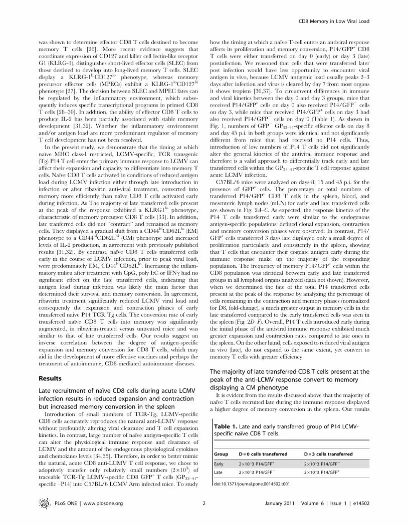
Table 1. Late and early transferred group of P14 LCMV-specific naıve CD8 T cells.
Group D = 0 cells transferred D = 3 cells transferred
Early 2610‘3 P14/GFP+ 2610‘3 P14/GFP2
Late 2610‘3 P14/GFP2 2610‘3 P14/GFP+
doi:10.1371/journal.pone.0014502.t001
CD8 Memory in Low Viral Load
PLoS ONE | www.plosone.org 2 January 2011 | Volume 6 | Issue 1 | e14502
suggest that the cells recruited later during the acute anti-LCMV
response can become memory cells at higher efficiency than naıve
CD8 cells that join the antiviral immune response early and
expand maximally. Next, we wanted to investigate whether the
relative contribution to the CM or EM CD8 T cell pool differs
between the early and late transferred cells. In order to compare
memory subset development between the early and late trans-
ferred cells, additional characterization based on CD44 and
CD62L expression was done. We chose CD62L since it is a key
marker that distinguishes EM from CM memory T cell subsets
along with the expression of CD44. Of note, all P14 cells
transferred late or early displayed CD44 upregulation at the peak
of the response in the spleen (data not shown). Later on, at the
contraction phase, more than 70% of the cells displayed an EM
phenotype CD44hiCD62Llo in both groups (Fig. 3A–B). Interest-
ingly though, while in the early transferred group almost one third
of the cells displayed CM phenotype at the memory phase, a much
greater proportion of cells (.70%) in the late transferred group
displayed CM features (Fig. 3D–E). These phenotypic character-
istics were somewhat different in the mLN and blood, since in both
early and late transferred cells, preferential high levels of both
CD44 and CD62L expression were seen at the contraction and
memory phase, indicating that these cells fall within the CM
population (data not shown).
In addition, in order to differentiate between the functional
capacities of CM and EM cells, IL-2 production from the
CD8+GFP+ population was measured 45 days after infection,
following ex-vivo stimulation with the class I, P14-specific peptide
GP33–41 (Fig. 3C&F). Indeed, IL-2 production was greatly
enhanced in terms of total number by late transferred cells,
consistent with their predominant CM phenotype. This preferen-
tial IL-2 production together with re-expression of CD62L by the
late transferred group suggests that the strength of antigen
stimulation received during the priming phase of the response
was reduced compared to the early transferred cells [20,21]. In
addition, memory cells generated from these late transferred CD8
T cells adopt homing properties, characteristic of the CM subset.
Early and late transferred CD8 cells display similarfunctional characteristics at the effector and memoryphases
As effector T cells differentiate into memory cells, they acquire a
CM or EM phenotype and retain the potential to rapidly produce
IFNc and TNF when exposed to antigen. We compared the ability
of P14 early and late transferred cells to secrete cytokines in
response to antigen during the primary antiviral effector and
memory phase. As shown in Fig. 4, intracellular cytokine staining
after gating on P14/GFP+ cells following stimulation with the class
I-restricted epitope GP33–41 showed no difference in IFNc and
TNF production at the peak of the response day 8 p.i. (D8)
between early and late transferred groups. Similar analysis
conducted at the memory stage day 45 p.i. (D45), in which early
transferred cells displayed a predominantly EM phenotype while
late transferred cells had become CM in the spleen, showed that
both groups exhibited similar cytokine production characteristics
indicative of functional memory. In conclusion, although cells
engaged late in the immune response are exposed to less
inflammatory signals and antigen and therefore proliferate and
contract less, they acquire normal CM characteristics and are fully
able to produce antiviral effector cytokines.
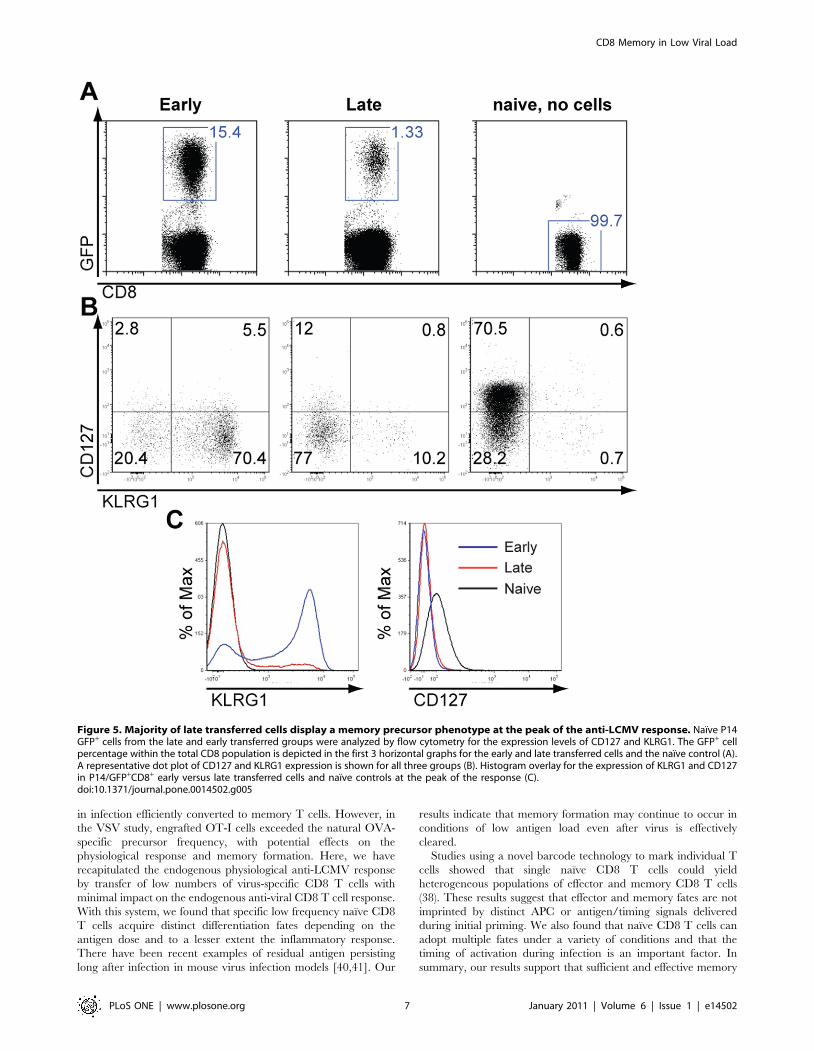
Late transferred cells display a memory precursorphenotype at the peak of the response
Recently it has been suggested that memory cell precursors can
already be identified at the peak of the response by high levels of
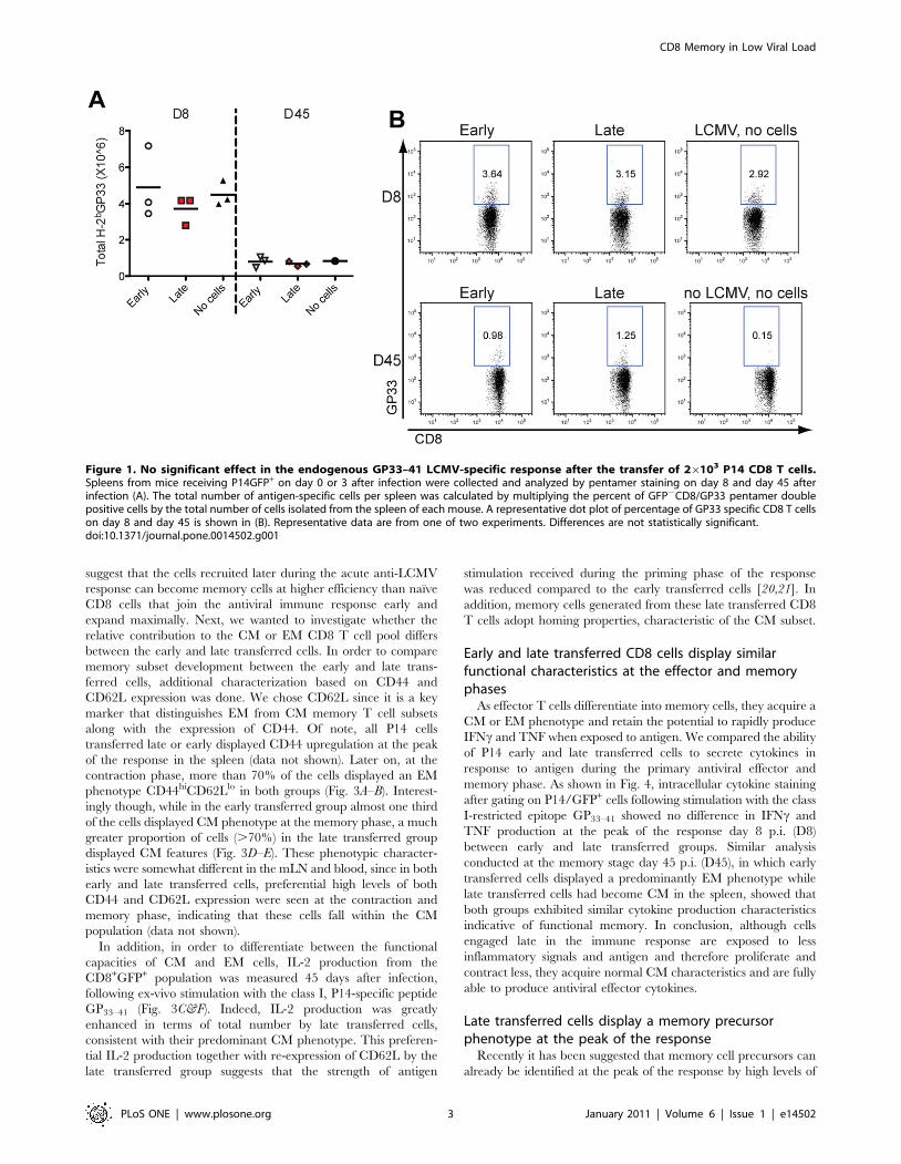
Figure 1. No significant effect in the endogenous GP33–41 LCMV-specific response after the transfer of 26103 P14 CD8 T cells.Spleens from mice receiving P14GFP+ on day 0 or 3 after infection were collected and analyzed by pentamer staining on day 8 and day 45 afterinfection (A). The total number of antigen-specific cells per spleen was calculated by multiplying the percent of GFP2CD8/GP33 pentamer doublepositive cells by the total number of cells isolated from the spleen of each mouse. A representative dot plot of percentage of GP33 specific CD8 T cellson day 8 and day 45 is shown in (B). Representative data are from one of two experiments. Differences are not statistically significant.doi:10.1371/journal.pone.0014502.g001
CD8 Memory in Low Viral Load
PLoS ONE | www.plosone.org 3 January 2011 | Volume 6 | Issue 1 | e14502
CD127 in conjunction with low levels of KLRG1 expression.
Since we observed that the majority of naıve P14 cells that enter
the immune response at a later time point remain as memory, we
performed a phenotypic analysis in order to examine whether the
late transferred cells display a memory precursor phenotype early,
by the eighth day after infection with LCMV. To this end, naıve
P14/GFP+ cells from late and early transferred groups were
analyzed by flow cytometry for the expression levels of CD127 and
KLRG1. Interestingly, the majority of P14 transferred cells had
downregulated the expression levels of CD127 (Fig. 5), which was
even more pronounced in the early transferred group. Important-
ly, comparison of KLRG1 levels detected within the P14/GFP+
cells at the peak of the response (Fig. 5A) between early and late
transferred cells showed a strong correlation of greater memory
formation in the later group with the lower expression of KLRG1
levels in these samples (Fig. 5B–C). Based on the recent
classification for memory precursor effector cells (MPECs), the
IL-7Rahi/KLRG1lo cell frequency was much greater in the late
(13.8%) than early (2.8%) transferred P14/GFP+ cells at the peak
of the anti-LCMV response (Fig. 5B). Collectively, these data
indicate that naıve CD8 T cells recruited later in the antiviral
immune response largely convert into memory and their fate is
determined as early as the initial stages of their activation.
Duration of antigen exposure is the decisive factor formemory T cell fate
Our results prompted us to investigate the signals that are
necessary to enhance memory conversion of the late transferred
cells. Initially we hypothesized that their greater memory
conversion was either due to differences in i) antigen load (less
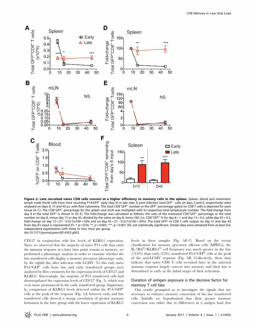
Figure 2. Late recruited naıve CD8 cells convert at a higher efficiency to memory cells in the spleen. Spleen, blood and mesentericlymph node (mLN) cells from mice receiving P14/GFP+ early (day 0) or late (day 3) post infection (and GFP2 cells on days 3 and 0, respectively) wereanalyzed on days 8, 15 and 45 p.i. with flow cytometry. The total CD8+GFP+ number or the GFP+ percentage gated on CD8 T cells is depicted for eachtissue (A–C). The CD8+GFP+ percentage for the spleen and mLN was multiplied with its respective total lymphocyte number. The fold-change fromday 8 in the total GFP+ is shown in (D–E). The fold-change was calculated as follows: the ratio of the measured CD8+GFP+ percentage or the totalnumber on day 8, minus day 15 or day 45, divided by the value on day 8, times 100. (i.e. CD8+GFP+ % for day 8 = 1 and day 15 = 0.5, while day 45 = 0.2,fold-change on day 15 = [(120.5)/1]x100 = 50% and on day 45 = [(120.2)/1]x100 = 20%). The total GFP+ in CD8 T cells output on day 15 and day 45from day 8’s input is represented (F). *, p,0.05, **, p,0.005, ***, p,0.001, NS, not statistically significant. Similar data were obtained from at least fiveindependent experiments with three to four mice per group.doi:10.1371/journal.pone.0014502.g002
CD8 Memory in Low Viral Load
PLoS ONE | www.plosone.org 4 January 2011 | Volume 6 | Issue 1 | e14502
cumulative antigen exposure over time ii) inflammatory stimuli/
activation status of the APCs or iii) due to greater IL-7Ra levels
expressed by P14 cells entering the response at a later stage. In
order to identify parameters other than viral antigenic load that
differ and could affect memory conversion of antiviral CD8 T
cells, mice that had received naıve P14/GFP+ cells late were
treated the day after the cell transfer with polyI:C (100 mg/mouse),
CpG (200 mg/mouse) or recombinant IFNc (50 ng/mouse). As
shown in Fig. S1, treatment with any of these three regiments did
not alter memory conversion. In parallel to these experiments, we
were able to recapitulate the enhanced memory conversion of P14
early transferred naıve CD8 T cells after recombinant IL-7
treatment as previously described (30) (data not shown).
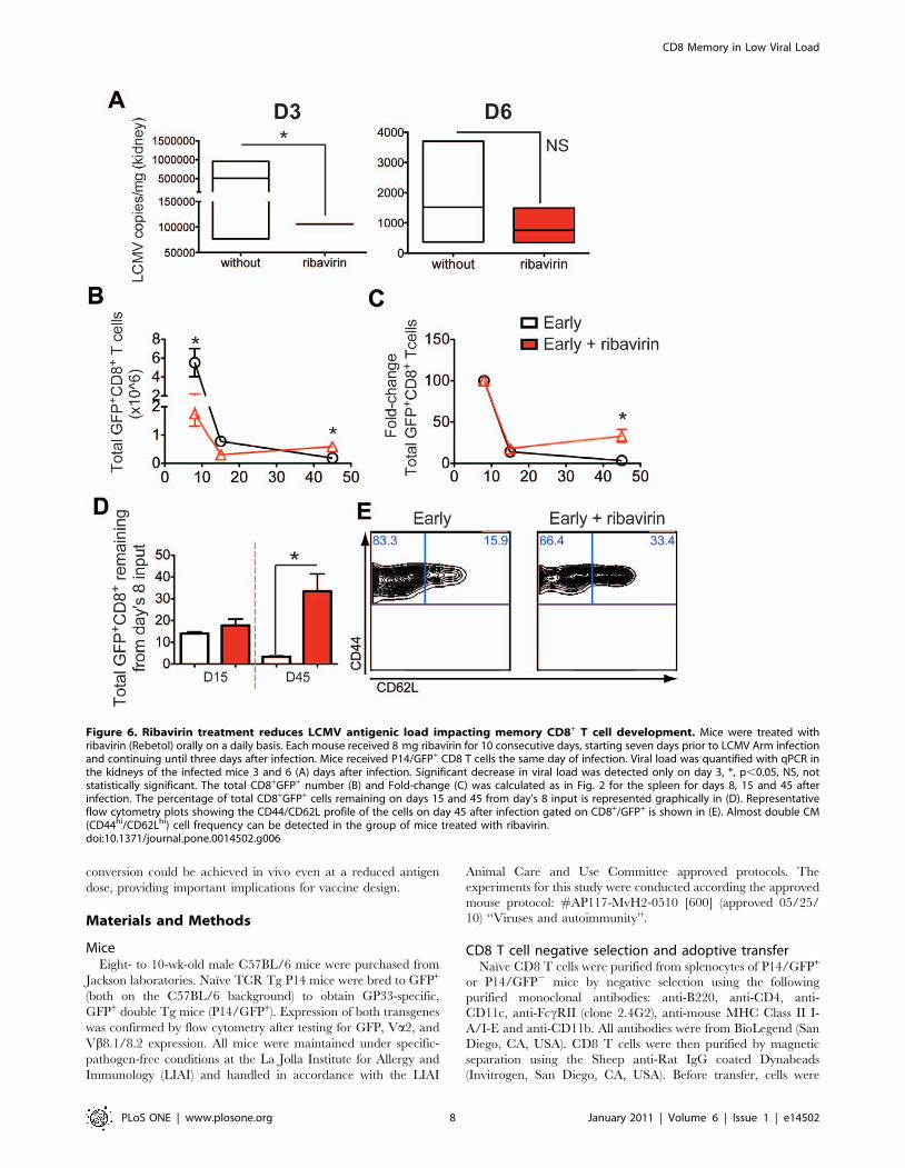
In order to address in more detail whether cumulative antigen
exposure over time was the decisive factor for T cell fate
determination, we took advantage of an additional approach.
P14/GFP+ cells were transferred to LCMV infected recipients
early, which were previously and continuously treated with
ribavirin (Rebetol) orally on a daily basis. Ribavirin is a nucleoside
analog that is an effective antiviral treatment against arenaviruses
[38]. Each recipient mouse received 8 mg ribavirin for 10
consecutive days, starting seven days prior to LCMV infection
and continuing until three days after infection. As shown in Fig. 6A,
ribavirin treatment reduced the LCMV copies in the kidney
significantly three days but not six days after infection. The
reduction in virus levels caused the endogenous GFP2/GP33-
pentamer+ and transferred GFP+/P14 cells to drop at the peak of
the response (Fig. 6B and data not shown). However, and in
agreement with our late versus early transfer approach, memory
development was favored (Fig. 6B–D). Although in this scenario
the contraction phase was not diminished compared to late
transfer experiments, cells contributed again more effectively to
the ensuing memory pool. Importantly, at the memory phase, P14
early transferred cells in the group of mice treated with ribavirin,
there was an increased proportion of CD62Lhi CM cells (Fig. 6E).
Altogether, our results suggest that by reducing the antigen load
alone, we do not compromise memory T cell development but
rather promote differentiation of the CM subset.
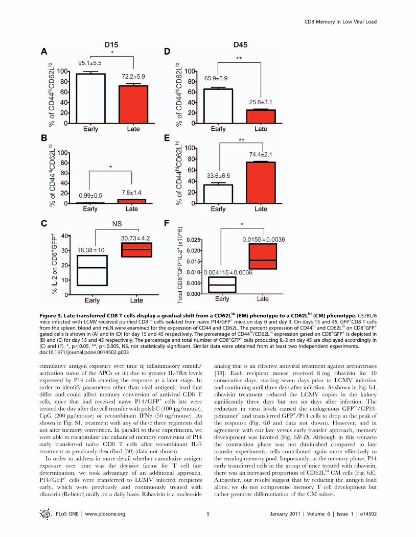
Figure 3. Late transferred CD8 T cells display a gradual shift from a CD62Llo (EM) phenotype to a CD62Lhi (CM) phenotype. C57BL/6mice infected with LCMV received purified CD8 T cells isolated from naıve P14/GFP+ mice on day 0 and day 3. On days 15 and 45, GFP+CD8 T cellsfrom the spleen, blood and mLN were examined for the expression of CD44 and CD62L. The percent expression of CD44hi and CD62Llo on CD8+GFP+
gated cells is shown in (A) and in (D) for day 15 and 45 respectively. The percentage of CD44hi/CD62Lhi expression gated on CD8+GFP+ is depicted in(B) and (E) for day 15 and 45 respectively. The percentage and total number of CD8+GFP+ cells producing IL-2 on day 45 are displayed accordingly in(C) and (F). *, p,0.05, **, p,0.005, NS, not statistically significant. Similar data were obtained from at least two independent experiments.doi:10.1371/journal.pone.0014502.g003
CD8 Memory in Low Viral Load
PLoS ONE | www.plosone.org 5 January 2011 | Volume 6 | Issue 1 | e14502
Discussion
In this study we demonstrated that LCMV-specific naıve CD8
T cells that experience reduced cumulative antigen exposure
during the anti-LCMV response convert more efficiently into
memory CD8 T cells. The majority of cells activated late exhibit
an activated phenotype with CD44 upregulation and CD62L
downregulation at the acute and contraction phases indicative of
previous antigen encounter (data not shown and Fig. 3A–B).
However, cells exposed to low levels of antigen become imprinted
with a distinct long-term differentiation program: such cells do not
expand and consequently do not contract to the same extent, while
they primarily survive as CM cells (Fig. 7). Most CD8 T cells
recruited at a later stage downregulate IL-7Ra levels, while
maintaining low levels of KLRG1 expression at the peak of the
response (Fig. 5). Given their high conversion rate into memory,
perhaps a significant proportion of KLRG1lo/IL-7Ralo cells
should be considered as memory precursor cells, which contradicts
the current classification of MPECs as IL-7Rahi cells. In earlier
studies, constitutive IL-7Ra expression had a minimal effect on the
formation and function of effector and memory CD8 cells,
suggesting that IL-7Ra levels do not identify memory CD8 T cell
precursors (31). KLRG1lo cells though, irrespective their IL-7Ralevels, seem to give rise to IL-7Rahi long-lived memory cells (33).
Taken together, KLRG1 and to a lesser extent IL-7Ra levels seem
to best define memory precursor frequency as early as at the peak
of the response.
Since naıve cells entering the immune response at a later time
point express higher CD127 levels, we investigated whether
blocking IL-7Ra during the contraction phase would affect the
outcome of memory T cell development. However, we did not
observe consistent effects after CD127 blockade on memory CD8
conversion (data not shown). Together, our results suggest that
anti-CD127 treatment at the contraction phase does not have a
clear impact on naıve CD8 T cell memory formation, similar to
what was previously described [12].
Exogenous treatment with inflammatory stimuli could not
reverse preferential memory conversion of the late recruited cells,
suggesting that the antigenic load is likely the main factor that
contributes to memory formation, possibly determined by the
number of T cell to APC contacts. Cells that encounter more
antigen or more recently infected APCs receive stronger antigenic
stimulation and co-stimulation, proliferate more vigorously and
therefore experience greater activation-induced cell death or
become imprinted as senescent effectors or SLECs, thus making
conversion to memory less likely. In agreement, treatment with
ribavirin prior to early P14/GFP+ cell transfer, while reducing the
viral load and the expansion of cells at the peak of the response,
had a positive impact on memory formation. Overall, our results
support the decreasing-potential model (6), which proposes that
one of the main factors controlling memory output of a given
population recruited in the immune response is the duration of
antigen exposure during priming. CD8 T cells recruited later in
the response, despite receiving a weak stimulation, become
imprinted with a memory differentiation program, perhaps
because the received signal is not adequate enough to trigger the
death pathway. In addition, full differentiation into effector cells is
not prerequisite for memory conversion [39]. In agreement with
our findings, during persistent viral infection, prolonged and
strong antigenic encounter decreases the contribution to the
memory pool by constantly eliminating effector T cells and
MPECs [4,40].
Recent reports have also examined the effect of timing and
antigen load on T cell priming and memory development
[21,34,35]. In one study using a vaccinia viral infection model,
the initial burst size of hemagglutinin (HA)-specific CD8 effector T
cells in response to recombinant vaccinia virus encoding HA (rVV-
HA) correlated with the magnitude of the long-term memory pool
size (37). The major caveat of this study was that rather large
numbers of cells were transferred (1610‘5) without the inclusion of
control co-transfers, which may have masked the endogenous
physiological response. Another study, using a recombinant
vesicular stomatitis virus (VSV) -expressing OVA (VSV-OVA)
infection model, found that ‘‘latecomer’’ OT-I cells were not
preferentially recruited to the surviving memory pool (21),
contrasting our results where naıve CD8 T cells activated later
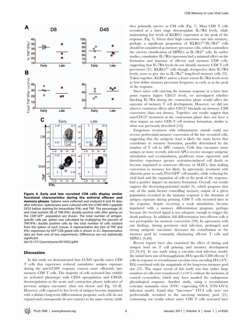
Figure 4. Early and late recruited CD8 cells display similarfunctional characteristics during the antiviral effector andmemory phases. Spleens were collected and analyzed 8 and 45 daysafter infection. Splenocytes were cultured with the LCMV MHC-I peptideGP33 before staining for intracellular IFNc and TNF. The percentage (A)and total number (B) of TNF/IFNc double positive cells after gating onthe CD8+GFP+ population are shown. The total number of antigen-specific cells per spleen was calculated by multiplying the percent ofTNF/IFNc double positive cells by the total number of cells isolatedfrom the spleen of each mouse. A representative dot plot of TNF andIFNc expression by GFP+CD8 gated cells is shown in (C). Representativedata are from one of two experiments. Differences are not statisticallysignificant.doi:10.1371/journal.pone.0014502.g004
CD8 Memory in Low Viral Load
PLoS ONE | www.plosone.org 6 January 2011 | Volume 6 | Issue 1 | e14502
in infection efficiently converted to memory T cells. However, in
the VSV study, engrafted OT-I cells exceeded the natural OVA-
specific precursor frequency, with potential effects on the
physiological response and memory formation. Here, we have
recapitulated the endogenous physiological anti-LCMV response
by transfer of low numbers of virus-specific CD8 T cells with
minimal impact on the endogenous anti-viral CD8 T cell response.
With this system, we found that specific low frequency naıve CD8
T cells acquire distinct differentiation fates depending on the
antigen dose and to a lesser extent the inflammatory response.
There have been recent examples of residual antigen persisting
long after infection in mouse virus infection models [40,41]. Our
results indicate that memory formation may continue to occur in
conditions of low antigen load even after virus is effectively
cleared.
Studies using a novel barcode technology to mark individual T
cells showed that single naıve CD8 T cells could yield
heterogeneous populations of effector and memory CD8 T cells
(38). These results suggest that effector and memory fates are not
imprinted by distinct APC or antigen/timing signals delivered
during initial priming. We also found that naıve CD8 T cells can
adopt multiple fates under a variety of conditions and that the
timing of activation during infection is an important factor. In
summary, our results support that sufficient and effective memory
Figure 5. Majority of late transferred cells display a memory precursor phenotype at the peak of the anti-LCMV response. Naıve P14GFP+ cells from the late and early transferred groups were analyzed by flow cytometry for the expression levels of CD127 and KLRG1. The GFP+ cellpercentage within the total CD8 population is depicted in the first 3 horizontal graphs for the early and late transferred cells and the naıve control (A).A representative dot plot of CD127 and KLRG1 expression is shown for all three groups (B). Histogram overlay for the expression of KLRG1 and CD127in P14/GFP+CD8+ early versus late transferred cells and naıve controls at the peak of the response (C).doi:10.1371/journal.pone.0014502.g005
CD8 Memory in Low Viral Load
PLoS ONE | www.plosone.org 7 January 2011 | Volume 6 | Issue 1 | e14502
conversion could be achieved in vivo even at a reduced antigen
dose, providing important implications for vaccine design.
Materials and Methods
MiceEight- to 10-wk-old male C57BL/6 mice were purchased from
Jackson laboratories. Naıve TCR Tg P14 mice were bred to GFP+
(both on the C57BL/6 background) to obtain GP33-specific,
GFP+ double Tg mice (P14/GFP+). Expression of both transgenes
was confirmed by flow cytometry after testing for GFP, Va2, and
Vb8.1/8.2 expression. All mice were maintained under specific-
pathogen-free conditions at the La Jolla Institute for Allergy and
Immunology (LIAI) and handled in accordance with the LIAI
Animal Care and Use Committee approved protocols. The
experiments for this study were conducted according the approved
mouse protocol: #AP117-MvH2-0510 [600] (approved 05/25/
10) ‘‘Viruses and autoimmunity’’.
CD8 T cell negative selection and adoptive transferNaıve CD8 T cells were purified from splenocytes of P14/GFP+
or P14/GFP2 mice by negative selection using the following
purified monoclonal antibodies: anti-B220, anti-CD4, anti-
CD11c, anti-FccRII (clone 2.4G2), anti-mouse MHC Class II I-
A/I-E and anti-CD11b. All antibodies were from BioLegend (San
Diego, CA, USA). CD8 T cells were then purified by magnetic
separation using the Sheep anti-Rat IgG coated Dynabeads
(Invitrogen, San Diego, CA, USA). Before transfer, cells were
Figure 6. Ribavirin treatment reduces LCMV antigenic load impacting memory CD8+ T cell development. Mice were treated withribavirin (Rebetol) orally on a daily basis. Each mouse received 8 mg ribavirin for 10 consecutive days, starting seven days prior to LCMV Arm infectionand continuing until three days after infection. Mice received P14/GFP+ CD8 T cells the same day of infection. Viral load was quantified with qPCR inthe kidneys of the infected mice 3 and 6 (A) days after infection. Significant decrease in viral load was detected only on day 3, *, p,0.05, NS, notstatistically significant. The total CD8+GFP+ number (B) and Fold-change (C) was calculated as in Fig. 2 for the spleen for days 8, 15 and 45 afterinfection. The percentage of total CD8+GFP+ cells remaining on days 15 and 45 from day’s 8 input is represented graphically in (D). Representativeflow cytometry plots showing the CD44/CD62L profile of the cells on day 45 after infection gated on CD8+/GFP+ is shown in (E). Almost double CM(CD44hi/CD62Lhi) cell frequency can be detected in the group of mice treated with ribavirin.doi:10.1371/journal.pone.0014502.g006
CD8 Memory in Low Viral Load
PLoS ONE | www.plosone.org 8 January 2011 | Volume 6 | Issue 1 | e14502
washed extensively with HBSS-HEPES buffer. 26103 of naıve
P14/GFP+ or P14/GFP2 CD8 T cells were adoptively transferred
into the tail vein (intravenously, i.v.) on days 0 and 3 post infection
(p.i.). Mice that received P14/GFP+ cells early, received P14/
GFP2 late, whereas the ones that received P14/GFP+ cells late,
received P14/GFP2 early.
VirusMice were infected with 104 PFU of LCMV strain Armstrong
(53b) by intraperitoneal (i.p.) injection.
TreatmentsMice were treated once on day 4 after infection with LCMV
with 100 mg/mouse polyI:C (Amersham Pharmacia) i.p. or 50 ng/
mouse recombinant mouse IFNc (BD Pharmingen, San Diego,
CA, USA) i.p. or with 200 mg/mouse CpG i.v. Anti-CD127
(125 mg/mouse) treatments (clone SB/14 BD Pharmingen or
clone A7R34 Biolegend) were conducted i.p. in the contraction
phase on days 8-10-12-14 after LCMV infection. Mice were
treated with ribavirin (Rebetol, USP- NDC 0085-1318-01) orally
on a daily basis. Each mouse received 8 mg ribavirin for 10
consecutive days, starting seven days prior to LCMV infection and
continuing until three days after infection.
Flow cytometrySingle cell suspensions were prepared from spleen, peripheral
blood and mesenteric lymph nodes (MLN) from all groups. After a
2.4G2 block step, cells were stained with the conjugated antibodies
for cell surface markers. PE-conjugated H2-Db/GP33 pentamers
were purchased from ProImmune and stained as previously
described [42]. Directly conjugated antibodies, CD8-PerCP,
CD62L-APC, CD127-PE or PeCy7 (BD Pharmingen), CD44-
APCCy7, CCR7-PeCy7, KLRG1-PE (e-Bioscience, San Diego,
CA, USA) and CD25-PB (Biolegend) were used. For surface
staining, cell suspensions were incubated at 4uC for 30 min. After
surface staining, cells were fixed in 4% paraformaldehyde (Sigma-
Aldrich). DbGP33 and class I pentamers were obtained as PE
conjugates from Proimmune and used as described previously. For
intracellular cytokine analysis, single cell suspensions were
stimulated in vitro for 3 hours with 1 mg/ml MHC class I-
restricted viral peptides GP33–41 (GP33) (Abgent, San Diego, CA,
USA). Cells were stained for surface expression of CD4 and CD8,
fixed, permeabilized, and stained for intracellular IL-2, IFNc and
TNF. After staining, cells were processed on LSRII (BD
Biosciences) and results were analyzed using FlowJo (Tree Star).
Quantitative PCRKidney samples were surgically removed and frozen at 280uC,
then weighed and homogenized. RNA was isolated using the
RNAqueous mini spin column based system (Ambion). RNA was
eluted from RNAaqueous spin columns in a volume of 20 ml. 8.5 ul
of RNA was used in a 10 ml cDNA reaction with SuperScript III
Reverse Transcriptase (SSIII) (Invitrogen, Carlsbad, CA) and a GP-
R primer (S pos. 970–991), GCAACTGCTGTGTTCCCGAAAC
GP-R at 55uC for 1 hr in a programmed PCR thermocycler. 10 ml
of cDNA was used as template for a 25 ml qPCR reaction using
SYBR Green kit (Roche), plated in 96 well plate format and run on
a LightCycler 480 (Roche). Amplification was done for 40 cycles,
with each cycle consisting of two steps: 95uC, 15 sec; 60uC, 30 sec.
All qPCR samples ran in triplicate, with water as a negative control
and LCMV as a positive control. Standard curves were generated
using linearized pSG5-GP plasmid.
Statistical analysisData are expressed as a mean 6 SD. The statistical significance
of the difference between means was determined using the two-
tailed Student’s t-test. *, p,0.05, **, p,0.005, ***, p,0.001.
Supporting Information
Figure S1 Recruitment of late transferred cells into the memory
T cell pool is not influenced by inflammatory agents. Mice were
infected with acute LCMV and received P14/GFP+ CD8 T cells
the same day or 3 days after infection. Groups of mice were
treated 1 day after cell transfer with recombinant mouse IFNc,
polyI:C, CpG, or no treatment, as described in the Materials and
Methods section. The percentage of GFP+ cells remaining on day
28 from day 8’s input is represented graphically.
Found at: doi:10.1371/journal.pone.0014502.s001 (1.01 MB TIF)
Acknowledgments
We sincerely thank Malina McClure for excellent husbandry help, Yang
Cheng for performing the qPCR and Priscilla Colby for administrative
assistance.
Author Contributions
Conceived and designed the experiments: GF AD AJ MGvH. Performed
the experiments: GF AD AJ TJ BM LT. Analyzed the data: GF AD AJ LT.
Contributed reagents/materials/analysis tools: GF AD AJ TJ BM LT DLF
MGvH. Wrote the paper: GF AD DLF MGvH.
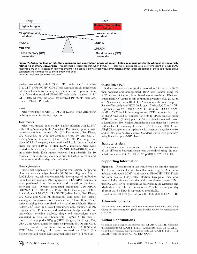
Figure 7. Antigenic load affects the expansion and contraction phase of an anti-LCMV response positively whereas it is inverselyrelated to memory conversion. This schematic represents that when P14/GFP+ T cells were introduced at a later time point of acute LCMVresponse a much less expansion followed by almost no contraction was observed. Nevertheless, a much larger proportion of those cells found on D8converted and contributed to the memory cell pool.doi:10.1371/journal.pone.0014502.g007
CD8 Memory in Low Viral Load
PLoS ONE | www.plosone.org 9 January 2011 | Volume 6 | Issue 1 | e14502
References
1. Badovinac VP, Harty JT (2006) Programming, demarcating, and manipulating
CD8+ T-cell memory. Immunol Rev 211: 67–80.2. Ahmed R, Gray D (1996) Immunological memory and protective immunity:
understanding their relation. Science 272: 54–60.3. Virgin HW, Wherry EJ, Ahmed R (2009) Redefining chronic viral infection.
Cell 138: 30–50.4. Mescher MF, Curtsinger JM, Agarwal P, Casey KA, Gerner M, et al. (2006)
Signals required for programming effector and memory development by CD8+T cells. Immunol Rev 211: 81–92.
5. Williams MA, Bevan MJ (2007) Effector and memory CTL differentiation. Annu
Rev Immunol 25: 171–192.6. Sprent J, Tough DF (2001) T cell death and memory. Science 293: 245–248.
7. Seder RA, Ahmed R (2003) Similarities and differences in CD4+ and CD8+effector and memory T cell generation. Nat Immunol 4: 835–842.
8. Jameson SC, Masopust D (2009) Diversity in T cell memory: an embarrassment
of riches. Immunity 31: 859–871.9. Bachmann MF, Schwarz K, Wolint P, Meijerink E, Martin S, et al. (2004)
Cutting edge: distinct roles for T help and CD40/CD40 ligand in regulating
differentiation of proliferation-competent memory CD8+ T cells. J Immunol173: 2217–2221.
10. Kolumam GA, Thomas S, Thompson LJ, Sprent J, Murali-Krishna K (2005)Type I interferons act directly on CD8 T cells to allow clonal expansion and
memory formation in response to viral infection. J Exp Med 202: 637–650.11. Langenkamp A, Messi M, Lanzavecchia A, Sallusto F (2000) Kinetics of
dendritic cell activation: impact on priming of TH1, TH2 and nonpolarized T
cells. Nat Immunol 1: 311–316.12. Ku CC, Murakami M, Sakamoto A, Kappler J, Marrack P (2000) Control of
homeostasis of CD8+ memory T cells by opposing cytokines. Science 288:675–678.
13. Murakami M, Sakamoto A, Bender J, Kappler J, Marrack P (2002)
CD25+CD4+ T cells contribute to the control of memory CD8+ T cells. ProcNatl Acad Sci U S A 99: 8832–8837.
14. Wherry EJ, Teichgraber V, Becker TC, Masopust D, Kaech SM, et al. (2003)Lineage relationship and protective immunity of memory CD8 T cell subsets.
Nat Immunol 4: 225–234.15. Lefrancois L, Marzo AL (2006) The descent of memory T-cell subsets. Nat Rev
Immunol 6: 618–623.
16. Sallusto F, Lenig D, Forster R, Lipp M, Lanzavecchia A (1999) Two subsets ofmemory T lymphocytes with distinct homing potentials and effector functions.
Nature 401: 708–712.17. Unsoeld H, Pircher H (2005) Complex memory T-cell phenotypes revealed by
coexpression of CD62L and CCR7. J Virol 79: 4510–4513.
18. Reiner SL, Sallusto F, Lanzavecchia A (2007) Division of labor with a workforceof one: challenges in specifying effector and memory T cell fate. Science 317:
622–625.19. Kaech SM, Ahmed R (2001) Memory CD8+ T cell differentiation: initial
antigen encounter triggers a developmental program in naive cells. NatImmunol 2: 415–422.
20. van Faassen H, Saldanha M, Gilbertson D, Dudani R, Krishnan L, et al. (2005)
Reducing the stimulation of CD8+ T cells during infection with intracellularbacteria promotes differentiation primarily into a central (CD62LhighCD44high)
subset. J Immunol 174: 5341–5350.21. D’Souza WN, Hedrick SM (2006) Cutting edge: latecomer CD8 T cells are
imprinted with a unique differentiation program. J Immunol 177: 777–781.
22. Schluns KS, Lefrancois L (2003) Cytokine control of memory T-celldevelopment and survival. Nat Rev Immunol 3: 269–279.
23. Schluns KS, Kieper WC, Jameson SC, Lefrancois L (2000) Interleukin-7mediates the homeostasis of naive and memory CD8 T cells in vivo. Nat
Immunol 1: 426–432.
24. Tan JT, Dudl E, LeRoy E, Murray R, Sprent J, et al. (2001) IL-7 is critical for
homeostatic proliferation and survival of naive T cells. Proc Natl Acad Sci U S A
98: 8732–8737.
25. Tanchot C, Lemonnier FA, Perarnau B, Freitas AA, Rocha B (1997) Differential
requirements for survival and proliferation of CD8 naive or memory T cells.
Science 276: 2057–2062.
26. Kaech SM, Tan JT, Wherry EJ, Konieczny BT, Surh CD, et al. (2003) Selective
expression of the interleukin 7 receptor identifies effector CD8 T cells that give
rise to long-lived memory cells. Nat Immunol 4: 1191–1198.
27. Wakim LM, Bevan MJ (2010) From the thymus to longevity in the periphery.
Curr Opin Immunol 22: 274–278.
28. Joshi NS, Cui W, Chandele A, Lee HK, Urso DR, et al. (2007) Inflammation
directs memory precursor and short-lived effector CD8(+) T cell fates via the
graded expression of T-bet transcription factor. Immunity 27: 281–295.
29. Crotty S, Johnston RJ, Schoenberger SP (2010) Effectors and memories: Bcl-6
and Blimp-1 in T and B lymphocyte differentiation. Nat Immunol 11: 114–120.
30. Kallies A, Xin A, Belz GT, Nutt SL (2009) Blimp-1 transcription factor is
required for the differentiation of effector CD8(+) T cells and memory responses.
Immunity 31: 283–295.
31. Mitchell DM, Ravkov EV, Williams MA (2010) Distinct roles for IL-2 and IL-15
in the differentiation and survival of CD8+ effector and memory T cells.
J Immunol 184: 6719–6730.
32. Williams MA, Tyznik AJ, Bevan MJ (2006) Interleukin-2 signals during priming
are required for secondary expansion of CD8+ memory T cells. Nature 441:
890–893.
33. Hand TW, Morre M, Kaech SM (2007) Expression of IL-7 receptor alpha is
necessary but not sufficient for the formation of memory CD8 T cells during
viral infection. Proc Natl Acad Sci U S A 104: 11730–11735.
34. Obar JJ, Khanna KM, Lefrancois L (2008) Endogenous naive CD8+ T cell
precursor frequency regulates primary and memory responses to infection.
Immunity 28: 859–869.
35. Badovinac VP, Haring JS, Harty JT (2007) Initial T cell receptor transgenic cell
precursor frequency dictates critical aspects of the CD8(+) T cell response to
infection. Immunity 26: 827–841.
36. Fung-Leung WP, Kundig TM, Zinkernagel RM, Mak TW (1991) Immune
response against lymphocytic choriomeningitis virus infection in mice without
CD8 expression. J Exp Med 174: 1425–1429.
37. Cerny A, Sutter S, Bazin H, Hengartner H, Zinkernagel RM (1988) Clearance
of lymphocytic choriomeningitis virus in antibody- and B-cell-deprived mice.
J Virol 62: 1803–1807.
38. Seiler P, Senn BM, Klenerman P, Kalinke U, Hengartner H, et al. (2000)
Additive effect of neutralizing antibody and antiviral drug treatment in
preventing virus escape and persistence. J Virol 74: 5896–5901.
39. Manjunath N, Shankar P, Wan J, Weninger W, Crowley MA, et al. (2001)
Effector differentiation is not prerequisite for generation of memory cytotoxic T
lymphocytes. J Clin Invest 108: 871–878.
40. Jelley-Gibbs DM, Dibble JP, Brown DM, Strutt TM, McKinstry KK, et al.
(2007) Persistent depots of influenza antigen fail to induce a cytotoxic CD8 T cell
response. J Immunol 178: 7563–7570.
41. Takamura S, Roberts AD, Jelley-Gibbs DM, Wittmer ST, Kohlmeier JE, et al.
(2010) The route of priming influences the ability of respiratory virus-specific
memory CD8+ T cells to be activated by residual antigen. J Exp Med 207:
1153–1160.
42. Juedes AE, Rodrigo E, Togher L, Glimcher LH, von Herrath MG (2004) T-bet
controls autoaggressive CD8 lymphocyte responses in type 1 diabetes. J Exp
Med 199: 1153–1162.
CD8 Memory in Low Viral Load
PLoS ONE | www.plosone.org 10 January 2011 | Volume 6 | Issue 1 | e14502