Increased frequencies of CD4+CD25high regulatory T cells in acute dengue infection
7
The Journal of Experimental Medicine BRIEF DEFINITIVE REPORT JEM © The Rockefeller University Press $15.00 Vol. 204, No. 5, May 14, 2007 979–985 www.jem.org/cgi/doi/10.1084/jem.20061381 979 Dengue infection is a major public health prob- lem in tropical areas, predominantly in chil- dren. The mosquito-borne disease is caused by four related serotypes of the dengue virus (DV), a flavivirus like West Nile and yellow fever virus. A wide range of disease severities is ob- served; 100 million cases each year have dengue fever, an acute febrile illness. Half a million patients suffer from the more severe dengue haemorrhagic fever (DHF) with plasma leakage, thrombocytopenia, and occasionally hypovole- mic shock (1). If untreated, the mortality rate of DHF is around 30% but can be reduced to 0.2–5% by fluid replacement. To date, no vac- cines or antiviral therapies are available. The pathogenesis of severe dengue disease is incompletely understood. Epidemiological stud- ies show an increased risk of severe DHF in a secondary infection with a heterologous sero- type. Preexisting nonneutralizing antibodies are thought to form immunocomplexes with the virus, thereby enhancing the infection of Fcγ receptor–bearing cells like monocytes/ macrophages (antibody-dependent enhance- ment; references 2, 3 ). However, it is still un- clear how this can lead to plasma leakage as the major symptom of DHF. Excessive activation of the innate and adaptive immune system, es- pecially of cross-reactive memory T cells, ap- pears to augment the secretion of vasoactive factors like TNF-α, IFN-γ, IL-2, and IL-6 (4–6). Indeed, increased numbers of activated CD8 + T cells and levels of cytokines have been reported Increased frequencies of CD4 + CD25 high regulatory T cells in acute dengue infection Kerstin Lühn, 1 Cameron P. Simmons, 2 Edward Moran, 1 Nguyen Thi Phuong Dung, 2 Tran Nguyen Bich Chau, 2 Nguyen Than Ha Quyen, 2 Le Thi Thu Thao, 3 Tran Van Ngoc, 3 Nguyen Minh Dung, 3 Bridget Wills, 2 Jeremy Farrar, 2 Andrew J. McMichael, 1 Tao Dong, 1 and Sarah Rowland-Jones 1 1 Weatherall Institute of Molecular Medicine, Medical Reasearch Council Human Immunology Unit, John Radcliffe Hospital, University of Oxford, Oxford OX3 9DS, UK 2 Oxford University Clinical Research Unit and 3 The Hospital for Tropical Diseases, District 5, Ho Chi Minh City, Vietnam Dengue virus infection is an increasingly important tropical disease, causing 100 million cases each year. Symptoms range from mild febrile illness to severe hemorrhagic fever. The pathogenesis is incompletely understood, but immunopathology is thought to play a part, with antibody-dependent enhancement and massive immune activation of T cells and monocytes/macrophages leading to a disproportionate production of proinflammatory cytokines. We sought to investigate whether a defective population of regulatory T cells (T reg cells) could be contributing to immunopathology in severe dengue disease. CD4 + CD25 high FoxP3 + T reg cells of patients with acute dengue infection of different severities showed a conventional phenotype. Unexpectedly, their capacity to suppress T cell proliferation and to secrete interleukin-10 was not altered. Moreover, T reg cells sup- pressed the production of vasoactive cytokines after dengue-specific stimulation. Further- more, T reg cell frequencies and also T reg cell/effector T cell ratios were increased in patients with acute infection. A strong indication that a relative rise of T reg cell/effector T cell ratios is beneficial for disease outcome comes from patients with mild disease in which this ratio is significantly increased (P < 0.0001) in contrast to severe cases (P = 0.2145). We conclude that although T reg cells expand and function normally in acute dengue infection, their relative frequencies are insufficient to control the immunopathology of severe disease. CORRESPONDENCE Kerstin Lühn: [email protected] T. Dong and S. Rowland-Jones contributed equally to this paper. The online version of this article contains supplemental material.
-
Upload
independent -
Category
Documents
-
view
2 -
download
0
Transcript of Increased frequencies of CD4+CD25high regulatory T cells in acute dengue infection
The
Journ
al o
f Exp
erim
enta
l M
edic
ine
BRIEF DEFINITIVE REPORT
JEM © The Rockefeller University Press $15.00
Vol. 204, No. 5, May 14, 2007 979–985 www.jem.org/cgi/doi/10.1084/jem.20061381
979
Dengue infection is a major public health prob-lem in tropical areas, predominantly in chil-dren. The mosquito-borne disease is caused by four related serotypes of the dengue virus (DV), a fl avivirus like West Nile and yellow fever virus. A wide range of disease severities is ob-served; 100 million cases each year have dengue fever, an acute febrile illness. Half a million patients suff er from the more severe dengue haemorrhagic fever (DHF) with plasma leakage, thrombocytopenia, and occasionally hypovole-mic shock (1). If untreated, the mortality rate of DHF is around 30% but can be reduced to 0.2–5% by fl uid replacement. To date, no vac-cines or antiviral therapies are available.
The pathogenesis of severe dengue disease is incompletely understood. Epidemiological stud-ies show an increased risk of severe DHF in a secondary infection with a heterologous sero-type. Preexisting nonneutralizing antibodies are thought to form immunocomplexes with the virus, thereby enhancing the infection of Fcγ receptor–bearing cells like monocytes/macrophages (antibody-dependent enhance-ment; references 2, 3). However, it is still un-clear how this can lead to plasma leakage as the major symptom of DHF. Excessive activation of the innate and adaptive immune system, es-pecially of cross-reactive memory T cells, ap-pears to augment the secretion of vasoactive factors like TNF-α, IFN-γ, IL-2, and IL-6 (4–6). Indeed, increased numbers of activated CD8+ T cells and levels of cytokines have been reported
Increased frequencies of CD4+CD25high regulatory T cells in acute dengue infection
Kerstin Lühn,1 Cameron P. Simmons,2 Edward Moran,1 Nguyen Thi Phuong Dung,2 Tran Nguyen Bich Chau,2 Nguyen Than Ha Quyen,2 Le Thi Thu Thao,3 Tran Van Ngoc,3 Nguyen Minh Dung,3 Bridget Wills,2 Jeremy Farrar,2 Andrew J. McMichael,1 Tao Dong,1 and Sarah Rowland-Jones1
1Weatherall Institute of Molecular Medicine, Medical Reasearch Council Human Immunology Unit, John Radcliffe Hospital,
University of Oxford, Oxford OX3 9DS, UK2Oxford University Clinical Research Unit and 3The Hospital for Tropical Diseases, District 5, Ho Chi Minh City, Vietnam
Dengue virus infection is an increasingly important tropical disease, causing 100 million
cases each year. Symptoms range from mild febrile illness to severe hemorrhagic fever. The
pathogenesis is incompletely understood, but immunopathology is thought to play a part,
with antibody-dependent enhancement and massive immune activation of T cells and
monocytes/macrophages leading to a disproportionate production of proinfl ammatory
cytokines. We sought to investigate whether a defective population of regulatory T cells
(T reg cells) could be contributing to immunopathology in severe dengue disease.
CD4+CD25highFoxP3+ T reg cells of patients with acute dengue infection of different
severities showed a conventional phenotype. Unexpectedly, their capacity to suppress T cell
proliferation and to secrete interleukin-10 was not altered. Moreover, T reg cells sup-
pressed the production of vasoactive cytokines after dengue-specifi c stimulation. Further-
more, T reg cell frequencies and also T reg cell/effector T cell ratios were increased in
patients with acute infection. A strong indication that a relative rise of T reg cell/effector
T cell ratios is benefi cial for disease outcome comes from patients with mild disease in which
this ratio is signifi cantly increased (P < 0.0001) in contrast to severe cases (P = 0.2145).
We conclude that although T reg cells expand and function normally in acute dengue
infection, their relative frequencies are insuffi cient to control the immunopathology of
severe disease.
CORRESPONDENCE
Kerstin Lühn:
T. Dong and S. Rowland-Jones contributed equally to this paper.
The online version of this article contains supplemental material.

980 REGULATORY T CELLS IN ACUTE DENGUE INFECTION | Lühn et al.
in DHF (7, 8). Thus, the pathogenesis of dengue infection is likely to be a result of the immune response rather than of the virus itself.
CD4+ regulatory T cells (T reg cells) have been described to play a key role in the control of the immune system. The most commonly used marker is CD25 (IL-2Rα; reference 9), although the recently identifi ed transcription factor FoxP3 is more specifi c (10). Suppression is mediated via direct cell–cell contact or via the secretion of IL-10 or TGF-β (11).
T reg cells play a role not only in autoimmunity, trans-plantation, and cancer but also in infections (12, 13). They can limit immunopathology, which was shown for mouse Helicobacter hepaticus and herpes infections, for example (14, 15). So far, they have only been described in chronic viral infections. This study is the fi rst to report on a functional T reg cell population in a nonpersistent viral infection.
As immunopathology is thought to play a major role in the pathogenesis of severe dengue disease, we hypothesized that the T reg cell response in acute infection may be inade-quate to control immune activation. However, we found that T reg cells are fully functional and expand in acute dengue infection. Moreover, they were able to suppress the produc-tion of vasoactive cytokines such as IFN-γ, TNF-α, and IL-6 in response to dengue antigens. Nevertheless, this expansion might be insuffi cient to control the immune response in patients with severe disease. Strong support for this hypothesis comes from our observation that the relative expansion of T reg cell/eff ector T cell ratios is only signifi cant in children with mild disease in contrast to severe cases (P < 0.0001 and P = 0.2145, respectively).
RESULTS AND D I S C U S S I O N
Characteristics of study cohort
89 serologically confi rmed dengue patients were enrolled and grouped based on their disease severity (Table S1, available at http://www.jem.org/cgi/content/full/jem.20061381/DC1). The acute phase of dengue infection was compared with a recovered time point. Age-matched healthy, uninfected con-trols were compared with the recovered samples (adults only) and showed no diff erence.
Identifi cation of CD4+CD25high T reg cells
Patients’ blood cells were stained ex vivo for CD4, 8, and 25. CD4+ and CD8+ cells were gated in forward/sideward scat-ter plots (Fig. 1 A). CD4+ monocytes were excluded (Fig. 1 B). Human CD25high T reg cells are not a distinct population and, therefore, are diffi cult to gate in a reproducible way. We observed that CD4+ T cells with the highest expression of CD25, which are believed to be T reg cells, show a slight re-duction of CD4 expression (bend of CD4 level). As this was a consistent fi nding, we gated this population of cells in all samples to describe CD4+CD25high T reg cells (Fig. 1 C).
FoxP3 staining of CD4+CD25high T cells
As naive T cells express CD25 upon activation, the CD25high cell population of acutely infected dengue patients might con-tain activated T cells beside T reg cells. The best marker for dis-tinction is FoxP3 (10). To control our CD25-based gating, we stained frozen PBMCs of acutely infected and recovered sub-jects for FoxP3 (Fig. 1 D) or used the corresponding isotype control (not depicted) and analyzed the percentage of FoxP3+
Figure 1. Identifi cation of CD25high T reg cells. Blood cells were
stained ex vivo for CD4 and CD25. (A and B) Lymphocytes were gated (A),
and monocytes were excluded (B). (C) CD25high cells with a slightly de-
creased CD4 expression were gated as T reg cells. (D–G) Frozen PBMCs of
acutely infected and recovered subjects were stained for CD4, CD25, and
FoxP3 (D), and FoxP3 levels of CD25neg, CD25int, and CD25high cells were
analyzed (E–G). FoxP3+ cells show decreased CD4 levels. (H) The percent-
age of FoxP3+ cells in the CD25high population is identical at acute and
recovered states. Error bars represent SEM. Applied gates are depicted as
pink lines.

JEM VOL. 204, May 14, 2007 981
BRIEF DEFINITIVE REPORT
cells in the CD4+CD25negative (CD25neg), CD4+CD25intermediate (CD25int), and CD25high gate (Fig. 1, E–G). If activated CD25+/FoxP3− T cells fell into the CD25high T reg cell gate, we would expect a drop of the percentage of FoxP3+ cells in acute disease in this gate. However, the percentage of FoxP3+ cells was com-parable in acutely infected and recovered subjects (Fig. 1 H). Thus, we confi rmed that even in acute dengue infection, the gated CD4+CD25high T cells are indeed T reg cells and not ac-tivated CD25+ T cells. Therefore, the terms CD25high cells and T reg cells will be used synonymously throughout the study.
In addition, FoxP3 analysis confi rmed our observation that the CD25high cells with less CD4 on their surface are human FoxP3+ T reg cells (Fig. 1, E–G). This reduction of CD4 ex-pression has already been observed on mouse T reg cells (16, 17). Based on this, we suggest that a reduced CD4 expres-sion could be used collaterally for the identifi cation of T reg cells in cases in which FoxP3 cannot be applied as a marker.
T reg cells in dengue infection show
the conventional phenotype
To describe the phenotype of T reg cells in acute dengue in-fection, fresh blood cells of acutely infected patients with dif-ferent severities of recovered as well as uninfected donors were stained ex vivo and analyzed using fl ow cytometry. Expression of the memory marker CD45RO, death receptor CD95, early activation marker CD69, homing receptor CD62L, and in-hibitory receptor CTLA-4 was addressed (Fig. 2, A–E). T reg
cells of uninfected donors and recovered study participants showed the same phenotype (not depicted).
At recovery, the highest percentage of CD45RO+, CD95+, CD62L+, or CTLA-4+ cells was detected in the T reg cell population. CD69 was not expressed. This is in agreement with what has been described previously in healthy donors (18, 19). Interestingly, the expression level of all of these markers was unchanged on T reg cells in acute dengue infection. Signs of activation were only detectable in the CD25int gate, where, for example, CD69 and CTLA-4 ex-pression was increased.
Collectively, the phenotype of T reg cells in acute den-gue infection did not diff er from that seen after recovery. This was the same for all of the diff erent categories of disease severity. As in healthy donors, T reg cells were CD45ROhigh, CD95high, CD69−, CD62Lhigh, and CTLA-4high. In addition, we observed that acutely activated T cells could be found in the CD25int and not in the CD25high gate. Based on those re-sults together with FoxP3 analysis, we suggest that in acute dengue infection, activated T cells express lower levels of CD25 than T reg cells and can clearly be distinguished.
T reg cells suppress the proliferation of CD4+ T cells
in acute dengue infection
We determined the functionality of T reg cells in acute den-gue infection by measuring their capacity to suppress the pro-liferation of CD4+CD25− responder T cells in vitro. Because
Figure 2. T reg cells in dengue infection show conventional pheno-
types. (A–E) Blood cells of acutely infected (n = 8) and recovered (n = 11)
subjects were stained ex vivo for CD4, 25, 45RO (A), 95 (B), 69 (C), 62L
(D), or CTLA-4 (E). CD25neg, CD25int, and CD25high cells were compared
(as indicated). Representative histograms of individual patients are
shown, and numbers indicate relevant percentages of positive cells.

982 REGULATORY T CELLS IN ACUTE DENGUE INFECTION | Lühn et al.
of the amounts of cells needed for the assay, we were re-stricted to samples of dengue-infected adults. CD4+CD25+ and CD4+CD25− T cells were separated from acutely in-fected, recovered, and uninfected subjects. The responder T cells were CFSE labeled and incubated with the same number or without T reg cells on plate-bound anti-CD3 and allogeneic APCs. After 5 d in culture, cells were analyzed by fl ow cytometry. As a negative control, cells were cultured without anti-CD3 (proliferation of <10%; not depicted). Analysis of the proliferation is shown for one acute sample in Fig. 3 A. Lymphocytes, including blasts, were gated and ana-lyzed for their CFSE content. No diff erence was detectable between recovered study participants and uninfected donors (not depicted). The proliferative capacity of CD4+CD25− T cells in acute dengue infection is comparable with that at convalescence: >55% of the cells proliferated (Fig. 3 B). Pro-liferation was reduced drastically upon the addition of T reg cells. The level of suppression was >85% in acute and recov-ered patients (Fig. 3 C). Reduction of the T reg cell/responder T cell ratio to 1:2 did not infl uence the level of suppression (not depicted). Based on these results, we conclude that T reg cells are fully functional in acute dengue infection (i.e., their suppressive capacity is not altered). We suggest that the im-munopathology in dengue disease is not caused by defective T reg cell function.
Culture supernatants were collected from the suppression assays on days 1 and 5, and the IL-10 content was analyzed. Only cultures with T reg cells were substantially positive for IL-10 (Fig. 3 D). The amount of IL-10 produced was comparable in cells of both acutely infected and recovered subjects. Although IL-10 might serve as a possible mean of suppression, determina-tion of the regulatory mechanisms needs further investigation.
T reg cells suppress the production of vasoactive cytokines
after dengue-specifi c stimulation
The eff ect of T reg cells on the dengue-specifi c production of vasoactive cytokines was determined by IFN-γ ELISPOT. PBMCs, CD25-depleted PBMCs (CD25− responder T cells), and T reg cells of 28 acute adult dengue patients were acti-vated with DV-4 peptides plus inactivated virus (DV-2; Fig. 4 A). Stimulation of PBMCs without any, with irrele-vant antigen, or of PBMCs of noninfected donors with den-gue antigen (not depicted) did not result in any notable IFN-γ response. Strikingly, the IFN-γ production of dengue patients’ CD25 responder T cells (CD25−, 1,402 ± 635 spot-forming units/106 cells) was signifi cantly decreased (by around 50%) in the presence of T reg cells (CD25+, T reg cell/responder T cells ≈ 1/10; P = 0.0005). In addition, even the mere depletion of T reg cells from PBMCs led to a signifi cant increase of IFN-γ release by nearly 20% (P = 0.0186). This suggests that T reg cells are eff ective in suppressing cytokine production after dengue-specifi c stimu-lation at cell ratios found physiologically in the blood without the need for in vitro enrichment procedures.
To determine the eff ect of T reg cells on the production of other cytokines, cells of some ELISPOT assays were cul-
tured for another 48 h (Fig. 4 B). Without stimulation, no notable amounts of cytokine were detected. A signifi cant re-duction of TNF-α (P = 0.0386) by 44% was detectable in the presence of enriched T reg cells.
To analyze the eff ect of T reg cells on other blood cells, monocytes, CD25−, and T reg cells were isolated from three
Figure 3. Suppressive capacity of T reg cells is unaltered in acute
dengue infection. (A–D) CD4+ cells were negatively sorted from acutely
infected (n = 7) and recovered (n = 3) subjects and split into CD25+
and CD25− cells. CFSE-labeled CD25− cells were incubated with or with-
out CD25+ cells on plate-bound anti-CD3 and allogeneic APCs. After
5 d, proliferation was analyzed in FACS analysis. (A) A representative
CFSE staining of an acutely infected patient. (B) The percentage of prolif-
erating cells was comparable in acutely infected and recovered patients
and decreased in the presence of CD25+ cells. (C) The suppressive capac-
ity of the T reg cells in acute infection was like after recovery. (D) At the
acute and recovered states, IL-10 was detected on days 1 and 5 in
supernatants of cultures with CD25+ cells. Means and SEM (error bars)
are shown.

JEM VOL. 204, May 14, 2007 983
BRIEF DEFINITIVE REPORT
acute adult patients. The monocytes were cocultured at a 1:1 ratio with CD25− cells or T reg cells and DV-4 peptides for T cell stimulation. After 40 h, monocytes were separated again and activated with antibody-coated inactivated DV-2 for 48 h. Subsequently, IL-6 levels were analyzed. Although the results were not signifi cant, a trend was observed for a decrease of IL-6 production (by 66%; P = 0.0779) by monocytes that were precultured with T reg cells instead of CD25− cells (Fig. 4 C).
From these data, we conclude that T reg cells that can suppress antigen-specifi c responses to dengue can be detected in the blood of acutely infected patients. As demonstrated in vitro, they have an impact on the expression of IFN-γ, TNF-α, and IL-6 produced by T cells and monocytes in response to dengue antigens. This is of major importance, as all of these cytokines have been linked to plasma leakage and, thus, to the severity of dengue disease (4).
T reg cell frequencies and T reg cell/effector T cell ratios are
increased in acute dengue infection, especially in mild cases
The frequency of CD4+CD25high T reg cells was determined in FACS analysis by staining patients’ fresh blood cells ex vivo. The acute samples were collected on days 4–13 of illness. T reg cells were gated, and frequencies were analyzed as a per-centage of CD4+ cells. Absolute CD4 counts were comparable
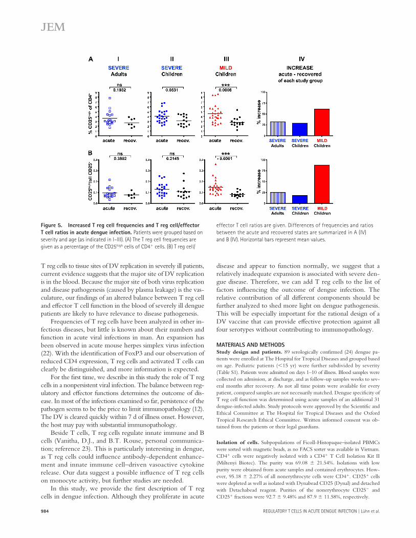
in severe and mild cases. In adult patients, the frequency of T reg cells at the recovered state was comparable with those of healthy controls (2.71 ± 0.88% and 2.37 ± 0.55%, respec-tively; not depicted). In acute infection, the T reg cell fre-quency was slightly increased by 32% to 3.59 ± 1.47%, although it is not statistically signifi cant (Fig. 5 A, I). A diff er-ence of 29% was detectable in children with severe disease (recovered, 3.12 ± 1.03% vs. acute, 4.01 ± 1.3%; Fig. 5 A, II). With P = 0.0531, this increase was close to signifi cance. However, in contrast to this, the T reg cell frequency of chil-dren with mild disease increased substantially by 61% from 2.8 ± 0.97 to 4.52 ± 1.79% (Fig. 5 A, III). This diff erence was highly statistically signifi cant (P = 0.0006; comparable n values for severe- and mild-diseased children). Fig. 5 A (IV) summarizes the results for all three study groups.
More important than the sole increase of T reg cells for the outcome of a disease is the ratio between T reg cells and eff ector T cells. We determined the percentage of all CD25+ cells as a measure for eff ector T cells and calculated the T reg cell/eff ector T cell ratios. In both severely sick adults and chil-dren, the ratios changed in acute infection by 25% and 18%, respectively (Fig. 5 B, I and II). These diff erences were not statistically signifi cant. However, in children with mild disease, the ratios increased highly signifi cantly by 88% at the acute state (P < 0.0001; Fig. 5 B, III). This increase was also noted to a lower extent (38%; P = 0.0192) in a small group of sam-ples collected at an early state of mild acute disease (days 1–3 of illness; not depicted). Fig. 5 B (IV) summarizes the devel-opment of the ratios for the diff erent groups of patients.
In summary, the T reg cell population that expands in acute dengue infection has the conventional phenotype and suppressive capacity. Our demonstration that T reg cells sup-press the dengue-specifi c secretion of vasoactive cytokines of eff ector T cells leads us to suggest that this population plays an active role in dengue infection. Numbers and also T reg cell/eff ector T cell ratios are increased. Despite this expan-sion of functionally intact T reg cells, patients with severe disease still develop strong immunopathology and symptoms that require hospital admission. We propose that in severe disease, the expansion of this T reg cell population is insuffi -cient to control the immune response. This hypothesis is sup-ported by our observation that T reg cell/eff ector T cell ratios are only signifi cantly increased in children with mild and not with severe disease (P < 0.0001 and P = 0.2145, respectively; Fig. 5 B, II–IV). A change in T reg cell/eff ector T cell ratios could have a direct eff ect on the disease severity, as we dem-onstrated that T reg cells in the blood control the eff ector T cell production of vasoactive cytokines, which is directly linked to plasma leakage and disease outcome.
An alternative explanation for the relative defi ciency of T reg cells in the blood of severely sick patients could be diff er-ential migration of these cells to the tissues. It has been dem-onstrated in HIV and hepatitis C virus infection that T reg cells can be detected at sites of preferential virus replication such as the lymphoid tissue or liver (20, 21). Although our data could be compatible with the increased migration of
Figure 4. T reg cells suppress dengue-specifi c vasoactive cytokine
production. (A) IFN-γ ELISPOT using CD25− cells at a 10:1 ratio with
CD25+ (+25+) or CD25− (+25−) cells, PBMCs, or PBMCs depleted of
CD25+ cells (Depl.) of 28 acute dengue patients. No peptide (wo) or DV-4
NS3 peptides and inactivated DV-2 were used as a stimulus for 12 h. In
the presence of CD25+ cells, spot-forming units (SFU)/106 cells are re-
duced (n = 28). (B) Cells of some ELISPOTs were cultured for an additional
48 h to analyze TNF-α levels in supernatants (n = 5). (C) After culturing
monocytes of three acute patients at a 1:1 ratio with CD25− (+25−) or
CD25+ (+25+) cells with DV-4 peptides for 40 h, they were reisolated and
incubated with antibody-coated inactivated DV-2 for another 48 h. IL-6
was measured in the supernatants (n = 3). Wo, no dengue antigens
added. Means and SEM (error bars) are shown.
984 REGULATORY T CELLS IN ACUTE DENGUE INFECTION | Lühn et al.
T reg cells to tissue sites of DV replication in severely ill patients, current evidence suggests that the major site of DV replication is in the blood. Because the major site of both virus replication and disease pathogenesis (caused by plasma leakage) is the vas-culature, our fi ndings of an altered balance between T reg cell and eff ector T cell function in the blood of severely ill dengue patients are likely to have relevance to disease pathogenesis.
Frequencies of T reg cells have been analyzed in other in-fectious diseases, but little is known about their numbers and function in acute viral infections in man. An expansion has been observed in acute mouse herpes simplex virus infection (22). With the identifi cation of FoxP3 and our observation of reduced CD4 expression, T reg cells and activated T cells can clearly be distinguished, and more information is expected.
For the fi rst time, we describe in this study the role of T reg cells in a nonpersistent viral infection. The balance between reg-ulatory and eff ector functions determines the outcome of dis-ease. In most of the infections examined so far, persistence of the pathogen seems to be the price to limit immunopathology (12). The DV is cleared quickly within 7 d of illness onset. However, the host may pay with substantial immunopathology.
Beside T cells, T reg cells regulate innate immune and B cells (Vanitha, D.J., and B.T. Rouse, personal communica-tion; reference 23). This is particularly interesting in dengue, as T reg cells could infl uence antibody-dependent enhance-ment and innate immune cell–driven vasoactive cytokine release. Our data suggest a possible infl uence of T reg cells on monocyte activity, but further studies are needed.
In this study, we provide the fi rst description of T reg cells in dengue infection. Although they proliferate in acute
disease and appear to function normally, we suggest that a relatively inadequate expansion is associated with severe den-gue disease. Therefore, we can add T reg cells to the list of factors infl uencing the outcome of dengue infection. The relative contribution of all diff erent components should be further analyzed to shed more light on dengue pathogenesis. This will be especially important for the rational design of a DV vaccine that can provide eff ective protection against all four serotypes without contributing to immunopathology.
MATERIALS AND METHODSStudy design and patients. 89 serologically confi rmed (24) dengue pa-
tients were enrolled at The Hospital for Tropical Diseases and grouped based
on age. Pediatric patients (<15 yr) were further subdivided by severity
(Table S1). Patients were admitted on days 1–10 of illness. Blood samples were
collected on admission, at discharge, and as follow-up samples weeks to sev-
eral months after recovery. As not all time points were available for every
patient, compared samples are not necessarily matched. Dengue specifi city of
T reg cell function was determined using acute samples of an additional 31
dengue-infected adults. Study protocols were approved by the Scientifi c and
Ethical Committee at The Hospital for Tropical Diseases and the Oxford
Tropical Research Ethical Committee. Written informed consent was ob-
tained from the patients or their legal guardians.
Isolation of cells. Subpopulations of Ficoll-Histopaque–isolated PBMCs
were sorted with magnetic beads, as no FACS sorter was available in Vietnam.
CD4+ cells were negatively isolated with a CD4+ T Cell Isolation Kit II
(Miltenyi Biotec). The purity was 69.08 ± 21.54%. Isolations with low
purity were obtained from acute samples and contained erythrocytes. How-
ever, 95.18 ± 2.27% of all nonerythrocyte cells were CD4+. CD25+ cells
were depleted as well as isolated with Dynabead CD25 (Dynal) and detached
with Detachabead reagent. Purities of the nonerythrocyte CD25− and
CD25+ fractions were 92.7 ± 9.48% and 87.9 ± 11.58%, respectively.
Figure 5. Increased T reg cell frequencies and T reg cell/effector
T cell ratios in acute dengue infection. Patients were grouped based on
severity and age (as indicated in I–III). (A) The T reg cell frequencies are
given as a percentage of the CD25high cells of CD4+ cells. (B) T reg cell/
effector T cell ratios are given. Differences of frequencies and ratios
between the acute and recovered states are summarized in A (IV)
and B (IV). Horizontal bars represent mean values.

JEM VOL. 204, May 14, 2007 985
BRIEF DEFINITIVE REPORT
In vitro coculture proliferation assay. 5 × 10³ CFSE-labeled CD4+CD25−
cells were incubated in RPMI/10% human serum with the same amount or
with 2.5 × 10³ CD4+CD25+ or CD4+CD25− cells in anti-CD3–coated
round-bottom 96-well plates (1 μg/ml OKT3; eBioscience). 3 × 10⁴
mitomycin-C–treated allogeneic EBV-immortalized B cells were added to
each well as APCs. After 5 d, proliferation was analyzed by FACS analysis.
IFN-𝛄 ELISPOT. ELISPOT was performed as described previously (24).
For activation, a pool of 124 peptides of the DV-4 NS3 protein (15 mers;
10-mer overlap; 1.5 μM each), 10 μg/ml of inactivated DV-2 (BiosPacifi c),
or a pool of irrelevant peptides (Mycobacterium tuberculosis ESAT6) were used.
Responses higher than four times the background were considered positive.
Cells and culture media were incubated for another 48 h before analyzing
TNF-α in the supernatant.
Monocyte coculture assay. CD14+ monocytes were sorted (CD14 beads;
Miltenyi Biotec). 105 monocytes and 105 CD25− or T reg cells were cul-
tured in 0.2% gelatine-coated round-bottom 96-well plates with or without
DV-4 NS3 peptide pool. After 40 h, CD14+ cells were sorted again and
cultured with or without inactivated DV-2 that was preincubated with auto-
logous plasma to coat virus particles with antibodies. Supernatants were ana-
lyzed for IL-6 after 48 h.
Flow cytometry. 150 μl of blood was incubated ex vivo with combinations
of four antibodies (BD Biosciences) targeting CD4, 8, 25, 45RO, 62L, 69, 95,
and CTLA-4. Suitable isotype controls were performed. Intracellular CTLA-4
was detected in parallel to FoxP3. Cells were analyzed with a fl ow cytometry
system (FACSCalibur; BD Biosciences) and FlowJo software (Tree Star).
FoxP3 analysis. FoxP3 analysis was performed by fl ow cytometry using the
FoxP3 staining set (clone PCH101; eBioscience).
Cytokine analysis. The levels of secreted IL-10, TNF-α, and IL-6 were
determined in culture supernatants using a Bio-Plex Cytokine Assay (Bio-
Rad Laboratories).
Statistical analysis. Statistical signifi cance was calculated with nonpara-
metric unpaired (Mann-Whitney) or paired (ELISPOT and monocyte assays)
Student’s t tests using Prism 4 software (GraphPad). P-values of <0.05
were regarded as signifi cant.
Online supplemental material. Table S1 presents summary characteristics
of serologically confi rmed dengue cases. Online supplemental material is
available at http://www.jem.org/cgi/content/full/jem.20061381/DC1.
The authors thank all patients and hospital staff as well as Hoang Truong Long,
Mike Salmon, Martin Wild, Jerome Feldmann, and Katja Simon for their help.
K. Luhn was supported by the Max Planck Society and the European Molecular
Biology Organization. C.P. Simmons, E. Moran, B. Willis, and J. Farrar were supported
by the Wellcome Trust. The study was funded by the Medical Research Council and
the Wellcome Trust.
The authors have no confl icting fi nancial interests.
Submitted: 28 June 2006
Accepted: 21 March 2007
R E F E R E N C E S 1. Gubler, D.J. 2002. Epidemic dengue/dengue hemorrhagic fever as a
public health, social and economic problem in the 21st century. Trends Microbiol. 10:100–103.
2. Halstead, S.B., and E.J. O’Rourke. 1977. Dengue viruses and mono-nuclear phagocytes. I. Infection enhancement by non-neutralizing anti-body. J. Exp. Med. 146:201–217.
3. Halstead, S.B. 1979. In vivo enhancement of dengue virus infec-tion in rhesus monkeys by passively transferred antibody. J. Infect. Dis. 140:527–533.
4. Kurane, I., A.L. Rothman, P.G. Livingston, S. Green, S.J. Gagnon, J. Janus, B.L. Innis, S. Nimmannitya, A. Nisalak, and F.A. Ennis. 1994. Immunopathologic mechanisms of dengue hemorrhagic fever and den-gue shock syndrome. Arch. Virol. Suppl. 9:59–64.
5. Beynon, H.L., D.O. Haskard, K.A. Davies, R. Haroutunian, and M.J. Walport. 1993. Combinations of low concentrations of cytokines and acute agonists synergize in increasing the permeability of endothelial monolayers. Clin. Exp. Immunol. 91:314–319.
6. Maruo, N., I. Morita, M. Shirao, and S. Murota. 1992. IL-6 increases endothelial permeability in vitro. Endocrinology. 131:710–714.
7. Mongkolsapaya, J., W. Dejnirattisai, X.N. Xu, S. Vasanawathana, N. Tangthawornchaikul, A. Chairunsri, S. Sawasdivorn, T. Duangchinda, T. Dong, S. Rowland-Jones, et al. 2003. Original antigenic sin and apoptosis in the pathogenesis of dengue hemorrhagic fever. Nat. Med. 9:921–927.
8. Bashyam, H.S., S. Green, and A.L. Rothman. 2006. Dengue virus-reactive CD8+ T cells display quantitative and qualitative diff erences in their response to variant epitopes of heterologous viral serotypes. J. Immunol. 176:2817–2824.
9. Sakaguchi, S. 2004. Naturally arising CD4+ regulatory t cells for immunologic self-tolerance and negative control of immune responses. Annu. Rev. Immunol. 22:531–562.
10. Hori, S., T. Nomura, and S. Sakaguchi. 2003. Control of regula-tory T cell development by the transcription factor Foxp3. Science. 299:1057–1061.
11. von Boehmer, H. 2005. Mechanisms of suppression by suppressor T cells. Nat. Immunol. 6:338–344.
12. Belkaid, Y., and B.T. Rouse. 2005. Natural regulatory T cells in infec-tious disease. Nat. Immunol. 6:353–360.
13. Mills, K.H. 2004. Regulatory T cells: friend or foe in immunity to infection? Nat. Rev. Immunol. 4:841–855.
14. Maloy, K.J., L. Salaun, R. Cahill, G. Dougan, N.J. Saunders, and F. Powrie. 2003. CD4+CD25+ T(R) cells suppress innate immune pathology through cytokine-dependent mechanisms. J. Exp. Med. 197:111–119.
15. Suvas, S., A.K. Azkur, B.S. Kim, U. Kumaraguru, and B.T. Rouse. 2004. CD4+CD25+ regulatory T cells control the severity of viral immunoinfl ammatory lesions. J. Immunol. 172:4123–4132.
16. Apostolou, I., and H. von Boehmer. 2004. In vivo instruction of sup-pressor commitment in naive T cells. J. Exp. Med. 199:1401–1408.
17. Curotto de Lafaille, M.A., A.C. Lino, N. Kutchukhidze, and J.J. Lafaille. 2004. CD25- T cells generate CD25+Foxp3+ regulatory T cells by peripheral expansion. J. Immunol. 173:7259–7268.
18. Baecher-Allan, C., J.A. Brown, G.J. Freeman, and D.A. Hafl er. 2001. CD4+CD25high regulatory cells in human peripheral blood. J. Immunol. 167:1245–1253.
19. Taams, L.S., J. Smith, M.H. Rustin, M. Salmon, L.W. Poulter, and A.N. Akbar. 2001. Human anergic/suppressive CD4(+)CD25(+) T cells: a highly diff erentiated and apoptosis-prone population. Eur. J. Immunol. 31:1122–1131.
20. Andersson, J., A. Boasso, J. Nilsson, R. Zhang, N.J. Shire, S. Lindback, G.M. Shearer, and C.A. Chougnet. 2005. The prevalence of regulatory T cells in lymphoid tissue is correlated with viral load in HIV-infected patients. J. Immunol. 174:3143–3147.
21. Cabrera, R., Z. Tu, Y. Xu, R.J. Firpi, H.R. Rosen, C. Liu, and D.R. Nelson. 2004. An immunomodulatory role for CD4(+)CD25(+) regulatory T lymphocytes in hepatitis C virus infection. Hepatology. 40:1062–1071.
22. Suvas, S., U. Kumaraguru, C.D. Pack, S. Lee, and B.T. Rouse. 2003. CD4+CD25+ T cells regulate virus-specifi c primary and memory CD8+ T cell responses. J. Exp. Med. 198:889–901.
23. Taams, L.S., J.M. van Amelsfort, M.M. Tiemessen, K.M. Jacobs, E.C. de Jong, A.N. Akbar, J.W. Bijlsma, and F.P. Lafeber. 2005. Modulation of monocyte/macrophage function by human CD4+CD25+ regula-tory T cells. Hum. Immunol. 66:222–230.
24. Simmons, C.P., T. Dong, N.V. Chau, N.T. Dung, T.N. Chau, T.T. Thao le, N.T. Dung, T.T. Hien, S. Rowland-Jones, and J. Farrar. 2005. Early T-cell responses to dengue virus epitopes in Vietnamese adults with secondary dengue virus infections. J. Virol. 79:5665–5675.