Phytochemical Constituents and Determination of Resveratrol from the Roots of Arachis hypogea L
Int.J.Curr.Microbiol.App.Sci (2015) 4(1): 604-613
604
Original Research Article
In vitro inhibition of Pectinolytic enzymes of Fusarium oxysporum by Trichoderma spp and Pseudomonas fluorescens on Arachis hypogaea.L
P.Rajeswari*
Department of Biochemistry and Molecular biology, Pondicherry University, Puducherry-605014, India
*Corresponding author
A B S T R A C T
Introduction
Arachis hypogaea L. (Groundnut) is one of the important crops all over the world.Fusarium oxysporum, the soil borne pathogen causes vascular wilt diseases in a wide variety of economically important crops (Beckman, 1987). To gain entrance to plant cells, fungi generally secrete a mixture of hydrolytic enzymes including cutinases, cellulases, pectinases and proteases .After penetration, the fungus often secretes toxins
or plant hormone- like compounds that manipulate the plants physiology to the benefit of the pathogen (Knogge, 1996). This is often achieved through the production of phytotoxins with varying degrees of specificity towards different plants (Walton, 1994). Fusarium oxysporum produces several enzymes that act upon the pectic and cellulose components of cell walls of host plant (Agrios, 1997).
ISSN: 2319-7706 Volume 4 Number 1 (2015) pp. 604-613 http://www.ijcmas.com
In an attempt to develop biocontrol system for management of Fusarium wilt in groundnut, Trichoderma viride, Trichoderma harzianum, and Pseudomonas fluorescens were evaluated for their antagonistic activity against Fusarium oxysporum in vitro .Fusarium wilt diseases caused by the fungus Fusarium oxysporum lead to significant yield losses of crops. Experiments were conducted on the effect of culture filtrates of T.viride (1%), T.harzianum (1.5%), and P. fluorescens (2%) on the in vitro inhibition of Pectinolytic enzymes of Fusarium oxysporum. The activity of Poly Methyl Esterase (PME), endo polymethylgalacturonase (PMG), and exopolymethylgalacturonase (PMG), Pectin transeliminases (PTE) produced by Fusariumoxysporum was higher, when compared to control . Maximum inhibition of above pectinolytic enzymes (PME, endo , exoPMG, endo and exo PTE) was shown by T. viride treatment was followed by T.harzianum and P.fluorescens. Of all the treatments, T. viride treatment showed higher rate of inhibition of Pectinolytic enzymes of Fusarium oxysporum followed by that of T.harzianum and P.fluorescens.This present study indicates that culture filtrate of T.viride(1%) is the best biocontrol agent in the inhibition of Fusarium oxysporum causing Fusarium wilt of Arachis hypogaea .L
K e y w o r d s
Fusarium oxysporum,
Poly methyl esterase, Polymethyl-galact uronase, Pectin transeliminases, Arachis hypogaea. .L

Int.J.Curr.Microbiol.App.Sci (2015) 4(1): 604-613
605
Pectinases catalyse the degradation of pectic polysaccharides, the main component of the middle lamella, i.e., the intercellular cement that holds in place the cells of plant tissues (Rombouts and Pilnik, 1980). PME is a pectin-degrading enzyme and its enzymatic reaction results in partially demethoxylated pectin chains and methanol (Verlent et al., 2004) Polygalacturonases (PGs) are important pectinolytic enzymes produced by phytopathogenic fungi during the process of infection and colonization of host plants (Posada et al., 2001). Exo- PGs are responsible for the release of soluble low molecular weight oligogalacturonides from highly polymeric substrates which can enter into the cell where they are catabolised and act as inducers of other pectic enzymes (Cooper , 1983 ). The role of endo- PGs in pathogenesis is tissue maceration and cell death (Bateman and Bashman, 1976) and the generation of oligogalacturonides that could act as elicitors of plant defence responses (Walton, 1994).. Trichoderma is a genus which include species of free-living soil fungi, opportunistic, avirulent plant symbionts (Harman et al., 2004), asymptomatic endophytes (Wilberforce et al., 2003) and parasites of other fungi (Harman, 2006).It is often the major component of the mycoflora in soils of various ecosystems, such as agricultural farm soil, grassland, forest, marshes, deserts and water (Danielson and Davey, 1973; Papavizas, 1985; Zhang et al., 2005). These fungi are well known for their ability to produce a wide range of antibiotic substances and for their ability to parasitize other fungi. These are highly interactive in roots, soil and foliar environments and have been used to control many crop pathogens. Trichoderma spp. produce at least three classes of compounds (viz. peptides, proteins and low molecular weight compounds) that elicit plant defense responses. Trichoderma
species have long been recognized as agents for the control of plant pathogenic fungi and have the ability of promoting plant growth and development (Samuels, 2006). Meena et al. (2001) studied the biological control of root rot of groundnut with antagonistic Pseudomonas fluorescens. There have been studies on the application of antagonistc microbes, such as Pseudomonas spp., for control of Fusaruium wilt (Tu and Chang, 1983; Duijff et al., 1999 and Leonardo et al., 2006) Thus, the present study was carried out to determine the in vitro inhibition of pectinolytic enzyme of F. oxysporum with the application of Trichoderma spp and Pseudomonas fluorescens on Arachis hypogaea.
Materials and Methods
Trichoderma viride,Trichoderma harzianum & Pseudomonas fluorescens were obtained from Institute of Microbial Technology (IMTECH),Chandigarh and were used for the present study.The pathogen Fusarium oxysporum was obtained from the infected leaves of Arachis hypogaea and was obtained from the infected leaves of Arachis hypogaea and was purified by single conidium isolation method. The purified culture was stored in the slants of PSA. Fusarium oxysporum was grown on PSA for 30 days and further grown in Czapek s medium for 7 days and filtrate was taken. Trichoderma viride & Trichoderma harzianum were grown on Malt Extract agar and Pseudomonas fluorescens on ABM Medium and further grown on Czapek s medium in conical flask. It was further centrifuged and culture filtrate was taken
For pectinolytic enzyme production the pathogens were grown in Czapek s broth, supplemented with pectin as carbon source replacing sucrose. Similarly for cellulolytic enzymes microcrystalline cellulose and
Int.J.Curr.Microbiol.App.Sci (2015) 4(1): 604-613
606
carboxy methyl cellulose were used.To 50ml sterilized Czapek s liquid media in a 250ml Erlenmeyer conical flask, the culture filtrate of T. viride, T. harzianum and P. fluorescens in their OIC were amended to the media separately. The two discs of 9 mm were cut from the growing tip of the 7 days old culture of F.oxysporum with the help of a cork borer. They were inoculated in each flask and incubated in the BOD incubator at 28°± 0.2C for 7 days. The control and treated flasks were all maintained in triplicates. After incubation, the fungal mat and the liquid media were separated by double layered Whatman No. 1 filter paper placed in Buchner funnel under suction by vacuum pump. The filtrates were further centrifuged in a high speed, cooling centrifuge at 5000 rpm for 10 min and the supernatant was used as the enzyme source
Poly methyl Esterase (PME)
The enzyme hydrolyses pectin to methanol and pectic acid. Increase in free carboxyl groups was monitored in a Control Dynamics pH meter. The PME was assayed by the titration method of Muse et al. (1972) with modification.
1.5 g of pectin dissolved in 100 ml of 0.2 M NaCl was blended with the help of the polytron homogenizer, then passed through two layers of cheese cloth and pH was adjusted to 7.
To 3 ml of enzyme, 10 ml of 1.5% pectin substrate was added and pH of this reaction mixture was immediately adjusted to 7. After 24 h of incubation at 28°± 0.2C, pH of the reaction mixture was measured in control dynamics pH meter and the solution was titrated back to pH 7 with 0.02 N NaOH. Control was maintained with boiled enzyme as enzyme source. The activity was expressed as specific activity units (SAU).
One unit = ml of 0.02 N NaOH required to maintain pH 7/h.
Polymethyl Galacturonase (PMG)
The activity of the Endo-PMG was assayed by measuring the reduction in the viscosity of the substrate caused by the enzyme. The activity of exo-PMG was assayed by measuring the mono galacturonic units and the activity was expressed as SAU (Mahadevan and Sridhar, 1986).
1g of pectin was dissolved in I00 ml of acetate buffer, pH 5.2, heated to 50-60°C in a water bath and mixed with the help of a polytron homogeniser (blender) and then passed through the two layered cheese-cloth. The pH was adjusted to 5.2 using 1N HCI or IN NaOH. Few drops of toluene was added to the substrate and stored at 4°C.
Viscosity Assay
To 4 ml of the substrate, 1 ml of the buffer and 2 ml of the enzyme was pipetted out into the Ostwald Viscometer-150. Suction was applied through the large arm of the viscometer to mix the contents and the suction was also applied to the small arm and to determine the viscosity of the mixture (i.e. zero time). The efflux time of the reaction mixture was measured at every 30 min intervals for 3 h and the loss in viscosity was calculated by the formula.
T0-T1
V= ×100 T0-TW
Exo-PMG
The activity of exo-PMG was assayed by measuring the monomeric galacturonic acids released by the enzyme by catalysing the
Int.J.Curr.Microbiol.App.Sci (2015) 4(1): 604-613
607
pectin degradation. The results were expressed as specific activity units. From the three hour incubated reaction mixture, 2.0 ml aliquots were pipetted out. To this 2 ml of DNS reagent was added and heated in boiling water bath for 10 minutes. Then cooled and diluted with 10 ml of distilled water. The orange red colour was read at 575 nm. Control was maintained with boiled enzyme reaction mixture.
The enzyme activity was expressed as specific activity units. One unit represents
g of maltose released/h.
Pectin Trans Eliminase (PTE)
The enzyme PTE cleaves pectin either randomly (Endo) or terminally (Exo), there by reducing viscosity of substrate and produces TBA reacting substances. Endo-PTE activity was determined by measuring the loss in viscosity of reaction mixture and Exo-PTE by determining the production of TBA reacting substances (Mahadevan and Sridhar, 1986).
Substrate preparation: 1% of Pectin was prepared in boric acid-borax buffer. The mixture was kept at 50-60°C in the water bath and then blended with the help of the polytron homogeniser. It was then passed through two layered cheese cloth and pH was adjusted to 8.7.
Viscosity Assay
Viscosity loss was determined with the Ostwald Viscometer 150 at intervals of 30 minutes starting from 0 to 180 min after preparing the reaction mixture.
To 4 ml of the substrate, 1ml of the buffer and 2 ml of the enzyme was added and were pipetted into the Ostwald Viscometer 150. The efflux time of the mixture was measured at every 30 min interval for 3 h
and the reduction in viscosity was expressed as percentage loss in viscosity and calculated by the formula.
T0-T1
V= ×100 T0-TW
Estimation of TBA Reacting Substances
3 ml of the reaction mixture incubated for 3 h was pipetted out into a 25 ml test tube, 10 ml of 0.01 M TBA and 5 ml of 0.5N HCl was added and placed in a boiling water bath for 60 min. This was cooled under running tap water and the volume of the solution was adjusted to 18 ml with distilled water. The absorbance of the supernatant was measured between 480 and 580 nm. The maximum absorbance of the solution was observed at 547 nm. Enzyme-substrate mixture drawn at zero time incubation and boiled enzyme was used as blank. The activity was expressed in specific activity units. One unit represents changes in the absorbance of 0.001/h
Result and Discussion
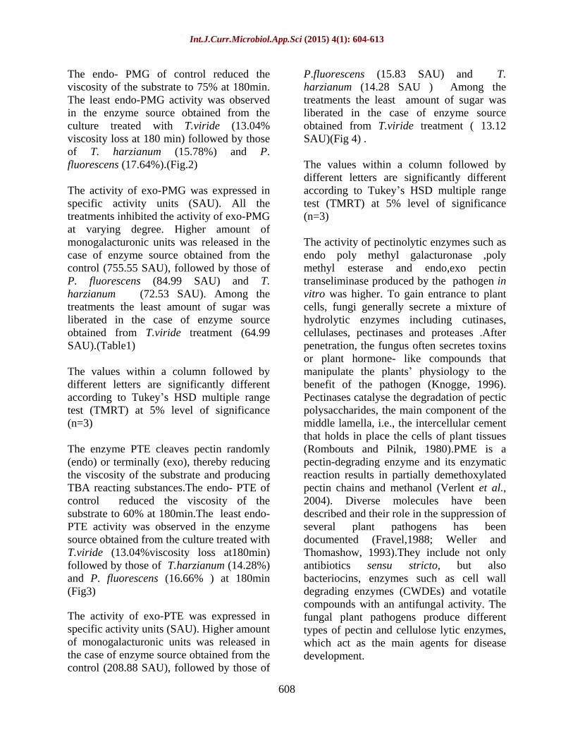
The highest PME activity was observed in enzyme source of control (59.44SAU) followed by those treated with P.fluorescens (17.31SAU) and T.harzianum (15.01SAU). The lowest rate of enzyme activity was observed in T.viride-treated enzyme source (8.83SAU). Maximum inhibition of enzyme activity was recorded in T.viride-treated culture (85.14%) followed by T.harzianum (74.74%) and P. fluorescens (70.87%). (Fig1)
The values within a column followed by different letters are significantly different according to Tukey s HSD multiple range test (TMRT) at 5% level of significance (n=3)
Int.J.Curr.Microbiol.App.Sci (2015) 4(1): 604-613
608
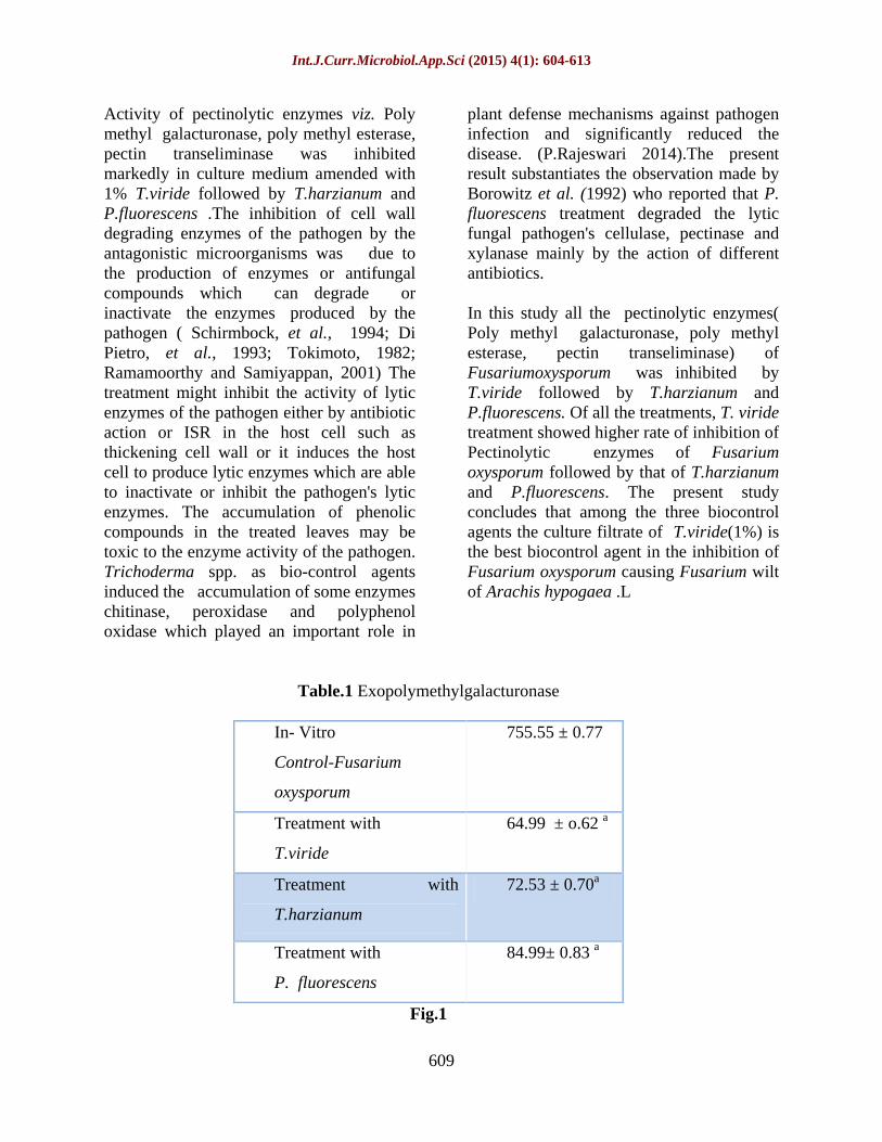
The endo- PMG of control reduced the viscosity of the substrate to 75% at 180min. The least endo-PMG activity was observed in the enzyme source obtained from the culture treated with T.viride (13.04% viscosity loss at 180 min) followed by those of T. harzianum (15.78%) and P. fluorescens (17.64%).(Fig.2)
The activity of exo-PMG was expressed in specific activity units (SAU). All the treatments inhibited the activity of exo-PMG at varying degree. Higher amount of monogalacturonic units was released in the case of enzyme source obtained from the control (755.55 SAU), followed by those of P. fluorescens (84.99 SAU) and T. harzianum (72.53 SAU). Among the treatments the least amount of sugar was liberated in the case of enzyme source obtained from T.viride treatment (64.99 SAU).(Table1)
The values within a column followed by different letters are significantly different according to Tukey s HSD multiple range test (TMRT) at 5% level of significance (n=3)
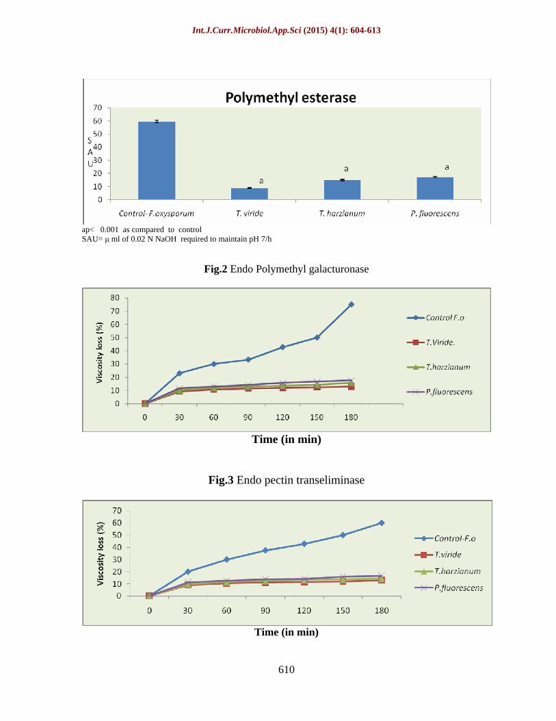
The enzyme PTE cleaves pectin randomly (endo) or terminally (exo), thereby reducing the viscosity of the substrate and producing TBA reacting substances.The endo- PTE of control reduced the viscosity of the substrate to 60% at 180min.The least endo-PTE activity was observed in the enzyme source obtained from the culture treated with T.viride (13.04%viscosity loss at180min) followed by those of T.harzianum (14.28%) and P. fluorescens (16.66% ) at 180min (Fig3)
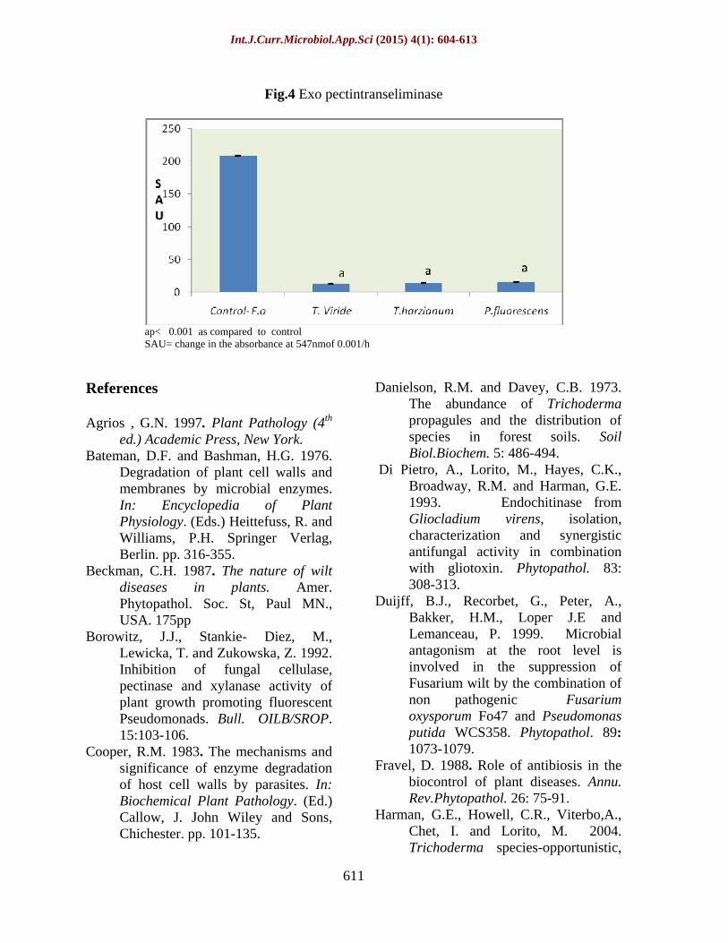
The activity of exo-PTE was expressed in specific activity units (SAU). Higher amount of monogalacturonic units was released in the case of enzyme source obtained from the control (208.88 SAU), followed by those of
P.fluorescens (15.83 SAU) and T. harzianum (14.28 SAU ) Among the treatments the least amount of sugar was liberated in the case of enzyme source obtained from T.viride treatment ( 13.12 SAU)(Fig 4) .
The values within a column followed by different letters are significantly different according to Tukey s HSD multiple range test (TMRT) at 5% level of significance (n=3)
The activity of pectinolytic enzymes such as endo poly methyl galacturonase ,poly methyl esterase and endo,exo pectin transeliminase produced by the pathogen in vitro was higher. To gain entrance to plant cells, fungi generally secrete a mixture of hydrolytic enzymes including cutinases, cellulases, pectinases and proteases .After penetration, the fungus often secretes toxins or plant hormone- like compounds that manipulate the plants physiology to the benefit of the pathogen (Knogge, 1996). Pectinases catalyse the degradation of pectic polysaccharides, the main component of the middle lamella, i.e., the intercellular cement that holds in place the cells of plant tissues (Rombouts and Pilnik, 1980).PME is a pectin-degrading enzyme and its enzymatic reaction results in partially demethoxylated pectin chains and methanol (Verlent et al., 2004). Diverse molecules have been described and their role in the suppression of several plant pathogens has been documented (Fravel,1988; Weller and Thomashow, 1993).They include not only antibiotics sensu stricto, but also bacteriocins, enzymes such as cell wall degrading enzymes (CWDEs) and votatile compounds with an antifungal activity. The fungal plant pathogens produce different types of pectin and cellulose lytic enzymes, which act as the main agents for disease development.
Int.J.Curr.Microbiol.App.Sci (2015) 4(1): 604-613
609
Activity of pectinolytic enzymes viz. Poly methyl galacturonase, poly methyl esterase, pectin transeliminase was inhibited markedly in culture medium amended with 1% T.viride followed by T.harzianum and P.fluorescens .The inhibition of cell wall degrading enzymes of the pathogen by the antagonistic microorganisms was due to the production of enzymes or antifungal compounds which can degrade or inactivate the enzymes produced by the pathogen ( Schirmbock, et al., 1994; Di Pietro, et al., 1993; Tokimoto, 1982; Ramamoorthy and Samiyappan, 2001) The treatment might inhibit the activity of lytic enzymes of the pathogen either by antibiotic action or ISR in the host cell such as thickening cell wall or it induces the host cell to produce lytic enzymes which are able to inactivate or inhibit the pathogen's lytic enzymes. The accumulation of phenolic compounds in the treated leaves may be toxic to the enzyme activity of the pathogen. Trichoderma spp. as bio-control agents induced the accumulation of some enzymes chitinase, peroxidase and polyphenol oxidase which played an important role in
plant defense mechanisms against pathogen infection and significantly reduced the disease. (P.Rajeswari 2014).The present result substantiates the observation made by Borowitz et al. (1992) who reported that P. fluorescens treatment degraded the lytic fungal pathogen's cellulase, pectinase and xylanase mainly by the action of different antibiotics.
In this study all the pectinolytic enzymes( Poly methyl galacturonase, poly methyl esterase, pectin transeliminase) of Fusariumoxysporum was inhibited by T.viride followed by T.harzianum and P.fluorescens. Of all the treatments, T. viride treatment showed higher rate of inhibition of Pectinolytic enzymes of Fusarium oxysporum followed by that of T.harzianum and P.fluorescens. The present study concludes that among the three biocontrol agents the culture filtrate of T.viride(1%) is the best biocontrol agent in the inhibition of Fusarium oxysporum causing Fusarium wilt of Arachis hypogaea .L
Table.1 Exopolymethylgalacturonase
In- Vitro
Control-Fusarium
oxysporum
755.55 ± 0.77
Treatment with
T.viride
64.99 ± o.62 a
Treatment with
T.harzianum
72.53 ± 0.70a
Treatment with
P. fluorescens
84.99± 0.83 a
Fig.1
Int.J.Curr.Microbiol.App.Sci (2015) 4(1): 604-613
610
ap< 0.001 as compared to control SAU= ml of 0.02 N NaOH required to maintain pH 7/h
Fig.2 Endo Polymethyl galacturonase
Time (in min)
Fig.3 Endo pectin transeliminase
Time (in min)
Int.J.Curr.Microbiol.App.Sci (2015) 4(1): 604-613
611
Fig.4 Exo pectintranseliminase
ap< 0.001 as compared to control SAU= change in the absorbance at 547nmof 0.001/h
References
Agrios , G.N. 1997. Plant Pathology (4th
ed.) Academic Press, New York. Bateman, D.F. and Bashman, H.G. 1976.
Degradation of plant cell walls and membranes by microbial enzymes. In: Encyclopedia of Plant Physiology. (Eds.) Heittefuss, R. and Williams, P.H. Springer Verlag, Berlin. pp. 316-355.
Beckman, C.H. 1987. The nature of wilt diseases in plants. Amer. Phytopathol. Soc. St, Paul MN., USA. 175pp
Borowitz, J.J., Stankie- Diez, M., Lewicka, T. and Zukowska, Z. 1992. Inhibition of fungal cellulase, pectinase and xylanase activity of plant growth promoting fluorescent Pseudomonads. Bull. OILB/SROP. 15:103-106.
Cooper, R.M. 1983. The mechanisms and significance of enzyme degradation of host cell walls by parasites. In: Biochemical Plant Pathology. (Ed.) Callow, J. John Wiley and Sons, Chichester. pp. 101-135.
Danielson, R.M. and Davey, C.B. 1973. The abundance of Trichoderma propagules and the distribution of species in forest soils. Soil Biol.Biochem. 5: 486-494.
Di Pietro, A., Lorito, M., Hayes, C.K., Broadway, R.M. and Harman, G.E. 1993. Endochitinase from Gliocladium virens, isolation, characterization and synergistic antifungal activity in combination with gliotoxin. Phytopathol. 83: 308-313.
Duijff, B.J., Recorbet, G., Peter, A., Bakker, H.M., Loper J.E and Lemanceau, P. 1999. Microbial antagonism at the root level is involved in the suppression of Fusarium wilt by the combination of non pathogenic Fusarium oxysporum Fo47 and Pseudomonas putida WCS358. Phytopathol. 89: 1073-1079.
Fravel, D. 1988. Role of antibiosis in the biocontrol of plant diseases. Annu. Rev.Phytopathol. 26: 75-91.
Harman, G.E., Howell, C.R., Viterbo,A., Chet, I. and Lorito, M. 2004. Trichoderma species-opportunistic,
Int.J.Curr.Microbiol.App.Sci (2015) 4(1): 604-613
612
avirulent plant symbionts. Nature reviews. Microbiology 2: 43-56.
Harman, G.E. 2006. Overview of mechanism and use of Trichoderma spp. Phytopathol. 96: 190-194.
Knogge, W. 1996. Fungal infection of plants. The Plant Cell 8:1711-1722.
Leonardo , D., Blanca L.F., Landa, B. and Weller D.M. 2006. Host crop affects rhizosphere colonization and competitiveness of 2,4- diacetylphloroglucinol-producing Pseudomonas fluoresens .Phytopathol. 96: 751-762.
Mahadevan, A. and Sridhar, R. 1986. Methods in physiological plant pathology (3rd ed.) Sivakami Publications, Madras, India.
Meena, B., Marimuthu, T., Vidyasekaran, P. and Velazhahan, R. 2001. Biological control of root rot of groundnut with antagonistic Pseudomonas fluorescens strains. J.Plant Dis.Protect. 108:368-381.
Muse, R.R., Couch. H. B., Moore, L.D. and Muse, B. D. 1972. Pectinolytic and cellulolytic enzymes associated with Helminthosporium leaf spot on Kentucky bluegrass. Can. J. Microbiol. 18: 1091-1098.
Papavizas, G.E. 1985. Trichoderma and Gliocladium: Biology, ecology and potential for biocontrol. Ann. Rev. Phytopathol. 23: 23-54.
Posada, M. L., Patino, B., Mirete. S., Munoz, M. C., Vazquez, C. and Gonzalez-Jaen, M.T. 2001. Comparative analysis of polygalacturonases in isolates of seven species of Fusarium from Pinus pinea. Mycol.Res.105:100-104
Rajeswari ,P. 2014. Role of phenols and antioxidant enzymes in the biocontrol of Fusarium oxysporum causing Fusarium wilt on Arachis hypogaea L.(Groundnut).
International Journal of Agricultural Science and Research 4:95-104
Ramamoorthy, V. and Samiyappan, R. 2001. Induction of defence related genes in Pseudomonas fluorescens treated chilli plants in response to infection by Colletotrichum capsici. J. Mol. Pl. Pathol. 31: 146-155.
Rombouts, F. M. and Pilnik, W. 1980. Pectic enzymes. In: Microbial Enzymes and Bioconversions, Economic Microbiology (Ed.) A.H.Rose. Academic Press, New York, pp.228-282.
Samuels, G.J. 2006. Trichoderma: Systematics, the sexual state, and ecology. Phytopathol. 96:195-206.
Schirmbock, M., Lorito, M., Wang, Y.L., Hayes, C.K., Arisan-atac, I., Scala, F., Harman, G.E. and Kubichek, C.P. 1994. Parallel formation and synergism of hydrolytic enzymes and peptaibol antibiotics, molecular mechanisms involved in the antagonistic action of Trichiderma harzianum against phytopathogenic fungi. Appl. Environ. Microbiol. 60: 4364-4370.
Tokimoto, K. 1982.Lysis of the mycelium of Lentinus edodes (Berk.) Sing. caused by mycolytic enzymes of Trichoderma harzianum when the two fungi were in an antagonistic state. Trans. Mycol. Soc. Jpn. 23:13-20.
Tu, C.C. and Chang, Y.H .1983.Soil microbial activity in relation to Fusarium wilt suppression soils and conductive soil. In: Proc Republic of China Federal Republic of Germany Seminar Plant Nutrition Soil Sciences. Natural Science Council, Taipei, pp.189-196.
Verlent, I., Van loey, CA., Smout, C., Duvetter, T. and Hendriclox, ME. 2004. Purified tomato
Int.J.Curr.Microbiol.App.Sci (2015) 4(1): 604-613
613
polygalacturonase activity during thermal and high pressure treatment. Biotechnol. Bioeng. 86: 63-71.
Walton, J.D. 1994. Deconstructing the cell wall. Plant Physiol. 104:191-196.
Weller, D.M. and Thomashow, L.S. 1993. Microbial metabolites with biological activity In: Pest management: biologically based technologies. (Eds.) Lumsden R.D. and Vaughn J.L. American Chemical Society, Washington, DC, USA. pp. 173-180.
Wilberforce, E.M., Boddy, L., Griffiths, R. and Griffith, G.W. 2003. Agricultural management affects communities of culturable root endophytic fungi in temperate grasslands. Soil Biol. Biochem.35:1143-1154.
Zhang, C., Druzhinina, I., Kubick , C.P. and Xu, T. 2005. Trichoderma biodiversity in China: evidence for a north to southern distribution of species in East Asia. FEMS Microbiol. Lett. 251: 251-257.
Copyright © 2022 FDOKUMEN