Enzymes with new biochemical properties in the pectinolytic complex produced by Aspergillus niger...
12
See discussions, stats, and author profiles for this publication at: https://www.researchgate.net/publication/6182809 Enzymes with new biochemical properties in the pectinolytic complex produced by Aspergillus niger MIUG 16 Article in Journal of Biotechnology · September 2007 DOI: 10.1016/j.jbiotec.2007.06.005 · Source: PubMed CITATIONS 59 READS 56 5 authors, including: Some of the authors of this publication are also working on these related projects: Gadolinium Nanohydrogels for Lymph Node Magnetic Resonance Imaging View project Diana Dinu University of Bucharest 26 PUBLICATIONS 239 CITATIONS SEE PROFILE Marina Nechifor University of Bucharest 10 PUBLICATIONS 204 CITATIONS SEE PROFILE Gheorghe Stoian 37 PUBLICATIONS 87 CITATIONS SEE PROFILE Marieta Costache University of Bucharest 271 PUBLICATIONS 1,070 CITATIONS SEE PROFILE All content following this page was uploaded by Diana Dinu on 27 April 2015. The user has requested enhancement of the downloaded file. All in-text references underlined in blue are added to the original document and are linked to publications on ResearchGate, letting you access and read them immediately.
Transcript of Enzymes with new biochemical properties in the pectinolytic complex produced by Aspergillus niger...

Seediscussions,stats,andauthorprofilesforthispublicationat:https://www.researchgate.net/publication/6182809
EnzymeswithnewbiochemicalpropertiesinthepectinolyticcomplexproducedbyAspergillusnigerMIUG16
ArticleinJournalofBiotechnology·September2007
DOI:10.1016/j.jbiotec.2007.06.005·Source:PubMed
CITATIONS
59
READS
56
5authors,including:
Someoftheauthorsofthispublicationarealsoworkingontheserelatedprojects:
GadoliniumNanohydrogelsforLymphNodeMagneticResonanceImagingViewproject
DianaDinu
UniversityofBucharest
26PUBLICATIONS239CITATIONS
SEEPROFILE
MarinaNechifor
UniversityofBucharest
10PUBLICATIONS204CITATIONS
SEEPROFILE
GheorgheStoian
37PUBLICATIONS87CITATIONS
SEEPROFILE
MarietaCostache
UniversityofBucharest
271PUBLICATIONS1,070CITATIONS
SEEPROFILE
AllcontentfollowingthispagewasuploadedbyDianaDinuon27April2015.
Theuserhasrequestedenhancementofthedownloadedfile.Allin-textreferencesunderlinedinblueareaddedtotheoriginaldocumentandarelinkedtopublicationsonResearchGate,lettingyouaccessandreadthemimmediately.
This article was published in an Elsevier journal. The attached copyis furnished to the author for non-commercial research and
education use, including for instruction at the author’s institution,sharing with colleagues and providing to institution administration.
Other uses, including reproduction and distribution, or selling orlicensing copies, or posting to personal, institutional or third party
websites are prohibited.
In most cases authors are permitted to post their version of thearticle (e.g. in Word or Tex form) to their personal website orinstitutional repository. Authors requiring further information
regarding Elsevier’s archiving and manuscript policies areencouraged to visit:
http://www.elsevier.com/copyright

Author's personal copy
Journal of Biotechnology 131 (2007) 128–137
Enzymes with new biochemical properties in the pectinolyticcomplex produced by Aspergillus niger MIUG 16
Diana Dinu a, Marina Tamara Nechifor b, Gheorghe Stoian a,Marieta Costache a, Anca Dinischiotu a,∗
a Department of Biochemistry, Faculty of Biology, University of Bucharest, 91-95 Splaiul Independentei, Bucharest 050095, Romaniab Department of Animal Biology, Faculty of Biology, University of Bucharest, 91-95 Splaiul Independentei, Bucharest 050095, Romania
Received 5 February 2007; received in revised form 21 May 2007; accepted 14 June 2007
Abstract
A comprehensive study on the purification and characterization of pectinolytic enzymes produced by Aspergillus niger MIUG 16 on raw materialssolid-state fermentation is reported. Five pectinolytic enzymes were purified using a combination of chromatographic techniques. The propertiesof these homogenous enzymes were analyzed. The purified enzymes were classified with respect to their biochemical properties and substratespecificity. Among these proteins, one revealed polygalacturonase activity, another appeared to be a pectin methylesterase and three were identifiedas pectate lyases. The capacity of the fungus A. niger to produce pectate lyases with optimum pH in acidic domain was reported for the first time.© 2007 Elsevier B.V. All rights reserved.
Keywords: Aspergillus niger; Acidic pectate lyases; Homogenous pectinases characterization; Pectin methylesterase; Polygalacturonase; Purification
1. Introduction
Pectin is a major component of primary cell walls ofall plants and encompasses a range of galacturonic acid(GalA)-rich polysaccharides. This complex heteropolysaccha-ride contains two different regions, the “smooth” and the“hairy” ones (Albersheim et al., 1996; Willats et al., 2001).The “smooth” regions consist of a homogalacturonan (HGA), alinear homopolymer composed of repeated d-galacturonic acidmonosaccharide subunits, that are methyl-esterified to a varyingdegree. In the “hairy” regions, the main structure is a branchedrhamnogalacturonan containing the d-galacturonic acid residuesin the backbone interrupted by �-1,2-linked l-rhamnose, towhich long arabinan and galactan chains can be attached at O-4(Albersheim et al., 1996).
Due to its complex structure, pectin modification or break-down are catalyzed by a variety of pectinolytic enzymes: (i)esterases that remove the methyl and acetyl groups from pectinand (ii) depolymerases that cleave the backbone of pectin.The main classes of depolymerizing enzymes, collectively
∗ Corresponding author. Tel.: +40 21 318 15 75; fax: +40 21 318 15 75/102.E-mail address: [email protected] (A. Dinischiotu).
called pectinases, are polygalacturonases, rhamnogalactur-onases, pectin lyase, pectate lyase and rhamnogalacturonanlyase (Sakai et al., 1993).
Pectinolytic enzymes are widely used for the disintegrationof plant tissues in the fruit and vegetable processing indus-tries, increasing the extraction of juice, decreasing the viscosityof concentrates and solubilizing pectic complexes to completesedimentation and clarification of juices (Kashyap et al., 2001;Gumamadi and Panda, 2003; Crook and Corredig, 2006) andwines (Perez-Maganino and Gonzalez-San Jose, 2001). Apartfrom these industrial applications, these enzymes possess bio-logical importance in protoplast fusion technology and plantpathology (Gumamadi and Panda, 2003).
Pectynolitic enzymes are found mainly in fungi and bac-teria (Jayani et al., 2005; Oliveira et al., 2006). The mostwidely occurring enzymes are polygalacturonases (PG), pec-tate lyases (PeL), pectin lyases (PL) and pectin methylesterases(PME).
Most commercial pectynolitic enzymes preparations are pro-duced by fermentation with Aspergillus spp. and are mixtures ofpectinolityic enzymes. Efficient degradation of polysaccharidesin food industry requires cooperative or synergistic interactionsbetween these enzymes responsible for cleaving the differ-ent linkages in pectin structure. PME and the depolymerising
0168-1656/$ – see front matter © 2007 Elsevier B.V. All rights reserved.doi:10.1016/j.jbiotec.2007.06.005

Author's personal copy
D. Dinu et al. / Journal of Biotechnology 131 (2007) 128–137 129
enzymes, PG and PeL, are known to operate in tandem todegrade the methylesterified pectins. The occurrence of de-esterified homogalacturonan region, after PME action, suppliesthe sites for the enzymes that can cleave the HGA backbone(Kester et al., 2000). Only a limited number of studies onsynergy between PME and the others pectinolytic enzymesare reported (de Vries and Visser, 2001; Semenova et al.,2003).
The fungus Aspergillus niger (A. niger) was intensively inves-tigated for pectinolytic enzymes production. PGs, the mostabundant and extensively studied of the pectinolytic enzymes,typically exist in multi-gene families and may have both endo(Benen et al., 1999; Parenicova et al., 2000) and exo activ-ities (Hara et al., 1984; Sakamoto et al., 2002). PLs (Vitaliet al., 1998) and PMEs (Khanh et al., 1991) were also puri-fied and characterized from some Aspergillus strains. Secretionof PeLs from Aspergillus spp. is less reported (Benen et al.,2000).
Solid-state fermentation (SSF) using wastes from food andagriculture industry was widely used for Aspergilus pectinolyticenzymes production (Patil and Dayanand, 2006; Botella et al.,2007; Ustok et al., 2007). Our previous studies showed that A.niger MIUG 16 is able to secrete many types of pectinolyticenzymes through solid-state fermentation on raw materials(Dinu, 1999). Since applications of pectinolytic enzymes invarious fields are widening, it is important to understand thenature and properties of these enzymes for efficient and effectiveusage. In this paper, a fractionation protocol, with good recoveryof major pectinolytic components, and a comprehensive studycomprising enzymatic and molecular properties are reported.The comprehensive study of the properties of the homogenouspectinolytic enzymes revealed three pectate lyase activities withacidic pH optima.
2. Materials and methods
2.1. Microorganism
The A. niger strain (A. niger MIUG 16) was obtained fromthe Laboratory of Industrial Microbiology, University of Galati,Roumania and it was routinely cultured on potato dextrose agar(PDA, Merck) slants and incubated at 25 ◦C for 7 days for inoc-ula preparation for SSF cultivation.
2.2. Solid-state fermentation
SSF medium containing 50 g wheat bran, 25 g beet marc and125 ml mineral solution with the following composition (w/v):2.5% ammonium sulphate, 0.33% monopotasium phosphate,and 0.17% magnesium sulphate was introduced in 2 l Erlen-meyer flasks and autoclaved at 120 ◦C for 60 min. The pH ofthe substrate solution was adjusted at pH 5.5 with 2 M HCl.This was inoculated by fungal stock cultures grown on PDA andincubated for 5 days at 28 ◦C. After incubation, the cultures weredehumidified in incubator for 24 h at 40 ◦C and were ground for2 min.
2.3. Extraction of enzymes
The powdered culture media were subjected to an extrac-tion in 0.05 M acetic acid–sodium acetate buffer, pH 5.2 (10buffer volumes for 1 g solid media) for 60 min. The extractionprocedure was repeated once again in the same conditions. Theextraction mixtures were centrifuged at 6000 × g for 30 min in arefrigerated Sigma centrifuge and then filtered with filter paper(Whatman No. 1). The final crude filtrate was used as a sourceof enzyme in all our studies.
2.4. Enzyme assays
The PG activity was determined by measuring the releaseof reducing sugars, according to the Nelson–Somogyi method(Nelson, 1944; Somogyi, 1952) using galacturonic acid as stan-dard, and sodium polygalacturonate (0.5%, w/v) as substrate.One unit (U) of PG activity was defined as the amount of enzymewhich produced 1 �mole of reducing groups per min at pH 4.6and 40 ◦C.
Pectin and pectate lyase activities were measured by moni-toring the unsaturated oligogalacturonates accumulated after theenzymatic cleavage of pectin or sodium pectate (0.2%, w/v inassay). Because Ca2+ is essential for in vitro activity of pec-tate lyase, calcium chloride (0.6 mM) was added in the reactionmedia. The unsaturated fragments were estimated with a modi-fied thiobarbituric acid spectrophotochemical method suitableto differentiate between polygalacturonase and pectate lyase(Nedjma et al., 2001). One unit (U) of lyase activity was definedas the amount of enzyme which produced an increase of 0.01 inabsorbance at 550 nm per 1 min, under optimal conditions.
PME activity was determined, as described by Wood andSiddiqui (1971), by measuring the release of methanol follow-ing incubation of 60% methylated (0.33%, w/v) with an aliquotof enzyme in 0.05 M sodium acetate buffer, pH 4.2. The PMEactivity was expressed as �g methanol released per minute andper ml enzymatic solution.
2.5. Protein estimation
Due to the presence of phenolic and other interfering sub-stances in the crude extracts and fractions obtained in the earlystages of purification, the protein concentration could be mea-sured with accuracy only after precipitation and redissolution.Because of this, proteins of each sample were precipitatedwith 12% (w/v) trichloric acid containing 0.02% (w/v) sodiumdeoxycolate (Bensadoun and Weinstein, 1976) redissolved in anappropriate volume of 0.1 M NaOH and measured by the methodof Lowry et al. (1951) using bovine serum albumin as a standard.
2.6. Enzymes purification
The procedure used for the purification of all pectinolyticenzymes produced by A. niger MIUG 16 is illustrated in Fig. 1.The details for the purification procedure are described below.
A. niger pectinolytic enzymes purification was carried out bya diversity of chromatographic procedures on a Pharmacia FPLC

Author's personal copy
130 D. Dinu et al. / Journal of Biotechnology 131 (2007) 128–137
Fig. 1. Schematic illustration of the procedure used in the purification of pectinolytic enzymes from Aspergillus niger.
system (Amersham Pharmacia Biotech, Uppsala, Sweden). Thecrude extract of whole solid-state culture obtained as describedabove was concentrated and desalted by ultrafiltration usingPLGCTM membrane with a molecular mass limit of 10000 Da(Millipore, Bedford, MA, USA). The initial enzymatic extractwas dialysed against 50 mM Tris–HCl buffer, pH 8.0. A sam-ple of 60 mg proteins was loaded on Fractogel TSK DEAE-650(M) (Merck, Frankfurt, Germany) column (12 cm × 4 cm) pre-equilibrated with the same buffer. The unadsorbed proteins wereeluted with 300 ml of starting buffer, 50 mM Tris–HCl, pH 8.0,at a flow rate of 30 ml per hour. The pectinolytic enzymes boundto anion exchanger were eluted by a step-wise procedure. The
volume of each step was 150 ml of starting buffer containing thefollowing concentrations of NaCl: 0.1, 0.2, 0.3 and 1 M (Fig. 2a).Several purification runs were carried out to obtain the enzymesin quantities sufficient for further purification procedures andextensive studies.
The proteins of peak A were concentrated and dialyzedagainst 50 mM sodium acetate buffer, pH 4.7, and thenloaded on to Fractogel TSK CM-650 (M) (Merck) column(6.8 cm × 2.6 cm) equilibrated with the same buffer. The columnwas eluted with 75 ml acetate buffer before starting the gradi-ent. A linear gradient of 0–0.3 M NaCl was used (90 ml in eachchamber) for the elution of adsorbed proteins. A part of loaded

Author's personal copy
D. Dinu et al. / Journal of Biotechnology 131 (2007) 128–137 131
Fig. 2. Purification of pectinolytic enzymes from A. niger MIUG 16 (a) anion-exchange chromatography on DEAE-Fractogel; (b) cation-exchange chromatographyof PG, PeL II and PeL III on CM-Fractogel; (c) chromatofocusing of PG and PeL III on Mono P; (d) gel filtration of PeL II on Bio-Gel P-100; (e) fractionation ofPME and PeL I on Bio-Gel P-100; (f) separation of PE from PeL I by hydrophobic chromatography on Phenyl-Sepharose. Protein (—); PG (–�–); PeL (–�–); PME(- - - -); molarity of NaCl (a and b) or (NH4)2SO4 (f) ( ).
PeL activity was unadsorbed on cation-exchanger and the otherpart of lyase activity was eluted together with the PG one, at0.11 M NaCl. The unadsorbed lyase was designated as PeL IIand the adsorbed protein as PeL III (Fig. 2b). The separation ofPG from PeL III was performed by chromatofocusing on MonoP HR 5/20 pre-packed column (Amersham Pharmacia Biotech).The column was equilibrated in 25 mM pyperazine–HCl buffer,pH 5.8 and the elution was performed with a decreased pH lin-ear gradient (5.5–4.5) achieved with diluted Polybuffer 74. Themaximum of PG activity was achieved at pH 5.13, whereas themaximum of PeL activity was obtained at pH 5.03 (Fig. 2c).Further purification of Pel II was achieved on Bio-Gel P-100(BioRad Labs, Richmond, CA, USA) in 50 mM acetate buffer,pH 4.7 (Fig. 2d).
The proteins of peak B from Fractogel TSK DEAE-650 separation were further fractionated on Bio-Gel P-100(72 cm × 1.6 cm). The elution profile for 40 mg loaded proteinsare showed in Fig. 2e. The separation of PME from PeL activitywas achieved by hydrophobic interaction chromatography onPhenyl-Sepharose High Performance from a Hi Trap HIC testkit (Amersham Pharmacia Biotech). The fractions eluted from
Bio-Gel with PME and lyase activity were reunited and concen-trated. Then, ammonium sulphate was added up to 1 M in 50 mMsodium phosphate, pH 7.0. The column, equilibrated with thesame saline solution, was loaded with 6 mg proteins of eachrun, and then washed with 15 ml sodium phosphate buffer with1 M ammonium sulphate. The proteins bounded at hydropho-bic matrix were eluted with 20 ml of a linear decreased gradientof ammonium sulphate concentration. Elution was performedunder a 3 bar pressure. The PME activity was not retained of thehydrophobic matrix, and PeL I bound activity was released at0.54 M ammonium sulphate concentration (Fig. 2f).
2.7. Gel electrophoresis and staining
The purity of the various fractions from the purificationprocedure was monitored by 5–15% (w/v) gradient sodiumdodecyl sulphate polyacrylamide gel electrophoresis (SDS-PAGE), according to Laemmli (1970). In order to locate proteinbands, gels were fixed by immersion in a solution of 12% (w/v)trichloric acid and stained with 0.1% (w/v) Coomassie BrillantBlue R 250 in water/methanol/acetic acid (4.5:4.5:1, v/v/v). The
Author's personal copy
132 D. Dinu et al. / Journal of Biotechnology 131 (2007) 128–137
gels were destained in 10% (v/v) acetic acid until the back-ground disappeared. The proteins with pectinolytic activitieswere separated under non-denaturing conditions on 12% (w/v)polyacrilamide (PAGE) slab gel containing 0.1% (w/v) pecticsubstances and 4% (w/v) stacking gel (Fonseca and Said, 1995).Pectinolytic enzymes bands were stained with Ruthenium Red(Cruickshank and Wade, 1980).
2.8. Enzymes characterization
2.8.1. Molecular mass estimation and pI valuesThe protein molecular masses were determined after SDS-
PAGE electrophoresis, calibrated by a low molecular weightmarkers kit (Amersham Pharmacia Biotech). The gels wereanalyzed by densitometry using a Bio-Profil system (VilberLourmat, France) with Bio-1D image analysis software and themolecular masses were calculated. The molecular masses werealso determined by gel chromatography on Bio-Gel P-100, usinga Sigma calibration kit and Blue Dextran (2000 kDa). The loga-rithms of standards molecular masses versus Ve/Vo (Ve, proteinelution volume; Vo, Dextran Blue volume elution) were plot-ted. With the linear regression output, the molecular masses ofpurified pectinolytic enzyme were estimated.
The pI value of each pectinolytic enzyme was determi-nated by chromatofocusing on Mono P HR 5/20 pre-packedcolumn. The column was equilibrated with 0.025 M bisTris–iminodiacetic acid, pH 7.1 as starting buffer. Elution wasdone with a decreased pH linear gradient from 7.0 to 4.0. Theelution of proteins with a pI below 4.0 was performed by elutionof the column with 1 M NaCl in starting buffer.
2.8.2. pH and temperature optimaPG and PME activities were determined at various pH values
(3.5–7.0) using sodium polygalacturonate and 60% methylatedpectin, respectively, in 0.05 M acetate buffer (3.5–5.5) or in0.05 M sodium phosphate buffer (5.5–7.0). For lyase activi-ties, sodium polygalacturonate, 9.5% methylated pectin, 30%methylated pectin and 60% methylated pectin were prepared indifferent pH buffers: 0.05 M sodium acetate (3.5–5.5), 0.05 Msodium phosphate (5.5–7.0) and 0.05 M Tris–HCl (7.0–9.0). Theoptima temperature of each enzyme was determined by incubat-ing the conveyable substrate at various temperatures between25 and 70 ◦C for 5 min before the addition of the enzyme andassaying at the same temperature.
2.8.3. Substrate specificityThe substrate specificity was determined by measuring the
activity at pH and temperature optima for each pectinolyticenzyme using different substrates. The substrates used were:sodium polygalacturonate, citrus pectin (9.5% esterification),citrus pectin (30% esterification), citrus pectin (60% esterifi-cation) and highly methylated pectin (90% esterification). Thesubstrates were purchased from Sigma, excepting the 9.5% ester-ified pectin which was prepared from methylesterifid pectinby tratament with 1.5 M NaOH. Samples were assayed formethanol release upon saponification using the method of Woodand Siddiqui (1971). Moles of methanol released were related
to moles of galacturonic acid equivalents present to calculatedegree of methylesterification. The activities of each enzymeusing these substrates were followed as described above. Thespecificity was also evaluated by electrophoresis studies. Pecti-nolytic enzymes diffuse from electrophoresis gels into ultrathinpectic polysaccharides–polyacrylamide overlays. After migra-tion, the overlays were placed in a 0.05% (w/v) Ruthenium Redsolution, which precipitates and stains the undegraded substrateleaving a clear zone in the overlay that corresponds to the loca-tion of the enzymes in the electrophoresis gel (Cruickshankand Wade, 1980). By manipulation of the reaction conditionsin the overlays, the polygalacturonases and pectate lyases maybe assayed.
2.8.4. Determination of Km, Vmax and kcat.
The Km and Vmax values of purified PG were determinatewith sodium polygalacturonate as the substrate (concentrationrange 0.25–6 mg/ml in the assay medium) in 0.05 M acetatebuffer, pH 4.6, at 40 ◦C. The kinetic parameters of hydrolysisof 60% methylated pectin under the action of purified PMEwere measured at pH 4.2 and 40 ◦C, where the substrate con-centration was varied in the range of 0.5–18 mg/ml medium ofreaction. For PeLs, the concentration of sodium polygalactur-onate, prepared at the optimum pH for each enzyme, was variedbetween 0.056 and 8.0 mg/ml assay medium and incubated withan appropriate amount of enzyme. A double-reciprocal plot wasused for estimating Km and Vmax. Then, the values of kcat werecalculated using the equation: kcat = Vmax/[E], where [E] is theconcentration of enzyme.
3. Results
3.1. Purification of pectinolytic enzymes
Isolation of pectinolytic enzymes secreted by A. niger wascarried out by using different types of chromatography (Fig. 1).The purification procedure leaded to the separation of one PG,one PME and three PeL. The PeL activities were designatedas PeL I, PeL II and PeL III, according to the increasingpI values determinated by chromatofocusing on Mono P (notshowed).
In the first step, the concentrated enzymatic extract waspurified by anion-exchange chromatography on Fractogel TSKDEAE column. The protein elution profile, shown in Fig. 2a,revealed several major peaks. The fraction eluted with 0.1 MNaCl, corresponding to the protein peak marked with A, con-tained the PG activity. In the peak marked with B, eluted with0.2 M NaCl, the PME activity was detected, while PeL activitieswere detected in both peaks.
The proteins of peak A were further separated by cation-exchange chromatography on Fractogel TSK CM (Fig. 2b). Theunadsorbed PeL activity, eluted with the starting buffer, wasdesignated as PeL II. The other PeL activity, designated as PeLIII, was eluted out together with PG activity, at 0.11 M of NaClgradient.
The separation of PG from PeL III activity was achieved bychromatofocusing on Mono P (Fig. 2c). Using this high perfor-

Author's personal copy
D. Dinu et al. / Journal of Biotechnology 131 (2007) 128–137 133
Table 1Summary of the purification of pectinolytic enzymes from Aspergillus niger
Purification step Total activity (U) Total protein (mg) Specific activity (U/mg) Fold purity Yield (%)
Crude extractPG 13359.0 667.9 20.0 1.0 100.0PeL 7402.3 667.9 11.1 1.0 100.0PME 11859.0 667.9 17.8 1.0 100.0
UltrafiltrationPG 12023.1 603.1 19.9 – 90.0PeL 7134.4 603.1 11.8 – 96.4PME 11324.3 603.1 18.8 – 95.5
Anion-exchange (DEAE)PG 8873.0 64.2 138.2 6.9 66.4PeL (peak A: PeL II and PeL III) 3193.5 64.2 49.7 4.5 43.1PeL (peak B: PeL I) 1357.0 209.6 6.5 – 18.3PME 8971.0 209.6 42.8 2.4 75.6
Cation-exchange (CM)PG 3604.0 4.1 879.0 43.9 27.0PeL II 3813.5 41.5 91.9 8.3 51.5PeL III 1686.7 4.1 411.4 37.1 22.8
Chromatofocusing (pH 5.5–4.5)PG 3238.7 2.1 1542.2 77.1 24.2PeL III 1610.2 1.3 1238.6 111.6 21.7
Gel-filtration (Bio-Gel P-100)PeL I 1279.0 33.2 38.5 3.5 17.3PeL II 2818.9 8.3 339.6 30.6 38.1PME 5674.8 33.2 170.9 9.6 47.8
Hydrophobic interactionPeL I 813.1 3.6 225.8 20.4 11.0PME 4420.4 8.2 539.1 30.3 37.3
mance chromatographic procedure we were able to separate thePG, enzyme with a pI of 5.13, from PeL III with a pI of 5.03.
The further purification of PeL II was performed by gel-filtration on Bio-Gel P-100 column. The elution profile, showedin Fig. 2d presented two protein peaks, the second of themcontaining the PeL II activity.
The proteins of peak B, from Fractogel TSK DEAE sep-aration, were first subjected to gel-filtration on Bio-Gel P-100,and then to hydrophobic interaction chromatography on Phenyl-Sepharose. The elution profiles, presented in Fig. 2e and f,respectively, showed that, only the second procedure, led to theseparation of PME from PeL I.
A summary of the purification of all five pectinolytic enzymesis shown in Table 1. With respect to the crude extract, it can beseen than the PG was obtained in 24.2 yield and represented77.1-fold purification. The PME was purified 30.3-fold with ayield of 37.3% with respect to the crude extract. The purificationof the three PeL activities was achieved with different yieldsand purification factor values. The best purification factor, witha 111.6 value, was obtained for PeL III with a yield of 21.7%.PeL I was purified 20.4-fold with a yield of 11%, whereas therelevant values for PeL II were 30.6 purification and 38.1% yield.
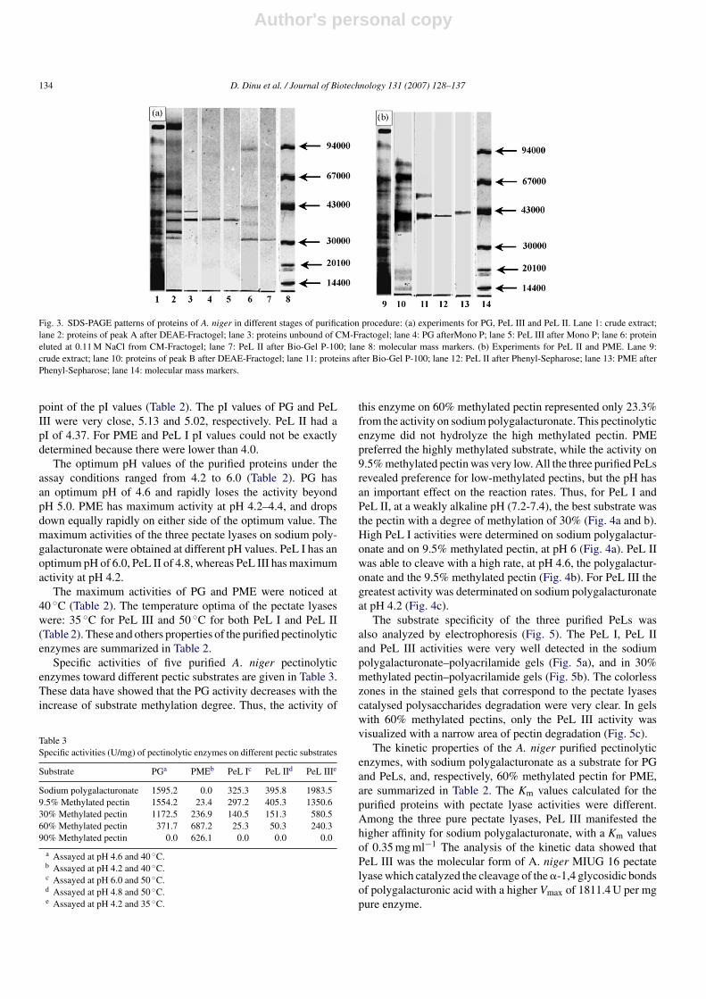
The purification of the pectinolytic enzymes from A. nigerwas checked during the all purification steps by SDS-PAGE(Fig. 3). The five enzymes appeared at single protein bands on5–15% (w/v) gradient SDS-PAGE. The electrophoretical pat-
terns are showed in Fig. 3a, for PG, PeL III and PeL II, and inFig. 3b, for PME and PeL I. The appearance of single proteinbands, on 5–15% (w/v) gradient gels, for each of five purifiedenzymes was a proof of their homogeneity.
3.2. Characteristics of enzymes
The Mr values of the five homogenous pectinolytic enzymeswere calculated by comparison with the behaviour of standardproteins The results obtained by SDS-PAGE were similar tothese of Bio-Gel P-100 evaluation (Table 2).
The chromatofocusing on Mono P HR 5/20 (not shown)revealed heterogeneity of these fungal enzymes from the view-
Table 2Properties of pectinolytic enzymes purified from Aspergillus niger
PG PME PeL I PeL II PeL III
Mr (SDS-PAGE) 36300 41700 41700 30200 36300Mr (gel-chromatography) 35500 38100 42600 30000 35500pI 5.13 <4.0 <4.0 4.37 5.02Optimum pH 4.6 4.2–4.4 6.0 4.6 4.2Optimum temperature (◦C) 40 40 50 50 35Km (mg ml−1)a 0.94 2.5 5.73 1.32 0.35Vmax (U/mg)a 3114.3 626.2 298.8 359.4 1811.4
a With sodium polygalacturonate as substrate for PG, PeL I, PeL II and PeLIII and 60% methylated pectin as substrate for PME.
Author's personal copy
134 D. Dinu et al. / Journal of Biotechnology 131 (2007) 128–137
Fig. 3. SDS-PAGE patterns of proteins of A. niger in different stages of purification procedure: (a) experiments for PG, PeL III and PeL II. Lane 1: crude extract;lane 2: proteins of peak A after DEAE-Fractogel; lane 3: proteins unbound of CM-Fractogel; lane 4: PG afterMono P; lane 5: PeL III after Mono P; lane 6: proteineluted at 0.11 M NaCl from CM-Fractogel; lane 7: PeL II after Bio-Gel P-100; lane 8: molecular mass markers. (b) Experiments for PeL II and PME. Lane 9:crude extract; lane 10: proteins of peak B after DEAE-Fractogel; lane 11: proteins after Bio-Gel P-100; lane 12: PeL II after Phenyl-Sepharose; lane 13: PME afterPhenyl-Sepharose; lane 14: molecular mass markers.
point of the pI values (Table 2). The pI values of PG and PeLIII were very close, 5.13 and 5.02, respectively. PeL II had apI of 4.37. For PME and PeL I pI values could not be exactlydetermined because there were lower than 4.0.
The optimum pH values of the purified proteins under theassay conditions ranged from 4.2 to 6.0 (Table 2). PG hasan optimum pH of 4.6 and rapidly loses the activity beyondpH 5.0. PME has maximum activity at pH 4.2–4.4, and dropsdown equally rapidly on either side of the optimum value. Themaximum activities of the three pectate lyases on sodium poly-galacturonate were obtained at different pH values. PeL I has anoptimum pH of 6.0, PeL II of 4.8, whereas PeL III has maximumactivity at pH 4.2.
The maximum activities of PG and PME were noticed at40 ◦C (Table 2). The temperature optima of the pectate lyaseswere: 35 ◦C for PeL III and 50 ◦C for both PeL I and PeL II(Table 2). These and others properties of the purified pectinolyticenzymes are summarized in Table 2.
Specific activities of five purified A. niger pectinolyticenzymes toward different pectic substrates are given in Table 3.These data have showed that the PG activity decreases with theincrease of substrate methylation degree. Thus, the activity of
Table 3Specific activities (U/mg) of pectinolytic enzymes on different pectic substrates
Substrate PGa PMEb PeL Ic PeL IId PeL IIIe
Sodium polygalacturonate 1595.2 0.0 325.3 395.8 1983.59.5% Methylated pectin 1554.2 23.4 297.2 405.3 1350.630% Methylated pectin 1172.5 236.9 140.5 151.3 580.560% Methylated pectin 371.7 687.2 25.3 50.3 240.390% Methylated pectin 0.0 626.1 0.0 0.0 0.0
a Assayed at pH 4.6 and 40 ◦C.b Assayed at pH 4.2 and 40 ◦C.c Assayed at pH 6.0 and 50 ◦C.d Assayed at pH 4.8 and 50 ◦C.e Assayed at pH 4.2 and 35 ◦C.
this enzyme on 60% methylated pectin represented only 23.3%from the activity on sodium polygalacturonate. This pectinolyticenzyme did not hydrolyze the high methylated pectin. PMEpreferred the highly methylated substrate, while the activity on9.5% methylated pectin was very low. All the three purified PeLsrevealed preference for low-methylated pectins, but the pH hasan important effect on the reaction rates. Thus, for PeL I andPeL II, at a weakly alkaline pH (7.2-7.4), the best substrate wasthe pectin with a degree of methylation of 30% (Fig. 4a and b).High PeL I activities were determined on sodium polygalactur-onate and on 9.5% methylated pectin, at pH 6 (Fig. 4a). PeL IIwas able to cleave with a high rate, at pH 4.6, the polygalactur-onate and the 9.5% methylated pectin (Fig. 4b). For PeL III thegreatest activity was determinated on sodium polygalacturonateat pH 4.2 (Fig. 4c).
The substrate specificity of the three purified PeLs wasalso analyzed by electrophoresis (Fig. 5). The PeL I, PeL IIand PeL III activities were very well detected in the sodiumpolygalacturonate–polyacrilamide gels (Fig. 5a), and in 30%methylated pectin–polyacrilamide gels (Fig. 5b). The colorlesszones in the stained gels that correspond to the pectate lyasescatalysed polysaccharides degradation were very clear. In gelswith 60% methylated pectins, only the PeL III activity wasvisualized with a narrow area of pectin degradation (Fig. 5c).
The kinetic properties of the A. niger purified pectinolyticenzymes, with sodium polygalacturonate as a substrate for PGand PeLs, and, respectively, 60% methylated pectin for PME,are summarized in Table 2. The Km values calculated for thepurified proteins with pectate lyase activities were different.Among the three pure pectate lyases, PeL III manifested thehigher affinity for sodium polygalacturonate, with a Km valuesof 0.35 mg ml−1 The analysis of the kinetic data showed thatPeL III was the molecular form of A. niger MIUG 16 pectatelyase which catalyzed the cleavage of the �-1,4 glycosidic bondsof polygalacturonic acid with a higher Vmax of 1811.4 U per mgpure enzyme.

Author's personal copy
D. Dinu et al. / Journal of Biotechnology 131 (2007) 128–137 135
Fig. 4. Pectate lyase activities at different pH values PeL I (a), PeL II (b) and PeL III (c) on: sodium polygalacturonate (—), 9.5% methylated pectin (–�–), 30%methylated pectin (- - -) and 60% methylated pectin (–�–).
Fig. 5. Pectate lyases zymograms after electrophoresis on different pectic polysaccharides encorporated in polyacrilamide gels (a) sodium polygalacturonate; (b)30% methylated pectin; (c) 60% methylated pectin. Lane 1: PeL I; lane 2: PeL II; lane 3: PeL III.
For PG and PME, the Vmax values were used for determiningof kcat, the enzyme concentration being calculated on the molec-ular mass estimation of each enzyme (Table 2). The kcat valueswere 1954.3 s−1, for PG, and, 12.0 s−1 for PE, respectively.Unfortunately, the assay used for PeL activities was not ableto express the Vmax as �mole min−1 mg−1 enzyme, modalityabsolute required to kcat calculation.
4. Discussions
Many studies are reported on pectinolytic enzymes purifica-tion. However, the majority of these studies have been based on
the purification of a single type of pectin degrading enzymes.For optimal uses of these enzymes in technological processes,biochemical properties and substrate specificity characterizationare required.
This paper reports the simultaneous purification of somepectinolytic enzymes produced by a MIUG 16 strain of A.niger. Due to the synergistic action and competition of pecti-nolytic enzymes it was necessary to purify and characterizethem. The synergism of endopolygalacturonase with the pectin-metylesterase during the hydrolysis of highly methylated pectinwas already demonstrated (de Vries et al., 2000). On the con-trary, an antagonism between polygalacturonase and pectate

Author's personal copy
136 D. Dinu et al. / Journal of Biotechnology 131 (2007) 128–137
lyases in degradation of low-methylated pectin was observed(de Vries and Visser, 2001).
The complexity of the purification procedure (Figs. 1 and 2)was necessary due to the resemblance in molecular properties ofthe pectinolytic enzymes of A. niger MIUG 16. Thus, the molec-ular weights of purified enzymes determined by SDS-PAGEwere comprised between 30200 Da for PeL II and 41700 Dafor PE and PeL I, while all pI values were in the acidic domain(Table 2).
The most interesting result obtained in our simultaneouspurification of the pectinolitic enzymes was related to the threeproteins with pectate lyase activities. These three proteins weredesignated to be lyases and not polygalacturonases becauseunsaturated fragments were detected, as reaction products, withthe thiobarbituric acid method (Nedjma et al., 2001). The capac-ity of Aspergillus spp. to produce pectate lyases was littleinvestigated (Benen et al., 2000). The detection of such activi-ties in the presence of PME and PG activities is difficult becauseof synergistic and competitive interaction effects. The analy-sis of the purification process (Table 1) showed these effects.The separation of proteins by DEAE-ion exchange chromatog-raphy led to a diminution of the total pectate lyase activity from7134.3 to 4550.5 U. This diminution could be explained throughthe separation of lyases from PME, process that stopped thesynergism between these enzymes. On the other hand, the sep-aration of PeLs from PG eliminated the competition for sodiumpolygalacturonate. Thus, the total activity of 5010.9 U calcu-lated for the three homogenous pectate lyases, was greater than4550.5 U, a value obtained after the first step of purification(Table 1).
The studies on substrate specificity (Table 3, Fig. 5) empha-sized the preferences of all three proteins for low-methylatedpectins, at acidic pH values (Fig. 4). As it is known, this study isthe first report that the fungus A. niger produces pectate lyaseswith optimum pH in acidic domain. Secreted pathogenicity fac-tors, such as cell wall degrading enzymes, were recognized tobe controlled by environmental conditions. pH is one of themajor ambient traits affecting the activity of pathogenicity fac-tors secreted by the pathogen, hence, a pH sensing–responsesystem was developed to enable the pathogen to tailor its arse-nal to best fit its host (Prusky and Yakoby, 2003). In this context,we consider the biosynthesis of PeLs with optimum pH in acidicdomain may be an adaptation of the strain MIUG 16 of A. nigerto the ambient conditions of its hosts. According to Herron et al.(2003), pectate lyases are active, even at low pH if the exogenousCa2+ concentration is sufficiently high.
PeL III have at pH 4.2 the higher affinity for sodiumpolygalacturonate, with a Km of 0.35 mg ml−1 and a Vmax of1811.4 U mg−1. PeL I manifested the lower affinity for sodiumpolygalacturonate, with a Km of 5.73 mg ml−1 at pH 6.0 and aVmax of 298.8 U mg−1 (Table 2).
A homogenous PG with a specific activity of 3238.7 U/mgwas identified. Homogenous PGs preparations are preferred inthe manufacture of baby foods (Lang and Dornenburg, 2000;Fanaro et al., 2005) and, in a novel field of application, the pro-duction of oligogalacturonides with prebiotics properties (Kitturet al., 2005). As the beneficial properties of oligosaccharide
products became more evident, the use of purified PGs prepara-tions will be necessary at large-scale production. This protein,with a molecular mass of about 36000 Da, evidently preferredsodium polygalacturonate as a substrate (Table 3). Its proper-ties, pH optima 4.6, maximum activity at 40 ◦C, Km value of0.94 mg ml−1 and Vmax of 3114.3 U mg−1, were similar withthose of other major endopolygalacturonase of A. niger (Singhand Rao, 2002). At the same time, this PG was distinct fromthe acidic polygalacturonase of Aspergillus kawachii (CentrerasEsquivel and Voget, 2004).
Unlike the previous studies that have reported multiple formsof polygalacturonases produced by different strains of A. niger(Benen et al., 1999; Parenicova et al., 2000), our study revealedonly a single protein with polygalacturonase activity. In our opin-ion this unexpected result may be explained in two ways: (a)only one of the PG-encoding genes of A. niger MIUG 16 wasexpressed in the ambient cultivation media; (b) some studiessuggested that the pectate lyases function as hydrolases at lowconcentration of Ca2+ (Herron et al., 2003). The previous stud-ies investigated only PG activity, in absence of calcium ionsand without PeL enzymatic activity assay. In these conditionssome proteins with pectate lyases activities could be detectedas hydrolases. The possibility of PGs to have complementaryactivities and to be expressed under different conditions wasalso suggested by Benen et al. (1999).
Another purified protein revealed a preference for highlymetylated pectins. This enzyme, which hydrolyzed with a highrate the 60 and 90% methylated pectin (Table 3), was desig-nated to be a pectinmethylesterase. In vivo, the preference ofthis enzyme for methyl esters of pectin was demonstrated byvan Alebeek et al. (2003). The main characteristics of PMEstudied by us were a maximum activity of 626.2 U mg−1, atpH 4.4 and 40 ◦C and a Km 2.5 mg ml−1 with 60% methylatedpectin (Table 2). A pectinesterase with similar properties wasalso purified from another strain of A. niger (Moldanado et al.,1994).
In conclusion, this study showed a new aspect on thepectinolytic enzymes produced by A. niger in solid-state fer-mentation. According to our knowledge the evidence and thecharacterization of three acidic pectate lyases have been reportedfor first time. The identification of these new pectinolyticenzymes in A. niger strain MIUG 16 was possible becausethe purification procedure led to the obtainance of homogenouspectinolytic enzyme, which were evaluated for the biochemicalcharacteristics.
References
Albersheim, P., Darvill, A., O’Neill, M.A., Schols, H.A., Voragen, A.G.J., 1996.Pectins and pectinases. In: Visser, J., Voregen, A.G.J. (Eds.), Progress inBiotechnology. Elsevier, Amsterdam, pp. 47–55.
Benen, J.A., Kester, H.C.M., Visser, J., 1999. Kinetic characterization ofAspergillus niger N400 endopolygalacturonases I, II and C. Eur. J. Biochem.259, 577–585.
Benen, J.A.E., Kester, H.C.M., Parenicova, L., Visser, J., 2000. Characterizationof Aspergillus niger pectate lyase A. Biochemistry 39, 15563–15569.
Bensadoun, A., Weinstein, D., 1976. Assay of proteins in the presence of inter-fering materials. Anal. Biochem. 70, 241–250.

Author's personal copy
D. Dinu et al. / Journal of Biotechnology 131 (2007) 128–137 137
Botella, C., Diaz, A., de Ory, A., Webb, C., Blandiano, A., 2007. Xylanase andpectinase production by Aspergillus awamori on grape pomace in solid statefermentation. Process Biochem. 42, 98–101.
Centreras Esquivel, J.C., Voget, C.E., 2004. Purification and partial characteriza-tion of an acidic polygalacturonase from Aspergillus kawachii. J. Biotechnol.110, 21–28.
Crook, S., Corredig, M., 2006. The role of pectin in orange juice stabilization.Effect of pectin methylesterase and pectinase activity on the size of cloudparticles. Food Hydrocolloides 20, 961–967.
Cruickshank, R.H., Wade, G.C., 1980. Detection of pectic enzymes in pectin-acrylamide gels. Anal. Biochem. 107, 177–181.
de Vries, R.P., Kester, H.C.M., Poulsen, C.H., Benen, J.A., Visser, J., 2000.Synergy between enzymes from Aspergillus involved in the degradation ofplant cell wall polysaccharides. Carbohydr. Res. 327, 401–410.
de Vries, R.P., Visser, J., 2001. Aspergillus enzymes involved in degradation ofplant cell wall polysaccharides. Microbiol. Mol. Biol. Rev. 65, 497–522.
Dinu, D., 1999. Pectic enzymes produced by Aspergillus niger MIUG 16 insolid-state fermentation on wheat bran and beet pulp. Roum. Biotechnol.Lett. 4, 113–118.
Fanaro, S., Jelinek, J., Stahl, B., Boehm, G., Kock, R., Vigi, V., 2005. Acidicoligosaccharides from pectin hydrolysate as new component for infant for-mulae: effect on intestinal flora, stool characteristics, and pH. J. Pediatr.Gastroenterol. Nutr. 41, 186–190.
Fonseca, M.J.V., Said, S., 1995. Clevage of structural preoteins during theassembly of the head of bacteriophage. T4. Nature 227, 680–685.
Gumamadi, S.N., Panda, T., 2003. Purification and biochemical properties ofmicrobial pectinases—a review. Process Biochem. 38, 987–996.
Hara, T.J.Y., Fujio, Y., Ueda, S., 1984. Purification and some properties of exo-polygalacturonase from Aspergillus niger cultured in the medium containingSatsuma mandarin peel. Nippon Shokuhin Kogyo Gakkaishi 31, 581–586.
Herron, S.R., Scavetta, R.D., Garrett, M., Legner, M., Juranak, F., 2003. Char-acterization and implications of Ca2+ binding to Pectate Lyase C. J. Biol.Chem. 278, 12271–12277.
Jayani, R.S., Saxena, S., Gupta, R., 2005. Microbial pectinolytical enzymes: areview. Process Biochem. 40, 2931–2944.
Kashyap, D.R., Volin, P.K., Chopra, S., Tewari, R., 2001. Applications of pecti-nases in the commercial sector: a review. Bioresour. Technol. 77, 215–227.
Kester, H.C.M., Benen, J.A., Visser, J., Warren, M.E., Orlando, R., Bergmann,C., Magaud, D., Anker, D., Douthean, A., 2000. Tandem mass spectrometricanalysis of Aspergillus niger pectin methylesterase: mode of action on fullymethyl-esterified oligogalacturonates. Biochem. J. 346, 469–474.
Khanh, N.Q., Ruttkowski, E., Leidinger, K., Albrecht, H.M., Gottschalk, M.,1991. Characterisation and expression of a genomic pectin methyl esterase-encoding gene in Aspergillus niger. Gene 106, 71–77.
Kittur, F.S., Visu Kumar, A.B., Varadaraj, M.C., Tharanathan, R.N., 2005.Chitooligosaccharides-preparation with the aid of pectinase isozyme fromAspergillus niger and their antibacterial activity. Carbohydr. Res. 340,1239–1245.
Laemmli, U.K., 1970. Clevage of structural proteins during the assembly of thehead of bacteriophage T4. Nature 227, 680–685.
Lang, C., Dornenburg, H., 2000. Perspectives in the biological function andthe technological application of polygalacturonases (Minireview). Appl.Microbiol. Biotechnol. 53, 366–375.
Lowry, O.H., Rosebrough, N.J., Farr, A.L., Randall, R.J., 1951. Protein mea-surement with the Folin phenol reagent. J. Biol. Chem. 193, 265–275.
Moldanado, M.C., Strasser de Saad, A.M., Callieri, D., 1994. Purification andcharacterization of pectinesterase by a strain of Aspergillus niger. Curr.Microbiol. 28, 193–196.
Nedjma, M., Hoffmann, N., Belarbi, A., 2001. Selective and sensitive detec-tion of pectin lyase activity using colorimetric test: application to screeningof microorganism possessing pectin lyase activity. Anal. Biochem. 291,290–296.
Nelson, N., 1944. A photometric adaptation of the Somogyi method for thedetermination of glucose. J. Biol. Chem. 153, 375–380.
Oliveira, K.F., Malavolta, L., Souza, C.S., Vicente, E.J., Laluce, C., 2006. Pecti-nolytic activity secreted by yeasts isolated from fermented citrus molasses.J. Appl. Microbiol. 100, 633–640.
Parenicova, L., Kester, H.C.M., Benen, J.A., Visser, J., 2000. Characterizationof a novel endopolygalacturonase from Aspergillus niger with unique kineticproperties. FEBS Lett. 467, 333–336.
Patil, S.R., Dayanand, A., 2006. Optimization of process for the production offungal pectinases from deseeded sunflower head in submerged and solid-state condition. Bioresour. Technol. 97, 2340–2344.
Perez-Maganino, S., Gonzalez-San Jose, M.L., 2001. Influence of commercialpectolytic preparations on the composition and storage evolution of Albillowhite wines. Int. J. Food Sci. Tech. 36, 789–796.
Prusky, D., Yakoby, N., 2003. Pathogenic fungi: leading or led by ambient pH?Mol. Plant Pathol. 4, 509–516.
Sakai, T., Sakamoto, T., Hallaert, J., Vandamme, E., 1993. Pectin, pectinase andprotopectinase: production, properties and applications. Adv. Appl. Micro-biol. 39, 213–294.
Sakamoto, T., Bonnin, E., Quemener, B., Thibault, J.F., 2002. Purification andcharacterization of two exo-polygalacturonase from Aspergillus niger ableto degrade xylogalacturonan and acetylated homogalacturonan. Biochem.Biophys. Acta 1572, 10–18.
Semenova, M.V., Grishutin, S.G., Gusakov, A.V., Okunev, O.N., Sinitsyn, A.P.,2003. Isolation and properties of pectinases from the fungus Aspergillusjaponicus. Biochemistry (Moscow) 68, 559–569.
Singh, S.A., Rao, A.G.A., 2002. A simple fractionation protocol for, and acompressive study of the molecular properties of, two major endopolygalac-turonase from Aspergillus niger. Biotechnol. Appl. Biochem. 35, 115–123.
Somogyi, M., 1952. A photometric adaptation of the Somogyi method for thedetermination of glucose. J. Biol. Chem. 153, 375–380.
Ustok, F.I., Tari, C., Gogus, N., 2007. Solid-state production of polygalactur-onase by Aspergillus sojae ATCC 20235. J. Biotechnol. 127, 322–334.
van Alebeek, G.J., van Scherpenzeel, K., Beldman, G., Schols, H.A., Vora-gen, A.G., 2003. Partially esterified oligogalacturonides are the preferredsubstrates for pectin methylesterase of Aspergillus niger. Biochem. J. 372,11–18.
Vitali, J., Schick, B., Kester, H.C.M., Visser, J., Jurnak, F., 1998. The three-dimensional structure of Aspergillus niger pectin lyase B at 1.7 A resolution.Plant Physiol. 116, 69–80.
Willats, W.G., McCartney, L., Mackie, W., Knox, J.P., 2001. Pectin: cell biologyand prospects for functional analysis. Plant Mol. Biol. 47, 9–27.
Wood, P.J., Siddiqui, I.R., 1971. Determination of methanol and its applicationsto measurement of pectin ester content and pectin methylesterase activity.Anal. Biochem. 39, 418–428.