Phytochemicals from Plant Foods as Potential Source ... - MDPI

1
In vitro Antioxidant Properties and Characterization in Nutrients and 1
Phytochemicals of Six Medicinal Plants from the Portuguese Folk Medicine 2
3
4
LILLIAN BARROS, SÓNIA OLIVEIRA, ANA MARIA CARVALHO, 5
ISABEL C.F.R. FERREIRA* 6
7
CIMO/Escola Superior Agrária, Instituto Politécnico de Bragança, Campus de Santa 8
Apolónia, Apartado 1172, 5301-855 Bragança, Portugal. 9
10
11
* Author to whom correspondence should be addressed (e-mail: [email protected] 12
telephone +351-273-303219; fax +351-273-325405). 13
14

2
ABSTRACT 15
Traditional ethnomedical use of plants is recognized as an important potential source of 16
compounds used in mainstream medicine. Herein, the in vitro antioxidant properties, 17
nutrients and phytochemical composition of six medicinal plants widely used in the 18
north-eastern Portuguese region were evaluated. The antioxidant activity was screened 19
through: radical scavenging effects, reducing power, and inhibition of lipid peroxidation 20
in brain homogenates. Nutrients and phytochemical characterization included 21
determination of sugars by HPLC-RI, fatty acids by GC-FID, tocopherols by HPLC-22
fluorescence, phenolics, flavonoids, carotenoids and ascorbic acid, by 23
spectrophotometric techniques. Rubus ulmifolius flowers revealed the highest 24
antioxidant activity, and the highest contents in phenolics, flavonoids, ascorbic acid and 25
lycopene. Castanea sativa flowers revealed the highest concentration of individual and 26
total tocopherols, and total sugars, including the reducing sugars glucose and fructose. 27
Helichrysum stoechas aerial parts gave the highest levels of β-carotene, Matricaria 28
recutita aerial parts revealed the highest PUFA levels, including linoleic acid. The 29
studied medicinal plants have interesting antioxidant properties and a phytochemical 30
composition that could provide scientific evidence for some folk uses in the treatment of 31
diseases related to the production of ROS and oxidative stress, but further experiments 32
are required to explore the mechanisms of action. 33
34
Keywords: Medicinal plants; Antioxidant properties; Phytochemicals; Portuguese 35
ethnobotany 36
3
1. Introduction 37 38 More and more chronic diseases are crippling the ageing population: cancers, arthritis 39
and arthrosis, cardiovascular and neurodegenerative diseases bring more people to 40
hospitals and retirement boarding houses (Montagnier, 2009). At the individual level, 41
there is a common biochemical denominator resulting from the summation of genetic, 42
behavioural and environmental factors: oxidative stress. There are intrinsic factors for 43
the generation of ROS (reactive oxygen species): dysfunction of mitochondria, thymic 44
involution favouring chronic inflammation and infections (Montagnier, 2009). 45
Natural products represent a rich source of biologically active compounds and are an 46
example of molecular diversity, with recognized potential in drug discovery and 47
development (Mishra et al., 2008). Particularly, the plant kingdom offers a wide range 48
of natural antioxidants. However, little is known about the practical usefulness of most 49
of them. Many herbal and plant infusions frequently used in folk medicine have 50
antioxidative and pharmacological properties connected with the presence of phenolic 51
compounds, especially flavonoids (Dawidowicz et al., 2006). The biological, 52
pharmacological and medicinal properties of this group of compounds have been 53
extensively reviewed (Marchand, 2002). 54
In rural areas, such as the north-eastern region of Portugal, folk medicine and traditional 55
healing practices often coexists with formalized and institutionalized medicine systems. 56
Since a long time ago, several species from the local flora have become very popular 57
and are widely consumed due to their pharmacological properties and therapeutic 58
effects. These species are mainly recommended for disorders of the respiratory system, 59
digestive system, inflammation, cholesterol and hypertension (Neves et al., 2009; 60
Carvalho, 2010). 61
4
Ethnobotanical surveys conducted in this Portuguese region highlighted (Frazão-62
Moreira et al., 2007; Carvalho, 2010; Carvalho and Morales, 2010) some widespread 63
examples of traditional plant use such as the decoction of upright catkins of the 64
Fagaceae Castanea sativa Miller for cold and caught, diarrhea and cholesterol; infusions 65
and decoctions of the flowering aerial parts of the Asteraceae Centaurea paniculata L. 66
for inflammation, Helichrysum stoechas (L.) Moench for the respiratory system and to 67
reduce fever, Matricaria recutita L. for cold, bronchitis, inflammation and as digestive; 68
decoctions of the inflorescences of the Fabaceae Trifolium angustifolium L. and of 69
flower buds and flowers of the Rosaceae Rubus ulmifollius Schott for stomachache and 70
diarrhea. 71
Although antioxidant properties of some of these plants, such as Helichrysum stoechas 72
(Carini et al., 2001; Albayrak et al., 2010), Matricaria recutita (Miliauskas et al., 2004; 73
Schempp et al., 2006) and Rubus ulmifolius (Dall’Acqua et al., 2008; Martini et al., 74
2009) have been investigated in different countries, there are no reports on material 75
collected in Portugal. Castanea sativa flowers antioxidant properties were reported by 76
our research group (Barreira et al., 2008), but without a complete characterization of the 77
chemical constituents. 78
This work aims to study the antioxidant potential of six medicinal plants traditionally 79
used in the northeastern region of Portugal (Castanea sativa flowers, Centaurea 80
paniculata, Helichrysum stoechas Matricaria recutita and Trifolium angustifolium 81
flowering aerial parts, and Rubus ulmifolius flower buds and flowers), characterize their 82
nutrients and phytochemical composition, and find correlations with their folk 83
medicinal uses. 84
85
5
2. Materials and methods 86
2.1. Samples 87
All the samples were collected in 2009, in the Natural Park of Montesinho territory, 88
Trás-os-Montes, North-eastern Portugal, considering the Portuguese folk 89
pharmacopoeia, the local medicinal criteria of use and the plants growth patterns. From 90
Castanea sativa, the upright catkins during anthesis in late summer; from Centaurea 91
paniculata, Helichrysum stoechas Matricaria recutita and Trifolium angustifolium, the 92
inflorescences and leafy flowering stems of about 15 cm long, in late spring and early 93
summer; the flower buds and fully opened flowers (each part constituting one different 94
sample according to folk uses) of Rubus ulmifollius in spring. 95
Morphological key characters from the Flora Iberica (Castroviejo 1991, 2001 and 2007) 96
and the “Nova Flora de Portugal” (Franco 1994) were used for plant identification. 97
Voucher specimens are deposited in the “Herbarium of Escola Superior Agrária de 98
Bragança” (BRESA). Each sample was lyophilized (Ly-8-FM-ULE, Snijders, Holland) 99
and kept in the best conditions for subsequent use. 100
101
2.2. Standards and Reagents 102
Acetonitrile 99.9%, n-hexane 95%, and ethyl acetate 99.8% were of HPLC grade (Lab-103
Scan, Lisbon, Portugal). The fatty acids methyl ester (FAME) reference standard 104
mixture 37 (standard 47885-U) was purchased from Sigma (St. Louis, MO), as also 105
other individual fatty acid isomers, ascorbic acid, tocopherols, and sugars standards, 106
Trolox (6-hydroxy-2,5,7,8-tetramethylchroman-2-carboxylic acid), gallic acid, and (+)-107
catechin. Racemic tocol, 50 mg/mL, was purchased from Matreya (Chalfont, PA). 2,2-108
Diphenyl-1-picrylhydrazyl (DPPH) was obtained from Alfa Aesar (Ward Hill, MA). All 109
6
other chemicals and solvents were of analytical grade and purchased from common 110
sources. Water was treated in a Milli-Q water purification system (Pure Water Systems, 111
Brea, CA). 112
113
2.3. In vitro evaluation of antioxidant properties 114
2.3.1. Preparation of the methanolic extracts 115
A fine dried powder (20 mesh; ~1 g) was extracted by stirring with 30 mL of methanol 116
at 25 ºC at 150 rpm for 1 h and filtered through Whatman No. 4 paper. The residue was 117
then extracted with one additional 30 mL portion of methanol. The combined 118
methanolic extracts were evaporated at 35ºC under reduced pressure, re-dissolved in 119
methanol at a concentration of 10 mg/mL, and stored at 4 ºC for further use. 120
In vitro assays which have already been described by the authors (Guimarães et al., 121
2010) were applied to evaluate the antioxidant activity of all the samples. Different 122
concentrations of the extracts (4.00 to 0.03 mg/mL) were used to find EC50 values. 123
124
2.3.2. DPPH radical-scavenging activity 125
This methodology was performed using an ELX800 Microplate Reader (BioTek 126
Instruments, Inc., Winooski, VT). The reaction mixture in each of the 96-wells 127
consisted of one of the different concentrations of the extracts (30 μL) and aqueous 128
methanolic solution (80:20, v/v, 270 μL) containing DPPH radicals (6 × 10⁻⁵ mol/L). 129
The mixture was left to stand for 60 min in the dark. The reduction of the DPPH radical 130
was determined by measuring the absorption at 515 nm. The radical scavenging activity 131
(RSA) was calculated as a percentage of DPPH discolouration using the equation: % 132
RSA = [(ADPPH-AS)/ADPPH] × 100, where AS is the absorbance of the solution when the 133
7
sample extract has been added at a particular level, and ADPPH is the absorbance of the 134
DPPH solution. The extract concentration providing 50% of radicals scavenging activity 135
(EC50) was calculated from the graph of RSA percentage against extract concentration. 136
Trolox was used as standard. 137
138
2.3.3. Reducing power 139
This methodology was performed using the Microplate Reader described above. The 140
different concentrations of the extracts (0.5 mL) were mixed with sodium phosphate 141
buffer (200 mmol/L, pH 6.6, 0.5 mL) and potassium ferricyanide (1%, w/v, 0.5 mL). 142
The mixture was incubated at 50 ºC for 20 min, and trichloroacetic acid (10%, w/v, 0.5 143
mL) was added. The mixture (0.8 mL) was poured in the 48-wells, as also deionised 144
water (0.8 mL) and ferric chloride (0.1%, w/v, 0.16 mL), and the absorbance was 145
measured at 690 nm. The extract concentration providing 0.5 of absorbance (EC50) was 146
calculated from the graph of absorbance at 690 nm against extract concentration. Trolox 147
was used as standard. 148
149
2.3.4. Inhibition of β-carotene bleaching 150
A solution of β-carotene was prepared by dissolving β-carotene (2 mg) in chloroform 151
(10 mL). Two millilitres of this solution were pipetted into a round-bottom flask. After 152
the chloroform was removed at 40ºC under vacuum, linoleic acid (40 mg), Tween 80 153
emulsifier (400 mg), and distilled water (100 mL) were added to the flask with vigorous 154
shaking. Aliquots (4.8 mL) of this emulsion were transferred into different test tubes 155
containing different concentrations of the extracts (0.2 mL). The tubes were shaken and 156
incubated at 50ºC in a water bath. As soon as the emulsion was added to each tube, the 157
8
zero time absorbance was measured at 470 nm using a 200-2004 spectrophotometer 158
(Analytikjena, Jena, Germany). A blank, devoid of β-carotene, was prepared for 159
background subtraction. β-Carotene bleaching inhibition was calculated using the 160
following equation: (β-carotene content after 2h of assay/initial β-carotene content) × 161
100. The extract concentration providing 50% antioxidant activity (EC50) was calculated 162
by interpolation from the graph of β-carotene bleaching inhibition percentage against 163
extract concentration. Trolox was used as standard. 164
165
2.3.5. Inhibition of lipid peroxidation using thiobarbituric acid reactive substances 166
(TBARS) 167
Brains were obtained from pig (Sus scrofa) of body weight ~150 Kg, dissected and 168
homogenized with a Polytron in ice-cold Tris–HCl buffer (20 mM, pH 7.4) to produce a 169
1:2 (w/v) brain tissue homogenate which was centrifuged at 3000g for 10 min. An 170
aliquot (0.1 mL) of the supernatant was incubated with the different concentrations of 171
the extracts (0.2 mL) in the presence of FeSO4 (10 μM; 0.1 mL) and ascorbic acid (0.1 172
mM; 0.1 mL) at 37ºC for 1 h. The reaction was stopped by the addition of 173
trichloroacetic acid (28%, w/v, 0.5 mL), followed by thiobarbituric acid (TBA, 2%, w/v, 174
0.38 mL), and the mixture was then heated at 80 ºC for 20 min. After centrifugation at 175
3000g (Centorion K24OR- 2003 refrigerated centrifuge) for 10 min to remove the 176
precipitated protein, the colour intensity of the malondialdehyde (MDA)-TBA complex 177
in the supernatant was measured by its absorbance at 532 nm. The inhibition ratio (%) 178
was calculated using the following formula: Inhibition ratio (%) = [(A – B)/A] x 100%, 179
where A and B were the absorbance of the control and the compound solution, 180
respectively. The extract concentration providing 50% lipid peroxidation inhibition 181
9
(EC50) was calculated from the graph of TBARS inhibition percentage against extract 182
concentration. Trolox was used as standard. 183
184
2.4. Characterization in nutrients and phytochemicals 185
2.4.1. Phenolic compounds 186
For total phenolics estimation an aliquot of the methanolic extract solution (1 mL) was 187
mixed with Folin-Ciocalteu reagent (5 mL, previously diluted with water 1:10, v/v) and 188
sodium carbonate (75 g/L, 4 mL). The tubes were vortexed for 15 s and allowed to stand 189
for 30 min at 40 °C for colour development. Absorbance was then measured at 765 nm 190
(Wolfe et al., 2003). Gallic acid was used to calculate the standard curve (0.0094-0.15 191
mg/mL), and the results were expressed as mg of gallic acid equivalents (GAE) per g of 192
extract. 193
194
2.4.2. Flavonoids 195
For total flavonoids content determination, an aliquot (0.5 mL) of the methanolic extract 196
solution was mixed with distilled water (2 mL) and subsequently with NaNO2 solution 197
(5%, 0.15 mL). After 6 min, AlCl3 solution (10%, 0.15 mL) was added and allowed to 198
stand further 6 min, thereafter, NaOH solution (4%, 2 mL) was added to the mixture. 199
Immediately, distilled water was added to bring the final volume to 5 mL. Then the 200
mixture was properly mixed and allowed to stand for 15 min. The intensity of pink 201
colour was measured at 510 nm (Jia et al., 1999). (+)-Catechin was used to calculate the 202
standard curve (0.0045-0.29 mg/mL) and the results were expressed as mg of (+)-203
chatechin equivalents (CE) per g of extract. 204
205
10
2.4.3. Ascorbic acid 206
A fine dried powder (20 mesh; 150 mg) was extracted with metaphosphoric acid (1%, 207
10 mL) for 45 min at room temperature and filtered through Whatman Nº 4 filter paper. 208
The filtrate (1 mL) was mixed with 2,6-dichloroindophenol (9 mL) and the absorbance 209
was measured within 30 min at 515 nm (Klein and Perry, 1982). Content of ascorbic 210
acid was calculated on the basis of the calibration curve of authentic L-ascorbic acid 211
(0.006-0.1 mg/mL), and the results were expressed as mg of ascorbic acid per 100 g of 212
dry weight. 213
214
2.4.4. Tocopherols 215
Tocopherols content was determined following a procedure previously optimized and 216
described by the authors (Barros et al., 2010). BHT solution in n-hexane (10 mg/mL; 217
100 μL) and IS solution in n-hexane (tocol; 50 μg/mL; 400 μL) were added to the 218
sample prior to the extraction procedure. The samples (~500 mg) were homogenized 219
with methanol (4 mL) by vortex mixing (1 min). Subsequently, n-hexane (4 mL) was 220
added and again vortex mixed for 1 min. After that, saturated NaCl aqueous solution (2 221
mL) was added, the mixture was homogenized (1 min), centrifuged (5 min, 4,000g) and 222
the clear upper layer was carefully transferred to a vial. The sample was re-extracted 223
twice with n-hexane. The combined extracts were taken to dryness under a nitrogen 224
stream, redissolved in 2 mL of n-hexane, dehydrated with anhydrous sodium sulphate, 225
filtered through 0.2 µm nylon filters from Whatman and transferred into a dark injection 226
vial. The equipment consisted of an integrated system with a Smartline 1000 pump 227
(Knauer, Berlin, Germany), a Smartline manager 5000 degasser, an AS-2057 auto-228
sampler (Jasco, Easton, MD) and an FP-2020 fluorescence detector (Jasco, Easton, MD) 229
11
programmed for excitation at 290 nm and emission at 330 nm. The column used was a 230
normal-phase 250 mm × 4.6 mm i.d., 5 μm, Polyamide II, with a 10 mm × 4 mm i.d. 231
guard column of the same material (YMC Waters, Dinslaken, Germany), operating at 232
30 ºC. The mobile phase used was a mixture of n-hexane and ethyl acetate (70:30, v/v) 233
at a flow rate of 1 mL/min. The compounds were identified by chromatographic 234
comparisons with authentic standards. Quantification was based on the fluorescence 235
signal response, using the internal standard method. Tocopherol contents in the samples 236
are expressed in mg per 100 g of dry weight. 237
238
2.4.5. Liposoluble pigments 239
A fine dried powder (150 mg) was vigorously shaken with 10 mL of acetone–hexane 240
mixture (4:6) for 1 min and filtered through Whatman No. 4 filter paper. The 241
absorbance of the filtrate was measured at 453, 505, 645 and 663 nm (Nagata and 242
Yamashita, 1992). Content of β-carotene was calculated according to the following 243
equation: β-carotene (mg/100 mL) = 0.216 × A663 – 1.220 × A645 - 0.304 × A505 + 0.452 244
× A453; Lycopene (mg/100 mL) = − 0.0458 × A663 + 0.204 × A645 - 0.304 × A505 + 0.452 245
× A453; Chlorophyll a (mg/100 mL) = 0.999 × A663 - 0.0989 × A645; Chlorophyll b 246
(mg/100 mL) = - 0.328 × A663 + 1.77 × A645, and further expressed in mg per 100 g of 247
dry weight. 248
249
2.4.6. Sugars 250
Free sugars were determined by high performance liquid chromatography coupled to a 251
refraction index detector (HPLC-RI) as previously described by the authors (Guimarães 252
et al., 2010). Dried sample powder (1.0 g) was spiked with the melezitose as internal 253
12
standard (IS, 5 mg/ml), and was extracted with 40 mL of 80% aqueous ethanol at 80 ºC 254
for 30 min. The resulting suspension was centrifuged at 15,000g for 10 min. The 255
supernatant was concentrated at 60 ºC under reduced pressure (rotary evaporator Büchi 256
R-210) and defatted three times with 10 mL of ethyl ether, successively. After 257
concentration at 40 ºC, the solid residues were dissolved in water to a final volume of 5 258
mL and filtered through 0.2 µm nylon filters from Whatman. The equipment described 259
above was connected to a Smartline 2300 RI detector. Data were analysed using Clarity 260
DataApex 2.4 Software. The column used was a 250 mm × 4.6 mm i.d., 5 μm, 261
Eurospher 100-5 NH2 with a 5 mm × 4mm i.d. guard column of the same material 262
(Knauer, Berlin, Germany), operating at 30 ºC in a 7971 R Grace oven. The mobile 263
phase was acetonitrile/deionized water, 7:3 (v/v) at a flow rate of 1 mL/min. Sugar 264
identification was made by comparing the relative retention times of sample peaks with 265
standards. Quantification was made by internal normalization of the chromatographic 266
peak area and the results are expressed in g per 100 g of dry weight. 267
268
2.4.7. Fatty Acids 269
Fat was extracted with petroleum ether in a Soxhlet apparatus. Fatty acids were 270
determined by gas chromatography with flame ionization detection (GC-FID) as 271
described previously by the authors (Guimarães et al., 2010), and after the following 272
transesterification procedure: fatty acids (obtained after Soxhlet extraction) were 273
methylated with 5 mL of methanol:sulphuric acid:toluene 2:1:1 (v/v/v), during at least 274
12 h in a water bath at 50 ºC and 160 rpm; then 3 mL of deionised water were added to 275
obtain phase separation; the FAME were recovered with 3 mL of diethyl ether by 276
shaking in vortex, and the upper phase was passed through a micro-column of sodium 277
13
sulphate anhydrous, in order to eliminate the water; the sample was recovered in a vial 278
with Teflon, and filtered with 0.2 µm nylon filter from Whatman. The equipment was a 279
DANI model GC 1000 with a split/splitless injector, and a FID. The column used was a 280
30 m × 0.32 mm i.d., 0.25 µm, 50% cyanopropyl-methyl-50% 281
phenylmethylpolysiloxane (Macherey-Nagel, Düren, Germany). The oven temperature 282
program was as follows: the initial temperature of the column was 50 ºC, held for 2 min, 283
then a 10ºC/min ramp to 240 ºC and held for 11 min. The carrier gas (hydrogen) flow-284
rate was 4.0 mL/min (0.61 bar), measured at 50 ºC. Split injection (1:40) was carried 285
out at 250 ºC. Fatty acid identification was made by comparing the relative retention 286
times of FAME peaks from samples with standards. The results were recorded and 287
processed using CSW DataApex 1.7 software and expressed in relative percentage of 288
each fatty acid. 289
290
2.5. Statistical analysis 291
For each species, three samples were analysed and the assays were carried out in 292
triplicate. The results are expressed as mean values and standard deviation (SD). The 293
results were analyzed using one-way analysis of variance (ANOVA) followed by 294
Tukey’s HSD Test with α = 0.05 (different letters mean significant differences; the 295
letter a is attributed to the highest value). This treatment was carried out using SPSS v. 296
16.0 program. 297
298
3. Results and discussion 299
3.1. In vitro evaluation of antioxidant properties 300
14
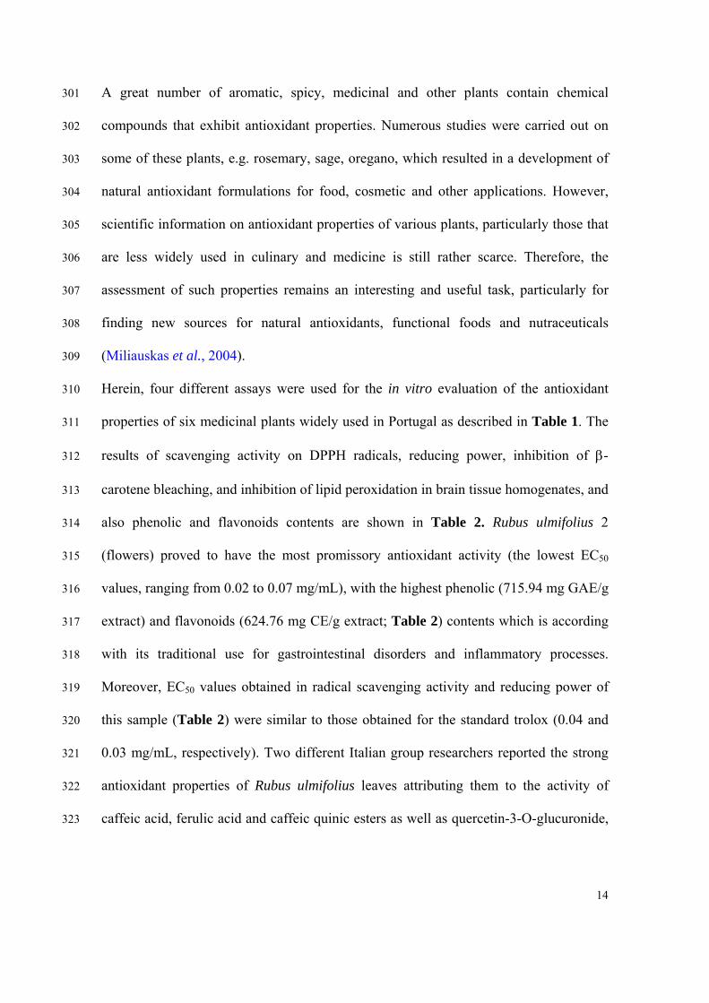
A great number of aromatic, spicy, medicinal and other plants contain chemical 301
compounds that exhibit antioxidant properties. Numerous studies were carried out on 302
some of these plants, e.g. rosemary, sage, oregano, which resulted in a development of 303
natural antioxidant formulations for food, cosmetic and other applications. However, 304
scientific information on antioxidant properties of various plants, particularly those that 305
are less widely used in culinary and medicine is still rather scarce. Therefore, the 306
assessment of such properties remains an interesting and useful task, particularly for 307
finding new sources for natural antioxidants, functional foods and nutraceuticals 308
(Miliauskas et al., 2004). 309
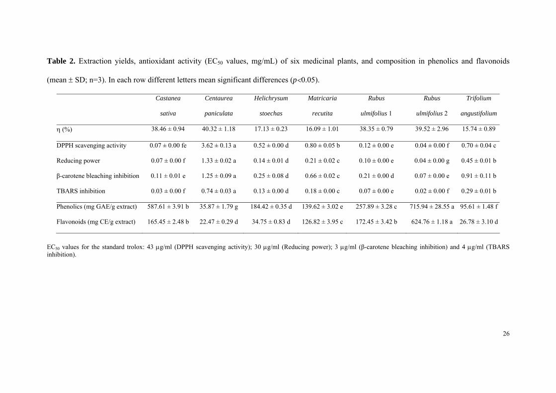
Herein, four different assays were used for the in vitro evaluation of the antioxidant 310
properties of six medicinal plants widely used in Portugal as described in Table 1. The 311
results of scavenging activity on DPPH radicals, reducing power, inhibition of β-312
carotene bleaching, and inhibition of lipid peroxidation in brain tissue homogenates, and 313
also phenolic and flavonoids contents are shown in Table 2. Rubus ulmifolius 2 314
(flowers) proved to have the most promissory antioxidant activity (the lowest EC50 315
values, ranging from 0.02 to 0.07 mg/mL), with the highest phenolic (715.94 mg GAE/g 316
extract) and flavonoids (624.76 mg CE/g extract; Table 2) contents which is according 317
with its traditional use for gastrointestinal disorders and inflammatory processes. 318
Moreover, EC50 values obtained in radical scavenging activity and reducing power of 319
this sample (Table 2) were similar to those obtained for the standard trolox (0.04 and 320
0.03 mg/mL, respectively). Two different Italian group researchers reported the strong 321
antioxidant properties of Rubus ulmifolius leaves attributing them to the activity of 322
caffeic acid, ferulic acid and caffeic quinic esters as well as quercetin-3-O-glucuronide, 323
15
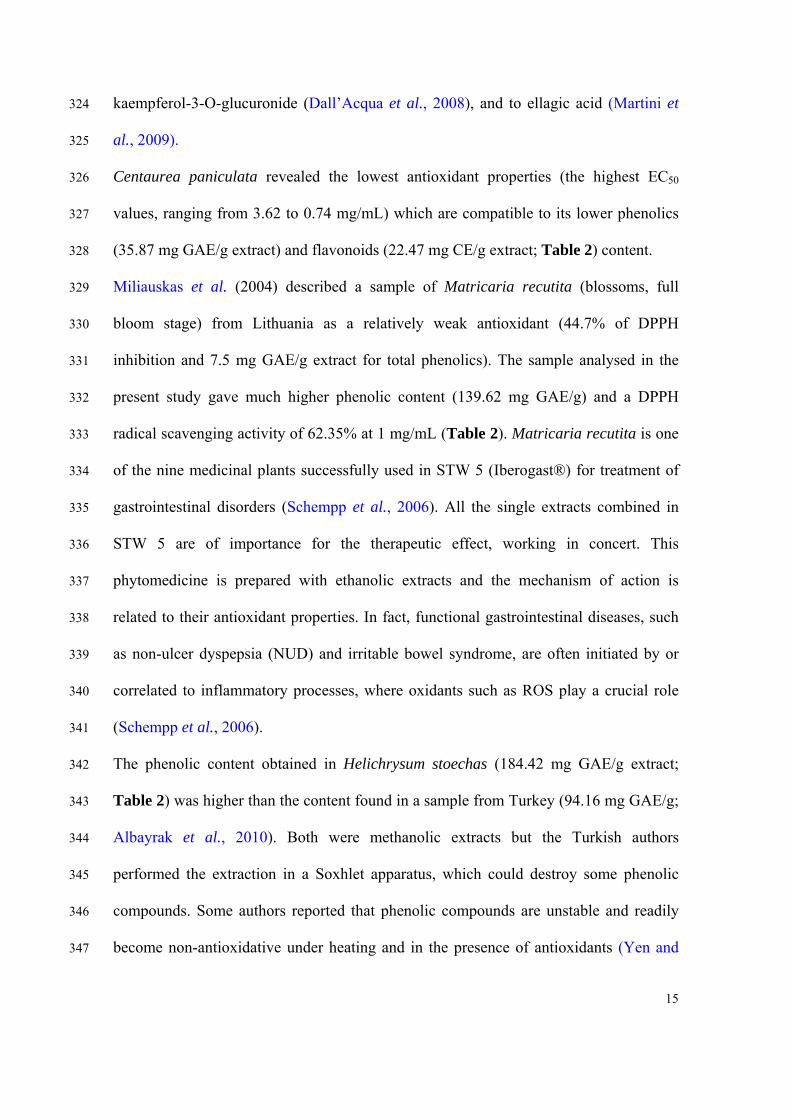
kaempferol-3-O-glucuronide (Dall’Acqua et al., 2008), and to ellagic acid (Martini et 324
al., 2009). 325
Centaurea paniculata revealed the lowest antioxidant properties (the highest EC50 326
values, ranging from 3.62 to 0.74 mg/mL) which are compatible to its lower phenolics 327
(35.87 mg GAE/g extract) and flavonoids (22.47 mg CE/g extract; Table 2) content. 328
Miliauskas et al. (2004) described a sample of Matricaria recutita (blossoms, full 329
bloom stage) from Lithuania as a relatively weak antioxidant (44.7% of DPPH 330
inhibition and 7.5 mg GAE/g extract for total phenolics). The sample analysed in the 331
present study gave much higher phenolic content (139.62 mg GAE/g) and a DPPH 332
radical scavenging activity of 62.35% at 1 mg/mL (Table 2). Matricaria recutita is one 333
of the nine medicinal plants successfully used in STW 5 (Iberogast®) for treatment of 334
gastrointestinal disorders (Schempp et al., 2006). All the single extracts combined in 335
STW 5 are of importance for the therapeutic effect, working in concert. This 336
phytomedicine is prepared with ethanolic extracts and the mechanism of action is 337
related to their antioxidant properties. In fact, functional gastrointestinal diseases, such 338
as non-ulcer dyspepsia (NUD) and irritable bowel syndrome, are often initiated by or 339
correlated to inflammatory processes, where oxidants such as ROS play a crucial role 340
(Schempp et al., 2006). 341
The phenolic content obtained in Helichrysum stoechas (184.42 mg GAE/g extract; 342
Table 2) was higher than the content found in a sample from Turkey (94.16 mg GAE/g; 343
Albayrak et al., 2010). Both were methanolic extracts but the Turkish authors 344
performed the extraction in a Soxhlet apparatus, which could destroy some phenolic 345
compounds. Some authors reported that phenolic compounds are unstable and readily 346
become non-antioxidative under heating and in the presence of antioxidants (Yen and 347

16
Hung, 2000; Barros et al., 2007. Nevertheless, Carini et al. (2001) obtained a very high 348
antioxidant activity with a sample from Italy. They reported that the polar fraction 349
isolated from the flowering tops of Helichrysum stoechas, displays radical scavenging 350
properties, with potency comparable to that of Trolox, the water-soluble analogue of 351
vitamin E. The extract included approximately 50% of polyphenols and 4% of 352
kaempferol-3-O-glucoside, the more prominent component of the extract (Carini et al., 353
2001). 354
Castanea sativa flowers (methanolic extract) gave highest DPPH scavenging activity 355
(0.07 mg/mL), reducing power (0.07 mg/mL), β-carotene bleaching inhibition (0.11 356
mg/mL), but lowest TBARS inhibition (0.03 mg/mL; Table 2) than a sample (aqueous 357
extract) also from Portugal (0.08 mg/mL, 0.09 mg/mL, 0.16 mg/mL and 0.01 mg/mL, 358
respectively; Barreira et al., 2008). This is in agreement with the higher phenolics 359
(587.61 mg GAE/g extract) and flavonoids (165.45 mg CE/g; Table 2) content found in 360
our sample in comparison to the other sample (298.18 mg GAE/g and 159.56 mg CE/g, 361
respectively; Barreira et al., 2008). This proves that the solvent used for phenolic 362
extraction has significant influence on the results. Herein, we decided to use methanol 363
according to previous results in experiments performed with different extraction 364
conditions to achieve the best procedure leading to highest contents in phenolics and 365
better antioxidant properties (Barros et al., 2010). Furthermore, chemical and biological 366
diversity of aromatic and medicinal plants depend on such factors, as growth habitat, 367
climatic conditions, vegetation phase and genetic modifications (Miliauskas et al., 368
2004). 369
Overall, among the medicinal plants analysed in the present study, Rubus ulmifolius 370
flowers revealed the highest antioxidant activity in all the tested assays. 371
17
372
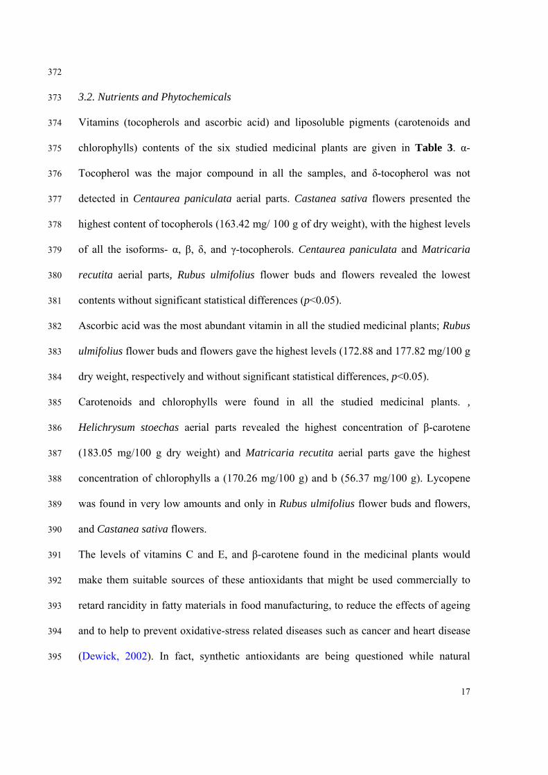
3.2. Nutrients and Phytochemicals 373
Vitamins (tocopherols and ascorbic acid) and liposoluble pigments (carotenoids and 374
chlorophylls) contents of the six studied medicinal plants are given in Table 3. α-375
Tocopherol was the major compound in all the samples, and δ-tocopherol was not 376
detected in Centaurea paniculata aerial parts. Castanea sativa flowers presented the 377
highest content of tocopherols (163.42 mg/ 100 g of dry weight), with the highest levels 378
of all the isoforms- α, β, δ, and γ-tocopherols. Centaurea paniculata and Matricaria 379
recutita aerial parts, Rubus ulmifolius flower buds and flowers revealed the lowest 380
contents without significant statistical differences (p<0.05). 381
Ascorbic acid was the most abundant vitamin in all the studied medicinal plants; Rubus 382
ulmifolius flower buds and flowers gave the highest levels (172.88 and 177.82 mg/100 g 383
dry weight, respectively and without significant statistical differences, p<0.05). 384
Carotenoids and chlorophylls were found in all the studied medicinal plants. , 385
Helichrysum stoechas aerial parts revealed the highest concentration of β-carotene 386
(183.05 mg/100 g dry weight) and Matricaria recutita aerial parts gave the highest 387
concentration of chlorophylls a (170.26 mg/100 g) and b (56.37 mg/100 g). Lycopene 388
was found in very low amounts and only in Rubus ulmifolius flower buds and flowers, 389
and Castanea sativa flowers. 390
The levels of vitamins C and E, and β-carotene found in the medicinal plants would 391
make them suitable sources of these antioxidants that might be used commercially to 392
retard rancidity in fatty materials in food manufacturing, to reduce the effects of ageing 393
and to help to prevent oxidative-stress related diseases such as cancer and heart disease 394
(Dewick, 2002). In fact, synthetic antioxidants are being questioned while natural 395
18
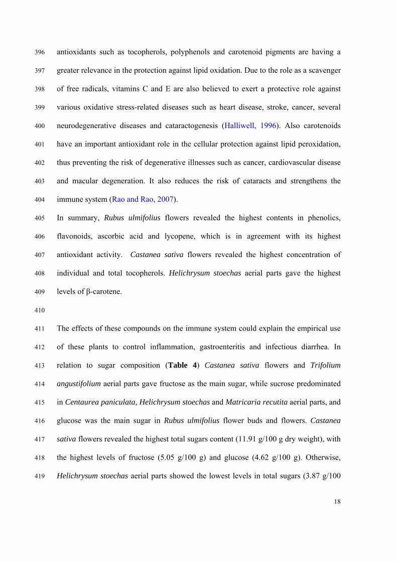
antioxidants such as tocopherols, polyphenols and carotenoid pigments are having a 396
greater relevance in the protection against lipid oxidation. Due to the role as a scavenger 397
of free radicals, vitamins C and E are also believed to exert a protective role against 398
various oxidative stress-related diseases such as heart disease, stroke, cancer, several 399
neurodegenerative diseases and cataractogenesis (Halliwell, 1996). Also carotenoids 400
have an important antioxidant role in the cellular protection against lipid peroxidation, 401
thus preventing the risk of degenerative illnesses such as cancer, cardiovascular disease 402
and macular degeneration. It also reduces the risk of cataracts and strengthens the 403
immune system (Rao and Rao, 2007). 404
In summary, Rubus ulmifolius flowers revealed the highest contents in phenolics, 405
flavonoids, ascorbic acid and lycopene, which is in agreement with its highest 406
antioxidant activity. Castanea sativa flowers revealed the highest concentration of 407
individual and total tocopherols. Helichrysum stoechas aerial parts gave the highest 408
levels of β-carotene. 409
410
The effects of these compounds on the immune system could explain the empirical use 411
of these plants to control inflammation, gastroenteritis and infectious diarrhea. In 412
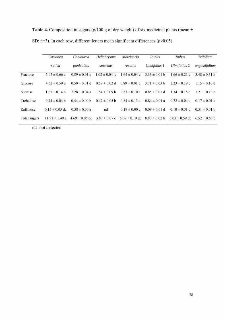
relation to sugar composition (Table 4) Castanea sativa flowers and Trifolium 413
angustifolium aerial parts gave fructose as the main sugar, while sucrose predominated 414
in Centaurea paniculata, Helichrysum stoechas and Matricaria recutita aerial parts, and 415
glucose was the main sugar in Rubus ulmifolius flower buds and flowers. Castanea 416
sativa flowers revealed the highest total sugars content (11.91 g/100 g dry weight), with 417
the highest levels of fructose (5.05 g/100 g) and glucose (4.62 g/100 g). Otherwise, 418
Helichrysum stoechas aerial parts showed the lowest levels in total sugars (3.87 g/100 419
19
g). Some of the identified sugars, mostly the reducing sugars fructose, glucose and 420
raffinose could also contribute to the antioxidant activity observed in the studied plants. 421
Overall, Castanea sativa flowers revealed the highest concentration of total sugars, 422
including the reducing sugars glucose and fructose. 423
424
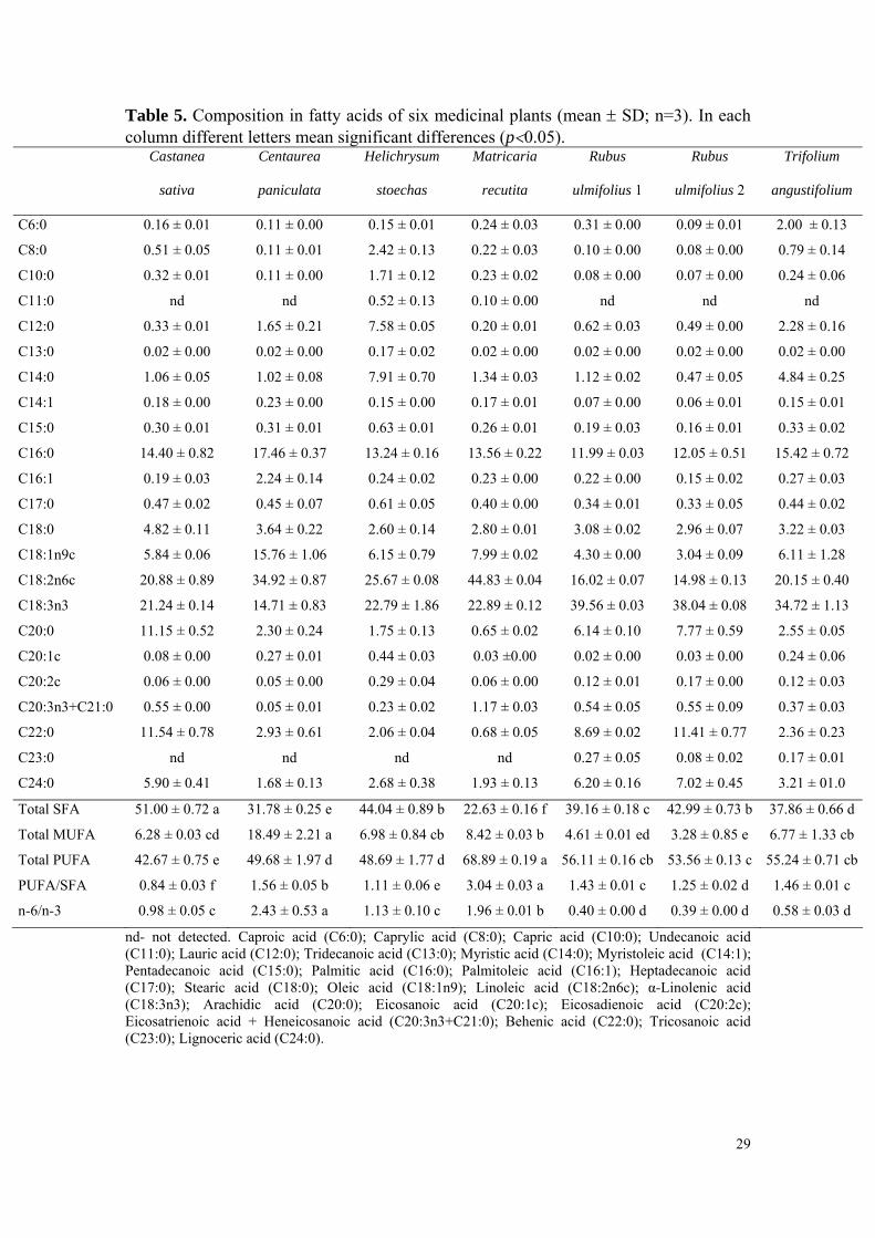
The results for fatty acid composition, total saturated fatty acids (SFA), 425
monounsaturated fatty acids (MUFA), polyunsaturated fatty acids (PUFA), and the 426
ratios of PUFA/SFA and n-6/n-3 of the studied medicinal plants are shown in Table 5. 427
Twenty three fatty acids were identified and quantified. The major fatty acids found 428
were linoleic acid (C18:2n6) and α-linolenic acid (C18:3n3) and contributing to the 429
prevalence of PUFA. Palmitic acid (C16:0) was also a main fatty acid in all the studied 430
plants, while arachidic acid (C20:0) and behenic acid (C22:0) were abundant in 431
Castanea sativa flowers, contributing to the highest SFA content found in this sample 432
(51%). Oleic acid (C18:1n9) was abundant in Centaurea paniculata aerial parts 433
contributing to the increase of its MUFA content (18.5%). Matricaria recutita aerial 434
parts revealed the highest PUFA contents (68.9%) with the highest levels of linoleic 435
acid (44.8%). 436
α-Linolenic and linoleic acids, as also high ratios of PUFA/SFA (> 0.45) and low n-6/n-437
3 fatty acids ratios (< 4.0) can decrease the total amount of fat in blood (cholesterol), 438
reducing the risk of cancer, cardiovascular, inflammatory and autoimmune diseases 439
(HMSO, 1994; Kanu et al., 2007). Once again these results are according to folk 440
recommendations. For instance, it is considered that chestnut catkins decoctions, drunk 441
for nine days, can lower blood cholesterol levels, and several informants pointed out 442
that German chamomile infusion may help reduce cholesterol. 443
20
Overall, Matricaria recutita aerial parts revealed the highest PUFA levels, including 444
linoleic acid. 445
446
In conclusion, the studied medicinal plants revealed interesting antioxidant properties, 447
nutrients and phytochemicals such as phenolics, flavonoids, vitamins, carotenoids, 448
sugars, and fatty acids that could provide scientific evidence for some folk uses in the 449
treatment of diseases related to the production of ROS and oxidative stress, but further 450
experiments are required to explore the mechanisms of action. Traditional medicines, 451
whose knowledge and practices have been orally transmitted over the centuries, are 452
important approaches for discovering therapeutic molecules and compounds. This study 453
provides information useful not only to chemical analysis, activity assays and 454
standardization of phytochemical composition of wild species traditionally used, but to 455
researchers in phytopharmacology, phytotherapy and phytotoxicology as well. As far as 456
we know this is the first report on chemical characterization of six medicinal plants 457
from the Portuguese folk medicine: Castanea sativa, Centaurea paniculata, 458
Helichrysum stoechas, Matricaria recutita, Rubus ulmifollius and Trifolium 459
angustifolium. 460
461
Acknowledgements 462
The authors are grateful to the Foundation for Science and Technology (Portugal) for 463
financial support to the research centre CIMO, L. Barros (SFRH/BPD/4609/2008) and 464
S. Oliveira (BII/CIMO/09/18) grants. 465
466
References 467
468
21
Albayrak, S., Aksoy, A., Sagdic, O., Hamzaoglu, E. 2010. Compositions, antioxidant 469
and antimicrobial activities of Helichrysum (Asteraceae) species collected from 470
Turkey. Food Chem. 119, 114–122. 471
Barreira, J.C.M., Ferreira, I.C.F.R., Oliveira, M.B.P.P., Pereira, J.A. 2008. Antioxidant 472
activities of the extracts from chestnut flower, leaf, skins and fruit. Food Chem. 473
107, 1106–1113. 474
Barros, L., Baptista, P., Correia, D.M., Morais, J.S., Ferreira, I.C.F.R. 2007. Effects of 475
conservation treatment and cooking on the chemical composition and antioxidant 476
activity of Portuguese wild edible mushrooms. J. Agric. Food Chem. 55, 4781-477
4788. 478
Barros, L., Heleno, S.A., Carvalho, A.M., Ferreira, I.C.F.R. 2010. Lamiaceae often used 479
in Portuguese folk medicine as a source of powerful antioxidants: Vitamins and 480
phenolics. LWT 43, 544–550. 481
Carini, M., Aldini, G., Furlanetto, S., Stefani, R., Facino, R.M. 2001. LC coupled to ion-482
trap MS for the rapid screening and detection of polyphenol antioxidants from 483
Helichrysum stoechas. J. Pharm. Biomed. Anal 24, 517–526. 484
Carvalho, A.M. 2010. Plantas y sabiduría popular del Parque Natural de Montesinho. 485
Un estudio etnobotánico en Portugal. Madrid: CSIC, Biblioteca de Ciencias. 486
Carvalho, A.M., Morales, R. 2010. ‘Persistence of Wild Food and Wild Medicinal Plant 487
Knowledge in a North-Eastern Region of Portugal’. In M. Pardo de Santayana, A. 488
Pieroni, & R. Puri (eds.), Ethnobotany in the New Europe: People, Health and 489
Wild Plant Resources. Oxford, UK: Berghahn Books. 490
Castroviejo, S. (coord.). 1991, 2001, 2007. Flora Iberica. Plantas vasculares de la 491
Península Ibérica e Islas Baleares. Real Jardín Botánico, CSIC, Madrid. 492
22
Dall’Acqua, S., Cervellati, R., Loi, M.C., Innocenti, G. 2008. Evaluation of in vitro 493
antioxidant properties of some traditional Sardinian medicinal plants: Investigation 494
of the high antioxidant capacity of Rubus ulmifolius. Food Chem. 106, 745–749. 495
Dawidowicza, A.L., Wianowska, D., Baraniak, B. 2006. The antioxidant properties of 496
alcoholic extracts from Sambucus nigra L. (antioxidant properties of extracts). 497
LWT 39, 308–315. 498
Dewick, P.M. 2002. Medicinal Natural Products. John Wiley & Sons, Lda. 499
Franco, J.A. 1984. Nova Flora de Portugal, Volume II. Lisboa. 500
Frazão-moreira, A., Carvalho, A.M., Martins, M.E. 2007. Conocimientos acerca de 501
plantas en la nueva ruralidad. Cambio social y agro ecología en el Parque Natural 502
de Montesinho (Portugal). Revista Periféria 7. 503
Guimarães, R., Barros, L., Carvalho, A.M., Ferreira, I.C.F.R. 2010. Studies on chemical 504
constituents and bioactivity of Rosa micrantha: An alternative antioxidants source 505
for food, pharmaceutical, or cosmetic applications. J. Agric. Food Chem. 58, 506
6277-6284. 507
Halliwell, B. 1996. Antioxidants in human health and disease. Annu. Rev. Nutr. 16, 33-508
50. 509
HMSO, U.K. 1994. Department of Health. Nutritional aspects of cardiovascular disease. 510
London, Report on Health and Social Subjects 46, 37–46. 511
Jia, Z., Tang, M., Wu, J. 1999. The determination of flavonoid contents in mulberry and 512
their scavenging effects on superoxide radicals. Food Chem. 64, 555-559. 513
Kanu, P.J., Zhu, K., Kanu, J.B., Zhou, H., Qian, H., Zhu, K. 2007. Biologically active 514
components and nutraceuticals in sesame and related products: a review and 515
prospect. Trends Food Sci. Technol. 18, 599-608. 516
23
Klein, B.P., Perry, A.K. 1982. Ascorbic acid and vitamin A activity in selected 517
vegetables from different geographical areas of the United States. J. Food Sci. 47, 518
941–945. 519
Marchand, L.L. 2002. Cancer preventive effects of flavonoids- a review. Biomed 520
Pharmacother 56, 296-301. 521
Martinia, S., D’Addario, C., Colacevich, A., Focardi, S., Borghinic, F., Santucci, A., 522
Figura, N., Rossi, C. 2009. Antimicrobial activity against Helicobacter pylori 523
strains and antioxidant properties of blackberry leaves (Rubus ulmifolius) and 524
isolated compounds. Int. J. Antimicrob. Agents 34, 50–59. 525
Miliauskas, G., Venskutonis, P.R., van Beek, T.A. 2004. Screening of radical scavenging 526
activity of some medicinal and aromatic plant extracts. Food Chem. 85, 231–237. 527
Mishra, K.P., Ganju, L., Sairam, M., Banerjee, P.K., Sawhney, R.C. 2008. A review of 528
high throughput technology for the screening of natural products. Biomed. 529
Pharmacother 62, 94-98. 530
Montagnier, L. 2009. Oxidative stress in preventive medicine. Free Rad. Res. 43, S27-531
97. 532
Nagata, M., Yamashita, I. 1992. Simple method for simultaneous determination of 533
chlorophyll and carotenoids in tomato fruit. Nippon Shokuhin Kogyo Gakkaish 39, 534
925–928. 535
Neves, J.M., Matosa, C., Moutinho, C., Queiroz, G., Gomes, L.R. 2009. 536
Ethnopharmacological notes about ancient uses of medicinal plants in Trás-os-537
Montes (northern of Portugal). J. Ethnopharmacol. 124, 270–283. 538
Rao, A.V, Rao, L.G. 2007. Carotenoids in human health. Pharmacol. Res. 55, 207-216. 539
24
Schemppa, H., Weiserb, D., Kelberb, O., Elstner, E.F. 2006. Radical scavenging and 540
anti-inflammatory properties of STW 5 (Iberogasts) and its components. 541
Phytomedicine 13, SV 36–44. 542
Wolfe, K., Wu, X., Liu, R.H. 2003. Antioxidant activity of apple peels. J. Agric. Food 543
Chem. 51, 609-614. 544
Yen, G-C, Hung, C-Y. 2000. Effects of alkaline and heat treatment on antioxidative 545
activity and total phenolics of extracts from Hsian-tsao (Mesona procumbens 546
Hemsl.). Food Res. Int. 33, 487-492. 547

25
Table 1. Medicinal uses of the studied plants reported in Portuguese ethnobotanical
studies conducted in the north-eastern region.
Scientific name English name Local name Medicinal uses
Castanea sativa Chestnut flower Flor-de-castanheiro Cold, cough, diarrhea, cholesterol
Centaurea paniculata starthistles, knapweeds Escalabriosa Inflammation
Helichrysum stoechas Shrubby everlasting Douradinha Cold, bronchitis, fever
Matricaria recutita German chamomile Maçiela Respiratory and digestive systems, inflammation
Rubus ulmifolius Elm-leaved blackberry; Silva, silva-gravanceira
Stomachache or abdmominal pane diarrhea
Trifolium angustifolium Narrow clover Rabo-de-gato Diarrhea
26
Table 2. Extraction yields, antioxidant activity (EC50 values, mg/mL) of six medicinal plants, and composition in phenolics and flavonoids
(mean ± SD; n=3). In each row different letters mean significant differences (p<0.05).
Castanea
sativa
Centaurea
paniculata
Helichrysum
stoechas
Matricaria
recutita
Rubus
ulmifolius 1
Rubus
ulmifolius 2
Trifolium
angustifolium
η (%) 38.46 ± 0.94 40.32 ± 1.18 17.13 ± 0.23 16.09 ± 1.01 38.35 ± 0.79 39.52 ± 2.96 15.74 ± 0.89
DPPH scavenging activity 0.07 ± 0.00 fe 3.62 ± 0.13 a 0.52 ± 0.00 d 0.80 ± 0.05 b 0.12 ± 0.00 e 0.04 ± 0.00 f 0.70 ± 0.04 c
Reducing power 0.07 ± 0.00 f 1.33 ± 0.02 a 0.14 ± 0.01 d 0.21 ± 0.02 c 0.10 ± 0.00 e 0.04 ± 0.00 g 0.45 ± 0.01 b
β-carotene bleaching inhibition 0.11 ± 0.01 e 1.25 ± 0.09 a 0.25 ± 0.08 d 0.66 ± 0.02 c 0.21 ± 0.00 d 0.07 ± 0.00 e 0.91 ± 0.11 b
TBARS inhibition 0.03 ± 0.00 f 0.74 ± 0.03 a 0.13 ± 0.00 d 0.18 ± 0.00 c 0.07 ± 0.00 e 0.02 ± 0.00 f 0.29 ± 0.01 b
Phenolics (mg GAE/g extract) 587.61 ± 3.91 b 35.87 ± 1.79 g 184.42 ± 0.35 d 139.62 ± 3.02 e 257.89 ± 3.28 c 715.94 ± 28.55 a 95.61 ± 1.48 f
Flavonoids (mg CE/g extract) 165.45 ± 2.48 b 22.47 ± 0.29 d 34.75 ± 0.83 d 126.82 ± 3.95 c 172.45 ± 3.42 b 624.76 ± 1.18 a 26.78 ± 3.10 d
EC50 values for the standard trolox: 43 μg/ml (DPPH scavenging activity); 30 μg/ml (Reducing power); 3 μg/ml (β-carotene bleaching inhibition) and 4 μg/ml (TBARS inhibition).

27
Table 3. Composition in vitamins and liposoluble pigments (mg/100 g dry weight) of six medicinal plants (mean ± SD; n=3). In each row
different letters mean significant differences (p<0.05).
nd- not detected.
Castanea
sativa
Centaurea
paniculata
Helichrysum
stoechas
Matricaria
recutita
Rubus
ulmifolius 1
Rubus
ulmifolius 2
Trifolium
angustifolium
α-tocopherol 124.64 ± 2.28 a 8.54 ± 0.46 dc 66.43 ± 3.61 b 3.52 ± 0.30 e 5.97 ± 0.20 de 5.84 ± 0.01 de 12.73 ± 1.14 c
β-tocopherol 3.62 ± 0.20 a 0.25 ± 0.05 c 1.70 ± 0.23 b 0.18 ± 0.02 c 0.28 ± 0.10 c 0.18 ± 0.00 c 0.60 ± 0.06 c
γ-tocopherol 20.26 ± 0.94 a 1.82 ± 0.15 d 4.20 ± 0.01 c 2.44 ± 0.06 d 4.05 ± 0.01 c 2.55 ± 0.01 d 5.41 ± 0.62 b
δ-tocopherol 14.90 ± 0.80 a nd 1.28 ± 0.01 b 1.20 ± 0.23 cb 1.98 ± 0.01 b 1.28 ± 0.05 b 0.39 ± 0.02 d
Total tocopherols 163.42 ± 4.22 a 10.60 ± 0.66 d 73.61 ± 3.85 b 7.35 ± 0.03 d 12.28 ± 0.21 d 9.86 ± 0.05 d 19.13 ± 2.00 c
Ascorbic acid 163.48 ± 8.20 b 40.45 ± 1.15 d 157.07 ± 10.48 b 61.57 ± 0.81 c 172.88 ± 1.89 a 177.82 ± 6.97 a 163.80 ± 2.48 b
β-carotene 43.53 ± 0.08 e 69.25 ± 0.08 d 183.05 ± 0.75 a 127.68 ± 0.29 c 34.22 ± 0.28 g 38.77 ± 0.15 f 160.69 ± 1.13 b
Lycopene 0.05 ± 0.00 b nd nd nd 0.01 ± 0.00 c 0.07 ± 0.00 a nd
Chlorophyll a 1.06 ± 0.00 e 1.23 ± 0.00 d 2.72 ± 0.00 c 170.26 ± 0.08 a 0.63 ± 0.00 f 1.09 ± 0.00 e 4.75 ± 0.00 b
Chlorophyll b 0.46 ± 0.00 d 0.35 ± 0.00 d 1.02 ± 0.00 c 56.37 ± 0.36 a 0.27 ± 0.00 d 0.59 ± 0.00 d 1.77 ± 0.00 b
28
Table 4. Composition in sugars (g/100 g of dry weight) of six medicinal plants (mean ±
SD; n=3). In each row, different letters mean significant differences (p<0.05).
nd- not detected
Castanea
sativa
Centaurea
paniculata
Helichrysum
stoechas
Matricaria
recutita
Rubus
Ulmifolius 1
Rubus
Ulmifolius 2
Trifolium
angustifolium
Fructose 5.05 ± 0.66 a 0.89 ± 0.01 c 1.02 ± 0.04 c 1.64 ± 0.04 c 3.33 ± 0.01 b 1.66 ± 0.21 c 3.48 ± 0.31 b
Glucose 4.62 ± 0.59 a 0.50 ± 0.01 d 0.59 ± 0.02 d 0.89 ± 0.01 d 3.71 ± 0.03 b 2.23 ± 0.19 c 1.15 ± 0.10 d
Sucrose 1.65 ± 0.14 b 2.28 ± 0.04 a 1.84 ± 0.09 b 2.53 ± 0.10 a 0.85 ± 0.01 d 1.34 ± 0.15 c 1.21 ± 0.13 c
Trehalose 0.44 ± 0.04 b 0.44 ± 0.00 b 0.42 ± 0.05 b 0.84 ± 0.13 a 0.84 ± 0.01 a 0.72 ± 0.04 a 0.17 ± 0.01 c
Raffinose 0.15 ± 0.05 dc 0.58 ± 0.00 a nd 0.19 ± 0.00 c 0.09 ± 0.01 d 0.10 ± 0.01 d 0.51 ± 0.01 b
Total sugars 11.91 ± 1.49 a 4.69 ± 0.05 de 3.87 ± 0.07 e 6.08 ± 0.19 dc 8.83 ± 0.02 b 6.03 ± 0.59 dc 6.52 ± 0.63 c
29
Table 5. Composition in fatty acids of six medicinal plants (mean ± SD; n=3). In each column different letters mean significant differences (p<0.05).
Castanea
sativa
Centaurea
paniculata
Helichrysum
stoechas
Matricaria
recutita
Rubus
ulmifolius 1
Rubus
ulmifolius 2
Trifolium
angustifolium
C6:0 0.16 ± 0.01 0.11 ± 0.00 0.15 ± 0.01 0.24 ± 0.03 0.31 ± 0.00 0.09 ± 0.01 2.00 ± 0.13
C8:0 0.51 ± 0.05 0.11 ± 0.01 2.42 ± 0.13 0.22 ± 0.03 0.10 ± 0.00 0.08 ± 0.00 0.79 ± 0.14
C10:0 0.32 ± 0.01 0.11 ± 0.00 1.71 ± 0.12 0.23 ± 0.02 0.08 ± 0.00 0.07 ± 0.00 0.24 ± 0.06
C11:0 nd nd 0.52 ± 0.13 0.10 ± 0.00 nd nd nd
C12:0 0.33 ± 0.01 1.65 ± 0.21 7.58 ± 0.05 0.20 ± 0.01 0.62 ± 0.03 0.49 ± 0.00 2.28 ± 0.16
C13:0 0.02 ± 0.00 0.02 ± 0.00 0.17 ± 0.02 0.02 ± 0.00 0.02 ± 0.00 0.02 ± 0.00 0.02 ± 0.00
C14:0 1.06 ± 0.05 1.02 ± 0.08 7.91 ± 0.70 1.34 ± 0.03 1.12 ± 0.02 0.47 ± 0.05 4.84 ± 0.25
C14:1 0.18 ± 0.00 0.23 ± 0.00 0.15 ± 0.00 0.17 ± 0.01 0.07 ± 0.00 0.06 ± 0.01 0.15 ± 0.01
C15:0 0.30 ± 0.01 0.31 ± 0.01 0.63 ± 0.01 0.26 ± 0.01 0.19 ± 0.03 0.16 ± 0.01 0.33 ± 0.02
C16:0 14.40 ± 0.82 17.46 ± 0.37 13.24 ± 0.16 13.56 ± 0.22 11.99 ± 0.03 12.05 ± 0.51 15.42 ± 0.72
C16:1 0.19 ± 0.03 2.24 ± 0.14 0.24 ± 0.02 0.23 ± 0.00 0.22 ± 0.00 0.15 ± 0.02 0.27 ± 0.03
C17:0 0.47 ± 0.02 0.45 ± 0.07 0.61 ± 0.05 0.40 ± 0.00 0.34 ± 0.01 0.33 ± 0.05 0.44 ± 0.02
C18:0 4.82 ± 0.11 3.64 ± 0.22 2.60 ± 0.14 2.80 ± 0.01 3.08 ± 0.02 2.96 ± 0.07 3.22 ± 0.03
C18:1n9c 5.84 ± 0.06 15.76 ± 1.06 6.15 ± 0.79 7.99 ± 0.02 4.30 ± 0.00 3.04 ± 0.09 6.11 ± 1.28
C18:2n6c 20.88 ± 0.89 34.92 ± 0.87 25.67 ± 0.08 44.83 ± 0.04 16.02 ± 0.07 14.98 ± 0.13 20.15 ± 0.40
C18:3n3 21.24 ± 0.14 14.71 ± 0.83 22.79 ± 1.86 22.89 ± 0.12 39.56 ± 0.03 38.04 ± 0.08 34.72 ± 1.13
C20:0 11.15 ± 0.52 2.30 ± 0.24 1.75 ± 0.13 0.65 ± 0.02 6.14 ± 0.10 7.77 ± 0.59 2.55 ± 0.05
C20:1c 0.08 ± 0.00 0.27 ± 0.01 0.44 ± 0.03 0.03 ±0.00 0.02 ± 0.00 0.03 ± 0.00 0.24 ± 0.06
C20:2c 0.06 ± 0.00 0.05 ± 0.00 0.29 ± 0.04 0.06 ± 0.00 0.12 ± 0.01 0.17 ± 0.00 0.12 ± 0.03
C20:3n3+C21:0 0.55 ± 0.00 0.05 ± 0.01 0.23 ± 0.02 1.17 ± 0.03 0.54 ± 0.05 0.55 ± 0.09 0.37 ± 0.03
C22:0 11.54 ± 0.78 2.93 ± 0.61 2.06 ± 0.04 0.68 ± 0.05 8.69 ± 0.02 11.41 ± 0.77 2.36 ± 0.23
C23:0 nd nd nd nd 0.27 ± 0.05 0.08 ± 0.02 0.17 ± 0.01
C24:0 5.90 ± 0.41 1.68 ± 0.13 2.68 ± 0.38 1.93 ± 0.13 6.20 ± 0.16 7.02 ± 0.45 3.21 ± 01.0
Total SFA 51.00 ± 0.72 a 31.78 ± 0.25 e 44.04 ± 0.89 b 22.63 ± 0.16 f 39.16 ± 0.18 c 42.99 ± 0.73 b 37.86 ± 0.66 d
Total MUFA 6.28 ± 0.03 cd 18.49 ± 2.21 a 6.98 ± 0.84 cb 8.42 ± 0.03 b 4.61 ± 0.01 ed 3.28 ± 0.85 e 6.77 ± 1.33 cb
Total PUFA 42.67 ± 0.75 e 49.68 ± 1.97 d 48.69 ± 1.77 d 68.89 ± 0.19 a 56.11 ± 0.16 cb 53.56 ± 0.13 c 55.24 ± 0.71 cb
PUFA/SFA 0.84 ± 0.03 f 1.56 ± 0.05 b 1.11 ± 0.06 e 3.04 ± 0.03 a 1.43 ± 0.01 c 1.25 ± 0.02 d 1.46 ± 0.01 c
n-6/n-3 0.98 ± 0.05 c 2.43 ± 0.53 a 1.13 ± 0.10 c 1.96 ± 0.01 b 0.40 ± 0.00 d 0.39 ± 0.00 d 0.58 ± 0.03 d
nd- not detected. Caproic acid (C6:0); Caprylic acid (C8:0); Capric acid (C10:0); Undecanoic acid (C11:0); Lauric acid (C12:0); Tridecanoic acid (C13:0); Myristic acid (C14:0); Myristoleic acid (C14:1); Pentadecanoic acid (C15:0); Palmitic acid (C16:0); Palmitoleic acid (C16:1); Heptadecanoic acid (C17:0); Stearic acid (C18:0); Oleic acid (C18:1n9); Linoleic acid (C18:2n6c); α-Linolenic acid (C18:3n3); Arachidic acid (C20:0); Eicosanoic acid (C20:1c); Eicosadienoic acid (C20:2c); Eicosatrienoic acid + Heneicosanoic acid (C20:3n3+C21:0); Behenic acid (C22:0); Tricosanoic acid (C23:0); Lignoceric acid (C24:0).
Copyright © 2022 FDOKUMEN