
IN THIS ISSUE: - PJP
83
Vol. 4 No. 1 June 2019 ISSN 2507-8364 (Online) IN THIS ISSUE: ORIGINAL ARTICLES Predictive Value of Histologic Characteristics on Hormone Receptor and HER-2 Status of Patients with Invasive Breast Carcinoma, No Special Type, in an Academic Medical Center Kevin Elomina and Ma. Carmen Cagampan The National External Quality Assessment Scheme for Diagnostic Medical Parasitology in the Philippines, 2009–2015 Sherwin Galit, Dave Tangcalagan, Julius Matt Rapanut, Alexander Sadiasa, Prince Ninja Bautista, Kim Joshua Dominguez, Jahra Bañaga, Celine Bernice Roxas, Rafael Navarro, Mark Philip Bugayong, Jennifer Luchavez, Melisa Mondoy, Lydia Sombrero, Socorro Lupisan Buccal Cell Micronuclei among Betel Quid Chewers and Non-Betel Quid Chewers from Selected Barangays in Zamboanga City Benkassar Abdurajak, Servando Halili Jr., Al-Zamzam Abubakar Immunohistochemical Expression of WT1 in Nasopharyngeal Carcinoma Among Filipino Patients in a Tertiary Hospital Criston Van Manasan, Jenny Maureen Atun, Jose Carnate Jr. CASE REPORT Oral Carcinoma Cuniculatum: A Case Report Jose Carnate Jr., Leila Salera, Cynthia del Castillo, Jay Hansel Tabije AUTOPSY VAULT Autopsy Findings in a Patient with Post-Obstructive Pulmonary Edema Marissa Krizelda Santos, Ann Margaret Chang, Jimmy Chang DIAGNOSTIC PERSPECTIVES Wire-Free Virtual Breast Localization Using Liquid Carbon Nanoparticles Ma. Theresa Buenaflor, Ricardo Victorio Quimbo, Norman Val San Agustin
-
Upload
khangminh22 -
Category
Documents
-
view
4 -
download
0
Transcript of IN THIS ISSUE: - PJP

Vol. 4 No. 1 June 2019ISSN 2507-8364 (Online)
IN THIS ISSUE:ORIGINAL ARTICLESPredictive Value of Histologic Characteristics on Hormone Receptor and HER-2 Status of Patients with Invasive Breast Carcinoma, No Special Type, in an Academic Medical CenterKevin Elomina and Ma. Carmen Cagampan
The National External Quality Assessment Scheme for Diagnostic Medical Parasitology in the Philippines, 2009–2015Sherwin Galit, Dave Tangcalagan, Julius Matt Rapanut, Alexander Sadiasa, Prince Ninja Bautista, Kim Joshua Dominguez, Jahra Bañaga, Celine Bernice Roxas, Rafael Navarro, Mark Philip Bugayong, Jennifer Luchavez, Melisa Mondoy, Lydia Sombrero, Socorro Lupisan
Buccal Cell Micronuclei among Betel Quid Chewers and Non-Betel Quid Chewers from Selected Barangays in Zamboanga CityBenkassar Abdurajak, Servando Halili Jr., Al-Zamzam Abubakar
Immunohistochemical Expression of WT1 in Nasopharyngeal Carcinoma Among Filipino Patients in a Tertiary HospitalCriston Van Manasan, Jenny Maureen Atun, Jose Carnate Jr.
CASE REPORTOral Carcinoma Cuniculatum: A Case ReportJose Carnate Jr., Leila Salera, Cynthia del Castillo, Jay Hansel Tabije
AUTOPSY VAULTAutopsy Findings in a Patient with Post-Obstructive Pulmonary EdemaMarissa Krizelda Santos, Ann Margaret Chang, Jimmy Chang
DIAGNOSTIC PERSPECTIVESWire-Free Virtual Breast Localization Using Liquid Carbon NanoparticlesMa. Theresa Buenaflor, Ricardo Victorio Quimbo, Norman Val San Agustin
ISSN
0118-3265PH
ILIPPIN
E JO
UR
NA
L O
F PA
TH
OL
OG
Y

* This advertisement is a complimentary service of the PJP for member societies/organizations.

This journal is OPEN ACCESS, providing immediate access to its content on the principle that making research freely available to the public supports a greater global exchange of knowledge. The Philippine Journal of Pathology is licensed under a Creative Commons Attribution-NonCommercial-ShareAlike 4.0 International License, which allows sharing, copying and redistributing the material in any medium or format, and adaptation, in which the material can be built upon, under strict terms of giving appropriate credit to the authors and this journal, and use for non-commercial purposes.
Philippine Journal of PathologyVol. 4 No. 1 June 2019 | ISSN 2507-8364 (Online)
http://philippinejournalofpathology.org

AMADO O. TANDOC IIIEditor-in-Chief
FRANCIS G. MORIAVice Editor-in-Chief
IVY A. ROSALESMARICEL REGINO-RIBO
FARRAH KRISTINE FONTILA-SANTIAGOANN MARGARET V. CHANGFRANCES SAURA-SANCHEZ
MARIE CHRISTINE F. BERNARDODAPHNE CHUA ANG
MA. LOURDES L. GOCOMARY JANE CARIAS-MARINES
Associate Editors
Editorial Board
AGUSTINA D. ABELARDO Cytopathology
JOSE M. CARNATE JR.Head & Neck Pathology
MA. RIZALINA F. CHUABlood Banking
NELSON T. GERALDINOBiochemistry
EVELINA N. LAGAMAYOMedical Microbiology
RAYMUNDO W. LOImmunology & Molecular Pathology
MIGUEL MARTIN N. MORENO IIBiosafety/Biosecurity
JANUARIO D. VELOSOHematopathology
SOCORRO C. YAÑEZLaboratory Quality Assurance
ROWEN T. YOLOSurgical Pathology
Editorial Advisers
JOSE MA. C. AVILAMARISSA A. ORILLAZA
MARITA V.T. REYES
Editorial Staff
MELISSA O. TANDOCEditorial Coordinator
PRESIDENT’S MESSAGEEDITORIAL
ORIGINAL ARTICLESPredictive Value of Histologic Characteristics on Hormone Receptor and HER-2 Status of Patients with Invasive Breast Carcinoma, No Special Type, in an Academic Medical CenterKevin Elomina and Ma. Carmen Cagampan
The National External Quality Assessment Scheme for Diagnostic Medical Parasitology in the Philippines, 2009–2015Sherwin Galit, Dave Tangcalagan, Julius Matt Rapanut, Alexander Sadiasa, Prince Ninja Bautista, Kim Joshua Dominguez, Jahra Bañaga, Celine Bernice Roxas, Rafael Navarro, Mark Philip Bugayong, Jennifer Luchavez, Melisa Mondoy, Lydia Sombrero, Socorro Lupisan
Buccal Cell Micronuclei among Betel Quid Chewers and Non-Betel Quid Chewers from Selected Barangays in Zamboanga CityBenkassar Abdurajak, Servando Halili Jr., Al-Zamzam Abubakar
Immunohistochemical Expression of WT1 in Nasopharyngeal Carcinoma Among Filipino Patients in a Tertiary HospitalCriston Van Manasan, Jenny Maureen Atun, Jose Carnate Jr.
CASE REPORTOral Carcinoma Cuniculatum: A Case ReportJose Carnate Jr., Leila Salera, Cynthia del Castillo, Jay Hansel Tabije
AUTOPSY VAULTAutopsy Findings in a Patient with Post-Obstructive Pulmonary EdemaMarissa Krizelda Santos, Ann Margaret Chang, Jimmy Chang
DIAGNOSTIC PERSPECTIVESWire-Free Virtual Breast Localization Using Liquid Carbon NanoparticlesMa. Theresa Buenaflor, Ricardo Victorio Quimbo, Norman Val San Agustin
Instructions to AuthorsAuthorship FormICMJE Form for Disclosure of Potential Conflicts of InterestPatient Consent FormEquator Network Reporting GuidelinesOJS Guide to AuthorsPeer Reviewers
34
6
12
18
25
34
37
43
47515255566777
The Philippine Journal of Pathology (PJP) is an open-access, peer-reviewed, English language, medical science journal published by the Philippine Society of Pathologists, Inc. Committee on Publications. It shall serve as the official platform for publication of high quality original articles, case reports or series, feature articles, and editorials covering topics on clinical and anatomic pathology, laboratory medicine and medical technology, diagnostics, laboratory biosafety and biosecurity, as well as laboratory quality assurance. The journal's primary target audience are laboratorians, diagnosticians, laboratory managers, pathologists, medical technologists, and all other medical and scientific disciplines interfacing with the laboratory. The PJP follows the ICMJE Recommendations for the Conduct, Reporting, Editing and Publication of Scholarly Work in Medical Journals, EQUATOR Network Guidelines, and COPE Guidelines. The PJP does not charge any article processing or submission fees from authors. It does not charge any subscription fees or download fees to access content.

http://philippinejournalofpathology.org | Vol. 4 No. 1 June 2019
PRESIDENT’S MESSAGE
Greetings!
Welcome to the June 2019 issue of the Philippine Journal of Pathology. My congratulations to the hardworking editorial team of PJP and to the supportive Board of Governors of the Philippine Society of Pathologists, Inc. for a job well done.
As we have encountered many challenges from the inception of our PJP to sustaining its publications, we are very thankful to all the support given by members of the PSP leading to its success. It was our dream to have our own journal where the scholarly works of our members can be published and shared with the other specialties both local and abroad. The researches of our members contribute to the advancement in the practice of pathology and on how we can improve our diagnostic acumen.
Let us look forward to many more issues and hope that you can join us as contributors. Goodluck to the Philippine Society of Pathologists, Inc. and the Philippine Journal of Pathology to its future endeavors.
Roberto D. Padua Jr., MD, FPSP, MHAPresident, Philippine Society of Pathologists, Inc.
EDITORIAL
http://philippinejournalofpathology.org | Vol. 4 No. 1 June 2019
This 2019, I was privileged to represent the Philippine Journal of Pathology in two local conventions: the 1st Philippine Association of Medical Journal Editors (PAMJE) Convention and the 68th Annual Convention of the Philippine Society of Pathologists, Inc., (PSP Inc.). Within weeks of each other,
the dual engagement made me realize how our appreciation for research has been evolving for the better. Local journals may not necessarily be in their best condition at this time, but there are signs of life at the very least.
At the PAMJE convention, I discussed the workflow of manuscripts from submission to publication as part of journal management. Editors from other local health journals joined the event to share not only best practices but also the collective travails of a small but growing lot of advocates for ethical scholarly publication. At this year's PSP convention, I was given an opportunity to give tips to our colleagues for maximizing publication potential, i.e., practical advice to guide pathologist researchers on how to increase the chances of becoming part of the scientific body of literature (Table 1).
The learning curve for local authors and journals, specifically in Pathology, is steep, but ultimately manageable. It is going to be a grueling climb, but always, my guiding principle is that “nothing worth doing is easy.”
PJP, as a prime example, was certainly not a low-hanging fruit.
Facing the difficulties head on, such as aiming to run a journal up to international editorial standards and daring to play in the same arena of giants such as Lancet, BMJ, and PLOS, is a strategic position that not all local journals will take.
Moreover, using an online editorial management platform, marking articles for permanent storage in the world wide web through digital object identifiers (DOIs), and maintaining a 24/7 virtual editorial office, may be the more efficient and effective alternative to the traditional print-only publishing methods, but these strategies certainly do not come for free.
In connection to this, we purposefully distanced ourselves from the usual subscription-based, industry-sponsored, or author-processing-fee-dependent economic sustainability models. We lobbied to the Board that PJP shall be open access and free for both authors and readers. This stand practically meant 100% subsidy by the Society, in order to let PJP focus more on the “ends” rather than the “means.”
We are now on our 4th volume since the revival of PJP and you are reading the 1st issue for 2019. To be honest, considering the trends in copy flow (i.e., the number of articles received versus the number of manuscripts published) for the last 2 years, I precariously oscillated between calling it a momentary hiccup and pulling the plug on the project. Truly, with excitement and relief over an issue published, there is worry and uncertainty. Are we already at that critical point? Should we admit defeat, fold up and move on? Is this our last issue? Will PJP still exist tomorrow?
Considering all the investments made and efforts exerted, the continued support of the Board, and the appreciation of authors and readers on what has so far been accomplished, the work is far from finished and I can but say one thing to the face of Death: "Not today."
Amado O. Tandoc III, MD, FPSPEditor-in-Chief
Not Today
Table 1. Tips to maximize publication potentialTip 1: Choose a topic of scientific interest.Tip 2: Determine article type and follow reporting guidelines.Tip 3: Select the most appropriate journal for your work.Tip 4: Read and re-read the Instructions to Authors.Tip 5: Consider the reader; be logical in writing the manuscript.Tip 6: Write clearly.Tip 7: Write briefly and to the point.Tip 8: Learn from the actual experience of the manuscript submission process.* Based on my presentation at the 68th Annual Convention (https://bit.ly/
PAMJESlides).
https://doi.org/10.21141/PJP.2019.01

* This advertisement is a complimentary service of the PJP for member societies/organizations.

Predictive Value of Histologic Characteristics on Hormone Receptor and HER-2 Status of Patients with Invasive Breast Carcinoma, No Special Type, in an Academic Medical Center
Kevin Elomina and Ma. Carmen Cagampan
Department of Laboratory Medicine, De La Salle University Medical Center, Dasmariñas City, Philippines
ABSTRACT
Objective. This study aims to assess the predictive value of histologic characteristics in determination of hormone receptor (ER/PR) and HER-2/Neu status in patients with invasive breast carcinoma of no special type (NST).
Methodology. A 4-year review of histopathology and immunohistochemistry reports of women diagnosed with invasive carcinoma NST, was done. Multiple logistic regression was used to determine the association between histologic characteristics and ER and PR status, while multinomial multiple logistic regression was used to determine the association between histologic characteristics and HER-2 status, and that between ER and PR expression, and HER-2 immunoreactivity. All analyses included age, pathologic tumor size, lymph node stage, and lymphovascular space invasion as covariates.
Results. A total of 137 cases were included in the study. Architectural grade is a significant positive predictor of equivocal HER-2 status (P=0.026). Nuclear grade is a significant negative predictor of ER status (P=0.031). Elston score and Nottingham histologic grade showed no significant association with hormone receptor and HER-2 status. ER status demonstrated no significant association with HER-2 expression, but PR status appears to be a significant negative predictor of a strongly positive HER-2 status (P=0.035). Lymph node stage seems to be a significant positive predictor of an equivocal HER-2 status.
Conclusion. Histologic characteristics can predict ER, PR, and HER-2 status, and interactions between expression of these markers provide some insights regarding the complex genetic interactions in the pathogenesis of breast cancer, and its translation into different histologic phenotypes.
Key words: breast carcinoma, histology, immunohistochemistry
INTRODUCTION
Current practices in the diagnostic workup of breast cancer include histopathologic examination of biopsy and mastectomy specimens, and determination of estrogen receptor (ER), progesterone receptor (PR), and HER-2/Neu (C-erb B2) status via immunohistochemistry (IHC), and Fluorescence in situ hybridization (FISH), as confirmatory test for HER-2 status, should the results be equivocal.1,2
Although the use of IHC has been increasing in the Philippines, it is still not widely available, especially in technologically challenged institutions. In some centers where IHC is available, the cost of the test continues to be a major impediment to its use. In both cases, diagnostic workup often stops at routine histopathologic examination. In such cases where IHC could not be performed or could not be availed, there is a pressing need to maximize the utility of the information written on a routine histopathology report, and possibly use it to predict hormonal receptor and HER-2 status in patients with breast cancer.
ISSN 2507-8364 (Online)Printed in the Philippines.Copyright© 2019 by the PJP.Received: 11 December 2018.Accepted: 4 February 2019.Published online first: 19 March 2019.https://doi.org/10.21141/PJP.2019.02 Corresponding author: Kevin A. Elomina, MDE-mail: [email protected]
http://philippinejournalofpathology.org | Vol. 4 No. 1 June 2019
OPEN ACCESS – ORIGINAL ARTICLE
Response of different grades of breast carcinomas to hormonal therapy has been observed as early as 1950s, demonstrating the possible correlation of tumor grade with the expression of hormone receptors.3 The increasing use of IHC as part of diagnostic workup of breast cancer paved the way to studying the pattern of hormone receptor and HER-2 expression across histologic grades of breast carcinoma.
In general, low-grade tumors express ER and/or PR, and increasing tumor grade is associated with a negative ER and/or PR phenotype.4–10 There is conflicting evidence as regards HER-2 immunoreactivity in relation to tumor grade, but high-grade tumors are observed to be associated with HER-2 overexpression.7 Age seems to influence ER/PR expression, in that younger patients are generally ER/PR negative, and older patients are generally ER/PR positive,5,7,10 while immunoreactivity to HER-2 appears to decrease with age.5,10 An inverse relationship seems to exist between ER/PR and HER-2 immunoreactivity.5,10 Interestingly, there is an apparent association between hormone receptor and HER-2 status and presence of axillary lymph node metastases, in that ER/PR-positive tumors are associated with a negative lymph node status,8 while HER-2-positive tumors are associated with a positive lymph node status.5 In the last decade, there is paucity of data regarding the correlation of the components of the Nottingham histologic grading system and hormone receptor and HER-2 expression.
The main objective of this study is to determine if histologic characteristics can predict hormone receptor and HER-2/Neu status of patients with invasive breast carcinoma of no special type (NST) in a local setting. Furthermore, the study aims to determine the relationship between ER, PR, and HER-2 expression in the said histologic type of breast cancer.
METHODOLOGY
This is an observational, analytic, retrospective study approved by the De La Salle Medical and Health Sciences Institute-Center for Clinical Epidemiology and Biostatistics (DLSMHSI-CCEB) on November 14, 2017, and DLSMHSI-Independent Ethics Committee (DLSMHSI-IEC) on January 18, 2018, and conducted at the department of Laboratory Medicine, De La Salle University Medical Center (DLSUMC), from October 9, 2017 to June 25, 2018.
Female patients who underwent modified radical mastectomy in DLSUMC within the January 1, 2014 to December 31, 2017 with a final histopathologic diagnosis of invasive ductal carcinoma or invasive carcinoma, NST were included in the study; provided that they met the following inclusion criteria: 1. the invasive carcinoma must not have a mixed component (e.g. invasive carcinoma with mucinous component, mixed invasive ductal and lobular carcinoma); 2. there must be no other malignant lesions or pathology indicating the presence of such malignant lesions, accompanying the invasive carcinoma (e.g. ductal and/or lobular carcinoma in situ, Paget disease of the nipple); the presence of benign lesions (e.g. fibrocystic changes, intraductal hyperplasia, and fibroadenoma)
does not preclude inclusion in the study; and 3. the patient must have undergone IHC for ER, PR, and HER-2 in DLSUMC, using samples from core needle biopsy, excision biopsy, or sections of the tumor from modified radical mastectomy; the aforementioned procedures should also have been performed in DLSUMC. The last criterion was included to ensure adherence to the preanalytic guidelines of handling breast specimens stated in the ASCO/CAP guidelines for IHC testing of ER, PR, and HER-2 for breast cancer.1,2 (See Appendix)
A minimum total sample size of 121 was computed using the method described by Peduzzi et al.,11 with K as the maximum number of predictor variables included in the analysis (K = 7), and p as the smallest among the proportions of ER- and PR-positive cases (0.68 and 0.58, respectively), and HER-2-negative cases (0.64). Values of p were taken from literature.12
Histopathology report forms of the patients included in the study were reviewed, and the Nottingham histologic grade, Elston-Ellis score, and its components were noted. Four trained pathologists assessed the histologic characteristics of all cases using the Nottingham histologic grading system.13 Primary tumor (pT) and lymph node stage (pN) were determined using the AJCC 8th edition cancer staging manual for breast cancer.14
IHC was performed, following epitope retrieval, with a polymer based detection system (EnVision+, Dako, Carpinteria, CA) using monoclonal rabbit antibodies for ER-α (Clone EP1, Ready-to-Use (RTU)), monoclonal mouse antibodies for PR (Clone PgR 636, RTU) (Dako, Carpinteria, CA), and Herceptin kit (HercepTest, Dako, Carpinteria, CA). IHC reports of included patients were reviewed, and the ER, PR, and HER-2 status were noted. A trained pathologist assessed all the cases following the ASCO/CAP guidelines for IHC testing of ER, PR, and HER-2 for breast cancer.1,2
Multivariate logistic regression analysis was used to assess the effect of histologic characteristics on ER and PR status, with the age, lymphovascular space invasion, T, and N as covariates. Multivariate multinomial logistic regression analysis was employed to assess the effect of histologic characteristics and covariates on HER-2 status. Multivariate multinomial logistic regression was used to determine the relationship between HER-2 and ER/PR status; the significant covariates that were included in the model are: age, lymphovascular space invasion, Elston-Ellis score, pT, and pN. Statistical analysis was performed at 95% level of significance, using STATA 14.2 (College Station, Texas, USA).
RESULTS
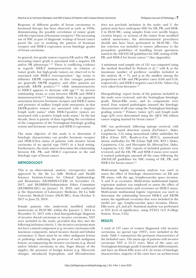
A total of 137 cases of women diagnosed with invasive carcinoma, no special type (NST), were included in the study. Table 1 summarizes the pertinent characteristics of the included cases. The mean age of patients with invasive carcinoma NST is 55.11 years. Most of the cases are Nottingham histologic grade 2 (moderately differentiated), and have an Elston-Ellis score of 6. In terms of histologic characteristics, majority of the cases have an architectural
http://philippinejournalofpathology.org | Vol. 4 No. 1 June 2019
Elomina et al, Hormone Receptor and HER-2 Status of Patients with Invasive Breast Carcinoma Philippine Journal of Pathology | 7
grade of 3, nuclear score of 2, and mitotic count score of 1. As per pathologic stage, most of the cases are T2 and N2. In terms of immunohistochemical phenotype, majority of the cases are ER-positive, PR-positive, and HER-2-negative.
Multiple logistic regression was performed to determine the effect of histologic characteristics on ER and PR status, while multinomial multiple logistic regression was used to determine the effect of the said predictors on HER-2 expression. The regression coefficients and the pertinent statistics are shown in Table 2. Architectural grade demonstrated no significant effect on ER and PR status; however, it appears to be a significant positive
predictor of HER-2 immunoreactivity, although only at the equivocal level (P=0.026). Nuclear pleomorphism is a significant negative predictor of ER immunoreactivity (P=0.031). Mitotic count is not a predictor of hormone receptor and HER-2 status.
Similar analyses were employed to identify the effect of Elston-Ellis score and Nottingham histologic grade on hormone receptor and HER-2 status. The regression coefficients and pertinent statistics are summarized in Table 3. Elston-Ellis score and Nottingham histologic grade are not predictors of hormone receptor and HER-2 immunoreactivity.
Multinomial logistic regression was performed to assess the effect of ER and PR status on HER-2 expression. The regression coefficients and pertinent statistics are shown in Table 4. ER immunoreactivity is not a predictor of HER-2 status, while PR immunoreactivity appears to be a significant negative predictor of a strongly positive HER-2 status (P=0.035).
Lymph node stage, while included in the models as covariate, appears to be a significant positive predictor of HER-2 immunoreactivity, albeit at an equivocal level. The regression coefficients and pertinent statistics including P values, where lymph node stage has served as covariate for each model are summarized in Table 5.
DISCUSSION
The Nottingham histologic grading system accounts three histologic characteristics – architectural grade, nuclear pleomorphism, and mitotic count, to classify breast cancers as to three histologic grades. The study suggests that increasing nuclear grade is associated with a negative ER status, and increasing architectural grade is associated with HER-2 expression. There are limited studies correlating the components of the Elston score with hormone receptor status. Increasing nuclear grade points to a more anaplastic morphology and indicates that a tumor is actively dividing. There is less time to assume the normal phenotype of mammary ductal epithelial cells to express steroid hormone receptors. Also, ER-independent breast cancers usually rely on other genetic mechanisms for growth, and are associated with high-grade histology; and these may be possible explanations behind the association.15 There is also scarce data as regards correlation between the components of the Elston score and HER-2 expression. Increasing architectural grade indicates rapid division of cells that result in formation of tumor nests, clusters, and sheets, rather than formation of tubular structures characteristic of normal mammary ductal epithelial cells. HER-2 is an oncogene that drives cellular proliferation, and HER-2-enriched breast cancers are usually high-grade.15,16 The nature of HER-2 as a driver of cellular proliferation may explain the correlation. In a limited-resource setting, breast cancer patients with high nuclear and architectural grade on routine histology, should prioritize determination of hormone receptor and HER-2 status via IHC, because there is high likelihood of a negative ER phenotype and HER-2 expression in these cancers, which determines treatment options for these patients.
Table 1. Patient characteristics in the studyPatient characteristics Mean SDAge (years) 55.11 11.55Nottingham histologic gradea n %
1 24 17.522 105 76.643 8 5.84
Lymphovascular invasion Negative 120 87.59Positive 17 12.41
Elston score 3 1 0.754 3 2.245 20 14.936 82 61.197 20 14.938 6 4.489 2 1.49
Histologic characteristicsa
Architectural grade 1 2 1.492 28 20.903 104 77.61
Nuclear pleomorphism 1 4 2.992 113 84.333 17 12.69
Mitotic count 1 115 85.822 10 7.463 9 6.72
Pathologic stageb
Tumor size T1 13 9.56T2 84 61.76T3 26 19.12T4 13 9.56
Lymph node stage N0 33 24.09N1 29 21.17N2 58 42.34N3 17 12.41
Immunohistochemical phenotypec
ERNegative 35 25.55Positive 102 74.45
PR Negative 43 31.39Positive 94 68.61
HER-2Negative 82 59.85Equivocal 28 20.44Strongly positive 27 19.71
a - Classification as per Elston and Ellis.13 b - Classification as per AJCC 8th edition Cancer staging manual for breast cancer.14 c - Classification as per American Society of Clinical Oncology/College of American
Pathologists (ASCO/CAP) guidelines.1,2
http://philippinejournalofpathology.org | Vol. 4 No. 1 June 2019
Elomina et al, Hormone Receptor and HER-2 Status of Patients with Invasive Breast Carcinoma Philippine Journal of Pathology | 8

The results of the study show that Elston score and Nottingham histologic grade do not predict hormone receptor and HER-2 status, which is conflicting with studies done previously.4–10 In an attempt to eliminate complex interactions between variables, a simple logistic regression model that includes only the dependent variable (ER, PR, and HER-2 status) and the main predictor variable (Elston score and Nottingham histologic grade) was run, and results were inconsistent (not shown). The association may become apparent with improved statistical power. The findings demonstrate the importance of IHC for ER, PR, and HER-2 in managing patients with breast cancer, since Elston score and histologic grade cannot predict such. In a limited-resource setting, clinicians must always advise patients to allot funds for these tests in order to determine amenability to hormonal and anti-HER-2 drugs, and to properly prognosticate their patients.
The study suggests that PR expression is associated with a negative HER-2 status. A similar relationship is expected for ER expression, albeit the model showed otherwise. A simple model including HER-2 as dependent variable, and ER as main predictor variable was run, and it showed that ER expression is also a negative predictor of a strong HER-2 status (P=0.037). The association may become apparent with improved statistical power. This finding is congruent with that of studies done previously.5,10 The pathogenesis of breast cancer is complex, but current evidence suggests that ER-positive breast cancers harbor distinct genetic abnormalities (16q deletions and 1q gains) that are generally not observed in ER-negative breast cancers, implying that the molecular pathogenesis of ER-positive breast cancers is different from ER-negative breast cancers. As previously mentioned, ER-independent breast cancers rely on different genetic mechanisms for growth; one of these being HER-2.15 This may explain the
Table 2. Effect of histologic characteristics on hormone receptor and HER-2 immunoreactivitya
Histologic characteristic Marker Coef SE z P 95% CI
Architectural grade
ER -0.755 0.588 -1.280 0.199 -1.906 0.397PR -0.683 0.500 -1.370 0.172 -1.664 0.297
HER-2b Equivocal 1.807 0.812 2.220 0.026 0.215 3.399Positive 0.522 0.550 0.950 0.342 -0.555 1.600
Nuclear pleomorphism
ER -1.150 0.532 -2.160 0.031 -2.193 -0.108PR -0.996 0.523 -1.910 0.057 -2.020 0.028
HER-2b Equivocal -0.864 0.706 -1.220 0.221 -2.248 0.520Positive -0.562 0.640 -0.880 0.380 -1.817 0.693
Mitotic count
ER 0.248 0.427 0.580 0.562 -0.590 1.085PR 0.415 0.430 0.970 0.335 -0.428 1.257
HER-2b Equivocal -0.151 0.498 -0.300 0.761 -1.126 0.824Positive -0.120 0.444 -0.270 0.787 -0.991 0.751
a - Logistic regression model includes age, lymphovascular space invasion, tumor size, and lymph node stage as covariatesb - Multinomial logistic regression model has negative HER-2 immunoreactivity as base outcome
Table 3. Effect of elston score and overall histologic grade on hormone receptor and HER-2 immunoreactivitya
Parameter Marker Coef SE z P 95% CI
Elston-Ellis score
ER -0.406 0.250 -1.620 0.105 -0.896 0.085PR -0.314 0.231 -1.360 0.174 -0.768 0.139
HER-2b Equivocal 0.143 0.281 0.510 0.612 -0.408 0.693Positive -0.030 0.277 -0.110 0.915 -0.573 0.514
Nottingham histologic grade
ER -0.282 0.473 -0.590 0.552 -1.209 0.646PR -0.214 0.430 -0.500 0.618 -1.058 0.629
HER-2b Equivocal 0.988 0.594 1.660 0.096 -0.177 2.153Positive 0.138 0.258 0.530 0.593 -0.368 0.644
a - Logistic regression model includes age, lymphovascular space invasion, tumor size, and lymph node stage as covariatesb - Multinomial logistic regression model has negative HER-2 immunoreactivity as base outcome
Table 5. Effect of lymph node stage on equivocal HER-2 immunoreactivity
Predictor variablea Coef SE z P 95% CIHistologic characteristics 0.554 0.253 2.190 0.028 0.058 1.049Elston score 0.519 0.242 2.140 0.032 0.044 0.994Overall histologic grade 0.539 0.241 2.230 0.026 0.066 1.012ER immunoreactivity 0.520 0.245 2.120 0.034 0.040 1.001PR immunoreactivity 0.508 0.245 2.080 0.038 0.029 0.988a - Model includes architectural grade, nuclear pleomorphism, and mitotic count as principal predictor variables, and includes age, lymphovascular space invasion,
tumor size, and lymph node stage as covariates.
Table 4. Effect of hormone receptor immunoreactivity on HER-2 immunoreactivitya
Marker HER2 Coef SE z P 95% CI
EREquivocal 0.586 0.627 0.940 0.350 -0.642 1.815Positive -0.884 0.500 -1.770 0.077 -1.864 0.097
PREquivocal 0.182 0.548 0.330 0.740 -0.891 1.255Positive -1.021 0.484 -2.110 0.035 -1.970 -0.072
a - Multinomial logistic regression model includes age, lymphovascular space invasion, tumor size, and lymph node stage as covariates, and has negative HER-2 immunoreactivity as base outcome
http://philippinejournalofpathology.org | Vol. 4 No. 1 June 2019
Elomina et al, Hormone Receptor and HER-2 Status of Patients with Invasive Breast Carcinoma Philippine Journal of Pathology | 9

inverse relationship between ER and HER-2 expression. PR expression can be regulated by estrogen bound to ER or by estrogen-independent mechanisms, and the estrogen-ER-dependent expression of PR may explain the same inverse relationship between PR and HER-2.17 Considering the results of the study, PR may be dropped from the usual IHC panel of breast cancer in a limited-resource setting, but there is evidence that ER(+), PR(-) breast cancers tend to respond poorly to Tamoxifen than those that are ER(+), PR(+).16 Such a finding underscores the importance of PR status in management of patients with breast cancer, and it is optimal to determine the status of all three markers in all breast cancer patients, if possible.
Interestingly, the results of the study show that increasing lymph node stage is an independent predictor of HER-2 expression, which is consistent with the findings of a previous study.5 Breast cancers expressing HER-2 are associated with high-grade histology, aggressive clinical behavior, and decreased survival, which may be attributed to the nature of HER-2 as a potent driver of cellular proliferation.16 While this study demonstrates that advancing lymph node stage is predictive of HER-2 expression, in practice, clinicians still need to advise patients as regards the importance of HER-2 testing via IHC and its confirmation via FISH, to determine amenability to treatment with Trastuzumab, which is an expensive drug.
The study is time-bound, which limits the number of cases included in the study, which, in turn limits statistical power. Extension of the duration of the study includes more patients and may improve statistical power, which may make some relationships apparent. Another limitation is the complexity of analysis of HER-2 because of three outcomes that can be dichotomized with confirmatory testing via FISH, which is not available in our institution. The possibility of discordance in histopathology and IHC of core needle biopsy and mastectomy specimens may also be a possible limitation. The unavailability of IHC for both core needle biopsy and mastectomy specimens is secondary to variations in clinical practice in consideration of patient-related factors. Nevertheless, one recent study demonstrated that histopathology and core needle biopsy IHC were known to have high concordance rates with those of mastectomy specimens.18 In order to minimize its potential effect, in future studies, we recommend doing IHC for both core needle biopsy and mastectomy specimens if patient and institutional factors permit.
CONCLUSION
The findings of this study demonstrate the predictive value of histologic characteristics on hormone receptor and HER-2 status in breast cancers, as well as the relationship between expression of hormone receptors and HER-2. These gave insights as regards the complex genetic mechanisms that are responsible in the development of breast carcinoma, and their influence on its histology. Because of the current limitations of this study, we still recommend that all three markers should be assessed in all breast cancer patients, even in a limited-resource setting, to optimize prognostication
and management, and to properly channel the patient's limited funds to more appropriate diagnostic and therapeutic procedures.
ACKNOWLEDGMENT
The authors thank Prof. Danaida Marcelo, Head of the Biostatistics unit of DLSMHSI-CCEB for her inputs on how to perform the statistical analysis for this study.
STATEMENT OF AUTHORSHIP
All authors certified fulfillment of ICMJE authorship criteria.
AUTHOR DISCLOSURE
The authors declared no conflict of interest.
FUNDING SOURCE
None.
REFERENCES
1. Hammond ME, Hayes DF, Dowsett M, et al. American Society of Clinical Oncology/College of American Pathologists guideline recommendations for immunohistochemical testing of estrogen and progesterone receptors in breast cancer. J Clin Oncol. 2010;28(16):2784–95. PMID: 20404251. PMCID: PMC2881855. https://doi.org/10.1200/JCO.2009.25.6529.
2. Wolff AC, Hammond ME, Hicks DG, et al. Recommendations for human epidermal growth factor receptor 2 testing in breast cancer: American Society of Clinical Oncology/College of American Pathologists clincal practice guideline update. J Clin Oncol. 2013;31(31):3997–4013. PMID: 24101045. https://di.org/10.1200/JCO.2013.50.9984.
3. Bloom HJ, Richardson WW. Histological grading and prognosis in breast cancer a study of 1409 cases of which 359 have been followed for 15 years. Br J Cancer. 1957;11(3):359–77. PMID: 13499785. PMCID: PMC2073885.
4. Mittal A, Prasad CSBR, Sreeramulu PN, Srinivasan D, Naveedahmed K, Ruta UJ. Histopathological grade versus estrogen and progestron receptor status in carcinoma breast- a single center study. Open Access J Surg. 2017;4(3):10–3. https://doi.org/ 10.19080/OAJS.2017.04.555639.
5. Dayal A, Shah RJ, Kothari S, Patel SM. Correlation of HER-2/neu status with estrogen, progesterone receptors and histologic features in breast carcinoma. Ann Pathol Lab Med. 2016;3(5):8.
6. Geethamala K, Srinivasa M V., Vani BR, Sudha R. Histopathological grade versus hormone receptor status in breast carcinoma-treasure the past. Int J Biomed Res. 2015;6(7):466–71. https://doi.org/ 10.7439/ijbr.v6i7.2203.
7. Iqbal J, Abukhatir M, Shafi AA, Alyahya GM, Alharthi BN. Hormone receptor status of breast cancer in patients of different age groups, lymph node status histological type and tumor grade, an experience at King Fahad Medical City, Riyadh. Pak J Surg. 2014;30(4):296–300. http://www.pjs.com.pk/journal_pdfs/oct-dec14/296.pdf.
http://philippinejournalofpathology.org | Vol. 4 No. 1 June 2019
Elomina et al, Hormone Receptor and HER-2 Status of Patients with Invasive Breast Carcinoma Philippine Journal of Pathology | 10

8. Mostafa MG, Larsen MT, Love RR. Estrogen receptor, progesterone receptor, and HER-2/neu oncogene expression in breast cancers among Bangladeshi women. J Bangladesh Coll Physicians Surg. 2010;28(3):157–62. PMID: 22279410. PMCID: PMC3263928. NIHMSID: NIHMS270134. https://doi.org/10.3329/jbcps.v28i3.6509
9. Pathak T, Bashyal R, Pun C, et al. Estrogen and progesterone receptors in breast cancer. J Pathol Nepal. 2011;1(2):100–3. https://doi.org/10.3126/jpn.v1i2.5401.
10. Azizun-Nisa, Bhurgi Y, Raza F, Kayani N. Comparison of ER, PR & HER-2/neu (C-erb B 2) reactivity pattern with histologic grade, tumor size and lymph node status in breast cancer. Asian Pacific J Cancer Prev. 2008;9(4):553–6. PMID: 19256737.
11. Peduzzi P, Concato J, Kemper E, Holford TR, Feinstem AR. A simulation study of the number of events per variable in logistic regression analysis. J Clin Epidemiol. 1996;49(12):1373–9. PMID: 8970487.
12. Gupta D, Gupta V, Marwah N, et al. Correlation of hormone receptor expression with histologic parameters in benign and malignant breast tumors. Iran J Pathol. 2015;10(1):23–34. PMID: 26516322. PMCID: PMC4539786.
13. Elston CW, Ellis IO. Pathological prognostic factors in breast cancer. I. The value of histological grade in breast cancer: experience from a large study with long-term follow-up. Histopathology. 1991;19:403–10. PMID: 1757079.
14. Giuliano AE, Connolly JL, Edge SB, et al. Breast cancer-major changes in the American Joint Committee on Cancer eighth edition cancer staging manual. CA Cancer J Clin. 2017;67(4):290–303. PMID: 28294295. https://doi.org/10.3322/caac.21393.
15. Allison KH. Molecular pathology of breast cancer: what a pathologist needs to know. Am J Clin Pathol. 2012;138(6):770–80. PMID: 23161709. https://doi.org/10.1309/AJCPIV9IQ1MRQMOO.
16. Kabel AM. Tumor markers of breast cancer: new prospectives. J Oncol Sci. 2017;3(1):5–11. https://doi.org/10.1016/j.jons.2017.01.001.
17. Jacobsen BM, Horwitz KB. Progesterone receptors, their isoforms and progesterone regulated transcription. Mol Cell Endocrinol. 2012;357(1–2):18–29. PMID: 21952082. PMCID: PMC3272316. https://doi.org/10.1016/j.mce.2011.09.016.
18. You K, Park S, Ryu JM, et al. Comparison of core needle biopsy and surgical specimens in determining intrinsic biological subtypes of breast cancer with immunohistochemistry. J Breast Cancer. 2017;20(3):297–303. PMID: 28970856. PMCID: PMC5620445. https://doi.org/10.4048/jbc.2017.20.3.297.
Disclaimer: This journal is OPEN ACCESS, providing immediate access to its content on the principle that making research freely available to the public supports a greater global exchange of knowledge. As a requirement for submission to the PJP, all authors have accomplished an AUTHOR FORM, which declares that the ICMJE criteria for authorship have been met by each author listed, that the article represents original material, has not been published, accepted for publication in other journals, or concurrently submitted to other journals, and that all funding and conflicts of interest have been declared. Consent forms have been secured for the publication of information about patients or cases; otherwise, authors have declared that all means have been exhausted for securing consent.
APPENDIX
Preanalytic guidelines in handling breast specimens for IHC testing of ER, PR, and HER2 for breast cancer (adapted from ASCO/CAP guideline recommendations, 2010 and 2014)1,2
1. The time from tumor removal to fixation (cold ischemia time) should be kept to 1 hour or less.2. The ideal fixative to be used is 10% neutral buffered formalin (NBF). The specimen should be fixed with an
adequate volume of fixative (i.e. at least ten-fold greater than specimen volume).3. The time of tissue fixation should be at least 6 hours but no greater than 72 hours.4. Sections made more than 6 weeks are not recommended for HER-2 analysis.
http://philippinejournalofpathology.org | Vol. 4 No. 1 June 2019
Elomina et al, Hormone Receptor and HER-2 Status of Patients with Invasive Breast Carcinoma Philippine Journal of Pathology | 11

The National External Quality Assessment Scheme forDiagnostic Medical Parasitology in the Philippines, 2009–2015Sherwin Galit,1 Dave Tangcalagan,2 Julius Matt Rapanut,2 Alexander Sadiasa,2 Prince Ninja Bautista,2
Kim Joshua Dominquez,2 Jahra Bañaga,2 Celine Bernice Roxas,2 Rafael Navarro,2 Mark Philip Bugayong,2 Jennifer Luchavez,2 Melisa Mondoy,2 Lydia Sombrero,2 Socorro Lupisan3
1Department of Parasitology, Research Institute for Tropical Medicine-Department of Health, Philippines2Department of Microbiology, Research Institute for Tropical Medicine-Department of Health, Philippines3Office of the Director, Research Institute for Tropical Medicine-Department of Health, Philippines
ABSTRACT
Background. The Research Institute for Tropical Medicine (RITM)–National Reference Laboratory (NRL) for Malaria and Other Parasites, mandated by the Department of Health–Philippines (DOH), administers an annual Proficiency Test (PT) in diagnostic medical parasitology to clinical laboratories throughout the Philippines through the National External Quality Assessment Scheme (NEQAS). The PT in Parasitology aims to monitor and evaluate the capability of Philippine laboratories in the identification of blood and intestinal parasites, and the estimation of malaria parasite density in malaria-infected blood films. As of 2018, participation in the NEQAS is an annual requirement by the Department of Health–Health Facilities and Services Regulatory Bureau (DOH-HFSRB) for each clinical laboratory to obtain a license to operate.
Objective. This report aims to summarize the results of the PT for Parasitology and assess the performance of participating laboratories in malaria and fecal parasite microscopy from 2009 to 2015.
Methodology. RITM–NRL oriented clinical laboratories in the NEQAS in 2008. Laboratories submitted their accomplished enrolment forms to RITM–NRL and paid fees to enroll in the PT in 2009 to 2015. Participating laboratories identified the species of malaria in blood films and the parasite/s in formalin-preserved fecal specimens. Estimation of parasite density in malaria blood films was performed as well.
Results. One thousand five hundred forty (1,540) laboratories participated from 2009 to 2015. Mean and median scores in all seven years were below the cut-off score of 80. Schistosoma japonicum was the most difficult to identify with only 7.7% of laboratories having correct identification result. Majority of participants from 2010 to 2014 gave malaria parasite density estimates outside the acceptable range.
Conclusion. Most participating laboratories performed poorly in the proficiency tests over the last seven years. Training and refresher courses for laboratorians are recommended in order to address the poor performance in the laboratory diagnosis of parasitic infections, especially the endemic and uncommon ones, in the country
Key words: laboratory proficiency testing, external quality assessment, medical parasitology, malaria, schistosomiasis, helminthiasis, protozoan infections
INTRODUCTION
Parasitic infections caused by a diverse range of helminths and protozoans affect millions of people living in the Philippines. Around 25 million Filipinos are at risk of soil transmitted helminthiases (STH), with a prevalence rate of six to 97 percent among Filipino children aged six to 12.1 Also, 12 million are at risk of schistosomiasis, with 2.5 million Filipinos directly exposed to the infection.1 In addition, around 33 million Filipinos are at risk of malaria.1,2 Control and elimination of these diseases depend on accurate and reliable diagnosis, of which diagnostic medical parasitology laboratories are responsible. In the Philippines, medical parasitology laboratories typically employ microscopy to demonstrate parasites in stool, blood, or other specimens.3 In order to ensure accurate and reliable diagnosis, laboratories must carry out quality assurance through a quality
ISSN 2507-8364 (Online)Printed in the Philippines.Copyright© 2019 by the PJP.Received: 28 February 2019.Accepted: 20 March 2019.Published online first: 22 March 2019.https://doi.org/10.21141/PJP.2019.03 Corresponding author: Rafael B. Navarro, BScE-mail: [email protected] ORCiD: https://orcid.org/0000-0003-3042-6021
http://philippinejournalofpathology.org | Vol. 4 No. 1 June 2019
OPEN ACCESS – ORIGINAL ARTICLE

management system, which encompasses documentation, implementation of standard operating procedures (SOPs), practice of quality control (QC), and participation in external quality assessment schemes (EQAS).4
The National External Quality Assessment Scheme (NEQAS) for Parasitology is one of the measures by the Department of Health (DOH) to assess the reliability of laboratory diagnosis and maintain quality assurance of licensed medical parasitology laboratories in the country. DOH, through the Department Order No. 393–E s. 2000, designated the Research Institute for Tropical Medicine (RITM) as the National Reference Laboratory (NRL) for Malaria and other Parasites,5 which maintains DOH-approved external quality assessment program by administering annual proficiency testing (PT) to diagnostic medical laboratories through NEQAS. The DOH Administrative Order No. 2007–0027 and Memorandum No. 2009–0086, required every diagnostic medical laboratory throughout the country to participate in the NEQAS,6–8 which allows each to obtain a license to operate (LTO) from the DOH Health Facilities and Services Regulatory Bureau (HFSRB, formerly Bureau of Health Facilities and Services).
This paper reports the results of the proficiency tests for diagnostic medical parasitology administered to participating laboratories in 2009–2015. The proficiency test was conducted to assess the capability of laboratorians to identify and quantify malaria parasite density in malaria blood films; and identify species of parasitic helminths and protozoans in formalin-preserved fecal suspensions.
METHODOLOGY
Laboratory participationRITM–NRL conducted orientation seminars on NEQAS implementation to diagnostic medical parasitology laboratories in 2008 and 2009. Laboratories required by DOH–HSFRB to participate in the annual proficiency test submitted their enrolment forms and paid fees before the scheduled testing event.
Preparation of blood and fecal specimensThe parasites used as analytes were obtained from blood or stool of infected patients with adequate number of parasites exhibiting characteristic morphological features. Malaria parasite-infected blood samples were collected from consenting individuals from malaria endemic areas in Palawan province. Thick and thin blood films were prepared for malaria microscopy.9
For the identification of intestinal helminths and protozoans, infected stools were collected and examined through Direct Fecal Smear (DFS), Kato-Katz technique10,11 and Formalin-Ether Concentration Technique (FECT).10,12,13 Samples were preserved in 10% formalin3 for storage and validated with DFS and FECT prior to preparation for packaging. Validated samples were pooled and the resulting concoction was validated with DFS and FECT. Around 500 µL of the concoction was transferred to each polypropylene vial.
Quality control and validation of parasite speciesTwo (2) trained microscopists from RITM–NRL for Malaria and other Parasites performed quality control of blood films and formalin-preserved fecal samples in vials; validation of these analytes was done through blinded crosschecking. All blood films were ensured to be stained properly and to contain consistent malaria parasite species identity and parasite density. Likewise, fecal samples were ensured to contain parasites with consistent species identity and with intact and recognizable morphological features. In addition, the identities of Plasmodium species were confirmed through nested polymerase chain reaction and agarose gel electrophoresis.14 In cases of discrepancies between the results of the two blinded examinations, a third microscopist who is a senior staff of the NRL would re-examine and validate the results.
Packaging of analytesAnalytes sent to participating laboratories were packed based on the international standard of transporting biohazard materials (IATA).15 Microscope slides were secured in plastic slide mailers. Each polypropylene vial was labeled and sealed with Parafilm M® (Bemis Co., Inc., Oshkosh, Wisconsin, USA), wrapped in a paper towel, and placed inside a 100 mm × 115 mm resealable propylene resin bag. The vials and slide mailers were encased in 600 mL polypropylene canister (Philtop Industries Inc., Valenzuela City, Metro Manila) and placed inside a 120 mm × 115 mm × 190 mm corrugated box (Thousand Oaks Packaging Corp., Parañaque City, Metro Manila) with necessary attachments and labels. The package included proficiency testing guidelines and answer sheets.
Proficiency testing and scoringDuring the testing event, participants received their package and were asked to identify the parasite or parasites in the fecal sample and in stained thick and thin blood films by microscopy. In 2010 to 2014, malaria parasite counting was included in the proficiency test. Each participant was asked to estimate the malaria parasite density by performing parasite counting on the thick and thin blood films. Results were submitted to RITM–NRL within 15-working days after the package had been received in the laboratory.
In 2009, the percentage score for parasite identification was calculated with a right-minus-wrong scheme which was modified to the percentage method in 2010. Parasite identification was calculated by determining the number of correctly identified organisms over the number of organisms in the actual analyte and additional organisms answered by the participant but excluded in the list of actual organisms in the analyte. For malaria parasite counting, the percentage score was calculated by determining the counts within ± 20% of the actual parasite count over the number of analytes given.
Statistical analysisGraphs were generated using Matplotlib version 2.0.0 pyplot module in Python16 and statistical analyses were done using SciPy version 0.19.0 scipy.stats module.17 Kruskal–Wallis one-way analysis of variance was performed to determine the differences between annual proficiency test scores.
http://philippinejournalofpathology.org | Vol. 4 No. 1 June 2019
Galit et al, NEQAS for Diagnostic Medical Parasitology in the Philippines, 2009–2015 Philippine Journal of Pathology | 13

RESULTS AND DISCUSSION
A total of 1,540 laboratories participated in the PT for parasitology in 2009-2015, of which 82% (1263/1540) were private and 18% (277/1540) were government facilities (Figure 1). In terms of laboratory type, the total number of participants is composed of 30.3% (467/1540) tertiary, 59.8% (921/1540) secondary, and 9.9% (152/1540) primary level clinical laboratories. National Capital Region holds the highest number of participating laboratories within the 7-year period (463/1540); followed by Western Visayas (Region VI; 284/1540); and CALABARZON (Region IV-A; 226/1540). Notably, one laboratory in the Autonomous Region in Muslim Mindanao or ARMM participated, for the first time, in the PT during 2015.
Scores ranged from zero to 100 in all years, except in 2009 where scores ranged from -125 to 100 because of the right-minus-wrong grading scheme (Figure 2). The mean scores and sample standard deviation per year were: 66.7 (41.0) in 2009, 70.3 (23.5) in 2010, 54.0 (24.1) in 2011, 52.7 (27.1) in 2012, 66.0 (23.6) in 2013, 60.3 (23.5) in 2014, and 61.9 (21.2) in 2015. Annual median scores were 75.0 in 2009 and 2010, 50.0 in 2011 and 2012, 66.7 in 2013, 62.5 in 2014, and 62.5 in 2015 (Figure 3). Mean and median scores in all years were below the cut-off score of 80.0. Annual PT scores were significantly different from each other (H = 192.14; p-value = 8.93x10 -39) based on the Kruskal–Wallis test.
Within the 7-year period, participants found the blood fluke, Schistosoma japonicum, to be the most difficult to identify—only 7.7% (15/196) of the laboratories that received the analyte identified it correctly. Following the schistosome was the intestinal protozoan, Blastocystis hominis, which 38.7% (592/1528); the pinworm, Enterobius vermicularis (42.3%; 202/478); the commensal and nonpathogenic amoebae, Endolimax nana (42.3%; 85/201) and Entamoeba coli (50.2%; 821/1635) (Table 1).
Figure 1. Number of participating laboratories from different regions in the Philippines in the PT for parasitology, 2009–2015.
Figure 2. Annual number of participants and proportion of those who obtained scores of 80 and above in parasite identification, in the 2009–2015 PT for parasitology.
Figure 3. Annual distribution, mean, and median of scores in the 2009-2015 PT for parasitology.
http://philippinejournalofpathology.org | Vol. 4 No. 1 June 2019
Galit et al, NEQAS for Diagnostic Medical Parasitology in the Philippines, 2009–2015 Philippine Journal of Pathology | 14

vacuolated and sometimes contains bacteria but no red blood cells. E. coli cysts are usually spherical with 10–35 µm in diameter. Each mature cyst usually contains five or more nuclei while immature cysts have two to four nuclei. Each of the nuclei contains large, discrete, and usually eccentric karyosomal chromatin and coarsely granulated peripheral chromatin. Additionally, the cytoplasm of immature cysts usually appears to be diffuse and contains glycogen mass, which stains reddish brown with iodine, and chromatoid bodies with splintered ends. In contrast, E. nana cysts, which are also usually spherical but are smaller, measures around 5–10 µm in diameter. Each cyst typically contains four nuclei with large, blot-like, and usually central karyosomal chromatin and no peripheral chromatin. Its cytoplasm usually contains diffuse glycogen and occasionally concentrated glycogen mass in young cysts.10,19
Artifacts in the stool such as fungal spore, algal spore, mite egg, plant cell, and pollen grain may be mistaken as helminth eggs. In addition, epithelial cells and white blood cells in stool may be mistaken as amoebae. Moreover, Howell–Jolly bodies and nucleated red blood cells in blood films may be mistaken as malaria parasites19. Laboratorians performing diagnostic parasitology should be able to recognize details that differentiate parasite components and non-parasite artifacts.
In 2009, participants were asked to perform malaria parasite count in malaria positive blood films merely as an initial survey to assess the capability of laboratories to determine the parasite density in blood but scoring was not done. Scores in malaria parasite count in 2010 to 2014 were below 50% owing to the majority of participating laboratories giving malaria parasite density estimates outside the acceptable range (Table 2). As a result, NEQAS removed malaria parasite counting in the proficiency test in 2015 since majority of laboratories were incapable of estimating malaria parasite density in blood films.
Participation in 2015 (1430 participants) rose to 230% from that in 2014 (623 participants). In addition, the number of participants in 2015 comprised around 93% (1430/1540) of all participating laboratories throughout seven years. Overall, only 19.4% (277/1430) obtained a
S. japonicum eggs are small with typically round to oval shape measuring 70–100 µm by 55–65 µm. Each egg contains a miracidium enclosed in a thin transparent shell with a small lateral spine, which usually is not clearly visible and often obscured by fecal debris adhering to the shell or by wrong orientation.10 Moreover, detection of S. japonicum eggs is enhanced by concentration of formalin-preserved fecal sample by FECT.18
The cyst-like form of the stramenopile B. hominis is generally round and measures around 6–40 µm. This form has a large central body that appears to be a large vacuole with a thin band, surrounded by multiple nuclei. To maximally recover the cyst-like forms, fecal samples must be concentrated through FECT before examining through a microscope.19 Lysis of trophozoites and central body forms after exposure to water prior to fixation yield false-negative results20. In addition, concentrated wet mount preparations often fail to display the distinguishable features of the parasite so smears permanently stained with trichrome or iron hematoxylin are preferably prepared.10
E. vermicularis eggs are typically recovered from the perianal area using a swab or using the “sticky tape” method, where a clear adhesive tape is put on the perianal area in the morning before bathing or defecation. The eggs are elongated, measuring 50–60 µm in length by 20–32 µm in width; and are asymmetrical, with one side flattened and the other side convex. They are colorless and the shells are thin.19
E. coli and E. nana are nonpathogenic amebae but they can colonize the intestine when a person ingests mature cysts in fecally contaminated food and water. The E. coli trophozoite, which measures around 15–50 µm in diameter, contains a single nucleus with large karyosome, and coarse and irregular peripheral nuclear chromatin. Its cytoplasm appears to be coarsely granular and often vacuolated, and sometimes includes bacteria, yeasts, and other materials but not red blood cells. On the other hand, the E. nana trophozoite, which measures around 6–12 µm in diameter, has a nucleus with large, irregular karyosome and does not have a peripheral nuclear chromatin. Its cytoplasm appears to be granular and
Table 1. Parasite species used as analytes in the 2009-2015 PT for Parasitology and frequency of correct identificationOrganism and Authority Total Frequency Frequency of Correct ID and PercentageSchistosoma japonicum Katsurada, 1904 196 15 (7.7%)Blastocystis hominis Brumpt, 1912 1528 592 (38.7%)Enterobius vermicularis Linnaeus, 1758 478 202 (42.3%)Endolimax nana Wenyon & O'Connor, 1917 201 85 (42.3%)Entamoeba coli (Grassi, 1879), Casagrandi & Barbagallo, 1895 1635 821 (50.2%)Plasmodium vivax Grassi & Feletti, 1890 1004 634 (63.2%)Plasmodium falciparum Welch, 1897 5388 3598 (66.8%)Hookworm
1222 836 (68.4%)Ancylostoma duodenale (Dubini, 1843)Necator americanus (Stiles, 1902)
Taenia spp. Linnaeus, 1758 1085 752 (69.3%)Giardia lamblia (Lambl, 1859) Kofoid & Christiansen, 1915* 1621 1159 (71.5%)Entamoeba histolytica Schaudinn, 1903 589 454 (77.1%)Trichuris trichiura Linnaeus 1771 962 843 (87.6%)Ascaris lumbricoides Linnaeus, 1758 1168 1033 (88.4%)Hymenolepis diminuta Rudolphi, 1819 1 1 (100.0%)Plasmodium malariae Feletti & Grassi, 1889 2 2 (100.0%)* also known as Giardia duodenalis Stiles, 1902 and more recently as Giardia intestinalis Kulda & Nohýnková, 1995
http://philippinejournalofpathology.org | Vol. 4 No. 1 June 2019
Galit et al, NEQAS for Diagnostic Medical Parasitology in the Philippines, 2009–2015 Philippine Journal of Pathology | 15

ACKNOWLEDGMENTS
The authors acknowledge the past and present staff of RITM–NEQAS: Donato Esparar, Jo-Anne Bibit, Grace Esparar, Ma. Theresa Kapawan, Daryl Joy Almonia, Jhobert Bernal, Armando Martinez, Sheila Joy Gonzaga, and Razaele Aguinaldo.
The authors also extend their gratitude to the DOH–Health Facilities and Services Regulatory Bureau (formerly Bureau of Health Facilities and Services) and the DOH–Health Facilities Development Bureau (formerly National Center for Health Facilities Development), for providing financial support in the 2009–2012 Proficiency Tests for Parasitology.
STATEMENT OF AUTHORSHIP
All authors certified fulfillment of ICMJE authorship criteria.
AUTHOR DISCLOSURE
The authors declared no conflict of interest.
FUNDING SOURCE
The 2009–2012 Proficiency Tests for Parasitology were funded by the Health Facilities and Services Regulatory Bureau and the Health Facilities Development Bureau—both under the Department of Health of the Republic of the Philippines.
REFERENCES 1. World Health Organization, Regional Office for
the Western Pacific. Western Pacific country health information profiles. Manila, Philippines: World Health Organization, Regional Office for the Western Pacific; 2011.
2. World Health Organization, Global Malaria Programme, University of California SF, et al. Progress towards subnational elimination in the Philippines. Geneva: World Health Organization; 2014.
3. Belizario VY Jr., De Leon W, editors. Medical parasitology in the Philippines. Diliman, Quezon City: University of the Philippines Press; 2015.
4. Belizario VY Jr., Plan AO, De Leon WU. Laboratory diagnosis of selected neglected parasitic diseases in the Philippines: can we do better? Acta Med Philipp. 2014;48:4-10. https://www.actamedicaphilippina.org/article/7066.
5. Romualdez AG Jr. Department Order No. 393-E s. 2000: Designation of National Reference Laboratories and transfer of corresponding equipment, instruments, supplies, specimens, records from the Bureau of Research and Laboratories to the designated National Reference Laboratories. November 14, 2000. http://mt-lectures.blogspot.com/2017/08/department-order-no-393-e-s-2000.html.
6. Duque F. Administrative Order No. 2007-0027: Revised rules and regulations governing the licensure and regulation of clinical laboratories in the
passing score of 80 and above (Table 3) and all regions got below 50% passing rate. Four regions in Mindanao (Davao, Caraga, Northern Mindanao, and SOCCSKSARGEN) outperformed all regions in Visayas and most regions in Luzon in the 2015 PT based on the percentage of passers. Southern Luzon regions (CALABARZON, National Capital Region, Bicol, and MIMAROPA), plus ARMM had the least percentage of passers. It should be noted that MIMAROPA and ARMM, both of which had zero passing rates in 2015, consisted of malaria- and schistosomiasis-endemic provinces.21,22
Belizario et al. noted several reports describing low rates of parasite recognition by laboratorians in different areas in the Philippines.4 Lack of resources and limited training opportunities in medical parasitology make for the poor performance of laboratorians in the diagnosis of parasitic diseases. Belizario et al. also proposed that emerging parasites should also be included in the proficiency testing.4
CONCLUSION
Overall, majority of participating laboratories performed poorly in both identification of parasites in preserved fecal samples and in malaria blood films, including estimation of malaria parasite density. More training opportunities in medical parasitology, especially in malaria microscopy, must be prioritized by the government. Government regulatory agencies may also consider setting cut off scores for the licensing of diagnostic parasitology laboratories. Laboratorians should also be evaluated on their capability to identify uncommon and emerging parasites like schistosomes, filarial nematodes, pathogenic protozoans, etc. since many of these diseases are endemic in the country.
Table 2. Number of blood films with acceptable estimated malaria parasite density
Year No. of Slides No. and Percentage of Acceptable Results2011 254 76 (28.5%)2012 387 53 (13.3%)2013 366 119 (31.2%)2014 606 251 (40.3%)
Table 3. Passing rate of participating laboratories per regionRegion Percentage of Passers in 2015Davao Region (XI) 45.9% (17/37)Central Luzon (III) 42.9% (27/63)Caraga (XIII) 40.0% (6/15)CAR 38.1% (8/21)Northern Mindanao (X) 37.9% (11/29)Soccsksargen (XII) 31.3% (10/32)Central Visayas (VII) 27.3% (6/22)Western Visayas (VI) 23.3% (60/258)Ilocos Region (I) 22.2% (6/27)Zamboanga Peninsula (IX) 16.1% (9/56)Eastern Visayas (VIII) 15.5% (11/71)Cagayan Valley (II) 15.0% (15/100)CALABARZON (IV-A) 14.0% (15/100)NCR 13.1% (59/450)Bicol Region (V) 4.8% (1/21)MIMAROPA (IV-B) 0.0% (0/6)ARMM 0.0% (0/1)Overall 19.4% (277/1430)
http://philippinejournalofpathology.org | Vol. 4 No. 1 June 2019
Galit et al, NEQAS for Diagnostic Medical Parasitology in the Philippines, 2009–2015 Philippine Journal of Pathology | 16

Philippines. August 22, 2007. http://lcp.gov.ph/images/Admin_Order_2007_0027.pdf.
7. Duque F. Department Memorandum No. 2009-0086: Implementation of External Quality Assessment Program as a regulatory requirement for licensing of clinical laboratories. February 2009.
8. Lutero N. Department Memorandum No. 2009-0086-B: Amendment to Department Memorandum No. 2009-0086-A entitled, “Implementation of External Quality Assessment Program as regulatory requirement for licensing of clinical laboratories.” September 8, 2014. http://lcp.gov.ph/images/Dept_Memo_2009_0086B.pdf.
9. World Health Organization, ed. Basic malaria microscopy, 2nd ed. Geneva: WHO; 2010.
10. Ash LR, ed. Bench Aids for the diagnosis of intestinal parasites. Switzerland: Geneva: World Health Organization; 1994. https://apps.who.int/iris/bitstream/handle/10665/37323/9789241544764_eng.pdf?sequence=1.
11. Katz N, Chaves A, Pellegrino J. A simple device for quantitative stool thick-smear technique in Schistosomiasis mansoni. Rev Inst Med Trop Sao Paulo. 1972;14(6):397-400. PMID:4675644.
12. Basic laboratory methods in medical parasitology. Switzerland: Geneva: World Health Organization; 1991. https://apps.who.int/iris/handle/10665/40793.
13. Ritchie LS. An ether sedimentation technique for routine stool examinations. Bull US Army Med Dep U S Army Med Dep. 1948;8(4):326. PMID:18911509.
14. Snounou G, Viriyakosol S, Xin Ping Zhu, et al. High sensitivity of detection of human malaria parasites by the use of nested polymerase chain reaction. Mol Biochem Parasitol. 1993;61(2):315-20. PMID:8364734.
15. Dangerous goods regulations (IATA Resolution 618 attachment “A”). Montreal International Air Transport Association; 2015.
16. Droettboom M, Caswell TA, Hunter J, et al. Matplotlib/matplotlib: v2.0.0. January 2017. https://zenodo.org/record/248351#.XJRZZCgzaUk.
17. Jones E, Oliphant T, Peterson P, et al. SciPy: Open Source Scientific Tools for Python. 2001.
18. United Kingdom National External Quality Assessment Service for Parasitology. Schistosoma japonicum. 2013.
19. DPDx - Laboratory Identification of parasites of public health concern. Diagn Proced. April 2018. https://www.cdc.gov/dpdx/monthlycasestudies/2018/case466.html.
20. Jorgensen JH, Pfaller MA, Carroll KC, American Society for Microbiology, eds. Manual of clinical microbiology, 11th ed. Washington, DC: ASM Press; 2015.
21. Leonardo L, Rivera P, Saniel O, et al. A national baseline prevalence survey of schistosomiasis in the Philippines using stratified two-step systematic cluster sampling design. J Trop Med. 2012; Article ID 936128. https://doi.org/10.1155/2012/936128.
22. EB-DOH. Malaria Surveillance Report: January 1–May 6, 2017. https://www.doh.gov.ph/sites/default/files/statistics/2017%20Malaria%20MW%201%20-%20MW-18.pdf.
Disclaimer: This journal is OPEN ACCESS, providing immediate access to its content on the principle that making research freely available to the public supports a greater global exchange of knowledge. As a requirement for submission to the PJP, all authors have accomplished an AUTHOR FORM, which declares that the ICMJE criteria for authorship have been met by each author listed, that the article represents original material, has not been published, accepted for publication in other journals, or concurrently submitted to other journals, and that all funding and conflicts of interest have been declared. Consent forms have been secured for the publication of information about patients or cases; otherwise, authors have declared that all means have been exhausted for securing consent.
http://philippinejournalofpathology.org | Vol. 4 No. 1 June 2019
Galit et al, NEQAS for Diagnostic Medical Parasitology in the Philippines, 2009–2015 Philippine Journal of Pathology | 17

Buccal Cell Micronuclei among Betel Quid Chewers and Non-Betel Quid Chewers from Selected Barangays in Zamboanga CityBenkassar Abdurajak,1 Servando Halili, Jr.,2,3 Al-Zamzam Abubakar4
1Ateneo de Zamboanga University School of Medicine, Zamboanga del Sur, Philippines2Zamboanga State College of Marine Sciences and Technology, Zamboanga del Sur, Philippines3Ateneo de Zamboanga University Graduate School, Zamboanga del Sur, Philippines4Ciudad Medical Zamboanga, Zamboanga del Sur, Philippines
ABSTRACT
Background. Betel quid chewing has been reported to have carcinogenic properties due to the presence of harmful compounds present in its ingredients. The oral mucosa is directly exposed to these carcinogenic compounds which could cause pathological changes and lead to malignancies. Micronucleus is a biomarker that indicates genetic alteration could form due to exposure from carcinogenic substances that can be attributed from betel quid chewing. Thus, a person’s oral health status can be gauged through the detection of micronucleus in buccal cells.
Objective. A cross-sectional study was done to compare the presence of micronuclei in buccal epithelial cells between betel quid chewers and non-betel quid chewers in Zamboanga City.
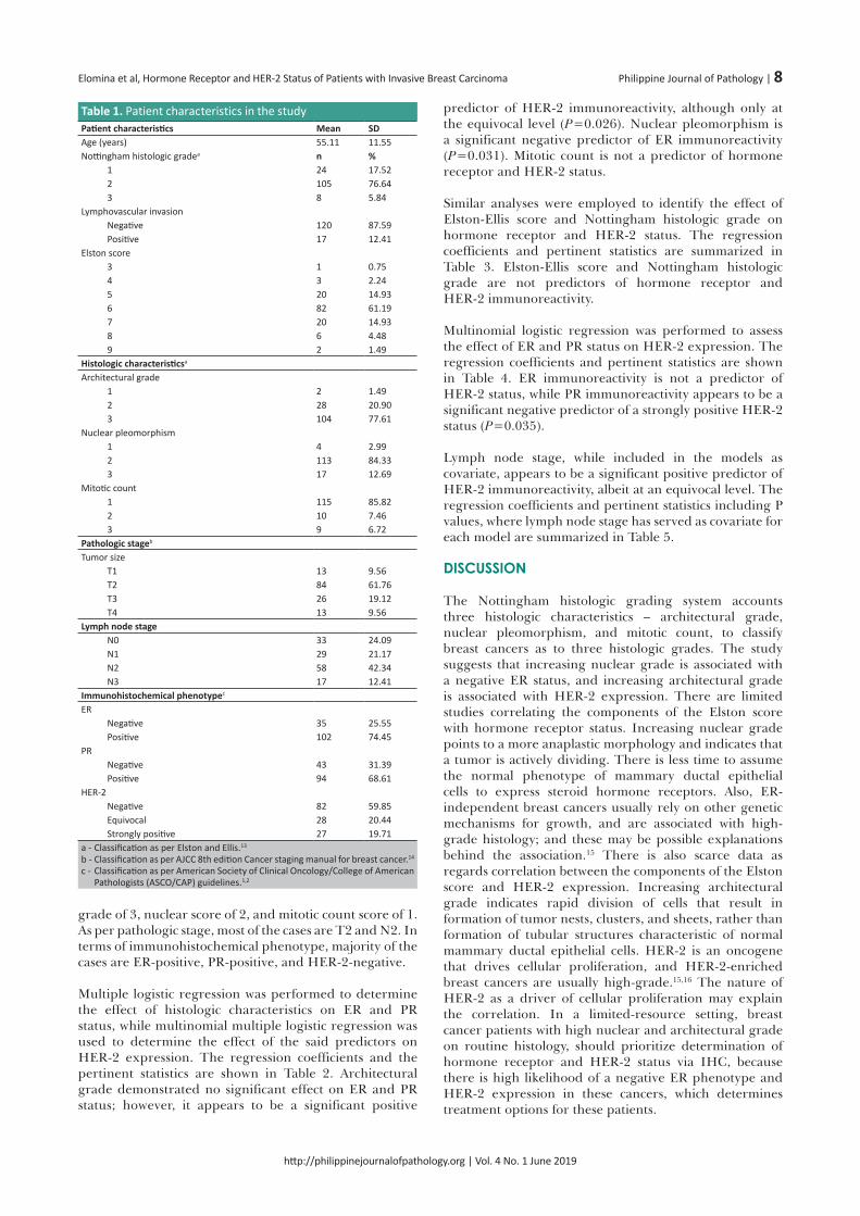
Methodology. Purposive sampling was used to enroll the 104 participants (52 betel quid chewers and 52 non-betel quid chewers). The demographic profiles and betel quid chewing habits of the participants were obtained using a questionnaire. Buccal cells samples were collected using clean and dry tongue depressors and were smeared directly onto pre-cleaned glass slides. Slides were processed for Papanicolaou staining by a medical technologist. For each slide, 1000 buccal cells were examined using a light microscope with an attached camera. Photomicrographs of buccal cells with micronuclei were taken. Two pathologists separately validated the results through the photomicrographs. Intraclass correlation coefficient for inter-rater reliability gave a value of 1 which indicates high reliability among observers.
Results. The median of the frequency of micronuclei among betel quid chewers and non-betel quid chewers were 56.5 and 36, respectively. Mann-Whitney U test revealed a significant difference (p=0.031) at α=0.05 in the Micronuclei frequency between the 2 groups. There were 36.5% of betel quid chewers who have Micronuclei frequency above the cut-off value and on the other hand, 15.4% among the non-betel quid chewers. Pearson’s correlation coefficient revealed that there was a very weak negative relationship (r=-0.072) between total Micronuclei frequency and length of time of betel quid exposure among the exposed group.
Conclusion. Betel-quid chewers have significantly higher frequency of micronuclei compared to non-betel quid chewers which puts them at higher risk for developing oral malignancies.
Key words: micronucleus, betel, quid, Areca, Papanicolaou
INTRODUCTION
Betel quid chewing is one of the habits practiced by some Filipinos. Accordingly, as early as 1915, betel quid chewing was already associated with oral cancer in 70% of cases and this was rampant among the elderly people around the Philippines.1 One of the noted ethnic groups to practice betel quid chewing in the country are the Ifugaos and it was found out that those who had this habit had 3.7% higher proportion of micronucleated cells compared to those who did not.2 A betel quid usually comprises of an Areca nut cut into sections, betel piper vine leaf, a lime made from ground and burnt sea shell, and tobacco leaves (Figure 1).
Its addictive potential is attributed to its parasympathetic agonist properties brought about by alkaloids arecoline and arecaidine which are independent of synergistic
ISSN 2507-8364 (Online)Printed in the Philippines.Copyright© 2019 by the PJP.Received: 24 December 2019.Accepted: 4 February 2019.Published online first:12 May 2019.https://doi.org/10.21141/PJP.2019.04 Corresponding author: Benkassar A. Abdurajak, MD, MPHE-mail: [email protected]
http://philippinejournalofpathology.org | Vol. 4 No. 1 June 2019
OPEN ACCESS – ORIGINAL ARTICLE

properties of other added substances.3 The withdrawal among betel quid chewers has been observed to be similar to those seen among users of nicotine and caffeine.
There are people who chew betel quid as alternative for smoking, because of their perception that it has no negative effect on a person’s health. Thus, some people would prefer to chew betel quid than to smoke because of the belief that it is safer.
Betel quid chewing has been linked to cause oral cancer in several studies. Frequent chewing of betel quid leads to oral submucous fibrosis which is attributed to the presence of the active alkaloid arecoline present in the betel nut.4 Leukoplakia is also a common finding. Individuals with oral submucous fibrosis are at high-risk for precancerous conditions5 which may develop into malignancy at a rate of 7.6%.6
The betel quid being sold in Zamboanga city has 4 main ingredients; 1) betel piper vine leaf, 2) betel nut, 3) calcium hydroxide, a lime made from ground and burnt sea shell and, 4) tobacco leaves. Among the major ingredients, 2 of these, betel nut and tobacco, have been reported to have harmful compounds that are deleterious to a person’s health, specifically the oral parts due to its direct exposure from chewing. A betel nut contains alkaloids in which arecoline is the most abundant. When arecoline undergoes the process of nitrosation, it gives rise to betel quid specific nitrosamine which is reported to have carcinogenic properties.7 Tobacco also contains nitrosamines which have clastogenic and mutagenic properties which cause the induction of chromatid and chromosomal aberrations giving rise to micronuclei in cells.8 The lime consists mainly of calcium hydroxide which stimulates oral mucosal fibroblast proliferation but doesnot contribute to genotoxicity by means of DNA strand break.9 However, slaked lime has been shown to promote carcinogenesis by inducing generation of reactive oxygen free radicals from betel nut. This makes both ingredients a toxic combination. On the good side, betel piper vine leaf is devoid of mutagenic and carcinogenic properties. The betel piper vine leaf possesses cancer-preventive properties. It contains various phytochemicals. In one study, aqueous extract of betel leaf did not induce tumor in mice by which they have concluded that is not
carcinogenic.10 Other studies have shown that betel leaf is effective for prevention of tobacco-specific nitrosamines that causes cancer.
Despite the advances in research on treating oral cancer, the outcome of such disease has not improved. Oral cancers are often diagnosed at advanced stages. Oral carcinogenesis involves multiple processes that progressively cause genetic damage. Early detection of oral cancer is an important factor in having a good prognosis for patients affected.
Oral cytology may aide in detecting patients with high risk for genotoxicity. One of the biomarkers described is the micronucleus. Micronuclei are cytoplasmic chromatin masses with the appearance of small nuclei arising from either lagging chromosomes at anaphase or from acentric chromosome fragments.11 These structures can be visualized in buccal epithelial cells using scrapings or brushings from the oral mucosa. Howel and Jolly were the first to mention and describe about micronuclei in the late 1800s and early 1900s.8 Its presence indicates mutagenetic stress in an individual.There are several factors contributing to the formation of micronuclei in cells such as genetic makeup, exposure to physical or chemical substances and habits such as chewing betel quid, tobacco use and alcoholic drinking.
METHODOLOGY
Research designThis is cross-sectional study comparing the presence of micronuclei in buccal epithelial cells between betel quid chewers and non-betel quid chewers in Zamboanga City.
Population and sampling designBetel quid chewers from selected areas in Zamboanga City were selected based on the following characteristics: male or female 18 years old and above, without any apparent oral lesions and must have been chewing at least 1 packet of betel quid per day for duration of at least 1 year or more.During the period of recruitment of participants, no female chewers were encountered. Thus, the betel quid chewer group was comprised totally of male participants. The control group was selected based on the following criteria: male or female 18 years old and above, has not been chewing betel quid in his/her entire lifetime and without any apparent oral lesions. The following were excluded: individuals taking medications (e.g. antibiotics, NSAIDS, or steroids for systemic diseases), alcoholic beverage drinkers, those who have undergone radiation therapy, and tobacco cigarette smokers.
Purposive sampling was used in enrolling the participants. A total of 52 betel quid chewers were recruited. For the control group, 52 participants were also recruited using the same method and from the same setting to match the exposed group.
Personal data sheets or questionnaires were used in recording the demographic data and chewing habits of participants who consented for the study. Materials needed prior to doing the staining technique were brought to collect and preserve the viability of the collected specimens
Figure 1. Components of betel quid; A. betel piper vine leaf, B. Areca nut, C. lime from ground and burnt sea shells, D. tobacco.
A B C
D
http://philippinejournalofpathology.org | Vol. 4 No. 1 June 2019
Abdurajak et al, Buccal Cell Micronuclei among Betel Quid Chewers and Non-Betel Quid Chewers Philippine Journal of Pathology | 19

(e.g. tongue depressors, clean gloves, glass slides, tap water and 95% ethyl alcohol for fixation). An electric light microscope was used in viewing the stained smears. Micronuclei frequency was quantified using a tally counter and recorded in a tally sheet. A camera attached to the microscope was used in taking photomicrographs.
Data gathering procedure
Collection of specimenThe collection of the buccal specimens was adapted from the paper of Celik et al.12 Each participant was interviewed and his/her buccal cell specimens were collected. Participants rinsed with tap water twice by gargling. Sterile and dry disposable tongue depressors were used to scrape off the buccal cells from each individual. The collected specimens were directly smeared onto pre-cleaned glass slides and were submerged immediately in 95% ethyl alcohol to prevent air drying (may cause enlargement of the nucleus). The fixation period was for at least 6-8 hours. One slide per participant was prepared.
Staining procedureThe slides were stained using regressive Papanicolaou staining technique by the medical technologists at Ciudad Medical Zamboanga Laboratory. The sequence of reagents or steps used were as follows: 1) 95% ethyl alcohol (fixed for at least 30 minutes), 2) 75% ethyl alcohol (10 dips), 3) 50% ethyl alcohol (10 dips), 4) distilled water (10 dips), 5) Harris hematoxylin (13 minutes), 6) rinsed with tap water (1 minute), 7) 0.25% HCl, 8) rinsed with tap water (1 minute), 9) blueing reagent (1 minute), 10) rinsed with tap water (1 minute), 11) 50% ethyl alcohol (10 dips), 12) 75% ethyl alcohol (10 dips), 13) 95% ethyl alcohol (10 dips), 14) OG-6 (10 dips), 15) 95% ethyl alcohol (10 dips), 16) 95% ethyl alcohol (10 dips), 17) EA-65 (5-7 minutes), 18) 95% ethyl alcohol (10 dips x 2), 19) 100% C2H3OH (1 minute), 20) Air dried, 21) Xylene (1 minute x 2), 22) mount.
Quantification of micronucleiEach slide was viewed under a light microscope at 1000x magnification. The slides were read following a zigzag pattern; cells were counted from right to left as done in the paper by Anila et al.13 A tally counter was utilized to quantify micronuclei seen and the data were recorded in a tally sheet. Micronuclei frequency was measured as the number of micronucleus per 1000 cells seen for each slide. Two pathologists separately validated the micronuclei seen by the researcher through the photomicrographs taken. Validation was performed as to whether the micronucleiseen wereactually micronuclei. Criteria set by Tolbert et al. (1992) (Table 1) were used. The following
degenerative nuclear changes were not counted and included for the analysis of data: binucleated cells, nuclear bud, karyolysis, pyknosis, and karyorrhexis.
Cut-off value for micronuclei frequencyFor this study, the cut-off value was computed using the receiver operating characteristic (ROC) test. This test shows the graphical connection between clinical sensitivity and specificity to determine the most appropriate cut-off value for micronuclei frequency. The test revealed that the appropriate cut-off value for micronuclei frequency was 56.5. However, we cannot directly state that genotoxicity is directly related to frequency.
Ethical consideration This study was approved by the Ethical Review Board of Ateneo de Zamboanga University Research Center.
Data analysisData were analyzed using Mann-Whitney U test to determine the significant difference in frequencies of micronucleus between the two groups at α=0.05. Intraclass correlation coefficient was used to determine the inter-rater reliability. Pearson’s correlation coefficient was used to determine the relationship between length of time of betel quid exposure and Micronuclei frequency among the betel quid chewers group.
RESULTS AND DISCUSSION
A total of 104 participants were enrolled in this study. The demographic characteristics of the participants are summarized in Table 2.
Majority of the participants belonged to the age group of 18-25 years old for both groups (22 or 42.3% for betel quid chewers and 26 or 50% in the non-betel quid chewers). Thus, it is important to educate the people especially the younger ones on the harmful effects of betel
Table 1. Criteria for identification of micronuclei by Tolbert et al.14
Parameters for cell inclusion in cells to be scored Suggested criteria for identifying micronuclei
Intact cytoplasm and relatively flat cell position
Rounded, smooth perimeter
Little or no overlap of cells Less than a third of the diameter of the nucleus but big enough to discern the shape
Little or no debris Staining intensity similar to nucleusNucleus normal and intact Same focal plane as nucleusNuclear perimeter smooth and distinct
Absence of overlap with or bridge of the nucleus
Table 2. Demographic characteristics of the participants
Characteristics Betel Quid Chewers(n=52)
Non-betel Quid Chewers (n=52)
Age18-25 years old 22 (42.3%) 26 (50%)26-35 years old 13 (25%) 11 (21.2%)36-45 years old 9 (17.3 %) 9 (17.3%)46-55 years old 3 (5.8%) 2 (3.8%)>55 years old 5 (9.6%) 4 (7.7%)Mean age±Standard Deviation 32±11.92 30 ±10.93
SexFemale 0 24 (46.2%)Male 52 (100%) 28 (53.8%)
ReligionIslam 52 (100%) 40 (76.9%)Protestant 0 1 (1.9%)Roman Catholic 0 9 (17.3%)
EthnicityBisaya 1 (1.9%) 3 (5.8%)Chavacano 0 6 (11.5%)Ilokano 0 1 (1.9%)Maranao 1(1.9%) 0Sama 2 (3.8%) 1 (1.9%)Tagalog 0 1 (1.9%)Tausug 47 (90.4%) 40 (76.9%)Yakan 1(1.9%) 0
http://philippinejournalofpathology.org | Vol. 4 No. 1 June 2019
Abdurajak et al, Buccal Cell Micronuclei among Betel Quid Chewers and Non-Betel Quid Chewers Philippine Journal of Pathology | 20
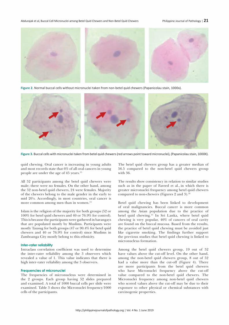
quid chewing. Oral cancer is increasing in young adults and most records state that 6% of all oral cancers in young people are under the age of 45 years.15
All 52 participants among the betel quid chewers were male; there were no females. On the other hand, among the 52 non-betel quid chewers, 24 were females. Majority of the chewers belong to the male gender in the early to mid 20’s. Accordingly, in most countries, oral cancer is more common among men than in women.15
Islam is the religion of the majority for both groups (52 or 100% for betel quid chewers and 40 or 76.9% for control). This is because the participants were gathered in barangays that are populated mostly by Muslims. Participants were mostly Tausug for both groups (47 or 90.4% for betel quid chewers and 40 or 76.9% for control) since Muslims in Zamboanga City mostly belong to this ethnicity.
Inter-rater reliabilityIntraclass correlation coefficient was used to determine the inter-rater reliability among the 3 observers which revealed a value of 1. This value indicates that there is high inter-rater reliability among the 3 observers.
Frequencies of micronucleiThe frequencies of micronucleus were determined in the 2 groups. Each group having 52 slides prepared and examined. A total of 1000 buccal cells per slide were examined. Table 3 shows the Micronuclei frequency/1000 cells of the participants.
The betel quid chewers group has a greater median of 56.5 compared to the non-betel quid chewers group with 36.
The results show consistency in relation to similar studies such as in the paper of Fareed et al., in which there is greater micronuclei frequency among betel quid chewers compared to non-chewers (Figures 2 and 3).16
Betel quid chewing has been linked to development of oral malignancies. Buccal cancer is more common among the Asian population due to the practice of betel quid chewing.15 In Sri Lanka, where betel quid chewing is very popular, 40% of cancers of oral cavity are found on the buccal mucosa. Based from the results, the practice of betel quid chewing must be avoided just like cigarette smoking. The findings further support the previous studies that betel quid chewing is linked to micronucleus formation.
Among the betel quid chewers group, 19 out of 52 have values above the cut-off level. On the other hand, among the non-betel quid chewers group, 8 out of 52 had a value more than the cut-off (Figure 4). There are more participants from the betel quid chewers who have Micronuclei frequency above the cut-off value compared to the non-betel quid chewers. The Micronuclei frequency among non-betel quid chewers who scored values above the cut-off may be due to their exposure to other physical or chemical substances with carcinogenic properties.
Figure 2. Normal buccal cells without micronuclei taken from non-betel quid chewers (Papanicolau stain, 1000x).
Figure 3. Buccal cells with micronuclei taken from betel quid chewers (red arrows point toward micronuclei), (Papanicolau stain, 1000X).
http://philippinejournalofpathology.org | Vol. 4 No. 1 June 2019
Abdurajak et al, Buccal Cell Micronuclei among Betel Quid Chewers and Non-Betel Quid Chewers Philippine Journal of Pathology | 21

Comparison of micronuclei frequencies between the groupsThe micronucleus frequencies between the 2 groups were analyzed using Mann-Whitney U test. It is shown in Table 3 that there is a significant difference in the frequencies of micronucleus between betel quid chewers and non-betel quid chewers (p value=0.031) at α=0.05.
This significant difference further supports the previous findings that betel quid chewing promotes micronuclei formation in buccal cells.
Relationship between micronuclei frequency and length of time of betel quid exposure (LBE)The betel quid chewing habits of the betel quid chewers group in terms of duration in years and the number of betel quid chewed per day is seen on Table 4. These numbers however are approximations due to difficulty among the participants in recalling their exact values. The length of time of betel quid exposure (LBE) was computed by multiplying the duration and number of betel quid chewed per day.
The scatter plot below (Figure 5) demonstrates the relationship between the Micronuclei frequency and length of time of betel quid exposure (LBE) variables. A very weak negative relationship is shown.
For this group of samples, the relationship between Micronuclei frequency and length of time of betel quid exposure (LBE) was analyzed using Pearson’s correlation coefficient which revealed a very weak negative relationship (r=-0.072). Although it is well established that the degree or intensity of consumption of betel quid increases the risk of developing oral malignancies which have been reported by several studies, there is a weak correlation between Micronuclei frequency and LBE in this study. This coincides with the study done by Nair et al., wherein there was no correlation between the number of betel
quid chewed per day, the number of years of chewing and the frequency of micronucleated oral mucosal cells.17 The same goes for the cases of Stich et al. and Suhas et al., wherein there was a very weak or no simple relationship observed between the number of betel nuts chewed and the frequency of micronucleated mucosa cells.18, 19
Nair et al. suggested that it would be more valid to link the micronucleus test to the cotinine (an alkaloid found in tobacco and a predominant metabolite of nicotine) levels in saliva or urine which is a more reliable marker of actual betel quid exposure.17
Figure 4. Bar graph of number of participants for each group who had Micronuclei frequency above the cut-off level of 56.5
Table 3. Comparison of frequencies of micronucleus between betel quid chewers and non-betel quid chewers using Mann-Whitney U test at α=0.05Group N Median p valueBetel Quid Chewers 52 56.5
0.031*Non-betel Quid Chewers 52 36*with significant difference: p ≤ 0.05
Table 4. Chewing habits and length of time of betel quid exposure among betel quid chewers
Duration(years)
No. of betel quid chewed per day
LBE = duration x no. of betel quid chewed per day
Micronuclei frequency/ 1000 cells
1 3 3 638 10 80 73 10 30 13
13 7 91 7019 10 190 3315 30 450 10710 4 40 65
2 5 10 1975 10 50 114 8 32 1259 10 90 1599 20 180 296 20 120 393 2 6 905 20 100 16
13 15 195 587 5 35 58
10 10 100 455 6 30 398 10 80 132 5 10 763 3 9 109
20 10 200 910 5 50 6710 3 30 118
3 10 30 9920 6 120 68
4 1 4 596 10 60 1115 5 25 442 10 20 136 5 30 78
12 7 84 410 50 500 6010 8 80 6818 10 180 11
8 8 64 1210 7 70 7710 4 40 63
6 5 30 672 10 20 115 10 50 233 20 60 461 4 4 513 3 9 88
10 4 40 3415 5 75 8
1 6 6 5813 10 130 9
3 11 33 3913 7 91 55
2 20 40 18
http://philippinejournalofpathology.org | Vol. 4 No. 1 June 2019
Abdurajak et al, Buccal Cell Micronuclei among Betel Quid Chewers and Non-Betel Quid Chewers Philippine Journal of Pathology | 22

The micronucleus test is a marker of the extent of chromosome breakage a few days to weeks previously, when the cells currently being exfoliated were dividing in the basal layer of oral mucosa.20 A betel chewer’s mucosa is said to be hyperplastic which means that there is an increase in the rate of proliferation in dividing cells.21 Thus, if there is an increase in the proliferation rate in dividing cells, there is an increase in the micronuclei frequency. With continuous exposure of the oral mucosa to betel quid, there is also continuous formation of micronucleus. Hence, an inference that the genotoxicity in the oral epithelium is local and acute due to the short turn-over period of 25 days in buccal cells from the basal layer to the epithelium.19 Furthermore, Nair et al. confirmed that this test can only reflect the current risk but not the cumulative risk over the years.17 Therefore, a person with a lower LBE may exhibit a higher micronuclei frequency than a person with a higher LBE at the time of sample collection of exfoliated buccal cells or may result the other way around or even equal. The only difference between the 2 individuals is that, the person with a higher LBE has higher number of oral lesions that developed over the years due to betel quid chewing, putting him at higher risk for developing oral malignancies. Furthermore, the average frequencies of the micronucleus are increased in precancerous lesions when compared to oral mucosa, with further increase in carcinomas, suggesting that micronucleus is a biomarker of neoplastic progression.19 This explains why there is a very weak or absence of correlation between the micronuclei frequency and LBE but when compared to non-betel quid chewers, betel quid chewers consistently exhibit higher average of micronuclei frequency as reported by several studies mentioned earlier.
To further determine the causal relationship between the carcinogenicity of betel quid chewing and its effect on the oral mucosa of individuals, studies such as ploidy studies and DNA adducts are highly recommended.
CONCLUSION
Between the 2 groups, the betel quid chewers exhibited significantly higher micronuclei frequencies compared to the non-betel quid chewers. The results of this study further support previous studies that the practice of betel quid chewing is associated with micronuclei formation and development of oral malignancies.
ACKNOWLEDGMENTS
The researchers thank the Department of Science and Technology Region IX, Zamboanga Consortium for Health Research and Development, Department of Health, and Ciudad Medical Zamboanga.
STATEMENT OF AUTHORSHIP
All authors certified fulfillment of ICMJE authorship criteria.
AUTHOR DISCLOSURE
The authors declared no conflict of interest.
FUNDING SOURCE
The study has been funded by the Department of Health IX Health Systems Research Fund Initiative.
Figure 5. Scatter plot diagram showing the relationship between Micronuclei frequency and length of time of betel quid exposure (LBE).
http://philippinejournalofpathology.org | Vol. 4 No. 1 June 2019
Abdurajak et al, Buccal Cell Micronuclei among Betel Quid Chewers and Non-Betel Quid Chewers Philippine Journal of Pathology | 23

REFERENCES
1. Davis GG. Buyo cheek cancer with special reference to etiology. JAMA. 1915; LXIV(9):711-8. https://doi.org/10.1001/jama.1915.02570350005002.
2. Stitch HF, Rosin MP, Vallejera MO. Reduction with vitamin A and beta-carotene administration of proportion of micronucleated buccal mucosa cells in Asian betal nut and tobacco chewers. Lancet. 1984;1(8388):1204-6. PMID: 6144923.
3. Nelson BS, Heischober B. Betel nut: a common drug used by naturalized citizens from India, Far East Asia, and the South Pacific Islands. Ann Emerg Med. 1999;34(2):238-43. PMID: 10424931.
4. Leghari MA, Ali S, Maqbool S. The prevalence of use of areca nut and its effect on oral health in school going children in Gadap town, Malir, Karachi, Pakistan. World J Dent. 2016;7(1):6-9.
5. Pindborg JJ. Epidemiological studies for oral cancer. Int Dent J. 1977;27(2);172-8. PMID: 267624.
6. Murti PR, Bhonsle RB, Gupta PC, Daftary DK, Pindborg JJ, Mehta FS. Etiology of oral submucous fibrosis with special reference to the role of areca nut chewing. J Oral Pathol Med. 1995;24(4):145-52. PMID: 7783003.
7. Adhikari A, De M. Toxic effects of betel quid. Int J Hum Genet. 2013;13(1):7-14.
8. Sabharwal R, Verma P, Syed MA, et al. Emergence of micronuclei as a genomic biomarker. Indian J Med Paediatr Oncol. 2015;36(4):212-8. PMID: 26811590. PMCID: PMC4711219. https://doi.org/10.4103/0971-5851.171541.
9. Jeng JH, Kuo ML, Hahn LJ, Kuo MYP. Genotoxic and no-genotoxic effects of betel quid ingredients on oral mucosal fibroblasts in vitro. J Dent Res. 1994;73(5):1043-9. PMID: 8006230. https://doi.org/10.1177/00220345940730050501.
10. Bhide SV, Padma PR, Amonkar AJ. Antimutagenic and anticarcinogenic effects of betel leaf extract against the tobacco-specific nitrosamine 4-(N-nitrosomethylamino)-1-(3-pyridyl)-1-butanone (NNK). IARC Sci Publ. 1991;(105):520-4. PMID: 1855909.
11. Jois HS, Kale AD, Mohan Kumar KP. Micronucleus as potential biomarker of oral carcinogenesis. Indian Journal of Dental Advancements. 2010;2:1-5.
12. Celik A, Cavas T, Ergene-Gözükara S. Cytogenetic monitoring in petrol station attendants: micronucleus test in exfoliated buccal cells. Mutagenesis. 2003;18(5):417-21. PMID: 12960409.
13. Anila K, Hallikeri K, Naikmasur VG. Comparative study of oral micronucleated cell frequency in oral submucous fibrosis patients and healthy individuals. J Clin Exp Dent. 2011;3(3): e201-6. https://doi.org/10.4317/jced.3.e201.
14. Tolbert PE, Shy CM, Allen JW. Micronuclei and other nuclear anomalies in buccal smears: methods development. Mutat Res. 1992;27(1):69-77. PMID: 1371831.
15. Warnakulasuriya S. Global epidemiology of oral and oropharyngeal cancer. Oral Oncol. 2009:45(4-5):309-16. PMID: 18804401. https://doi.org/10.1016/j.oraloncology.2008.06.002.
16. Fareed M, Afzal M, Siddique YH. Micronucleus investigation in buccal cells among pan masala/gutkha chewers and its relevance for oral cancer. Biol Med. 2011;3:8-15.
17. Nair U, Obe G, Nair J, et al. Evaluation of frequency of micronucleated oral mucosa cells as a marker for genotoxic damage in chewers of betel quid with or without tobacco. Mutat Res. 1991;261(3):163-8.
18. Stich HF, Stich W, Farida BB. Elevated frequency of micronucleated cells in the buccal mucosa of individuals at high risk for oral cancer: betel quid chewers. Cancer Lett. 1982;17(2):125-34. PMID: 6187434.
19. Suhas S, Ganapathy KS, Gayatri Devi, Ramesh C. Application of the micronucleus test to exfoliated epithelial cells from the oral cavity of beedi smokers, a high-risk group for oral cancer. Mutat Res. 2004;561(1-2):15-21. PMID: 15238226. https://doi.org/10.1016/j.mrgentox.2004.03.001.
20. Stich HF, Rosin MP. Quantitating the synergistic effect of smoking on alcohol consumption with the micronucleus test on human buccal mucosa cells. Int J Cancer. 1983;31(3):305-8. PMID: 6826255.
21. Reichart PA, Philipsen HP. Betel chewer’s mucosa-a review. J Oral Pathol Med. 1998; 27(6):239-42. PMID: 9707274.
Disclaimer: This journal is OPEN ACCESS, providing immediate access to its content on the principle that making research freely available to the public supports a greater global exchange of knowledge. As a requirement for submission to the PJP, all authors have accomplished an AUTHOR FORM, which declares that the ICMJE criteria for authorship have been met by each author listed, that the article represents original material, has not been published, accepted for publication in other journals, or concurrently submitted to other journals, and that all funding and conflicts of interest have been declared. Consent forms have been secured for the publication of information about patients or cases; otherwise, authors have declared that all means have been exhausted for securing consent.
http://philippinejournalofpathology.org | Vol. 4 No. 1 June 2019
Abdurajak et al, Buccal Cell Micronuclei among Betel Quid Chewers and Non-Betel Quid Chewers Philippine Journal of Pathology | 24

Immunohistochemical Expression of WT1 in Nasopharyngeal Carcinoma Among Filipino Patients in a Tertiary Hospital*Criston Van Manasan, Jenny Maureen Atun, Jose Carnate Jr.
Department of Laboratories, Philippine General Hospital, Taft Avenue, Manila
ABSTRACT
Background. Nasopharyngeal carcinoma (NPC) is endemic in Southeast Asia and the Philippines. Novel treatments are desirable due to the high disease burden and adverse effects of existing modalities. Detection of WT1 expression via immunohistochemistry has been reported in many tumors. Moreover, immunotherapy via WT1 peptide vaccination has shown promising results in a wide range of malignancies. No studies on WT1 expression in NPC have been published in any population.
Objective. Documenting WT1 expression in NPC via immunohistochemistry may provide insight into the possibility of using WT1 vaccination for this disease.
Methodology. This was a retrospective descriptive study. All newly-diagnosed cases of NPC from 2016 to 2017 with samples stored in the Department of Laboratories of the Philippine General Hospital were considered. Cases were included based on specific criteria. The tumor classification of each case was reviewed and WT1 immunohistochemistry staining was performed. Assessment of the strength of WT1 immunostaining was conducted. The results were analyzed using Chi-square test for association with fisher exact correction.
Results. A total of 57 cases were included, all of which were non-keratinizing squamous cell carcinomas (NK-SCCs). Forty-nine were undifferentiated type while eight were differentiated type. The mean age was 48 years. Two thirds were male, one third were female. Seventeen of the 57 cases (29.8%) were positive for WT1 immunostaining, and all were undifferentiated type. The majority (82.32%) of the positive cases showed cytoplasmic expression. There was a significant association between tumor classification and WT1 staining.
Conclusion. Similar to studies conducted in other carcinomas, a considerable subset of NPCs express WT1. This finding opens other avenues for exploration, including the feasibility of WT1 peptide vaccination as a treatment option. Further studies on the associations between WT1 and NPC are recommended.
Key words: nasopharyngeal cancer, Wilms’ tumor, wt1, immunohistochemistry, immunotherapy
INTRODUCTION
Nasopharyngeal carcinoma (NPC), while rare in most parts of the world (<1 per 100,000), is endemic in Southeast Asia, with an estimated incidence ranging from three to 30 per 100,000.1 The highest incidences (15-50 per 100,000) have been recorded in China, particularly in the southern regions, and it is uncommon among Caucasians.2 The pathogenesis is complex, but Epstein-Barr virus (EBV) infection is a major predisposing factor especially in endemic areas. EBV-LMP1 is the primary oncogene identified and is present in up to 90% of tumors.3 EBV infection along with genetic predisposition and environmental factors altogether contribute to tumorigenesis.4
A preliminary analysis by Mejia and Sarmiento in 2014 based on data from 49 patients from four centers in one year estimated the disease burden in the Philippines to be 2.07 per 100,000.1 This study did not include data from the Philippine General Hospital (PGH). The mainstay of treatment in NPC is radiotherapy, with 10-year survival rates of up to 43% overall.3 Country-specific data on survival and remission rates for the Philippines is lacking.
ISSN 2507-8364 (Online)Printed in the Philippines.Copyright© 2019 by the PJP.Received: 3 April 2019.Accepted: 19 May 2019.Published online first: 22 May 2019.https://doi.org/10.21141/PJP.2019.05 Corresponding author: Criston Van C. Manasan, MDE-mail: [email protected]
* Presented during the 24th Annual Residents’ Research forum of the Department of Laboratories of the Philippine General Hospital last November 22, 2018.
http://philippinejournalofpathology.org | Vol. 4 No. 1 June 2019
OPEN ACCESS – ORIGINAL ARTICLE

Given the high disease prevalence and the side effects of radiotherapy, including the risk for development of second primary tumors,5 new anti-cancer treatments are desirable. The role of the immune system in cancer progression and control has been known for years.6 The field of immunotherapy has emerged as an important front in the development of novel anti-cancer therapies. Interest in cancer immunotherapy has grown considerably since the discovery of the T-cell receptor in 1982.7 Subsequent research and clinical trials gave way to the approval by the United States Food and Drug Administration of anti-cancer immune checkpoint antibodies targeting CTLA-4 (Ipilimumab) in 20117 and PD-1 (Pembrolizumab) in 2014.8
Historically, immunotherapeutic research on NPC has been focused on the EBV antigens LMP1, LMP2, EBNA1, EBER and EBV-encoded RNA.9 These antigens have shown limited immunogenicity, play a role in tumor oncogenesis and contribute to viral latency and immune evasion. Together, these attributes constitute major challenges in harnessing EBV-related antigens as immunotherapy targets.9 Studies on non-EBV antigens, such as immune checkpoint antibodies, have also been performed. Recently, various clinical trials using EBV-related (anti-LAG3, anti-LMP2 vaccine) and non-EBV immunotherapeutic agents (Pembrolizumab, Nivolumab) have started. A phase Ib trial published in 2017 showed an objective response rate of 25.9% to Pembrolizumab in 27 patients. Eligibility in this trial included unresectable or metastatic disease, failure of prior standard therapy, and immunohistochemical PD-L1 expression in 1% or more of either the tumor cells or tumor-infiltrating lymphocytes.10 Even more recently, cell-based treatments using autologous cytotoxic T-lymphocytes (CTLs) are underway.9
Wilms’ Tumor 1 (WT1) peptide vaccines are among the immunotherapies in development outside of current NPC-related research. The earliest was developed in Japan by Oka et al.,11 which was followed by a series of further investigations and clinical trials by various authors.12,13 The vaccine-based therapy indirectly enhances the immune response against malignant tumors through stimulating the action of WT1-specific CD8+ CTLs.11,12 The WT1-specific CTLs do not induce cell lysis in normal tissues that express wild-type WT1 due to complex immunologic factors,11,12 thus greatly limiting toxicity. Central to the selection of cases for WT1 peptide immunotherapy is the detection of WT1 protein in tumor cells using polymerase chain reaction, Western blot, immunohistochemistry and/or other methods.11,14,15 Evidence accrued from early trials have solidified immunohistochemistry, an accessible and relatively inexpensive technique, as a reliable index of WT1 expression.11,13,15
The preliminary clinical trials of WT1 peptide vaccination focused on hematologic malignancies. The results for leukemia and myelodysplastic syndromes were promising in terms of slowing disease progression.12,13 WT1 was found to be highly-immunogenic in these trials. Trials have also been conducted on cases of glial tumors, soft tissue malignancies and various solid cancers.6,7,13 In 2009, the American National Cancer Institute identified WT1 as the top priority antigen in cancer treatment
research among 75 different antigens.16 There has been considerable interest in documenting WT1 expression in various tumors for the purpose of identifying possible candidates for WT1 peptide immunotherapy.6,7,12,15
At the time that this study was conducted, there were no published studies in the English literature documenting WT1 expression in NPC using immunohistochemistry. Literature search was performed in PubMed and Google Scholar which yielded no published articles. Given the high immunogenicity of WT1 as an immunotherapeutic target in other malignancies, it would be worthwhile to determine the degree of WT1 expression in NPC. Subsequently, this could potentially provide a rationale for utilizing WT1 immunotherapy for the treatment of this malignancy.
Review of Related Literature The Wilms’ Tumor 1 gene (WT1) was the first discovered gene associated with Wilms’ Tumor (WT).17 Located at chromosome 11p13 and initially cloned in 1990,18 the gene plays an important role in normal human embryonic development.19 Mutations are associated with Wilms tumor-aniridia-genitourinary anomalies-mental retardation (WAGR) syndrome, Denys-Drash syndrome and Frasier syndrome.17 Initially discovered as a tumor suppressor, mutations in WT1 are found in up to 15% of sporadic cases of WTs.19 Successive studies revealed that WT1 is overexpressed in a range of benign20 and malignant neoplasms such as hematologic malignancies,19,21 a broad range of carcinomas,15,22 soft tissue tumors23-25 and glial neoplasms.26 Genomic sequencing in these cases did not reveal mutations in the WT1 gene. This evidence suggests that wild-type WT1 may have a possible oncogenic role in malignancies aside from WT.15,19,25 WT1 is widely-regarded to function as a regulator of transcription but it has become apparent that its full function is more complex.27 Evidence has accumulated that WT1 can be a tumor suppressor and an oncogene depending on which cell types express it.19
In routine histopathology, the WT1 gene product is detected using immunohistochemistry. In established practice, it has been used as a supportive marker in the diagnosis of Wilms’ tumor,3 ovarian serous tumors,28 mesothelioma,29 and various other tumors. Generally, only nuclear staining was considered positive, and cytoplasmic expression was initially thought of as due to cross-reactivity of the antibody with unknown proteins.29 Subsequent studies have increasingly uncovered evidence that both nuclear and cytoplasmic WT1 immunostaining in a wide range of neoplasms is in fact due to the presence of the WT1 protein,15,23,24 and thus can be used as an index of WT1 protein expression. This finding accounted for the cytoplasmic expression previously seen in tumors that were generally regarded to express only nuclear staining such as in malignant mesothelioma. The presence of the WT1 peptide within the cytoplasm was confirmed in several studies using Western Blot and other methods.15,23,27 Ye et al. discussed that WT1 expression in the cytoplasm is mainly due to post-translational phosphorylation at zinc fingers leading to loss of the ability to bind DNA.30 This then results to retention of WT1 in the cytoplasm. Niksic et al.27 also reported that WT1 shuttles between the nucleus and the cytoplasm in association with active polyribosomes, suggesting a role for it in translation regulation.
http://philippinejournalofpathology.org | Vol. 4 No. 1 June 2019
Manasan et al, Immunohistochemical Expression of WT1 in Nasopharyngeal Carcinoma Among Filipino Philippine Journal of Pathology | 26

Nuclear and/or cytoplasmic expression of WT1 has been documented using immunohistochemistry in gastrointestinal, breast, lung, prostatic, kidney, urothelial and gynecologic cancers,15,22,31 as well as soft tissue sarcomas,15,23-25 pediatric small round blue cell tumors,14 and gliomas.26 Among soft tissue tumors, rhabdomyosarcomas have shown consistent cytoplasmic expression.24
Yang et al.19 reviewed several studies of WT1 immunohistochemistry expression in hematologic malignancies. WT1 was found to be increased in 354 of 476 (74%) cases of acute myeloid leukemia (AML) and 86 of 131 (66%) cases of acute lymphoblastic leukemia (ALL). Some types of myelodysplastic syndromes (MDS) also had increased levels of WT1.
Studies documenting WT1 expression in head and neck carcinomas are more limited. Mikami et al.32 analyzed tissues from six cell lines of oral squamous cell carcinoma (SCC) and one showed overexpression of WT1 protein while two out of 29 cases showed positive WT1 expression using immunohistochemistry. Xingru et al.33 demonstrated that WT1 promotes cell proliferation in vitro in a study that used cells derived from hypopharyngeal SCC. Leader et al.,20 in a study of 80 salivary gland tumors, found that WT1 was expressed in most benign salivary gland neoplasms while it is lost in malignant neoplasms with the exception of polymorphous low-grade adenocarcinomas (now called polymorphous adenocarcinoma in the 2017 WHO classification34) in which 11 out of 12 cases were positive. In 2002, a study by Oji et al.35 showed WT1 gene expression in 42 out of 56 (75%) head and neck SCCs using real-time reverse transcriptase-polymerase chain reaction (RT-PCR); none of which were nasopharyngeal cancers. In addition, only six cases underwent immunohistochemical staining, and all were positive. A study by Fattahi et al.36 in 2015 contrasted with the initial findings of Oji et al., wherein only three out of 45 (6.2%) cases of oral SCCs stained positive for WT1.
Across all the studies mentioned, gene sequencing findings in cases with positive immunohistochemistry results did not show mutations of the WT1 gene. This suggests a role for wild-type WT1 in tumorigenesis or possible epigenetic modifications which led to increased WT1 expression in various tumors.
In summary, increased wild-type WT1 expression has been demonstrated in a wide range of malignant tumors, with promising implications in the realm of cancer immunotherapy. There were no published studies analyzing the extent of WT1 expression via immunohistochemistry in NPCs in the English literature, even among studies focused on head and neck cancers. This dearth of information is what this study aimed to address, and demonstration of WT1 activity in NPC would put forward the possibility of WT1-specific cancer immunotherapy for this tumor.
ObjectivesThe study aimed to evaluate the immunohistochemistry staining patterns for WT1 in nasopharyngeal carcinomas diagnosed in the Philippine General Hospital from 2016 to 2017.
Specifically, the study aimed to:1. Determine the basic demographic information of
patients diagnosed with nasopharyngeal carcinomas in the hospital, namely: age, sex and tumor histologic classification.
2. Determine the rates of positive and negative expression of WT1 among the various histologic classifications.
3. Determine the sub-cellular localization, extent and intensity of WT1 staining among nasopharyngeal carcinomas according to histologic classification.
4. Identify and describe the predominant WT1 staining patterns for each histologic classification of nasopharyngeal carcinoma.
METHODOLOGY
Ethical ConsiderationsThe study was approved by the University of the Philippines-Manila Research Ethics Board (UPM REB) prior to being conducted. A waiver of consent was requested and approved as there were no risks to the study participants. The methods of data collection, handling and storage ensured anonymity and confidentiality of the participants.
Study DesignThe study was a descriptive, retrospective study and involved slide reviews of patients who were diagnosed with NPC in accordance with the inclusion criteria below. All recent cases (2016 to 2017) were included. As this is a preliminary study, the use of recent tissue samples ensured the most optimal immunohistochemistry staining results.
Only the patients’ age and sex were collected. The formalin-fixed, paraffin-embedded tissue blocks of each case were retrieved and processed for immunohistochemistry staining with WT1. Inclusion CriteriaThe study included all newly-diagnosed cases of nasopharyngeal carcinoma from 2016 to 2017 at the Philippine General Hospital that have been confirmed using histomorphologic assessment and immunohistochemistry staining with at least a Pan-Cytokeratin.
Exclusion Criteria The following were excluded: cases of recurrent or persistent nasopharyngeal carcinoma that have already undergone radiotherapy and/or other treatments; cases of nasopharyngeal carcinoma metastatic to other sites that do not have a nasopharyngeal tissue sample in storage; cases that have concomitant malignant tumors elsewhere; cases that have deteriorated and unsalvageable slides and paraffin-embedded tissue blocks.
Data Collection All diagnosed NPCs in the PGH from 2016 to 2017 that fulfilled the criteria were included in the study. The patients’ age and sex were collected from the records of the surgical pathology and outpatient sections of the Department of Laboratories. All patients were anonymized.
http://philippinejournalofpathology.org | Vol. 4 No. 1 June 2019
Manasan et al, Immunohistochemical Expression of WT1 in Nasopharyngeal Carcinoma Philippine Journal of Pathology | 27

Slide Review The diagnosis for each case was classified in accordance with the WHO Classification of Tumors recommended by 8th edition (2017) of the American Joint Committee on Cancer (AJCC) Staging Manual34 (Table 1).
The stained biopsy slides and blocks were retrieved for review. Each case had at least a Hematoxylin and Eosin (H&E) stained slide and an immunohistochemistry slide for Pan-Cytokeratin. A consensus diagnosis was generated by three pathologists, with at least two out of three (2 out of 3) pathologists concurring.
Immunohistochemistry with WT1New slides were prepared from the formalin-fixed and paraffin-embedded tissue blocks for immunohistochemistry. Immunohistochemistry staining was performed using the standardized protocols established by the section of surgical pathology (Autostainer Link 48, DAKO, CALIFORNIA, USA). 3-um sections were prepared and placed on glass slides. Each slide was deparaffinized, rehydrated and subjected to heat-induced epitope retrieval for 10 minutes using an automated system (PT Link Instrument, DAKO, CALIFORNIA, USA). The sections were then treated with a peroxidase-blocking solution (FLEX) for five minutes. Subsequent incubation with the ready-to-use anti-WT1 antibody was done at room temperature for 15 minutes. A monoclonal antibody against WT1 (6F-H2 DAKO, CALIFORNIA, USA) was used. Visualization of signals was done using HRP Labelled Polymer (DAKO, CALIFORNIA, USA) for 20 minutes, followed by washing with a buffer for 10 minutes, and then incubation in DAB+Chromogen (DAKO, CALIFORNIA, USA) for 10 minutes. The slides were counterstained with Hematoxylin. Positive controls were included with each case: either Wilms’ tumor
Table 1. Classification of tumors according to the 8th edition of the AJCC staging manualAJCC/WHO 2017 Classification Former TerminologyKeratinizing squamous cell carcinoma WHO Type I (squamous cell carcinoma)Non-keratinizing squamous cell carcinoma, Differentiated
WHO Type II (transitional cell carcinoma)
Non-keratinizing squamous cell carcinoma, Undifferentiated
WHO Type III (lymphoepithelial carcinoma)
Basaloid squamous cell carcinoma No synonym exists (recently described)
Figure 1. Sample photomicrograph of a non-keratinizing, squamous cell carcinoma of undifferentiated type (H&E, 400X).
Figure 3. Sample photomicrograph of a case which showed granular, cytoplasmic immunostaining for WT1 (400X).
Figure 2. Sample photomicrograph of a case which was negative for WT1 immunostaining (400X).
Figure 4. Sample photomicrograph of a case which showed positive WT1 nuclear immunostaining (400X).
http://philippinejournalofpathology.org | Vol. 4 No. 1 June 2019
Manasan et al, Immunohistochemical Expression of WT1 in Nasopharyngeal Carcinoma Among Filipino Philippine Journal of Pathology | 28

or ovarian serous carcinoma. The WT1 antibody also stained lymphocytes and endothelial cells which served as internal controls.
Assessment of WT1 ImmunostainingAssessment of WT1 immunostaining was performed by three different pathologists, all of whom were blinded in terms of clinicopathologic information. Criteria for assessment was based on a modified version of the assessment done by Kim et al.23 The intensity of the staining and the proportion of the positively-staining area were considered together and evaluated semi-quantitatively.
Both nuclear and cytoplasmic staining were considered. For staining intensity, a score of 0 was assigned if there was no staining or if it is barely perceptible. Clearly-perceptible but faint staining was assigned a score of 1. Distinct staining that is not as strong as the control was assigned a score of 2. Staining intensity equal to or stronger than the positive controls was assigned a score of 3. In cases where the staining pattern was heterogeneous, the more frequent intensity was considered. Afterwards, the percentage of tumors cells that stained positive were estimated (1-100). The intensity and percentage were multiplied and assigned a final strength based on the product. The final strengths were negative (0-20), weak (21-80), moderate (81-180) and strong (181-300).
RESULTS
A total of 79 new cases of NPC were diagnosed from 2016 to 2017. Fifty-seven (57) out of these 79 cases were eligible for review and immunohistochemistry testing. Twenty-two (22) cases were not included in the review due to the following: (a) irretrievable tissue blocks (n=15), (b) cases that were sampled from outside hence the tissue blocks were not available (n=6) and (c) one case with a non-usable block. The diagnosis of each case was reviewed using the H&E slide and corresponding immunohistochemistry slide for Pan-Cytokeratin.
Of the 57 cases reviewed, exactly two-thirds (n=38; 67%) were male and the remaining one-third (n=19; 33%) were female. The median age was 48 years old. The females averaged older at 53.53 years while the average age of males was 45.2 years. The youngest patient was a 10-year-old male while the oldest was a 79-year-old female. All of the cases were non-keratinizing squamous cell carcinomas (NK-SCC). The majority of cases (n=49, 86%) were of the undifferentiated subtype while the remainder (n=8; 14%) were of the differentiated subtype. None of the cases were keratinizing squamous cell carcinomas (K-SCC) or basaloid squamous cell carcinomas (B-SCC) (Table 2).
Analysis of WT1 StainingSeventeen out of 57 cases (29.82%) stained positive for WT1 while 40 cases (70.18%) were negative (Table 3). Among the 40 cases which were negative, 32 (80%) were undifferentiated while eight (20%) were differentiated. Conversely, all 17 (100%) positive cases were undifferentiated. The distribution of the 17 positive cases in terms of sex followed the overall distribution: 11 (64.7%) were male, and 6 (35.3%) were female. The positive cases accounted for a third (34.7%) of all the cases classified as undifferentiated type (17 out of 49). In terms of intensity, most cases were assigned scores of 1 and 2. In terms of tumor cell population stained, an average of 45% of tumor cells expressed WT1. Five cases expressed the protein in more than 70% of the tumor cell population. Fourteen out of the 17 (82.35%) positive cases showed diffuse to granular cytoplasmic WT1 expression. Two cases showed nuclear expression, and one showed both nuclear and cytoplasmic expression (mixed). In terms of strength of staining, the positive cases were distributed almost evenly between weak (n=8) and moderate (n=9) expression. None of the cases showed strong expression as defined by the assessment protocol.
Statistical tests were performed using Chi-square test of association with fisher exact correction. STATA 14 (StataCorp, Texas, USA) was used for all statistical analysis. There was a statistically significant association (p-value=0.047) between the presence of staining and tumor classification (Table 4).
There was no statistically significant association between tumor type and strength of staining (p-value=0.221). There was also no statistically significant association between the strength of staining and localization (p-value=0.329) among the positive cases classified as undifferentiated-type, NK-SCC (Table 5).
Table 2. Overview of results per tumor differentiation and sex
Tumor ClassificationSex
SubtotalMale Female
Keratinizing squamous cell carcinoma 0 0 0Non-keratinizing squamous cell carcinoma, Differentiated 6 2 8Non-keratinizing squamous cell carcinoma, Undifferentiated 32 17 49Basaloid squamous cell carcinoma 0 0 0Grand Total 38 19 57
Table 3. WT1 Immunohistochemistry staining profile of nasopharyngeal carcinoma per histologic classification and localization of staining
Classification LocalizationPositive Total
positiveTotal
negativeGrand totalWeak Moderate
Non-keratinizing squamous cell carcinoma, Differentiated
Cytoplasmic 0 0
0 8 8
Mixed 0 0
Nuclear 0 0Non-keratinizing squamous cell carcinoma, Undifferentiated
Cytoplasmic 8 6
17 32 49
Mixed 0 1
Nuclear 0 2Grand Total 8 9 17 40 57
Table 4. Association of WT1 Immunostaining with tumor classification
Tumor ClassificationStaining p-value
Positive Negative Fisher’s ExactNon-keratinizing squamous cell carcinoma, Differentiated 0 8 (20.00)
0.047Non-keratinizing squamous cell carcinoma, Undifferentiated 17 (100.00) 32 (80.00)
http://philippinejournalofpathology.org | Vol. 4 No. 1 June 2019
Manasan et al, Immunohistochemical Expression of WT1 in Nasopharyngeal Carcinoma Philippine Journal of Pathology | 29

DISCUSSION
Cancer immunotherapy using WT1 peptide vaccination has been undergoing trials over the past decade. Taking the results of several investigations together, researchers have acknowledged the challenges in assessing the potential of WT1 as an anti-cancer antigen. Most of these factors are inherent to the time-consuming nature of clinical trials and the slow action of anti-cancer vaccines, in general, relative to other anti-cancer treatments such as chemotherapy.6,37 Nevertheless, considerable developments have been achieved. Limited early trials in hematologic and other malignancies have found the method to be safe and efficacious.11 The trials showed that the treatment enhances the body’s immune response against cancer cells through the action of WT1-specific CD8+ CTLs.6,13 Experience with the vaccinations for AML, MDS and other hematologic malignancies in Japan has advanced to the point where WT1 levels in peripheral blood are being utilized as a marker for minimal residual disease. Complete remission (CR) has been achieved via WT1 peptide vaccination in combination with other treatments for some cases of AML and MDS.12,38 Recent proposals for further trials focusing on hematologic malignancies have called for cure-oriented approaches.38
Trials in carcinomas have also been ongoing. A Phase I trial conducted by Ohno et al. in 2012 with 28 patients showed that WT1 peptide vaccination combined with other treatments was well-tolerated and showed 60% improved clinical response in patients with advanced cervical, ovarian, lung, colorectal, pancreatic or biliary tract cancers.37 More recently in 2018, a Phase II randomized study of a WT1 vaccine combined with Gemcitabine conducted by Nishida et al. showed improved one-year progression-free survival in 85 evaluated patients with advanced pancreatic ductal adenocarcinoma versus Gemcitabine alone.39 Overall survival was not significantly altered.
Aside from its therapeutic potential in cancer immunotherapy, the interest in WT1 has extended to its value as a prognostic marker. Kim et al. examined the prognostic value of WT1 expression in 63 patients with soft tissue sarcomas. They found that strong WT1 expression was associated with improved outcomes among patients with high-grade soft tissue sarcomas, but not in other groups.23 A 2015 meta-analysis conducted by Qi et al.40 on the association of WT1 and prognosis in patients with solid cancers included 22 studies and 3,620 patients.
Their findings contrasted with Kim et al., as they found that WT1 expression seemed connected to increased risk for disease relapse and progression. The differences in results illustrates the limited knowledge regarding WT1 activity in various tumors de novo. The data on the usefulness of WT1 as a prognostic marker in cancers is still being accumulated and remains controversial. There have been no published studies on WT1 expression in NPC. Our study of 57 cases of NPC all consisted of NK-SCCs. Eight cases were of the differentiated subtype, and 49 were of the undifferentiated subtype in accordance with the WHO classification. The undifferentiated subtype of NK-SCC is the most common in endemic areas2 and so the distribution in our study seems consistent with trends observed in the literature. K-SCCs are less frequent in endemic areas, and are less frequently associated with EBV.2 B-SCCs of the nasopharynx are similar to basaloid SCCs elsewhere in the head and neck and may also be associated with EBV in endemic areas. The literature on the differences in behavior, tumor spread and prognosis among the different tumor classifications of NPC has been inconclusive thus far.2
Seventeen out of 57 cases (29.82%) stained positive for WT1, and all were of the undifferentiated subtype. In addition, the majority (n=14; 82.35%) of the positive cases showed diffuse to granular cytoplasmic expression. Two cases showed nuclear expression, and one case exhibited both nuclear and cytoplasmic expression. Approximately half of cases (n=8) stained weakly for WT1, and a slightly higher number stained with a moderate intensity (n=9). None of the cases stained with a strong intensity.
No equivalent comparisons of the results can be made in the literature due to the lack of studies documenting WT1 immunostaining in NPC. As such, the results were compared with studies done on cancers of the head and neck as well as cancers in various other organ systems. The degree of WT1 expression in NPC seen in our study was higher than the results obtained by Leader et al.20 in their study on salivary gland neoplasms. The exception is polymorphous adenocarcinoma which showed positive WT1 expression in 11 out of 12 cases. Our study also showed higher WT1 expression rates in NPC compared with oral SCCs as studied by Mikami et al. (6.9%)32 and Fattahi et al. (6.2%).36 Conversely, our results differ from the findings of Oji et al., wherein six out of six cases of oral SCCs were reportedly positive for WT1 via immunohistochemistry.35
A study conducted by Nakatsuka et al.15 in 2006 included a wide variety of cancers. They used polyclonal (C-19) and monoclonal (6F-H2) antibodies for assessing WT1 immunostaining; this monoclonal antibody was the same used in our study. Both nuclear and cytoplasmic staining were considered. They found discrepant immunostaining results between the two antibodies in 129 out of 338 cases (38%) studied. For the 6F-H2 antibody, they found a wide range of expression rates (5-81%). The cancer types that showed less than 50% expression rates among cases were cervical (5%), prostate (25%), lung (30%), urothelial (33%), renal (36%), gastric (42%) and esophageal (45%). The cancer types which showed greater than 50% expression
Table 5. Association of WT1 immunostaining with strength of staining and localization
VariableStrength of Staining p-value
Weak (n=8)
Moderate (n=9)
Negative (n=40)
Fisher’s Exact
Tumor ClassificationDifferentiatedUndifferentiated
08 (100.00)
09 (100.00)
8 (20.00)32 (80.00)
0.221
Localization (Differentiated)CytoplasmicMixedNuclear
000
000
000
–
Localization (Undifferentiated)CytoplasmicMixedNuclear
8 (100.00)00
6 (66.67)1 (11.11)2 (22.22)
000
0.329
http://philippinejournalofpathology.org | Vol. 4 No. 1 June 2019
Manasan et al, Immunohistochemical Expression of WT1 in Nasopharyngeal Carcinoma Among Filipino Philippine Journal of Pathology | 30

rates were breast (52%), pancreatic (65%), ovarian (66%), biliary (68%), colorectal (69%) and endometrial (81%).
Overall, the degree of WT1 expression in NPC is higher compared to head and neck cancers and lower compared to cancers of other organ systems; exceptions being cancers of the uterine cervix, lung and prostate.
Our study also found a significant association between tumor classification and positive WT1 immunostaining. Current evidence shows no clinical difference in the behavior between the differentiated and undifferentiated subtypes of NK-SCC of the nasopharynx.2 As such, whether or not the association has any relevance needs further study given the small sample size and limited knowledge regarding the role of WT1 in NPC. A comprehensive NPC-related genomic survey conducted by Hu et al.41 did not specifically-include WT1.
No other statistically significant associations were found.
LimitationsThe study was inherently limited by its retrospective nature. Due to the dearth of information on WT1 expression in nasopharyngeal carcinoma, the study aimed to offer only an initial glimpse into a possible role of the gene in this subset of head and neck cancers. The small sample size (N=57) also limited the analysis which could be made due to the limited NPC tumor classifications represented. The study is also limited to documenting the strength and localization of WT1 immunostaining and the number of positive versus negative cases. Correlating these results with clinical factors, morphologic features and other variables is beyond the scope of this study.
CONCLUSION
To the best of our knowledge, this is the first study in the English literature that has studied the immunohistochemical expression of WT1 in NPC. Expression of WT1 was documented in a considerable proportion (29.82%) of NPC cases included in our study. All of the positive cases were NK-SCCs of undifferentiated subtype. The vast majority of the positive cases showed cytoplasmic staining.
RecommendationsAdditional data collection is needed to expand on the preliminary information from this study. Prospective studies with larger sample sizes are strongly recommended.
Specifically, the results and observations made during the conduct of the study point to meaningful avenues for further exploration along several possible routes:
1. Confirmation of WT1 gene products – Previous studies have shown that WT1 immunostaining is specific for detecting WT1 protein in malignant tumors. This was in the form of mRNA detected via RT-PCR or WT1 peptide isolated via Western Blot. This has yet to be proven for NPC specifically, and confirmation would be ideal.
2. Antibody type – The 6F-H2 antibody used in this study recognizes the N terminus of the WT1 protein. If the N terminus is lacking, then sensitivity would
be affected. Nakatsuka et al. found different staining results between polyclonal and monoclonal WT1 antibodies in 129 out of 338 cases (38%) studied.15 Further studies using a polyclonal antibody to detect other isoforms of the WT1 protein may provide additional useful information.
3. WT1 allele – In prior studies of WT1 expression in other cancers, the WT1 gene was usually of the wild-type allele. This was revealed through genomic sequencing of the WT1 gene in cancers with WT1 overexpression. Genomic sequencing of the WT1 gene in cases of NPC, in correlation with immunohistochemistry results, might provide useful information. Current knowledge about the complex nature of NPC oncogenesis does not mention a role for WT1.41 The complex pathology of NPC is still unravelling and yet to be fully understood.
4. Tumor classification and WT1 staining – Our study showed a statistically significant association between tumor classification and WT1 immunostaining. The limited literature regarding the role of WT1 in NPC and the study limitations preclude further interpretation of this finding. Additional studies investigating this association are recommended.
5. Histomorphology and WT1 staining – Our study did not correlate between WT1 staining and histomorphologic features. Analyzing the extent, localization and intensity of WT1 staining and their association with morphologic features is recommended.
6. Recurrent and resistant cases of NPC – Our study included only newly-diagnosed cases of NPC that have not yet undergone treatment. The pattern of WT1 expression may be different in cases of metastatic, recurrent or cases that are non-responsive to conventional treatment. Given the possible changes at the genetic and molecular level that have occurred in this subset of NPCs, studying WT1 expression in this population may provide valuable insight.
7. Tumor microenvironment – We have observed WT1 staining among some tumor-related elements, namely the endothelial cells of the tumor blood vessels and the associated lymphocytes. Given the lymphocyte-rich morphology of NPC and association with EBV, further study of this observation and its possible therapeutic implications is recommended. Other authors have observed similar WT1 staining in the tumor-related elements in other tumor types.15,23,24
8. Prognosis and correlation with other antigens – To date, only the presence of EBV viral DNA has been incorporated clinically as a distinct prognostic marker for NPC.2 Previous studies have suggested a correlation between WT1 expression in solid cancers and poorer prognosis, though this remains unsettled. Further study may be done to determine if WT1 expression in NPC is connected with tumor behavior and whether correlation with EBV-related antigens (such as LMP1, LMP2, EBNA1 and EBER) is present.
9. WT1 peptide vaccination in NPC – Early trials using various immunotherapy agents for cases of advanced NPC are ongoing.9,10 None are currently for WT1. Our study has shown that some cases of NPC express WT1. Further data is needed in order to determine the feasibility of WT1 peptide vaccination for NPC.
http://philippinejournalofpathology.org | Vol. 4 No. 1 June 2019
Manasan et al, Immunohistochemical Expression of WT1 in Nasopharyngeal Carcinoma Philippine Journal of Pathology | 31

This study provided a glimpse into the role of WT1 in NPC. The results indicate that a subset of NPCs express WT1. Additional studies examining this relationship in larger populations are recommended. In addition, the results presented here provided potential rationales for the further study of WT1 and its association with NPC.
ACKNOWLEDGMENTS
The authors thank the Surgical Pathology Section of the Department of laboratories of the Philippine General Hospital for their invaluable contribution to this research. Specifically, Dr. Michele Hernandez-Diwa (section head), Dr. Karen Cybelle Sotalbo (vice section head), Esmeralda Talplacido (section supervisor) and the following medical technologists and administrative staff: Renee Rose Martos, Ma. Theresa Cargo, Ma. Belinda Cabanes, Jick Tarrayo, Jameson Malabanan, Jogie Qiambao, Mary Rose Hallig and Mr. Al Ela.
STATEMENT OF AUTHORSHIP
All authors certified fulfillment of ICMJE authorship criteria.
AUTHOR DISCLOSURE
The authors declared no conflict of interest.
FUNDING SOURCE
None.
REFERENCES
1. Sarmiento MP, Mejia MB. Preliminary assessment of nasopharyngeal carcinoma incidence in the Philippines: a second look at published data from four centers. Chin J Cancer. 2014; 33(3):159-64. PMID: 23958058. PMCID: PMC3966143. https://doi.org/ 10.5732/cjc.013.10010.
2. El-Naggar AK, Chan JKC, Grandis JR, Takata T, Slootweg PJ, eds. World Health Organization classification of head and neck tumours, 4th ed, Vol. 9. Lyon: International Agency for Research on Cancer (IARC), 2017.
3. Goldblum J, Lamps L, McKenney J, Myers J. In: Rosai and Ackermann’s surgical pathology, 11th ed. USA: Mosby, Elsevier Inc., 2018.
4. Hildesheim A, Wang CP. Genetic predisposition factors and nasopharyngeal carcinoma risk: a review of epidemiological association studies, 2000-2011: Rosetta Stone for NPC: genetics, viral infection, and other environmental factors. Semin Cancer Biol. 2012;22(2):107-16. PMID: 22300735. PMCID: PMC3296903. https://doi.org/10.1016/j.semcancer.2012.01.007.
5. Kong L, Lu JJ, Guo X, Wu Y, Zhang Y. The risk of second primary tumors in patients with nasopharyngeal carcinoma after definitive radiotherapy. Cancer. 2006;107(6):1287-93. PMID: 16909425. https://doi.org/10.1002/cncr.22119.
6. Oka Y, Sugiyama H. WT1 peptide vaccine, one of the most promising cancer vaccines: its present status and
the future prospects. Immunotherapy. 2010;2(5):591-4. PMID: 20874639. https://doi.org/10.2217/imt.10.58.
7. Oiseth SJ, Aziz MS. Cancer immunotherapy: a brief review of the history, possibilities, and challenges ahead. J Cancer Metastasis Treat. 2017;3:250-61. https://doi.org/10.20517/2394-4722.2017.41.
8. Gong J, Chehrazi-Raffle A, Reddi S, Salgia R. Development of PD-1 and PD-L1 inhibitors as a form of cancer immunotherapy: a comprehensive review of registration trials and future considerations. J Immunother Cancer. 2018;6(1):8. PMID: 29357948. PMCID: PMC5778665. https://doi.org/10.1186/s40425-018-0316-z.
9. Jain A, Chia WK, Toh HC. Immunotherapy for nasopharyngeal cancer – a review. Chin Clin Oncol. 2016;5(2):22. PMID: 27121882. https://doi.org/10.21037/cco.2016.03.08.
10. Hsu C, Lee SH, Ejadi S, et al. Safety and antitumor activity of pembrolizumab in patients with programmed-death-ligand 1-positive nasopharyngeal carcinoma: results of the KEYNOTE-028 study. J Clin Oncol 2017;35(36):4050-6. PMID: 28837405. https://doi.org/10.1200/JCO.2017.73.3675.
11. Oka Y, Tsuboi A, Taguchi T, et al Induction of WT1 (Wilms' tumor gene)-specific cytotoxic T lymphocytes by WT1 peptide vaccine and the resultant cancer regression. Proc Natl Acad Sci USA. 2004;101(38):13885-90. PMID: 15365188. PMCID: PMC518848. https://doi.org/10.1073/pnas.0405884101.
12. Di Stasi A, Jimenez AM, Minagawa K, Al-obaidi M, Rezvani K. Review of the results of WT1 peptide vaccination strategies for myelodysplastic syndromes and acute myeloid leukemia from nine different studies. Front Immunol. 2015;6:36. PMID: 25699052. PMCID: PMC4316779. https://doi.org/10.3389/fimmu. 2015.00036.
13. Oka Y, Tsuboi A, Kawakami M, et al. Development of WT1 peptide cancer vaccine against hematopoietic malignancies and solid cancers. Curr Med Chem. 2006;13(20):2345-52. PMID: 16918359.
14. Carpentieri DF, Nichols K, Chou PM, Matthews M, Pawel B, Huff D. The expression of WT1 in the differentiation of rhabdomyosarcoma from other pediatric small round blue cell tumors. Mod Pathol. 2002;15(10):1080-6. PMID: 12379755. https://doi.org/10.1097/01.MP.0000028646.03760.6B.
15. Nakatsuka S, Oji Y, Horiuchi T, et al Immunohistochemical detection of WT1 protein in a variety of cancer cells. Mod Pathol. 2006;19(6)804-14. PMID: 16547468. https://doi.org/10.1038/modpathol.3800588.
16. Cheever MA, Allison JP, Ferris AS, et al. The prioritization of cancer antigens: a national cancer institute pilot project for the acceleration of translational research. Clin Cancer Res. 2009;15(17):5323-17. PMID: 19723653. PMCID: PMC5779623. https://doi.org/10.1158/1078-0432.CCR-09-0737.
17. Kumar V, Abbas A, Aster J. Robbins and Cotran pathologic basis of disease, 9th ed. Philadelphia, PA: Elsevier/Saunders, 2015.
18. Call KM, Glaser T, Ito CY, et al Isolation and characterization of a zinc finger polypeptide gene at the human chromosome 11 Wilms’ tumor locus. Cell. 1990;50(3):509-20. PMID: 2154335.
19. Yang L, Han Y, Suarez Saiz F, Minden MD. A tumor suppressor and oncogene: the WT1 story. Leukemia.
http://philippinejournalofpathology.org | Vol. 4 No. 1 June 2019
Manasan et al, Immunohistochemical Expression of WT1 in Nasopharyngeal Carcinoma Among Filipino Philippine Journal of Pathology | 32
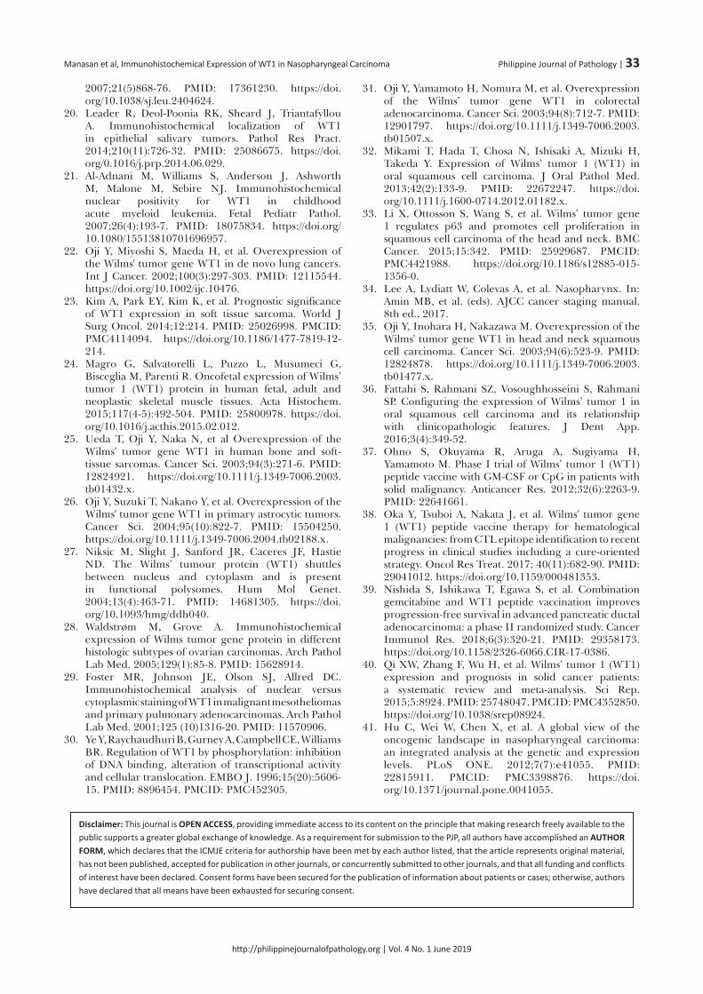
2007;21(5)868-76. PMID: 17361230. https://doi.org/10.1038/sj.leu.2404624.
20. Leader R, Deol-Poonia RK, Sheard J, Triantafyllou A. Immunohistochemical localization of WT1 in epithelial salivary tumors. Pathol Res Pract. 2014;210(11):726-32. PMID: 25086675. https://doi.org/0.1016/j.prp.2014.06.029.
21. Al-Adnani M, Williams S, Anderson J, Ashworth M, Malone M, Sebire NJ. Immunohistochemical nuclear positivity for WT1 in childhood acute myeloid leukemia. Fetal Pediatr Pathol. 2007;26(4):193-7. PMID: 18075834. https://doi.org/ 10.1080/15513810701696957.
22. Oji Y, Miyoshi S, Maeda H, et al. Overexpression of the Wilms' tumor gene WT1 in de novo lung cancers. Int J Cancer. 2002;100(3):297-303. PMID: 12115544. https://doi.org/10.1002/ijc.10476.
23. Kim A, Park EY, Kim K, et al. Prognostic significance of WT1 expression in soft tissue sarcoma. World J Surg Oncol. 2014;12:214. PMID: 25026998. PMCID: PMC4114094. https://doi.org/10.1186/1477-7819-12-214.
24. Magro G, Salvatorelli L, Puzzo L, Musumeci G, Bisceglia M, Parenti R. Oncofetal expression of Wilms’ tumor 1 (WT1) protein in human fetal, adult and neoplastic skeletal muscle tissues. Acta Histochem. 2015;117(4-5):492-504. PMID: 25800978. https://doi.org/10.1016/j.acthis.2015.02.012.
25. Ueda T, Oji Y, Naka N, et al Overexpression of the Wilms' tumor gene WT1 in human bone and soft-tissue sarcomas. Cancer Sci. 2003;94(3):271-6. PMID: 12824921. https://doi.org/10.1111/j.1349-7006.2003.tb01432.x.
26. Oji Y, Suzuki T, Nakano Y, et al. Overexpression of the Wilms' tumor gene WT1 in primary astrocytic tumors. Cancer Sci. 2004;95(10):822-7. PMID: 15504250. https://doi.org/10.1111/j.1349-7006.2004.tb02188.x.
27. Niksic M, Slight J, Sanford JR, Caceres JF, Hastie ND. The Wilms’ tumour protein (WT1) shuttles between nucleus and cytoplasm and is present in functional polysomes. Hum Mol Genet. 2004;13(4):463-71. PMID: 14681305. https://doi.org/10.1093/hmg/ddh040.
28. Waldstrøm M, Grove A. Immunohistochemical expression of Wilms tumor gene protein in different histologic subtypes of ovarian carcinomas. Arch Pathol Lab Med. 2005;129(1):85-8. PMID: 15628914.
29. Foster MR, Johnson JE, Olson SJ, Allred DC. Immunohistochemical analysis of nuclear versus cytoplasmic staining of WT1 in malignant mesotheliomas and primary pulmonary adenocarcinomas. Arch Pathol Lab Med. 2001;125 (10)1316-20. PMID: 11570906.
30. Ye Y, Raychaudhuri B, Gurney A, Campbell CE, Williams BR. Regulation of WT1 by phosphorylation: inhibition of DNA binding, alteration of transcriptional activity and cellular translocation. EMBO J. 1996;15(20):5606-15. PMID: 8896454. PMCID: PMC452305.
31. Oji Y, Yamamoto H, Nomura M, et al. Overexpression of the Wilms’ tumor gene WT1 in colorectal adenocarcinoma. Cancer Sci. 2003;94(8):712-7. PMID: 12901797. https://doi.org/10.1111/j.1349-7006.2003.tb01507.x.
32. Mikami T, Hada T, Chosa N, Ishisaki A, Mizuki H, Takeda Y. Expression of Wilms’ tumor 1 (WT1) in oral squamous cell carcinoma. J Oral Pathol Med. 2013;42(2):133-9. PMID: 22672247. https://doi.org/10.1111/j.1600-0714.2012.01182.x.
33. Li X, Ottosson S, Wang S, et al. Wilms’ tumor gene 1 regulates p63 and promotes cell proliferation in squamous cell carcinoma of the head and neck. BMC Cancer. 2015;15:342. PMID: 25929687. PMCID: PMC4421988. https://doi.org/10.1186/s12885-015-1356-0.
34. Lee A, Lydiatt W, Colevas A, et al. Nasopharynx. In: Amin MB, et al. (eds). AJCC cancer staging manual. 8th ed., 2017.
35. Oji Y, Inohara H, Nakazawa M. Overexpression of the Wilms' tumor gene WT1 in head and neck squamous cell carcinoma. Cancer Sci. 2003;94(6):523-9. PMID: 12824878. https://doi.org/10.1111/j.1349-7006.2003.tb01477.x.
36. Fattahi S, Rahmani SZ, Vosoughhosseini S, Rahmani SP. Configuring the expression of Wilms’ tumor 1 in oral squamous cell carcinoma and its relationship with clinicopathologic features. J Dent App. 2016;3(4):349-52.
37. Ohno S, Okuyama R, Aruga A, Sugiyama H, Yamamoto M. Phase I trial of Wilms’ tumor 1 (WT1) peptide vaccine with GM-CSF or CpG in patients with solid malignancy. Anticancer Res. 2012;32(6):2263-9. PMID: 22641661.
38. Oka Y, Tsuboi A, Nakata J, et al. Wilms' tumor gene 1 (WT1) peptide vaccine therapy for hematological malignancies: from CTL epitope identification to recent progress in clinical studies including a cure-oriented strategy. Oncol Res Treat. 2017; 40(11):682-90. PMID: 29041012. https://doi.org/10.1159/000481353.
39. Nishida S, Ishikawa T, Egawa S, et al. Combination gemcitabine and WT1 peptide vaccination improves progression-free survival in advanced pancreatic ductal adenocarcinoma: a phase II randomized study. Cancer Immunol Res. 2018;6(3):320-21. PMID: 29358173. https://doi.org/10.1158/2326-6066.CIR-17-0386.
40. Qi XW, Zhang F, Wu H, et al. Wilms’ tumor 1 (WT1) expression and prognosis in solid cancer patients: a systematic review and meta-analysis. Sci Rep. 2015;5:8924. PMID: 25748047. PMCID: PMC4352850. https://doi.org/10.1038/srep08924.
41. Hu C, Wei W, Chen X, et al. A global view of the oncogenic landscape in nasopharyngeal carcinoma: an integrated analysis at the genetic and expression levels. PLoS ONE. 2012;7(7):e41055. PMID: 22815911. PMCID: PMC3398876. https://doi.org/10.1371/journal.pone.0041055.
Disclaimer: This journal is OPEN ACCESS, providing immediate access to its content on the principle that making research freely available to the public supports a greater global exchange of knowledge. As a requirement for submission to the PJP, all authors have accomplished an AUTHOR FORM, which declares that the ICMJE criteria for authorship have been met by each author listed, that the article represents original material, has not been published, accepted for publication in other journals, or concurrently submitted to other journals, and that all funding and conflicts of interest have been declared. Consent forms have been secured for the publication of information about patients or cases; otherwise, authors have declared that all means have been exhausted for securing consent.
http://philippinejournalofpathology.org | Vol. 4 No. 1 June 2019
Manasan et al, Immunohistochemical Expression of WT1 in Nasopharyngeal Carcinoma Philippine Journal of Pathology | 33

Oral Carcinoma Cuniculatum: A Case ReportJose Carnate, Jr.,1 Leila Salera,2 Cynthia del Castillo,3 Jay Hansel Tabije3
1Department of Pathology, College of Medicine, University of the Philippines Manila2College of Dentistry, University of the East, Manila, Philippines3Section of Oral and Maxillofacial Surgery and Implantology, Saint Jude Hospital and Medical Center, Manila, Philippines
ABSTRACT
We report a case of oral carcinoma cuniculatum, an exophytic variant of oral squamous cell carcinoma that has bland cytomorphologic features, and a peculiar and characteristic growth pattern. Despite the lack of cytologic atypia, the tumor exhibited locally aggressive and infiltrative behavior with bone and cutaneous involvement. Pertinent benign and malignant mimics, and helpful differentiating features are also discussed.
Key words: oral squamous cell carcinoma, oral squamous cell carcinoma variant, oral carcinoma, mouth neoplasms
CASE
A 75-year-old female consulted for a warty mass at the lower right premolar-molar area of 6 months’ duration. Examination showed a sessile papillomatous gingival mass on the right posterior mandible with intra-osseous extension that produced a defect on the mandibular ridge (Figure 1). There was also an extra-oral fluctuant and draining communication to the overlying skin (Figure 2). An excision of the lesion was performed. Histological sections from the mass showed an exophytic superficial component composed of blunt papillomatous sqamous proliferations with surface orthokeratosis (Figure 3). There was also an underlying invasive component composed of burrowing and branching channels that were lined by the same neoplastic squamous epithelium, and that contained keratinaceous debris within these channels (Figure 4). The tumor cells displayed minimal cytological atypia (Figure 4, inset). Based on these features, we signed the case out as Oral Carcinoma Cuniculatum (OCC).
DISCUSSION
OCC is a rather unusual morphological variant of Oral Cavity Squamous Cell Carcinoma (OSCC) included in the current World Health Organization (WHO) Classification of Head and Neck Tumors and College of American Pathologists (CAP) Cancer Checklist for Lip and Oral Cavity.1,2 It is chiefly characterized by an exophytic papillary surface with a blunt, “cobble-stone” appearance, a deeply invasive underlying component composed of anastomosing keratin-filled channels that are likened to “rabbit burrows,” and minimal cytological atypia of the neoplastic squamous cell lining.3,4 Because of the exophytic surface with minimal histological atypia, cogent differential diagnoses include innocous benign lesions such as Squamous Papilloma, and another variant of OSCC characterized by a bland cytology - Verrucous Carcinoma (VC).1 Squamous papillomas are benign and have limited growth potential. These are entirely superficial lesions that should be devoid of an invasive component. Both
ISSN 2507-8364 (Online)Printed in the Philippines.Copyright© 2019 by the PJP.Received: 7 April 2019.Accepted: 13 May 2019.Published online first: 13 May 2019.https://doi.org/10.21141/PJP.2019.06 Corresponding author: Jose M. Carnate Jr., MDE-mail: [email protected]
http://philippinejournalofpathology.org | Vol. 4 No. 1 June 2019
OPEN ACCESS – CASE REPORT
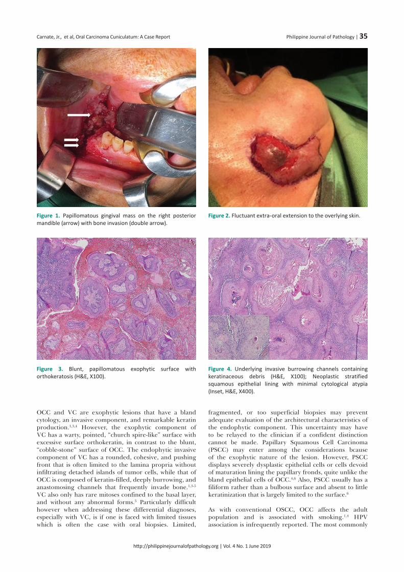
OCC and VC are exophytic lesions that have a bland cytology, an invasive component, and remarkable keratin production.1,3,4 However, the exophytic component of VC has a warty, pointed, “church spire-like” surface with excessive surface orthokeratin, in contrast to the blunt, “cobble-stone” surface of OCC. The endophytic invasive component of VC has a rounded, cohesive, and pushing front that is often limited to the lamina propria without infiltrating detached islands of tumor cells, while that of OCC is composed of keratin-filled, deeply burrowing, and anastomosing channels that frequently invade bone.1,3-5 VC also only has rare mitoses confined to the basal layer, and without any abnormal forms.5 Particularly difficult however when addressing these differential diagnoses, especially with VC, is if one is faced with limited tissues which is often the case with oral biopsies. Limited,
fragmented, or too superficial biopsies may prevent adequate evaluation of the architectural characteristics of the endophytic component. This uncertainty may have to be relayed to the clinician if a confident distinction cannot be made. Papillary Squamous Cell Carcinoma (PSCC) may enter among the considerations bcause of the exophytic nature of the lesion. However, PSCC displays severely dysplastic epithelial cells or cells devoid of maturation lining the papillary fronds, quite unlike the bland epithelial cells of OCC.4,6 Also, PSCC usually has a filiform rather than a bulbous surface and absent to little keratinization that is largely limited to the surface.6
As with conventional OSCC, OCC affects the adult population and is associated with smoking.1,4 HPV association is infrequently reported. The most commonly
Figure 1. Papillomatous gingival mass on the right posterior mandible (arrow) with bone invasion (double arrow).
Figure 3. Blunt, papillomatous exophytic surface with orthokeratosis (H&E, X100).
Figure 2. Fluctuant extra-oral extension to the overlying skin.
Figure 4. Underlying invasive burrowing channels containing keratinaceous debris (H&E, X100); Neoplastic stratified squamous epithelial lining with minimal cytological atypia (Inset, H&E, X400).
http://philippinejournalofpathology.org | Vol. 4 No. 1 June 2019
Carnate, Jr., et al, Oral Carcinoma Cuniculatum: A Case Report Philippine Journal of Pathology | 35

reported site of involvement of OCC is the mandibular gingiva, as with our patient. Prognosis is suggested to be worse than that of VC but better than that of conventional OSCC.3 Although metastasis is rarely reported, OCC is locally aggressive and infiltrative.3,4 Our patient manifested with bone invasion and draining cutaneous communication. Two months after surgery, the patient had clinical findings indicative of a recurrence despite clear surgical margins.
CONCLUSION
Because of the unusual growth pattern and bland cytology, the unacquainted pathologist may find it disconcerting to render a diagnosis of malignancy in OCC, especially if faced with limited tissues. Clinical and ancillary parameters, familiarity with the entity, and histologic clues of locally aggressive behavior, will certainly aid in the correct diagnosis.
ETHICAL CONSIDERATION
Patient consent was obtained before submission of the manuscript.
STATEMENT OF AUTHORSHIP
All authors certified fulfillment of ICMJE authorship criteria.
AUTHOR DISCLOSURE
The authors declared no conflict of interest.
FUNDING SOURCE
None.
REFERENCES
1. Sloan P, Gale N, Hunter K, et al. Malignant surface epithelial tumours: squamous cell carcinoma. In: El-Naggar AK, Chan JKC, Grandis JR, Takata T, Slootweg PJ, eds. WHO classification of tumours of the head and neck. 4th ed. Lyon: IARC Press; 2017.
2. Raja RS, Weinreb I, Bullock MJ, et al. College of American Pathologists Protocol for the examination of specimens from patients with cancers of the lip and oral cavity Version LipOralCavity 4.0.0.1; 2017. Available from: https://cap.objects.frb.io/protocols/cp-headandneck-lip-oralcavity-17protocol-4001.pdf. Accessed May 9, 2019.
3. Datar UV, Kale A, Mane D. Oral carcinoma cuniculatum: a new entity in the clinicopathological spectrum of oral squamous cell carcinoma. J Clin Diagn Res. 2017;11(1): ZD37-9. PMID: 28274074. PMCID: PMC5324519. https://doi/org/10.7860/JCDR/2017/23437.9226.
4. Farag AF, Abou-Alnour DA, Abu-Taleb NS. Oral carcinoma cuniculatum, an unacquainted variant of oral squamous cell carcinoma: a systematic review. Imaging Sci Dent. 2018;48(4):233-44. PMID: 30607347. PMCID: PMC6305781. https://doi.org/10.5624/isd.2018.48.4.233.
5. Zidar N, Cardesa A, Gillison M, Hekkiwekk T, Hille J, Nadal A. Verrucous carcinoma. In: El-Naggar AK, Chan JKC, Grandis JR, Takata T, Slootweg PJ eds. WHO classification of head and neck tumorus. 4th ed. Lyon: IARC Press; 2017.
6. El-Mofty SK, Cardesa A, Helliwell T, et al. Papillary squamous cell carcinoma. In: El-Naggar AK, Chan JKC, Grandis JR, Takata T, Slootweg PJ, eds. WHO classification of head and neck tumours. 4th ed. Lyon: IARC Press; 2017.
Disclaimer: This journal is OPEN ACCESS, providing immediate access to its content on the principle that making research freely available to the public supports a greater global exchange of knowledge. As a requirement for submission to the PJP, all authors have accomplished an AUTHOR FORM, which declares that the ICMJE criteria for authorship have been met by each author listed, that the article represents original material, has not been published, accepted for publication in other journals, or concurrently submitted to other journals, and that all funding and conflicts of interest have been declared. Consent forms have been secured for the publication of information about patients or cases; otherwise, authors have declared that all means have been exhausted for securing consent.
http://philippinejournalofpathology.org | Vol. 4 No. 1 June 2019
Carnate, Jr., et al, Oral Carcinoma Cuniculatum: A Case Report Philippine Journal of Pathology | 36

Autopsy Findings in a Patient with Post-Obstructive Pulmonary EdemaMarissa Krizelda Santos,1 Ann Margaret Chang,1 Jimmy Chang2
1Institute of Pathology, St. Luke’s Medical Center, Quezon City, Philippines2Comprehensive Sleep Disorder Center, St. Luke’s Medical Center, Quezon City, Philippinese
ABSTRACT
Post-obstructive pulmonary edema (POPE), a form of non-cardiogenic pulmonary edema, is a significant entity in anesthesiology and head/neck surgery. This rapidly developing and life-threatening condition occurs following the relief of the obstruction in the upper airways. This condition has two main categories with distinct etiology. We report the case of a 62-year-old Filipino female who developed POPE after the removal of the endotracheal tube following a routine biopsy of her maxillary mass. Immediately after the removal of the endotracheal tube, she presented with episodes of hypotension and desaturation. Chest x-ray post-re-intubation revealed bilateral lung opacities. The autopsy findings of the respiratory and cardiovascular system are presented.
Key words: sleep apnea, obstructive, pulmonary edema, autopsy
INTRODUCTION
Post-obstructive pulmonary edema (POPE) is a rare condition that in most cases, is associated with complications of general anesthesia. It is categorized into Type I and Type II.1 In the Type I setting, post-obstructive pulmonary edema occurs once there is sudden and severe episode of upper airway obstruction, while in Type II, pulmonary edema sets in upon the relief of a chronic obstruction. Post-anesthetic laryngospasm is the most frequent cause of POPE in adults.2 It is possible that in our case, both types of POPE were exhibited. The laryngospasm probably developed as an effect of the general anesthesia and the relief of the obstruction during the reintubation represent Type I and Type II POPE, respectively.
CASE
A 62-year-old Filipino female, known diabetic and non-hypertensive, was admitted for routine biopsy of a maxillary mass. She was presenting with progressive nasal congestion and intermittent shortness of breath over the last year. Paranasal sinus Computed Tomography (CT) scan revealed a large enhancing irregular soft tissue mass (9.7x7.8x5.9 cm) centered in the left maxillary region with intranasal extension (Figure 1).
History of loud snoring was also elicited in the patient’s medical history but was not thoroughly explored. Consequently, no sleep study was performed prior to the operation.
Pre-operative work-up such as electrocardiogram, chest x-ray and hematological studies were normal. The intraoperative course was unremarkable until the time of extubation, when the patient again was noted with loud snoring and with persistent oxygen desaturation as low as 14-40% for 3 to 5 minutes followed by hypotensive
ISSN 2507-8364 (Online)Printed in the Philippines.Copyright© 2019 by the PJP.Received: 6 February 2019.Accepted: 21 March 2019.Published online first: 25 March 2019.https://doi.org/10.21141/PJP.2019.07 Corresponding author: Ann Margaret V. Chang, MDE-mail: [email protected]: https://orcid.org/0000-0002-9036-3678
http://philippinejournalofpathology.org | Vol. 4 No. 1 June 2019
OPEN ACCESS – AUTOPSY VAULT

episodes (blood pressure of 80/40 mmHg). She was reintubated and was given inotropes. She was transferred to the intensive care unit and was managed accordingly. The chest x-ray post-intubation revealed bilateral lung opacities attributed to air-space opacification with no appreciable pneumothorax (Figure 2).
Serum electrolytes and blood urea nitrogen were relatively within normal range, while creatinine and white blood cell counts were elevated (Table 1). Despite continuous medical management, she expired after the 2nd post-operative day and a post-mortem examination was performed.
AUTOPSY FINDINGS
A partial autopsy limited to the thoraco-abdominal organs was performed with the consent of the relatives of the decedent and the findings recorded within the institutions’ ethical proceedings for documentation and academic purposes.
The decedent was normocephalic, short-necked with endomorphic body habitus. There was a soft to rubbery ill-defined mass (6 cm in single widest dimension) located in the inferior auricular to lateral neck area diagnosed through biopsy as clear-cell tumor with
Figure 1. Paranasal Sinus CT Scan after contrast. (A) PNS Axial plane. (B) FESS Axial plane. A large enhancing irregular soft tissue mass lesion (9.7x7.8x5.9 cm) is seen in the left maxillary region extending into the left nasal cavity, left masticator space, left buccal mucosa, both sides of the hard palate, left soft palate, both sides of the nasopharyngeal roof, clivus, both sphenoid sinuses and left posterior ethmoid sinus.
Figure 2. Chest X-Ray. (A) Pre-operative exam showing normal lung parenchyma, normal cardiac silhouette and elongated aorta. (B) Post-operative exam showing bilateral air-space filling opacities.
A
A
B
B
http://philippinejournalofpathology.org | Vol. 4 No. 1 June 2019
Santos et al, Autopsy Findings in a Patient with Post-Obstructive Pulmonary Edema Philippine Journal of Pathology | 38

recommendation for immunohistochemical staining for proper histologic classification.
Examination of the head and neck area showed partial left nasal cavity obstruction. No similar obstruction or deformity is noted in the glottis or larynx. The hyoid bone and laryngeal cartilage are normally formed and intact without evidence of fractures or hemorrhage.
A standard Y-shaped thoraco-abdominal incision was done to reveal areas of visceral pleural adhesions in the right lung. Though the weight of the lungs were within normal range (right 750 g; mean reference range, MRR: 185-967 g; left-700 g; MRR: 186-885 grams), frothy material was noted to extrude copiously from both sides (Figure 3A).
Microsections from the bilateral lung lobes showed alveoli filled with red blood cells and proteinaceous material consistent with pulmonary congestion (Figure 3B). There was neither mass on gross examination nor microscopic evidence of carcinomatosis. The major pulmonary vessels and airways were likewise patent and free of any blood clot or emboli.
The heart was normal in size and weight (280 grams). Gross examination of the heart showed no evidence of lesion or old scar (Figure 4). Microsections showed diffuse interstitial fibrosis transecting the myocardial fibers with only focal mild lymphocytic infiltrates. No evidence of infarction was appreciated (Figure 5). The major coronary vessels were patent, thin-walled and devoid of calcifications.
DISCUSSION
Although the Troponin I was elevated, the postmortem examination of our case revealed that the heart was grossly unremarkable. Microscopically, no evidence of myocardial infarction such as myocardial fiber waviness, geographic necrosis nor neutrophilic infiltrates were appreciated. The absence of the microscopic findings of the typical myocardial infarction rules this out as the most likely cause of demise. Instead, diffuse interstitial fibrosis in the absence of cardiac hypertrophy and dilatation are seen transecting the myocardial fibers with only focal, mild lymphocytic infiltrates. Though myocarditis could have resulted in the heart failure as evidenced by the severe pulmonary congestion and edema, cardiomegaly and dilatation of the chambers were not appreciated. Indubitably, cardiac markers remain the cornerstone for the diagnosis of acute coronary syndromes (ACS), although troponin elevation has also been appreciated in non-ACS conditions. A subset of which are seen in patients with chronic airway obstruction and its concomitant negative intrathoracic pressure effect.3 These findings, together with the ventricular tachycardia with limb lead low voltage complexes on the electrocardiogram should elicit a more thorough search for other factors of death causality.
The elevated WBC in our patient in the absence of infection and hematologic disorder are believed to be a secondary reaction to acute lung injury4 with neutrophils being elevated in obstructive sleep apnea.5
Table 1. Laboratory results of patientAnalyte Result RR Analyte Result RRSodium 147 136-145 mEq/L Hemoglobin 14.7 11.6-15.5 g/dlPotassium 3.4 3.5-5.1 mEq/L Hematocrit 46.2 36-47%Magnesium 2.0 1.8-2.4 mg/dl RBC 4.59 4.4-5.4x10³/mm3
Ionized Ca+ 1.06 1.09-1.30 mg/dl WBC 34980 4800-10800 mm3Creatinine 1.08 0.55-1.02 mg/dl Neutrophil 82% 40-74%Urea 11 7-18 mg/dl Bands 5% 2-6%Troponin I 101.9 0-0.30 ng/ml Lymphocyte 5% 19-48%INR 1.76 0.9-1.19 Monocyte 8% 3-9%PT 46.9 29.5-39.9 secs Platelet 271000 150-400x103/mm3
Figure 3. (A) Both lungs were red and appeared heavy grossly. (B) Photomicrograph of lung showing alveoli filled with smooth to slightly floccular pink material and capillaries in the alveolar walls congested with many red blood cells (H&E, 100X).
A B
http://philippinejournalofpathology.org | Vol. 4 No. 1 June 2019
Santos et al, Autopsy Findings in a Patient with Post-Obstructive Pulmonary Edema Philippine Journal of Pathology | 39

A B
A
C
C
B
D
Figure 4. Gross anatomy of heart. (A) Anterior view of the heart. (B) Posterior view of the heart. (C) Cross sections of the heart showed absence of gross lesions and scars.
Figure 5. Microsections from the right and left ventricles of the heart. (A) Microsections from the left ventricle revealed diffuse interstitial fibrosis transecting the myocardial fibers. (B) Microsections from the left ventricle showed focal inflammatory cells which are comprised of lymphocytes. Individual myocardial cell necrosis is also noted. No myocardial fiber waviness and geographic necrosis are appreciated. (C) Microsections from the right ventricle also revealed diffuse interstitial fibrosis. (D) The presence of mild lymphocytic infiltrates in the left ventricle is appreciated.
http://philippinejournalofpathology.org | Vol. 4 No. 1 June 2019
Santos et al, Autopsy Findings in a Patient with Post-Obstructive Pulmonary Edema Philippine Journal of Pathology | 40

The significant gross and microscopic findings in this case are bilateral pulmonary congestion and edema (Figure 3). Based on the CT scan, the patient was noted to have a mass in the left maxillary region which also extends into the left nasal cavity, left masticator space, left buccal mucosa, both sides of the hard palate, soft palate more towards the left side, both sides of the nasopharyngeal roof, clivus, both sphenoid sinuses and left posterior ethmoid sinus. Hence, there was already obstruction in the upper airways. The timeline of the events during the surgical procedure, normal cardiac evaluation, the presence of bilateral pulmonary edema in the absence of any cardiac pathology, favor the diagnosis of post-obstructive pulmonary edema in our case. There are two forms of post-obstructive pulmonary edema. Type I follows a sudden severe episode of upper airway obstruction while Type II develops after surgical relief of chronic upper airway obstruction. In our case, both forms may be present. For the Type I, there is the possibility of post-extubation as well as general anesthesia complications. Based on Ahsayan et al.,6 post-obstructive pulmonary edema occurs in 0.1% of all patients undergoing general anesthesia. The patient was noted to have episodes of desaturation after the extubation and this can be attributed to several causes such as the following: laryngospasm, endotracheal tube biting, tongue falling back and extubation before the patient had been sufficiently awakened.7,1 On the other hand, the relief of the upper airway obstruction contributes to the development of Type II post-obstructive pulmonary edema. Of note, while patient was still under the effects of anesthesia, she was observed to be snoring loudly. It is possible that aside from the neoplastic obstruction of the upper airway, the patient most likely has undiagnosed obstructive sleep apnea and once she underwent general endotracheal anesthesia, there was transient relief of the upper airway obstruction.
The pathophysiology of post-obstructive pulmonary edema is multifactorial. Initially there is a decrease in interstitial pressure favoring transudation. Alveolar membrane injury can occur sequentially which may consequently result in more transudation. Physiological positive end-expiratory pressure (PEEP) from the obstructed airway counterbalances this positive intravascular pressure, but when relieved of the obstruction, the lack of PEEP permits the transudation of fluid into the alveolar spaces resulting in pulmonary edema. Microscopically, we can appreciate engorgement of the alveolar capillaries as well as the presence of finely granular pale pink material representing the intra-alveolar transudate. The hypoxia from the airway obstruction exacerbates the physiology of post-obstructive pulmonary edema. The hypoxia results in a systemic adrenergic release which then leads to systemic vasoconstriction further increasing venous return. In the lungs, this vasoconstriction further elevates the intravascular pressure thereby encouraging the transudative process.8 In chronic airway obstruction, the repetitive chronic negative pulmonary intravascular pressures are counterbalanced by reactive pulmonary vasoconstriction and an increase in pulmonary artery pressure. Eventually, pulmonary hypertension and systemic hypertension develop. Once this occurs, the right-sided heart failure can ensue. In obstructive sleep apnea, the heart also dilates acutely during negative intrathoracic
pressures and systemic blood pressure escalates. The snoring history of the patient and Asian anatomy present an appropriate background for obstructive sleep apnea to confound the already compromised airway. This is why patients with obstructive sleep apnea are at risk for developing POPE after tracheotomy.9
CONCLUSION
In a patient with signs of compromised airway, thorough evaluation of compounding factors prior to any procedure should always be performed. Awareness of entities such as post-obstructive pulmonary edema and obstructive sleep apnea may aid in uncovering causes of death. But more importantly, provide an avenue for more stringent pre-anesthetic evaluation to prevent untoward morbidities and mortalities even in seemingly straightforward routine surgical procedures.
ACKNOWLEDGEMENT
The authors acknowledge the invaluable contribution of Dr. Francisco V. Narciso for his heart dissection.
STATEMENT OF AUTHORSHIP
All authors certified fulfillment of ICMJE authorship criteria. All authors have equally contributed to this work, proofread and approved the manuscript for publication.
AUTHOR DISCLOSURE
The authors declared no conflict of interest.
FUNDING SOURCE
None.
REFERENCES
1. Mehta KK, Ahmad SQ, Shah V, Lee H. Postobstructive pulmonary edema after biopsy of a nasopharyngeal mass. Respir Med Case Rep. 2015; 16:166-8.
2. Herrick IA, Mahendran B, Penny FJ. Postobstructive pulmonary edema following anesthesia. J Clin Anesth. 1990;2(2):116-20. PMID: 2189449.
3. Tanindi A, Cemri M. Troponin elevation in conditions other than acute coronary syndromes. Vasc Health Risk Manag. 2011;597-603. PMID: 22102783. PMCID: PMC3212425. https://doi.org/10.2147/VHRM.S24509.
4. Butt Y, Kurdowska A, Allen TC. Acute lung injury: a clinical and molecular review. Arch Pathol Lab Med. 2016;140(4):345-50. PMID: 27028393. https://doi.org/10.5858/arpa.2015-0519-RA.
5. Geovanini GR, Wang R, Weng J, et al. Elevations in neutrophils with obstructive sleep apnea: the multi-ethnic study of atherosclerosis (MESA). 2018;257:318-23. PMID: 29506719. PMCID: PMC5842816 [Available on 2019-04-15]. https://doi.org/10.1016/j.ijcard.2017.10.121.
6. Ahsayan K, Ghalem A, Ouafi NE, Housni B. Post obstructive pulmonary edema: a rare and preventable etiology of postoperative respiratory distress. J Cardiol
http://philippinejournalofpathology.org | Vol. 4 No. 1 June 2019
Santos et al, Autopsy Findings in a Patient with Post-Obstructive Pulmonary Edema Philippine Journal of Pathology | 41

Cardiovasc Ther. 2017;6(5):555696. https://doi.org/10.19080/JOCCT.2017.06.555696.
7. Granados-Romero J, Valderrama-Treviño AI, Arauz-Peña G, et al. Acute pulmonary non-cardiogenic edema after extubation with laryngospasm: a case report. Int J Res Med Sci. 2018; 6(7):2528-32. https://doi.org/10.18203/2320-6012.ijr,s20182850.
8. Lemyze M, Mallat J. Understanding negative pressure pulmonary edema. Intensive Care Med. 2014;40(8):1140-3. PMID: 24797685. PMCID: PMC4148265. https://doi.org/10.1007/s00134-014-3307-7.
9. Burke AJ, Duke SG, Clyne S, Khoury SA, Chiles C, Mathews BL. Incidence of pulmonary edema after tracheotomy for obstructive sleep apnea. Otolaryngol Head Neck Surg. 2001;125(4):319-23. PMID: 11593165. https://doi.org/10.1067/mhn.2001.117713.
Publish in the new PJP.Visit our website:
http://philippinejournalofpathology.org
Disclaimer: This journal is OPEN ACCESS, providing immediate access to its content on the principle that making research freely available to the public supports a greater global exchange of knowledge. As a requirement for submission to the PJP, all authors have accomplished an AUTHOR FORM, which declares that the ICMJE criteria for authorship have been met by each author listed, that the article represents original material, has not been published, accepted for publication in other journals, or concurrently submitted to other journals, and that all funding and conflicts of interest have been declared. Consent forms have been secured for the publication of information about patients or cases; otherwise, authors have declared that all means have been exhausted for securing consent.
http://philippinejournalofpathology.org | Vol. 4 No. 1 June 2019
Santos et al, Autopsy Findings in a Patient with Post-Obstructive Pulmonary Edema Philippine Journal of Pathology | 42

Wire-Free Virtual Breast Localization Using Liquid Carbon NanoparticlesMa. Theresa Buenaflor, Ricardo Victorio Quimbo, Norman Val San Agustin
Asian Breast Center, Centuria Medical Makati, Metro Manila, Philippines
ABSTRACT
The emergence of improved multi-modal diagnostics including functional imaging has enabled the diagnosis of more nonpalpable breast lesions. Lesions diagnosed as early unifocal breast cancers are amenable to breast-conserving surgery (BCS). The precise localization of these lesions is a caveat to its complete removal along with sufficient surgical margins and the preservation of normal breast tissues. Carbon marking is an alternative to needle wire localization that is easy to perform and simplifies the workflow of the multidisciplinary team involved in breast cancer care.
Key words: liquid carbon nanoparticles, non-wire breast localization, carbon nanoparticles suspension injection (CNSI), molecular breast imaging (MBI)
INTRODUCTION
Carbon is a naturally occurring element known since ancient times. Antoine-Laurent Lavoisier, a French chemist, proposed carbon in 1789 from the Latin carbo meaning “charcoal”.1 Carbon, likewise, is a pigment used to produce black ink, as when used in tattoos, leaving an indelible mark because it is biologically inert.2 When a tattoo needle punctures the skin, a tiny wound is produced. The body responds to injury by signaling macrophages to address the injury and engulf or phagocytose the foreign body. In tattoo ink, the pigment particles are too large to be destroyed, hence, remain fixed or permanent.3
The same principle applies to using carbon as an alternative wire-free method to mark lesions in the breast. It is an FDA-approved substance that provides direct visual aid to the surgeon and pathologist. Grossly and microscopically, it points to the precise location of the lesion of interest.
Svane was the first one to utilize carbon particles suspended in an aqueous solution to preoperatively mark 56 non-palpable breast lesions. The stability of carbon over time remains one of its strengths as well as its characteristic to remain in the area where it is injected without dispersing into the surrounding tissues because it is water-insoluble.4 This is in contradistinction to vital dyes such as toluidine blue, methylene blue, green isocyanate and India ink.5 Although vital dyes are low cost, they need to be injected in the immediate preoperative period because they diffuse easily and present an impediment in the proper identification of the area to be removed.6
Xie et al., studied the bioaccumulation of carbon nanoparticle suspension injection (CNSI) among mice after intratumoral injection and found that its toxicity is low and confirmed its biosafety when it entered blood circulation.7 In a study by Jiang et al., using carbon marking for preoperative marking of 16 cases, no allergic reaction was observed. The particle diameter size range
ISSN 2507-8364 (Online)Printed in the Philippines.Copyright© 2019 by the PJP.Received: March 30, 2019.Accepted: April 29, 2019.Published online first: 13 May 2019.https://doi.org/10.21141/PJP.2019.08 Corresponding author: Ma. Theresa S. Buenaflor, MD, FPCRE-mail: [email protected]: https://orcid.org/0000-0001-7257-1265
http://philippinejournalofpathology.org | Vol. 4 No. 1 June 2019
OPEN ACCESS – DIAGNOSTIC PERSPECTIVES

is 150-200 nm,8,9 diluted in a saline solution and injected into the area of concern. The carbon marking can be done 10-14 days8 before the operation and thus limit scheduling conflict.
As a method of preoperative marking, carbon localization can improve the success rate of breast-conserving surgery. Rose et al., did a head to head comparison of 219 carbon localizations and 292 hookwire localizations. Their study showed the rate of complete excision of nonpalpable lesions – with adequate surgical margins in carbon marking at 81.1% compared with wire-guided localization which is 70.8%.10 In another study by Cavalcanti et al., 135 surgical specimens that were carbon marked showed that in all cases containing detectable lesions (98.52%), no impairment to histologic analysis was shown and the final histopathologic diagnosis was straightforward.6
Local experienceAt our institution, carbon marking was used in 2 cases. The first case (Figures 1 and 2) involved marking the abnormal axillary lymph node prior to sentinel lymph node biopsy. The liquid carbon was injected directly into
the substance of the abnormal lymph node using a Gauge 23 needle. This was to maximize the identification of the lymph node on frozen section.
The second patient (Figure 3 and 4) had carbon marking at the medial margin of the tumor to facilitate breast-conserving surgery after a 50% reduction tumor size post-neoadjuvant chemotherapy. In this case, the needle was positioned perpendicular to the probe because the direction was straight down, marking the shortest distance for the surgeon from the skin down to the lesion. Upon slow withdrawal of the needle going up, slow infiltration was done to create a track for the surgeon to follow.
The advantages of carbon marking are its ease of use and effectivity in facilitating the complete excision of nonpalpable lesions. It can withstand histological analysis and presents no diagnostic difficulty to the pathologist.6
Carbon marking is a valuable and accurate alternative to wire localization and has the added value of improving service delivery.
Figure 1. (A) Digital mammogram of a 65-year-old female with a palpable area at the upper outer right breast with nipple retraction and skin thickening showing heterogeneously dense breasts showing a large non-mass area of increased density with associated pleomorphic microcalcifications (marked with arrows). There are at least two hyperdense ipsilateral lymph nodes seen. (B) Image of Molecular Breast Imaging (MBI), a functional imaging modality using a radiopharmaceutical, 99m-Technetium sestamibi. It shows the large area of concern with marked tracer uptake (appears as black with tumor/background ratio of 11.6). The ipsilateral axilla likewise shows focus of tracer uptake with tumor/background ratio of 2.9 corresponding to the abnormal lymph node. (C) Correlate ultrasound image of the axilla showing the abnormal lymph node prior to and during carbon marking.
A B
C
http://philippinejournalofpathology.org | Vol. 4 No. 1 June 2019
Buenaflor et al, Wire-Free Virtual Breast Localization Using Liquid Carbon Nanoparticles Philippine Journal of Pathology | 44

FUNDING SOURCE
None.
REFERENCES
1. http: / / sc ience.marshal l .edu/castel la /chm448/elements2.pdf [Internet]. Accessed April 29, 2019 .
2. Sperry K. Tattoos and tattooing. Part I: history and methodology. Am J Forensic Med Pathol. 1991;12(4);313-9. PMID: 1807141.
3. Wood-Black F. Going skin deep: the culture and chemistry of tattoos. Inchemistry. https://inchemistry.acs.org/content/inchemistry/en/atomic-news/tattoo-ink.html. Accessed March 18, 2019.
4. Svane G. A stereotaxic technique for preoperative marking of non-palpable breast lesions. Acta Radiol Diagn (Stockh). 1983;24(2):145-51. PMID: 6353873.
5. Patrikeos A, Wylie EJ, Bourke A, Frost F. Imaging of carbon granulomas of the breast following carbon track localization. Clin Radiol. 1998;53(11):845-8. PMID:9833790.
CONCLUSION
Preoperative marking is both a science and an art. It involves meticulous planning and precise identification of lesions. Our local experience suggests that carbon marking is a viable alternative and easily adapted method of localizing impalpable breast lesions. This technique can streamline the work of the radiologist, surgeon and pathologist.
ETHICAL CONSIDERATION
Patient consent was obtained before submission of the manuscript.
STATEMENT OF AUTHORSHIP
All authors certified fulfillment of ICMJE authorship criteria.
AUTHOR DISCLOSURE
The authors declared no conflict of interest.
Figure 2. (A) Low power view of the abnormal lymph node with a 2 mm focus of micrometastasis showing black specks (red box) at the 12 o’clock position corresponding to the dye (Hematoxylin-Eosin, 10x). (B) High power view (Hematoxylin-Eosin, 40x). (C, D) Carbon nanoparticles are noted without obscuration of the cellular details (Hematoxylin-Eosin, 10x & 40x).
A
C
B
D
http://philippinejournalofpathology.org | Vol. 4 No. 1 June 2019
Buenaflor et al, Wire-Free Virtual Breast Localization Using Liquid Carbon Nanoparticles Philippine Journal of Pathology | 45
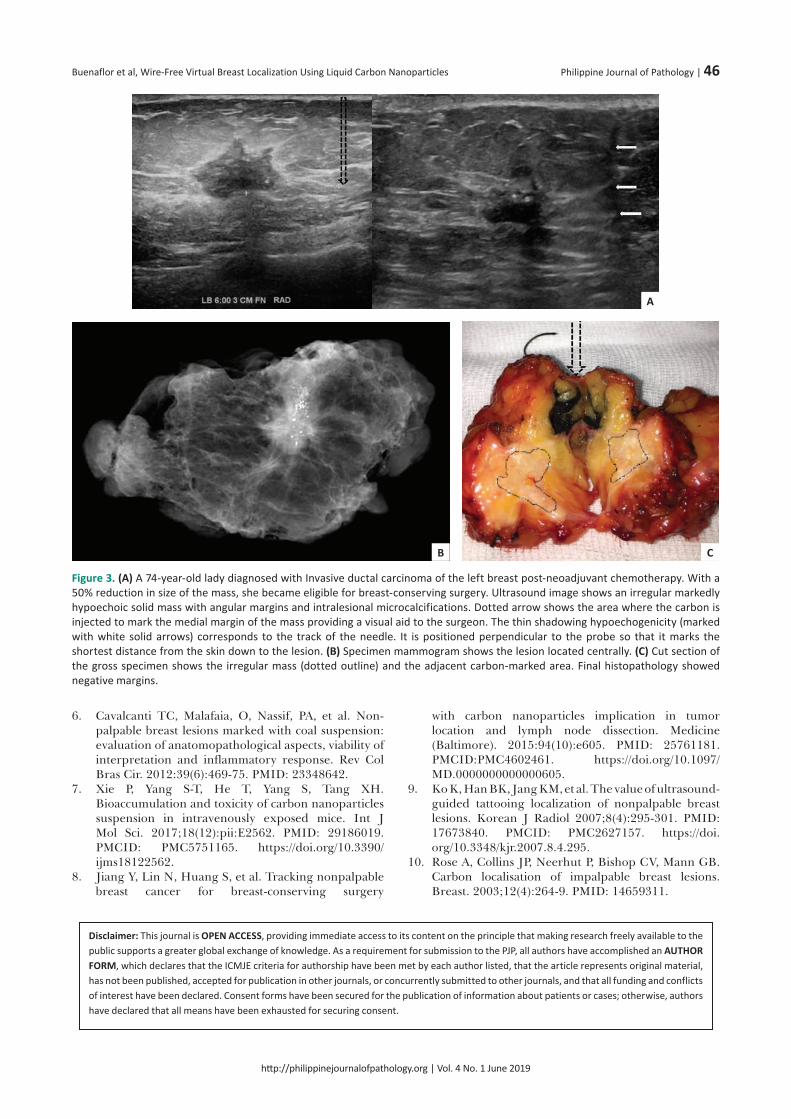
Disclaimer: This journal is OPEN ACCESS, providing immediate access to its content on the principle that making research freely available to the public supports a greater global exchange of knowledge. As a requirement for submission to the PJP, all authors have accomplished an AUTHOR FORM, which declares that the ICMJE criteria for authorship have been met by each author listed, that the article represents original material, has not been published, accepted for publication in other journals, or concurrently submitted to other journals, and that all funding and conflicts of interest have been declared. Consent forms have been secured for the publication of information about patients or cases; otherwise, authors have declared that all means have been exhausted for securing consent.
6. Cavalcanti TC, Malafaia, O, Nassif, PA, et al. Non-palpable breast lesions marked with coal suspension: evaluation of anatomopathological aspects, viability of interpretation and inflammatory response. Rev Col Bras Cir. 2012:39(6):469-75. PMID: 23348642.
7. Xie P, Yang S-T, He T, Yang S, Tang XH. Bioaccumulation and toxicity of carbon nanoparticles suspension in intravenously exposed mice. Int J Mol Sci. 2017;18(12):pii:E2562. PMID: 29186019. PMCID: PMC5751165. https://doi.org/10.3390/ijms18122562.
8. Jiang Y, Lin N, Huang S, et al. Tracking nonpalpable breast cancer for breast-conserving surgery
with carbon nanoparticles implication in tumor location and lymph node dissection. Medicine (Baltimore). 2015:94(10):e605. PMID: 25761181. PMCID:PMC4602461. https://doi.org/10.1097/MD.0000000000000605.
9. Ko K, Han BK, Jang KM, et al. The value of ultrasound-guided tattooing localization of nonpalpable breast lesions. Korean J Radiol 2007;8(4):295-301. PMID: 17673840. PMCID: PMC2627157. https://doi.org/10.3348/kjr.2007.8.4.295.
10. Rose A, Collins JP, Neerhut P, Bishop CV, Mann GB. Carbon localisation of impalpable breast lesions. Breast. 2003;12(4):264-9. PMID: 14659311.
Figure 3. (A) A 74-year-old lady diagnosed with Invasive ductal carcinoma of the left breast post-neoadjuvant chemotherapy. With a 50% reduction in size of the mass, she became eligible for breast-conserving surgery. Ultrasound image shows an irregular markedly hypoechoic solid mass with angular margins and intralesional microcalcifications. Dotted arrow shows the area where the carbon is injected to mark the medial margin of the mass providing a visual aid to the surgeon. The thin shadowing hypoechogenicity (marked with white solid arrows) corresponds to the track of the needle. It is positioned perpendicular to the probe so that it marks the shortest distance from the skin down to the lesion. (B) Specimen mammogram shows the lesion located centrally. (C) Cut section of the gross specimen shows the irregular mass (dotted outline) and the adjacent carbon-marked area. Final histopathology showed negative margins.
A
CB
http://philippinejournalofpathology.org | Vol. 4 No. 1 June 2019
Buenaflor et al, Wire-Free Virtual Breast Localization Using Liquid Carbon Nanoparticles Philippine Journal of Pathology | 46

http://philippinejournalofpathology.org | Vol. 4 No. 1 June 2019
Instructions to Authors
The Philippine Journal of Pathology (PJP) is an open-access, peer-reviewed, English language, medical and healthscience journal that is published continuously online and semi-annually in print by the Philippine Society of Pathologists,Inc. (PSP, Inc). All manuscripts must be submitted through the PJP Official Website (Open Journal Systems)( ). All other correspondences and other editorial matters should be sent viaelectronic mail to .
Articles and any other material published in the PJP represent the work of the author(s) and do not reflect the opinions ofthe Editors or the Publisher. Articles that do not subscribe to the Instructions to Authors shall be promptly returned.
ARTICLE SECTIONSThe PJP welcomes manuscripts on all aspects ofpathology and laboratory medicine, to includecytology, histopathology, autopsy, forensic pathology,clinical chemistry, clinical microscopy, medicalmicrobiology, parasitology, immunology, hematology,blood banking, medical technology, laboratorydiagnostics, laboratory biosafety and biosecurity,laboratory management, and quality assurance.
The PJP accepts original articles, review articles, casereports, feature articles, brief communications, autopsycases, editorials, or letters to the Editor.
Original articlesThe research must have received institutional review board approval that is explicitly stated in the methodology. The abstract should contain no more than 200 words with a structured format consisting of the objective/s, methodology, results and conclusion. A manuscript for original articles should not exceed 25 typewritten pages (including tables, figures, illustrations and maximum of 30 references) or 6000 words.ReviewsReview articles, both solicited and unsolicited, provide information on the “state of the art.” PJP reviews not only summarize current understanding of a particular topic but also critically appraise relevant literature and data sources, describe significant gaps in the research, and future directions. The abstract should be from 50 to 75 words and should not be structured. A manuscript for reviews should not exceed 15 typewritten pages (including tables, figures, illustrations and maximum of 50 references) or 4000 words.Case ReportsThis type of article pertains to single or multiple reports of well-characterized cases that are highly unusual, novel, or rare; or with a unique or variant presentation, evolution or course; or that represent an unexpected or uncommon association of two or more diseases or disorders that may represent a previously unsuspected causal relationship; or that are underreported in the literature. The abstract should be from 50 to 75 words and should not be structured. A manuscript for case reports should not exceed 10 typewritten pages (including tables, figures, illustrations and maximum of 15 references) or 3000 words.Feature articlesThe PJP may feature articles, either as part of an issue themeor a special topic on pathology by a local or international expert or authority. The abstract should be from 50 to 75 words and should not be structured. A manuscript for feature articles should not exceed 25 typewritten pages (including tables, figures, illustrations and maximum of 30 references) or 6000 words.Autopsy VaultThe PJP highly welcomes articles on autopsy protocols of cases. The article must include a summary presentation of the history, evaluation and work-up, clinical course of a case, followed by the autopsy procedure performed, gross and
microscopic findings, discussion, learning points and conclusion. The PJP recognizes the instructional and educational value of articles under this section. The abstract should be from 50 to 75 words and should not be structured. A manuscript for the Autopsy Vault should not exceed 25 typewritten pages (including tables, figures, illustrations and maximum of 30 references) or 6000 words.Images in PathologyImages of unique, interesting, or highly educational cases encountered in hematology, cytology, histopathology, or medical microbiology, may be submitted under this section, and may include photomicrographs, gross pictures, machine read-outs, among others. A brief history, the photograph(s) and short discussion of the case. No abstract is required. A manuscript for Images in Pathology should not exceed 500 words, with maximum of 10 references. This is distinct from the Case Report which is a full write up. Brief CommunicationsBrief Communications are short reports intended to either extend or expound on previously published research orpresent new and significant findings which may have a major impact in current practice. If the former, authors must acknowledge and cite the research which they are building upon. The abstract should be from 50 to 75 words and should not be structured. A manuscript for brief communications should not exceed 5 typewritten pages (including tables, figures, illustrations and maximum of 10 references) or 1500 words.EditorialsRecognized leaders in the field of pathology and laboratory medicine may be invited by the Editor-in-Chief/Editorial Board to present their scientific opinion and views of a particular topic within the context of an issue theme or issues on scholarly publication. No abstract or keywords necessary.Letters to the EditorPJP welcomes feedback and comments on previously published articles in the form of Letters to the Editor. No abstract or keywords are necessary. A Letter to the Editor must not exceed 2 typewritten pages or 500 words.Special AnnouncementsSpecial announcements may include upcoming conventions, seminars or conferences relevant to pathology. The Editors shall deliberate and decide on acceptance and publication of special announcements. Please coordinate with the Editorial Coordinator for any request for special announcements.
COVER LETTERA cover letter must accompany each manuscript citingthe complete title of the manuscript, the list of authors(complete names, position/designation and institutionalaffiliations), with one (1) author clearly designated ascorresponding author, providing his/her completeinstitutional mailing address, institutional telephone/faxnumber, and work e-mail address. The PJP Cover LetterTemplate must be used.
http://philippinejournalofpathology.orgphilippinepathologyjournal@gmail.com

PJP AUTHOR FORMFor submissions to the PJP to be accepted, all authors must read and sign the PJP Author Form consisting of:(1) the Authorship Certification, (2) the Author Declaration, (3) the Statement of Copyright Transfer, and (4) the Statement of Disclosure of Conflicts of Interest. The completely accomplished PJP Author Form shall be scanned and submitted along with the manuscript. No manuscript shall be received without the PJP Author Form.
GENERAL FORMATTING GUIDELINESAuthors must use the standard PJP templates foreach type of manuscript. These templates arealigned with the most current versions of theEQuaToR Network guidelines and checklists( ).The manuscript should be encoded on the templateusing Microsoft Word (2007 version or later version),single-spaced, 2.54 cm margins throughout, on A4size paper. Preferred fonts may include CenturyGothic (template default), Times New Roman, orArial.The manuscript should be arranged in sequence asfollows: (1) Title Page, (2) Abstract, (3) Text, (4)References, (5) Tables, and (6) Figures & Illustrations.All the sheets of the manuscript should be labelledwith the page number (in Hindu-Arabic Numerals)printed on the upper right corner.References should pertain directly to the work beingreported. Within the text, references should beindicated using Hindu-Arabic numerals insuperscripts.
SPECIFIC FORMATTING GUIDELINESTitle and Authors
The title should be as concise as possible.A running title (less than 50 characters) shall also berequired. The running title is the abbreviated versionof the title that will be placed in the header. Therunning title should capture the essence of themanuscript title.The full name of the author(s) directly affiliated withthe work should be included (First name, Middle initialand Last name). The order of authorship shall be theprerogative of the author(s).There are 4 criteria for authorship (ICMJErecommendations). These are captured in the PJPAuthor Form.
Substantial contributions to the conception or design ofthe work; or the acquisition, analysis, or interpretation ofdata for the work; ANDDrafting the work or revising it critically for importantintellectual content; ANDFinal approval of the version to be published; ANDAgreement to be accountable for all aspects of the workin ensuring that questions related to the accuracy orintegrity of any part of the work are appropriatelyinvestigated and resolved.
The highest educational attainment or title of theauthors should be included as an attachmentwhenever appropriate (MD, PhD, et cetera).Name and location of no more than one (1)institutional affiliation per author may be included.If the paper has been presented in a scientific forumor convention, a note should be provided indicatingthe name of the forum or convention, location(country), and date of its presentation.
AbstractFor manuscripts under the “Original Article” section:the abstract should contain no more than 300 wordswith a structured format consisting of the followingstandard headings: objective/s, methodology, resultsand conclusion.For manuscripts under the “Feature Article,” “ReviewArticle,” “Case Report,” “Brief Communications,” and“Autopsy Vault” sections: the abstract should be nomore than 200 words and need not be structured.Letters to the Editor and editorials do not require anabstract.
KeywordsAt least three (3) keywords but no more than six (6),preferably using terms from the Medical SubjectHeadings (MeSH) list of Index Medicus, should be listedhorizontally under the abstract for cross-indexing of thearticle.
TextThe text should be organized consecutively asfollows: Introduction, Methodology, Resultsand Discussion, Conclusion (IMRaD format), followedby Disclosures, Acknowledgments and References.All references, tables, figures and illustrations shouldbe cited in the text, in numerical order.All abbreviations should be spelled out once (the firsttime they are mentioned in the text) followed by theabbreviation enclosed in parentheses. The sameabbreviation may then be used subsequently insteadof the full names.All measurements and weights should be in SystemInternational (SI) units.Under Methodology, information should be providedon institutional review board/ethics committeeapproval or informed consent taking (if appropriate).Acknowledgements to individuals/groups of persons,or institution/s who have contributed to themanuscript but did not qualify as authors based onthe ICMJE criteria, should be included at the end ofthe text just before the references. Grants andsubsidies from government or private institutionsshould also be acknowledged.
ReferencesReferences in the text should be identified by Hindu-Arabic Numerals in superscript on the same line as thepreceding sentence.References should be numbered consecutively in theorder by which they are mentioned in the text. Theyshould not be alphabetized.All references should provide inclusive pagenumbers.Journal abbreviations should conform to those usedin PubMed.A maximum of six authors per article can becited; beyond that, name the first three and add “etal.”The style/punctuation approved by PJP conforms tothat recommended by the International Committeeof Medical Journal Editors (ICMJE) availableat . Examples are shown below:
One to Six AuthorsKrause RM. The origin of plagues: old and new. Science.1992;257:1073-1078.
http://equatornetwork.org
http://www.icmje.org
o
o
oo
http://philippinejournalofpathology.org | Vol. 4 No. 1 June 2019
Instruction to Authors Philippine Journal of Pathology | 48

Mokdad AH, Bowman BA, Ford ES, Vinicor F, Marks JS,Koplan JP. The continuing epidemics of obesity anddiabetes in the US. JAMA. 2001;286(10):1195-1200.More than Six AuthorsRhynes VK, McDonald JC, Gelder FB, et al. Soluble HLA class I in the serum of transplant recipients. Ann Surg. 1993; 217 (5): 485–9.Authors Representing a GroupMoher D, Schulz KF, Altman D; for the CONSORT Group.The CONSORT statement: revised recommendations forimproving the quality of reports of parallel-grouprandomized trials. JAMA. 2001;285(15):1987-1991.BookByrne, DW. Publishing your medical research paper:What they don't teach in medical school. Baltimore:Williams & Wilkins, 1998.World Wide WebBarry JM. The site of origin of the 1918 influenzapandemic and its public health implications.[Commentary]. JTranslational Med. January 20,2004;2(3):1-4. http://www.translational-medicine.com/content/2/1/3. Accessed November18, 2005.
TablesCite all tables consecutively in the text and numberthem accordingly.Create tables preferably using Microsoft Excel withone table per worksheet.Tables should not be saved as image files.The content of tables should include a table number(Hindu-Arabic) and title in capital letters above thetable.Place explanatory notes and legends, as well asdefinitions of abbreviations used below the table. Forlegends, use small letters (i.e., a, b, c, d).Each table must be self-explanatory, being asupplement rather than a duplicate of information inthe text.Up to a maximum of five (5) tables are allowed.
Figures and GraphsFigures or graphs should be identified by Hindu-Arabic Numeral/s with titles and explanationsunderneath.The numbers should correspond to the order in whichthe figures/graphs occur in the text.Figures & graphs should not be saved as image files.For illustrations and photographs, see next section.Provide a title and brief caption for each figure orgraph. Caption should not be longer than 15-20words.All identifying data of the subject/s or patient/s understudy such as name or case numbers, should beremoved.Up to a maximum of five (5) figures and graphs areallowed.
Illustrations and PhotographsWhere appropriate, all illustrations/photographicimages should be at least 800 x 600 dpi andsubmitted as image files (preferably as .png, .jpeg or.gif files).For photomicrographs, the stain used (e.g. H & E) andmagnification (e.g. X400) should be included in thedescription.Computer-generated illustrations which are notsuited for reproduction should be professionallyredrawn or printed on good quality laserprinters. Photocopies are not acceptable.All letterings for illustration should be of adequate sizeto be readable even after size reduction.Place explanatory notes and legends, as well asdefinitions of abbreviations used below theillustration/photograph.Up to a maximum of five (5) illustrations/ photographsare allowed.
N.B.: For tables, figures, graphs, illustrations and photographsthat have been previously published in another journal or book,a note must be placed under the specific item stating that suchhas been adapted or lifted from the original publication.This should also be referenced in the References portion.
EDITORIAL PROCESS (Figure 1)The Editorial Coordinator shall review each submission to check if it has met aforementioned criteria and providefeedback to the author within 24 hours.Once complete submission is acknowledged, the manuscript undergoes Editorial Board Deliberation to decidewhether it shall be considered or not for publication in the journal. Within five (5) working days, authors shall be notifiedthrough e-mail that their manuscript either (a) has been sent to referees for peer-review or (b) has been declinedwithout review.The PJP implements a strict double blind peer review policy. For manuscripts that are reviewed, authors can expecta decision within ten (10) working days from editorial deliberation. There may be instances when decisions can takelonger: in such cases, the Editorial Coordinator shall inform the authors.The editorial decision for manuscripts shall be one of the following: (a) acceptance without further revision, (b)acceptance with minor revisions, (c) major manuscript revision and resubmission, or (d) non-acceptance.Accepted manuscripts are subject to editorial modifications to bring them in conformity with the style of the journal. Copyediting and layout shall take five (5) working days, after which the manuscript is published online. All online articles from the last six (6) months shall be collated and published in print as a full issue.
EDITORIAL OFFICE CONTACT INFORMATION:The Philippine Journal of Pathology2nd Floor, Laboratory Research DivisionResearch Institute for Tropical MedicineFilinvest Corporate CityAlabang, Muntinlupa City 1781Editor-in-Chief: Amado O. Tandoc III, MD, FPSPTelefax number: (+632)8097120E-mail: Website:
[email protected]://philippinejournalofpathology.org
http://philippinejournalofpathology.org | Vol. 4 No. 1 June 2019
Instruction to Authors Philippine Journal of Pathology | 49

Figure 1. Editorial Process Flow.
noissi
mbus
no
pusru
oh42
5 w
ork
ing
da
ys10
wo
rkin
g d
ays
no
pusy
ad
gnikro
w5
re-s
ubm
issi
on
5-10
wo
rkin
g d
ays
http://philippinejournalofpathology.org | Vol. 4 No. 1 June 2019
Instruction to Authors Philippine Journal of Pathology | 50

http://philippinejournalofpathology.org | Vol. 4 No. 1 June 2019
PJP AUTHOR FORM
For submissions to the PJP to be accepted, all authors must read and sign this PJP Author Form consisting of: (1) the Authorship Certification, (2) the Author Declaration, (3) the Statement of Copyright Transfer, and (4) the Statement of Disclosure of Conflicts of Interest. The completely accomplished PJP Author Form shall be scanned and submitted along with the manuscript. No manuscript shall be received without the PJP Author Form.
COMPLETE TITLE OF MANUSCRIPT
________________________________________________________________________________________________________________________
________________________________________________________________________________________________________________________
________________________________________________________________________________________________________________________
AUTHORSHIP CERTIFICATIONIn consideration of our submission to the Philippine Journal of Pathology (PJP), the undersigned author(s) of the manuscript hereby certify, that all of us have actively and sufficiently participated in (1) the conception or design of the work, the acquisition, analysis and interpretation of data for the work; AND (2)drafting the work, revising it critically for important intellectual content; AND (3) that we are all responsible for the final approval of the version to be published; AND (4) we all agree to be accountable for all aspects of the work in ensuring that questions related to the accuracy or integrity of any part of the work are appropriately investigated and resolved.
AUTHOR DECLARATIONS The undersigned author(s) of the manuscript hereby certify, that the submitted manuscript represents original, exclusive and unpublished material. It is not under simultaneous consideration for publication elsewhere. Furthermore, it will not be submitted for publication in another journal, until a decision is conveyed regarding its acceptability for publication in the PJP. The undersigned hereby certify, that the study on which the manuscript is based had conformed to ethical standards and/or had been reviewed by the appropriate ethics committee. The undersigned likewise hereby certify that the article had written/informed consent for publication from involved subjects (for case report/series only) and that in case the involved subject/s can no longer be contacted (i.e., retrospective studies, no contact information, et cetera), all means have been undertaken by the author(s) to obtain the consent.
AUTHOR STATEMENT OF COPYRIGHT TRANSFER Furthermore, the undersigned author(s) recognize that the PJP is an OPEN-ACCESS publication which licenses all published manuscripts to be used for building on and expanding knowledge, for non-commercial purposes, so long as the manuscripts are properly cited and recognized (Attribution-NonCommercial-ShareAlike 4.0 International Creative Commons License [CC BY-NC-SA 4.0]. The undersigned author(s) hereby, transfer/assign or otherwise convey all copyright ownership of the manuscript to the PJP.
AUTHOR DISCLOSURE OF CONFLICTS OF INTEREST In order to ensure scientific objectivity and independence, the PJP requires all authors to make a full disclosure of areas of potential conflict of interest. Such disclosure will indicate whether the person and/or his/her immediate family has any financial relationship with pharmaceutical companies, medical equipment manufacturers, biomedical device manufacturers, or any companies with significant involvement in the field of health care. Place all disclosures in the table below. An extra form may be used if needed. Examples of disclosures include but not limited to: ownership, employment, research support (including provision of equipment ormaterials), involvement as speaker, consultant, or any other financial relationship or arrangement with manufacturers, companies or suppliers. With respect to any relationships identified, author(s) must provide sufficiently detailed information to permit assessment of the significance of the potential conflict of interest (for example, the amount of money involved and/or the identification of any value of
YNAPMOC /REILPPUS /RERUTCAFUNAM PIHSNOITALER EMAN ROHTUA
determined by the PJP. If there are no conflicts of interest to disclose, the author(s) should check the box below.
I/We do not have any conflicts of interest to disclose.
etaD erutangiS emaN rohtuA (MM/DD/YYYY)
______________________________________________________ ________________________ ___________________________
______________________________________________________ ________________________ ___________________________
______________________________________________________ ________________________ ___________________________
______________________________________________________ ________________________ ___________________________
______________________________________________________ ________________________ ___________________________
goods and services).
All disclosures shall remain confidential during the review process and the nature of any final printed disclosure will be

http://philippinejournalofpathology.org | Vol. 4 No. 1 June 2019
ICMJE Form for Disclosure of Potential Con�icts of Interest
The purpose of this form is to provide readers of your manuscript with information about your other interests that could in�uence how they receive and understand your work. The form is designed to be completed electronically and stored electronically. It contains programming that allows appropriate data display. Each author should submit a separate form and is responsible for the accuracy and completeness of the submitted information. The form is in six parts.
Identifying information.
The work under consideration for publication. This section asks for information about the work that you have submitted for publication. The time frame for this reporting is that of the work itself, from the initial conception and planning to the present. The requested information is about resources that you received, either directly or indirectly (via your institution), to enable you to complete the work. Checking "No" means that you did the work without receiving any �nancial support from any third party -- that is, the work was supported by funds from the same institution that pays your salary and that institution did not receive third-party funds with which to pay you. If you or your institution received funds from a third party to support the work, such as a government granting agency, charitable foundation or commercial sponsor, check "Yes".
Relevant �nancial activities outside the submitted work. This section asks about your �nancial relationships with entities in the bio-medical arena that could be perceived to in�uence, or that give the appearance of potentially in�uencing, what you wrote in the submitted work. You should disclose interactions with ANY entity that could be considered broadly relevant to the work. For example, if your article is about testing an epidermal growth factor receptor (EGFR) antagonist in lung cancer, you should report all associations with entities pursuing diagnostic or therapeutic strategies in cancer in general, not just in the area of EGFR or lung cancer.
Report all sources of revenue paid (or promised to be paid) directly to you or your institution on your behalf over the 36 months prior to submission of the work. This should include all monies from sources with relevance to the submitted work, not just monies from the entity that sponsored the research. Please note that your interactions with the work's sponsor that are outside the submitted work should also be listed here. If there is any question, it is usually better to disclose a relationship than not to do so.
For grants you have received for work outside the submitted work, you should disclose support ONLY from entities that could be perceived to be a�ected �nancially by the published work, such as drug companies, or foundations supported by entities that could be perceived to have a �nancial stake in the outcome. Public funding sources, such as government agencies, charitable foundations or academic institutions, need not be disclosed. For example, if a government agency sponsored a study in which you have been involved and drugs were provided by a pharmaceutical company, you need only list the pharmaceutical company.
Intellectual Property. This section asks about patents and copyrights, whether pending, issued, licensed and/or receiving royalties.
Relationships not covered above. Use this section to report other relationships or activities that readers could perceive to have in�uenced, or that give the appearance of potentially in�uencing, what you wrote in the submitted work.
De�nitions.
Instructions
1.
2.
3.
4.
5.
Other: Anything not covered under the previous three boxes Pending: The patent has been �led but not issued Issued: The patent has been issued by the agency Licensed: The patent has been licensed to an en�ty, whether earning royal�es or not Royalties: Funds are coming in to you or your institution due toyour patent
Entity: government agency, foundation, commercial sponsor, academic institution, etc. Grant: A grant from an entity, generally [but not always] paid to your organization Personal Fees: Monies paid to you for services rendered, generally honoraria, royalties, or fees for consulting , lectures, speakers bureaus, expert testimony, employment, or other a�liations Non-Financial Support: Examples include drugs/equipment supplied by the entity, travel paid by the entity, writing assistance, administrative support, etc.
SAVE

http://philippinejournalofpathology.org | Vol. 4 No. 1 June 2019
ICMJE Form for Disclosure of Potential Con�icts of Interest
Identifying Information Section 1.
1. Given Name (First Name) 2. Surname (Last Name)
4. Are you the corresponding author? Yes No
3. Date
5. Manuscript Title
6. Manuscript Identifying Number (if you know it)
The Work Under Consideration for PublicationSection 2.
Did you or your institution at any time receive payment or services from a third party (government, commercial, private foundation, etc.) for any aspect of the submitted work (including but not limited to grants, data monitoring board, study design, manuscript preparation, statistical analysis, etc.)?Are there any relevant con�icts of interest? Yes No
Relevant �nancial activities outside the submitted work.Section 3.
Are there any relevant con�icts of interest? Yes No
Place a check in the appropriate boxes in the table to indicate whether you have �nancial relationships (regardless of amount of compensation) with entities as described in the instructions. Use one line for each entity; add as many lines as you need by clicking the "Add +" box. You should report relationships that were present during the 36 months prior to publication.
Intellectual Property -- Patents & CopyrightsSection 4.
Do you have any patents, whether planned, pending or issued, broadly relevant to the work? Yes No
SAVE
ADD
ADD

http://philippinejournalofpathology.org | Vol. 4 No. 1 June 2019
ICMJE Form for Disclosure of Potential Con�icts of Interest
Are there other relationships or activities that readers could perceive to have in�uenced, or that give the appearance of potentially in�uencing, what you wrote in the submitted work?
Yes, the following relationships/conditions/circumstances are present (explain below):
No other relationships/conditions/circumstances that present a potential con�ict of interest
Relationships not covered aboveSection 5.
At the time of manuscript acceptance, journals will ask authors to con�rm and, if necessary, update their disclosure statements. On occasion, journals may ask authors to disclose further information about reported relationships.
Based on the above disclosures, this form will automatically generate a disclosure statement, which will appear in the box below.
Disclosure StatementSection 6.
Evaluation and Feedback
Please visit http://www.icmje.org/cgi-bin/feedback to provide feedback on your experience with completing this form.
Generate Disclosure Statement
SAVE

http://philippinejournalofpathology.org | Vol. 4 No. 1 June 2019
For case report and image submissions to the PJP to be accepted, the author/s must ensure that patients or patients’ legal guardian/relative have provided informed consent to publish information about them in the journal. The completely accomplished PJP Patient Consent Form shall be scanned and submitted along with the manuscript. No case report and image shall be received without the PJP Consent Form.
Name of person described in article or shown in photograph:_________________________________________________________________________________________________________Subject matter of photograph or article (brief description):______________________________________________________________________________________________________________________________________________________________________________________________________________________________________________________(The Subject matter of the photograph or article is hereafter termed as the “INFORMATION.”)Title of article:____________________________________________________________________________________________________________________________________________________________________
I, _________________________________________ , give my consent for this information
about MYSELF/MY CHILD OR WARD/MY RELATIVE relating to the subject matter
above to appear in the Philippine Journal of Pathology (PJP) subject to its
publication policies and ethical standards.
I have seen and read the material to be submitted to the PJP and thoroughly understand the following: • The Information will be published in the PJP without my name. It is the obligation of the PJP to make
all attempts, within its reasonable jurisdiction and authority, to ensure my anonymity.• The Information may also be placed on the PJP website.• The PJP shall not allow the Information to be used for advertising or packaging or to be used out of
context (i.e., used to accompany an entirely different article or topic).• I can withdraw my consent at any time before publication, but once the Information has already
been sent to press, it is my understanding that it will not be possible to revoke the consent.
Signed:__________________________________ Date:______________________
Witness:Signed:__________________________________ Date:______________________
[please insert your full name]
[please underline correct description]
[signature over complete name]
[signature over complete name]
PATIENT CONSENT FORM

http://philippinejournalofpathology.org | Vol. 4 No. 1 June 2019http://philippinejournalofpathology.org | Vol. 1 No. 1 April 2016
Consolidated criteria for reporting qualitative research (COREQ): A 32-item checklist for interviews and focus groups
No Item Guide questions / descriptionDomaIN 1: ReseaRch team aND ReflexIvItyPersonal Characteristics12345
Interviewer/facilitator CredentialsOccupationGender Experience and training
Which author/s conducted the interview or focus group?What were the researcher’s credentials? E.g. PhD, MDWhat was their occupation at the time of the study?Was the researcher male or female?What experience or training did the researcher have?
Relationship with participants678
RelationshipParticipant knowledge of the interviewerInterviewer characteristics
Was a relationship established prior to study commencement?What did the participants know about the researcher? e.g. personal goals, reasons for doing the researchWhat characteristics were reported about the interviewer/facilitator? e.g. Bias, assumptions, reasons and interests in the research topic
DomaIN 2: stuDy DesIGNTheoretical framework9 Methodological orientation and Theory What methodological orientation was stated to underpin the study? e.g. grounded theory, discourse analysis,
ethnography, phenomenology, content analysisParticipant selection10111213
SamplingMethod of approachSample sizeNon-participation
How were participants selected? e.g. purposive, convenience, consecutive, snowballHow were participants approached? e.g. face-to-face, telephone, mail, emailHow many participants were in the study?How many people refused to participate or dropped out? Reasons?
Setting141516
Setting of data collectionPresence of non-participantsDescription of sample
Where was the data collected? e.g. home, clinic, workplaceWas anyone else present besides the participants and researchers?What are the important characteristics of the sample? e.g. demographic data, date
Data Collection17181920212223
Interview guideRepeat interviewAudio/visual recordingField notesDurationData saturationTranscripts returned
Were questions, prompts, guides provided by the authors? Was it pilot tested?Were repeat interviews carried out? If yes, how many?Did the research use audio or visual recording to collect the data?Were field notes made during and/or after the interview or focus group?What was the duration of the interviews or focus group?Was data saturation discussed?Were transcripts returned to participants for comment and/or correction?
DomaIN 3: aNalysIs aND fINDINGsData analysis2425262728
Number of data codersDescription of the coding treeDerivation of themesSoftwareParticipant checking
How many data coders coded the data?Did authors provide a description of the coding tree?Were themes identified in advance or derived from the data?What software, if applicable, was used to manage the data?Did participants provide feedback on the findings?
Reporting29
303132
Quotations presented
Data and findings consistentClarity of major themesClarity of minor themes
Were participant quotations presented to illustrate the themes / findings? Was each quotation identified? e.g. participant numberWas there consistency between the data presented and the findings?Were major themes clearly presented in the findings?Is there a description of diverse cases or discussion of minor themes?
EQUATOR stands for Enhancing the QUAlity and Transparency Of health Research. It is an international initiative that started in 2008 whose main objective is to improve the reliability and value of scholarly publication of health research through promotion of transparent, complete, and accurate reporting. The Network promotes standards, guidelines and checklists of reporting requirements for various types of studies, from clinical trials and observational studies to reviews and case reports.
The complete checklists and full guidelines are available at http://equator-network.org.
http://philippinejournalofpathology.org | Vol. 1 No. 1 April 2016
CARE Checklist (2013) of Information to include when Writing a Case Report
topic Item no. checklist item description Reported on page no.
Title
Key Words
Abstract
Introduction
Patient Information
Clinical Findings
Timeline
Diagnostic Assessment
Therapeutic Intervention
Follow-up and Outcomes
Discussion
Patient Perspective
Informed Consent
1
2
3a
3b
3c
3d
4
5a
5b
5c
5d
6
7
8a
8b
8c
8d
9a
9b
9c
10a
10b
10c
10d
11a
11b
11c
11d
12
13
The words “case report” should be in the title along with the area of focus
2 to 5 key words that identify areas covered in this case report
Introduction—What is unique about this case? What does it add to the medical literature?
The main symptoms of the patient and the important clinical findings
The main diagnoses, therapeutics interventions, and outcomes
Conclusion—What are the main “take-away” lessons from this case?
One or two paragraphs summarizing why this case is unique with references
De-identified demographic information and other patient specific information
Main concerns and symptoms of the patient
Medical, family, and psychosocial history including relevant genetic information
(also see timeline)
Relevant past interventions and their outcomes
Describe the relevant physical examination (PE) and other significant clinical findings
Important information from the patient’s history organized as a timeline
Diagnostic methods (such as PE, laboratory testing, imaging, surveys)
Diagnostic challenges (such as access, financial, or cultural)
Diagnostic reasoning including other diagnoses considered
Prognostic characteristics (such as staging in oncology) where applicable
Types of intervention (such as pharmacologic, surgical, preventive, self-care)
Administration of intervention (such as dosage, strength, duration)
Changes in intervention (with rationale)
Clinician and patient-assessed outcomes (when appropriate)
Important follow-up diagnostic and other test results
Intervention adherence and tolerability (How was this assessed?)
Adverse and unanticipated events .
Discussion of the strengths and limitations in your approach to this case
Discussion of the relevant medical literature
The rationale for conclusions (including assessment of possible causes)
The primary “take-away” lessons of this case report
When appropriate the patient should share their perspective on the treatments they received
Did the patient give informed consent? Please provide if requested
____________
____________
____________
____________
____________
____________
____________
____________
____________
____________
____________
____________
____________
____________
____________
____________
____________
____________
____________
____________
____________
____________
____________
____________
____________
____________
____________
____________
____________
Yes No
EQUATOR stands for Enhancing the QUAlity and Transparency Of health Research. It is an international initiative that started in 2008 whose main objective is to improve the reliability and value of scholarly publication of health research through promotion of transparent, complete, and accurate reporting. The Network promotes standards, guidelines and checklists of reporting requirements for various types of studies, from clinical trials and observational studies to reviews and case reports.
The complete checklists and full guidelines are available at http://equator-network.org.
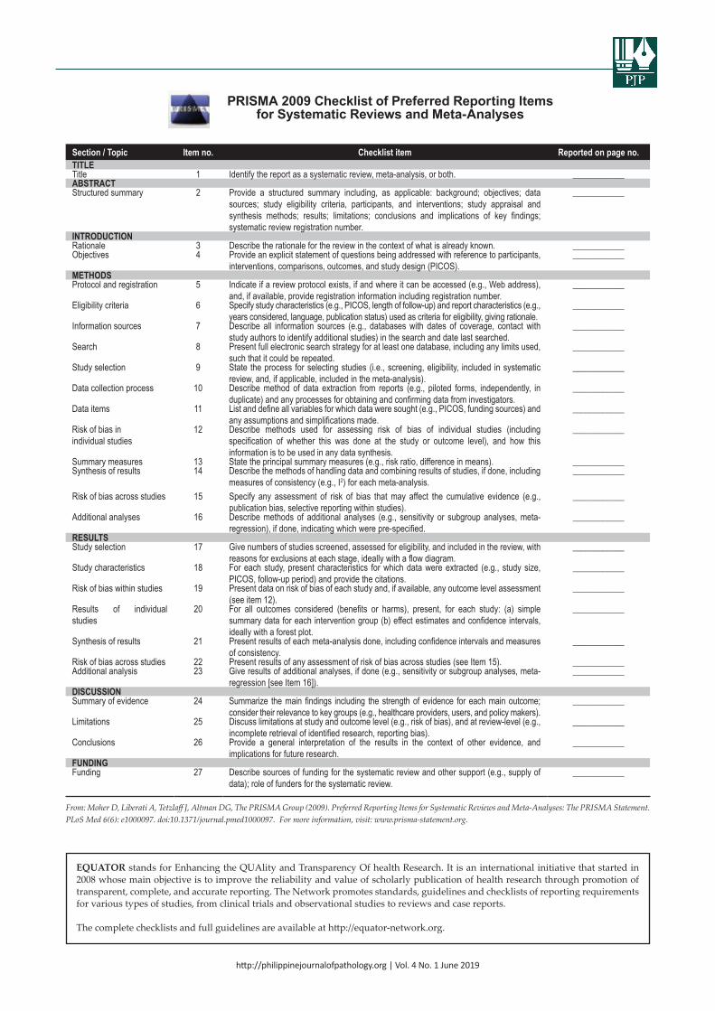
http://philippinejournalofpathology.org | Vol. 4 No. 1 June 2019http://philippinejournalofpathology.org | Vol. 1 No. 1 April 2016
From: Moher D, Liberati A, Tetzlaff J, Altman DG, The PRISMA Group (2009). Preferred Reporting Items for Systematic Reviews and Meta-Analyses: The PRISMA Statement. PLoS Med 6(6): e1000097. doi:10.1371/journal.pmed1000097. For more information, visit: www.prisma-statement.org.
PRISMA 2009 Checklist of Preferred Reporting Items for Systematic Reviews and Meta-Analyses
section / topic Item no. checklist item Reported on page no.tItle Title 1 Identify the report as a systematic review, meta-analysis, or both. ____________abstRact Structured summary 2 Provide a structured summary including, as applicable: background; objectives; data
sources; study eligibility criteria, participants, and interventions; study appraisal and synthesis methods; results; limitations; conclusions and implications of key findings; systematic review registration number.
____________
INtRoDuctIoN Rationale 3 Describe the rationale for the review in the context of what is already known. ____________Objectives 4 Provide an explicit statement of questions being addressed with reference to participants,
interventions, comparisons, outcomes, and study design (PICOS). ____________
methoDs Protocol and registration 5 Indicate if a review protocol exists, if and where it can be accessed (e.g., Web address),
and, if available, provide registration information including registration number. ____________
Eligibility criteria 6 Specify study characteristics (e.g., PICOS, length of follow-up) and report characteristics (e.g., years considered, language, publication status) used as criteria for eligibility, giving rationale.
____________
Information sources 7 Describe all information sources (e.g., databases with dates of coverage, contact with study authors to identify additional studies) in the search and date last searched.
____________
Search 8 Present full electronic search strategy for at least one database, including any limits used, such that it could be repeated.
____________
Study selection 9 State the process for selecting studies (i.e., screening, eligibility, included in systematic review, and, if applicable, included in the meta-analysis).
____________
Data collection process 10 Describe method of data extraction from reports (e.g., piloted forms, independently, in duplicate) and any processes for obtaining and confirming data from investigators.
____________
Data items 11 List and define all variables for which data were sought (e.g., PICOS, funding sources) and any assumptions and simplifications made.
____________
Risk of bias in individual studies
12 Describe methods used for assessing risk of bias of individual studies (including specification of whether this was done at the study or outcome level), and how this information is to be used in any data synthesis.
____________
Summary measures 13 State the principal summary measures (e.g., risk ratio, difference in means). ____________Synthesis of results 14 Describe the methods of handling data and combining results of studies, if done, including
measures of consistency (e.g., I2) for each meta-analysis. ____________
Risk of bias across studies 15 Specify any assessment of risk of bias that may affect the cumulative evidence (e.g., publication bias, selective reporting within studies).
____________
Additional analyses 16 Describe methods of additional analyses (e.g., sensitivity or subgroup analyses, meta-regression), if done, indicating which were pre-specified.
____________
Results Study selection 17 Give numbers of studies screened, assessed for eligibility, and included in the review, with
reasons for exclusions at each stage, ideally with a flow diagram. ____________
Study characteristics 18 For each study, present characteristics for which data were extracted (e.g., study size, PICOS, follow-up period) and provide the citations.
____________
Risk of bias within studies 19 Present data on risk of bias of each study and, if available, any outcome level assessment (see item 12).
____________
Results of individual studies
20 For all outcomes considered (benefits or harms), present, for each study: (a) simple summary data for each intervention group (b) effect estimates and confidence intervals, ideally with a forest plot.
____________
Synthesis of results 21 Present results of each meta-analysis done, including confidence intervals and measures of consistency.
____________
Risk of bias across studies 22 Present results of any assessment of risk of bias across studies (see Item 15). ____________Additional analysis 23 Give results of additional analyses, if done (e.g., sensitivity or subgroup analyses, meta-
regression [see Item 16]). ____________
DIscussIoN Summary of evidence 24 Summarize the main findings including the strength of evidence for each main outcome;
consider their relevance to key groups (e.g., healthcare providers, users, and policy makers). ____________
Limitations 25 Discuss limitations at study and outcome level (e.g., risk of bias), and at review-level (e.g., incomplete retrieval of identified research, reporting bias).
____________
Conclusions 26 Provide a general interpretation of the results in the context of other evidence, and implications for future research.
____________
fuNDING Funding 27 Describe sources of funding for the systematic review and other support (e.g., supply of
data); role of funders for the systematic review. ____________
EQUATOR stands for Enhancing the QUAlity and Transparency Of health Research. It is an international initiative that started in 2008 whose main objective is to improve the reliability and value of scholarly publication of health research through promotion of transparent, complete, and accurate reporting. The Network promotes standards, guidelines and checklists of reporting requirements for various types of studies, from clinical trials and observational studies to reviews and case reports.
The complete checklists and full guidelines are available at http://equator-network.org.
http://philippinejournalofpathology.org | Vol. 1 No. 1 April 2016
* Give information separately for cases and controls in case-control studies and, if applicable, for exposed and unexposed groups in cohort and cross-sectional studies.
Note: An Explanation and Elaboration article discusses each checklist item and gives methodological background and published examples of transparent reporting. The STROBE checklist is best used in conjunction with this article (freely available on the Web sites of PLoS Medicine at http://www.plosmedicine.org/, Annals of Internal Medicine at http://www.annals.org/, and Epidemiology at http://www.epidem.com/). Information on the STROBE Initiative is available at www.strobe-statement.org.
STROBE Statement - Checklist of Items that should be included in Reports of Observational Studies
section / topic Item no. RecommendationtItleTitle and abstract 1 (a) Indicate the study’s design with a commonly used term in the title or the abstract
(b) Provide in the abstract an informative and balanced summary of what was done and what was foundINtRoDuctIoN Background / rationaleObjectives
23
Explain the scientific background and rationale for the investigation being reported State specific objectives, including any prespecified hypotheses
methoDs Study Design Setting Participants
Variables
Data Sources / measurement Bias Study Size Quantitative variablesStatistical methods
Participants
Descriptive data
Outcome data
Main Results
Other analyses
456
7
8*
9101112
13*
14*
15*
16
17
Present key elements of study design early in the paper Describe the setting, locations, and relevant dates, including periods of recruitment, exposure, follow-up, and data collection(a) Cohort study—Give the eligibility criteria, and the sources and methods of selection of participants. Describe methods
of follow-up Case-control study—Give the eligibility criteria, and the sources and methods of case ascertainment and control
selection. Give the rationale for the choice of cases and controls Cross-sectional study—Give the eligibility criteria, and the sources and methods of selection of participants (b) Cohort study—For matched studies, give matching criteria and number of exposed and unexposed Case-control study—For matched studies, give matching criteria and the number of controls per case Clearly define all outcomes, exposures, predictors, potential confounders, and effect modifiers. Give diagnostic criteria, if applicable For each variable of interest, give sources of data and details of methods of assessment (measurement). Describe comparability of assessment methods if there is more than one group Describe any efforts to address potential sources of bias Explain how the study size was arrived atExplain how quantitative variables were handled in the analyses. If applicable, describe which groupings were chosen and why(a) Describe all statistical methods, including those used to control for confounding(b) Describe any methods used to examine subgroups and interactions(c) Explain how missing data were addressed(d) Cohort study—If applicable, explain how loss to follow-up was addressed Case-control study—If applicable, explain how matching of cases and controls was addressed Cross-sectional study—If applicable, describe analytical methods taking account of sampling strategy(e) Describe any sensitivity analyses(a) Report numbers of individuals at each stage of study—eg numbers potentially eligible, examined for eligibility,
confirmed eligible, included in the study, completing follow-up, and analysed(b) Give reasons for non-participation at each stage(c) Consider use of a flow diagram(a) Give characteristics of study participants (eg demographic, clinical, social) and information on exposures and
potential confounders(b) Indicate number of participants with missing data for each variable of interest(c) Cohort study—Summarise follow-up time (eg, average and total amount)Cohort study—Report numbers of outcome events or summary measures over timeCase-control study—Report numbers in each exposure category, or summary measures of exposureCross-sectional study—Report numbers of outcome events or summary measures(a) Give unadjusted estimates and, if applicable, confounder-adjusted estimates and their precision (eg, 95% confidence
interval). Make clear which confounders were adjusted for and why they were included(b) Report category boundaries when continuous variables were categorized(c) If relevant, consider translating estimates of relative risk into absolute risk for a meaningful time periodReport other analyses done—eg analyses of subgroups and interactions, and sensitivity analyses
DIscussIoN Key ResultsLimitations
Interpretation
Generalisability
1819
20
21
Summarise key results with reference to study objectivesDiscuss limitations of the study, taking into account sources of potential bias or imprecision. Discuss both direction and magnitude of any potential biasGive a cautious overall interpretation of results considering objectives, limitations, multiplicity of analyses, results from similar studies, and other relevant evidenceDiscuss the generalisability (external validity) of the study results
otheR INfoRmatIoN Funding 22 Give the source of funding and the role of the funders for the present study and, if applicable, for the original study on
which the present article is based
EQUATOR stands for Enhancing the QUAlity and Transparency Of health Research. It is an international initiative that started in 2008 whose main objective is to improve the reliability and value of scholarly publication of health research through promotion of transparent, complete, and accurate reporting. The Network promotes standards, guidelines and checklists of reporting requirements for various types of studies, from clinical trials and observational studies to reviews and case reports.
The complete checklists and full guidelines are available at http://equator-network.org.

http://philippinejournalofpathology.org | Vol. 4 No. 1 June 2019http://philippinejournalofpathology.org | Vol. 1 No. 1 April 2016
This STARD list was released in 2015. The 30 items were identified by an international expert group of methodologists, researchers, and editors. The guiding principle in the development of STARD was to select items that, when reported, would help readers to judge the potential for bias in the study, to appraise the applicability of the study findings and the validity of conclusions and recommendations. The list represents an update of the first version, which was published in 2003.
More information can be found on http://www.equator-network.org/reporting-guidelines/stard.
STARD 2015 Checklist of Essential Items for Reporting Diagnostic Accuracy Studies
section and topic No. ItemtItle oR abstRact
1 Identification as a study of diagnostic accuracy using at least one measure of accuracy(such as sensitivity, specificity, predictive values, or AUC)
abstRact 2 Structured summary of study design, methods, results, and conclusions (for specific guidance, see STARD for Abstracts)
INtRoDuctIoN 34
Scientific and clinical background, including the intended use and clinical role of the index test Study objectives and hypotheses
methoDsStudy design
Participants
Test Methods
Analysis
5
67
89
10a10b11
12a
12b
13a13b1415161718
Whether data collection was planned before the index test and reference standard were performed (prospective study) or after (retrospective study)Eligibility criteriaOn what basis potentially eligible participants were identified (such as symptoms, results from previous tests, inclusion in registry)Where and when potentially eligible participants were identified (setting, location and dates)Whether participants formed a consecutive, random or convenience seriesIndex test, in sufficient detail to allow replicationReference standard, in sufficient detail to allow replicationRationale for choosing the reference standard (if alternatives exist)Definition of and rationale for test positivity cut-offs or result categories of the index test, distinguishing pre-specified from exploratoryDefinition of and rationale for test positivity cut-offs or result categories of the reference standard, distinguishing pre-specified from exploratoryWhether clinical information and reference standard results were available to the performers/readers of the index testWhether clinical information and index test results were available to the assessors of the reference standardMethods for estimating or comparing measures of diagnostic accuracyHow indeterminate index test or reference standard results were handledHow missing data on the index test and reference standard were handledAny analyses of variability in diagnostic accuracy, distinguishing pre-specified from exploratoryIntended sample size and how it was determined
ResultsParticipants
Test Results
1920
21a21b22232425
Flow of participants, using a diagramBaseline demographic and clinical characteristics of participantsDistribution of severity of disease in those with the target conditionDistribution of alternative diagnoses in those without the target conditionTime interval and any clinical interventions between index test and reference standardCross tabulation of the index test results (or their distribution) by the results of the reference standardEstimates of diagnostic accuracy and their precision (such as 95% confidence intervals)Any adverse events from performing the index test or the reference standard
DIscussIoN2627
Study limitations, including sources of potential bias, statistical uncertainty, and generalisabilityImplications for practice, including the intended use and clinical role of the index test
otheR INfoRmatIoN 282930
Registration number and name of registryWhere the full study protocol can be accessedSources of funding and other support; role of funders
EQUATOR stands for Enhancing the QUAlity and Transparency Of health Research. It is an international initiative that started in 2008 whose main objective is to improve the reliability and value of scholarly publication of health research through promotion of transparent, complete, and accurate reporting. The Network promotes standards, guidelines and checklists of reporting requirements for various types of studies, from clinical trials and observational studies to reviews and case reports.
The complete checklists and full guidelines are available at http://equator-network.org.
http://philippinejournalofpathology.org | Vol. 1 No. 1 April 2016
section / Item Item no. Recommendation Reported on page no. / line no.
tItle aND abstRactTitle
Abstract
1
2
Identify the study as an economic evaluation or use more specific terms such as “cost-effectiveness analysis”, and describe the interventions compared.Provide a structured summary of objectives, perspective, setting, methods (including study design and inputs), results (including base case and uncertainty analyses), and conclusions.
____________
____________
INtRoDuctIoN Background and objectives 3 Provide an explicit statement of the broader context for the study.
Present the study question and its relevance for health policy or practice decisions.____________
methoDsTarget population and subgroupsSetting and locationStudy PerspectiveComparatorsTime horizonDiscount rateChoice of health outcomes
Measurement of effectiveness
Measurement and valuation of preference based outcomesEstimating resources and costs
Currency, price date,and conversion
Choice of model
AssumptionsAnalytical methods
4
56789
10
11a
11b
12
13a
13b
14
15
1617
Describe characteristics of the base case population and subgroups analysed, including why they were chosen.
State relevant aspects of the system(s) in which the decision(s) need(s) to be made.Describe the perspective of the study and relate this to the costs being evaluated.Describe the interventions or strategies being compared and state why they were chosen.State the time horizon(s) over which costs and consequences are being evaluated and say why appropriate.Report the choice of discount rate(s) used for costs and outcomes and say why appropriate.Describe what outcomes were used as the measure(s) of benefit in the evaluation and their relevance for the type of analysis performed.Single study-based estimates: Describe fully the design features of the single effectiveness study and why the single study was a sufficient source of clinical effectiveness data.Synthesis-based estimates: Describe fully the methods used for identification of included studies and synthesis of clinical effectiveness data.If applicable, describe the population and methods used to elicit preferences for outcomes.
Single study-based economic evaluation: Describe approaches used to estimate resource use associated with the alternative interventions. Describe primary or secondary research methods for valuing each resource item in terms of its unit cost. Describe any adjustments made to approximate to opportunity costs.Model-based economic evaluation: Describe approaches and data sources used to estimate resource use associated with model health states. Describe primary or secondary research methods for valuing each resource item in terms of its unit cost. Describe any adjustments made to approximate to opportunity costs.Report the dates of the estimated resource quantities and unit costs. Describe methods for adjusting estimated unit costs to the year of reported costs if necessary. Describe methods for converting costs into a common currency base and the exchange rate.Describe and give reasons for the specific type of decision analytical model used. Providing a figure to show model structure is strongly recommended.Describe all structural or other assumptions underpinning the decision-analytical model.Describe all analytical methods supporting the evaluation. This could include methods for dealing with skewed, missing, or censored data; extrapolation methods; methods for pooling data; approaches to validate or make adjustments (such as half cycle corrections) to a model; and methods for handling population heterogeneity and uncertainty.
____________
________________________________________________________________________
____________
____________
____________
____________
____________
____________
____________
________________________
ResultsStudy parameters
Incremental costs andoutcomes
Characterisinguncertainty
Characterisingheterogeneity
18
19
20a
20b
21
Report the values, ranges, references, and, if used, probability distributions for all parameters. Report reasons or sources for distributions used to represent uncertainty where appropriate. Providing a table to show the input values is strongly recommended.For each intervention, report mean values for the main categories of estimated costs and outcomes of interest, as well as mean differences between the comparator groups. If applicable, report incremental cost-effectiveness ratios.Single study-based economic evaluation: Describe the effects of sampling uncertainty for the estimated incremental cost and incremental effectiveness parameters, together with the impact Consolidated Health Economic Evaluation Reporting Standards – CHEERS Checklist 3 of methodological assumptions (such as discount rate, study perspective).Model-based economic evaluation: Describe the effects on the results of uncertainty for all input parameters, and uncertainty related to the structure of the model and assumptions.If applicable, report differences in costs, outcomes, or costeffectiveness that can be explained by variations between subgroups of patients with different baseline characteristics or other observed variability in effects that are not reducible by more information.
____________
____________
____________
____________
____________
DIscussIoNStudy findings, limitations,generalisability, and current knowledge
22 Summarise key study findings and describe how they support the conclusions reached. Discuss limitations and the generalisability of the findings and how the findings fit with current knowledge.
____________
otheR INfoRmatIoN Source of funding
Conflicts of interest
23
24
Describe how the study was funded and the role of the funder in the identification, design, conduct, and reporting of the analysis. Describe other non-monetary sources of support.Describe any potential for conflict of interest of study contributors in accordance with journal policy. In the absence of a journal policy, we recommend authors comply with International Committee of Medical Journal Editors recommendations.
____________
____________
CHEERS Checklist - Items to include when Reporting Economic Evaluations of Health Interventions
EQUATOR stands for Enhancing the QUAlity and Transparency Of health Research. It is an international initiative that started in 2008 whose main objective is to improve the reliability and value of scholarly publication of health research through promotion of transparent, complete, and accurate reporting. The Network promotes standards, guidelines and checklists of reporting requirements for various types of studies, from clinical trials and observational studies to reviews and case reports.
The complete checklists and full guidelines are available at http://equator-network.org.

http://philippinejournalofpathology.org | Vol. 4 No. 1 June 2019http://philippinejournalofpathology.org | Vol. 1 No. 1 April 2016
This STARD list was released in 2015. The 30 items were identified by an international expert group of methodologists, researchers, and editors. The guiding principle in the development of STARD was to select items that, when reported, would help readers to judge the potential for bias in the study, to appraise the applicability of the study findings and the validity of conclusions and recommendations. The list represents an update of the first version, which was published in 2003.
More information can be found on http://www.equator-network.org/reporting-guidelines/stard.
STARD 2015 Checklist of Essential Items for Reporting Diagnostic Accuracy Studies
section and topic No. ItemtItle oR abstRact
1 Identification as a study of diagnostic accuracy using at least one measure of accuracy(such as sensitivity, specificity, predictive values, or AUC)
abstRact 2 Structured summary of study design, methods, results, and conclusions (for specific guidance, see STARD for Abstracts)
INtRoDuctIoN 34
Scientific and clinical background, including the intended use and clinical role of the index test Study objectives and hypotheses
methoDsStudy design
Participants
Test Methods
Analysis
5
67
89
10a10b11
12a
12b
13a13b1415161718
Whether data collection was planned before the index test and reference standard were performed (prospective study) or after (retrospective study)Eligibility criteriaOn what basis potentially eligible participants were identified (such as symptoms, results from previous tests, inclusion in registry)Where and when potentially eligible participants were identified (setting, location and dates)Whether participants formed a consecutive, random or convenience seriesIndex test, in sufficient detail to allow replicationReference standard, in sufficient detail to allow replicationRationale for choosing the reference standard (if alternatives exist)Definition of and rationale for test positivity cut-offs or result categories of the index test, distinguishing pre-specified from exploratoryDefinition of and rationale for test positivity cut-offs or result categories of the reference standard, distinguishing pre-specified from exploratoryWhether clinical information and reference standard results were available to the performers/readers of the index testWhether clinical information and index test results were available to the assessors of the reference standardMethods for estimating or comparing measures of diagnostic accuracyHow indeterminate index test or reference standard results were handledHow missing data on the index test and reference standard were handledAny analyses of variability in diagnostic accuracy, distinguishing pre-specified from exploratoryIntended sample size and how it was determined
ResultsParticipants
Test Results
1920
21a21b22232425
Flow of participants, using a diagramBaseline demographic and clinical characteristics of participantsDistribution of severity of disease in those with the target conditionDistribution of alternative diagnoses in those without the target conditionTime interval and any clinical interventions between index test and reference standardCross tabulation of the index test results (or their distribution) by the results of the reference standardEstimates of diagnostic accuracy and their precision (such as 95% confidence intervals)Any adverse events from performing the index test or the reference standard
DIscussIoN2627
Study limitations, including sources of potential bias, statistical uncertainty, and generalisabilityImplications for practice, including the intended use and clinical role of the index test
otheR INfoRmatIoN 282930
Registration number and name of registryWhere the full study protocol can be accessedSources of funding and other support; role of funders
EQUATOR stands for Enhancing the QUAlity and Transparency Of health Research. It is an international initiative that started in 2008 whose main objective is to improve the reliability and value of scholarly publication of health research through promotion of transparent, complete, and accurate reporting. The Network promotes standards, guidelines and checklists of reporting requirements for various types of studies, from clinical trials and observational studies to reviews and case reports.
The complete checklists and full guidelines are available at http://equator-network.org.
http://philippinejournalofpathology.org | Vol. 1 No. 1 April 2016
section / Item Item no. Recommendation Reported on page no. / line no.
tItle aND abstRactTitle
Abstract
1
2
Identify the study as an economic evaluation or use more specific terms such as “cost-effectiveness analysis”, and describe the interventions compared.Provide a structured summary of objectives, perspective, setting, methods (including study design and inputs), results (including base case and uncertainty analyses), and conclusions.
____________
____________
INtRoDuctIoN Background and objectives 3 Provide an explicit statement of the broader context for the study.
Present the study question and its relevance for health policy or practice decisions.____________
methoDsTarget population and subgroupsSetting and locationStudy PerspectiveComparatorsTime horizonDiscount rateChoice of health outcomes
Measurement of effectiveness
Measurement and valuation of preference based outcomesEstimating resources and costs
Currency, price date,and conversion
Choice of model
AssumptionsAnalytical methods
4
5678910
11a
11b
12
13a
13b
14
15
1617
Describe characteristics of the base case population and subgroups analysed, including why they were chosen.
State relevant aspects of the system(s) in which the decision(s) need(s) to be made.Describe the perspective of the study and relate this to the costs being evaluated.Describe the interventions or strategies being compared and state why they were chosen.State the time horizon(s) over which costs and consequences are being evaluated and say why appropriate.Report the choice of discount rate(s) used for costs and outcomes and say why appropriate.Describe what outcomes were used as the measure(s) of benefit in the evaluation and their relevance for the type of analysis performed.Single study-based estimates: Describe fully the design features of the single effectiveness study and why the single study was a sufficient source of clinical effectiveness data.Synthesis-based estimates: Describe fully the methods used for identification of included studies and synthesis of clinical effectiveness data.If applicable, describe the population and methods used to elicit preferences for outcomes.
Single study-based economic evaluation: Describe approaches used to estimate resource use associated with the alternative interventions. Describe primary or secondary research methods for valuing each resource item in terms of its unit cost. Describe any adjustments made to approximate to opportunity costs.Model-based economic evaluation: Describe approaches and data sources used to estimate resource use associated with model health states. Describe primary or secondary research methods for valuing each resource item in terms of its unit cost. Describe any adjustments made to approximate to opportunity costs.Report the dates of the estimated resource quantities and unit costs. Describe methods for adjusting estimated unit costs to the year of reported costs if necessary. Describe methods for converting costs into a common currency base and the exchange rate.Describe and give reasons for the specific type of decision analytical model used. Providing a figure to show model structure is strongly recommended.Describe all structural or other assumptions underpinning the decision-analytical model.Describe all analytical methods supporting the evaluation. This could include methods for dealing with skewed, missing, or censored data; extrapolation methods; methods for pooling data; approaches to validate or make adjustments (such as half cycle corrections) to a model; and methods for handling population heterogeneity and uncertainty.
____________
________________________________________________________________________
____________
____________
____________
____________
____________
____________
____________
________________________
ResultsStudy parameters
Incremental costs andoutcomes
Characterisinguncertainty
Characterisingheterogeneity
18
19
20a
20b
21
Report the values, ranges, references, and, if used, probability distributions for all parameters. Report reasons or sources for distributions used to represent uncertainty where appropriate. Providing a table to show the input values is strongly recommended.For each intervention, report mean values for the main categories of estimated costs and outcomes of interest, as well as mean differences between the comparator groups. If applicable, report incremental cost-effectiveness ratios.Single study-based economic evaluation: Describe the effects of sampling uncertainty for the estimated incremental cost and incremental effectiveness parameters, together with the impact Consolidated Health Economic Evaluation Reporting Standards – CHEERS Checklist 3 of methodological assumptions (such as discount rate, study perspective).Model-based economic evaluation: Describe the effects on the results of uncertainty for all input parameters, and uncertainty related to the structure of the model and assumptions.If applicable, report differences in costs, outcomes, or costeffectiveness that can be explained by variations between subgroups of patients with different baseline characteristics or other observed variability in effects that are not reducible by more information.
____________
____________
____________
____________
____________
DIscussIoNStudy findings, limitations,generalisability, and current knowledge
22 Summarise key study findings and describe how they support the conclusions reached. Discuss limitations and the generalisability of the findings and how the findings fit with current knowledge.
____________
otheR INfoRmatIoN Source of funding
Conflicts of interest
23
24
Describe how the study was funded and the role of the funder in the identification, design, conduct, and reporting of the analysis. Describe other non-monetary sources of support.Describe any potential for conflict of interest of study contributors in accordance with journal policy. In the absence of a journal policy, we recommend authors comply with International Committee of Medical Journal Editors recommendations.
____________
____________
CHEERS Checklist - Items to include when Reporting Economic Evaluations of Health Interventions
EQUATOR stands for Enhancing the QUAlity and Transparency Of health Research. It is an international initiative that started in 2008 whose main objective is to improve the reliability and value of scholarly publication of health research through promotion of transparent, complete, and accurate reporting. The Network promotes standards, guidelines and checklists of reporting requirements for various types of studies, from clinical trials and observational studies to reviews and case reports.
The complete checklists and full guidelines are available at http://equator-network.org.

http://philippinejournalofpathology.org | Vol. 4 No. 1 June 2019http://philippinejournalofpathology.org | Vol. 1 No. 1 April 2016
The ARRIVE (Animal Research: Reporting of In Vivo Experiments) guidelines were developed as part of an NC3Rs initiative to improve the design, analysis and reporting of research using animals – maximising information published and minimising unnecessary studies. The guidelines were published in the online journal PLOS Biology in June 2010 and are currently endorsed by scientific journals, major funding bodies and learned societies. More information can be found on www.nc3rs.org.uk/ARRIVE
The ARRIVE Guidelines(Animal Research: Reporting of In Vivo Experiments)
section / topic Item no. checklist itemtItle aND abstRactTitleAbstract
12
Provide as accurate and concise a description of the content of the article as possible.Provide an accurate summary of the background, research objectives, including details of the species or strain of animal used, key methods, principal findings and conclusions of the study.
INtRoDuctIoNBackground Objectives
3
4
a. Include sufficient scientific background (including relevant references to previous work) to understand the motivation and context for the study, and explain the experimental approach and rationale.
b. Explain how and why the animal species and model being used can address the scientific objectives and, where appropriate, the study’s relevance to human biology.
Clearly describe the primary and any secondary objectives of the study, or specific hypotheses being tested.methoDsEthical statement 5 DIndicate the nature of the ethical review permissions, relevant licences (e.g. Animal [Scientific Procedures] Act 1986), and national or
institutional guidelines for the care and use of animals, that cover the research.Study design 6 For each experiment, give brief details of the study design including:
a. The number of experimental and control groups.b. Any steps taken to minimise the effects of subjective bias when allocating animals to treatment (e.g. randomisation procedure) and when
assessing results (e.g. if done, describe who was blinded and when).c. The experimental unit (e.g. a single animal, group or cage of animals). A time-line diagram or flow chart can be useful to illustrate how complex study designs were carried out.
Experimental procedures 7 For each experiment and each experimental group, including controls, provide precise details of all procedures carried out.For example:a. How (e.g. drug formulation and dose, site and route of administration, anaesthesia and analgesia used [including monitoring], surgical
procedure, method of euthanasia). Provide details of any specialist equipment used, including supplier(s).b. When (e.g. time of day).c. Where (e.g. home cage, laboratory, water maze).d. Why (e.g. rationale for choice of specific anaesthetic, route of administration, drug dose used).
Experimental animals 8 a. Provide details of the animals used, including species, strain, sex, developmental stage (e.g. mean or median age plus age range) and weight (e.g. mean or median weight plus weight range).
b. Provide further relevant information such as the source of animals, international strain nomenclature, genetic modification status (e.g. knock-out or transgenic), genotype, health/immune status, drug or test naïve, previous procedures, etc.
Housing and husbandry 9 Provide details of:a. Housing (type of facility e.g. specific pathogen free [SPF]; type of cage or housing; bedding material; number of cage companions; tank
shape and material etc. for fish).b. Husbandry conditions (e.g. breeding programme, light/dark cycle, temperature, quality of water etc for fish, type of food, access to food
and water, environmental enrichment).c. Welfare-related assessments and interventions that were carried out prior to, during, or after the experiment.
Sample size 10 a. Specify the total number of animals used in each experiment, and the number of animals in each experimental group.b. Explain how the number of animals was arrived at. Provide details of any sample size calculation used.c. Indicate the number of independent replications of each experiment, if relevant.
Allocating animals to experimental groups
11 a. Give full details of how animals were allocated to experimental groups, including randomisation or matching if done.b. Describe the order in which the animals in the different experimental groups were treated and assessed.
Experimental outcomes 12 Clearly define the primary and secondary experimental outcomes assessed (e.g. cell death, molecular markers, behavioural changes).Statistical methods 13 a. Provide details of the statistical methods used for each analysis.
b. Specify the unit of analysis for each dataset (e.g. single animal, group of animals, single neuron).c. Describe any methods used to assess whether the data met the assumptions of the statistical approach.
ResultsBaseline data 14 For each experimental group, report relevant characteristics and health status of animals (e.g. weight, microbiological status, and drug or test
naïve) prior to treatment or testing (this information can often be tabulated).Numbers analysed 15 a. Report the number of animals in each group included in each analysis. Report absolute numbers (e.g. 10/20, not 50%).
b. If any animals or data were not included in the analysis, explain why.Outcomes and estimation 16 Report the results for each analysis carried out, with a measure of precision (e.g. standard error or confidence interval).Adverse events 17 a. Give details of all important adverse events in each experimental group.
b. Describe any modifications to the experimental protocols made to reduce adverse events.DIscussIoNInterpretation/scientific implications
18 a. Interpret the results, taking into account the study objectives and hypotheses, current theory and other relevant studies in the literature.b. Comment on the study limitations including any potential sources of bias, any limitations of the animal model, and the imprecision
associated with the results.c. Describe any implications of your experimental methods or findings for the replacement, refinement or reduction (the 3Rs) of the use of
animals in research.Generalisability/translation 19 Comment on whether, and how, the findings of this study are likely to translate to other species or systems, including any relevance to human
biology.Funding 20 List all funding sources (including grant number) and the role of the funder(s) in the study.
EQUATOR stands for Enhancing the QUAlity and Transparency Of health Research. It is an international initiative that started in 2008 whose main objective is to improve the reliability and value of scholarly publication of health research through promotion of transparent, complete, and accurate reporting. The Network promotes standards, guidelines and checklists of reporting requirements for various types of studies, from clinical trials and observational studies to reviews and case reports.
The complete checklists and full guidelines are available at http://equator-network.org.
http://philippinejournalofpathology.org | Vol. 1 No. 1 April 2016
Revised Standards for Quality Improvement Reporting Excellence (SQUIRE 2.0)
No Item Guide questions / descriptiontItle aND abstRact1
2
Title
Abstract
Indicate that the manuscript concerns an initiative to improve healthcare (broadly defined to include the quality, safety, effectiveness, patient-centeredness, timeliness, cost, efficiency, and equity of healthcare)a. Provide adequate information to aid in searching and indexingb. Summarize all key information from various sections of the text using the abstract format of the intended publication or
a structured summary such as: background, local problem, methods, interventions, results, conclusionsINtRoDuctIoN Why DID you staRt?345
6
Problem DescriptionAvailable knowledgeRationale
Specific aims
Nature and significance of the local problemSummary of what is currently known about the problem, including relevant previous studiesInformal or formal frameworks, models, concepts, and/or theories used to explain the problem, any reasons or assumptions that were used to develop the intervention(s), and reasons why the intervention(s) was expected to workPurpose of the project and of this report
methoDs What DID you Do?78
9
10
11
12
ContextIntervention(s)
Study of the Intervention(s)
Measures
Analysis
Ethical Considerations
Contextual elements considered important at the outset of introducing the intervention(s)a. Description of the intervention(s) in sufficient detail that others could reproduce itb. Specifics of the team involved in the worka. Approach chosen for assessing the impact of the intervention(s)b. Approach used to establish whether the observed outcomes were due to the intervention(s)a. Measures chosen for studying processes and outcomes of the intervention(s), including rationale for choosing them,
their operational definitions, and their validity and reliabilityb. Description of the approach to the ongoing assessment of contextual elements that contributed to the success, failure,
efficiency, and costc. Methods employed for assessing completeness and accuracy of dataa. Qualitative and quantitative methods used to draw inferences from the datab. Methods for understanding variation within the data, including the effects of time as a variableEthical aspects of implementing and studying the intervention(s) and how they were addressed, including, but not limited to, formal ethics review and potential conflict(s) of interest
Results What DID you fIND?13 Results a. Initial steps of the intervention(s) and their evolution over time (e.g., time-line diagram, flow chart, or table), including
modifications made to the intervention during the projectb. Details of the process measures and outcomec. Contextual elements that interacted with the intervention(s)d. Observed associations between outcomes, interventions, and relevant contextual elements e. Unintended consequences such as unexpected benefits, problems, failures, or costs associated with the intervention(s).f. Details about missing data
DIscussIoN What Does It meaN?14
15
16
17
Summary
Interpretation
Limitations
Conclusions
a. Key findings, including relevance to the rationale and specific aimsb. Particular strengths of the projecta. Nature of the association between the intervention(s) and the outcomesb. Comparison of results with findings from other publicationsc. Impact of the project on people and systemsd. Reasons for any differences between observed and anticipated outcomes, including the influence of contexte. Costs and strategic trade-offs, including opportunity costsa. Limits to the generalizability of the workb. Factors that might have limited internal validity such as confounding, bias, or imprecision in the design, methods,
measurement, or analysisc. Efforts made to minimize and adjust for limitationsa. Usefulness of the workb. Sustainabilityc. Potential for spread to other contextsd. Implications for practice and for further study in the fielde. Suggested next steps
otheR INfoRmatIoN18 Funding Sources of funding that supported this work. Role, if any, of the funding organization in the design, implementation,
interpretation, and reporting
EQUATOR stands for Enhancing the QUAlity and Transparency Of health Research. It is an international initiative that started in 2008 whose main objective is to improve the reliability and value of scholarly publication of health research through promotion of transparent, complete, and accurate reporting. The Network promotes standards, guidelines and checklists of reporting requirements for various types of studies, from clinical trials and observational studies to reviews and case reports.
The complete checklists and full guidelines are available at http://equator-network.org.

http://philippinejournalofpathology.org | Vol. 4 No. 1 June 2019http://philippinejournalofpathology.org | Vol. 1 No. 1 April 2016
section / topic Item no. DescriptionaDmINIstRatIve INfoRmatIoNTitleTrial registration
Protocol versionFundingRoles and responsibilities
12a2b34
5a5b5c
5d
Descriptive title identifying the study design, population, interventions, and, if applicable, trial acronymTrial identifier and registry name. If not yet registered, name of intended registryAll items from the World Health Organization Trial Registration Data SetDate and version identifierSources and types of financial, material, and other supportNames, affiliations, and roles of protocol contributorsName and contact information for the trial sponsorRole of study sponsor and funders, if any, in study design; collection, management, analysis, and interpretation of data; writing of the report; and the decision to submit the report for publication, including whether they will have ultimate authority over any of these activitiesComposition, roles, and responsibilities of the coordinating centre, steering committee, endpoint adjudication committee, data management team, and other individuals or groups overseeing the trial, if applicable (see Item 21a for data monitoring committee)
INtRoDuctIoNBackground and rationale
ObjectivesTrial design
6a
6b78
Description of research question and justification for undertaking the trial, including summary of relevant studies (published and unpublished) examining benefits and harms for each interventionExplanation for choice of comparatorsSpecific objectives or hypothesesDescription of trial design including type of trial (eg, parallel group, crossover, factorial, single group), allocation ratio, and framework (eg, superiority, equivalence, noninferiority, exploratory)
methoDs: PaRtIcIPaNts, INteRveNtIoNs, aND outcomesStudy setting
Eligibility criteria
Interventions
Outcomes
Participant timeline
Sample size
Recruitment
9
10
11a11b
11c
11d12
13
14
15
Description of study settings (eg, community clinic, academic hospital) and list of countries where data will be collected. Reference to where list of study sites can be obtainedInclusion and exclusion criteria for participants. If applicable, eligibility criteria for study centres and individuals who will perform the interventions (eg, surgeons, psychotherapists)Interventions for each group with sufficient detail to allow replication, including how and when they will be administeredCriteria for discontinuing or modifying allocated interventions for a given trial participant (eg, drug dose change in response to harms, participant request, or improving/worsening disease)Strategies to improve adherence to intervention protocols, and any procedures for monitoring adherence (eg, drug tablet return, laboratory tests)Relevant concomitant care and interventions that are permitted or prohibited during the trialPrimary, secondary, and other outcomes, including the specific measurement variable (eg, systolic blood pressure), analysis metric (eg, change from baseline, final value, time to event), method of aggregation (eg, median, proportion), and time point for each outcome. Explanation of the clinical relevance of chosen efficacy and harm outcomes is strongly recommendedTime schedule of enrolment, interventions (including any run-ins and washouts), assessments, and visits for participants. A schematic diagram is highly recommended (see Figure)Estimated number of participants needed to achieve study objectives and how it was determined, including clinical and statistical assumptions supporting any sample size calculationsStrategies for achieving adequate participant enrolment to reach target sample size
methoDs: assIGNmeNt of INteRveNtIoNs (foR coNtRolleD tRIals)Allocation:Sequence generation
Allocation concealment mechanismImplementationBlinding (masking)
16a
16b
16c17a
17b
Method of generating the allocation sequence (eg, computer-generated random numbers), and list of any factors for stratification. To reduce predictability of a random sequence, details of any planned restriction (eg, blocking) should be provided in a separate document that is unavailable to those who enrol participants or assign interventionsMechanism of implementing the allocation sequence (eg, central telephone; sequentially numbered, opaque, sealed envelopes), describing any steps to conceal the sequence until interventions are assignedWho will generate the allocation sequence, who will enrol participants, and who will assign participants to interventionsWho will be blinded after assignment to interventions (eg, trial participants, care providers, outcome assessors, data analysts), and howIf blinded, circumstances under which unblinding is permissible, and procedure for revealing a participant’s allocated intervention during the trial
SPIRIT 2013 Checklist: Recommended items to address in a clinical trial protocol and related documents*
http://philippinejournalofpathology.org | Vol. 1 No. 1 April 2016
*It is strongly recommended that this checklist be read in conjunction with the SPIRIT 2013 Explanation & Elaboration for important clarification on the items. Amendments to the protocol should be tracked and dated. The SPIRIT checklist is copyrighted by the SPIRIT Group under the Creative Commons “Attribution-NonCommercial-NoDerivs 3.0 Unported” license.
methoDs: Data collectIoN, maNaGemeNt, aND aNalysIsData collection methods
Data management
Statistical methods
18a
18b
19
20a
20b20c
Plans for assessment and collection of outcome, baseline, and other trial data, including any related processes to promote data quality (eg, duplicate measurements, training of assessors) and a description of study instruments (eg, questionnaires, laboratory tests) along with their reliability and validity, if known. Reference to where data collection forms can be found, if not in the protocolPlans to promote participant retention and complete follow-up, including list of any outcome data to be collected for participants who discontinue or deviate from intervention protocolsPlans for data entry, coding, security, and storage, including any related processes to promote data quality (eg, double data entry; range checks for data values). Reference to where details of data management procedures can be found, if not in the protocolStatistical methods for analysing primary and secondary outcomes. Reference to where other details of the statistical analysis plan can be found, if not in the protocolMethods for any additional analyses (eg, subgroup and adjusted analyses)Definition of analysis population relating to protocol non-adherence (eg, as randomised analysis), and any statistical methods to handle missing data (eg, multiple imputation)
methoDs: moNItoRINGData monitoring
Harms
Auditing
21a
21b
22
23
Composition of data monitoring committee (DMC); summary of its role and reporting structure; statement of whether it is independent from the sponsor and competing interests; and reference to where further details about its charter can be found, if not in the protocol. Alternatively, an explanation of why a DMC is not neededDescription of any interim analyses and stopping guidelines, including who will have access to these interim results and make the final decision to terminate the trialPlans for collecting, assessing, reporting, and managing solicited and spontaneously reported adverse events and other unintended effects of trial interventions or trial conductFrequency and procedures for auditing trial conduct, if any, and whether the process will be independent from investigators and the sponsor
ethIcs aND DIssemINatIoNResearch ethics approvalProtocol amendments
Consent or assent
Confidentiality
Declaration of interestsAccess to data
Ancillary and post-trial careDissemination policy
2425
26a26b
27
2829
3031a
31b31c
Plans for seeking research ethics committee/institutional review board (REC/IRB) approvalPlans for communicating important protocol modifications (eg, changes to eligibility criteria, outcomes, analyses) to relevant parties (eg, investigators, REC/IRBs, trial participants, trial registries, journals, regulators)Who will obtain informed consent or assent from potential trial participants or authorised surrogates, and how (see Item 32)Additional consent provisions for collection and use of participant data and biological specimens in ancillary studies, if applicableHow personal information about potential and enrolled participants will be collected, shared, and maintained in order to protect confidentiality before, during, and after the trialFinancial and other competing interests for principal investigators for the overall trial and each study siteStatement of who will have access to the final trial dataset, and disclosure of contractual agreements that limit such access for investigatorsProvisions, if any, for ancillary and post-trial care, and for compensation to those who suffer harm from trial participationPlans for investigators and sponsor to communicate trial results to participants, healthcare professionals, the public, and other relevant groups (eg, via publication, reporting in results databases, or other data sharing arrangements), including any publication restrictionsAuthorship eligibility guidelines and any intended use of professional writersPlans, if any, for granting public access to the full protocol, participant-level dataset, and statistical code
aPPeNDIcesInformed consent materialsBiological specimens
3233
Model consent form and other related documentation given to participants and authorised surrogatesPlans for collection, laboratory evaluation, and storage of biological specimens for genetic or molecular analysis in the current trial and for future use in ancillary studies, if applicable
SPIRIT 2013 Checklist Philippine Journal of Pathology | 69
EQUATOR stands for Enhancing the QUAlity and Transparency Of health Research. It is an international initiative that started in 2008 whose main objective is to improve the reliability and value of scholarly publication of health research through promotion of transparent, complete, and accurate reporting. The Network promotes standards, guidelines and checklists of reporting requirements for various types of studies, from clinical trials and observational studies to reviews and case reports.
The complete checklists and full guidelines are available at http://equator-network.org.
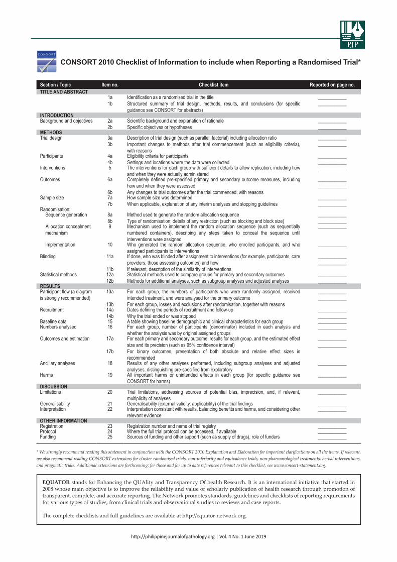
http://philippinejournalofpathology.org | Vol. 4 No. 1 June 2019http://philippinejournalofpathology.org | Vol. 1 No. 1 April 2016
* We strongly recommend reading this statement in conjunction with the CONSORT 2010 Explanation and Elaboration for important clarifications on all the items. If relevant, we also recommend reading CONSORT extensions for cluster randomised trials, non-inferiority and equivalence trials, non-pharmacological treatments, herbal interventions, and pragmatic trials. Additional extensions are forthcoming: for those and for up to date references relevant to this checklist, see www.consort-statement.org.
CONSORT 2010 Checklist of Information to include when Reporting a Randomised Trial*
section / topic Item no. checklist item Reported on page no.tItle aND abstRact
1a1b
Identification as a randomised trial in the titleStructured summary of trial design, methods, results, and conclusions (for specific guidance see CONSORT for abstracts)
________________________
INtRoDuctIoNBackground and objectives 2a
2bScientific background and explanation of rationaleSpecific objectives or hypotheses
________________________
methoDsTrial design 3a
3bDescription of trial design (such as parallel, factorial) including allocation ratioImportant changes to methods after trial commencement (such as eligibility criteria), with reasons
________________________
Participants 4a4b
Eligibility criteria for participantsSettings and locations where the data were collected
________________________
Interventions 5 The interventions for each group with sufficient details to allow replication, including how and when they were actually administered
____________
Outcomes 6a
6b
Completely defined pre-specified primary and secondary outcome measures, including how and when they were assessedAny changes to trial outcomes after the trial commenced, with reasons
________________________
Sample size 7a7b
How sample size was determinedWhen applicable, explanation of any interim analyses and stopping guidelines
________________________
Randomisation: Sequence generation 8a
8bMethod used to generate the random allocation sequenceType of randomisation; details of any restriction (such as blocking and block size)
________________________
Allocation concealment mechanism
9 Mechanism used to implement the random allocation sequence (such as sequentially numbered containers), describing any steps taken to conceal the sequence until interventions were assigned
____________
Implementation 10 Who generated the random allocation sequence, who enrolled participants, and who assigned participants to interventions
____________
Blinding 11a
11b
If done, who was blinded after assignment to interventions (for example, participants, care providers, those assessing outcomes) and howIf relevant, description of the similarity of interventions
________________________
Statistical methods 12a12b
Statistical methods used to compare groups for primary and secondary outcomesMethods for additional analyses, such as subgroup analyses and adjusted analyses
________________________
ResultsParticipant flow (a diagram is strongly recommended)
13a
13b
For each group, the numbers of participants who were randomly assigned, received intended treatment, and were analysed for the primary outcomeFor each group, losses and exclusions after randomisation, together with reasons
________________________
Recruitment 14a14b
Dates defining the periods of recruitment and follow-upWhy the trial ended or was stopped
________________________
Baseline data 15 A table showing baseline demographic and clinical characteristics for each group ____________Numbers analysed 16 For each group, number of participants (denominator) included in each analysis and
whether the analysis was by original assigned groups____________
Outcomes and estimation 17a
17b
For each primary and secondary outcome, results for each group, and the estimated effect size and its precision (such as 95% confidence interval)For binary outcomes, presentation of both absolute and relative effect sizes is recommended
________________________
Ancillary analyses 18 Results of any other analyses performed, including subgroup analyses and adjusted analyses, distinguishing pre-specified from exploratory
____________
Harms 19 All important harms or unintended effects in each group (for specific guidance see CONSORT for harms)
____________
DIscussIoNLimitations 20 Trial limitations, addressing sources of potential bias, imprecision, and, if relevant,
multiplicity of analyses____________
Generalisability 21 Generalisability (external validity, applicability) of the trial findings ____________Interpretation 22 Interpretation consistent with results, balancing benefits and harms, and considering other
relevant evidence____________
otheR INfoRmatIoNRegistration 23 Registration number and name of trial registry ____________Protocol 24 Where the full trial protocol can be accessed, if available ____________Funding 25 Sources of funding and other support (such as supply of drugs), role of funders ____________
EQUATOR stands for Enhancing the QUAlity and Transparency Of health Research. It is an international initiative that started in 2008 whose main objective is to improve the reliability and value of scholarly publication of health research through promotion of transparent, complete, and accurate reporting. The Network promotes standards, guidelines and checklists of reporting requirements for various types of studies, from clinical trials and observational studies to reviews and case reports.
The complete checklists and full guidelines are available at http://equator-network.org.
PJP Online Journal System User Guide for Authors
(Updated: January 2016)
Page 2
Getting Started
From the PJP website (http://www.philippinejournalofpathology.org), navigate to ‘For Authors’. (add screenshot of PJP landing page, circle ‘for authors’ on right column).
Select ‘FOR AUTHORS’.
Log in
New user:
o If you are a new user of the PJP website, please register by clicking the link ‘Not a user, Register with this site’.

http://philippinejournalofpathology.org | Vol. 4 No. 1 June 2019
PJP Online Journal System - User Guide for Authors Philippine Journal of Pathology | 68
Page 2
Getting Started
From the PJP website (http://www.philippinejournalofpathology.org), navigate to ‘For Authors’. (add screenshot of PJP landing page, circle ‘for authors’ on right column).
Select ‘FOR AUTHORS’.
Log in
New user:
o If you are a new user of the PJP website, please register by clicking the link ‘Not a user, Register with this site’.
Page 3
o Complete the online form then select ‘Register’. A confirmation email with your username and password will be sent to your email address.
Existing user:
o Log in to your OJS account using the username and password from original registration.
o If you have forgotten your log in details, please click the ‘Forgot the password?’ and an email will be sent to your registered email address.

http://philippinejournalofpathology.org | Vol. 4 No. 1 June 2019
PJP Online Journal System - User Guide for Authors Philippine Journal of Pathology | 69
Page 3
o Complete the online form then select ‘Register’. A confirmation email with your username and password will be sent to your email address.
Existing user:
o Log in to your OJS account using the username and password from original registration.
o If you have forgotten your log in details, please click the ‘Forgot the password?’ and an email will be sent to your registered email address.
Page 4
The Submission Process
To start the submission process, click ‘New Submission’
Step 1: Starting the submission From the drop-down menu, please select the most appropriate section to describe your
submission article type. If you are not sure what section to select, click ‘About’ to find out more information.

http://philippinejournalofpathology.org | Vol. 4 No. 1 June 2019
PJP Online Journal System - User Guide for Authors Philippine Journal of Pathology | 70
Page 6
Please ensure the items listed in the checklist are ready then tick each box.

http://philippinejournalofpathology.org | Vol. 4 No. 1 June 2019
PJP Online Journal System - User Guide for Authors Philippine Journal of Pathology | 71
Page 7
Read the ‘Copyright Notice’ and add comments to the editor (optional). Select ‘Save and continue’.
Step 2: Uploading the Submission Please follow the instructions on this page to upload your file, then select ‘Save and continue’.
This is where you upload the manuscript only. (You will be asked to upload other required documents at Step 4.

http://philippinejournalofpathology.org | Vol. 4 No. 1 June 2019
PJP Online Journal System - User Guide for Authors Philippine Journal of Pathology | 72
Page 8
Step 3: Entering the Submission’s Metadata Complete author(s)’s information as much as you can. Fields marked with * are mandatory. If
you have more than one author for your submission, click ‘Add author’ for each of these.
Please note the system will automatically select the first-recorded author as the principal contact for editorial correspondence. If you want to change this, choose the following option listed at the bottom of the author details for the author you want to be the principal contact.

http://philippinejournalofpathology.org | Vol. 4 No. 1 June 2019
PJP Online Journal System - User Guide for Authors Philippine Journal of Pathology | 73
Page 9
Complete ‘Title’, ‘Abstract’, ‘Indexing’ and ‘Supporting Agencies’ of your submission. Select ‘Save and continue’. These can be pasted from a word document.
Page 10
Step 4: Uploading Supplementary Files This is where you upload your supplementary documents, including the cover letter, title page,
and scanned copy of the WPSAR publication license signed by all authors.
You will need to upload each document separately. Once you press ‘Upload’, you will be asked to fill in additional information on this file. Then select ‘Save and continue’, the system will take you back to the previous page to continue uploading the other file.

http://philippinejournalofpathology.org | Vol. 4 No. 1 June 2019
PJP Online Journal System - User Guide for Authors Philippine Journal of Pathology | 74
Page 11
Once all files are uploaded, if you need to you can edit or delete them by clicking the links. To continue to next step, select ‘Save and continue’.

http://philippinejournalofpathology.org | Vol. 4 No. 1 June 2019
PJP Online Journal System - User Guide for Authors Philippine Journal of Pathology | 75
Page 12
Step 5: Confirming the Submission Please check that all required files have been uploaded and are listed on the ‘File Summary’.
Select ‘Finish Submission’ to submit your manuscript.
The principle contact of the submission will then receive an acknowledgement email.
Dear xxx: Thank you for submitting the manuscript, "xxxxxx" to Philippine Journal of Pathology. With the online journal management system that we are using, you will be able to track its progress through the editorial process by logging in to the journal web site: Manuscript URL: http://philippinejournalofpathology.org/index.php/PJP/........... Username: xxxxxx If you have any questions, please contact me. Thank you for considering this journal as a venue for your work. Amado O. Tandoc III, MD, DPSP Philippine Journal of Pathology __________________________________________ Philippine Journal of Pathology http://philippinejournalofpathology.org
Page 13
Status of Submission
During the review and editing process, the principal contact can log in to the PJP website to check the status of the submission. Follow the log in instructions on Page (?) and then click the ‘Active’ tab.
Responding to reviewer’s comments
You will receive an email from the Editor-in-Chief after the peer review process which will indicate the outcome of the review and provide the reviewer’s comments.
Dear xxx: Your manuscript "xxxxxx" submitted to Philippine Journal of Pathology has undergone peer review. The manuscript has been accepted subject to major / minor revisions. Please find attached the comments from the peer reviewers. Please take the following actions: 1. Review the manuscript according to the reviewers' comments using the track changes facility in Word. 2. Provide a response to each of the reviewers' comments in a separate Word document. 3. Upload both the revised manuscript and the response to the reviewers' comments. The due date for these revisions is Friday, xx month. If you have any queries regarding this please contact me. Thank you and kind regards, Amado O. Tandoc III, MD, DPSP Philippine Journal of Pathology __________________________________________ Philippine Journal of Pathology http://philippinejournalofpathology.org

http://philippinejournalofpathology.org | Vol. 4 No. 1 June 2019
PJP Online Journal System - User Guide for Authors Philippine Journal of Pathology | 76
Page 13
Status of Submission
During the review and editing process, the principal contact can log in to the PJP website to check the status of the submission. Follow the log in instructions on Page (?) and then click the ‘Active’ tab.
Responding to reviewer’s comments
You will receive an email from the Editor-in-Chief after the peer review process which will indicate the outcome of the review and provide the reviewer’s comments.
Dear xxx: Your manuscript "xxxxxx" submitted to Philippine Journal of Pathology has undergone peer review. The manuscript has been accepted subject to major / minor revisions. Please find attached the comments from the peer reviewers. Please take the following actions: 1. Review the manuscript according to the reviewers' comments using the track changes facility in Word. 2. Provide a response to each of the reviewers' comments in a separate Word document. 3. Upload both the revised manuscript and the response to the reviewers' comments. The due date for these revisions is Friday, xx month. If you have any queries regarding this please contact me. Thank you and kind regards, Amado O. Tandoc III, MD, DPSP Philippine Journal of Pathology __________________________________________ Philippine Journal of Pathology http://philippinejournalofpathology.org
Page 14
Please make the required changes to manuscript and in a separate file provide responses to each of the reviewer’s comments.
These can then be uploaded onto the system.
o Login (see instructions on Page (?))
o Click ‘Active’ tab.
o Click on your submission listed below ‘TITLE’.
o Select the ‘Review’ tab.
o In the ‘Editor Decision’ section at the bottom of the page, you can upload your revised manuscript and responses to reviewer’s comments.
o Once you have uploaded your files, you can view them at the ‘Author’s Version’ section.

PEER REVIEWERS
http://philippinejournalofpathology.org | Vol. 4 No. 1 June 2019
Jose Jasper L. Andal, MDSt. Luke’s Medical Center – Quezon City and Global City, Philippines
Mark Angelo C. Ang, MD, MoSCollege of Medicine, University of the Philippines Manila
Ronald N. Araneta, MDHartford Hospital /Connecticut Children’s Medical Center, USA
Elizabeth Y. Arcellana-Nuqui, MDThe Medical City, Pasig City, Philippines
Florido A. Atibagos Jr., MDPhilippine Heart Center
Jose Maria C. Avila, MDUniversity of the Philippines-Philippine General Hospital
Marie Christine F. Bernardo, MDSt. Luke’s Medical Center – Quezon City and Global City, Philippines
Jose M. Carnate Jr., MDCollege of Medicine, University of the Philippines Manila
Chrystalle Katte T. Carreon, MDUniversity of Pennsylvania / The Children’s Hospital of Philadelphia, USA
Ann Margaret V. Chang, MDSt. Luke’s Medical Center – Quezon City and Global City, Philippines
Ma. Rizalina F. Chua, MT (ASCP)Metropolitan Medical Center, Philippines
Leonides M. De Vera, MDSt. Louis University, Baguio City, Philippines
Arvin C. Faundo, MDSt. Luke’s Medical Center – Global City, Philippines
Arnold Joseph M. Fernandez, MDNational Kidney and Transplant Institute, Quezon City, Philippines
Elizabeth M. Gillies, MDUniversity of Oklahoma Health Sciences Center, USA
Jan A. Graw, MDCharite University, Berlin, Germany
Yael K. Heher, MD, MPHBeth Israel Deaconess Medical Center, USA
Lisa Maria HillenMaastricht Universitair Medisch Centrum, Maastricht, the Netherlands
Marianette T. Inobaya, MSPH, PhDResearch Institute for Tropical Medicine, Alabang, Muntinlupa City, Philippines
Jundelle Romulo K. Jalique, RN, MSPH, Biostatistics (c)Veterans Memorial Medical Center, Philippines
Evelina N. Lagamayo, MDUniversity of Santo Tomas, Manila, Philippines
Edna May Lasap-Go, MDUniversity of the Philippines-Philippine General Hospital
Catherine Jessica M. Lazaro, MDThe Medical City, Pasig City, Philippines
Frederick R. Llanera, MDPhilippine Heart Center

http://philippinejournalofpathology.org | Vol. 4 No. 1 June 2019
Peer Reviewers Philippine Journal of Pathology | 78
Raymundo W. Lo, MDSt. Luke’s Medical Center – Quezon City, Philippines
Manuelito A. Madrid, MDPhilippine Children’s Medical Center, Quezon City, Philippines
Herbert Manaois, MDDelos Santos Medical Center, Quezon City, Philippines
Paulo Giovanni L. Mendoza, MDMakati Medical Center, Philippines
Prof. Emeritus Florinia E. Merca, PhDUniversity of the Philippines – Los Baños, Philippines
Edelwisa S. Mercado, PhDResearch Institute for Tropical Medicine, Alabang, Muntinlupa City, Philippines
Miguel Martin N. Moreno II, MD, CBP, CIFBABioRisk Association of the Philippines 2015, Inc.
Francis G. Moria, MDCollege of Medicine, St. Luke’s Medical Center, Philippines
Minnie Jane A. Pineda, MDPhilippine Heart Center
Glenda Lyn Y. Pua, MDSt. Luke’s Medical Center – Quezon City and Global City
Bernadette G. Reyna Asuncion, MDNational University of Singapore
Ivy A. Rosales, MDMassachusetts General Hospital, USA
Ava Kristy D. Sy, RMT, MScResearch Institute for Tropical Medicine, Alabang, Muntinlupa City, Philippines
Pedrito Y. Tagayuna, MDThe Medical City, Pasig City, Philippines
Linda D. Tamesis, MDFar Eastern University-Nicanor Reyes Medical Foundation Institute of Medicine
Carmela D. Tan, MDCleveland Clinic, USA
Amado O. Tandoc III, MDResearch Institute for Tropical Medicine, Alabang, Muntinlupa City, Philippines
Enrico D. Tangco, MDThe Medical City, Pasig City, Philippines
Rogelio V. Tangco, MDNational Kidney and Transplant Institute, Quezon City, Philippines
Merva Soluk Tekkeşin, DDS, PhDInstitute of Oncology, Istanbul University, Turkey
Felipe S. Templo Jr., MDPhilippine Heart Center
Edith S. Tria, MDSLH Ministry of Health, Manila, Philippines
Paula Andrea Rodriguez Urrego, MDUniversity Hospital Fundación Santa Fe de Bogotá, Colombia
† Demetrio L. Valle Jr., MD
Rowen T. Yolo, MDUniversity of Santo Tomas, Manila, Philippines
Januario D. Veloso, MDNational Kidney and Transplant Institute, Quezon City, Philippines

* This advertisement is a complimentary service of the PJP for member societies/organizations.

http://philippinejournalofpathology.org
GET PUBLISHED IN THE NEWPHILIPPINE JOURNAL OF PATHOLOGY!
The Philippine Journal of Pathology (PJP) is an open-access, peer-reviewed, English language, medical science journal published by the Philippine Society of Pathologists, Inc. It shall serve as the official platform for publication of high quality original articles, case reports or series, feature articles, and editorials covering topics on clinical and anatomic pathology, laboratory medicine and medical technology, diagnostics, laboratory biosafety and biosecurity, as well as laboratory quality assurance.
The journal's primary target audience are laboratorians, diagnosticians, laboratory managers, pathologists, medical technologists, and all other medical and scientific disciplines interfacing with the laboratory. For instructions and more information, visit our Official Website at:
Online and Printed 100% Open Access Peer Reviewed Continuous
Publication model No Author
Processing Fees Streamlined process
from submission to publication
24/7 web-based technical support

Philippine Journal of PathologyCommittee on Publications | Philippine Society of Pathologists, Inc.
E-mail: [email protected]: http://philippinejournalofpathology.org