In planta gene expression analysis of Xanthomonas oryzae pathovar oryzae, African strain MAI1
15
Soto-Suárez et al. BMC Microbiology 2010, 10:170 http://www.biomedcentral.com/1471-2180/10/170 Open Access RESEARCH ARTICLE © 2010 Soto-Suárez et al; licensee BioMed Central Ltd. This is an Open Access article distributed under the terms of the Creative Com- mons Attribution License (http://creativecommons.org/licenses/by/2.0), which permits unrestricted use, distribution, and reproduc- tion in any medium, provided the original work is properly cited. Research article In planta gene expression analysis of Xanthomonas oryzae pathovar oryzae, African strain MAI1 Mauricio Soto-Suárez 1 , Diana Bernal 2 , Carolina González 3 , Boris Szurek 1 , Romain Guyot 1 , Joe Tohme 2 and Valérie Verdier* 1 Abstract Background: Bacterial leaf blight causes significant yield losses in rice crops throughout Asia and Africa. Although both the Asian and African strains of the pathogen, Xanthomonas oryzae pv. oryzae (Xoo), induce similar symptoms, they are nevertheless genetically different, with the African strains being more closely related to the Asian X. oryzae pv. oryzicola (Xoc). Results: Changes in gene expression of the African Xoo strain MAI1 in the susceptible rice cultivar Nipponbare were profiled, using an SSH Xoo DNA microarray. Microarray hybridization was performed comparing bacteria recovered from plant tissues at 1, 3, and 6 days after inoculation (dai) with bacteria grown in vitro. A total of 710 bacterial genes were found to be differentially expressed, with 407 up-regulated and 303 down-regulated. Expression profiling indicated that less than 20% of the 710 bacterial transcripts were induced in the first 24 h after inoculation, whereas 63% were differentially expressed at 6 dai. The 710 differentially expressed genes were one-end sequenced. 535 sequences were obtained from which 147 non-redundant sequences were identified. Differentially expressed genes were related to metabolism, secretion and transport, pathogen adherence to plant tissues, plant cell-wall degradation, IS elements, and virulence. In addition, various other genes encoding proteins with unknown function or showing no similarity to other proteins were also induced. The Xoo MAI1 non-redundant set of sequences was compared against several X. oryzae genomes, revealing a specific group of genes that was present only in MAI1. Numerous IS elements were also found to be differentially expressed. Quantitative real-time PCR confirmed 86% of the identified profile on a set of 14 genes selected according to the microarray analysis. Conclusions: This is the first report to compare the expression of Xoo genes in planta across different time points during infection. This work shows that as-yet-unidentified and potentially new virulence factors are appearing in an emerging African pathogen. It also confirms that African Xoo strains do differ from their Asian counterparts, even at the transcriptional level. Background Xanthomonas oryzae pv. oryzae (Xoo) is the causal agent of bacterial leaf blight in rice. Bacterial cells on leaf sur- faces enters the rice leaf by either swimming passively through the fluid oozing from hydathodes in the morning and spreading systemically in the plant through the xylem, or it enters directly into the xylem through wounds [1]. In Asia, this disease is the most economically important within the irrigated environment. It appeared in Africa in the 1980s, and has since been growing in importance [2]. The use of varietal resistance is a highly efficient way of controlling the disease in Asia, but, in Africa, adequate control methods and deployment of resistant varieties are still lacking. Among the prerequi- sites for finding adequate control strategies are an under- standing of the biology of the host-pathogen interaction and the characterization of those genes involved in pathogenicity. Numerous studies [1] have been carried out on the interaction between both host (rice) and pathogen (Asian Xoo strains). In Asia, Xoo shows important variations, as revealed by virulence and DNA fingerprinting analyses [3-5]. A race is a group of strains sharing common pheno- * Correspondence: [email protected] 1 UMR 5096 IRD-CNRS-Université de Perpignan, Laboratoire Génome et Développement des Plantes, Institut de Recherche pour le Développement, 911 Avenue Agropolis BP 64501, 34394 Montpellier Cedex 5, France Full list of author information is available at the end of the article
-
Upload
independent -
Category
Documents
-
view
0 -
download
0
Transcript of In planta gene expression analysis of Xanthomonas oryzae pathovar oryzae, African strain MAI1

Soto-Suárez et al. BMC Microbiology 2010, 10:170http://www.biomedcentral.com/1471-2180/10/170
Open AccessR E S E A R C H A R T I C L E
Research articleIn planta gene expression analysis of Xanthomonas oryzae pathovar oryzae, African strain MAI1Mauricio Soto-Suárez1, Diana Bernal2, Carolina González3, Boris Szurek1, Romain Guyot1, Joe Tohme2 and Valérie Verdier*1
AbstractBackground: Bacterial leaf blight causes significant yield losses in rice crops throughout Asia and Africa. Although both the Asian and African strains of the pathogen, Xanthomonas oryzae pv. oryzae (Xoo), induce similar symptoms, they are nevertheless genetically different, with the African strains being more closely related to the Asian X. oryzae pv. oryzicola (Xoc).
Results: Changes in gene expression of the African Xoo strain MAI1 in the susceptible rice cultivar Nipponbare were profiled, using an SSH Xoo DNA microarray. Microarray hybridization was performed comparing bacteria recovered from plant tissues at 1, 3, and 6 days after inoculation (dai) with bacteria grown in vitro. A total of 710 bacterial genes were found to be differentially expressed, with 407 up-regulated and 303 down-regulated. Expression profiling indicated that less than 20% of the 710 bacterial transcripts were induced in the first 24 h after inoculation, whereas 63% were differentially expressed at 6 dai. The 710 differentially expressed genes were one-end sequenced. 535 sequences were obtained from which 147 non-redundant sequences were identified. Differentially expressed genes were related to metabolism, secretion and transport, pathogen adherence to plant tissues, plant cell-wall degradation, IS elements, and virulence. In addition, various other genes encoding proteins with unknown function or showing no similarity to other proteins were also induced. The Xoo MAI1 non-redundant set of sequences was compared against several X. oryzae genomes, revealing a specific group of genes that was present only in MAI1. Numerous IS elements were also found to be differentially expressed. Quantitative real-time PCR confirmed 86% of the identified profile on a set of 14 genes selected according to the microarray analysis.
Conclusions: This is the first report to compare the expression of Xoo genes in planta across different time points during infection. This work shows that as-yet-unidentified and potentially new virulence factors are appearing in an emerging African pathogen. It also confirms that African Xoo strains do differ from their Asian counterparts, even at the transcriptional level.
BackgroundXanthomonas oryzae pv. oryzae (Xoo) is the causal agentof bacterial leaf blight in rice. Bacterial cells on leaf sur-faces enters the rice leaf by either swimming passivelythrough the fluid oozing from hydathodes in the morningand spreading systemically in the plant through thexylem, or it enters directly into the xylem throughwounds [1]. In Asia, this disease is the most economicallyimportant within the irrigated environment. It appeared
in Africa in the 1980s, and has since been growing inimportance [2]. The use of varietal resistance is a highlyefficient way of controlling the disease in Asia, but, inAfrica, adequate control methods and deployment ofresistant varieties are still lacking. Among the prerequi-sites for finding adequate control strategies are an under-standing of the biology of the host-pathogen interactionand the characterization of those genes involved inpathogenicity.
Numerous studies [1] have been carried out on theinteraction between both host (rice) and pathogen (AsianXoo strains). In Asia, Xoo shows important variations, asrevealed by virulence and DNA fingerprinting analyses[3-5]. A race is a group of strains sharing common pheno-
* Correspondence: [email protected] UMR 5096 IRD-CNRS-Université de Perpignan, Laboratoire Génome et Développement des Plantes, Institut de Recherche pour le Développement, 911 Avenue Agropolis BP 64501, 34394 Montpellier Cedex 5, FranceFull list of author information is available at the end of the article
© 2010 Soto-Suárez et al; licensee BioMed Central Ltd. This is an Open Access article distributed under the terms of the Creative Com-mons Attribution License (http://creativecommons.org/licenses/by/2.0), which permits unrestricted use, distribution, and reproduc-tion in any medium, provided the original work is properly cited.
Soto-Suárez et al. BMC Microbiology 2010, 10:170http://www.biomedcentral.com/1471-2180/10/170
Page 2 of 15
type of virulence to a set of host cultivars. In the case ofXoo near isogenic lines (IRBB lines) are being used andmore than 30 Xoo races have been reported in Asia so far.New ones are emerging that overcome deployed resis-tance [6]. Identification of the genes used by the bacteriato colonize plants may give new insights into the plantdefence pathways that are vulnerable to pathogen attackand provide better understanding of the processes inboth bacterial pathogenesis and plant immunity.
Microarray technology has been widely used to exploretranscriptional profiles in plant pathogenic bacteria suchas Pseudomonas syringae, Ralstonia solanacearum, Xan-thomonas axonopodis, X. campestris, and Xylella fastid-iosa [7-15]. These analyses were conducted to studyresponses to environmental factors such as heat shock,changes in iron bioavailability or carbon sources [7-9],expression changes related to pathogenesis [10-13], andbiofilm formation [13]. Another significant field ofmicroarray analysis is that of genome diversity [14] andhorizontal gene transfer events [15], using comparativegenome hybridization. One example was the recentdevelopment of an Xanthomonas oryzae 5K oligoarray,with oligos designed according to the sequences of thegenomes of Asian strains of Xoo and X. oryzae pv. oryzi-cola (Xoc) [16]. Xoc is the causal agent of bacterial leafstreak, a non-vascular counterpart of Xoo [1]. Xoo andXoc showed differentially expressed genes when grown inenriched versus minimal media [16]. For example, theminimal medium XOM2 induces the in vitro expressionof the hrp genes in Xoo but not in Xoc, presumably bymimicking the pH and nutrient content in the apoplast[17]. The great potential of microarray technology wasalso demonstrated by several other studies that used thetechnique based on whole or partial plant-bacterialgenomes [18-20]. Most analyses addressing bacterial geneexpression were conducted under in vitro conditions.
Strain variations were recently documented in whole-genome analyses of three Xoo strains: the Korean Xoostrain KACC10331 [21], the Japanese Xoo strainMAFF311018 [22], and the Philippine Xoo strain PXO99A
[23]. A whole-genome sequence is also available for oneAsian Xoc strain BLS256. Several characteristics differen-tiate the Xoo genome from those of other xanthomonads:a higher abundance of IS elements, and prevalence ofTAL effector genes of the avrBs3/pthA family [1,22]. TALgenes are widespread among Xanthomonas spp., but thisfamily of effectors has expanded specifically in thegenomes of Asian X. oryzae pathovars. Recent studiesidentified African Xoo strains as a significantly differentgenetic group that appears more closely related to theAsian Xoc than to Asian Xoo [24]. In contrast to AsianXoo strains, African Xoo strains show a reduced numberof both TAL genes and IS elements in their genomes [24].African Xoo strains induce a non-host hypersensitive
response (HR) in tobacco leaves suggesting that thesestrains display one or several specific non-host HR elici-tors, such as type III effectors or harpins. Finally, threenew races have been determined among the Africanstrains [24]. However, except for the role of one TALeffector, almost nothing is known about the specificgenetic determinants of pathogenicity in Xoo Africanstrains (Yu Y., Szurek B., Mathieu T, Feng X., Verdier V.2009, unpublished data). Much remains to be learnedabout the genes involved in the pathogenicity and viru-lence of this African pathogen. Identification of suchgenes can improve understanding of how Xoo causes dis-ease.
Efficient methods for recovering bacterial cells directlyfrom plant tissues permit analyses of in vivo expression inplant-pathogen interactions [25,26]. Conducting geneexpression analyses of bacterial pathogens in planta mayimprove the understanding of the mechanisms underly-ing plant-pathogen interactions and may help in the earlydetection of genes involved in pathogenicity [25,27].Because whole genome is not yet available for AfricanXoo strains, we used SSH libraries of Xoo strain MAI1[28] that were then spotted onto a microarray and used toanalyse in planta gene expression at different time pointsduring infection. Combining the SSH method, in vivoanalysis, and microarrays to study the Xoo MAI1-riceinteraction offers considerable advantages, particularly asin vitro approaches are frequently limited in their abilityto mimic all aspects of the in vivo state. Aditionally, con-structing an Xoo MAI1 microarray, based on SSH DNAlibraries, allows the enrichment of Xoo MAI1 sequences.Hence, the likelihood is higher that the microarray willreveal novel genes involved in Xoo-rice infection.Although the Xoo MAI1 SSH-microarray does not allowanalyses of genome-wide gene expression profiles, spe-cific biological questions can be answered more effi-ciently, for example, identification of virulencedeterminants in African Xoo strains. In contrast withother large-scale approaches to the study of gene expres-sion in the Xanthomonas genus [10,16,25,29,30], this isthe first report to compare bacterial gene expression inplanta and at different time points during infection.
Results and discussionBacterial recovery from plant tissues, and RNA isolationWe determined Xoo MAI1 multiplication in planta atseven time points after infection into five 2-cm leaf sec-tions (A-E, Figure 1). The Xoo strain MAI1 multiplied to apopulation size of almost 10-4 colony-forming units (cfu)in section A within 12 h after inoculation (hai). Thereaf-ter, the population continued increasing until it reached asize of more than 10-12 cfu within 15 days after inocula-tion (dai; Figure 1). That is, colonization along the leafwas fast. Initially, Xoo bacterial cells were concentrated in

Soto-Suárez et al. BMC Microbiology 2010, 10:170http://www.biomedcentral.com/1471-2180/10/170
Page 3 of 15
the first 2 cm behind the inoculation point but, within 3dai, they were found in section B. By day 6, the bacteriumhad colonized more than 8 cm, reaching section D. Levelsof Xoo MAI1 populations increased gradually from sec-tions A to D, reaching 10-9 to 10-13 cfu per section of leafby 15 dai. By that time, visible lesions were about 10 cmlong. We selected three time points (1, 3, and 6 dai) andthe first 2-cm lesion to perform bacterial RNA extrac-tions from leaf tissues for subsequent microarray experi-ments. Possible genomic DNA contamination was testedby PCR, using primers corresponding to the genomicregion flanking the hrpX (hypersensitive reaction andpathogenesis) gene and purified RNA as PCR template.No DNA contamination was found (data not shown).
Differentially expressed genes were identified at late stages of infectionThe DNA microarray constructed consists of about 4708randomly selected clones. The quality of PCR amplifica-tion was verified for 20% of the amplified genes (1330clones), with sizes ranging from 600 to 900 bp. The arrayswere hybridized with Cy labelled cDNA probes preparedfrom total RNA from plant-grown bacteria at 1, 3, and 6
dai, or from bacteria cultured in media and re suspendedin water.
We used bootstrap analysis with SAM to identify differ-entially expressed genes. Significance Analysis ofMicroarrays (SAM) calculates the fold change and signifi-cance of differences in expression. The delta-delta Ct val-ues ranged from 1.21 to 2.37 for each time point. Thefalse significant number (FSN) ranged between 0.80 and4.99, while the false discovery rate (FDR) ranged from0.25 to 3.80. Of the 4708 Xoo strain MAI1 clones analy-sed, 710 genes were found to be differentially expressedwith 407 up- and 303 down-regulated. The proportions ofdifferentially expressed genes (up- or down-regulated)remained relatively constant over the first 3 days afterinoculation but had changed markedly by day 6, with theproportions reversing (Table 1).
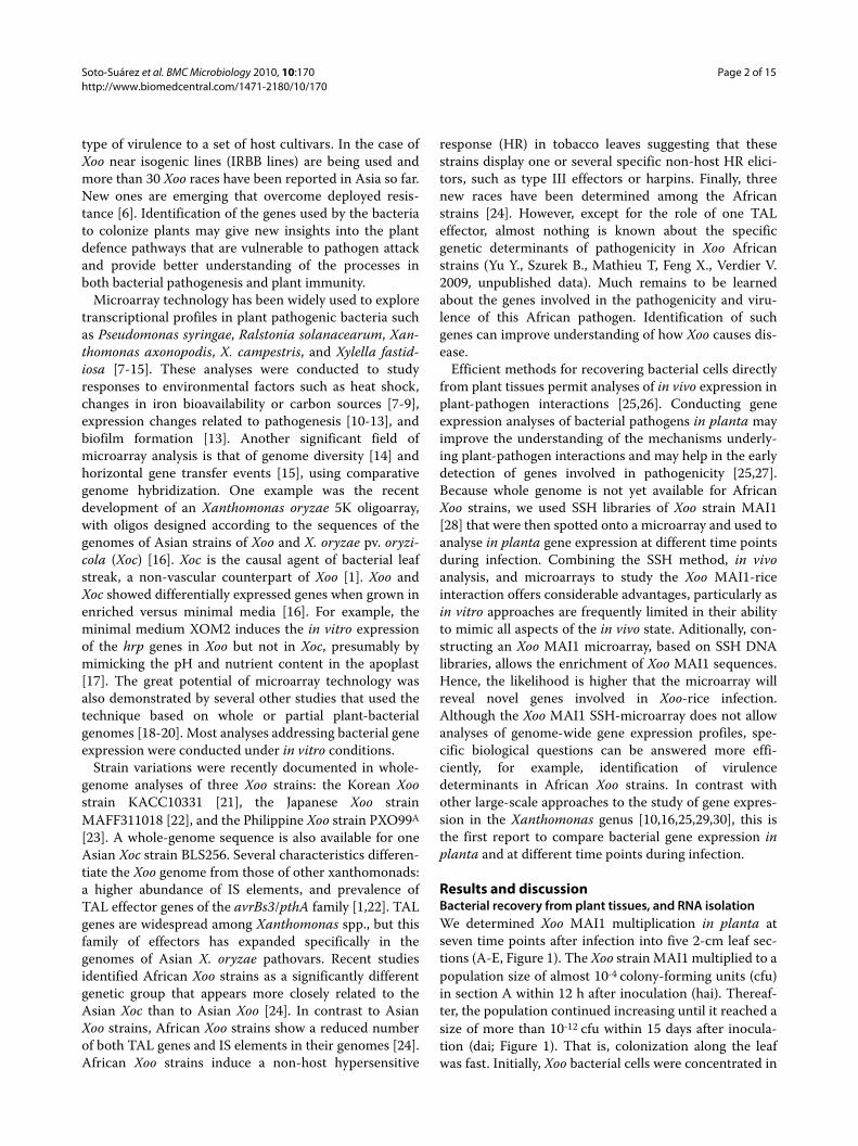
Identification of differentially expressed genesA total of 710 differentially expressed genes were one-endsequenced. After eliminating for low quality and vectorcontamination, 535 sequences were obtained. Insert sizevaried between 112 and 1902 bp, with an average of 660bp. The initial data set of 535 good sequences wasreduced to 147 unique consensus sequences, comprising57 contigs and 90 singletons. To annotate the Xoo MAI1non-redundant sequences, we used the Gene Ontology(GO) functional classification scheme [31]. Most func-tionally assigned non-redundant sequences (52%) fell intotwo classes: proteins with unknown function and biologi-cal process unknown (Figure 2). Mobile genetic elements,such as phage-related and IS elements, were well repre-sented (18%). Secretion, transport, and binding proteins,together with virulence-related sequences, represented14% of the differentially regulated genes (Figure 2).
Thirty genes are specifically regulatedThe set of 147 unique consensus sequences differentiallyexpressed during infection, was searched against thegenomes of all available sequenced strains of X. oryzae
Table 1: Statistical summary of Significance Analyses of Microarrays (SAM)
Gene expression Days after inoculation
1 3 6
Delta-delta Ct value 1.21 2.12 2.37
False significant number (FSN) 4.99 0.80 1.35
False discovery rate (FDR) 3.80 0.48 0.25
Up-regulated 58 (47%) 96 (40%) 253 (57%)
Down-regulated 66 (53%) 43 (60%) 194 (43%)
Total 124 239 447
The number of up- and down-regulated genes that are differentially expressed at different time points during infection by Xanthomonas oryzae pv. oryzae, African strain MAI1.
Figure 1 In planta quantification of bacteria. Bacterial growth in 8-week old rice variety Nipponbare, in sections A, B, C, D, and E of the leaf at 0 and 12 h, and 1, 3, 6, 10, and 15 days after inoculation. The experi-ment was repeated three times with three leaves per time point. Error bars indicate standard errors.
0
2
4
6
8
10
12
14
0d 1/2d 1d 3d 4d 5d 6d 10d 15d
Time points
LO
G10
(cfu
/cm
) A
B
C
D
E
Soto-Suárez et al. BMC Microbiology 2010, 10:170http://www.biomedcentral.com/1471-2180/10/170
Page 4 of 15
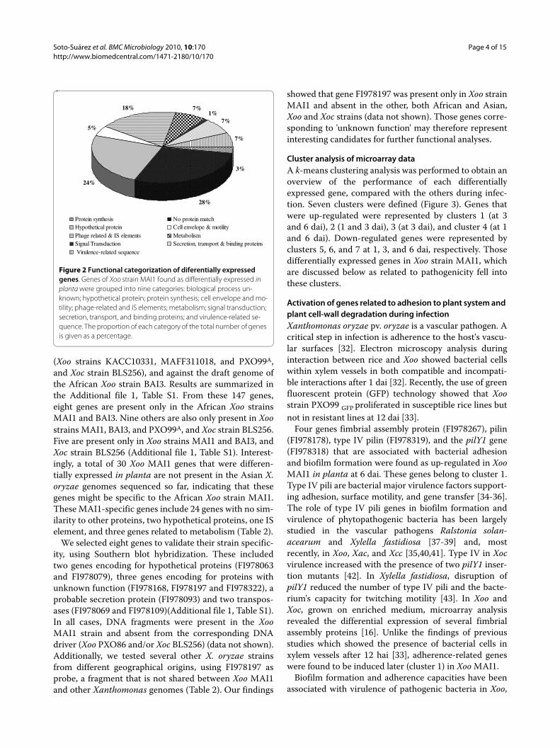
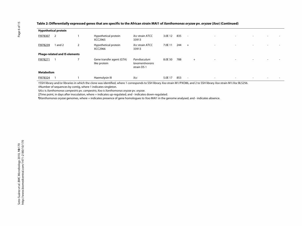
(Xoo strains KACC10331, MAFF311018, and PXO99A,and Xoc strain BLS256), and against the draft genome ofthe African Xoo strain BAI3. Results are summarized inthe Additional file 1, Table S1. From these 147 genes,eight genes are present only in the African Xoo strainsMAI1 and BAI3. Nine others are also only present in Xoostrains MAI1, BAI3, and PXO99A, and Xoc strain BLS256.Five are present only in Xoo strains MAI1 and BAI3, andXoc strain BLS256 (Additional file 1, Table S1). Interest-ingly, a total of 30 Xoo MAI1 genes that were differen-tially expressed in planta are not present in the Asian X.oryzae genomes sequenced so far, indicating that thesegenes might be specific to the African Xoo strain MAI1.These MAI1-specific genes include 24 genes with no sim-ilarity to other proteins, two hypothetical proteins, one ISelement, and three genes related to metabolism (Table 2).
We selected eight genes to validate their strain specific-ity, using Southern blot hybridization. These includedtwo genes encoding for hypothetical proteins (FI978063and FI978079), three genes encoding for proteins withunknown function (FI978168, FI978197 and FI978322), aprobable secretion protein (FI978093) and two transpos-ases (FI978069 and FI978109)(Additional file 1, Table S1).In all cases, DNA fragments were present in the XooMAI1 strain and absent from the corresponding DNAdriver (Xoo PXO86 and/or Xoc BLS256) (data not shown).Additionally, we tested several other X. oryzae strainsfrom different geographical origins, using FI978197 asprobe, a fragment that is not shared between Xoo MAI1and other Xanthomonas genomes (Table 2). Our findings
showed that gene FI978197 was present only in Xoo strainMAI1 and absent in the other, both African and Asian,Xoo and Xoc strains (data not shown). Those genes corre-sponding to 'unknown function' may therefore representinteresting candidates for further functional analyses.
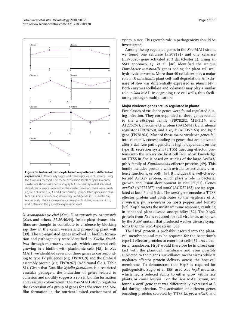
Cluster analysis of microarray dataA k-means clustering analysis was performed to obtain anoverview of the performance of each differentiallyexpressed gene, compared with the others during infec-tion. Seven clusters were defined (Figure 3). Genes thatwere up-regulated were represented by clusters 1 (at 3and 6 dai), 2 (1 and 3 dai), 3 (at 3 dai), and cluster 4 (at 1and 6 dai). Down-regulated genes were represented byclusters 5, 6, and 7 at 1, 3, and 6 dai, respectively. Thosedifferentially expressed genes in Xoo strain MAI1, whichare discussed below as related to pathogenicity fell intothese clusters.
Activation of genes related to adhesion to plant system and plant cell-wall degradation during infectionXanthomonas oryzae pv. oryzae is a vascular pathogen. Acritical step in infection is adherence to the host's vascu-lar surfaces [32]. Electron microscopy analysis duringinteraction between rice and Xoo showed bacterial cellswithin xylem vessels in both compatible and incompati-ble interactions after 1 dai [32]. Recently, the use of greenfluorescent protein (GFP) technology showed that Xoostrain PXO99 GFP proliferated in susceptible rice lines butnot in resistant lines at 12 dai [33].
Four genes fimbrial assembly protein (FI978267), pilin(FI978178), type IV pilin (FI978319), and the pilY1 gene(FI978318) that are associated with bacterial adhesionand biofilm formation were found as up-regulated in XooMAI1 in planta at 6 dai. These genes belong to cluster 1.Type IV pili are bacterial major virulence factors support-ing adhesion, surface motility, and gene transfer [34-36].The role of type IV pili genes in biofilm formation andvirulence of phytopathogenic bacteria has been largelystudied in the vascular pathogens Ralstonia solan-acearum and Xylella fastidiosa [37-39] and, mostrecently, in Xoo, Xac, and Xcc [35,40,41]. Type IV in Xocvirulence increased with the presence of two pilY1 inser-tion mutants [42]. In Xylella fastidiosa, disruption ofpilY1 reduced the number of type IV pili and the bacte-rium's capacity for twitching motility [43]. In Xoo andXoc, grown on enriched medium, microarray analysisrevealed the differential expression of several fimbrialassembly proteins [16]. Unlike the findings of previousstudies which showed the presence of bacterial cells inxylem vessels after 12 hai [33], adherence-related geneswere found to be induced later (cluster 1) in Xoo MAI1.
Biofilm formation and adherence capacities have beenassociated with virulence of pathogenic bacteria in Xoo,
Figure 2 Functional categorization of diferentially expressed genes. Genes of Xoo strain MAI1 found as differentially expressed in planta were grouped into nine categories: biological process un-known; hypothetical protein; protein synthesis; cell envelope and mo-tility; phage-related and IS elements; metabolism; signal transduction; secretion, transport, and binding proteins; and virulence-related se-quence. The proportion of each category of the total number of genes is given as a percentage.
3%
28%
24%
5%
18% 7%1%
7%
7%
Protein synthesis No protein matchHypothetical protein Cell envelope & motilityPhage related & IS elements MetabolismSignal Transduction Secretion, transport & binding proteins Virulence-related sequence
e (Xoo)
nthomonas oryzae genome¶
FF 311018 KACC 10331 PXO 99A BLS 256 BAI3
- - - - -
- - - - -
- - - - -
- - - - -
- - - - -
- - - - -
- - - - -
- - - - -
- - - - -
- - - - -
- - - - -
- - - - -
- - - - -
- - - - -
- - - - -
- - - - -
- - - - -
- - - - -
- - - - -
- - - - -
- - - - -
- - - - -
- - - - -
- - - - -
- - - - -
- - - - -
Soto
-Suá
rez
et a
l. BM
C M
icro
biol
ogy
2010
, 10:
170
http
://w
ww
.bio
med
cent
ral.c
om/1
471-
2180
/10/
170
Page
5 o
f 15
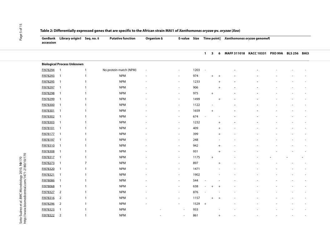
Table 2: Differentially expressed genes that are specific to the African strain MAI1 of Xanthomonas oryzae pv. oryza
GenBank accession
Library origin† Seq. no. ‡ Putative function Organism § E-value Size Time point|| Xa
1 3 6 MA
Biological Process Unknown
FI978294 1 1 No protein match (NPM) - - 1203 -
FI978293 1 1 NPM - - 974 + +
FI978295 1 1 NPM - - 1233 +
FI978297 1 1 NPM - - 906 +
FI978298 1 1 NPM - - 975 +
FI978299 1 1 NPM - - 1499 +
FI978300 1 1 NPM - - 1122 -
FI978301 1 1 NPM - - 1659 +
FI978302 1 1 NPM - - 674 - -
FI978303 1 1 NPM - - 1232 +
FI978101 1 1 NPM - - 409 +
FI978177 1 1 NPM - - 399 +
FI978197 1 1 NPM - - 248 -
FI978310 1 1 NPM - - 942 +
FI978308 1 1 NPM - - 931 +
FI978317 1 1 NPM - - 1175 +
FI978273 1 7 NPM - - 897 +
FI978320 1 1 NPM - - 1471 -
FI978321 1 1 NPM - - 1902 -
FI978086 1 1 NPM - - 544 - -
FI978068 1 1 NPM - - 638 - + +
FI978327 2 1 NPM - - 876 - -
FI978316 2 1 NPM - - 1157 + +
FI978296 2 1 NPM - - 1529 +
FI978323 1 1 NPM - - 933 -
FI978322 2 1 NPM - - 861 +
- - - - -
- - - - -
- - - - -
- - - - -
rary Xoo strain M1/Xoc BLS256.
absence.
e (Xoo) (Continued)
Soto-S
uáre
z et
al.
BMC
Mic
robi
olog
y 20
10, 1
0:17
0ht
tp://
ww
w.b
iom
edce
ntra
l.com
/147
1-21
80/1
0/17
0Pa
ge 6
of 1
5
Hypothetical protein
FI978307 2 1 Hypothetical protein XCC2965
Xcc strain ATCC 33913
3.0E 12 835 -
FI978239 1 and 2 2 Hypothetical protein XCC2966
Xcc strain ATCC 33913
7.0E 11 244 +
Phage-related and IS elements
FI978271 1 7 Gene transfer agent (GTA) like protein
Parvibaculum lavamentivorans strain DS 1
8.0E 50 788 +
Metabolism
FI978324 1 1 Haemolysin III Xcc 5.0E 17 853 -
†SSH library and/or libraries in which the clone was identified, where 1 corresponds to SSH library Xoo strain M1/PXO86, and 2 to SSH lib‡Number of sequences by contig, where 1 indicates singleton.§Xcc is Xanthomonas campestris pv. campestris; Xoo is Xanthomonas oryzae pv. oryzae.||Time point, in days after inoculation, where + indicates up-regulated, and - indicates down-regulated.¶Xanthomonas oryzae genomes, where + indicates presence of gene homologues to Xoo MAI1 in the genome analysed, and - indicates
Table 2: Differentially expressed genes that are specific to the African strain MAI1 of Xanthomonas oryzae pv. oryza
Soto-Suárez et al. BMC Microbiology 2010, 10:170http://www.biomedcentral.com/1471-2180/10/170
Page 7 of 15
X. axonopodis pv. citri (Xac), X. campestris pv. campestris(Xcc), and others [35,36,40,44]. Inside plant tissues, bio-films are thought to contribute to virulence by blockingsap flow in the xylem vessels and promoting plant wilt[39]. The up-regulated genes involved in biofilm forma-tion and pathogenicity were identified in Xylella fastid-iosa through microarray analysis, which compared cellsgrowing in a biofilm with planktonic cells [45]. In XooMAI1, we identified several of these genes as correspond-ing to type IV pili genes (e.g. FI978319) and the fimbrialassembly protein (e.g. FI978267) (Additional file 1, TableS1). Given that Xoo, like Xylella fastidiosa, is a restrictedvascular pathogen, the induction of genes related toadhesion and motility suggests a role in biofilm formationand vascular colonization. The Xoo MAI1 strain regulatesthe expression of a group of genes for adherence and bio-film formation in the nutrient-limited environment of
xylem in rice. This group's role in pathogenicity should beinvestigated.
Among the up-regulated genes in the Xoo MAI1 strain,we found one cellulase (FI978181) and one xylanase(FI978325) gene activated at 3 dai (cluster 1). Using anSSH approach, Qi et al. [46] identified the uniqueFibrobacter intestinalis genes coding for plant cell-wallhydrolytic enzymes. More than 40 cellulases play a majorrole in F. intestinalis plant cell-wall degradation. An xyla-nase of Xoo was differentially expressed in planta [47].Both enzymes (cellulase and xylanase) may play a similarrole in Xoo MAI1 in degrading rice cell walls, thus facili-tating pathogen multiplication.
Major virulence genes are up-regulated in plantaFive classes of virulence genes were found regulated dur-ing infection. They corresponded to three genes relatedto the avrBs3/pth family (FI978282, M1P3I15, andAF275267), a leucin-rich protein (BAE68417), a virulenceregulator (FI978260), and a xopX (ACD57163) and hrpFgene (FI978263). Most of these major virulence genes fellinto cluster 1, corresponding to genes that are activatedafter 3 dai. Xoo pathogenicity is highly dependent on thetype III secretion system (TTSS) injecting effector pro-teins into the eukaryotic host cell [48]. Most knowledgeon TTSS in Xoo is based on studies of the large AvrBs3/pthA family of Xanthomonas effector proteins [49]. Thisfamily includes proteins with avirulence activities, viru-lence functions, or both [48]. It includes the well-charac-terized AvrXa7 protein, which plays a role in bacterialgrowth and lesion development in rice [50,51]. GenesavrXa7 (AF275267) and xopX (ACD57163) are up-regu-lated at both 3 and 6 dai. The xopX gene encodes a TTSSeffector protein and contributes to the virulence of X.campestris pv. vesicatoria on hosts pepper and tomato[52]. XopX targets the innate immune response, resultingin enhanced plant disease susceptibility [52]. The XopXprotein from Xcc is required for full virulence, as shownby the XccN mutant that produced weaker disease symp-toms than the wild-type strain [53].
The HrpF protein is probably inserted into the plant-cell membrane and may be required for the bacterium'stype III effector proteins to enter host cells [54]. As a bac-terial translocon, HrpF would therefore be in direct con-tact with the plant-cell membrane and even possiblysubjected to the plant's surveillance mechanisms while itmediates effector protein delivery across the host-cellmembrane. To demonstrate that HrpF is required forpathogenicity, Sugio et al. [55] used Xoo hrpF mutants,which had a reduced ability to either grow within riceplants or cause lesions. For the Xoo MAI1 strain, wefound a hrpF gene that was differentially expressed at 3dai during infection. The activation of different genesencoding proteins secreted by TTSS (hrpF, avrXa7, and
Figure 3 Clusters of transcripts based on patterns of differential expression. Differentially expressed transcripts were clustered, using the k-means method. The mean expression levels of genes in each cluster are shown as a centroid graph. Error bars represent standard deviations of expression within the cluster. Seven clusters were creat-ed, with clusters 1, 2, 3, and 4 comprising up-regulated genes and clus-ters 5, 6, and 7 comprising down-regulated genes at 1, 3, and 6 dai, respectively. The x axis represents time-points during infection (1, 3, and 6 dai) and the y axis the expression level.
Cluster 1 Cluster2
1 3 6 1 3 6 Cluster 3 Cluster4
1 3 6 1 3 6 Cluster 5 Cluster 6
1 3 6 1 3 6 Cluster 7
1 3 6
Soto-Suárez et al. BMC Microbiology 2010, 10:170http://www.biomedcentral.com/1471-2180/10/170
Page 8 of 15
xopX genes) during Xoo MAI1-rice interaction was con-sistent with TTSS being essential for Xoo pathogenicity.
Expression of IS elements in Xoo MAI1 during infectionInsertion sequence (IS) elements have recently beenshown to play a role in plant pathogenicity [56-59]. Theseelements may inactivate genes on insertion or activateand/or enhance the expression of nearby genes[57,60,61]. One characteristic of the Xoo genomessequenced to date is the accumulation of many IS ele-ments, representing as much as 10% of the Xoo genomesize [23]. In Xanthomonas spp., virulence and pathoge-nicity islands are commonly associated with mobilegenetic elements such as phages and transposons [56,58].By comparing gene expression of both Xoo and Xocgrown in enriched versus minimal medium, Seo et al. [16]determined that IS elements are differentially expressedin minimal medium.
In our study, we identified 27 IS elements in Xoo MAI1that are up- or down-regulated in planta. Most of theseIS elements belong to cluster 1, corresponding to genesthat are activated after 3 dai. Twelve elements were classi-fied into the following IS families: IS30 (4 elements), IS5(7), and IS3 (1), with 15 IS elements unclassified. Mem-bers of the IS5 family have been reported previously inbacterial pathogens and it has been speculated thatexpression of some pathogenicity genes might be con-trolled by the expression/insertion of IS5 family elements[58,62,63]. Expression of IS5 members in the neighboringregion of their hrp gene cluster was observed inPseudomonas syringae [64] It has been also demonstratedthat IS elements (among them some IS5 family members)can act as a mobile switch for the downstream genes, cre-ating new transcriptional promoters and increasing theexpression levels of downstream genes [65]. Members ofthe IS3 and IS30 families have also been reported in bac-terial pathogens, some of them controlling the expressionof other genetic elements [60,66]. The expression of ISelements in Xoo MAI1 in planta suggests that these ele-ments may play a significant role in bacterial pathogenic-ity or may be associated with genes related topathogenicity.
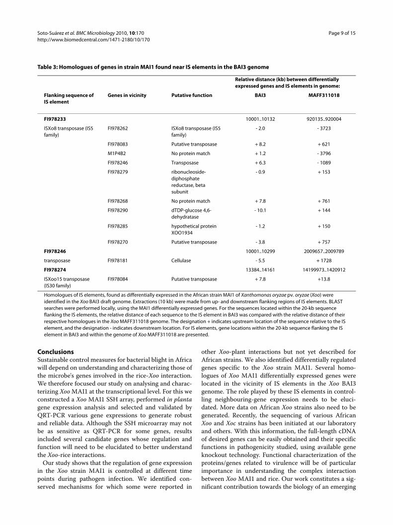
To establish a correlation between the presence of ISelements and adjacent genes differentially expressed inMAI1, we used the draft genome of Xoo African strainBAI3 (Genoscope project 154/AP 2006-2007 and our lab-oratory, 2009, unpublished data) and the publishedgenome of Xoo strain MAFF311018 [22]. We comparedthe location of the 147 Xoo MAI1 differentially expressedgenes with the presence of adjacent IS elements in theXoo BAI3 and MAFF311018 genomes. For this, homolo-gous sequences of IS elements, found as differentiallyexpressed in the Xoo strain MAI1, were first identified inthe BAI3 draft genome. We then extracted 10 kb fromeach of up- and downstream flanking regions of IS ele-
ments. BLAST searches were performed against theseflanking regions, using the Xoo MAI1 non-redundant setof sequences. For the sequences located within 20 kb ofsequences flanking the IS elements, we compared the rel-ative distance of each sequence to the IS element in BAI3with the relative distance of their respective homologuesin the Xoo MAFF311018 genome (Table 3).
Results showed that homologues of the 11 selected XooMAI1 differentially expressed genes are located in thevicinity of IS elements in BAI3 genome, within the same20-kb region (Table 3). In the Xoo MAFF311018 genome,Xoo MAI1 differentially expressed genes are not locatedin a vicinity of 20 kb of the IS elements. Given that theAfrican Xoo strain BAI3 is more closely related to XooMAI1 than Xoo MAFF311018, a similar organization ofIS elements and presence of neighbour genes areexpected for MAI1. Correlation between differentialexpression of IS elements, genome location, and roleplayed in the control of expression of nearby genes inAfrican Xoo strains need further study.
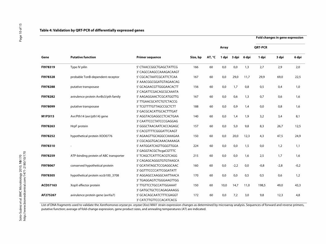
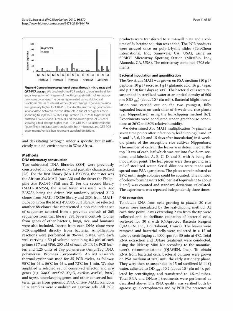
Validation of differentially expressed genes, using QRT-PCRTo validate the Xoo MAI1 microarray results, QRT-PCRwas performed on a set of 14 genes of different functionsand which were up- or down-regulated during infection.Table 4 lists the primers, putative function, and averagefold-change expression of genes used for QRT-PCR vali-dation. The genes selected for QRT-PCR correspond tofour hypothetical proteins (FI978067, FI978252,FI978305, and FI978328), one gene showing no similarityto known proteins (FI978310), two putative transposases(FI978288 and FI978099), two genes related to transportand motility (FI978259 and FI978319), one hrpF gene(FI978263), and one avirulence protein from the AvrBs3/pthA family (F1978282), the avr/pth14 gene (M1P3I15),the xopX gene (ACD57163), and the avrXa7 gene(AF275267). Figure 4 shows five genes out of the 14 testedthat were up-regulated by QRT-PCR and having a largerthan 4-fold change. Of the 14 genes selected according tothe microarray data (Table 4), 13 were up-regulated and 1(F1978067) was down-regulated. The QRT-PCR resultssupported these data, and also showed that the geneexpression pattern was identical for all genes tested,except two (FI978259 and FI978319). Gene expressionvalues, however, differed between microarrays and QRT-PCR. As shown in Figure 4, the expression values for thefive genes FI978252, FI978263, FI978328, AF275267, andACD57163 were higher in QRT-PCR than for microarray,indicating that QRT-PCR may be more sensitive thanmicroarray analysis. The xopX gene (ACD57163) washighly up-regulated in Xoo strain MAI1 in planta, indi-cating that induction of this gene is important duringinteraction between Xoo strain MAI1 and rice. These fivegenes belonged to cluster 1.
Soto-Suárez et al. BMC Microbiology 2010, 10:170http://www.biomedcentral.com/1471-2180/10/170
Page 9 of 15
ConclusionsSustainable control measures for bacterial blight in Africawill depend on understanding and characterizing those ofthe microbe's genes involved in the rice-Xoo interaction.We therefore focused our study on analysing and charac-terizing Xoo MAI1 at the transcriptional level. For this weconstructed a Xoo MAI1 SSH array, performed in plantagene expression analysis and selected and validated byQRT-PCR various gene expressions to generate robustand reliable data. Although the SSH microarray may notbe as sensitive as QRT-PCR for some genes, resultsincluded several candidate genes whose regulation andfunction will need to be elucidated to better understandthe Xoo-rice interactions.
Our study shows that the regulation of gene expressionin the Xoo strain MAI1 is controlled at different timepoints during pathogen infection. We identified con-served mechanisms for which some were reported in
other Xoo-plant interactions but not yet described forAfrican strains. We also identified differentially regulatedgenes specific to the Xoo strain MAI1. Several homo-logues of Xoo MAI1 differentially expressed genes werelocated in the vicinity of IS elements in the Xoo BAI3genome. The role played by these IS elements in control-ling neighbouring-gene expression needs to be eluci-dated. More data on African Xoo strains also need to begenerated. Recently, the sequencing of various AfricanXoo and Xoc strains has been initiated at our laboratoryand others. With this information, the full-length cDNAof desired genes can be easily obtained and their specificfunctions in pathogenicity studied, using available geneknockout technology. Functional characterization of theproteins/genes related to virulence will be of particularimportance in understanding the complex interactionbetween Xoo MAI1 and rice. Our work constitutes a sig-nificant contribution towards the biology of an emerging
Table 3: Homologues of genes in strain MAI1 found near IS elements in the BAI3 genome
Relative distance (kb) between differentially expressed genes and IS elements in genome:
Flanking sequence of IS element
Genes in vicinity Putative function BAI3 MAFF311018
FI978233 10001..10132 920135..920004
ISXo8 transposase (IS5 family)
FI978262 ISXo8 transposase (IS5 family)
- 2.0 - 3723
FI978083 Putative transposase + 8.2 + 621
M1P4B2 No protein match + 1.2 - 3796
FI978246 Transposase + 6.3 - 1089
FI978279 ribonucleoside-diphosphate reductase, beta subunit
- 0.9 + 153
FI978268 No protein match + 7.8 + 761
FI978290 dTDP-glucose 4,6-dehydratase
- 10.1 + 144
FI978285 hypothetical protein XOO1934
- 1.2 + 150
FI978270 Putative transposase - 3.8 + 757
FI978246 10001..10299 2009657..2009789
transposase FI978181 Cellulase - 5.5 + 1728
FI978274 13384..14161 14199973..1420912
ISXoo15 transposase (IS30 family)
FI978084 Putative transposase + 7.8 +13.8
Homologues of IS elements, found as differentially expressed in the African strain MAI1 of Xanthomonas oryzae pv. oryzae (Xoo) were identified in the Xoo BAI3 draft genome. Extractions (10 kb) were made from up- and downstream flanking regions of IS elements. BLAST searches were performed locally, using the MAI1 differentially expressed genes. For the sequences located within the 20-kb sequence flanking the IS elements, the relative distance of each sequence to the IS element in BAI3 was compared with the relative distance of their respective homologues in the Xoo MAFF311018 genome. The designation + indicates upstream location of the sequence relative to the IS element, and the designation - indicates downstream location. For IS elements, gene locations within the 20-kb sequence flanking the IS element in BAI3 and within the genome of Xoo MAFF311018 are presented.
Soto
-Suá
rez
et a
l. BM
C M
icro
biol
ogy
2010
, 10:
170
http
://w
ww
.bio
med
cent
ral.c
om/1
471-
2180
/10/
170
Page
10
of 1
5
Table 4: Validation by QRT-PCR of differentially expressed genes
Fold changes in gene expression
Array QRT-PCR
Gene Putative function Primer sequence Size, bp AT, °C 1 dpi 3 dpi 6 dpi 1 dpi 3 dpi 6 dpi
FI978319 Type IV pilin 5' CTAACCGGCTGAGCTATTCG 166 60 0,0 0,0 1,3 2,7 2,9 2,0
3' CAGCCAAGCCAAAGACAAGT
FI978328 probable TonB-dependent receptor 5' CGCACTAATCGCATTCTCAA 167 60 0,0 29,0 11,7 29,9 69,0 22,5
3' AAACGGCGGATGTAGAACAG
FI978288 putative transposase 5' GCAGAACGTTGGGAACACTT 156 60 0,0 1,7 0,8 0,5 0,4 1,0
3' CAGATTCGACAGCGCAAATA
FI978282 avirulence protein AvrBs3/pth family 5' AAGAGGAACTCGCATGGTTG 167 60 0,0 0,6 1,3 0,7 0,6 1,6
3' TTGAACGCATCTGTCTACCG
FI978099 putative transposase 5' TCGTTTTGTTAGCCGCTCTT 188 60 0,0 0,9 1,4 0,0 0,8 1,6
3' GACGCACATTGCACTTTGAT
M1P3I15 Avr/Pth14 (avr/pth14) gene 5' AGGTACGAGGCCTCACTGAA 140 60 0,0 1,4 1,9 3,2 3,4 8,1
3' CAATTCCCTATCCCGAGGAG
FI978263 HrpF protein 3' GGGCTAACAATCACCAGAGC 157 60 0,0 5,0 9,8 8,3 26,7 12,5
5' CACGTTTTCGGGATTCAAGT
FI978252 hypothetical protein XOO0776 5' AGAAGTTGCAGGCCAAAGAA 150 60 0,0 20,0 12,3 4,3 47,5 24,9
3' CGCAGGTGACAAACAAAAGA
FI978310 - 5' AATGGATCAGTTGGGTTGGA 224 60 0,0 0,0 1,5 0,0 1,2 1,1
3' GAGGTACGCTtcgaCGTTTC
FI978259 ATP-binding protein of ABC transporter 5' TCAGCTCATTTCACGTCAGG 215 60 0,0 0,0 1,6 2,5 1,7 1,6
3' CAGAGCAGGGTGTGTAAGCA
FI978067 conserved hypothetical protein 5' GCATATAGCTCCGAGGCAAC 160 60 0,0 -2,2 0,0 -0,8 -2,8 -0,2
3' GGTTTCCCCATTCGGATATT
FI978305 hypothetical protein xccb100_3708 5' AGGAGCCAAGGCAATTAACA 170 60 0,0 0,0 0,5 0,5 0,6 1,2
3' TGAGGAGTCTGGGAAGTTGG
ACD57163 XopX effector protein 5' TTGTTCCTGCCATTGGAAAT 150 60 10,0 14,7 11,0 198,5 49,0 43,3
3' GATGCTGCTCCAGAGAAAGG
AF275267 avirulence protein gene (avrXa7) 5' GCACAGCAATCTTTCGAGGT 172 60 0,0 7,2 3,0 9,8 12,3 4,8
3' CATCTTGTTCCCACATCACG
List of DNA fragments used to validate the Xanthomonas oryzae pv. oryzae (Xoo) MAI1 strain expression changes as determined by microarray analysis. Sequences of forward and reverse primers, putative function; average of fold-change expression, gene product sizes, and annealing temperatures (AT) are indicated.
Soto-Suárez et al. BMC Microbiology 2010, 10:170http://www.biomedcentral.com/1471-2180/10/170
Page 11 of 15
and devastating pathogen under a specific, but insuffi-ciently studied, environment in West Africa.
MethodsDNA microarray constructionTwo subtracted DNA libraries (SSH) were previouslyconstructed in our laboratory and partially characterized[28]. For the first library (MAI1-PXO86), the tester wasthe African Xoo MAI1 (race A3) and the driver the Philip-pine Xoo PXO86 (Phil race 2). For the second library(MAI1-BLS256), the same tester was used, with XocBLS256 being the driver. We randomly selected 2112clones from MAI1-PXO86 library and 2304 from MAI1-BLS256. From the MAI1-PXO86 SSH library, we selectedanother 88 clones that represented a non-redundant setof sequences selected from a previous analysis of 265sequences from that library [28]. Several controls (clonesfrom genes of other bacteria, fungi, rice, and humans)were also included. Inserts from each DNA clone werePCR-amplified directly from bacteria. Amplificationreactions were performed in 96-well plates, with eachwell carrying a 50-μl volume containing 0.2 μM of eachprimer (T7 and SP6), 200 μM of each dNTP, 1× PCR buf-fer, and 1.25 units of Taq polymerase (AmpliTaq® DNApolymerase, Promega Corporation). An MJ Researchthermal cycler was used for 35 PCR cycles, as follows:95°C for 45 s, 56°C for 45 s, and 72°C for 1 min. We alsoamplified a selected set of conserved effector and hrpgenes (e.g. XopX, avrXa7, XopD, avrRxv, avrXv3, hpaF,and hrpx), housekeeping genes, and other conserved bac-terial genes from genomic DNA of Xoo MAI1. RandomPCR samples were visualized on agarose gels. All PCR
products were transferred to a 384-well plate and a vol-ume of 2× betaine solution was added. The PCR productswere arrayed once on poly-L-lysine slides (TeleChemInternational, Inc., Sunnyvale, CA, USA), using anSPBIO™ Microarray Spotting Station (MiraiBio, Inc.,Alameda, CA, USA). The microarray contained 4708 ele-ments.
Bacterial inoculation and quantificationThe Xoo strain MAI1 was grown on PSA medium (10 g l-1
peptone, 10 g l-1 sucrose, 1 g l-1 glutamic acid, 16 g l-1 agar,and pH 7.0) for 2 days at 30°C. The bacterial cells were re-suspended in sterilized water at an optical density of 600nm (OD 600) (about 10-9 cfu ml-1). Bacterial blight inocu-lation was carried out on the two youngest, fullyexpanded leaves on each tiller of 6-week-old rice plants(var. Nipponbare), using the leaf-clipping method [67].Experiments were conducted under greenhouse condi-tions at 26°C and 80% relative humidity.
We determined Xoo MAI1 multiplication in planta atseven time points after infection by leaf clipping (0 and 12h, and 1, 3, 6, 10, and 15 days after inoculation) in 8-week-old plants of the susceptible rice cultivar Nipponbare.The number of cells in the leaves was determined at thetop 10 cm of each leaf which was cut into five 2-cm sec-tions, and labelled A, B, C, D, and E, with A being theinoculation point. The leaf pieces were then ground in 1ml of sterilized water. Serial dilutions were made andspread onto PSA agar plates. The plates were incubated at28°C until single colonies could be counted. The numberof colony-forming units (cfu) per leaf (equivalent to about2 cm2) was counted and standard deviations calculated.The experiment was repeated independently three times.
RNA extractionTo obtain RNA from cells growing in planta, 30 riceleaves were inoculated by the leaf-clipping method. Ateach time point, leaves extending 2 cm from the tip werecollected and, to facilitate exudation of bacterial cells,vortexed for 30 s with RNAprotect Bacteria Reagent(QIAGEN, Inc., Courtaboeuf, France). The leaves wereremoved and bacterial cells were collected in a 15-mltube by centrifuging at 4000 rpm for 30 min at 4°C. TotalRNA extraction and DNase treatment were conducted,using the RNeasy Mini Kit according to the manufac-turer's recommendations (QIAGEN, Inc.). To obtainRNA from bacterial cells, bacterial cultures were grownon PSA medium at 28°C until the early stationary phase.They were then re-suspended in 15 ml sterilized Milli-Qwater, adjusted to OD 600 of 0.2 (about 10-8 cfu ml-1), pel-leted by centrifuging, and transferred to 1.5-ml tubes.Total RNA and DNase I treatments were performed asdescribed above. The RNA quality was verified both byagarose-gel electrophoresis and by PCR (for presence of
Figure 4 Comparing expression of genes through microarray and QRT-PCR assays. We used real-time PCR analysis to confirm the differ-ential expression of 14 genes of the African strain MAI1 of Xanthomo-nas oryzae pv. oryzae. The genes represented various biological functional classes of interest. Although fold change in gene expression was generally higher for QRT-PCR than for the microarray, good corre-lation existed between the two data sets. A subset of 5 genes corre-sponding to xopX (ACD57163), HrpF protein (FI978263), hypothetical proteins (FI978252 and FI978328), and the avrXa7 gene (AF275267) showing a fold-change higher than 10 in QRT-PCR is illustrated in the figure. Three replicates were analysed in both microarray and QRT-PCR experiments. Vertical bars represent standard deviations.
0
50
100
150
200
250
1d 3d 6d 1d 3d 6d 1d 3d 6d 1d 3d 6d 1d 3d 6d
FI978263 FI978252 FI978328 AF275267 ACD57163
Rel
ativ
e ex
pres
sion
qRT-PCR
Array
Soto-Suárez et al. BMC Microbiology 2010, 10:170http://www.biomedcentral.com/1471-2180/10/170
Page 12 of 15
genomic DNA), using the genomic region flanking thehrpX gene as control and purified RNA as the PCR tem-plate. About 1 μg of Xoo MAI1 total RNA, obtained fromcells grown in culture medium or in planta and treatedwith DNase I, were used individually to synthesize single-stranded cDNA. The SMART™ PCR cDNA Synthesis Kit(BD Biosciences Clontech) was used, following the manu-facturer's instructions. The cDNA was then quantified,using the PicoGreen® reagent (Invitrogen, Ltd., Paisley,UK), an ultra-sensitive, fluorescent, and nucleic dye.
DNA microarray hybridizationFluorescent-labelled probes were prepared, following theKlenow labelling method (indirect labelling). Briefly, 500ng of cDNA were labelled, using 1 μl of either Cy3- orCy5-dUTP (Amersham Pharmacia Biotech, Little Chal-font, UK), 10 U Exonuclease-Free Klenow (USB Corpora-tion, Cleveland, OH, USA), and 3 μg random primers(Invitrogen Life Technologies, Carlsbad, CA, USA), andincubated 2 h at 37°C. Unincorporated nucleotides wereremoved, using a QIAquick PCR Purification Kit (QIA-GEN, Inc.). Cleaned probes were concentrated in aspeedvac (Eppendorf® Vacufuge Concentrator 5301,Hamburg, Germany).
Before hybridization, glass slides were snap-dried on a95°C heating block for 10 s. DNA was crosslinked to theslides, using 65 mJ of 254-nm UV-C radiation from aStratalinker® UV Crosslinker (model 2400, Stratagene, LaJolla, CA, USA). Slides were incubated 2 h at 70°C andpre-hybridized with 1% BSA, 5× SSC buffer, and 0.1% (w/v) SDS for 45 min at 54°C. The hybridization mixtureconsisted of 500 ng labelled cDNA and 4.5 μg μl-1 ofsalmon sperm DNA (Invitrogen Life Technologies) in afinal volume of 35 μl. This volume was mixed with 35 μlof 2× hybridization buffer (1× formamide, 1× SSC, and0.04× SDS). The mixture was denatured at 95°C for 2 minand transferred to ice. The hybridization mixture wasapplied to a microarray slide, transferred immediately toa hybridization chamber (Corning, Inc., Lowell, MA,USA), and incubated overnight (15-17 h) at 42°C. Theslide was then washed for 5 min successively in each of 2×SSC, 0.1% (w/v) SDS at 54°C, 1× SSC, and 0.1× SSC atroom temperature. Slides were immediately dried by cen-trifuging at 1000 rpm for 4 min.
At each time point, cDNA, obtained from bacteria usedas inoculum and re-suspended in water (time 0), wascompared with bacteria recovered from inoculated plantsat 1, 3, and 6 dai. The comparisons made were 0 - 1, 0 - 3,and 0 - 6 dai. To maximize the statistical reliability of thedata, three biological replicates were carried out.
In addition, for each time point comparison and eachbiological replicate, three technical replicates (cDNAobtained from the same mRNA extraction) were used forhybridization. For one of the three technical replicates,
the labelling of the two cDNA samples with either Cy5 orCy3 fluorescent dye was reversed to prevent potentialdye-related differences in labelling efficiency. Overall, 27images were analysed, 9 for each time point during Xooinfection. The nine data points obtained for each genewere used in the analyses.
Microarray data analysisThe slides were scanned, using a chip reader/scanner(Virtek Vision International, Inc., Waterloo, ON, Can-ada). The signal was initially normalized during imagescanning to adjust the average ratio between the twochannels, using control spots. Spot intensities fromscanned slides were quantified, using the Array-Pro 4.0software (Media Cybernetics, Inc., Silver Spring, MD,USA). With this program, local corner background cor-rection was carried out. Array-Pro 4.0 output data files(in Excel) were used to perform the lowest intensity nor-malization, standard deviation regularization, low inten-sity filtering, and dye-swap analysis, using the MIDAScomputer program [68]. Normalization between differentslides was carried out by centring [69]. MIDAS [68] wasalso used for replicate analysis and dye-swap filtering.
Bootstrap analyses with SAM enabled us to identify thedifferentially expressed genes, using a cut-off of two andadjusting the delta-delta Ct value, FDR, and FSN to mini-mize the number of false positives genes [70]. We con-ducted k-means clustering analysis to group the cDNAclones according to the similarity of their expression pat-terns, using MeV software available from TIGR and thedefault options [68].
Sequence data analysisThe 710 genes identified as differentially expressed wereone-end sequenced. Sequence data were processed, usinga PerlScript pipeline, to remove vector and low-qualitysequences and to assemble sequences into a non-redun-dant set of sequences [71]. The Xoo MAI1 non-redundantset of sequences was deposited at GenBank's GSS Data-base http://www.ncbi.nlm.nih.gov/dbGSS/[72], underaccession numbers FI978231-FI978329.
Processed sequences were initially searched against theNCBI database with BLASTN and TBLASTX http://blast.ncbi.nlm.nih.gov/Blast.cgi[73], setting BLASTparameters to search against the complete non-redun-dant database and the genomes of Xoo strainsKACC10331, MAFF311018, and PXO99A, and Xoc strainBLS256. A BLAST search was also performed with thepartial genome of the African Xoo strain BAI3, which iscurrently being sequenced (Genoscope project 154/AP2006-2007 and our laboratory, 2009, unpublished data).Results of these comparisons are summarized in theAdditional file 1, Table S1. The Xoo MAI1 non-redundantset of sequences was grouped into functional categories,
Soto-Suárez et al. BMC Microbiology 2010, 10:170http://www.biomedcentral.com/1471-2180/10/170
Page 13 of 15
using Gene Ontology (GO), a functional classificationscheme http://www.geneontology.org[74].
To establish if differentially expressed genes are locatedin the vicinity of the IS elements in the genomes of XooAfrican strain BAI3 and Xoo Asian strain MAFF311018,we selected a region of 20 kb that flanked the IS elementsin both the MAI1 and BAI3 genomes. BLAST searcheswere performed against these flanking sequences, usingthe Xoo MAI1 non-redundant set of sequences. For thesequences located within the 20-kb sequence flanking theIS elements, the relative distance of each sequence to theIS element was calculated and compared between the twogenomes.
Southern blot analysis of differentially expressed genesSouthern blot analysis was used to confirm that the DNAfragments derived from individual clones were present inthe initial tester (Xoo MAI1 strain) and absent in thedriver DNA (Xoo PXO86 or Xoc BLS256 strain). Eightgenes (FI978063, FI978069, FI978079, FI978093,FI978109, FI978168, FI978197 and FI978322) wereselected according to sequence similarities and libraryorigin. Additionally, the gene FI978197 was selected toscreen genomic DNA from different Xoo Asian strains(HN35, PXO339, PXO341, and PXO86), Xoo Africanstrains (MAI1, BAI3, NAI8, and BAI4), Xoc Africanstrains (MAI11 and MAI3), and the Xoc Asian strainBLS256 (Figure 2).
Briefly, for each strain, 5 μg of genomic DNA wasdigested with 10 units of RsaI and run on 0.8% agarosegels. The DNA was transferred to Hybond-N+ nylonmembranes (Amersham Pharmacia Biotech) by capillarytransfer. The insert DNA was amplified by PCR, using thenested primers provided with the PCR-Select™ BacterialGenome Subtraction Kit (Clontech Laboratories, Inc.).The amplified DNA fragment was gel purified, using theQIAquick Gel Extraction Kit (QIAGEN, Inc.), as recom-mended by the manufacturer. The DNA fragments werelabelled with [α32P] dCTP by random priming(MegaPrime labelling kit, Amersham Biosciences). Con-ditions of hybridization and washes were done at 65°C.Filters were washed with three solutions: the first of 2×SSC and 0.1% SDS for 20 min, followed by two washingswith 1× SSC and 0.1% SDS for 10 min each, and a finalwash with 0.1× SSC and 0.1% SDS for 20 min. Blots wereexposed on a PhosphorImager (model Storm 860, Amer-sham Pharmacia Biotech Inc.-Molecular Dynamics Divi-sion, Piscataway, NJ, USA).
Validation by quantitative QRT-PCRWe selected 14 genes that had been differentiallyexpressed at various time points during infection by XooMAI1 for confirmation by QRT-PCR. The primers forquantitative detection were designed, using the Beacon
Designer™ software (PREMIER Biosoft International,Palo Alto, CA, USA) (Table 4). All experiments were per-formed in triplicate. PCR mixtures were prepared, usingFullVelocity® SYBR® Green QPCR Master Mix (Strata-gene). PCR was performed on an Mx3005P thermalcycler (Stratagene), with the following cycling program:95°C for 5 min, followed by 40 cycles of 10 s at 95°C and30 s at 60°C.
Analysis was performed, using the delta-delta Ctmethod. The gene expression levels obtained by QRT-PCR were normalized, using the 16S ribosomal gene,which showed similar expression levels at different timepoints after infection. The gene expression level wascompared between microarray and QRT-PCR. Similarly,the microarray ratio for each gene analysed was norma-lised against the microarray ratio obtained for 16S ribo-somal gene. This allowed direct comparison between the16S ribosomal gene-normalized QRT-PCR ratio and the16S ribosomal gene microarray ratio for each transcriptinvestigated.
Additional material
Authors' contributionsMS JT and VV designed the research project. MS DB and CG constructed theSSH, prepared samples for microarray studies and performed the microarrayexperiments. MS and DB analyzed microarray data. MS and RG carried outsequence analysis, MS and BS designed QRT-PCR experiments. MS and VVdrafted the manuscript. All authors read and approved the final manuscript.
AcknowledgementsThe authors thank J.Leach (CSU) and CM.Vera Cruz (IRRI) for providing us with DNA of Xoo strains. We thank B.Piégu for his help with sequence analyses. We thank Thierry Mathieu for his help during greenhouse experiments. We are very grateful to Michèle Laudié for her help in preparing the materials for sequencing and Richard Cooke for access to the Montpellier Languedoc-Rous-sillon Génopole sequencing facilities. The authors thank Ralf Koebnik for his critical reading on the first draft of the manuscript and his helpful suggestions. We thank anonymous reviewers for their valuable suggestions to improve the manuscript. We thank Elizabeth McAdam for editing. MS was supported by a doctoral fellowship awarded by Programme Alβan of the European Commis-sion (grant E05D057941CO).
Author Details1UMR 5096 IRD-CNRS-Université de Perpignan, Laboratoire Génome et Développement des Plantes, Institut de Recherche pour le Développement, 911 Avenue Agropolis BP 64501, 34394 Montpellier Cedex 5, France, 2Biotechnology Research Unit, Centro Internacional de Agricultura Tropical (CIAT), A.A. 6713, Cali, Colombia and 3Laboratorio de Microbiología Molecular, Centro de Biotecnología y Bioindustria, Corporación Colombiana de Investigación Agropecuaria (Corpoica), Km 14 via a Mosquera, Cundinamarca, Colombia
Additional file 1 Xoo strain MAI1 genes identified as differentially expressed in planta by microarray analysis. The non-redundant set of sequences, composed of 147 Xoo strain MAI1 genes differentially expressed during infection, was searched against the genomes of all available sequenced strains of X. oryzae (Xoo strains KACC10331, MAFF311018, and PXO99A, and Xoc strain BLS256), and against the draft genome of the Afri-can Xoo strain BAI3. Changes in gene expression across different time points during infection are also presented.
Soto-Suárez et al. BMC Microbiology 2010, 10:170http://www.biomedcentral.com/1471-2180/10/170
Page 14 of 15
References1. Nino-Liu D, Ronald P, Bogdanove A: Xanthomonas oryzae pathovars:
model pathogens of a model crop. Mol Plant Pathol 2006, 7:303-324.2. Séré Y, Onasanya A, Verdier V, Akator K, Ouédraogo L, Segda Z, Coulibaly
M, Sido A, Basso A: Rice Bacterial Leaf Blight in West Africa: Preliminary Studies on Disease in Farmers' Fields and Screening Released Varieties for Resistance to the Bacteria. Asian Journal of Plant Sciences 2005, 4:577-579.
3. Leach J, Rhoads M, Vera Cruz C, White F, Mew T, Leung H: Assessment of genetic diversity and population structure of Xanthomonas oryzae pv. oryzae with a repetitive DNA element. Appl Environ Microbiol 1992, 58:2188-2195.
4. Nelson R, Baraoidan M, Vera Cruz C, Yap I, Leach J, Mew T, Leung H: Relationship between phylogeny and pathotype for the bacterial blight pathogen of rice. Appl Environ Microbiol 1994, 60:3275-3283.
5. Adhikari T, Mew T, Leach J: Genotypic and pathotypic diversity in Xanthomonas oryzae pv. oryzae in Nepal. Phytopathology 1999, 89:687-694.
6. Vera Cruz CM, Bai J, Oña I, Leung H, Nelson RJ, Mew T-W, Leach JE: Predicting durability of a disease resistance gene based on an assessment of the fitness loss and epidemiological consequences of avirulence gene mutation. Proc Natl Acad Sci USA 2000, 97:13500-13505.
7. Koide T, Vencio R, Gomes S: Global gene expression analysis of the heat shock response in the phytopathogen Xylella fastidiosa. Journal of bacteriology 2006, 188:5821-5830.
8. Bronstein P, Filiatrault M, Myers C, Rutzke M, Schneider D, Cartinhour S: Global transcriptional responses of Pseudomonas syringae DC3000 to changes in iron bioavailability in vitro. BMC Microbiology 2008, 8:209.
9. Serrania J, Vorhölter F-J, Niehaus K, Pühler A, Becker A: Identification of Xanthomonas campestris pv. campestris galactose utilization genes from transcriptome data. Journal of Biotechnology 2008, 135:309-317.
10. Ferreira AO, Myers CR, Gordon JS, Martin GB, Vencato M, Collmer A, Wehling MD, Alfano JR, Moreno-Hagelsieb G, Lamboy WF, DeClerck G, Schneider DJ, Cartinhour SW: Whole-Genome Expression Profiling Defines the HrpL Regulon of Pseudomonas syringae pv. tomato DC 3000, Allows de novo Reconstruction of the Hrp cisElement and Identifies Novel Coregulated Genes. Mol Plant Microbe Interact 2006, 19:1167-1179.
11. Lan L, Deng X, Xiao Y, Zhou J-M, Tang X: Mutation of Lon Protease Differentially Affects the Expression of Pseudomonas syringae Type III Secretion System Genes in Rich and Minimal Media and Reduces Pathogenicity. Mol Plant Microbe Interact 2007, 20:682-696.
12. He Y, Xu M, Lin K, Ng Y, Wen C, Wang L, Liu Z, Zhang H, Dong Y, Dow J, Zhang L: Genome scale analysis of diffusible signal factor regulon in Xanthomonas campestris pv. campestris: identification of novel cell-cell communication-dependent genes and functions. Molecular microbiology 2006, 59:610-622.
13. Shi XY, Dumenyo CK, Hernandez-Martinez R, Azad H, Cooksey DA: Characterization of Regulatory Pathways in Xylella fastidiosa: Genes and Phenotypes Controlled by gacA. Appl Environ Microbiol 2009, 75:2275-2283.
14. He Y, Zhang L, Jiang B, Zhang Z, Xu R, Tang D, Qin J, Jiang W, Zhang X, Liao J, Cao J, Zhang S, Liang X, Wei M, Lu G, Feng J, Chen B, Cheng J, Tang J: Comparative and functional genomics reveals genetic diversity and determinants of host specificity among reference strains and a large collection of Chinese isolates of the phytopathogen Xanthomonas campestris pv. campestris. Genome Biol 2007, 8:R218.
15. Guidot A, Coupat B, Fall S, Prior P, Bertollaq F: Horizontal gene transfer between Ralstonia solanacearum strains detected by comparative genomic hybridization on microarrays. The ISME J 2009, 3:549-562.
16. Seo Y-S, Sriariyanun M, Wang L, Pfeiff J, Phetsom J, Lin Y, Jung K-H, Chou HH, Bogdanove A, Ronald P: A two-genome microarray for the rice pathogens Xanthomonas oryzae pv. oryzae and X. oryzae pv. oryzicola and its use in the discovery of a difference in their regulation of hrp genes. BMC Microbiology 2008, 8:99.
17. Tsuge S, Ayako F, Rie F, Takashi O, Kazunori T, Hirokazu O, Yasuhiro I, Hisatoshi K, Yasuyuki K: Expression of Xanthomonas oryzae pv. oryzae
hrp Genes in XOM2, a Novel Synthetic Medium. J Gen Plant Path 2002, 68:363.
18. Lu S, Wang N, Wang J, Chen Z, Gross D: Oligonucleotide microarray analysis of the salA regulon controlling phytotoxin production by Pseudomonas syringae pv. syringae. Mol Plant Microbe Interact 2005, 18:324-333.
19. Valls M, Genin S, Boucher C: Integrated regulation of the type III secretion system and other virulence determinants in Ralstonia solanacearum. PLoS pathogens 2006, 2:e82.
20. Wang N, Lu S, Wang J, Chen Z, Gross D: The expression of genes encoding lipodepsipeptide phytotoxins by Pseudomonas syringae pv. syringae is coordinated in response to plant signal molecules. Mol Plant Microbe Interact 2006, 19:257-269.
21. Lee BM, Park YJ, Park DS, Kang HW, Kim JG, Song ES, Park IC, Yoon UH, Hahn JH, Koo BS, Lee GB, Kim H, Park HS, Yoon KO, Kim JH, Jung CH, Koh NH, Seo JS, GoS J: The genome sequence of Xanthomonas oryzae pathovar oryzae KACC10331 the bacterial blight pathogen of rice. Nucleic Acids Res 2005, 33:577-586.
22. Ochiai H, Inoue Y, Takeya M, Sasaki A, Kaku H: Genome sequence of Xanthomonas oryzae pv. oryzae suggests contribution of large numbers of effector genes and insertion sequences to its race diversity. Jpn Agri Res Quart 2005, 39:275-287.
23. Salzberg SL, Sommer DD, Schatz MC, Phillippy AM, Rabinowicz PD, Tsuge SA, Furutani A, Ochiai H, Delcher AL, Kelley D, Madupu R, Puiu D, Radune D, Shumway M, Trapnell C, Aparna G, Jha G, Pandey A, Patil PB, Ishihara H, Meyer DF, Szurek B, Verdier V, Koebnik R, Dow JM, Ryan RP, Hirata H, Tsuyumu S, Won Lee S, Seo YS, Sriariyanum M, Ronald PC, Sonti RV, Van Sluys MA, Leach JE, White FF, Bogdanove AJ: Genome sequence and rapid evolution of the rice pathogen Xanthomonas oryzae pv. oryzae PXO99A. BMC Genomics 2008, 9:204.
24. Gonzalez C, Szurek B, Manceau C, Mathieu T, Sere Y, Verdier V: Molecular and pathotypic characterization of new Xanthomonas oryzae strains from West Africa. Mol Plant Microbe Interact 2007, 20:534-546.
25. Astua-Monge G, Freitas-Astua J, Bacocina G, Roncoletta J, Carvalho S, Machado M: Expression profiling of virulence and pathogenicity genes of Xanthomonas axonopodis pv. citri. Journal of bacteriology 2005, 187:1201-1205.
26. Mehta A, Rosato Y: A simple method for in vivo expression studies of Xanthomonas axonopodis pv. citri. Curr Microbiol 2003, 47:400-403.
27. Thwaites R, Spanu PD, Panopoulos NJ, Stevens C, Mansfield JW: Transcriptional Regulation of Components of the Type III Secretion System and Effectors in Pseudomonas syringae pv. phaseolicola. Mol Plant Microbe Interact 2004, 17:1250-1258.
28. Soto-Suárez M, González C, Piégu B, Tohme J, Verdier V: Genomic comparison between Xanthomonas oryzae pv. oryzae and Xanthomonas oryzae pv. oryzicola, using suppression subtractive hybridization. FEMS Microbiol Lett 2010, 308:16-23.
29. Metha A, Rosato Y: Identification of differentially expressed genes of Xanthomonas axonopodis pv. citri by representational difference analysis. Genetics and Molecular Biology 2005, 28:140-149.
30. Tamir-Ariel D: Identification of genes in Xanthomonas campestris pv. vesicatoria induced during its interaction with tomato. J Bacteriol 2007, 189:6359-6371.
31. Ashburner AM, Ball CA, Blake JA, Botstein D, Butler H, Cherry JM, Davis AP, Dolinski K, Dwight SS, Eppig JT, Harris MA, Hill DP, Issel-Tarver L, Kasarskis A, Suzanna L, Matese JC, Richardson JE, Ringwald M, Rubin GM, Sherlock G: Gene Ontology: tool for the unification of biology. Nature genetics 2000, 25:25-29.
32. Hilaire E, Young SA, Willard LH, McGee JD, Sweat T, Chittoor JM, Guikema JA, Leach JE: Vascular Defense Responses in Rice: Peroxidase Accumulation in Xylem Parenchyma Cells and Xylem Wall Thickening. Mol Plant Microbe Interact 2001, 14:1411-1419.
33. Han S-W, Park C-J, Lee S-W, Ronald P: An efficient method for visualization and growth of fluorescent Xanthomonas oryzae pv. oryzae in planta. BMC Microbiology 2008, 8:164.
34. Clausen M, Koomey M, Maier B: Dynamics of Type IV Pili Is Controlled by Switching Between Multiple States. Biophysical Journal 2009, 96:1169-1177.
35. Lim S-H, So B-H, Wang J-C, Song E-S, Park Y-J, Lee B-M, Kang H-W: Functional analysis of pilQ gene in Xanthomonas oryzae pv. oryzae, bacterial blight pathogen of rice. The Journal of Microbiology 2008, 46:214-220.
Received: 28 September 2009 Accepted: 11 June 2010 Published: 11 June 2010This article is available from: http://www.biomedcentral.com/1471-2180/10/170© 2010 Soto-Suárez et al; licensee BioMed Central Ltd. This is an Open Access article distributed under the terms of the Creative Commons Attribution License (http://creativecommons.org/licenses/by/2.0), which permits unrestricted use, distribution, and reproduction in any medium, provided the original work is properly cited.BMC Microbiology 2010, 10:170

Soto-Suárez et al. BMC Microbiology 2010, 10:170http://www.biomedcentral.com/1471-2180/10/170
Page 15 of 15
36. Mccarthy Y, Ryan R, O'dovan K, He Y, Jiang B, Feng J, Tang J, Dow J: The role of PilZ domain proteins in the virulence of Xanthomonas campestris pv. campestris. Molecular Plant Pathology 2008, 9:819-824.
37. Kang Y, Liu H, Genin S, Schell MA, Denny TP: Ralstonia solanacearum requires type 4 pili to adhere to multiple surfaces and for natural transformation and virulence. Molecular microbiology 2002, 46:427-437.
38. Liu H, Kang Y, Genin S, Schell MA, Denny TP: Twitching motility of Ralstonia solanacearum requires a type IV pilus system. Microbiology 2001, 147:3215-3229.
39. Meng Y, Li Y, Galvani C, Hao G, Turner J, Burr T, Hoch H: Upstream Migration of Xylella fastidiosa via Pilus-Driven Twitching Motility. J Bacteriol 2005, 187:5560-5567.
40. Gottig N, Garavaglia BS, Garofalo CG, Orellano EG, Ottado J: A Filamentous Hemagglutinin-Like Protein of Xanthomonas axonopodis pv. citri, the Phytopathogen Responsible for Citrus Canker, Is Involved in Bacterial Virulence. PLoS ONE 2009, 4:e4358.
41. Guzzo CR, Salinas RK, Andrade MO, Farah CS: PILZ Protein Structure and Interactions with PILB and the FIMX EAL Domain: Implications for Control of Type IV Pilus Biogenesis. J Mol Biol 2009, 393:848-866.
42. Wang L, Makino S, Subedee A, Bogdanove AJ: Novel Candidate Virulence Factors in Rice Pathogen Xanthomonas oryzae pv. oryzicola as Revealed by Mutational Analysis. Appl Environ Microbiol 2007, 73:8023-8027.
43. Lerouge I, Vanderleyden J: O-antigen structural variation: mechanisms and possible roles in animal/plant-microbe interactions. FEMS Microbiol Rev 2002, 26:17-47.
44. Darsonval A, Darrasse A, Durand K, Bureau C, Cesbron S, Jacques M-A: Adhesion and Fitness in the Bean Phyllosphere and Transmission to Seed of Xanthomonas fuscans subsp. fuscans. Mol Plant Microbe Interact 2009, 22:747-757.
45. de Souza AA TM, Coletta-Filho HD, Caldana C, Yanai GM, Muto NH, de Oliveira RC, Nunes LR, Machado MA: Gene expression profile of the plant pathogen Xylella fastidiosa during biofilm formation in vitro. FEMS Microbiol Lett 2004, 237:341-353.
46. Qi M, Nelson KE, Daugherty SC, Nelson WC, Hance IR, Morrison M, Forsberg CW: Novel Molecular Features of the Fibrolytic Intestinal Bacterium Fibrobacter intestinalis Not Shared with Fibrobacter succinogenes as Determined by Suppressive Subtractive Hybridization. J Bacteriol 2005, 187:3739-3751.
47. Rajeshwari R, Jha G, Sonti RV: Role of an In Planta-Expressed Xylanase of Xanthomonas oryzae pv. oryzae in Promoting Virulence on Rice. Mol Plant Microbe Interact 2005, 18:830-837.
48. White FF, Yang B: Host and Pathogen Factors Controlling the Rice-Xanthomonas oryzae Interaction. Plant Physiol 2009, 150:1677-1686.
49. Kay S, Bonas U: How Xanthomonas type III effectors manipulate the host plant. Current Opinion in Microbiology 2009, 12:37-43.
50. Yang B, Zhu W, Johnson LB, White FF: The virulence factor AvrXa7 of Xanthomonas oryzae pv. oryzae is a type III secretion pathway-dependent nuclear-localized double-stranded DNA-binding protein. Proc Natl Acad Sci USA 2000, 97:9807-9812.
51. Yang B, White F: Diverse members of the AvrBs3/PthA family of type III effectors are major virulence determinants in bacterial blight disease of rice. Mol Plant Microbe Interact 2004, 17:1192-1200.
52. Metz M, Dahlbeck D, Morales CQ, Al Sady B, Clark ET, Staskawicz BJ: The conserved Xanthomonas campestris pv. vesicatoria effector protein XopX is a virulence factor and suppresses host defense in Nicotiana benthamiana. The Plant Journal 2005, 41:801-814.
53. Jiang B-L, He Y-Q, Cen W-J, Wei H-Y, Jiang G-F, Jiang W, Hang X-H, Feng J-X, Lu G-T, Tang D-J, Tang J-L: The type III secretion effector XopXccN of Xanthomonas campestris pv. campestris is required for full virulence. Research in Microbiology 2008, 159:216-220.
54. Büttner D, Bonas U: Port of entry - the type III secretion translocon. Trends in Microbiology 2002, 10:186-192.
55. Sugio A, Yang B, White FF: Characterization of the hrpF Pathogenicity Peninsula of Xanthomonas oryzae pv. oryzae. Mol Plant Microbe Interact 2005, 18:546-554.
56. Lima W, Paquola A, Varani A, Van Sluys M, Menck C: Laterally transferred genomic islands in Xanthomonadales related to pathogenicity and primary metabolism. FEMS Microbiol Lett 2008, 281:87-97.
57. Zerillo M, Van Sluys M-A, Camargo L, Monteiro-Vitorello C: Characterization of new IS elements and studies of their dispersion in two subspecies of Leifsonia xyli. BMC Microbiology 2008, 8:127.
58. Monteiro-Vitorello C, de Oliveira M, Zerillo M, Varani A, Civerolo E, Van Sluys M: Xylella and Xanthomonas Mobil'omics. Omics 2005, 9:146-159.
59. Nierman WC, DeShazer D, Kim HS, Tettelin H, Nelson KE, Feldblyum T, Ulrich RL, Ronning CM, Brinkac LM, Daugherty SC, Davidsen TD, Deboy RT, Dimitrov G, Dodson RJ, Durkin AS, Gwinn ML, Haft DH, Khouri H, Kolonay JF, Madupu R, Mohammoud Y, Nelson WC, Radune D, Romero CM, Sarria S, Selengut J, Shamblin C, Sullivan SA, White O, Yu Y, Zafar N, Zhou L, Fraser CM: Structural flexibility in the Burkholderia mallei genome. Proc Natl Acad Sci USA 2004, 101:14246-14251.
60. Nagy Z, Chandler M: Regulation of transposition in bacteria. Res Microbiol 2004, 155:387-398.
61. Mahillon J, Leonard C, Chandler M: IS elements as constituents of bacterial genomes. Res Microbiol 1999, 150:675-687.
62. Thieme F, Koebnik R, Bekel T, Berger C, Boch J, Buttner D, Caldana C, Gaigalat L, Goesmann A, Kay S: Insights into genome plasticity and pathogenicity of the plant pathogenic bacterium Xanthomonas campestris pv. vesicatoria revealed by the complete genome sequence. J Bacteriol 2005, 187:7254-7266.
63. Jeong E-L, Timmis JN: Novel Insertion Sequence Elements Associated with Genetic Heterogeneity and Phenotype Conversion in Ralstonia solanacearum. J Bacteriol 2000, 182:4673-4676.
64. Inoue Y, Takikawa Y: Investigation of Repeating Sequences in hrpL Neighboring Region of Pseudomonas syringae Strains. Ann Phytopathol Soc Japan 1999, 65:100-109.
65. Felipe López de F, Magni C, de Mendoza D, López P: Transcriptional activation of the citrate permease P gene of Lactococcus lactis biovar diacetylactis by an insertion sequence-like element present in plasmid pCIT264. Mol Gen Genet 1996, 250:428-436.
66. Hasebe A, Iida S: The Novel Insertion Sequences IS1417 IS1418 and IS1419 from Burkholderia glumae and Their Strain Distribution. Plasmid 2000, 44:44-53.
67. Kauffman H, Reddy A, Hsieh S, Merca S: An improved technique for evaluating resistance of rice varieties to Xanthomonas oryzae. Plant Dis Rep 1973, 57:537-541.
68. Saeed AI, Sharov V, White J, Li J, Liang W, Bhagabati N, Braisted J, Klapa M, Currier T, Thiagarajan M, Sturn A, Snuffin M, Rezantsev A, Popov D, Ryltsov A, Kostukovich E, Borisovsky I, Liu Z, Vinsavich A, Trush V, Quackenbush J: TM4: a free open-source system for microarray data management and analysis. Biotechniques 2003, 34:374-378.
69. Stekel D: Microarray Bioinformatics. Cambridge University Press Cambridge; 2003.
70. Tusher VG, Tibshirani R, Chu G: Significance analysis of microarrays applied to the ionizing radiation response. Proc Natl Acad Sci USA 2001, 98:5116-5121.
71. Lopez C, Jorge V, Piégu B, Mba C, Cortes D, Restrepo S, Soto M, Laudie M, Berger C, Cooke R, Delseny M, Tohme J, Verdier V: A unigene catalogue of 5700 expressed genes in cassava. Plant Molecular Biology 2004, 56:541-554.
72. Genome Survey Sequences Database [http://www.ncbi.nlm.nih.gov/dbGSS/]
73. BLAST (Basic Local Alignment Search Tool), BLAST Assembled Genomes [http://blast.ncbi.nlm.nih.gov/Blast.cgi]
74. The Gene Ontology [http://www.geneontology.org/]
doi: 10.1186/1471-2180-10-170Cite this article as: Soto-Suárez et al., In planta gene expression analysis of Xanthomonas oryzae pathovar oryzae, African strain MAI1 BMC Microbiology 2010, 10:170