Improving thermostability and catalytic activity of pyranose 2-oxidase from Trametes multicolor by...
17
Improving thermostability and catalytic activity of pyranose 2-oxidase from Trametes multicolor by rational and semi-rational design Oliver Spadiut 1 , Christian Leitner 1 , Clara Salaheddin 1 , Bala ´ zs Varga 2 , Beata G. Vertessy 2 , Tien-Chye Tan 3 , Christina Divne 3 and Dietmar Haltrich 1 1 Department of Food Sciences and Technology, BOKU–University of Natural Resources and Applied Life Sciences, Vienna, Austria 2 Institute of Enzymology, Biological Research Center, Hungarian Academy of Sciences, Budapest, Hungary 3 School of Biotechnology, Royal Institute of Technology KTH, Albanova University Center, Stockholm, Sweden The flavoenzyme pyranose 2-oxidase (P2Ox; pyra- nose:oxygen 2-oxidoreductase; EC 1.1.3.10), a member of the glucose–methanol–choline family of FAD- dependent oxidoreductases [1], catalyses the oxidation of several aldopyranoses at position C-2 to yield the corresponding 2-ketoaldoses and H 2 O 2 as products. The enzyme is found in wood-degrading basidiomyce- tes, where it is localized in the hyphal periplasmic space. Presumably, P2Ox supplies lignin and manga- nese peroxidases with H 2 O 2 , an essential cosubstrate Keywords enzyme engineering; pyranose oxidase; stability; stabilization; subunit interaction Correspondence D. Haltrich, Department of Food Sciences and Technology, Universita ¨t fu ¨ r Bodenkultur Wien, Muthgasse 18, A-1190 Wien, Austria Fax: +43 1 36006 6251 Tel: +43 1 36006 6275 E-mail: [email protected] Database Structural data are available in the Protein Data Bank under the accession numbers 3BG6, 3BG7 and 3BLY (Received 25 June 2008, revised 19 November 2008, accepted 1 December 2008) doi:10.1111/j.1742-4658.2008.06823.x The fungal homotetrameric flavoprotein pyranose 2-oxidase (P2Ox; EC 1.1.3.10) catalyses the oxidation of various sugars at position C2, while, concomitantly, electrons are transferred to oxygen as well as to alternative electron acceptors (e.g. oxidized ferrocenes). These properties make P2Ox an interesting enzyme for various biotechnological applications. Random mutagenesis has previously been used to identify variant E542K, which shows increased thermostability. In the present study, we selected position Leu537 for saturation mutagenesis, and identified variants L537G and L537W, which are characterized by a higher stability and improved cata- lytic properties. We report detailed studies on both thermodynamic and kinetic stability, as well as the kinetic properties of the mutational variants E542K, E542R, L537G and L537W, and the respective double mutants (L537G ⁄ E542K, L537G ⁄ E542R, L537W ⁄ E542K and L537W ⁄ E542R). The selected substitutions at positions Leu537 and Glu542 increase the melting temperature by approximately 10 and 14 ŶC, respectively, relative to the wild-type enzyme. Although both wild-type and single mutants showed first-order inactivation kinetics, thermal unfolding and inactivation was more complex for the double mutants, showing two distinct phases, as revealed by microcalorimetry and CD spectroscopy. Structural information on the variants does not provide a definitive answer with respect to the sta- bilizing effects or the alteration of the unfolding process. Distinct differ- ences, however, are observed for the P2Ox Leu537 variants at the interfaces between the subunits, which results in tighter association. Abbreviations ABTS, azino-bis-(3-ethylbenzthiazolin-6-sulfonic acid); DSC, differential scanning calorimetry; Fc + , ferricenium ion; IMAC, immobilized metal affinity chromatography; Mes, 2-(N-morpholino) ethane sulfonic acid (4-morpholine ethane sulfonic acid); P2Ox, pyranose 2-oxidase; PDB, Protein Data Bank; PsP2Ox, pyranose oxidase from Peniophora sp.; TLS, translation, libration, screw-rotation; T m , melting temperature; TmP2Ox, pyranose oxidase from Trametes multicolor; TvP2Ox, pyranose oxidase from Trametes (Coriolus) versicolor; s 1 ⁄ 2 , half-life of activity. 776 FEBS Journal 276 (2009) 776–792 ª 2008 The Authors Journal compilation ª 2008 FEBS
Transcript of Improving thermostability and catalytic activity of pyranose 2-oxidase from Trametes multicolor by...

Improving thermostability and catalytic activity ofpyranose 2-oxidase from Trametes multicolor by rationaland semi-rational designOliver Spadiut1, Christian Leitner1, Clara Salaheddin1, Balazs Varga2, Beata G. Vertessy2,Tien-Chye Tan3, Christina Divne3 and Dietmar Haltrich1
1 Department of Food Sciences and Technology, BOKU–University of Natural Resources and Applied Life Sciences, Vienna, Austria
2 Institute of Enzymology, Biological Research Center, Hungarian Academy of Sciences, Budapest, Hungary
3 School of Biotechnology, Royal Institute of Technology KTH, Albanova University Center, Stockholm, Sweden
The flavoenzyme pyranose 2-oxidase (P2Ox; pyra-
nose:oxygen 2-oxidoreductase; EC 1.1.3.10), a member
of the glucose–methanol–choline family of FAD-
dependent oxidoreductases [1], catalyses the oxidation
of several aldopyranoses at position C-2 to yield the
corresponding 2-ketoaldoses and H2O2 as products.
The enzyme is found in wood-degrading basidiomyce-
tes, where it is localized in the hyphal periplasmic
space. Presumably, P2Ox supplies lignin and manga-
nese peroxidases with H2O2, an essential cosubstrate
Keywords
enzyme engineering; pyranose oxidase;
stability; stabilization; subunit interaction
Correspondence
D. Haltrich, Department of Food Sciences
and Technology, Universitat fur Bodenkultur
Wien, Muthgasse 18, A-1190 Wien, Austria
Fax: +43 1 36006 6251
Tel: +43 1 36006 6275
E-mail: [email protected]
Database
Structural data are available in the Protein
Data Bank under the accession numbers
3BG6, 3BG7 and 3BLY
(Received 25 June 2008, revised
19 November 2008, accepted 1 December
2008)
doi:10.1111/j.1742-4658.2008.06823.x
The fungal homotetrameric flavoprotein pyranose 2-oxidase (P2Ox; EC
1.1.3.10) catalyses the oxidation of various sugars at position C2, while,
concomitantly, electrons are transferred to oxygen as well as to alternative
electron acceptors (e.g. oxidized ferrocenes). These properties make P2Ox
an interesting enzyme for various biotechnological applications. Random
mutagenesis has previously been used to identify variant E542K, which
shows increased thermostability. In the present study, we selected position
Leu537 for saturation mutagenesis, and identified variants L537G and
L537W, which are characterized by a higher stability and improved cata-
lytic properties. We report detailed studies on both thermodynamic and
kinetic stability, as well as the kinetic properties of the mutational variants
E542K, E542R, L537G and L537W, and the respective double mutants
(L537G ⁄E542K, L537G ⁄E542R, L537W ⁄E542K and L537W ⁄E542R). The
selected substitutions at positions Leu537 and Glu542 increase the melting
temperature by approximately 10 and 14 �C, respectively, relative to the
wild-type enzyme. Although both wild-type and single mutants showed
first-order inactivation kinetics, thermal unfolding and inactivation was
more complex for the double mutants, showing two distinct phases, as
revealed by microcalorimetry and CD spectroscopy. Structural information
on the variants does not provide a definitive answer with respect to the sta-
bilizing effects or the alteration of the unfolding process. Distinct differ-
ences, however, are observed for the P2Ox Leu537 variants at the
interfaces between the subunits, which results in tighter association.
Abbreviations
ABTS, azino-bis-(3-ethylbenzthiazolin-6-sulfonic acid); DSC, differential scanning calorimetry; Fc+, ferricenium ion; IMAC, immobilized metal
affinity chromatography; Mes, 2-(N-morpholino) ethane sulfonic acid (4-morpholine ethane sulfonic acid); P2Ox, pyranose 2-oxidase; PDB,
Protein Data Bank; PsP2Ox, pyranose oxidase from Peniophora sp.; TLS, translation, libration, screw-rotation; Tm, melting temperature;
TmP2Ox, pyranose oxidase from Trametes multicolor; TvP2Ox, pyranose oxidase from Trametes (Coriolus) versicolor; s1 ⁄ 2, half-life
of activity.
776 FEBS Journal 276 (2009) 776–792 ª 2008 The Authors Journal compilation ª 2008 FEBS

for ligninolysis by wood-rotting fungi [2]. To date,
P2Ox from Trametes multicolor and Peniophora gigan-
tea comprises the best studied enzyme, both from a
biochemical and structural point of view [3–6]. Native
P2Ox from T. multicolor (TmP2Ox) is composed of
four identical 68 kDa subunits, resulting in a 270 kDa
homotetramer [7]. It contains the prosthetic group
FAD bound covalently via its 8a-methyl group to each
His167 Ne2 (i.e. N3) per subunit [8], which was also
confirmed from the crystal structure of TmP2Ox deter-
mined at 1.8 A resolution [3]. Structurally, the homo-
tetramer is described more accurately as a dimer of
dimers (i.e. dimers formed by the subunits A and B, as
well as C and D) (Fig. 1). Interaction between the
interfaces is most extensive between these two dimers
A–B and C–D, with a large number of hydrogen
bonds and hydrophobic contacts. These interactions
occur mainly via two distinct regions of the subunit
termed the oligomerization loop and oligomerization
arm. The latter is also involved in the interactions
between subunits A and D (B and C, respectively),
whereas the weakest interaction surfaces are observed
at the interface of the A–C (and B–D) pair. These lat-
ter interactions occur mainly via hydrophobic contacts
between residues 508–528 and 532–540 (segments H8
and B6, respectively) [3].
In accordance with other flavoprotein oxidoreducta-
ses, the reaction mechanism of P2Ox is of the typical
Ping Pong Bi Bi type [9,10]. In the reductive half-reac-
tion, an aldopyranose is oxidized at position C-2 to
yield a 2-ketoaldose (aldos-2-ulose), whereas FAD is
reduced to FADH2 (reaction 1) [11,12]. During the
ensuing oxidative half-reaction, FADH2 is re-oxidized
by the second substrate oxygen, yielding the oxidized
prosthetic group and H2O2 (reaction 2). In addition,
alternative electron acceptors, including either two-
electron acceptors such as benzoquinones (reaction 3)
or one-electron acceptors such as chelated metal ions
(e.g. the ferricenium ion or radicals), are used effi-
ciently by P2Ox instead of oxygen [7].
FADþaldopyranose!FADH2þ2-keto-aldopyranose ð1Þ
FADH2 þO2 ! FADþH2O2 ð2Þ
FADH2 þ benzoquinone! FADþ hydroquinone ð3Þ
P2Ox comprises an interesting biocatalyst in the bio-
transformations of carbohydrates because it can be
used to synthesize various carbohydrate derivates and
rare sugars [12]. Amongst others, the oxidation of
d-glucose and d-galactose to 2-keto-d-glucose and
2-keto-d-galactose is of applied interest because these
oxidized intermediates can be subsequently reduced at
position C-1 to obtain the ketoses d-fructose and
d-tagatose [13,14], which are of interest in the food
industry. P2Ox is not only useful for biotransforma-
tions of carbohydrates, but also for applications in
sensors or biofuel cells [15,16]. Recently, we demon-
strated the electrical wiring of P2Ox with an osmium
redox polymer serving as a redox mediator on graphite
electrodes [15]. Here, the redox polymer collects the
electrons from the prosthetic groups of the enzyme
and transfers them to the electrode. Other mediators
that have been investigated for providing contact
between P2Ox and the electrode include ruthenium or
modified ferrocenes [16]. For this bioelectrochemical
application, the reactivity of P2Ox with alternative
electron acceptors, and notably with (complexed) metal
ions such as the ferrocenes, is of significant impor-
tance.
As for many other enzymes applied in industry
[17,18], there is the need for more stable and active
P2Ox. To date, few attempts to improve P2Ox by
enzyme engineering have been reported. Studies on
P2Ox from Coriolus (Trametes) versicolor (TvP2Ox)
using random mutagenesis revealed the importance of
position Glu542, both for improved thermostability
and catalysis, with variant E542K showing an increase
in optimum temperature by 5 �C and a decrease in the
Michaelis constant Km for the two substrates d-glucose
and 1,5-anhydro-d-glucitol [19]. Subsequent studies on
P2Ox from Peniophora gigantea (PgP2Ox) and Penio-
Fig. 1. Ribbon drawing illustrating the tetrameric assembly of func-
tional P2Ox. The model 2IGO [4] is shown. The subunits A, B, C
and D are colored yellow, blue, red and green, respectively. The
tetramer molecule is overlaid with a gray solvent-accessible
surface.
O. Spadiut et al. Stabilization of pyranose oxidase
FEBS Journal 276 (2009) 776–792 ª 2008 The Authors Journal compilation ª 2008 FEBS 777

phora sp. (PsP2Ox) confirmed the beneficial effects of
the Glu fi Lys mutation at position 542 (540 for
PgP2Ox), and identified additional amino acid residues
(e.g. Thr158 in PsP2Ox), which affect the Km values
positively for a range of carbohydrate substrates
[20,21]. In the present study, we report novel muta-
tions of P2Ox at position Leu537, which affect benefi-
cially both turnover number and thermal stability,
and, for the first time, provide a detailed analysis of
the effects of several mutations, including the E542K
variant, on the kinetic and thermodynamic stability of
TmP2Ox.
Results
Generation of mutants
Based on previously obtained results [19,21], we
selected position Glu542 for mutational studies towards
improved thermostability because replacement of this
residue by Lys was shown to be beneficial, increasing
the temperature optimum of activity and lowering the
Michaelis constant. In addition to variant E542K,
which was shown previously to be advantageous, we
also produced the variant E542R, again replacing Glu
by a basic amino acid. DNA sequence analysis con-
firmed the presence of the correct mutations at the
amino acid position 542 in the TmP2Ox sequence with
no undesired mutations. Furthermore, we selected posi-
tion Leu537 for mutational studies using saturation
mutagenesis. As evident from the structure of TmP2Ox
[3], Leu537 is located on the surface of the P2Ox sub-
unit as part of b-strand B6. Presumably, it takes part in
the (weak) interaction between subunits A and C, as
well as B and D with Leu537 of monomer A positioned
opposite Leu537¢ of monomer C (Fig. 2A,B). Replace-
ment of this amino acid by a more suitable residue
might therefore increase the interaction between the
subunits and stabilize the quaternary structure of
P2Ox. Saturation mutagenesis was performed as
described in the Experimental procedures. After screen-
ing of 190 colonies using a microtiter plate-based assay,
we selected the most thermostable mutants for sequenc-
ing; these were identified as variants L537G and
L537W. Different codons for these two amino acids
were found in the selected variants at position 537,
which confirmed the successful procedure of saturation
mutagenesis. After characterization of these four single
mutants, the double-mutants L537G ⁄E542K, L537G ⁄E542R, L537W ⁄E542K and L537W ⁄E542R were con-
structed by site-directed mutagenesis aiming to combine
the positive effects of the different single mutations on
thermostability and catalytic activity. Again, DNA
sequence analysis confirmed the presence of the cor-
rect replacements in the P2Ox gene with no undesired
mutations.
Protein expression and purification
To express active P2Ox variants, the different transfor-
mants were cultivated in 2 L shaken flasks and recom-
binant protein expression was induced by the addition
A
B
C
D
Fig. 2. Ribbon drawings showing the position 537 at the A ⁄ C inter-
face. The A and C subunits are colored yellow and red, respec-
tively. For clarity, subunits B and D have been omitted. (A) Model
2IGO with Leu537 at the dyad axis between monomers A and C in
the A ⁄ C interface. (B) Magnified view of (A). Magnified views of
(C) the L537G variant lacking a side chain at position 537 and (D)
the E542K ⁄ L537W mutant with tryptophan at position 537 are also
shown.
Stabilization of pyranose oxidase O. Spadiut et al.
778 FEBS Journal 276 (2009) 776–792 ª 2008 The Authors Journal compilation ª 2008 FEBS

of lactose (0.5%) to the culture medium. Routinely,
approximately 30 mg of P2Ox protein was obtained
per litre of culture medium in these cultivations. P2Ox
variants were purified from the crude extracts by
immobilized metal affinity chromatography (IMAC)
followed by ultrafiltration. This two-step purification
procedure resulted into proteins that were apparently
homogenous (> 98%) as judged by native PAGE and
SDS ⁄PAGE (Fig. 3).
Kinetic characterization of mutational variants
Steady-state kinetic constants for the different muta-
tional variants of TmP2Ox were determined for the
two sugar substrates, d-glucose and d-galactose, which
were varied over the range 0.1–50 and 0.1–200 mm,
respectively, using the standard azino-bis-(3-ethylbenz-
thiazolin-6-sulfonic acid) (ABTS) assay and oxygen
(air saturation). Prior to determination of the kinetic
constants, it was confirmed that introduction of the
amino acid substitutions in the different variants did
not affect the pH profile of P2Ox activity (data not
shown). Table 1 provides a summary of the kinetic
data for both d-glucose and d-galactose. For the pre-
sumed natural substrate of P2Ox, d-glucose, the two
Leu537 variants studied showed slightly decreased Km
and increased kcat values. Mutations at Glu542 low-
ered the Michaelis constant significantly, whereas kcatwas also decreased to some extent, especially for the
E542R variant, compared to the wild-type enzyme.
These effects could be combined in the double
mutants, which all showed notably reduced Km values
and turnover numbers that are comparable to wt
P2Ox. Variant L537W ⁄E542K showed the highest
increase in catalytic efficiency, kcat ⁄Km, which was
more than doubled relative to the wild-type (Table 1).
d-Galactose is a relatively poor substrate of P2Ox;
apparently, the axial hydroxyl group at position C-4 is
sterically hindered by the side chain of Thr169 in the
active site [22]. In accordance with the results obtained
for d-glucose, the Glu542 variants showed lower Km
values, whereas kcat is hardly affected by the mutations
A
B
Fig. 3. Native PAGE (A) and SDS ⁄ PAGE (B) of different variants of
P2Ox from T. multicolor. Lane 1, molecular mass standards [High
Molecular Weight Calibration Kit for native electrophoresis (Amer-
sham) and Precision Plus Protein Dual Color (Bio-Rad), respec-
tively]; lane 2, wild-type TmP2Ox; lane 3, variant L537G; lane 4,
L537W; lane 5, E542K; lane 6, E542R; lane 7, L537G ⁄ E542K;
lane 8, L537G ⁄ E542R; lane 9, L537W ⁄ E542K; lane 10, L537W ⁄E542R.
Table 1. Apparent kinetic constants of wild-type recombinant pyranose 2-oxidase from T. multicolor and mutational variants for either D-glu-
cose or D-galactose as substrate, with the concentration of O2 as electron acceptor held constant. Kinetic data were determined at 30 �Cusing the standard ABTS assay and air saturation.
Variant
D-Glucose D-Galactose
Km (mM) kcat (s)1)
kcat ⁄ Km
(M)1Æs)1)
Rel.
kcat ⁄ Km (%) Km (mM) kcat (s)1)
kcat ⁄ Km
(M)1Æs)1)
Relative
kcat ⁄ Km (%)
Wild-type P2Ox 0.939 ± 0.037 48.1 ± 0.53 51 200 100 8.79 ± 0.54 2.51 ± 0.046 286 100
L537G 0.851 ± 0.035 52.1 ± 0.59 61 200 119 9.47 ± 0.34 2.46 ± 0.021 260 90.8
L537W 0.749 ± 0.022 59.0 ± 0.48 78 800 154 9.40 ± 0.44 2.90 ± 0.034 309 108
E542K 0.521 ± 0.019 35.9 ± 0.33 68 900 135 3.87 ± 0.30 2.59 ± 0.041 670 234
E542R 0.489 ± 0.032 28.5 ± 0.46 58 100 114 4.26 ± 0.26 1.99 ± 0.025 467 163
L537G ⁄ E542K 0.487 ± 0.027 43.9 ± 0.61 90 200 176 6.01 ± 0.20 2.34 ± 0.017 389 136
L537G ⁄ E542R 0.441 ± 0.021 33.1 ± 0.38 75 000 146 5.77 ± 0.28 2.36 ± 0.021 409 143
L537W ⁄ E542K 0.432 ± 0.012 46.5 ± 0.32 107 600 210 5.19 ± 0.16 2.51 ± 0.016 483 170
L537W ⁄ E542R 0.419 ± 0.015 31.7 ± 0.32 75 600 148 5.49 ± 0.31 2.48 ± 0.031 452 158
O. Spadiut et al. Stabilization of pyranose oxidase
FEBS Journal 276 (2009) 776–792 ª 2008 The Authors Journal compilation ª 2008 FEBS 779

considered in the present study. Variants E542K and
L537W ⁄E542K resulted in the highest increase in cata-
lytic efficiency (2.3- and 1.7-fold, respectively) com-
pared to the wild-type enzyme; this is mainly due to
the decrease in Km (Table 1).
Steady-state kinetic constants were furthermore
determined for alternative electron acceptors of P2Ox
[i.e. the one-electron acceptor substrate ferricenium ion
(Fc+) and the two-electron acceptor substrate 1,4-
benzoquinone] using both d-glucose and d-galactose as
the saturating substrate. The data obtained are sum-
marized in Tables 2 and 3. Replacing Leu537 with
either Trp or Gly resulted in a significant increase in
kcat for both substrates, which is more pronounced for
variant L537W than for L537G. Interestingly, all other
variants had lower kcat values for Fc+ as substrate
than the wild-type enzyme. Furthermore, all of
the variants studied showed lower Km values for 1,4-
benzoquinone. As a result, the catalytic efficiencies
increased considerably for some of these variants,
which is most noteworthy for L537W, where kcat ⁄Km
increased 2.2- and 2.5-fold for Fc+ and 1,4-benzo-
quinone with d-glucose as electron donor substrate
(Tables 2 and 3).
Thermodynamic stability
Wild-type TmP2Ox and its variants were investigated
by differential scanning calorimetry (DSC) aiming to
acquire thermodynamic data on heat-induced unfold-
ing of these proteins and hence on their thermo-
dynamic stability [23]. For each protein sample,
cooperative unfolding peaks were observed for the first
heating cycle (Fig. 4). Samples after the first heating
cycle showed considerable precipitation, suggesting
irreversible aggregation, and therefore no cooperative
melting peaks could be observed in the second heating
cycle. Because of the irreversible nature of the
Table 2. Apparent kinetic constants of wild-type recombinant pyranose 2-oxidase from T. multicolor and mutational variants for the ferriceni-
um ion Fc+ as varied substrate, with the concentration of D-glucose or D-galactose as electron donor held constant at 100 mM. Kinetic data
were determined at 30 �C.
Variant
D-Glucose D-Galactose
Km (mM) kcat (s)1)
kcat ⁄ Km
(M)1Æs)1)
Rel.
kcat ⁄ Km (%) Km (mM) kcat (s)1)
kcat ⁄ Km
(M)1Æs)1)
Relative
kcat ⁄ Km (%)
Wild-type P2Ox 0.254 ± 0.099 151 ± 35 592 000 100 0.070 ± 0.008 5.34 ± 0.22 77 000 100
L537G 0.289 ± 0.097 282 ± 49 975 000 164 0.086 ± 0.017 7.07 ± 0.63 82 600 107.4
L537W 0.253 ± 0.093 334 ± 61 1 320 000 223 0.063 ± 0.017 8.18 ± 0.91 130 200 169.2
E542K 0.290 ± 0.096 54.4 ± 9.2 187 000 31.6 0.068 ± 0.014 1.44 ± 0.18 21 200 27.6
E542R 0.319 ± 0.105 46.7 ± 8.1 147 000 24.8 0.183 ± 0.029 2.08 ± 0.15 11 400 14.8
L537G ⁄ E542K 0.296 ± 0.097 86.7 ± 14 294 000 49.6 0.072 ± 0.012 2.11 ± 0.12 29 300 38.1
L537G ⁄ E542R 0.328 ± 0.141 102 ± 23 309 000 52.2 0.054 ± 0.011 1.81 ± 0.14 33 800 43.9
L537W ⁄ E542K 0.408 ± 0.168 127 ± 29 310 000 52.4 0.090 ± 0.009 2.68 ± 0.11 29 800 38.8
L537W ⁄ E542R 0.281 ± 0.103 86.3 ± 16 307 000 51.9 0.074 ± 0.020 2.47 ± 0.28 33 400 43.4
Table 3. Apparent kinetic constants of wild-type recombinant pyranose 2-oxidase from T. multicolor and mutational variants for 1,4-benzo-
quinone as varied substrate, with the concentration of D-glucose or D-galactose as electron donor held constant at 100 mM. Kinetic data
were determined at 30 �C.
Variant
D-Glucose D-Galactose
Km (mM) kcat (s)1)
kcat ⁄ Km
(M)1Æs)1)
Rel.
kcat ⁄ Km (%) Km (mM) kcat (s)1)
kcat ⁄ Km
(M)1Æs)1)
Relative
kcat ⁄ Km (%)
Wild-type P2Ox 0.241 ± 0.025 152 ± 5.9 633 000 100 0.065 ± 0.003 4.79 ± 0.055 74 200 100
L537G 0.176 ± 0.025 184 ± 7.6 1.042 000 165 0.048 ± 0.002 4.64 ± 0.051 96 200 129.7
L537W 0.130 ± 0.013 205 ± 6.1 1.579 000 250 0.036 ± 0.004 5.37 ± 0.129 150 100 202.3
E542K 0.182 ± 0.025 189 ± 9.2 1.039 000 164 0.049 ± 0.009 5.52 ± 0.22 113 300 152.7
E542R 0.136 ± 0.015 127 ± 4.0 932 000 147 0.040 ± 0.003 4.37 ± 0.075 109 100 147.0
L537G ⁄ E542K 0.150 ± 0.015 173 ± 5.1 1.157 000 183 0.040 ± 0.005 4.72 ± 0.125 118 400 159.6
L537G ⁄ E542R 0.155 ± 0.032 173 ± 10.3 1.118 000 177 0.037 ± 0.005 4.75 ± 0.144 127 000 171.2
L537W ⁄ E542K 0.140 ± 0.018 181 ± 7.8 1.292 000 204 0.038 ± 0.007 5.09 ± 0.21 135 400 182.6
L537W ⁄ E542R 0.137 ± 0.024 175 ± 10.4 1.278 000 202 0.032 ± 0.004 4.77 ± 0.147 148 000 199.5
Stabilization of pyranose oxidase O. Spadiut et al.
780 FEBS Journal 276 (2009) 776–792 ª 2008 The Authors Journal compilation ª 2008 FEBS

unfolding under the present circumstances, the thermo-
dynamic values associated with the heat absorption
curves, as calculated by equations based on reversible
thermodynamic criteria, are only indicative. However,
the melting temperature, Tm, can be taken as an infor-
mative value because irreversible aggregation is
expected to occur only once the unfolding is complete,
after the melting point has been reached. Wild-type
P2Ox from T. multicolor shows a Tm of 60.7 �C, andall variants are characterized by significantly increased
Tm values and thermal stability (Fig. 4). The clear dif-
ferences between the melting points of the single
Leu537 and the Glu542 mutants (approximately 70
and 75 �C, respectively) indicate that the replacement
of Glu542 with a basic residue might introduce an
ionic interaction exerting a greater stabilizing effect
on the tetramer than the mere alteration of an apolar
residue by residues of comparable hydrophobicity
(Fig. 4A).
Interestingly, the double mutants L537G ⁄E542K,
L537G ⁄E542R, L537W ⁄E542K and L537W ⁄E542R all
showed more complex melting curves with a first,
shouldered peak at approximately 65 �C and a second
peak at approximately 75–77 �C (Fig. 4B). Immedi-
ately after the second peak had been reached, a sudden
drop was observed in the heat absorption signal, pos-
sibly indicating major aggregation, which was also
confirmed by visual inspection of the samples. This
behaviour prevented full analysis of the second heat
absorption step; however, the two steps are clearly dif-
ferent from the single mutant and the wild-type pro-
teins. The first transition appears to be cooperative,
although irreversible, as determined by repeated heat
cycles. However, the two peaks can also be measured
in two subsequent heating cycles if the heating process
is stopped once the end of the first transition has been
reached, suggesting that the conformation associated
with this first transition remains stable and does not
undergo any irreversible changes at lower temp-
eratures.
Kinetic stability
Kinetic stability (i.e. the length of time an enzyme
remains active before undergoing irreversible inactiva-
tion) [23] was measured for wild-type P2Ox and
TmP2Ox variants at different temperatures and at a
constant pH of 6.5, and the inactivation constants,
kin, and half-life of denaturation, s1 ⁄ 2, were deter-
mined (Table 4). The single mutants showed first-
order inactivation kinetics when analysed in the
ln(residual activity) versus time plot (Fig. 5). The
selected substitutions at both positions 537 and 542
resulted in considerably stabilized P2Ox variants, with
the replacement of Glu542 by either Lys or Arg
showing a stronger effect (decreased kin and increased
s1 ⁄ 2 values) than the Leu fi Gly and Leu fi Trp
replacements at position 537. At 60 �C, the s1 ⁄ 2
values were increased for the Leu537 and Glu542
variants by approximately 200- and 250-fold, respec-
tively, compared to the wild-type enzyme. Inactivation
of the double mutants L537G ⁄E542K, L537G ⁄E542R,
L537W ⁄E542K and L537W ⁄E542R was a more com-
plex process, showing two distinct phases: a first
phase of relatively rapid inactivation that apparently
followed first-order kinetics and, after an intermediate
phase, a second phase of first-order decay, with inac-
A
B
Fig. 4. (A) Denaturation thermograms of wild-type P2Ox from
T. multicolor (solid line) and the single mutants L537W (dotted line),
L537G (dashed line), E542R (dash-dotted line) and E542K (thick
solid line). (B) Heat-induced unfolding of TmP2Ox double mutant
variants L537G ⁄ E542K (solid line), L537G ⁄ E542R (dashed line),
L537W ⁄ E542K (thick solid line) and L537W ⁄ E542R (dash-dotted
line). Melting temperatures are indicated directly in the figure. As
for the double mutants, the peaks of the second transitions occur
at: L537G ⁄ E542K, 77.4 �C; L537G ⁄ E542R, 75.0 �C; L537W ⁄ E542K,
77.5 �C; and L537W ⁄ E542R, 76.4 �C.
O. Spadiut et al. Stabilization of pyranose oxidase
FEBS Journal 276 (2009) 776–792 ª 2008 The Authors Journal compilation ª 2008 FEBS 781

tivation constants that were much lower than for the
first phase. This complex behaviour is in excellent
agreement with the results obtained by microcalori-
metry. At 60 �C, this first phase of inactivation lasted
for approximately 45 min, whereas it was instanta-
neous (< 2.5 min) at 70 �C (Fig. 5). Interestingly, the
second phase was characterized by inactivation con-
stants that were even lower than those found for the
single mutants. This is especially pronounced at 70 �Cwith kin values for the double mutants being lower by
one or two orders of magnitude than those of the
single mutants. Because of this complex behaviour,
no true s1 ⁄ 2 can be given, yet the values calculated
by using the obtained inactivation constants show
significant stabilization, especially at higher tem-
peratures.
Table 4. Kinetic stability of pyranose oxidase from T. multicolor at various temperatures. ND, not determined.
Variant
60 �C 70 �C 75 �C
Inactivation
constant
kin,1 (min)1)
Inactivation
constant
kin,2 (min)1)
Half-life
s1 ⁄ 2 (min)
Inactivation
constant
kin (min)1)
Half-life
s1 ⁄ 2 (min)
Inactivation
constant
kin (min)1)
Half-life
s1 ⁄ 2 (min)
Wild-type P2Ox )1040 · 10)4 — 6.66 ND < 1 min ND ND
L537G )5.87 · 10)4 — 1180 )24.4 · 10)2 2.84 ND ND
L537W )5.20 · 10)4 — 1330 )46.4 · 10)2 1.49 ND ND
E542K )4.22 · 10)4 — 1640 )1.26 · 10)2 55.0 ND ND
E542R )4.12 · 10)4 — 1680 )2.25 · 10)2 30.8 ND ND
L537G ⁄ E542K )81.2 · 10)4 )11.3 · 10)4 241a )0.242 · 10)2 5.5a )2.90 · 10)1 2.39
L537G ⁄ E542R )154 · 10)4 )4.34 · 10)4 132a )0.349 · 10)2 7.2a )3.98 · 10)1 1.74
L537W ⁄ E542K )61.1 · 10)4 )3.37 · 10)4 934a )0.207 · 10)2 105a )2.35 · 10)1 2.95
L537W ⁄ E542R )75.9 · 10)4 )3.10 · 10)4 727a )0.435 · 10)2 71.1a )3.43 · 10)1 2.02
a Inactivation did not follow apparent first-order kinetics but showed two distinct phases; s1 ⁄ 2 values were calculated using the inactivation
constant calculated by the regression analysis for the second phase, but are not true half-life values.
A C
B D
Fig. 5. Inactivation kinetics of pyranose oxidase from T. multicolor at (A,C) 60 �C and (B,D) 70 �C and pH 6.5. (A,B) , wild-type pyranose
oxidase; d, variant L537G; m, variant L537W; r, variant E542K; h, variant E542R; (C,D): , variant L537G ⁄ E542K; d, variant L537G ⁄ E542R;
, variant L537W ⁄ E542K; r, variant L537W ⁄ E542R.
Stabilization of pyranose oxidase O. Spadiut et al.
782 FEBS Journal 276 (2009) 776–792 ª 2008 The Authors Journal compilation ª 2008 FEBS
CD spectroscopy
To learn more about heat-induced conformational
changes of the proteins studied and the nature of the
residual fraction obtained for the double mutants after
the first melting step, CD spectroscopy was applied
using wild-type P2Ox as well as the L537W ⁄E542Kand L537W ⁄E542R double mutants as protein sam-
ples. The far-UV CD spectrum of wild-type P2Ox at
25 �C was typical for a protein composed of both
a-helical and b-strand secondary structure elements, as
also expected from the crystal structure of TmP2Ox
[3]. This spectrum was essentially unchanged when the
temperature was increased up to 55 �C (Fig. 6),
whereas a sharp loss in intensity was obtained near the
melting point of wild-type P2Ox (60.7 �C). The highest
CD signal in the CD spectrum was observed at
209 nm, and thermal unfolding was followed at this
wavelength in a separate experiment. The intensity at
209 nm did not change significantly until approxi-
mately 60 �C was reached, upon which it quickly
diminished and became zero (Fig. 6, inset). This is in
good agreement with the spectral CD measurements,
as well as with the results of the DSC.
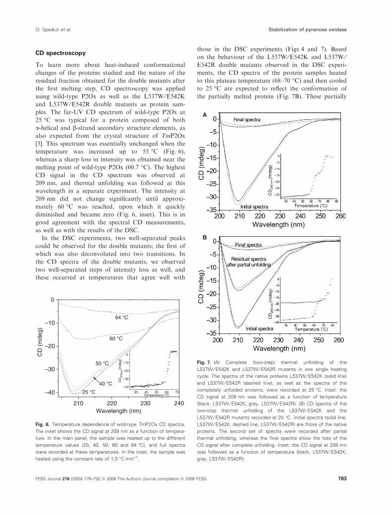
In the DSC experiments, two well-separated peaks
could be observed for the double mutants; the first of
which was also deconvoluted into two transitions. In
the CD spectra of the double mutants, we observed
two well-separated steps of intensity loss as well, and
these occurred at temperatures that agree well with
those in the DSC experiments (Figs 4 and 7). Based
on the behaviour of the L537W ⁄E542K and L537W ⁄E542R double mutants observed in the DSC experi-
ments, the CD spectra of the protein samples heated
to this plateau temperature (68–70 �C) and then cooled
to 25 �C are expected to reflect the conformation of
the partially melted protein (Fig. 7B). These partially
210 220 230 240
–40
–30
–20
–10
0
64 °C
60 °C
50 °C
40 °C
CD
(m
deg)
Wavelength (nm)
25 °C 30 40 50 60 70
–40
–30
–20
–10
0
CD
209n
m(m
deg)
Temperature (°C)
Fig. 6. Temperature dependence of wild-type TmP2Ox CD spectra.
The inset shows the CD signal at 209 nm as a function of tempera-
ture. In the main panel, the sample was heated up to the different
temperature values (25, 40, 50, 60 and 64 �C), and full spectra
were recorded at these temperatures. In the inset, the sample was
heated using the constant rate of 1.0 �CÆmin)1.
A
B
Fig. 7. (A) Complete (two-step) thermal unfolding of the
L537W ⁄ E542K and L537W ⁄ E542R mutants in one single heating
cycle. The spectra of the native proteins L537W ⁄ E542K (solid line)
and L537W ⁄ E542R (dashed line), as well as the spectra of the
completely unfolded proteins, were recorded at 25 �C. Inset: the
CD signal at 209 nm was followed as a function of temperature
(black, L537W ⁄ E542K; gray, L537W ⁄ E542R). (B) CD spectra of the
two-step thermal unfolding of the L537W ⁄ E542K and the
L537W ⁄ E542R mutants recorded at 25 �C. Initial spectra (solid line,
L537W ⁄ E542K; dashed line, L537W ⁄ E542R) are those of the native
proteins. The second set of spectra were recorded after partial
thermal unfolding, whereas the final spectra show the loss of the
CD signal after complete unfolding. Inset: the CD signal at 209 nm
was followed as a function of temperature (black, L537W ⁄ E542K;
gray, L537W ⁄ E542R).
O. Spadiut et al. Stabilization of pyranose oxidase
FEBS Journal 276 (2009) 776–792 ª 2008 The Authors Journal compilation ª 2008 FEBS 783

melted samples showed a profile identical to that of
native P2Ox, but with a lower intensity, suggesting
that no drastic change in the composition of the sec-
ondary structural elements occurred in the partially
melted sample compared to the native one. Because no
stable dimeric or monomeric form of P2Ox is available
for comparative CD studies, we cannot unambiguously
decide the oligomeric state of the species possessing
the residual CD spectrum and activity associated with
the first DSC transition.
Structure of the P2Ox variants
Data collection and model statistics are given in
Table 5. The final L537G and E542K models include
two complete tetramers per asymmetric unit, with each
monomer consisting of residues 43–619, and one FAD
molecule per monomer. The L537W ⁄E542K mutant
contains one monomer per asymmetric unit comprising
residues 46–618 with one FAD and one Mes [2-(N-
morpholino) ethane sulfonic acid (4-morpholine ethane
sulfonic acid)] molecule per monomer. As shown in
Table 5, all models have good R values, with residues
that fall within the allowed regions of the Ramachan-
dran plot [24].
The overall tetramer structure (Fig. 1) of all mutants
is identical to that previously reported for wild-type
and recombinant P2Ox from T. multicolor [3,4]. Typi-
cal rmsd values using all Ca atoms from all monomers
of the tetramer fall within the range 0.2–0.3 A. The
structures are also almost identical to the models of
Peniophora P2Ox [Protein Data Bank (PDB) codes
1TZL, 2F5V and 2F6C] [5,6] with rmsd values of
approximately 0.9 A for the monomer structure. The
only major difference observed between all Trametes
and Peniophora P2Ox models is the precise conforma-
tion of the substrate loop. As discussed in detail else-
where, we have shown that this loop is in an open
conformation when no substrate is bound (e.g. unli-
ganded recombinant P2Ox; PDB codes 2IGK, 2IGM,
2IGN) [4] or when an electron-donor substrate is
bound (e.g. monosaccharide as in P2Ox H167A in
complex with 2-fluoro-2-deoxy-d-glucose, 2FG; PDB
code 2IGO) [4], and in a closed conformation when
small electron-acceptor substrates (i.e. dioxygen) or
small inhibitor molecules (e.g. acetate as in wild-type
Table 5. Data collection and refinement statistics.
E542K L537G E542K ⁄ L537W
Data collectiona
Wavelength, k (A) 0.918 1.042 0.931
Beamline ⁄ temperature (�K) BESSY 14.1 ⁄ 100 MAX-lab I911-2 ⁄ 100 ESRF ID14-3 ⁄ 100
Cell constants a, b, c (A);
b (�) ⁄ space group
168.9, 103.7, 169.3, 106.31 ⁄ P21 168.5, 103.2, 169.3, 106.45 ⁄ P21 103.4, 103.4, 118.6 ⁄ P42212
Resolution range, nominal (A) 40–1.70 (1.75–1.70) 40–2.10 (2.20–2.10) 51–1.90 (2.00–1.90)
Unique reflections 603 616 (49 624) 321 136 (39 548) 51 240 (7193)
Multiplicity 3.8 (3.2) 4.4 (3.3) 12.6 (12.7)
Completeness (%) 98.2 (97.4) 99.0 (94.0) 99.9 (100)
<I ⁄ rI> 9.7 (2.2) 17.2 (6.2) 17.2 (4.8)
Rsym (%)b 13.7 (58.8) 6.6 (26.2) 12.1 (62.7)
Refinement
Resolution range (A) 40–1.70 40–2.10 51–1.90
Completeness, all % (highest bin) 98.3 (97.4) 99.1 (94.6) 100.0 (100)
Rfactorc ⁄ work reflns, all 16.6 ⁄ 597 554 15.6 ⁄ 317 871 14.9 ⁄ 50 735
Rfree ⁄ free reflns, all 19.8 ⁄ 6061 20.4 ⁄ 3262 18.1 ⁄ 504
Non-hydrogen atoms all ⁄ protein 39 388 ⁄ 36 320 38 655 ⁄ 36 287 4943 ⁄ 4524
Mean B (A2) protein all ⁄ mc ⁄ sc 26.6 ⁄ 25.4 ⁄ 27.8 38.5 ⁄ 37.4 ⁄ 39.7 12.9 ⁄ 11.6 ⁄ 14.2
Mean B (A2) solvent ⁄ number
of molecules
29.8 ⁄ 2564 38.8 ⁄ 1864 35.4 ⁄ 354
Mean B (A2) cofactor ⁄ number
of atoms
17.5 ⁄ 424 27.4 ⁄ 424 14.8 ⁄ 53
rmsd bond lengths (A), angles (�) 0.022, 1.89 0.022, 1.86 0.022, 1.91
Ramachandran: favored ⁄ allowed (%)d 97.4 ⁄ 100 97.1 ⁄ 100 97.9 ⁄ 100
PDB codee 3BG6 3BG7 3BLY
a The outer shell statistics of the reflections are given in parenthesis. Shells were selected as defined in XDS [32] by the user. b Rsym = [Rhkl
RI|I – <I>| ⁄ Rhkl RI |I] · 100%. c Rfactor = Rhkl| |Fo| – |Fc| | ⁄ Rhkl|Fo|. d As determined by MOLPROBITY [24]. e PDB accession codes for atomic coor-
dinates and structure factors are deposited with the Research Collaboratory for Structural Bioinformatics Protein Data Bank.
Stabilization of pyranose oxidase O. Spadiut et al.
784 FEBS Journal 276 (2009) 776–792 ª 2008 The Authors Journal compilation ª 2008 FEBS

P2Ox in complex with acetate; PDB code 1TT0) [3]
are bound. In the existing PsP2Ox models (recombi-
nant wild-type P2Ox and P2Ox E542K mutant; PDB
codes 1TZL, 2F5V and 2F6C, respectively) [5,6], the
substrate loop assumes a disordered conformation
intermediate to the ordered open and closed conform-
ers seen for TmP2Ox.
As expected for the active site in the absence of elec-
tron-donor monosaccharide substrate or electron-
acceptor substrate, the substrate loop in the E542K
and L537G variants is open and slightly disordered, as
indicated by partly weak electron density and elevated
temperature factors. In the L537W ⁄E542K variant,
however, the substrate loop is open and fully ordered.
In the E542K structure, the introduced Lys side chain
has unambiguous electron density and points into the
internal cavity at the centre of the homotetramer. In
the L537G mutant structure, the elimination of the rel-
atively large and hydrophobic Leu side chain results in
remarkably small changes. In wild-type P2Ox, Leu537
is located in strand B6 close to the dyad axis between
monomers A and C (or B and D) where the Cb atoms
of Leu537 of each monomer interact via a hydropho-
bic packing interaction (Fig. 2A,B). Upon replacement
of the Leu side chain by Gly (Fig. 2C), the Ca–Ca dis-
tance at position 537 between monomers A and C (or
B and D) increases from 6.2 to 6.4 A. The mutation
produces a relative Ca displacement at position 537
within the monomer of 0.6–0.7 A. The largest displace-
ment, however, is seen two residues away, where the
backbone Ca atom of Gly535 is shifted 0.9–1.0 A as a
result of the Leu537 fi Gly substitution in the L537G
mutant. At the interface between subunits A and C,
solvent molecules substitute for the missing Leu side
chain. In addition, the small, but distinct, displacement
around position 537 is accompanied by backbone
displacements in the substrate loop (0.8–1.0 A at Caposition 453).
We chose to use P2Ox H167A in complex with 2FG
(PDB code 2IGO) [4] as a reference for comparisons
because this model has the substrate loop in an open
and ordered conformation, with the open conformer
being observed also in the three P2Ox variants
described here. The mutants show minor but distinct
differences compared to 2IGO. With Trp residues
introduced at position 537, as in L537W ⁄E542K(Fig. 2D), the 537 backbone of monomers A and C
move 0.2 A closer together (with a concomitant move-
ment of helices H8 in A and C closer by 0.4 A),
whereas, with Gly replacements at this position (as in
L537G), the monomers move 0.4 A further apart.
However, two residues away at position 535, the back-
bones of the A and C monomers show tighter associa-
tion in L537G by 1.4 A, and only by 0.9 A in
L537W ⁄E542K, compared to model 2IGO. As a result
of these movements, the L537W ⁄E542K variant also
shows a concomitant displacement of the substrate
loop by 0.4–0.6 A, as well as tighter association
between the oligomerization arm in monomers A and
D by 0.6 A at position 121. In the E542K and L537G
mutants, the corresponding position is shifted 0.1 and
0.3 A further apart, respectively, thus possibly weaken-
ing the A–D interaction compared with 2IGO. At the
more detailed structural level, we observe that, com-
pared with 2IGO, the A ⁄C interface of the
L537W ⁄E542K variant shows improved hydrophobic
stacking interactions between Trp537 of monomer A
and Gln539 of monomer C, with a possibility of addi-
tional amino-aromatic interaction between Gln539 Ne2and the Trp537 ring. In addition, this arrangement
allows a shorter and more aligned hydrogen bond
between Gln539 Ne2 and Trp537 O, which ought to
be more stable.
When comparing the three mutants and 2IGO, the
largest difference observed is the position of the ‘head’
domain (Fig. 8). In the thermostable L537W ⁄E542Kdouble mutant, differences in the backbone position of
the exposed head domain of up to 4.3 A, and of
exposed parts of the Rossmann domain of up to
2.7 A, are observed. For the rest of the homotetramer-
ic assembly, only smaller backbone displacements of
up to 1 A occur. Although these differences might
arise from different packing in the tetragonal space
group of the double mutant, the amino-acid replace-
ments may also be of importance.
Fig. 8. Ribbon drawing showing the superpositioning of the tetra-
mers of 2IGO (red), L537G (yellow) and E542K ⁄ L537W (blue). As
discussed in the text, the only significant difference in the overall
tetramer structure is the relative displacement of the head domain
in E542K ⁄ L537W.
O. Spadiut et al. Stabilization of pyranose oxidase
FEBS Journal 276 (2009) 776–792 ª 2008 The Authors Journal compilation ª 2008 FEBS 785
Discussion
Pyranose oxidase is an enzyme of applied interest and,
hence, previous studies have aimed at improving this
biocatalyst [19–22]. One residue that was identified as
being important for both stability and reactivity is
Glu542, which is located on the surface of the internal
cavity, close to the entrance to the active site [6].
Replacement of that residue by Lys resulted in
increased thermostability (i.e. for TvP2Ox, an increase
in temperature at which activity is reduced by 50% at
30 min from � 55 �C for the wild-type to � 65 �C [19]
or, for PsP2Ox, an increase in the optimum tempera-
ture from 50 �C for the wild-type to 58 �C) [20], as wellas in improved catalytic properties. However, the effect
of this Glu fi Lys replacement on stability has not
been studied in full. In the present study, we present
for the first time detailed investigations for the E542K
and E542R variants pertaining to their stability. In
addition, we selected position Leu537, which is located
at the interface of two subunits, for mutational studies.
Leu537 of one subunit (A or B) interacts with Leu537¢of another subunit (C or D) via hydrophobic packing.
Strengthening this interaction by the introduction of a
better-suited residue might therefore improve subunit–
subunit interactions and hence stability. Saturation
mutagenesis at this position and subsequent screening
for thermostable variants identified the replacements
Leu537 fi Gly and Leu537 fi Trp as being beneficial.
DSC measurements of both the wild-type enzyme
and these four single mutants (L537G, L537W, E542K
and E542R) showed a significant increase in the Tm
for the variants. The replacements at position Glu542
proved to be more efficient for stabilization because
Tm was increased by approximately 14 �C for both
E542K and E542R, whereas this increase was 10.4 and
8.3 �C for L537G and L537W, respectively. These
improvements in thermostability were further con-
firmed by inactivation studies at 60–75 �C, where,
again, the Glu542 variants showed smaller inactivation
constants and hence higher s1 ⁄ 2 than the two Leu537
variants. Interestingly, not only the Glu542 fi Lys
replacement, but also the corresponding exchange with
the basic residue Arg resulted in considerably
improved stabilization to an approximately equal
extent (e.g. Tm of 74.7 and 74.3 �C, s1 ⁄ 2, 60 �C of 1640
and 1680 min for E542K and E542R, respectively).
Previous studies had concluded that only the Lys (and
no other amino acid substitution at this position) has
a similar positive effect [21]. The four single mutants
of TmP2Ox showed considerably increased s1 ⁄ 2 and
hence much slower inactivation at higher temperatures
than the wild-type enzyme. To possibly combine these
stabilizing effects, we constructed the double mutants
L537G ⁄E542K, L537G ⁄E542R, L537W ⁄E542K and
L537W ⁄E542R. Again, these P2Ox variants were more
thermostable than the wild-type but, interestingly, the
concomitant introduction of the substitutions at both
Glu542 and Leu537 altered the unfolding process sig-
nificantly. The single mutants showed one unfolding
peak by DSC, and the inactivation kinetics followed a
first-order equation, indicative of a simple one-step
inactivation process, where the native, active form is
transformed directly into the denatured, inactive form
[25]. All of the double mutants studied showed two
separate unfolding peaks in the DSC measurements.
Furthermore, the inactivation curves did not follow
first-order kinetics but showed two distinct phases that
can be described as two subsequent first-order reac-
tions: a first phase of rapid activity loss and, after a
short transition, a prolonged second phase of moderate
activity loss. This behaviour could indicate an inactiva-
tion procedure consisting of two consecutive processes,
with the native, active form of the P2Ox double
mutants being first transformed rapidly into an active
intermediate species, which then inactivates slowly in a
second, independent reaction. The second melting tem-
perature Tm,2, resulting in the final denaturation step
of the P2Ox double mutants, was increased by 14.3–
16.8 �C compared to the wild-type enzyme, which is
even higher than for the P2Ox mutational variants
with only one amino acid substitution. Based on CD
studies, the first inactivation process leading to the
active intermediate is not reversible. The nature of this
intermediate species is yet unknown. Because the
mutations mainly affect the interactions between the
subunits, it is conceivable that either active dimers or
monomers of P2Ox are formed in the first denatur-
ation process. This is further corroborated by the CD
measurements of protein samples of the L537W ⁄E542K and L537W ⁄E542R variants heated only to the
first transition temperature, thus obtaining the interme-
diate species, and then cooled to 25 �C. These samples
showed spectra identical to those of the native enzyme,
but with lower intensity. This demonstrates that no
drastic changes in the secondary structure elements
occurred in this first unfolding process.
In addition, we studied the effects of the replace-
ments at Leu537 and Glu542 on the reactivity of
P2Ox. In accordance with previous studies [19,21], the
introduction of a basic amino acid at position Glu542
results in a decrease of the Michaelis constant for the
two sugar substrates, d-glucose and d-galactose, but
also in a decrease in the kcat to some extent. The vari-
ants L537G and L537W show almost unaltered Km
values for both sugar substrates, whereas kcat was
Stabilization of pyranose oxidase O. Spadiut et al.
786 FEBS Journal 276 (2009) 776–792 ª 2008 The Authors Journal compilation ª 2008 FEBS
improved specifically for the substrate d-glucose. These
improvements of the catalytic properties could be com-
bined advantageously in the double mutants, especially
those containing the E542K replacement, which were
characterized by considerably reduced Km values com-
bined with practically unchanged kcat, and therefore
improved catalytic efficiencies (kcat ⁄Km), notably for
d-glucose. One aspect that has not been studied to
date is the effect of these mutations on the second
half-reaction of P2Ox, the oxidative half-reaction, in
which electrons are transferred to an acceptor. P2Ox
not only can transfer these electrons to oxygen, but
also to a range of other electron acceptors, including
substituted quinones, complex metal ions or certain
organic radical species [7], some of which are consider-
ably better substrates than oxygen. When benzoqui-
none was used as the substrate, these mutations
affected mainly Km, which decreased by a factor of
two for some of these variants. Most of the substitu-
tions had a negative effect when the ferricenium ion
was the varied substrate. It is conceivable that the
introduction of a positive charge in the internal cavity
of P2Ox, close to the entrance of the tunnel leading to
the active site [3], as in the E542K and E542R vari-
ants, results in the repulsion of the positively charged
ferricenium ion Fc+. By contrast, the replacements at
position Leu537 showed a significant, positive effect
on kcat, Fc+, especially for L537W where kcat, Fc+
was increased by more than two-fold when d-glucose
was the saturating substrate. This could be of interest
for the application of P2Ox in the anodic reaction in
biofuel cells based on mediated electron transfer. For
mediated electron transfer, certain mediators, such as
ferrocenes, Os-redox polymers or other complexed
metal ions, collect electrons, resulting from the oxida-
tion of the sugar, at the active site of the enzyme and
transfer these to the electrode. In a biofuel cell based
on an enzyme that is electrically wired to the electrode
in this way, the current measured as an analytical
response signal represents the actual turnover rate of
the enzyme [15], and therefore a P2Ox variant with an
increased kcat for the mediator (electron acceptor) will
certainly be of interest. Recently, it was shown that
TmP2Ox can communicate efficiently with electrodes
by using either ferrocenes or other complexed metal
ions [15,16]. It was further demonstrated that the
E542K variant, which is characterized by a lower kcatfor Fc+ than the wild-type enzyme, also performs sig-
nificantly worse in bioelectrochemical studies than the
wild-type, confirming the results of the kinetic charac-
terization using Fc+ in the present study.
The crystal structures of TmP2Ox, both in the unli-
ganded recombinant form and in complex with an
electron-donor substrate, have been studied in detail
[3,4]. One characteristic feature is the substrate loop,
which is in an open conformation when no substrate
or an electron-donor substrate such as 2-fluoro-2-
deoxy-d-glucose is bound, and in a closed conforma-
tion when small electron-acceptor substrates are
bound. The transition from the open to the closed
active site involves a major reorganization of the sub-
strate loop (residues 451–461). Two aromatic residues,
Phe454 and Tyr456, which have no interaction with
the active site in the open conformation, undergo
major structural rearrangements during this transition.
Notably, Tyr456 moves 9 A (together with Ser455) to
completely close off the active site from the internal
cavity of the homotetramer. Concomitantly, Phe454
rotates and moves some 7 A to fill the active site and
pack against the FAD cofactor. In the loop between
b-strand B6 in the substrate-binding domain and
strand E2 in the hinge domain, two residues appear to
act, at least partly, as structural determinants for the
closed conformation of the substrate loop. The side
chains of Met541 and Leu545 create a flat surface onto
which Tyr456 stacks in its ‘swung-in’ conformation
observed in the closed state of the P2Ox active site
[3,4]. These two residues are intervened by Glu542, the
Oe2 atom of which forms a hydrogen bond to Ser153
Oc located in the nearby loop preceding b-strand D2
at the start of the oligomerization arm. The Oc atom
of Ser153 in monomer A is also involved in a hydro-
gen-bond interaction with Asp124 in monomer D. The
A-Ser153–d-Asp124 interaction helps to stabilize the
association between the oligomerization arms in mono-
mers A and D, as well as B and C. Moreover, it
appears as if the interaction between Glu542 and
Ser153 serves to secure the position of the hydrophobic
Met ⁄Leu platform for Tyr456 when the active site
adopts its closed state. However, the Met ⁄Leu plat-
form does not appear to have any apparent function
when the active site is in the open conformation
because Met541 and Leu545 are then completely
exposed to the solvent of the internal void, and distant
from the aromatic side chain of Tyr456 in the
substrate loop.
When replacing Glu542 by Lys, the Glu542-Ser153
hydrogen bond is lost, and no additional hydrogen
bond is offered to Lys542. The loss of the hydrogen
bond may or may not affect the precise positioning of
the aromatic ring of Tyr456 in the closed state. In the
absence of a closed complex of E542K P2Ox, the effect
of the loss of this hydrogen bond on the packing of
the substrate loop is difficult to assess. However, any
mutation that affects the structure and function of the
substrate loop and ⁄or the local environment of the
O. Spadiut et al. Stabilization of pyranose oxidase
FEBS Journal 276 (2009) 776–792 ª 2008 The Authors Journal compilation ª 2008 FEBS 787

flavin cofactor is likely to affect the kinetics of cataly-
sis of either the reductive or oxidative half-reaction, or
both. Such an effect could be either due to altered
redox power of the cofactor and ⁄or discrete conforma-
tional changes of amino acids critical for substrate
binding and catalysis.
As mentioned above, the Lys side chain points into
the internal cavity of the tetramer and forms no hydro-
gen bonds to either protein or ordered solvent. Fur-
thermore, the Lys replacement does not introduce any
significant structural changes in either the monomer or
tetramer structure of TmP2Ox in the folded state, and
the corresponding mutation in PsP2Ox shows a Lys
with the same side-chain conformation as that
observed in the present study. For PsP2Ox, it was pro-
posed that the increased thermostability of the E540K
mutant may be due to an ionic effect assigned to the
ability of the Lys side chain to relieve possible electro-
static repulsion between Glu542 and Asp124 in the
wild-type enzyme at higher pH values [6]; however,
because neither the pKa of Asp124 is known, nor the
dependence of thermostability on pH, this hypothesis,
although probable, remains unproven.
As for the increased thermostability of P2Ox vari-
ants where Leu537 is replaced by either Gly or Trp,
we observed distinct differences at the A ⁄C interface.
By introducing either Trp or Gly at position 537, tigh-
ter association between the A and C monomers can be
achieved, although at different locations in the inter-
face. Compared with L537G, additional favourable
interactions are seen in E542K ⁄L537W, where the
A ⁄D interface also is strengthened significantly by the
tighter association of the oligomerization arms of
monomers A and D. This is likely to explain why,
although both Gly and Trp replacements increase sta-
bility to thermal unfolding, the Trp replacement is
more thermostable overall. The largest difference
between the structures is observed in the relative posi-
tion of the head domain (Fig. 8); however, this is most
likely due to differences in packing environment of the
monoclinic and tetragonal crystal lattices rather than
due to the amino-acid replacements per se. Thus, the
subtle differences observed at the subunit interfaces
discussed above are more likely determinants of
increased thermostability.
Experimental procedures
Bacterial strains, plasmids and media
E. coli strain BL21 Star DE3 (Invitrogen, Carlsbad, CA,
USA), well suited for the bacteriophage T7 promoter-based
expression system pET, was used as host for the expression
plasmids and, consequently, for the production of active
P2Ox protein. The vector pET21d(+) was used throughout
the study to express wild-type P2Ox and P2Ox variants
containing a C-terminal His6-tag. Construction of the plas-
mid pHL2, which expresses the His-tagged wild-type
TmP2Ox under the control of the T7 promoter, has been
described previously [4]. E. coli cells were grown in TBamp-
media (yeast extract 24 gÆL)1, peptone from casein
12 gÆL)1, glycerol 4 mLÆL)1; phosphate buffer 1 m, pH 7.5)
under appropriate selective conditions (ampicillin was
added to 100 mgÆL)1). The chemicals used were of the pur-
est grade available and were purchased from Sigma
(Vienna, Austria). Nucleotides, buffers and enzymes
for molecular biology were from Fermentas (St Leon-Rot,
Germany).
Generation of mutants
The TmP2Ox gene was mutated by a two-step site-directed
mutagenesis procedure using PCR and digestion with DpnI
[26]. For the replacement of Glu542 with Lys, the primers
used were P2OE542K-for (5¢-GCAATTCATGAAGCCT
GGT-3¢) and P2OE542K-rev (5¢-ACCAGGCTTCATGAA
TTGC-3¢). The primers for constructing variant E542R
were P2OE542R-for (5¢-GCAATTCATGCGGCCTGGT-
3¢) and P2OE542R-rev (5¢-ACCAGGCCGCATGAATTG
C-3¢). For saturation mutagenesis of position Leu537, we
used the primers P2O-Wobble-537-for (5¢-TCCTAC
CCGGCTCCNNSCCGCAA-3¢) and P2O-Wobble-537-rev
(5¢-TTGCGGSNNGGAGCCGGGTAGGA-3¢), where N =
A, G, C or T, and S = G or C. To create double mutants
at positions 537 and 542, we used the plasmids of variants
L537G and L537W as templates for the PCR reactions, the
forward primers P2OE542K-for and P2OE542R-for, and
the reverse primers Double-G-rev (5¢-CATGAATT
GCGGGCCGGAGCCG-3¢) and Double-W-rev (5¢-CATGAATTGCGGCCAGGAGCCG-3¢). All primers were
purchased from VBC Biotech (Vienna, Austria). The muta-
genic PCR was performed under the conditions: 95 �C for
4 min; then 30 cycles of 94 �C for 30 s, 58 �C for 30 s and
72 �C for 16 min; with a final extension at 72 �C for
10 min. Each reaction contained 1· buffer (Fermentas),
0.1 lg of plasmid DNA, 2.5 U Pfu DNA polymerase (Fer-
mentas), 10 lm of each dNTP and 5 pmol of each primer
in a total volume of 50 lL. After PCR, the methylated tem-
plate DNA was degraded by digestion with 10 U of DpnI
at 37 �C for 3 h. The remaining PCR products were sepa-
rated by agarose gel electrophoresis and purified using the
Wizard SV Gel and PCR-Clean-Up System (Promega,
Madison, WI, USA). Five microliters of each purified PCR
product were transformed into chemically competent E. coli
BL21 Star DE3 cells. The successful introduction of the
desired mutations and the absence of further mutations
were confirmed by DNA sequencing, which was performed
as a commercial service (VBC-Biotech, Vienna, Austria).
Stabilization of pyranose oxidase O. Spadiut et al.
788 FEBS Journal 276 (2009) 776–792 ª 2008 The Authors Journal compilation ª 2008 FEBS

Plasmidic DNA was extracted and used as template for
DNA sequencing of the complete P2Ox-encoding sequence
using the forward primer T7promfwd (5¢-AATACGACT
CACTATAGGGG-3¢) and the reverse primer T7termrev
(5¢-GCTAGTTATTGCTCAGCGG -3¢).
Screening for improved P2Ox variants
Position Leu537 of TmP2Ox was mutated by saturation
mutagenesis, which allows the creation of a mutant library
containing all possible codons at the target position. The
size of the library, which subsequently has to be screened
to cover all possible mutants, is determined by the muta-
genic codon and the number of target sites. For mutation
of position Leu537 by saturation mutagenesis, the primers
used were of the NNS type, which defines the minimum
library size to be screened to statistically cover 95% of all
possible substitutions as 95 colonies [27]. Accordingly, a
screening assay based on 96-well plates was used. Trans-
formed E. coli BL21 Star DE3 cells were transferred from
LB-ampicillin plates into microtiter wells containing
200 lL of liquid LBamp medium (‘masterplates’). Cells
were grown on a shaking incubator (150 r.p.m.) at 25 �Cfor 20 h. To induce protein expression, 10 lL of the cell
suspension of each well were transferred into another
96-well plate containing 200 lL of 2· LBamp ⁄ lactose (yeast
extract 10 gÆL)1, peptone from casein 20 gÆL)1, sodium
chloride 5 gÆL)1, lactose 5 gÆL)1; ampicillin 100 mgÆL)1)
per well, and incubated at 25 �C for another 20 h (‘work-
ing plates’). The growth of the cells was measured in a
plate reader (Sunrise Remote, Tecan, Groding, Austria) at
600 nm. Induced cells were sedimented in the wells by
centrifugation (2300 g for 15 min) and the supernatant
was discarded. The cells were lysed by adding 200 lL of
1· lysis buffer (CelLytic B Cell Lysis Reagent; Sigma) and
incubated at 4 �C for 30 min. Subsequently, the plates
were frozen at –70 �C for 1 h and then thawed at room
temperature to increase the efficiency of the lysis. Cell
debris was removed by centrifugation, and 10 lL of the
supernatant were added to 80 lL of chromogenic assay
mixture (0.035 mgÆmL)1 of horseradish peroxidase and
0.7 mgÆmL)1 of ABTS in 50 mm phosphate buffer,
pH 6.5). The reaction was started by adding either 10 lLof d-glucose or d-galactose (each 1 m), and recorded auto-
matically at 420 nm and 30 �C by the plate reader. To test
for increased thermostability, the microtiter plates contain-
ing the cell extracts were incubated at 65 �C for 10 min
before performing the activity assay.
Protein expression and purification
Cultures (2 L) of E. coli BL21 Star DE3 transformants
were grown in TBamp in shaken flasks at 37 �C and
160 r.p.m. When D600 of 0.5–0.6 was reached, recombinant
protein expression was induced by adding lactose to a final
concentration of 0.5%. After cultivation at 25 �C for an
additional 20 h, cells were harvested by centrifugation
(4200 g for 20 min), resuspended in phosphate buffer
(50 mm, pH 6.5) containing phenylmethylsulfonyl fluoride
(0.1%) and lysed by using a continuous homogenizer (APV
Systems, Silkeborg, Denmark). The crude cell extract was
obtained by centrifugation (150 000 g for 30 min at 4 �C)and then used for protein purification by IMAC using the
AKTA purifier system (Pharmacia, Uppsala, Sweden) and
the Bio-Rad Profinity IMAC Ni-Charged Resin (10 mL;
Bio-Rad, Vienna, Austria). The column was pre-equili-
brated with ten column volumes of buffer (50 mm KH2PO4,
0.5 m NaCl, 20 mm imidazole; pH 6.5). After applying the
sample to the column, it was washed with five column
volumes of the same buffer at a flow rate of 2 mLÆmin)1.
Proteins were then eluted with a linear gradient (flow rate
1.5 mLÆmin)1 for 60 min) of the same buffer containing
1 m imidazole and monitored at 280 nm (protein content)
and 456 nm (FAD content). The fractions containing eluted
enzyme were pooled, and imidazole was removed by ultra-
filtration using an Amicon Ultra Centrifugal Filter Device
(Millipore, Billerica, MA, USA) with a 10 kDa cut-off
membrane. The eluted, concentrated enzymes were washed
three times with 10 mL of phosphate buffer (50 mm,
pH 6.5), and finally diluted in this buffer to a protein
concentration of 10–20 mgÆL)1.
Electrophoresis
Electrophoresis was performed principally as described by
Laemmli [28]. Both native PAGE and SDS ⁄PAGE were
performed using a 5% stacking gel and a 10% separating
gel on the PerfectBlue vertical electrophoresis system
(Peqlab, Erlangen, Germany) and using the molecular mass
standards High Molecular Weight Calibration Kit for
native electrophoresis (Amersham Pharmacia, Piscataway,
NJ, USA) and the Precision Plus Protein Dual Color
Kit (Bio-Rad) for SDS ⁄PAGE. Gels were stained with
Coomassie brilliant blue.
Enzyme activity assays
P2Ox activity was measured with the standard chromogenic
ABTS assay [29]. A sample of diluted enzyme (10 lL) wasadded to 980 lL of assay mixture containing horseradish
peroxidase (142 U), ABTS (14.7 mg) and phosphate buffer
(50 mm, pH 6.5). The reaction was started by adding d-glu-
cose (20 mm). A420 was recorded at 30 �C for 180 s
(e420 = 42.3 mm)1Æcm)1). One unit of P2Ox activity was
defined as the amount of enzyme necessary for oxidation of
2 lmolÆmin)1 of ABTS (which equals the consumption of
1 lmolÆmin)1 of O2) under assay conditions. Protein con-
centrations were determined by the Bradford assay [30]
using the Bio-Rad Protein Assay Kit (Bio-Rad) with BSA
as standard.
O. Spadiut et al. Stabilization of pyranose oxidase
FEBS Journal 276 (2009) 776–792 ª 2008 The Authors Journal compilation ª 2008 FEBS 789

Steady-state kinetic measurements
kcat were measured for the two electron donors d-glucose
(0.1–50 mm) and d-galactose (0.1–200 mm) using the stan-
dard ABTS assay and oxygen (air saturation). In addition,
steady-state kinetic constants were determined for the alter-
native electron acceptors 1,4-benzoquinone and the ferrice-
nium ion Fc+ (using ferricenium hexafluorophosphate,
Fc+PF6), as substrate; Aldrich, Steinheim, Germany).
Appropriately diluted enzyme sample (10 lL) was added to
990 lL of assay buffer containing either d-glucose or
d-galactose in a constant concentration of 100 mm, phos-
phate buffer (50 mm, pH 6.5) and 1,4-benzoquinone, which
was varied in the range 0.01–1.5 mm. A290 was recorded at
30 �C for 180 s (e290 = 2.24 mm)1Æcm)1). FcPF6 was varied
in the range 0.005–0.5 mm and A300 was recorded at 30 �Cfor 180 s (e300 = 4.3 mm
)1Æcm)1). Kinetic constants were
calculated by nonlinear least-square regression, fitting the
data to the Henri–Michaelis–Menten equation.
Thermal stability
Kinetic stability of the TmP2Ox variants was determined by
incubating the enzymes in appropriate dilutions in 50 mm
phosphate buffer (pH 6.5) at 60, 70 and 75 �C, respectively,and by subsequent measurements of the enzyme activity (A)
at various time points (t) using the standard ABTS assay and
glucose as the substrate. A thermal cycler (thermocycler T3;
Biometra, Gottingen, Germany) and thin-wall PCR tubes
were used for all thermostability measurements. Residual
activities (At ⁄A0, where At is the activity measured at time
t and A0 is the initial P2Ox activity) were plotted versus the
incubation time. For those experiments where the inactiva-
tion followed apparent first-order kinetics, the inactivation
constant kin was obtained by linear regression of ln (activity)
versus time. The half-life values of thermal inactivation s1 ⁄ 2
were calculated using s1 ⁄ 2 = ln 2 ⁄kin [23].Thermodynamic stability (i.e. Tm) [23], was measured by
DCS, as described previously [31,32], using a MicroCal
VP-DSC instrument (MicroCal, Northampton, MA, USA)
in the range 15–80 �C at a scan rate of 1 �CÆmin)1 on
0.2 gÆL)1 protein samples in 50 mm phosphate buffer
(pH 6.4). Solutions were degassed by stirring under vacuum
for 15 min at room temperature immediately before mea-
surements. The solutions in the measuring cells were kept
under pressure to prevent degassing during heating. The
baseline was determined in an identical experiment with
buffer in both cells and was subtracted. Data processing
and evaluation were performed using origin 7.5 software
(OriginLab Corporation, Northampton, MA, USA).
CD measurements
Far-UV CD spectra (190–240 nm) were recorded as
described previously [33,34] at 25 �C on a Jasco J-720 spec-
tropolarimeter (Jasco International Co., Tokyo, Japan)
using protein samples at 8 lm concentration in 50 mm
phosphate buffer (pH 6.4), 1 mm pathlength, thermostatted
cuvettes and a Neslab RTE-100 computer-controlled ther-
mostat (Neslab Inc., Portsmouth, NH, USA). Spectra were
averaged over three scans. Processing of spectral data was
performed by using the built-in jasco software of the
spectropolarimeter. Thermal unfolding measurements were
measured at 209 nm over the temperature range 25–80 �C.After reaching 80 �C, samples were cooled back to 25 �C.Data processing and evaluation were performed using the
origin 7.5 software.
Crystallization, data collection and refinement of
the E542K and L537G mutants
Crystals of the E542K, L537G and E542K ⁄L537Wmutants were produced using the hanging drop vapour
diffusion method [35], essentially as described previously
for recombinant P2Ox [4]. Drops were set up by mixing
equal volumes of protein [4 mgÆmL)1 in 20 mm Mes
(pH 5.2)] and reservoir [12–16% (w ⁄ v) monomethylether
polyethylene glycol 2000, 0.1 m Mes (pH 5.2), 50 mm
MgCl2, 25% glycerol]. Microseeding was used routinely.
Prior to data collection, the crystals were frozen and vitri-
fied in liquid nitrogen. Data for the E542K mutant were
collected using synchrotron radiation (k = 0.918 A) at
beamline 14.1 at BESSY (Berlin, Germany) and data for
the L537G mutant (k = 1.042 A) were collected at beam-
line I911-2 (100 �K) at MAX-lab (Lund, Sweden). Data
for the E542K ⁄L537W mutant (k = 0.931 A) were col-
lected at beamline ID14-3 at ESRF (Grenoble, France)
(100 �K). All data were processed using xds [36]. The
E542K and L537G mutants crystallize in space group P21with eight monomers forming two tetramers in the asym-
metric unit, whereas the E542K ⁄L537W mutant crystallizes
in space group P42212 with one monomer in the asymmet-
ric unit. Phases were obtained by means of Fourier syn-
thesis using the H167A P2Ox variant (PDB code 2IGO)
[4] as the starting model. Crystallographic refinement was
performed with refmac5 [37], and included anisotropic
scaling, calculated hydrogen scattering from riding hydro-
gens, and atomic displacement parameter refinement using
the translation, libration, screw-rotation (TLS) model. In
the case of E542K and L537G, for each of the eight
monomers (two tetramers) in the asymmetric unit, individ-
ual TLS groups were defined: the Rossmann domain (resi-
dues 44–79, 254–353, 552–618); the substrate-binding
domain (residues 159–253, 354–551); the oligomerization
arm (residues 111–158); and the lid (residues 80–110). For
the E542K ⁄L537W mutant, the TLS model was deter-
mined using the TLS Motion Determination server [38].
Corrections of the models were performed manually with
the guidance of rA-weighted 2Fo–Fc and Fo–Fc electron-
density maps. The same set of Rfree reflections was used
Stabilization of pyranose oxidase O. Spadiut et al.
790 FEBS Journal 276 (2009) 776–792 ª 2008 The Authors Journal compilation ª 2008 FEBS

throughout all refinements, and noncrystallographic simi-
larity restraints ⁄ constraints were not used. Model building,
coordinate manipulation and least-squares comparisons
were performed with the o [39] and coot [40] software.
Figures 1 and 8 were prepared using pymol, version
0.93 [41].
Acknowledgements
D.H. was supported by grants from the Austrian Sci-
ence Fund (Translational Project L213-B11), whereas
C.D. was supported by grants from the Swedish
Research Council for Environment, Agricultural Sci-
ences and Spatial Planning (Formas), the Swedish
Research Council, the CF Lundstroms Foundation,
and the Carl Tryggers Foundation. B.G.V. was sup-
ported by grants from Hungarian Scientific Research
Fund (K68229); Howard Hughes Medical Institutes
(#55005628 and #55000342), USA; Alexander von
Humboldt Foundation, Germany; JAP_TSZ_071128_
TB_INTER from the National Office for Research
and Technology, Hungary; FP6 STREP 012127; FP6
SPINE2c LSHG-CT-2006-031220; TEACH-SG LSSG-
CT-2007-037198; INSTRUCT FP7-211252 from the
EU; and Aktion Austria-Hungary #78OU3. We thank
the beamline staff scientists at MAX-lab (Lund,
Sweden), BESSY (Berlin, Germany) and ESRF (Gre-
noble, France) for support during data collection.
References
1 Zamocky M, Ludwig R, Peterbauer C, Hallberg BM,
Divne C, Nicholls P & Haltrich D (2006) Cellobiose
dehydrogenase – a flavocytochrome from wood-degrad-
ing, phytopathogenic and saprotropic fungi. Curr
Protein Pept Sci 7, 255–280.
2 Daniel G, Volc J & Kubatova E (1994) Pyranose oxi-
dase, a major source of H2O2 during wood degradation
by Phanerochaete chrysosporium, Trametes versicolor,
and Oudemansiella mucida. Appl Environ Microbiol 60,
2524–2532.
3 Hallberg BM, Leitner C, Haltrich D & Divne C (2004)
Crystal structure of the 270 kDa homotetrameric lignin-
degrading enzyme pyranose 2-oxidase. J Mol Biol 341,
781–796.
4 Kujawa M, Ebner H, Leitner C, Hallberg BM, Prongjit
M, Sucharitakul J, Ludwig R, Rudsander U, Peterbauer
C, Chaiyen P et al. (2006) Structural basis for substrate
binding and regioselective oxidation of monosaccharides
at C3 by pyranose 2-oxidase. J Biol Chem 281, 35104–
35115.
5 Bannwarth M, Bastian S, Heckmann-Pohl D, Giffhorn
F & Schulz GE (2004) Crystal structure of pyranose
2-oxidase from the white-rot fungus Peniophora sp.
Biochemistry 43, 11683–11690.
6 Bannwarth M, Heckmann-Pohl D, Bastian S, Giffhorn
F & Schulz GE (2006) Reaction geometry and ther-
mostable variant of pyranose 2-oxidase from the
white-rot fungus Peniophora sp. Biochemistry 45, 6587–
6595.
7 Leitner C, Volc J & Haltrich D (2001) Purification and
characterization of pyranose oxidase from the white-rot
fungus Trametes multicolor. Appl Environ Microbiol 67,
3636–3644.
8 Halada P, Leitner C, Sedmera P, Haltrich D & Volc J
(2003) Identification of the covalent flavin adenine dinu-
cleotide-binding region in pyranose 2-oxidase from
Trametes multicolor. Anal Biochem 314, 235–242.
9 Ghisla S & Massey V (1989) Mechanisms of flavopro-
tein-catalyzed reactions. Eur J Biochem 181, 1–17.
10 Artolozaga MJ, Kubatova E, Volc J & Kalisz HM
(1997) Pyranose 2-oxidase from Phanerochaete chrysos-
porium – further biochemical characterisation. Appl
Microbiol Biotechnol 47, 508–514.
11 Volc J & Eriksson K-E (1988) Pyranose 2-oxidase from
Phanerochaete chrysosporium. Meth Enzymol 161, 316–
322.
12 Freimund S, Huwig A, Giffhorn F & Kopper S (1998)
Rare keto-aldoses from enzymatic oxidation: substrates
and oxidation products of pyranose 2-oxidase. Chem
Eur J 4, 2442–2455.
13 Haltrich D, Leitner C, Neuhauser W, Nidetzky B,
Kulbe KD & Volc J (1998) A convenient enzymatic
procedure for the production of aldose-free d-tagatose.
Anal NY Acad Sci 864, 295–299.
14 Leitner C, Neuhauser W, Volc J, Kulbe KD, Nidetzky
B & Haltrich D (1998) The Cetus process revisited: a
novel enzymatic alternative for the production of
aldose-free d-fructose. Biocatal Biotrans 16, 365–382.
15 Tasca F, Timur S, Ludwig R, Haltrich D, Volc J, An-
tiochia R & Gorton L (2007) Amperometric biosensors
for detection of sugars based on the electrical wiring of
different pyranose oxidases and pyranose dehydrogenas-
es with osmium redox polymers on graphite electrodes.
Electroanalysis 19, 294–302.
16 Nazaruk E & Bilewicz R (2007) Catalytic activity of
oxidases hosted in lipidic cubic phases on electrodes.
Bioelectrochemistry 71, 8–14.
17 Eijsink VG, Bjork A, Gaseidnes S, Sirevag R, Synstad
B, van den Burg B & Vriend G (2004) Rational engi-
neering of enzyme stability. J Biotechnol 113, 105–120.
18 Unsworth LD, van der Oost J & Koutsopoulos S
(2008) Hyperthermophilic enzymes – stability, activity
and implementation strategies for high temperature
applications. FEBS J 274, 4044–4056.
19 Masuda-Nishimura I, Minamihara T & Koyama Y
(1999) Improvement in thermal stability and reactivity
O. Spadiut et al. Stabilization of pyranose oxidase
FEBS Journal 276 (2009) 776–792 ª 2008 The Authors Journal compilation ª 2008 FEBS 791

of pyranose oxidase from Coriolus versicolor by random
mutagenesis. Biotechnol Lett 21, 203–207.
20 Heckmann-Pohl DM, Bastian S, Altmeier S & Antes I
(2006) Improvement of the fungal enzyme pyranose
2-oxidase using protein engineering. J Biotechnol 124,
26–40.
21 Bastian S, Rekowski MJ, Witte K, Heckmann-Pohl
DM & Giffhorn F (2005) Engineering of pyranose
2-oxidase from Peniophora gigantea towards improved
thermostability and catalytic efficiency. Appl Microbiol
Biotechnol 67, 654–663.
22 Spadiut O, Leitner C, Tan TC, Ludwig R, Divne C &
Haltrich D (2008) Mutations of Thr169 affect substrate
specificity of pyranose 2-oxidase from Trametes multi-
color. Biocatal Biotrans 26, 120–127.
23 Polizzi KM, Bommarius AS, Broering JM & Chaparro-
Riggers JF (2007) Stability of biocatalysts. Curr Opin
Chem Biol 11, 220–225.
24 Lovell SC, Davis IW, Arendall WB, de Bakker PI,
Word JM, Prisant MG, Richardson JS & Richardson
DC (2003) Structure validation by Ca geometry: /, wand Cb deviation. Proteins 50, 437–450.
25 Aymard C & Belarbi A (2000) Kinetics of thermal deac-
tivation of enzymes: a simple three parameters phenom-
enological model can describe the decay of enzyme
activity, irrespectively of the mechanism. Enzyme
Microb Technol 27, 612–618.
26 Li S & Wilkinson MF (1997) Site-directed mutagenesis:
a two-step method using PCR and DpnI. Biotechniques
4, 588–590.
27 Georgescu R, Bandara G & Sun L (2003) Saturation
mutagenesis. In Directed Evolution Library Creation:
Methods and Protocols (Arnold FH & Georgiou G,
eds), pp. 75–83. Springer Verlag, New York, NY.
28 Laemmli UK (1970) Cleavage of structural proteins
during the assembly of the head of bacteriophage T4.
Nature 227, 680–685.
29 Danneel H-J, Rossner E, Zeeck A & Giffhorn F (1993)
Purification and characterization of a pyranose oxidase
from the basidiomycete Peniophora gigantea and chemi-
cal analyses of its reaction products. Eur J Biochem
214, 795–802.
30 Bradford MM (1976) A rapid and sensitive method for
the quantitation of microgram quantities of protein
utilizing the principle of protein-dye binding. Anal Bio-
chem 72, 248–254.
31 Takacs E, Grolmusz VK & Vertessy BG (2004) A
tradeoff between protein stability and conformational
mobility in homotrimeric dUTPases. FEBS Lett 566,
48–54.
32 Varga B, Barabas O, Kovari J, Toth J, Hunyadi-Gulyas
E, Klement E, Medzihradszky KF, Tolgyesi F, Fidy J
& Vertessy BG (2007) Active site closure facilitates jux-
taposition of reactant atoms for initiation of catalysis
by human dUTPase. FEBS Lett 581, 4783–4788.
33 Molnar A, Liliom K, Orosz F, Vertessy BG & Ovadi J
(1995) Anti-calmodulin potency of indol alkaloids in in
vitro systems. Eur J Pharmacol 291, 73–82.
34 Kovari J, Barabas O, Takacs E, Bekesi A, Dubrovay Z,
Pongracz V, Zagyva I, Imre T, Szabo P & Vertessy BG
(2004) Altered active site flexibility and a structural
metal-binding site in eukaryotic dUTPase: kinetic char-
acterization, folding, and crystallographic studies of the
homotrimeric Drosophila enzyme. J Biol Chem 279,
17932–17944.
35 McPherson A (1982) Preparation and Analysis of Pro-
tein Crystals. John Wiley & Sons, New York, NY.
36 Kabsch W (1993) Automatic processing of rotation dif-
fraction data from crystals of initially unknown symme-
try and cell constants. J Appl Cryst 26, 795–800.
37 Murshudov GN, Vagin AA & Dodson EJ (1997) Refine-
ment of macromolecular structures by the maximum-like-
lihood method. Acta Crystallogr Sect D 53, 240–255.
38 Painter J & Merritt EA (2006) Optimal description of a
protein structure in terms of multiple groups undergo-
ing TLS motion. Acta Crystallogr Sect D 62, 439–450.
39 Jones TA, Zou J-Y, Cowan SW & Kjeldgaard M (1991)
Improved methods for building protein models in elec-
tron density maps and the location of errors in these
models. Acta Crystallogr Sect A 47, 110–119.
40 Emsley P & Cowtan K (2004) Coot: model-building
tools for molecular graphics. Acta Crystallogr Sect D
60, 2126–2132.
41 DeLano WL (2002) The PyMOL Molecular Graphics
System. DeLano Scientific, Palo Alto, CA. http://
www.pymol.org.
Stabilization of pyranose oxidase O. Spadiut et al.
792 FEBS Journal 276 (2009) 776–792 ª 2008 The Authors Journal compilation ª 2008 FEBS